
Review

Phthalates and Bisphenols as Endocrine-Disrupting Chemicals: Possible Determinants of Mood Disorders


1
Department of Economic, Psychological and Communication Sciences, Niccolò Cusano University, 00191 Rome, Italy
2
Department of Medical Sciences and Public Health, University of Cagliari, 09100 Cagliari, Italy
3
Department of Surgical Sciences, Neonatal Intensive Care Unit (NICU) Center, University of Cagliari, 09100 Cagliari, Italy
4
Section of Psychiatry, Department of Medical Sciences and Public Health, University of Cagliari, 09124 Cagliari, Italy
5
Department of Biomedical Sciences, University of Cagliari, 09100 Cagliari, Italy
6
Section of Molecular Biology, Department of Surgical Sciences, University of Cagliari, 09100 Cagliari, Italy
7
Department of Pharmacology, Dalhousie University, Halifax, NS B3H 4R2, Canada
8
Department of Biology, College of Science and Technology, Temple University, Philadelphia, PA 19104, USA
*
Author to whom correspondence should be addressed.
Environments 2026, 13(8), 425; https://doi.org/10.3390/environments13080425

Submission received: 20 June 2026 / Revised: 18 July 2026 / Accepted: 22 July 2026 / Published: 28 July 2026
(This article belongs to the Special Issue Biomonitoring of Environmental Pollutants)

Abstract

Major depressive disorder (MDD) accounts for a substantial share of global disability-adjusted life years and remains inadequately treated despite pharmacological advances. Growing evidence implicates endocrine-disrupting chemicals (EDCs)—particularly phthalates and bisphenols—as environmental contributors to the onset of these conditions. This narrative review examines evidence from PubMed, Scopus, and Web of Science (January 2000–March 2025) on the relationship between early-life exposure to these compounds and the development of mood disorders, with emphasis on molecular and neurobiological mechanisms. Phthalates such as di(2-ethylhexyl) phthalate (DEHP), and bisphenols such as bisphenol A (BPA), are detected ubiquitously in human urine, blood, placenta, and umbilical cord blood. Key mechanisms identified include Nrf2/HO-1-driven oxidative stress and neuronal apoptosis, disruption of calcium signalling and synaptic plasticity via CREB phosphorylation deficits, epigenetic suppression of brain-derived neurotrophic factor (BDNF) via promoter hypermethylation, NF-κB/NLRP3/IL-1β neuroinflammatory cascades, interference with thyroid hormone bioavailability through transthyretin competition, and PPAR-mediated disruption of brain lipid metabolism. Prenatal and early-life exposure has been associated with ADHD, cognitive impairment, autism spectrum disorder, and elevated risk of depressive and anxiety phenotypes in epidemiological cohorts. Psychological vulnerability factors—perceived stress, deficient emotion regulation, and adverse childhood experiences—likely amplify this biological susceptibility through HPA axis sensitisation. Methodological limitations of current evidence, including reliance on single-spot urine samples and residual confounding, are critically appraised. Future research priorities include longitudinal biomonitoring cohorts, brain organoid mechanistic models, and integration of validated psychiatric assessments into environmental health study designs.

1. Introduction

Major depressive disorder (MDD) ranks among the leading contributors to the global burden of disease. The World Health Organization estimates that depression alone affects more than 280 million people worldwide and is the primary driver of disability when burden is expressed as years lived with disability [1]. Measured in disability-adjusted life years (DALYs), unipolar depressive disorders place consistently in the top ten causes of global disease burden; the associated economic toll, which integrates direct healthcare costs, lost productivity, and premature mortality, has been estimated at over USD 1 trillion per year [2]. In high-income countries, lifetime prevalence of MDD sits between 15% and 18%, and epidemiological modelling projects a continued rise in absolute case numbers across coming decades [3]. Pharmacotherapy and psychotherapy have undeniably advanced, yet a sizable share of patients never reach full remission—a gap that calls for renewed scrutiny of aetiological factors beyond the classical monoamine framework.
Mood disorder aetiology is multifactorial by nature. Genetic heritability accounts for roughly 30–40% of MDD risk, with the remaining variance explained by psychosocial stress, early-life adversity, epigenetic modification, neuroinflammatory processes, and—more recently recognised—environmental chemical exposures [4]. Over the past two decades, a converging body of epidemiological, clinical, and experimental work has positioned the exposome—the totality of environmental exposures across the lifespan—as a meaningful determinant of mental health trajectories [5]. Within this framework, environmental pollutants that interfere with hormonal signalling have drawn intensifying scientific and regulatory scrutiny.
Endocrine-disrupting chemicals (EDCs) are exogenous substances that interfere with the synthesis, secretion, transport, metabolism, binding, or clearance of endogenous hormones, thereby disturbing homeostasis, reproduction, development, and behaviour [6]. The class is chemically heterogeneous: it spans phthalates, bisphenols, alkylphenols, organochlorine pesticides, polychlorinated dibenzo-p-dioxins (PCDDs), polybrominated flame retardants, and per- and polyfluoroalkyl substances (PFAS). What these structurally diverse compounds have in common is an ability to engage nuclear hormone receptors—among them ERα, ERβ, the androgen receptor (AR), peroxisome proliferator-activated receptors (PPARs), the aryl hydrocarbon receptor (AHR), and thyroid hormone receptors—thus disrupting endocrine control across virtually every organ system, the central nervous system (CNS) included [7].
Among EDC subclasses, phthalates and bisphenols stand out because of their sheer ubiquity in the built environment. Phthalates are di-esters of phthalic acid used principally as plasticisers in polyvinyl chloride (PVC) products; bisphenols—primarily bisphenol A (BPA) and its structural analogues bisphenol S (BPS) and bisphenol F (BPF)—function as monomers in polycarbonate plastics and as cross-linkers in epoxy resin coatings. Together, these compounds are found in food packaging, beverage containers, medical devices, cosmetics, thermal paper, toys, and flooring materials. Data from human biomonitoring programmes leave little room for doubt: metabolites of both chemical families appear routinely in urine, blood, breast milk, amniotic fluid, placenta, and umbilical cord blood across all age groups studied [8,9].
Early brain development is a window of heightened vulnerability to EDC exposure. The prenatal and early postnatal periods are defined by rapid neurogenesis, synaptic formation, myelination, and hormonal programming, and each of these processes can be disrupted by exogenous hormone-mimicking compounds [10]. The experimental and epidemiological evidence reviewed here indicates that early-life exposure to phthalates and bisphenols perturbs neurodevelopmental trajectories, reshapes neuroendocrine programming, and raises vulnerability to mood disorders in later life. These biological pathways do not act alone: adverse childhood experiences, maladaptive coping strategies, and deficient emotion regulation interact with chemical exposures to shape individual susceptibility.
This narrative review pursues several interconnected aims. It first characterises the sources, exposure routes, and biomonitoring landscape for phthalates and bisphenols, then synthesises the epidemiological and experimental literature on their neurodevelopmental and neuroinflammatory effects. The molecular pathways through which these compounds may promote mood disorder onset are delineated in detail, and the modulating role of psychological vulnerability factors is examined separately. The review also addresses the “cocktail effect” arising from simultaneous mixed EDC exposures and closes by identifying priorities for future research and public health intervention.

2. Literature Search Strategy

This work is a narrative review of the published literature. Searches were conducted in the PubMed, Scopus, and Web of Science electronic databases, covering the period from January 2000 to March 2025. The following keyword combinations were used: “endocrine-disrupting chemicals AND mood disorders”, “phthalates AND neurodevelopment”, “phthalates AND depression”, “bisphenol A AND brain”, “bisphenol AND neurotoxicity”, “DEHP AND neuronal apoptosis”, “BPA AND BDNF”, “EDC AND neuroinflammation”, “phthalates AND ADHD”, “bisphenols AND autism”, “EDC AND anxiety”, “phthalates AND epigenetics”, “PFAS AND microglia”, and “cocktail effect AND endocrine disruptors”.
Inclusion criteria were peer-reviewed articles published in English; human epidemiological studies (cross-sectional, case–control, prospective cohort); animal experimental studies; in vitro mechanistic studies; studies focused on phthalates, bisphenols, or related EDCs (PFAS, alkylphenols) as exposures; and studies reporting outcomes relevant to neurodevelopment, neuroinflammation, cognitive function, depression, anxiety, or mood disorders. Exclusion criteria were conference abstracts, letters, editorials, and opinion pieces; studies not specifically addressing neurological or psychiatric outcomes; and grey literature.
An initial pool of approximately 620 articles was identified through database searches. After removal of duplicates, title and abstract screening, and full-text review against the criteria above, 57 articles were retained. Additional references were identified through manual review of reference lists of selected papers. Quality assessment was performed informally, with preference given to studies from well-characterised birth cohorts, studies with validated exposure biomarkers, and mechanistic studies using established neural cell lines or in vivo models. In accordance with the narrative review format, no formal meta-analytic pooling was performed.

3. Exposure Burden of Phthalates and Bisphenols: Sources, Routes, and Biomonitoring

3.1. Phthalates

Phthalates constitute a large class of di-esters derived from phthalic acid (benzene-1,2-dicarboxylic acid), synthesised by esterification with mono- or branched-chain alcohols to yield compounds ranging from low-molecular-weight (LMW) species such as dibutyl phthalate (DBP) and diethyl phthalate (DEP) to high-molecular-weight (HMW) species such as di(2-ethylhexyl) phthalate (DEHP) and diisononyl phthalate (DINP). DEHP is the most widely used industrial plasticiser, constituting up to 40–50% by weight of flexible PVC products including medical tubing, blood bags, flooring, cables, food packaging films, and children’s toys [11]. Its physicochemical properties—an octanol/water partition coefficient (log Kou) of approximately 7.6, low vapour pressure, and semi-volatility—reflect its strongly lipophilic character. Because DEHP is not covalently bonded to the PVC polymer matrix but is rather physically embedded within it, it leaches continuously into contacting media including food, indoor air, and dust [12].
Human exposure to phthalates occurs primarily via three routes: dietary ingestion (the dominant pathway for DEHP), inhalation of indoor particulate matter and dust, and dermal absorption (relevant for cosmetic and personal care product-associated phthalates such as DEP) [13]. Children are disproportionately exposed relative to adults owing to their hand-to-mouth behaviour, greater intake of floor dust relative to body weight, and the presence of phthalates in toys and childcare articles [14].
Following absorption, DEHP is rapidly hydrolysed to mono(2-ethylhexyl) phthalate (MEHP), which undergoes further oxidative biotransformation in the liver to secondary metabolites including MEHHP, MEOHP, and MECPP. These urinary metabolites serve as the principal biomarkers in epidemiological studies. Data from the US National Health and Nutrition Examination Survey (NHANES) document the detection of DEHP metabolites in virtually all urine samples collected from the general population [9]. Critically, placental transfer of phthalate metabolites has been confirmed: MEHP, MEHHP, and MEOHP have been detected in human placental tissue and umbilical cord blood, establishing that the developing foetus is exposed during critical windows of prenatal neurodevelopment [15,16].

3.2. Bisphenols

Bisphenol A (BPA; 2,2-bis(4-hydroxyphenyl)propane) is produced in quantities exceeding four million tonnes annually for use as a monomer in polycarbonate plastics and as a cross-linking agent in epoxy resins [17]. Its physicochemical properties—a log Kou of approximately 3.4, moderate water solubility (~300 mg/L), and susceptibility to hydrolysis at elevated temperatures—account for its migration from polycarbonate containers and epoxy resin coatings of metal cans into foodstuffs and beverages, particularly under conditions of heat, mechanical stress, or acidic pH [18].
BPA’s estrogenic activity arises from its structural resemblance to 17β-estradiol: the two para-hydroxyphenyl groups positioned on a central carbon at an inter-hydroxyl distance of approximately 12 Å closely mirror the geometry of the natural ligand. The dissociation constant (K_d) for BPA–ERα binding is approximately 2.2 nM, some 20-fold lower in affinity than estradiol (K_d ~0.1 nM), but sufficient to exert meaningful agonistic and antagonistic effects at physiologically relevant concentrations [19]. In addition to genomic ERα/ERβ-mediated transcriptional activation, BPA activates membrane-bound receptors including GPER/GPR30 and membrane-associated ERα (mERα), triggering rapid non-genomic signalling cascades involving cAMP, PKA, and intracellular calcium mobilisation [20].
Regulatory restrictions on BPA have driven market substitution with structural analogues including BPS and BPF. Toxicological evidence indicates that both retain comparable estrogenic activity to BPA, suggesting this substitution may constitute a regrettable trade-off [21]. NHANES data demonstrate detectable urinary BPA in over 90% of the US general population [8]. BPA has been detected in human umbilical cord blood, amniotic fluid, breast milk, and placental tissue, confirming transplacental and lactational transfer to the developing foetus and neonate [22].

4. Neurodevelopmental Effects of EDC Exposure

4.1. EDCs and Neurodevelopment

The developing nervous system is a uniquely sensitive target for EDC-mediated toxicity. Neurogenesis, neuronal migration, axonal and dendritic elongation, synaptogenesis, myelination, and synaptic pruning proceed in a temporally regulated sequence governed in part by thyroid hormones and gonadal steroids [23]. Perturbation of these processes during sensitive developmental windows may not manifest clinically until years or decades after the initial exposure. A summary of key epidemiological and experimental studies is provided in Table 1.
Epidemiological evidence for adverse neurodevelopmental effects of phthalate exposure is now substantial. Cho et al. reported an inverse association between prenatal urinary phthalate metabolite concentrations and child IQ scores at 3 years of age in a prospective Korean birth cohort [24]. Factor-Litvak et al. demonstrated dose-dependent associations between multiple phthalate metabolites in maternal urine and lower scores on tests of motor function, social cognition, and attention at age 7 [25]. Gascon et al. found that prenatal MBP and MEHP concentrations were associated with reduced psychomotor development scores in children enrolled in the INMA cohort [26]. Associations between prenatal phthalate exposure and ADHD symptomatology have been reported by Engel et al. [27] and confirmed in a meta-analytic synthesis by Hu et al. [28]. Chopra et al. documented associations between urinary phthalate concentrations and parental reports of attention deficit disorder and learning disabilities in US children [29].
With respect to autism spectrum disorder (ASD), Kardas et al. reported significantly elevated urinary concentrations of both bisphenols and phthalates in children with ASD compared with typically developing controls [30]. Kim et al. demonstrated that gestational co-exposure to a mixture of phthalates, heavy metals, and persistent organic pollutants was associated with neurodevelopmental impairment in offspring, providing evidence for cumulative mixture effects on the developing brain [31].
Table 1. Summary of Key Studies on EDCs and Their Effects on Neurodevelopment, Neuroinflammation, and Mood Disorders.
Author(s) & YearEDC(s) StudiedStudy TypePopulation/ModelMain FindingOutcome Relevant to Mood Disorders
Cho et al., 2010 [24]MEHP, MBP, MBzPProspective cohortKorean mother-child pairs (n = 667)Higher prenatal phthalate levels → lower child IQ at age 3Cognitive impairment linked to mood vulnerability
Chopra et al., 2014 [29]Multiple phthalatesCross-sectional (NHANES)US children (ages 6–15)Urinary metabolites → increased odds of ADD and learning disabilityNeurocognitive deficits as mood disorder risk factor
Factor-Litvak et al., 2014 [25]Multiple phthalatesProspective birth cohortUS children, CCCEHPrenatal phthalate → reduced motor function, attention at age 7Broad neurodevelopmental impairment predisposing to mood disorders
Kardas et al., 2016 [30]BPA and phthalatesCase–controlASD children vs. controlsElevated urinary bisphenols and phthalates in ASD childrenShared neuroimmune mechanisms with mood dysregulation
Gascon et al., 2015 [26]MEHP, MBPProspective cohortINMA cohort, SpainPrenatal MBP and MEHP inversely associated with psychomotor scoresDelayed neurodevelopment increasing mood disorder risk
Kim et al., 2018 [31]Phthalates, heavy metals, POPsBirth cohortKorean neonatesGestational co-exposure → neurodevelopmental impairment in offspringMixture neurotoxicity impairing affect-regulation circuits
Engel et al., 2018 [27]Multiple phthalatesProspective cohortNorwegian Mother and Child CohortPrenatal phthalate exposure → ADHD symptom severityADHD shares neurobiological substrate with MDD
Hu et al., 2017 [28]PhthalatesMeta-analysisMulti-cohort childrenPooled OR > 1.3 for ADHD with phthalate exposureNeurodevelopmental disruption linked to mood vulnerability
Ran et al., 2019 [32]DEHPAnimal (rat)Sprague-Dawley ratsDEHP impaired spatial learning/memory via Ca2+ dysregulation and LTP suppressionHippocampal circuit impairment central to mood and memory
Min et al., 2014 [33]BBPAnimal (rat)Rat hippocampal slicesBBP reduced CREB phosphorylation → impaired associative learningCREB-BDNF deficits mechanistically linked to depression
Amara et al., 2019 [34]DEHPIn vitroNeuro-2a and NE-4C cellsDEHP activated Nrf2/HO-1 with paradoxical pro-apoptotic outcomeOxidative neuronal death reducing hippocampal cell density
Kundakovic et al., 2015 [35]BPAAnimal (mouse) + human cohortBALB/c mice + cord bloodPrenatal BPA → lasting Bdnf DNA methylation changes; replicated in human cord bloodEpigenetic BDNF suppression directly linked to MDD neurotrophic model
Cheong et al., 2018 [36]BPAAnimal (rat), CLARITY-BPARat offspring (female)Developmental BPA → Bdnf promoter hypermethylation in hippocampusEpigenetic programming of mood-relevant gene expression
Caporale et al., 2022 [37]EDC mixture (34 compounds)In vivo/in vitroZebrafish and chicken embryosMixture effects on thyroid signalling at sub-NOEL concentrationsCocktail effect amplifying neuroendocrine risk for mood disorders
Al-Shami et al., 2025 [38]BPAIn vitro/in vivoNeuronal cells; rodent hippocampusBPA activated NF-κB/NLRP3/IL-1β/Caspase-1 pyroptotic pathwayNeuroinflammatory cell death relevant to depression substrate
Cheng et al., 2025 [39]PFASIn vitro (microglia)Human iPSC-derived microgliaPFAS disrupted microglial phagocytosis and induced neuroinflammatory epigenetic reprogrammingPersistent microglial dysfunction elevating neuroinflammatory depression risk
Cui et al., 2025 [40]MBPIn vitro/in vivoMouse neural stem cellsMaternal MBP → IRE1α/XBP1s → phagocytic astrocytes → synapse engulfment → cognitive dysfunctionSynaptic loss contributing to mood disorder risk
Kondolot et al., 2016 [41]BPACase–controlPDD-NOS children vs. controlsElevated BPA with increased SOD and GR activity indicating oxidative stressOxidative neuroinflammatory burden shared with MDD pathophysiology

4.2. EDCs and Neurodegeneration/Neuroinflammation

Beyond neurodevelopmental outcomes, accumulating evidence implicates EDCs in neuroinflammatory and potentially neurodegenerative processes relevant to mood disorder pathophysiology. Microglial cells—the resident innate immune cells of the CNS—serve as key sensors of environmental perturbation and as orchestrators of neuroinflammatory responses. Cheng et al. demonstrated that PFAS exposure disrupts microglial phagocytic function and induces extensive transcriptional and epigenetic reprogramming of microglial gene expression, including upregulation of neuroinflammatory mediators [39]. Al-Shami et al. reported that BPA exposure activates the NF-κB/NLRP3/IL-1β/Caspase-1 pyroptotic pathway in hippocampal neurons, leading to inflammatory cell death—a mechanistic substrate of particular relevance to the neuroinflammatory theory of depression [38].
The association between benzyl butyl phthalate (BBP) and neurodegenerative processes has been highlighted through molecular docking analyses showing that BBP-derived moieties in microplastics and nanoplastics interact with PRKN (Parkin RBR E3 ubiquitin protein ligase) and PDK1 (pyruvate dehydrogenase kinase 1), two proteins whose dysfunction is implicated in mitochondrial quality control and Parkinson’s disease-related neurodegeneration [42]. The mechanistic link between neuroinflammation and mood disorders is well established: activated microglia release pro-inflammatory cytokines including IL-1β, IL-6, and TNF-α, which dysregulate HPA axis function, reduce hippocampal neurogenesis, and impair serotonergic neurotransmission—cardinal features of the neurobiological substrate of depression [43].

4.3. Critical Appraisal of the Evidence

Despite the growing body of evidence linking EDC exposure to adverse neurodevelopmental and psychiatric outcomes, several methodological limitations must be acknowledged. First, most epidemiological studies rely on single spot urine measurements, which may not accurately capture long-term or cumulative exposure due to the short half-lives of non-persistent compounds such as phthalates. Second, residual confounding represents a major challenge: socioeconomic status, dietary patterns, and co-exposures to other environmental pollutants are difficult to fully account for in observational designs. Third, the extrapolation of findings from animal models to human psychiatric conditions requires caution, given the known differences in neurodevelopmental timelines and hormonal regulation across species. Fourth, the heterogeneity of case definitions for mood disorders across studies further complicates comparison and meta-analytic synthesis. Future research should prioritise longitudinal cohort designs with repeated biological sampling, comprehensive covariate adjustment, and validated psychiatric endpoints to establish causal relationships between EDC exposure and mood disorder development.

5. Molecular Mechanisms Linking EDCs to Mood Disorders

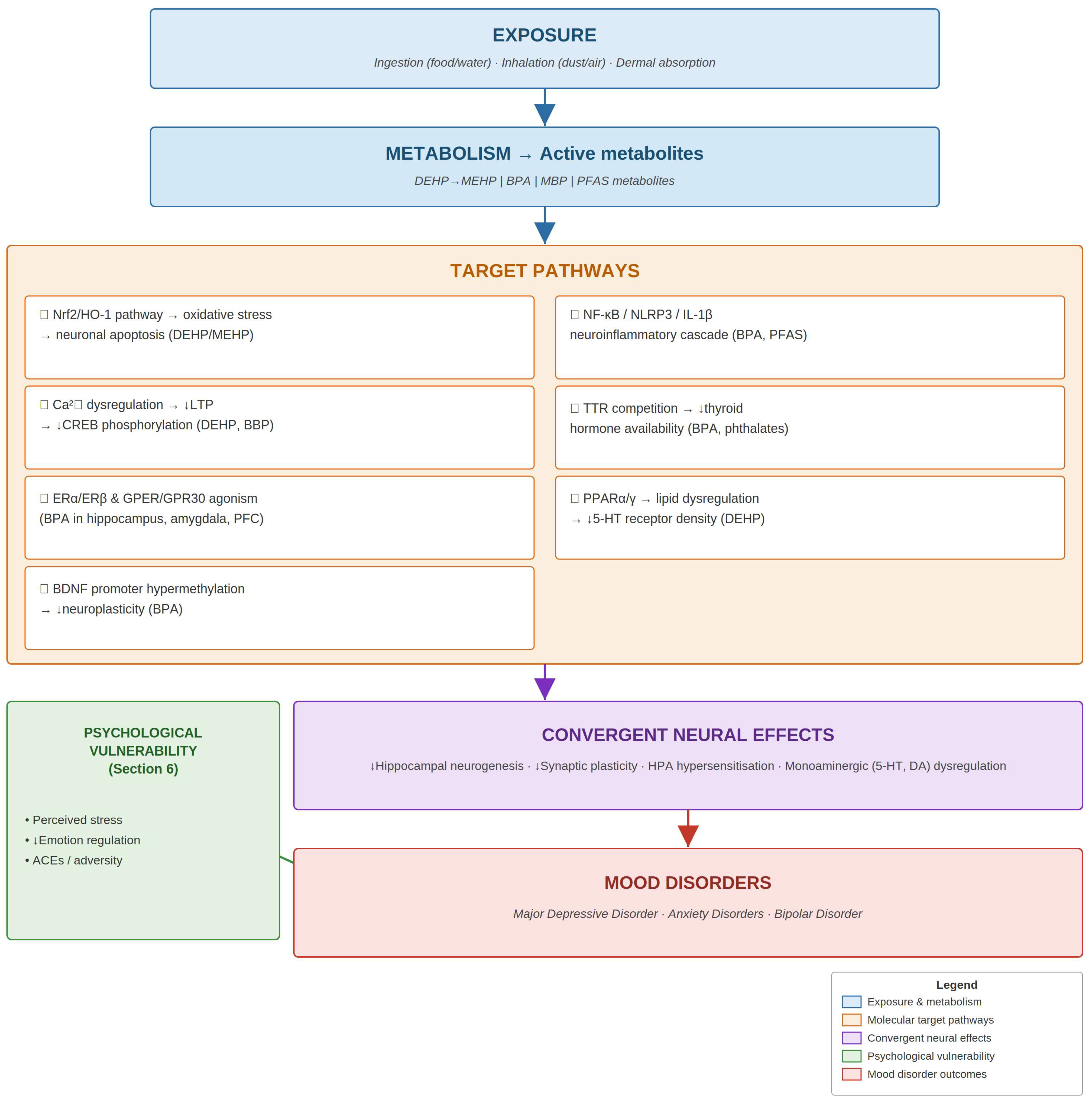
The molecular mechanisms through which phthalates and bisphenols may disrupt the neurobiological substrates of mood regulation are multiple, partially overlapping, and incompletely characterised. As schematically illustrated in Figure 1, they converge on hippocampal neurogenesis, synaptic plasticity, HPA axis dysregulation, and monoaminergic neurotransmission. The main pathways are described below.

5.1. Neuroinflammatory Pathways

The neuroinflammatory theory of depression proposes that microglial and astrocytic activation, with consequent release of pro-inflammatory cytokines, drives HPA axis hypersensitisation, reduces hippocampal neurogenesis, and increases tryptophan catabolism through the kynurenine pathway [43]. BPA activates NF-κB in neuronal and microglial cells, upregulating IL-1β, IL-6, TNF-α, and cyclooxygenase-2 (COX-2) [38]. Activation of the NLRP3 inflammasome by BPA initiates Caspase-1-dependent pyroptosis, releasing bioactive IL-1β into the extracellular space [38]. PFASs promote microglial release of neuroinflammatory mediators and impair phagocytic clearance of cellular debris, allowing accumulation of DAMPs that sustain chronic low-grade neuroinflammation [39].
Neuroinflammatory cytokines exert direct effects on monoamine systems: IL-6 and TNF-α activate indoleamine 2,3-dioxygenase (IDO), diverting tryptophan from serotonin synthesis towards the kynurenine pathway and generating neurotoxic quinolinic acid. These cytokine-mediated effects on neurotransmitter metabolism provide a molecular bridge between EDC-induced neuroinflammation and the monoaminergic deficits central to depressive psychopathology.

5.2. Oxidative Stress and Mitochondrial Dysfunction

Among the molecular mechanisms of phthalate neurotoxicity, oxidative stress has been the most consistently documented. DEHP and its primary metabolite MEHP generate reactive oxygen species (ROS) in neuronal cells, triggering activation of the Nrf2 transcription factor and subsequent upregulation of cytoprotective genes, notably haem oxygenase-1 (HO-1). Studies in Neuro-2a neuroblastoma cells and NE-4C neural stem cells have shown that the relationship between Nrf2/HO-1 activation and cell viability is dose- and duration-dependent: at low or brief exposures, the pathway is protective, but under sustained DEHP challenge, it paradoxically drives neuronal apoptosis through HO-1-derived haem degradation products that impair mitochondrial electron transport chain function [34,44,45]. Downstream mitochondrial dysfunction manifests as reduced membrane potential, impaired ATP synthesis, and elevated mitochondrial superoxide production in neural tissue [46], a bioenergetic failure that ultimately contributes to the synaptic deficits and HPA axis dysregulation characteristic of MDD.

5.3. Disruption of Calcium Signalling and Synaptic Plasticity

Intracellular calcium (Ca2+) serves as a pivotal second messenger in neuronal signalling, governing neurotransmitter release, synaptic potentiation, and the activation of transcription factors essential for synaptic plasticity and neuronal survival. Ran et al. demonstrated that chronic DEHP exposure in rats produced significant dysregulation of intracellular Ca2+ homeostasis in hippocampal neurons, accompanied by suppression of neuronal excitability and disruption of long-term potentiation (LTP), ultimately impairing spatial learning and memory in the Morris water maze [32].
BBP impairs synaptic plasticity through a convergent but distinct mechanism: Min et al. showed that BBP attenuates hippocampal glutamatergic neurotransmission and reduces phosphorylation of the cAMP response element-binding protein (CREB) at Ser133—a post-translational modification essential for consolidation of LTP into long-term memory—thereby impairing associative learning and memory in rodent models [33]. Given that deficient hippocampal LTP and impaired CREB-BDNF signalling are established features of MDD and anxiety disorders, these findings provide direct mechanistic plausibility for the epidemiological associations between phthalate exposure and mood-related outcomes.

5.4. Hormonal Receptor Interactions and Estrogenic Effects

BPA disrupts endocrine function primarily through direct engagement of oestrogen receptors, particularly ERα. X-ray crystallography has resolved the structural basis of this interaction: BPA’s two hydroxylated aromatic rings fit within the ERα ligand-binding domain in a conformation that resembles that of estradiol, with the hydroxyl groups contacting key residues Glu353, Arg394, and His524 [19]. In brain regions with dense ERα and ERβ expression—the hippocampus, amygdala, hypothalamus, and prefrontal cortex—this oestrogenic activation alters the transcription of genes that govern neuronal differentiation, synaptogenesis, and monoaminergic neurotransmitter regulation [47].
Bisphenols additionally interfere with thyroid hormone homeostasis through competitive inhibition of thyroxine (T4) binding to transthyretin (TTR), a major plasma thyroid hormone carrier protein [48]. Because thyroid hormones are indispensable regulators of neuronal migration, myelination, and synaptic maturation, TTR competition by bisphenols provides a mechanism through which these compounds can impair neural circuit formation independent of their oestrogenic activity. Phthalates similarly exert anti-thyroid effects through PPARα/γ-mediated alterations in thyroid hormone-regulated gene expression and displacement of T4 from TTR. Furthermore, phthalate-mediated anti-androgenic effects via competitive AR antagonism and suppression of testosterone biosynthesis may contribute to mood dysregulation, given that androgen receptor signalling in the brain influences dopaminergic tone and stress reactivity [49].

5.5. Epigenetic Mechanisms

The persistence of EDC-induced neurobiological effects well beyond the period of active exposure has prompted investigation of epigenetic mechanisms as mediators of long-term programming. Kundakovic et al. demonstrated that prenatal BPA exposure induces lasting DNA methylation changes in the transcriptionally relevant region of the Bdnf gene in the hippocampus and blood of BALB/c mice and that these changes were consistent with BDNF alterations observed in cord blood of human infants exposed to high maternal BPA levels in utero [35]. BDNF is a critical regulator of neuroplasticity, neurogenesis, and synaptic maintenance; its epigenetic suppression constitutes a well-validated mechanistic link between early-life adversity and susceptibility to mood disorders in the neurotrophic theory of depression [50]. Clinically, baseline BDNF levels and their restoration over the course of treatment are associated with antidepressant efficacy, and selective loss of BDNF in the hippocampal dentate gyrus attenuates the behavioural response to conventional antidepressants, including SSRIs, in animal models [50,51]; this reinforces the plausibility that EDC-induced suppression of BDNF signalling could blunt both mood regulation and pharmacological treatment response. Complementary evidence from Cheong et al. in the CLARITY-BPA consortium study showed that developmental BPA exposure resulted in hypermethylation of the Bdnf promoter region in the hippocampus of female rat offspring, associated with altered BDNF expression and spatial cognition [36].
Maternal exposure to mono-n-butyl phthalate (MBP) activates the IRE1α/XBP1s endoplasmic reticulum stress pathway in neural stem cells, redirecting their differentiation towards phagocytic astrocytes at the expense of neuronal and oligodendrocytic fates, with downstream excessive synapse engulfment and cognitive dysfunction in offspring [40]. PFASs additionally induce extensive epigenetic reprogramming in microglia, altering histone modification patterns and DNA methylation at loci governing inflammatory gene expression [39].

5.6. Alterations in Lipid Metabolism

Phthalates are ligands of PPARα and PPARγ, nuclear receptors that serve as master regulators of lipid and glucose metabolism. Activation of these receptors in neural cells by DEHP and MEHP alters the expression of genes governing fatty acid oxidation, cholesterol biosynthesis, and phospholipid remodelling [52]. In the CNS, lipid homeostasis is critical for myelin synthesis and maintenance, and disruption of oligodendrocyte PPARγ signalling by phthalates has been proposed as a mechanism contributing to impaired myelination and reduced white matter connectivity. Furthermore, alterations in the phospholipid composition of neuronal plasma membranes affect membrane fluidity and the signalling efficiency of serotonin, dopamine, and glutamate receptor systems—neurotransmitter pathways whose dysregulation is central to the pathophysiology of mood disorders [53].

6. The Role of Psychological Vulnerability Factors

EDC exposure is unlikely to determine mood disorder onset in any simple, deterministic sense; individual psychological and psychosocial factors clearly shape the biological response to chemical challenge. The diathesis–stress model offers a useful lens here. EDC-induced alterations in neuroendocrine programming and neuroinflammatory tone may function as a form of chemical–biological diathesis: they prime the system without necessarily precipitating illness, but they raise the probability of disorder considerably when they co-occur with independent psychological vulnerabilities [54]. This framing ties the molecular evidence from Section 5—HPA axis sensitisation, BDNF suppression-driven impairment of neuroplasticity, and elevated neuroinflammatory tone—directly to the clinically observable vulnerability profiles discussed below.
Perceived chronic stress is among the more clinically relevant moderating variables in this context. Individuals with a sensitised HPA axis show amplified cortisol responses to psychological stressors, and EDC exposure may create precisely that sensitised state during developmental windows. BPA, acting through epigenetic suppression of the Bdnf gene promoter (Section 5.5), and phthalates, via interference with adrenocortical steroidogenesis, can both shift HPA axis set points during critical periods. Individuals who carry elevated urinary phthalate or bisphenol loads and simultaneously report high chronic stress may therefore face a compounded—and disproportionate—risk of MDD onset [55].
Emotion regulation capacities—defined as the ability to modulate the intensity, duration, and expression of emotional states through cognitive and behavioural strategies—emerge as another critical moderating variable. Deficient emotion regulation, as operationalised by instruments such as the Difficulties in Emotion Regulation Scale (DERS), has been independently associated with depression, anxiety, and borderline personality disorder. Animal models of prenatal EDC exposure consistently report alterations in emotional reactivity and stress coping that phenocopy regulatory deficits documented in mood disorder patients, suggesting that the biological substrate of emotion regulation may itself be vulnerable to early-life chemical programming [56]. Adverse childhood experiences (ACEs)—including early abuse, neglect, and household dysfunction—are among the strongest known risk factors for adult-onset mood disorders. The co-occurrence of high ACE burden and elevated EDC exposure represents a plausible compounding risk scenario, particularly relevant given that socioeconomically disadvantaged populations tend to experience both higher rates of ACEs and greater environmental pollutant burden [57].
A critical methodological implication is that biomonitoring studies should routinely incorporate standardised psychometric instruments: the Patient Health Questionnaire-9 (PHQ-9) and Beck Depression Inventory-II (BDI-II) for depressive symptoms, the Positive and Negative Affect Schedule (PANAS) for affective state, the DERS for emotion regulation capacities, and the Perceived Stress Scale (PSS) for chronic stress appraisal. Integration of validated psychological assessments with biological exposure measures in future nested case–control and prospective cohort studies would substantially strengthen the evidence base for causal inference.

7. The Cocktail Effect: Exposure to Multiple EDCs

Human populations are not exposed to individual EDCs in isolation but rather to complex, time-varying mixtures. The “cocktail effect”—the aggregate biological impact of simultaneous exposure to multiple EDCs—represents a major regulatory and scientific challenge that single-chemical toxicology has historically failed to address.
A landmark study by Caporale et al. published in Science demonstrated that a mixture of 34 EDCs selected to represent real-world European exposure patterns produced adverse effects on thyroid hormone signalling and neuronal gene expression at concentrations below the individual no-observed-effect levels (NOELs) of each constituent compound [37]. This finding fundamentally challenges the assumption—embedded in current regulatory frameworks—that risk can be adequately assessed compound by compound.
Kim et al. showed that gestational co-exposure to phthalates, heavy metals, and POPs was associated with greater neurodevelopmental impairment in offspring than any single compound class alone [31]. Kondolot et al. documented significantly elevated urinary BPA alongside increased activities of superoxide dismutase (SOD) and glutathione reductase (GR) in children with pervasive developmental disorder not otherwise specified (PDD-NOS), consistent with the hypothesis that co-exposure to multiple EDCs augments neuronal oxidative stress through convergent mechanisms [41]. Future epidemiological studies should employ mixture analysis approaches, including weighted quantile sum (WQS) regression and Bayesian kernel machine regression (BKMR), to characterise the contributions of individual compounds and their interactions. Current regulatory frameworks, including the EU REACH regulation, evaluate chemical safety on a substance-by-substance basis and do not adequately capture mixture risks; cumulative exposure index approaches are urgently needed [58].

8. Conclusions and Future Perspectives

The evidence reviewed here, taken together, builds a credible—if not yet conclusive—case for a causal relationship between phthalate and bisphenol exposure and the development of mood disorders. Three converging lines of evidence underpin this reading: biomonitoring data confirming near-universal human exposure; experimental findings demonstrating plausible neurobiological mechanisms; and epidemiological signals connecting prenatal and childhood EDC exposure to adverse neurodevelopmental and affective outcomes. The molecular mechanisms examined span oxidative stress, calcium dysregulation, oestrogenic receptor agonism, epigenetic BDNF suppression, neuroinflammatory activation, and disruption of thyroid and lipid signalling pathways. That these pathways operate simultaneously in exposed individuals, and interact with independent psychological vulnerabilities, warrants serious attention from both researchers and regulators.
At the molecular level, the mechanisms by which these compounds may precipitate mood disorder are multiple and partially overlapping. Oxidative stress is the most robustly documented: DEHP/MEHPs activate the Nrf2/HO-1 pathway, which at sustained exposures shifts from cytoprotective to pro-apoptotic. Separately, phthalates and bisphenols disturb intracellular calcium homeostasis, undermining synaptic LTP through deficits in CREB phosphorylation. BPA engages ERα and ERβ as well as the membrane receptor GPER/GPR30 in limbic and prefrontal circuits. At the epigenetic level, promoter hypermethylation suppresses BDNF expression, with lasting consequences for neuroplasticity. Neuroinflammatory cascades—specifically NF-κB/NLRP3/IL-1β signalling—are also activated. Beyond these, competition with thyroid hormone at TTR binding sites disrupts thyroid signalling, and PPARα/γ-mediated changes in brain lipid metabolism reduce serotonergic receptor density. No single pathway is likely to account for the full phenotype; the coexistence of all seven in chronically exposed individuals is what makes the risk biologically plausible.
Three priority domains for future research can be identified. In the mechanistic domain, three-dimensional brain organoid models derived from human iPSCs offer unprecedented opportunities to study the effects of physiologically relevant EDC concentrations on human neural circuits in a developmental context that more accurately recapitulates the human brain than traditional cell line or rodent models. In the epidemiological domain, large prospective birth cohorts with serial collection of biological samples for EDC biomonitoring, comprehensive psychiatric phenotyping using validated diagnostic instruments, and longitudinal follow-up into adulthood are needed; existing cohorts such as HELIX (Human Early Life Exposome) and ECHO (Environmental influences on Child Health Outcomes) offer valuable platforms. In the translational domain, the incorporation of standardised psychometric instruments—including the PHQ-9, BDI-II, DERS, and PSS—into environmental health cohort protocols would enable the characterisation of psychological moderators of EDC-associated psychiatric risk.
From a public health perspective, current regulatory thresholds for DEHP in food contact materials and BPA are based primarily on reproductive and developmental toxicity endpoints and do not adequately reflect accumulating neuropsychiatric risk evidence. Biomonitoring programmes such as HBM4EU and its successor PARC should explicitly incorporate neurodevelopmental and psychiatric outcome measures. Public education campaigns targeting high-risk exposure scenarios—the use of PVC food packaging under thermal conditions, the handling of thermal receipts, and dietary patterns favouring canned foods—could contribute to primary prevention at the population level.

Author Contributions

Conceptualization, M.L. and G.F.; methodology, M.L., A.F., M.P. (Monica Piras) and A.D.; investigation, M.L., P.P., M.P. (Marco Piludu) and G.O.; writing—original draft preparation, M.L.; writing—review and editing, A.F., M.M., G.F. and V.F.; supervision, G.F. and V.F. All authors have read and agreed to the published version of the manuscript.

Funding

This research received no external funding.

Data Availability Statement

No new data were created or analyzed in this study. Data sharing is not applicable to this article.

Conflicts of Interest

The authors declare no conflicts of interest.

References

  1. World Health Organization. Depression and Other Common Mental Disorders: Global Health Estimates; WHO: Geneva, Switzerland, 2017; Available online: https://www.who.int/publications/i/item/depression-global-health-estimates (accessed on 10 March 2025).
  2. Chisholm, D.; Sweeny, K.; Sheehan, P.; Rasmussen, B.; Smit, F.; Cuijpers, P.; Saxena, S. Scaling-up treatment of depression and anxiety: A global return on investment analysis. Lancet Psychiatry 2016, 3, 415–424. [Google Scholar] [CrossRef] [PubMed]
  3. Kessler, R.C.; Bromet, E.J. The epidemiology of depression across cultures. Annu. Rev. Public Health 2013, 34, 119–138. [Google Scholar] [CrossRef] [PubMed]
  4. Sullivan, P.F.; Neale, M.C.; Kendler, K.S. Genetic epidemiology of major depression: Review and meta-analysis. Am. J. Psychiatry 2000, 157, 1552–1562. [Google Scholar] [CrossRef] [PubMed]
  5. Wild, C.P. Complementing the genome with an “exposome”: The outstanding challenge of environmental exposure measurement in molecular epidemiology. Cancer Epidemiol. Biomark. Prev. 2005, 14, 1847–1850. [Google Scholar] [CrossRef] [PubMed]
  6. Zoeller, R.T.; Brown, T.R.; Doan, L.L.; Gore, A.C.; Skakkebaek, N.E.; Soto, A.M.; Woodruff, T.J.; Vom Saal, F.S. Endocrine-disrupting chemicals and public health protection: A statement of principles from the Endocrine Society. Endocrinology 2012, 153, 4097–4110. [Google Scholar] [CrossRef] [PubMed]
  7. Gore, A.C.; Chappell, V.A.; Fenton, S.E.; Flaws, J.A.; Nadal, A.; Prins, G.S.; Toppari, J.; Zoeller, R.T. EDC-2: The Endocrine Society’s second scientific statement on endocrine-disrupting chemicals. Endocr. Rev. 2015, 36, E1–E150. [Google Scholar] [CrossRef] [PubMed]
  8. Calafat, A.M.; Ye, X.; Wong, L.Y.; Reidy, J.A.; Needham, L.L. Exposure of the US population to bisphenol A and 4-tertiary-octylphenol: 2003–2004. Environ. Health Perspect. 2008, 116, 39–44. [Google Scholar] [CrossRef] [PubMed]
  9. Silva, M.J.; Barr, D.B.; Reidy, J.A.; Malek, N.A.; Hodge, C.C.; Caudill, S.P.; Brock, J.W.; Needham, L.L.; Calafat, A.M. Urinary levels of seven phthalate metabolites in the US population from NHANES 1999–2000. Environ. Health Perspect. 2004, 112, 331–338. [Google Scholar] [CrossRef] [PubMed]
  10. Schug, T.T.; Janesick, A.; Blumberg, B.; Heindel, J.J. Endocrine disrupting chemicals and disease susceptibility. J. Steroid Biochem. Mol. Biol. 2011, 127, 204–215. [Google Scholar] [CrossRef] [PubMed]
  11. Staples, C.A.; Peterson, D.R.; Parkerton, T.F.; Adams, W.J. The environmental fate of phthalate esters: A literature review. Chemosphere 1997, 35, 667–749. [Google Scholar] [CrossRef]
  12. Koch, H.M.; Calafat, A.M. Human body burdens of chemicals used in plastic manufacture. Philos. Trans. R. Soc. B Biol. Sci. 2009, 364, 2063–2078. [Google Scholar] [CrossRef] [PubMed]
  13. Wormuth, M.; Scheringer, M.; Vollenweider, M.; Hungerbühler, K. What are the sources of exposure to eight frequently used phthalic acid esters in Europeans? Risk Anal. 2006, 26, 803–824. [Google Scholar] [CrossRef] [PubMed]
  14. Praveena, S.M.; Fong, C.S.; Amaruddin, A.F. Phthalates in children’s toys available in Malaysian market: Quantification and potential human health risk. J. Steroid Biochem. Mol. Biol. 2021, 213, 105955. [Google Scholar] [CrossRef] [PubMed]
  15. Warner, G.R.; Dettogni, R.S.; Bagchi, I.C.; Flaws, J.A.; Graceli, J.B. Placental outcomes of phthalate exposure. Reprod. Toxicol. 2021, 103, 1–17. [Google Scholar] [CrossRef] [PubMed]
  16. Minatoya, M.; Araki, A.; Miyashita, C.; Ait Bamai, Y.; Itoh, S.; Yamamoto, J.; Onoda, Y.; Ogasawara, K.; Matsumura, T.; Kishi, R. Association between prenatal bisphenol A and phthalate exposures and fetal metabolic related biomarkers: The Hokkaido Study. Environ. Res. 2018, 161, 505–511. [Google Scholar] [CrossRef] [PubMed]
  17. Rochester, J.R. Bisphenol A and human health: A review of the literature. Reprod. Toxicol. 2013, 42, 132–155. [Google Scholar] [CrossRef] [PubMed]
  18. Vandenberg, L.N.; Hauser, R.; Marcus, M.; Olea, N.; Welshons, W.V. Human exposure to bisphenol A (BPA). Reprod. Toxicol. 2007, 24, 139–177. [Google Scholar] [CrossRef] [PubMed]
  19. Kurosawa, T.; Hiroi, H.; Tsutsumi, O.; Ishikawa, T.; Osuga, Y.; Fujiwara, T.; Inoue, S.; Muramatsu, M.; Momoeda, M.; Taketani, Y. The activity of bisphenol A depends on both the estrogen receptor subtype and the cell type. Endocr. J. 2002, 49, 465–471. [Google Scholar] [CrossRef] [PubMed]
  20. Dong, S.; Terasaka, S.; Kiyama, R. Bisphenol A induces a rapid activation of Erk1/2 through GPR30 in human breast cancer cells. Environ. Pollut. 2011, 159, 212–218. [Google Scholar] [CrossRef] [PubMed]
  21. Rosenmai, A.K.; Dybdahl, M.; Pedersen, M.; Alice van Vugt-Lussenburg, B.M.; Wedebye, E.B.; Taxvig, C.; Vinggaard, A.M. Are structural analogues to bisphenol A safe alternatives? Toxicol. Sci. 2014, 139, 35–47. [Google Scholar] [CrossRef] [PubMed]
  22. Schönfelder, G.; Wittfoht, W.; Hopp, H.; Talsness, C.E.; Paul, M.; Chahoud, I. Parent bisphenol A accumulation in the human maternal-fetal-placental unit. Environ. Health Perspect. 2002, 110, A703–A707. [Google Scholar] [CrossRef] [PubMed]
  23. Berbel, P.; Navarro, D.; Ausó, E.; Varea, E.; Rodriguez, A.E.; Ballesta, J.J.; Salinas, M.; Flores, E.; Faura, C.C.; de Escobar, G.M. Role of late maternal thyroid hormones in cerebral cortex development. Cereb. Cortex 2010, 20, 1462–1475. [Google Scholar] [CrossRef] [PubMed]
  24. Cho, S.C.; Bhang, S.Y.; Hong, Y.C.; Shin, M.S.; Kim, B.N.; Kim, J.W.; Yoo, H.J.; Cho, I.H.; Kim, H.W. Relationship between environmental phthalate exposure and the intelligence of school-age children. Environ. Health Perspect. 2010, 118, 1027–1032. [Google Scholar] [CrossRef] [PubMed]
  25. Factor-Litvak, P.; Insel, B.; Calafat, A.M.; Liu, X.; Perera, F.; Rauh, V.A.; Whyatt, R.M. Persistent associations between maternal prenatal exposure to phthalates on child IQ at age 7 years. PLoS ONE 2014, 9, e114003. [Google Scholar] [CrossRef] [PubMed]
  26. Gascon, M.; Valvi, D.; Forns, J.; Casas, M.; Martínez, D.; Júlvez, J.; Monfort, N.; Ventura, R.; Sunyer, J.; Vrijheid, M. Prenatal exposure to phthalates and neuropsychological development during childhood. Int. J. Hyg. Environ. Health 2015, 218, 550–558. [Google Scholar] [CrossRef] [PubMed]
  27. Engel, S.M.; Villanger, G.D.; Nethery, R.C.; Thomsen, C.; Sakhi, A.K.; Drover, S.S.M.; Hoppin, J.A.; Zeiner, P.; Knudsen, G.P.; Reichborn-Kjennerud, T.; et al. Prenatal phthalates, maternal thyroid function, and risk of ADHD in the Norwegian Mother and Child Cohort. Environ. Health Perspect. 2018, 126, 057004. [Google Scholar] [CrossRef] [PubMed]
  28. Hu, D.; Wang, Y.X.; Chen, W.J.; Zhang, Y.; Li, H.H.; Xiong, L.; Zhu, H.P.; Chen, H.Y.; Peng, S.X.; Wan, Z.H.; et al. Associations of phthalates exposure with attention deficits hyperactivity disorder: A case-control study among Chinese children. Environ. Pollut. 2017, 229, 375–385. [Google Scholar] [CrossRef] [PubMed]
  29. Chopra, V.; Harley, K.; Lahiff, M.; Eskenazi, B. Association between phthalates and attention deficit disorder and learning disability in US children, 6–15 years. Environ. Res. 2014, 128, 64–69. [Google Scholar] [CrossRef] [PubMed]
  30. Kardas, F.; Bayram, A.K.; Demirci, E.; Akin, L.; Ozmen, S.; Kendirci, M.; Canpolat, M.; Gumus, H.; Kumandas, S.; Per, H. Increased serum phthalates (MEHP, DEHP) and bisphenol A concentrations in children with autism spectrum disorder. J. Child Neurol. 2016, 31, 629–635. [Google Scholar] [CrossRef] [PubMed]
  31. Kim, S.; Eom, S.; Kim, H.J.; Lee, J.J.; Choi, G.; Choi, S.; Kim, S.; Kim, S.Y.; Cho, G.; Kim, Y.D.; et al. Association between maternal exposure to major phthalates, heavy metals, and POPs, and the neurodevelopmental performances of their children at 1 to 2 years of age—CHECK cohort study. Sci. Total Environ. 2018, 624, 377–384. [Google Scholar] [CrossRef] [PubMed]
  32. Ran, D.; Luo, Y.; Gan, Z.; Liu, J.; Yang, J. Neural mechanisms underlying the deficit of learning and memory by exposure to di(2-ethylhexyl) phthalate in rats. Ecotoxicol. Environ. Saf. 2019, 174, 58–65. [Google Scholar] [CrossRef] [PubMed]
  33. Min, A.; Liu, F.; Yang, X.; Chen, M. Benzyl butyl phthalate exposure impairs learning and memory and attenuates neurotransmission and CREB phosphorylation in mice. Food Chem. Toxicol. 2014, 71, 81–89. [Google Scholar] [CrossRef] [PubMed]
  34. Amara, I.; Timoumi, R.; Graiet, I.; Ben Salem, I.; Adelou, K.; Abid-Essefi, S. Di(2-ethylhexyl) phthalate induces cytotoxicity in HEK-293 cell line: Implication of the Nrf-2/HO-1 antioxidant pathway. Environ. Toxicol. 2019, 34, 1034–1042. [Google Scholar] [CrossRef] [PubMed]
  35. Kundakovic, M.; Gudsnuk, K.; Herbstman, J.B.; Tang, D.; Perera, F.P.; Champagne, F.A. DNA methylation of BDNF as a biomarker of early-life adversity. Proc. Natl. Acad. Sci. USA 2015, 112, 6807–6813. [Google Scholar] [CrossRef] [PubMed]
  36. Cheong, A.; Johnson, S.A.; Howald, E.C.; Ellersieck, M.R.; Camacho, L.; Lewis, S.M.; Howard, P.C.; Guillette, L.J., Jr.; Rosenfeld, C.S. Gene expression and DNA methylation changes in the hypothalamus and hippocampus of adult rats developmentally exposed to bisphenol A or ethinyl estradiol: A CLARITY-BPA consortium study. Epigenetics 2018, 13, 704–720. [Google Scholar] [CrossRef] [PubMed]
  37. Caporale, N.; Leemans, M.; Birgersson, L.; Germain, P.L.; Cheroni, C.; Borbély, G.; Engdahl, E.; Lindh, C.; Bressan, R.B.; Cavallo, F.; et al. From cohorts to molecules: Adverse impacts of endocrine disrupting mixtures. Science 2022, 375, eabe8244. [Google Scholar] [CrossRef] [PubMed]
  38. Al-Shami, A.S.; Abd Elkader, H.T.A.E.A.; Moussa, N.; Essawy, A.E.; Haroun, M. Early-life bisphenol A exposure causes neuronal pyroptosis in juvenile and adult male rats through the NF-κB/IL-1β/NLRP3/Caspase-1 signalling pathway: Exploration of age and dose as effective covariates using an in vivo and in silico modeling approach. Mol. Cell. Biochem. 2025, 480, 2301–2330. [Google Scholar] [CrossRef] [PubMed]
  39. Cheng, Y.; Li, J.R.; Yu, H.; Li, S.; Tychhon, B.; Cheng, C.; Weng, Y.L. Perfluoroalkyl substance pollutants disrupt microglia function and trigger transcriptional and epigenomic changes. Toxicology 2025, 517, 154198. [Google Scholar] [CrossRef] [PubMed]
  40. Cui, F.; Deng, S.; Fu, Y.; Xu, T.; Bao, S.; Wang, S.; Lin, Y.; Wang, X.; Zhao, F.; Zhang, T.; et al. Maternal phthalates exposure promotes neural stem cell differentiation into phagocytic astrocytes and synapse engulfment via the IRE1α/XBP1s pathway. Cell Rep. 2025, 44, 115126. [Google Scholar] [CrossRef] [PubMed]
  41. Kondolot, M.; Ozmert, E.N.; Ascı, A.; Erkekoglu, P.; Oztop, D.B.; Gumus, H.; Kocer-Gumusel, B.; Yurdakok, K. Plasma phthalate and bisphenol A levels and oxidant–antioxidant status in autistic children. Environ. Toxicol. Pharmacol. 2016, 43, 149–158. [Google Scholar] [CrossRef] [PubMed]
  42. Alhamyani, A. Investigating the link between microplastic exposure (benzyl butyl phthalate) and neurodegenerative diseases using high-performance computational toxicology. Toxicol. Res. 2025, 14, tfae211. [Google Scholar] [CrossRef] [PubMed]
  43. Dantzer, R.; O’Connor, J.C.; Freund, G.G.; Johnson, R.W.; Kelley, K.W. From inflammation to sickness and depression: When the immune system subjugates the brain. Nat. Rev. Neurosci. 2008, 9, 46–56. [Google Scholar] [CrossRef] [PubMed]
  44. Aung, K.H.; Win-Shwe, T.T.; Kanaya, M.; Takano, H.; Tsukahara, S. Involvement of hemeoxygenase-1 in di(2-ethylhexyl) phthalate (DEHP)-induced apoptosis of Neuro-2a cells. J. Toxicol. Sci. 2014, 39, 217–229. [Google Scholar] [CrossRef] [PubMed]
  45. Wu, M.; Xu, L.; Teng, C.; Xiao, X.; Hu, W.; Chen, J.; Tu, W. Involvement of oxidative stress in di(2-ethylhexyl) phthalate (DEHP)-induced apoptosis of mouse NE-4C neural stem cells. NeuroToxicology 2019, 70, 41–47. [Google Scholar] [CrossRef] [PubMed]
  46. Kabekkodu, S.P.; Gladwell, L.R.; Choudhury, M. The mitochondrial link: Phthalate exposure and cardiovascular disease. Biochim. Biophys. Acta Mol. Cell Res. 2024, 1871, 119708. [Google Scholar] [CrossRef] [PubMed]
  47. Patisaul, H.B.; Adewale, H.B. Long-term effects of environmental endocrine disruptors on reproductive physiology and behavior. Front. Behav. Neurosci. 2009, 3, 10. [Google Scholar] [CrossRef] [PubMed]
  48. Hamers, T.; Kamstra, J.H.; Sonneveld, E.; Murk, A.J.; Kester, M.H.; Andersson, P.L.; Legler, J.; Brouwer, A. In vitro profiling of the endocrine-disrupting potency of brominated flame retardants. Toxicol. Sci. 2006, 92, 157–173. [Google Scholar] [CrossRef] [PubMed]
  49. Habert, R.; Muczynski, V.; Grisin, T.; Moison, D.; Messiaen, S.; Frydman, R.; Benachi, A.; Delbes, G.; Lambrot, R.; Lehraiki, A.; et al. Concerns about the widespread use of rodent models for human risk assessments of endocrine disruptors. Reproduction 2014, 147, R119–R129. [Google Scholar] [CrossRef] [PubMed]
  50. Castrén, E.; Rantamäki, T. The role of BDNF and its receptors in depression and antidepressant drug action: Reactivation of developmental plasticity. Dev. Neurobiol. 2010, 70, 289–297. [Google Scholar] [CrossRef] [PubMed]
  51. Adachi, M.; Barrot, M.; Autry, A.E.; Theobald, D.; Monteggia, L.M. Selective loss of brain-derived neurotrophic factor in the dentate gyrus attenuates antidepressant efficacy. Biol. Psychiatry 2008, 63, 642–649. [Google Scholar] [CrossRef] [PubMed]
  52. Feige, J.N.; Gerber, A.; Casals-Casas, C.; Yang, Q.; Winkler, C.; Bedu, E.; Bueno, M.; Gelman, L.; Auwerx, J.; Gonzalez, F.J.; et al. The pollutant diethylhexyl phthalate regulates hepatic energy metabolism via species-specific PPARα-dependent mechanisms. Environ. Health Perspect. 2010, 118, 234–241. [Google Scholar] [CrossRef] [PubMed]
  53. Chalon, S. Omega-3 fatty acids and monoamine neurotransmission. Prostaglandins Leukot. Essent. Fat. Acids 2006, 75, 259–269. [Google Scholar] [CrossRef] [PubMed]
  54. Monroe, S.M.; Simons, A.D. Diathesis-stress theories in the context of life stress research: Implications for the depressive disorders. Psychol. Bull. 1991, 110, 406–425. [Google Scholar] [CrossRef] [PubMed]
  55. Liu, W.; He, Y.; Zhang, L.; Tao, F.; Huang, Y.; Wang, G.; Su, P. Environmental endocrine disruptors and depression in adolescence: A missing link? Ecotoxicol. Environ. Saf. 2026, 309, 119568. [Google Scholar] [CrossRef] [PubMed]
  56. Al-Shami, A.S.; Haroun, M.; Essawy, A.E.; Moussa, N.; Abd Elkader, H.T.A.E.A. Early-life bisphenol A exposure causes detrimental age-related changes in anxiety, depression, learning and memory in juvenile and adult male rats. NeuroToxicology 2025, 106, 17–36. [Google Scholar] [CrossRef] [PubMed]
  57. Brody, G.H.; Yu, T.; Chen, Y.-F.; Kogan, S.M.; Evans, G.W.; Beach, S.R.H.; Windle, M.; Simons, R.L.; Gerrard, M.; Gibbons, F.X.; et al. Cumulative socioeconomic status risk, allostatic load, and adjustment: A prospective latent profile analysis. Dev. Psychol. 2013, 49, 913–927. [Google Scholar] [CrossRef] [PubMed]
  58. European Chemicals Agency (ECHA). Guidance on Information Requirements and Chemical Safety Assessment; Chapter R.19: Uncertainty Analysis; ECHA: Helsinki, Finland, 2016; Available online: https://echa.europa.eu/guidance-documents/guidance-on-information-requirements-and-chemical-safety-assessment (accessed on 10 March 2025).
Figure 1. Schematic representation of the molecular pathways linking phthalate and bisphenol exposure to mood disorders. Phthalates (e.g., DEHP, BBP) and bisphenols (e.g., BPA) enter the body via ingestion, inhalation, and dermal absorption. Following metabolism to bioactive moieties, seven convergent pathways are activated: ① Nrf2/HO-1-driven oxidative stress and neuronal apoptosis; ② calcium dysregulation impairing synaptic LTP and CREB phosphorylation; ③ ERα/ERβ and GPER/GPR30 agonism in limbic regions; ④ epigenetic BDNF promoter hypermethylation; ⑤ NF-κB/NLRP3/IL-1β neuroinflammation; ⑥ transthyretin (TTR) competition reducing thyroid hormone availability; ⑦ PPARα/γ-mediated lipid dysregulation reducing serotonergic receptor density. Converging with psychological vulnerability factors (Section 6), these pathways ultimately contribute to mood disorder development. ACEs: adverse childhood experiences; 5-HT: serotonin; DA: dopamine; LTP: long-term potentiation; PFC: prefrontal cortex.
Environments 13 00425 g001
Disclaimer/Publisher’s Note: The statements, opinions and data contained in all publications are solely those of the individual author(s) and contributor(s) and not of MDPI and/or the editor(s). MDPI and/or the editor(s) disclaim responsibility for any injury to people or property resulting from any ideas, methods, instructions or products referred to in the content.