Article

Environmental Drivers of Pesticide Toxicity: Temperature and pH Shift Azoxystrobin’s Effects on Zebrafish (Danio rerio) Early Development

1
Animal Physiological Ecology, University of Tübingen, Auf der Morgenstelle 5, D-72076 Tübingen, Germany
2
Steinbeis Transfer Center for Ecotoxicology and Ecophysiology, Blumenstraße 13, D-72108 Rottenburg, Germany
*
Author to whom correspondence should be addressed.
Environments 2025, 12(9), 334; https://doi.org/10.3390/environments12090334
Submission received: 14 August 2025 / Revised: 12 September 2025 / Accepted: 16 September 2025 / Published: 18 September 2025

Abstract

Azoxystrobin, a widely used strobilurin fungicide, poses a potential risk to aquatic ecosystems due to its frequent detection in surface waters. Although its toxicity to non-target organisms has been extensively studied under standardized conditions, few investigations have considered how environmental factors can modulate the adverse effects of this chemical. In this study, we examined the toxicity of azoxystrobin to zebrafish (Danio rerio) embryos under different pH (5, 7, 9) and temperature (21 °C, 26 °C, 31 °C) conditions. Embryos were exposed to azoxystrobin concentrations ranging from 0 to 1000 μg/L, and endpoints such as survival, hatching rate, heart rate, malformations, developmental delay, and Hsp70 expression were assessed over 96 h post-fertilization. Our results demonstrate that azoxystrobin induces significant malformations (including edema, eye, tail, and spinal defects) and developmental delays at 1000 μg/L across all environmental conditions. Furthermore, both pH and temperature were found to modulate azoxystrobin toxicity: elevated temperature and alkaline pH partly alleviated mortality at high concentrations. The hsp70 expression patterns revealed complex interactions between the effects of the chemical and environmental factors. These findings highlight the importance of incorporating environmental variables into ecotoxicological risk assessments of pesticides to better reflect realistic exposure scenarios and potential ecological impacts.

1. Introduction

The importance of fungicides as widely used chemicals with high sales volumes and their potential impact on target and non-target organisms was recently addressed in an article by Mohr et al. 2025 [1]. Azoxystrobin is a strobilurin fungicide widely applied to control fungal diseases in crops. As a broad-spectrum, systemic, and soil-applied fungicide, azoxystrobin is extensively used in agriculture worldwide [2,3]. However, its widespread application has raised concerns regarding contamination of the aquatic environment. Due to its persistence, mobility, and the associated relevance for humans and the environment, azoxystrobin was added to the EU Water Framework Directive watch list in 2022 and is, therefore, regularly monitored in European surface and groundwaters (https://op.europa.eu/en/publication-detail/-/publication/a531a7ff-d3b9-11ef-be2a-01aa75ed71a1/language-en, accessed on 13 August 2025). Currently, azoxystrobin concentrations in surface water vary from 0.06 to 11.10 μg/L in industrialized countries such as the United States, France, Germany, Greece, China, and Brazil, with peak levels reaching up to 29.7 μg/L [4,5,6,7]. In groundwater, concentrations between 0.01 and 0.1 μg/L were found [8,9]. Azoxystrobin inhibits the respiratory chain in fungi by blocking the electron transfer from quinol to cytochrome c1, thereby reducing ATP production [10,11]. However, evidence suggests that it can also induce toxicity in a wide range of non-target aquatic animals [5]. For example, azoxystrobin has been reported to reduce survival, delay growth, and impair reproduction in Daphnia [12,13]; to hinder tadpole development [5]; and to affect behavior and oxidative stress in zebrafish [7,14].
Despite extensive research that has documented the toxicity of pesticides like azoxystrobin, most chemical toxicity assessments are conducted under standardized laboratory conditions that do not reflect the complexity of natural environments [15,16]. In reality, abiotic factors such as temperature and pH vary considerably across habitats and can modulate both the action of pollutants and the physiological responses of organisms [17,18,19]. For instance, temperature influences metabolic rates and chemical uptake, while pH can affect the chemical speciation and stability of contaminants [17,18,20,21]. Therefore, ignoring environmental variability may result in inaccurate estimates of toxicity, undermining the ecological relevance of laboratory-based risk assessments.
The zebrafish (Danio rerio) has emerged as a widely used model organism in biological and environmental research [22,23,24]. Its early-life stages, particularly embryos, are widely used for toxicity testing because they are cost-efficient, quick, and comply with animal welfare principles as non-protected organisms [25,26]. These tests leverage the transparency of zebrafish eggs, allowing for direct observation of developmental effects like malformations. Since 2013, the OECD has officially recommended zebrafish embryo toxicity testing for environmental risk assessments [27].
In this study, our aim is to perform a hazard assessment under controlled laboratory conditions, focusing on identifying the developmental toxicity of azoxystrobin in zebrafish embryos and assessing whether this toxicity is modulated by environmental factors, specifically temperature and pH. To this end, we examined both apical endpoints (e.g., survival, hatching, malformations) and molecular responses (e.g., Hsp70 expression) under varying environmental conditions. This approach is designed to identify potential hazards and mechanistic interactions, rather than to provide a quantitative environmental risk assessment.

2. Materials and Methods

2.1. Test Substance

Azoxystrobin is a lipophilic organic ionizable compound whose lipophilicity remains stable between pH 4 and pH 14. Relevant physicochemical information is provided in Supplementary Table S1 and Figure S1.
A stock solution of azoxystrobin (CAS131860-33-8, purity ≥ 98%, Merck, Darmstadt, Germany) was prepared by dissolving it in reconstituted water composed of 25 mL stock solutions of 0.23 g/L KCl, 2.59 g/L NaHCO3, 4.93 g/L MgSO4·7 H2O, and 11.76 g/L CaCl2·2 H2O, and 900 mL double-distilled water. Other test concentrations were obtained by stepwise dilution of the stock solution.
The pH of each test solution was measured using a pH meter (SevenCompactDuo; Mettler Toledo, Gießen, Germany) and adjusted by 1M HCl or 1M NaOH as needed.

2.2. Test Organisms

Zebrafish (Danio rerio, USA Westaquarium strain) were maintained in 200 L tanks at the Animal Physiological Ecology section, University of Tübingen, Germany. Fish were fed three times daily with commercial flake food (TertraMin®, Tetra GmbH, Melle, Germany), and feces were removed prior to feeding. A water change (change 30–50%) was performed weekly. All procedures followed permission granted from the Animal Welfare Committee of the Regional Council of Tübingen, Germany (approval numbers ZO 2/16 and ZO 02/21 G).
The tanks were kept under a controlled 12 h light/12 h dark photoperiod. Water quality was regularly monitored, with temperature maintained at 26 ± 1 °C, pH at 7.4 ± 0.2, total hardness ranging from 8 to 12 °dH, and dissolved oxygen at 100 ± 5% saturation. Nitrate and nitrite levels were consistently below 1 mg/L and 5 mg/L, respectively.

2.3. Fish Embryo Test

Three separate experiments were conducted to assess azoxystrobin toxicity under varying pH and temperature conditions (Figure 1). Experiment 1: Embryos were exposed to 0, 0.1, 1, 10, 100, 1000 μg/L azoxystrobin at 26 °C, pH 7. As there was no apparent effect of 0.1 and 1 mg/L azoxystrobin in test 1, we decided to investigate only azoxystrobin concentrations of higher than 1 µg/L (0, 10, 100, 1000 mg/L) in subsequent tests. Experiment 2: With a focus on pH effects, embryos were exposed to azoxystrobin at pH 5 and 9, maintaining the temperature at 26 °C. Experiment 3: With a focus on temperature effects, embryos were exposed to azoxystrobin at 21 °C and 31 °C, maintaining pH constant at 7.
Early-life stage (ELS) tests followed our previous work based on OECD 236 [23,24]. Spawning and test setups were prepared one day prior to the start of the experiment. Breeding boxes included 1.5 mm mesh grids and artificial seagrass to stimulate spawning. Eggs were collected one hour after light onset and placed in Petri dishes containing the appropriate test solutions for a 2 h pre-exposure at the respective experimental temperature.
For each test concentration, eight small Petri dishes (30 mm in diameter) were prepared, each containing 3 mL of the corresponding solution, along with one larger dish (90 mm in diameter) holding 30 mL. Embryos at the 128–256 cell stage were randomly chosen and allocated to the small dishes, with four embryos per dish, resulting in a total of 32 embryos per experimental group. Incubation lasted for 96 h under the designated temperature. Each experiment was conducted in two independent runs, with 32 embryos per treatment group per run. This design follows the OECD Test Guideline 236 and exceeds the recommended minimum sample size, thereby providing sufficient statistical power to detect significant effects.
Embryonic development was monitored using a stereo microscope (Stemi 2000-C; Zeiss, Oberkochen, Germany) at 12, 24, 48, 60, 72, and 96 h post-fertilization (hpf). Endpoints included survival, developmental delay, heart rates, hatching success, and morphological abnormalities (Table 1). Coagulated embryos were recorded and removed throughout. Heart rates were recorded over 20 s and extrapolated to beats per minute. Hatching was monitored at 60 and 96 hpf. To better reflect the teratogenic potential while avoiding bias from high mortality, malformation rates were calculated based on surviving embryos only. This metric is hereafter referred to as the “malformation rates.”

2.4. Hsp70 Determination

For Hsp70 analysis, 40 freshly laid fertilized zebrafish eggs were allocated per group and incubated for 96 h. After exposure, 10 embryos were pooled per sample, frozen in liquid nitrogen, and stored at −80 °C (3 replicates for each group). Hsp70 levels were measured following the protocol of Vincze et al. (2014) [28]. Briefly, samples were homogenized in 20 µL extraction buffer (80 mM potassium acetate, 4 mM magnesium acetate, 20 mM Hepes, pH 7.5). The homogenates were then centrifuged at 20,000× g for 10 min at 4 °C, and the resulting supernatants were collected for analysis. The total protein concentration was determined according to Bradford (1976) [29], and 20 µg of protein per sample was separated via SDS-PAGE (80 V for 30 min, then 120 V for ~90 min). Immunostaining was performed using a monoclonal mouse α-human antibody (Santa Cruz Biotechnology, clone 3A3) and a secondary horseradish peroxidase-coupled goat α-mouse IgG (H + L) antibody (Dianova). Bands were visualized, digitized, and analyzed via densitometry (Herolab E.A.S.Y., Wiesloch, Germany). Hsp70 expression was normalized to two internal adult Danio rerio standards on the same blotting membrane.
Representative SDS-PAGE images of Hsp70 are provided in Supplementary Figure S2. Although some bands appear diffuse, all quantifications were based on densitometric analysis of raw images to ensure consistency and reproducibility.

2.5. Statistical Analysis

All statistical analyses were conducted using SAS JMP version 16.0. Results are expressed as means ± standard error (SE). Normality and homogeneity of variances were tested before statistical tests. If assumptions were met, one-way ANOVA followed by Tukey’s HSD post hoc test was used. If not, the non-parametric Steel–Dwass test was applied. LC50 values were determined by non-linear regression analysis using TableCurve software version 5.01 for azoxystrobin treatments at pH 7 and 26 °C.

3. Results

3.1. Survival Rate

Figure 2 and Figure 3 show the survival rates of zebrafish embryos exposed to azoxystrobin under different pH (Figure 2) and temperature conditions (Figure 3). At pH 5, pH 7, and pH 9, embryos exposed to 1000 μg/L azoxystrobin exhibited the lowest survival rates, significantly lower than all other treatment groups (p < 0.05). At pH 9, the survival rate in the 1000 μg/L group was significantly higher than that at pH 7 (p < 0.05).
At 21 °C, the survival rates of all groups were significantly lower than those exposed at 26 °C or 31 °C. In addition, the survival rate in the 1000 μg/L group was significantly lower than in the control group (p < 0.05). No significant differences were observed between the 100 and 1000 μg/L groups and the 0 μg/L group (p > 0.05).
At 26 °C and 31 °C, the 1000 μg/L treatment groups consistently showed significantly lower survival rates than other treatment groups (p < 0.05). The survival rate of the 1000 μg/L group at 31 °C was significantly higher than that at 26 °C (p < 0.05).

3.2. Heart Rates

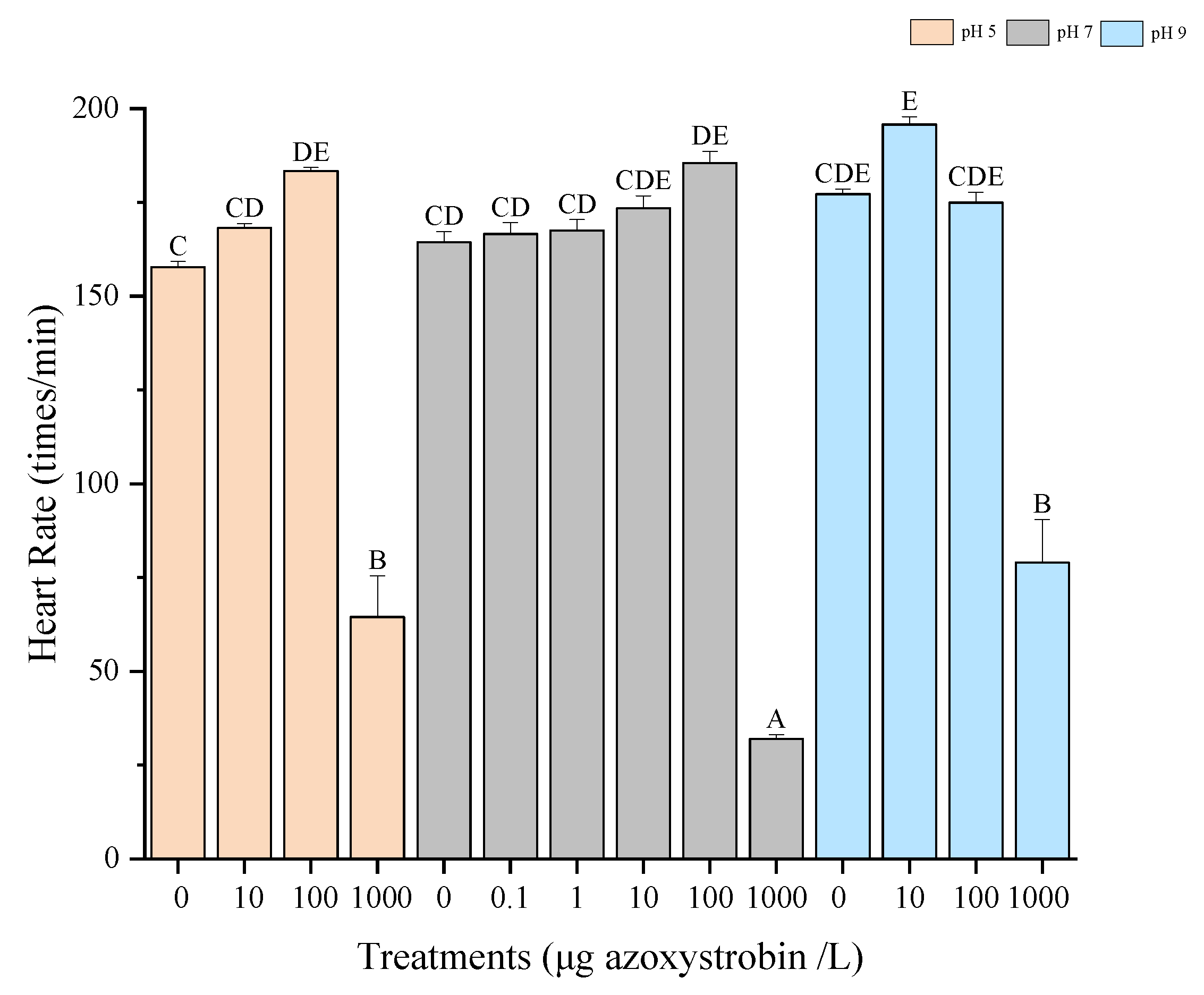
Heart rate responses at different pH levels are shown in Figure 4. At pH 5, pH 7, and pH 9, heart rates followed non-significant non-linear trends, initially increasing and then declining with rising azoxystrobin concentrations (Figure 4). At 1000 μg/L, the heart rates at pH 5 and pH 9 were significantly higher than those at pH 7 (p < 0.05).
Heart rates significantly increased in the control group with rising temperature (p < 0.05). Across all temperatures, the heart rates were significantly lower in embryos exposed to 1000 µg/L compared to the respective other exposure groups at the same temperature (Figure 5). No significant differences were observed between control and exposure groups at 21 °C (p > 0.05).

3.3. Hatching Rates

The effect of azoxystrobin on the hatching rates of zebrafish embryos at different pH and temperature is exhibited in Supplementary Figures S3 and S4. Across all temperature and pH conditions, embryonic development in the 1000 μg/L treatment groups was consistently delayed compared to the control at the same time points (Figures S3 and S4) (p < 0.05). No embryos hatched within 96 hpf at 21 °C in any treatment group.

3.4. Malformation

As shown in Figure 6, malformation rates were significantly elevated (p < 0.05) in the 1000 μg/L treatment groups under all pH and temperature conditions.
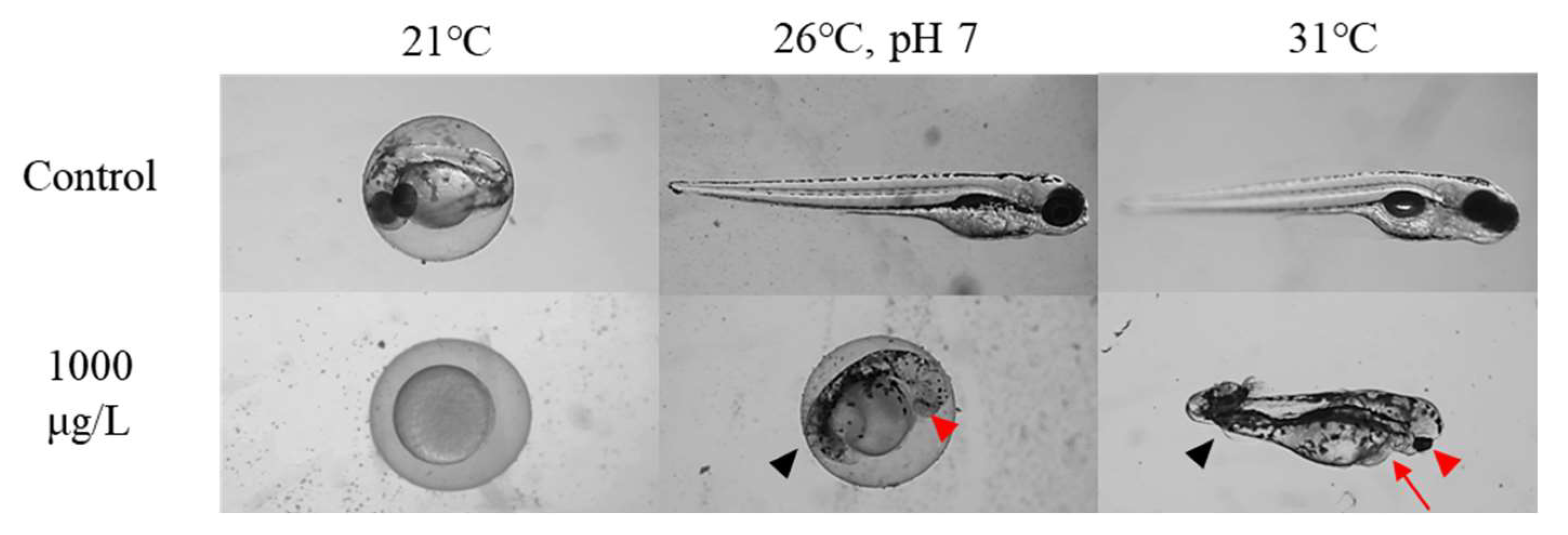
Embryonic development from 24 to 96 hpf is shown in Figure 7 and Figure 8 and Figures S5–S9. At 96 hpf, all 1000 μg/L groups exhibited varying malformations, including edema, ocular abnormalities, tail deformities, and spinal curvature (Figure 7 and Figure 8). At 21 °C, embryos in the 1000 μg/L group showed complete developmental arrest within 96 hpf (Figure 7, Figure 8 and Figures S5–S9).

3.5. Hsp70

Figure 9 and Figure 10 show the Hsp70 expression patterns in zebrafish embryos. At 26 °C and pH 7, Hsp70 levels initially increased and then decreased with rising azoxystrobin concentrations. Hsp70 expression in the 100 μg/L and 1000 μg/L groups was significantly lower than in the 0.1 μg/L group (p < 0.05). At pH 5, Hsp70 expression remained unchanged across all treatments with a trend to lower values after exposure to 1000 µg/L (p > 0.05). At pH 9, Hsp70 levels declined with increasing azoxystrobin concentration, and the 1000 μg/L group exhibited significantly lower expression than the control group (p < 0.05). At both 21 °C and 31 °C, there were no significant differences in Hsp70 expression between the treatment groups (p > 0.05). This section may be divided by subheadings. It should provide a concise and precise description of the experimental results, their interpretation, as well as the experimental conclusions that can be drawn.

4. Discussion

The current study investigated the impact of varying temperature and pH conditions on apical toxicity and proteotoxicity of azoxystrobin in zebrafish embryos. Our data resulted in the calculation of a 96 h-LC50 value of 988.2 (95% confidence interval from 987.7 to 988.7) μg/L at pH 7 and 26 °C. This value is similar to the findings of Jia et al. (2018) and Jiang et al. (2019) [30,31], who reported the 96 h-LC50 of zebrafish embryos exposed to azoxystrobin as 810 μg/L and 1230 μg/L, respectively. Zhang et al. (2020) reported a lower 96 h-LC50 of 488 μg/L [32]. This discrepancy may stem from the fact that Zhang et al. (2020) [32] used a commercial product, whereas all other studies, including ours, employed azoxystrobin of ≥95% purity. In addition, Cao et al. (2018) reported no significant increase in mortality in groups exposed to less than 200 μg/L azoxystrobin, which aligns with our findings that have shown low concentrations (0–100 μg/L) of azoxystrobin to exert only limited effects on zebrafish embryo survival [33].
Azoxystrobin is known to inhibit mitochondrial respiration, thereby reducing metabolic activity [5,11]. Notably, we observed a biphasic effect of azoxystrobin on heart rates. At the lower concentrations (10–100 μg/L), a moderate increase in heart rates was observed for all temperatures and all pH values tested, possibly due to compensatory mitochondrial stress responses or hormetic stimulation of cardiometabolic activity [18,23]. At the highest concentration (1000 μg/L), however, heart rates were significantly decreased, likely attributable to severe mitochondrial dysfunction, energy depletion, and oxidative damage to cardiac tissues [31]. Similarly, decreased heart rates in embryos and grass carp (Ctenopharyngodon idella) caused by cardiac depressants have been observed in the studies of Liu et al. (2013) and Zhang et al. (2020) [32,34].
Azoxystrobin has also been shown to inhibit embryonic development, as evidenced by delayed hatching and reduced growth. For instance, Cao et al. (2018) and Kumar et al. (2020) found shortened post-developmental body lengths, while Jiang et al. (2019) and Zhang et al. (2020) reported delayed hatching and decreased hatching success [11,31,32,33]. Consistently, our study revealed a negative impact of azoxystrobin on the hatching rate and the embryonic development. These developmental impairments are likely related to insufficient energy and reduced metabolism. Azoxystrobin inhibits mitochondrial complex III by binding to the Qo site of cytochrome b, disrupting electron transport and reducing proton gradient formation. This leads to impaired oxidative phosphorylation and decreased ATP production [13,32]. Zhang et al. (2020) also provided data that are likely to be related to this mechanism. They observed reduced yolk depletion in exposed embryos, which might also be indicative of decreased energy utilization resulting from impaired ATP production [32].
Even though sublethal concentrations will not cause immediate mortality, they can result in population-relevant effects, e.g., delayed development, reduced mobility, and prolonged vulnerability to predation [34,35]. Moreover, energy deficits during early-life stages may compromise reproductive capacity in later life stages [2,14].
Furthermore, our study observed morphological abnormalities in embryos such as edema, eye defects, spinal curvature, and tail defects, indicating the teratogenic potential of azoxystrobin. These abnormalities are likely associated with the production of excessive reactive oxygen species (ROS) and the induction of apoptosis, which may involve mitochondrial dysfunction among other pathways [13,32]. Jia et al. (2018) also reported azoxystrobin (1500 μg/L) to cause morphological abnormalities to zebrafish embryos, including tail defects, edema, and spinal curvature [30]. In our study, significant morphological changes were absent at medium to low concentrations (≤100 μg/L), which is consistent with Zhang et al. (2020) and Kumar et al. (2020) [11,32]. Similarly, Jiang et al. (2019) found that teratogenic effects appeared only at concentrations ≥ 500 μg/L [31].
The Hsp70 family is a well-known and highly conserved class of stress proteins that plays essential roles in maintaining cellular homeostasis. It assists in the refolding of misfolded proteins, inhibits apoptosis, and is indirectly involved in the regulation of ROS levels [36,37,38]. Since azoxystrobin causes mitochondrial dysfunction, oxidative stress, and apoptosis [13,32], it is reasonable to infer that Hsp70 may be upregulated during embryo exposure to azoxystrobin as part of a cellular defense mechanism. Specifically, Hsp70 likely contributes to preserving proteostasis, protecting cellular structures, and facilitating mitochondrial stress recovery.
The cellular Hsp70 response to increasing chemical toxicity can be broadly divided into three phases. In the initial phase, cells maintain homeostasis, and Hsp70 expression remains at baseline or only marginally elevated. During the second phase, activation of the stress response leads to a rapid increase in Hsp70 levels, culminating in peak expression. In the final phase, when toxic exposure surpasses the cellular tolerance threshold, pathological impairment of the protein synthesis machinery is likely to occur, resulting in a subsequent decline in Hsp70 expression [24,39]. At 26 °C, pH 7.0, the expression pattern of Hsp70 in response to azoxystrobin exposure aligns well with its typical stress-induced response pattern [24,39]. Hsp70 levels initially increased with rising azoxystrobin concentrations but declined at higher concentrations. This probably occurs between the reaction and destruction stages [24,39]. Specifically, cells respond to toxicity by sharply upregulating Hsp70 expression. When the toxicity of the azoxystrobin exceeds the cellular tolerance threshold, it may lead to structural and functional damage to the protein synthesis machinery, thereby reducing Hsp70 expression. Notably, in our study, Hsp70 showed a rapid response to azoxystrobin already at a very low concentration (0.1 μg/L), indicating its potential as a highly sensitive and early biomarker for proteotoxicity in aquatic organisms.
Temperature-dependent changes in azoxystrobin toxicity were evident in this study. At elevated temperatures (31 °C), an antagonistic effect was observed compared to 26 °C. Specifically, in the 1000 μg/L group at 31 °C, the survival rate increased significantly, the heart rates were elevated and equal to the normal level, the hatching rate increased, the deformity rate was reduced, and the animals hatched earlier than at 26 °C. No significant differences in Hsp70 levels were observed across the concentration groups at 31 °C. Elevated temperatures are often associated with increased chemical toxicity, as they can enhance passive diffusion, stimulate active uptake, and elevate oxygen demand—factors that collectively promote metabolic stress and ROS accumulation [18,24,40]. Nonetheless, this trend is not consistent across all compounds or organisms. In this study, azoxystrobin toxicity decreased at higher temperatures, suggesting temperature-dependent mitigation. To minimize potential methodological artifacts, a 2-h acclimation period was included prior to the onset of formal exposure. During this period, embryos were maintained at the respective experimental temperature in the corresponding incubators before selection. Therefore, the temperature-dependent toxicity observed is unlikely to result from abrupt thermal shock. One possible explanation for the alleviation of toxicity at higher temperatures is the result of physiological adaptations. Elevated temperatures can accelerate metabolic processes, potentially enhancing mitochondrial oxidative capacity and ATP production, which may counteract the respiratory inhibition caused by azoxystrobin [18,24,40]. Cominassi et al. (2022) reported an increased metabolic rate with increased acclimation temperature in the threespine stickleback (Gasterosteus aculeatus) [41]. Similarly, Voituron et al. (2022) reported that acclimation to high temperature improved mitochondrial efficiency (on average > 15%) of sea bass (Dicentrarchus labrax) juveniles, and improved ATP/O efficiency in European sea bass mitochondria at 26 °C, compared to 18–22 °C [42]. In addition, heat acclimation upregulates antioxidant enzymes (e.g., SOD, CAT), heat shock proteins (e.g., Hsp70), and other pathways to enhance ROS scavenging capacity [42,43]. In contrast, at 21 °C, we observed a marked increase in toxicity across all treatment groups: survival rates declined, Hsp70 levels were suppressed, and embryonic development was largely arrested. Moreover, within both the high (31 °C) and low (21 °C) temperature treatments, there were no significant differences among the groups that had been exposed to different concentrations of azoxystrobin, suggesting that temperature had a more dominant effect than the azoxystrobin concentration under these, for D. rerio, extreme conditions.
Compared to temperature, the effect of pH on azoxystrobin toxicity was less pronounced and showed no clear gradient-dependent pattern. In our study, the survival rate in the 1000 μg/L treatment group increased to varying degrees when pH changed from neutral to acidic or alkaline conditions; however, sublethal effects were significantly exacerbated under both conditions, such as increased malformation rates, altered Hsp70 expression, and accelerated heart rate. This indicates a possible toxicity shift from lethal to sublethal effects on embryo development under pH modulation, whereby embryos experience more biologically meaningful developmental and physiological disruptions. This shift is also supported by Bittner et al. (2018) and Schweizer et al. (2019) [44,45]. In their zebrafish embryo acute toxicity experiments, LC50 levels increased, and LOECs of heart rates, a sublethal concentration indicator, decreased as pH changed from neutral to acidic. The phenomenon was more pronounced at pH 9 in our study. In addition to the increase in malformation rate, we also observed a faster heart rate response to azoxystrobin and an earlier destructive stage of Hsp70 at pH 9 compared with that at pH 7.
Based on the logD and chemical structure of azoxystrobin (Figure S1), the compound remains non-ionized between pH 5 and pH 9, indicating that pH does not affect its ionization state or hydrophobicity. Therefore, the observed variations in toxicity across different pH conditions are unlikely to be due to altered uptake or membrane permeability [15,16,20]. Interestingly, elevated heart rates were observed at both pH 5 and pH 9. In one of our previous studies, we also found that both acidic and alkaline external environments increased the heart rates of zebrafish embryos exposed to pesticides [24]. These results suggest that pH does not alter azoxystrobin chemistry directly but rather modifies the physiological state of embryos. However, this compensation may come at the cost of enhanced oxidative stress, as ATP production and ROS generation are tightly coupled. At high azoxystrobin concentrations, the collapse of proteostasis further indicates that the combined stress of chemical exposure and pH imbalance overwhelms cellular defense capacity.
Although this study was designed as a hazard assessment rather than a quantitative risk assessment, the observed sublethal impairments (e.g., delayed development, altered cardiac function, reduced proteostatic capacity) may translate into population-relevant outcomes, including reduced recruitment, impaired growth, and compromised reproductive success [46,47]. Such effects, while not immediately lethal, can ultimately affect population dynamics by increasing predation vulnerability and lowering lifetime fitness. These findings suggest that current risk assessment frameworks (e.g., OECD fish embryo tests, EU WFD guidelines), which often emphasize lethality and nominal exposure concentrations, may underestimate ecological hazards under realistic environmental conditions where multiple abiotic stressors act simultaneously [46,47,48].
Moreover, organisms in natural ecosystems are rarely exposed to a single stressor. Factors such as oxygen fluctuations, temperature variability, pH shifts, and co-occurring pesticides act simultaneously and may interact in non-additive ways [49,50]. Our results show that even single abiotic variables like temperature and pH can shift toxicity thresholds, implying that combined stressors in the field may further modulate toxic responses. To better capture such complexity, future assessments could benefit from factorial laboratory designs and mechanistic modeling approaches, integrated with environmental monitoring, to improve the ecological realism of pesticide hazard evaluation [51].

5. Conclusions

This study reveals that the toxicity of azoxystrobin to zebrafish embryos is not only concentration-dependent but also influenced by confounding environmental factors such as temperature and pH. While traditional assessments typically rely on optimal laboratory conditions, our findings demonstrate that deviations in environmental parameters can significantly reshape the toxicity, altering both the severity and the nature of toxic effects.
Importantly, our findings highlight that environmental parameters, especially temperature and pH, can significantly alter the toxicity of azoxystrobin. Elevated temperature (31 °C) alleviated toxicity, possibly through enhanced mitochondrial efficiency and upregulation of stress–response mechanisms, whereas low temperature (21 °C) aggravated toxic outcomes. pH changes from neutral to acidic or alkaline conditions, despite not affecting the chemical’s ionization or uptake, caused a toxicity shift, reducing mortality but intensifying sublethal developmental impairments, particularly at pH 9. This suggests that abiotic factors can redirect toxic pressure from survival endpoints toward more subtle but ecologically significant physiological disruptions. These sublethal effects, though frequently overlooked, can have ecologically meaningful consequences by impairing individual fitness and population stability.
From an ecotoxicological perspective, these results provide valuable hazard assessment insights. They highlight that environmental parameters can substantially modulate toxic pressure, underscoring the need to account for realistic abiotic variability in toxicity testing. While our study does not constitute a full risk assessment, it supplies mechanistic and hazard-level evidence that can improve the ecological relevance of future risk evaluations. Incorporating such context-dependent responses into regulatory frameworks (e.g., OECD FET, EU WFD) will enhance predictive accuracy and ensure that pesticide hazards are more reliably characterized under changing environmental conditions.

Supplementary Materials

The following supporting information can be downloaded at: https://www.mdpi.com/article/10.3390/environments12090334/s1, Figure S1: Chemical structure and logD of azoxystrobin; Figure S2: Representative SDS-PAGE images of Hsp70; Figure S3: The effect of azoxystrobin on the hatching rate of zebrafish embryos at 26 °C and 31 °C; Figure S4: The effect of azoxystrobin on the hatching rate of zebrafish embryos at different pH; Figure S5: Embryonic development in the 0 and 1000 µg/L Azoxystrobin treatment groups at 24–96 hpf at 26 °C, pH 7; Figure S6: Embryonic development in the 0 and 1000 µg/L Azoxystrobin treatment groups at 24–96 hpf at pH 5; Figure S7: Embryonic development in the 0 and 1000 µg/L Azoxystrobin treatment groups at 24–96 hpf at pH 9; Figure S8: Embryonic development in the 0 and 1000 µg/L Azoxystrobin treatment groups at 24–96 hpf at 21 °C; Figure S9: Embryonic development in the 0 and 1000 µg/L Azoxystrobin treatment groups at 24–96 hpf at 31 °C; Table S1: Physicochemical properties of azoxystrobin.

Author Contributions

Conceptualization, Z.L.; methodology, Z.L.; software, Z.L.; validation, Z.L., R.T. and H.-R.K.; formal analysis, Z.L.; data curation, Z.L.; writing—original draft preparation, Z.L.; writing—review and editing, Z.L., R.T. and H.-R.K.; supervision, R.T.; funding acquisition, Z.L. All authors have read and agreed to the published version of the manuscript.

Funding

Z. Li received a personal grant from the Chinese Research Council (202108210104).

Institutional Review Board Statement

According to German legislation and the Animal Welfare law, zebrafish embryos up to 96 h post-fertilization are not considered protected animals, and thus embryo assays (OECD TG 236) do not require animal ethics approval.

Data Availability Statement

The original contributions presented in this study are included in the article/supplementary material.

Acknowledgments

We are grateful to Stefanie Krais, Tübingen University, for assistance with the Hsp70 assay, and we give our thanks to Leila Es-Sadafy for the daily zebrafish management.

Conflicts of Interest

The authors declare no conflict of interest.

References

  1. Mohr, S.; Antony, M.; Contardo-Jara, V.; Scholz, U.; Bader, S.; Polleichtner, C.; Arts, G. Evaluating Herbicidal Risks of the Fungicide Tebuconazole: Differential Sensitivity of Dicot and Monocot Macrophytes in Freshwater Mesocosms. Environ. Sci. Eur. 2025, 37, 119. [Google Scholar] [CrossRef]
  2. Cao, F.; Zhu, L.; Li, H.; Yu, S.; Wang, C.; Qiu, L. Reproductive toxicity of azoxystrobin to adult zebrafish (Danio rerio). Environ. Pollut. 2016, 219, 1109–1121. [Google Scholar] [CrossRef] [PubMed]
  3. Amador, P.; Vega, C.; Navarro Pacheco, N.I.; Moratalla-López, J.; Palacios, J.; Crettaz Minaglia, M.C.; López, I.; Díaz, M.; Rico, A. Effects of the Fungicide Azoxystrobin in Two Habitats Representative of Mediterranean Coastal Wetlands: A Mesocosm Experiment. Aquat. Toxicol. 2024, 267, 106828. [Google Scholar] [CrossRef] [PubMed]
  4. Berenzen, N.; Lentzen-Godding, A.; Probst, M.; Schulz, H.; Schulz, R.; Liess, M. A Comparison of Predicted and Measured Levels of Runoff-Related Pesticide Concentrations in Small Lowland Streams on a Landscape Level. Chemosphere 2005, 58, 683–691. [Google Scholar] [CrossRef]
  5. Rodrigues, E.T.; Lopes, I.; Pardal, M.Â. Occurrence, fate and effects of azoxystrobin in aquatic ecosystems: A review. Environ. Internat. 2013, 53, 18–28. [Google Scholar] [CrossRef]
  6. Peluso, J.; Pérez Coll, C.S.; Rojas, D.E.; Cristos, D.; Aronzon, C.M. Ecotoxicological Assessment of Complex Environmental Matrices from the Lower Paraná River Basin. Chemosphere 2022, 305, 135385. [Google Scholar] [CrossRef]
  7. Guo, X.; Zhang, R.; Li, C.; Duan, M.; Cao, N.; Jin, Q.; Chen, X.; Li, L.; Li, X.; Pang, S. Environmental Levels of Azoxystrobin Disturb Male Zebrafish Behavior: Possible Roles of Oxidative Stress, Cholinergic System, and Dopaminergic System. Ecotoxicol. Environ. Saf. 2024, 269, 115744. [Google Scholar] [CrossRef]
  8. Filho, A.M. Development, Validation and Application of a Method Based on DI-SPME and GC–MS for Determination of Pesticides of Different Chemical Groups in Surface and Groundwater Samples. Microchem. J. 2010, 96, 139–145. [Google Scholar] [CrossRef]
  9. Jørgensen, L.F.; Kjær, J.; Olsen, P.; Rosenbom, A.E. Leaching of Azoxystrobin and Its Degradation Product R234886 from Danish Agricultural Field Sites. Chemosphere 2012, 88, 554–562. [Google Scholar] [CrossRef]
  10. Bartlett, D.W.; Clough, J.M.; Godwin, J.R.; Hall, A.A.; Hamer, M.; Parr-Dobrzanski, B. The Strobilurin Fungicides. Pest Manag. Sci. 2002, 58, 649–662. [Google Scholar] [CrossRef]
  11. Kumar, N.; Willis, A.; Satbhai, K.; Ramalingam, L.; Schmitt, C.; Moustaid-Moussa, N.; Crago, J. Developmental toxicity in embryo-larval zebrafish (Danio rerio) exposed to strobilurin fungicides (azoxystrobin and pyraclostrobin). Chemosphere 2020, 241, 124980. [Google Scholar] [CrossRef]
  12. Cui, F.; Chai, T.; Liu, X.; Wang, C. Toxicity of three strobilurins (kresoxim-methyl, pyraclostrobin, and trifloxystrobin) on Daphnia magna. Environ. Toxicol. Chem. 2016, 36, 182–189. [Google Scholar] [CrossRef]
  13. Wang, X.; Li, X.; Wang, Y.; Qin, Y.; Yan, B.; Martyniuk, C.J. A comprehensive review of strobilurin fungicide toxicity in aquatic species: Emphasis on mode of action from the zebrafish model. Environ. Pollut. 2021, 275, 116671. [Google Scholar] [CrossRef]
  14. Cao, F.; Li, H.; Zhao, F.; Wu, P.; Qian, L.; Huang, L.; Pang, S.; Martyniuk, C.J.; Qiu, L. Parental exposure to azoxystrobin causes developmental effects and disrupts gene expression in F1 embryonic zebrafish (Danio rerio). Sci. Total Environ. 2019, 646, 595–605. [Google Scholar] [CrossRef] [PubMed]
  15. Köhler, H.-R.; Gräff, T.; Schweizer, M.; Blumhardt, J.; Burkhardt, J.; Ehmann, L.; Hebel, J.; Heid, C.; Kundy, L.; Kuttler, J.; et al. LogD-based modelling and ΔlogD as a proxy for pH-dependent action of ionizable chemicals reveal the relevance of both neutral and ionic species for fish embryotoxicity and possess great potential for practical application in the regulation of chemicals. Water Res. 2023, 235, 119864. [Google Scholar] [CrossRef] [PubMed]
  16. Kroll, A.; Von Der Ohe, P.C.; Köhler, H.-R.; Sellier, O.; Junghans, M. Aquatic thresholds for ionisable substances, such as diclofenac, should consider pH-specific differences in uptake and toxicity. Sci. Total Environ. 2024, 908, 168222. [Google Scholar] [CrossRef] [PubMed]
  17. Heugens, E.H.W.; Hendriks, A.J.; Dekker, T.; Straalen, N.M.V.; Admiraal, W. A Review of the Effects of Multiple Stressors on Aquatic Organisms and Analysis of Uncertainty Factors for Use in Risk Assessment. Crit. Rev. Toxicol. 2001, 31, 247–284. [Google Scholar] [CrossRef]
  18. Osterauer, R. Temperature-dependent effects of the pesticides thiacloprid and diazinon on the embryonic development of zebrafish (Danio rerio). Aquat. Toxicol. 2008, 86, 485–494. [Google Scholar] [CrossRef]
  19. Mishra, P.; Gong, Z.; Kelly, B.C. Assessing pH-dependent toxicity of fluoxetine in embryonic zebrafish using mass spectrometry-based metabolomics. Sci. Total Environ. 2019, 650, 2731–2741. [Google Scholar] [CrossRef]
  20. Bittner, L.; Teixidó, E.; Keddi, I.; Escher, B.I.; Klüver, N. pH-Dependent Uptake and Sublethal Effects of Antihistamines in Zebrafish (Danio rerio) Embryos. Environ. Toxicol. Chemi. 2019, 38, 1012–1022. [Google Scholar] [CrossRef]
  21. Andrade, T.S.; Henriques, J.F.; Almeida, A.R.; Soares, A.M.V.M.; Scholz, S.; Domingues, I. Zebrafish embryo tolerance to environmental stress factors—Concentration–dose response analysis of oxygen limitation, pH, and UV-light irradiation. Environ. Toxicol. Chem. 2017, 36, 682–690. [Google Scholar] [CrossRef]
  22. Nagel, R. DarT: The embryo test with the zebrafish Danio rerio-a general model in ecotoxicology and toxicology. ALTEX 2002, 19, 38–48. [Google Scholar]
  23. Schweizer, M.; Von Der Ohe, P.C.; Gräff, T.; Kühnen, U.; Hebel, J.; Heid, C.; Kundy, L.; Kuttler, J.; Moroff, F.-M.; Schlösinger, A.-F.; et al. Heart rate as an early warning parameter and proxy for subsequent mortality in Danio rerio embryos exposed to ionisable substances. Sci. Total Environ. 2022, 818, 151744. [Google Scholar] [CrossRef]
  24. Li, Z.; Köhler, H.-R.; Triebskorn, R. Proteotoxicity and Apical Toxicity of Nicosulfuron to Danio rerio Embryos: A Comprehensive Assessment at Different Temperatures and pH. Pollutants 2024, 4, 359–372. [Google Scholar] [CrossRef]
  25. Yang, L.; Ho, N.Y.; Alshut, R.; Legradi, J.; Weiss, C.; Reischl, M.; Mikut, R.; Liebel, U.; Müller, F.; Strähle, U. Zebrafish embryos as models for embryotoxic and teratological effects of chemicals. Reprod. Toxicol. 2009, 28, 245–253. [Google Scholar] [CrossRef]
  26. Su, T.; Lian, D.; Bai, Y.; Wang, Y.Y.L.; Zhang, D.; Wang, Z.; You, J. The feasibility of the zebrafish embryo as a promising alternative for acute toxicity test using various fish species: A critical review. Sci. Total Environ. 2021, 787, 147705. [Google Scholar] [CrossRef]
  27. Organisation for Economic Co-operation and Development (OECD). Test No. 236: Fish Embryo Acute Toxicity (FET) Test; OECD Publishing: Paris, France, 2013; Available online: https://www.oecd-ilibrary.org/environment/test-no-236-fish-embryo-acutetoxicity-fet-test_9789264203709-en (accessed on 26 July 2013).
  28. Vincze, K.; Graf, K.; Scheil, V.; Köhler, H.-R.; Triebskorn, R. Embryotoxic and proteotoxic effects of water and sediment from the Neckar River (Southern Germany) to zebrafish (Danio rerio) embryos. Environ. Sci. Eur. 2014, 26, 3. [Google Scholar] [CrossRef]
  29. Bradford, M.M. A Rapid and Sensitive Method for the Quantitation of Microgram Quantities of Protein Utilizing the Principle of Protein-Dye Binding. Anal. Biochem. 1976, 72, 248–254. [Google Scholar] [CrossRef]
  30. Jia, W.; Mao, L.; Zhang, L.; Zhang, Y.; Jiang, H. Effects of two strobilurins (azoxystrobin and picoxystrobin) on embryonic development and enzyme activities in juveniles and adult fish livers of zebrafish (Danio rerio). Chemosphere 2018, 207, 573–580. [Google Scholar] [CrossRef] [PubMed]
  31. Jiang, J.; Wu, S.; Lv, L.; Liu, X.; Chen, L.; Zhao, X.; Wang, Q. Mitochondrial dysfunction, apoptosis and transcriptomic alterations induced by four strobilurins in zebrafish (Danio rerio) early life stages. Environ. Pollut. 2019, 253, 722–730. [Google Scholar] [CrossRef]
  32. Zhang, Y.; Sheedy, C.; Nilsson, D.; Goss, G.G. Evaluation of Interactive Effects of UV Light and Nano Encapsulation on the Toxicity of Azoxystrobin on Zebrafish. Nanotoxicology 2020, 14, 232–249. [Google Scholar] [CrossRef] [PubMed]
  33. Cao, F.; Wu, P.; Huang, L.; Li, H.; Qian, L.; Pang, S.; Qiu, L. Short-term developmental effects and potential mechanisms of azoxystrobin in larval and adult zebrafish (Danio rerio). Aquat. Toxicol. 2018, 198, 129–140. [Google Scholar] [CrossRef]
  34. Liu, L.; Jiang, C.; Wu, Z.-Q.; Gong, Y.-X.; Wang, G.-X. Toxic effects of three strobilurins (trifloxystrobin, azoxystrobin and kresoxim-methyl) on mRNA expression and antioxidant enzymes in grass carp (Ctenopharyngodon idella) juveniles. Ecotoxicol. Environ. Saf. 2013, 98, 297–302. [Google Scholar] [CrossRef]
  35. Pompermaier, A.; Kirsten, K.; Soares, S.M.; Fortuna, M.; Kalichak, F.; Idalencio, R.; Koakoski, G.; Barreto, R.E.; Barcellos, L.J.G. Waterborne agrichemicals compromise the anti-predatory behavior of zebrafish. Environ. Sci. Pollut. Res. 2020, 27, 38559–38567. [Google Scholar] [CrossRef]
  36. Könemann, S.; Meyer, S.; Betz, A.; Županič, A.; Vom Berg, C. Sub-Lethal Peak Exposure to Insecticides Triggers Olfaction-Mediated Avoidance in Zebrafish Larvae. Environ. Sci. Technol. 2021, 55, 11835–11847. [Google Scholar] [CrossRef]
  37. Westerheide, S.D.; Morimoto, R.I. Heat Shock Response Modulators as Therapeutic Tools for Diseases of Protein Conformation. J. Biol. Chem. 2005, 280, 33097–33100. [Google Scholar] [CrossRef]
  38. Bonomo, J.; Welsh, J.P.; Manthiram, K.; Swartz, J.R. Comparing the functional properties of the Hsp70 chaperones, DnaK and BiP. Biophys. Chem. 2010, 149, 58–66. [Google Scholar] [CrossRef]
  39. Gupta, S.C.; Sharma, A.; Mishra, M.; Mishra, R.K.; Chowdhuri, D.K. Heat Shock Proteins in Toxicology: How Close and How Far? Life Sci. 2010, 86, 377–384. [Google Scholar] [CrossRef]
  40. Eckwert, H.; Alberti, G.; Kohler, H.-R. The induction of stress proteins (hsp) in Oniscus asellus (Isopoda) as a molecular marker of multiple heavy metal exposure: I. Principles and toxicological assessment. Ecotoxicology 1997, 6, 249–262. [Google Scholar] [CrossRef]
  41. Cominassi, L.; Ressel, K.N.; Brooking, A.A.; Marbacher, P.; Ransdell-Green, E.C.; O’Brien, K.M. Metabolic rate increases with acclimation temperature and is associated with mitochondrial function in some tissues of threespine stickleback. J. Exp. Biol. 2022, 225, jeb244659. [Google Scholar] [CrossRef] [PubMed]
  42. Voituron, Y.; Roussel, D.; Teulier, L.; Vagner, M.; Ternon, Q.; Romestaing, C.; Dubillot, E.; Lefrancois, C. Warm Acclimation Increases Mitochondrial Efficiency in Fish: A Compensatory Mechanism to Reduce the Demand for Oxygen. Physiol. Biochem. Zool. 2022, 95, 15–21. [Google Scholar] [CrossRef]
  43. Abdel-Tawwab, M.; Omar, A.A.; Khalil, R.H.; Selema, T.A.M.A.; Elsamanooudy, S.I.; El-Saftawy, H.A.M.; Sabry, E.A.; Fawzy, R.M.; Abdel-Razek, N. Influences of thermal stress on the growth biometrics, stress indicators, oxidative stress biomarkers, and histopathological alterations in European seabass, Dicentrarchus labrax, juveniles. Fish Physiol. Biochem. 2025, 51, 70. [Google Scholar] [CrossRef]
  44. Bittner, L.; Teixido, E.; Seiwert, B.; Escher, B.I.; Klüver, N. Influence of pH on the uptake and toxicity of β-blockers in embryos of zebrafish, Danio rerio. Aquat. Toxicol. 2018, 201, 129–137. [Google Scholar] [CrossRef]
  45. Schweizer, M.; Brilisauer, K.; Triebskorn, R.; Forchhammer, K.; Köhler, H.-R. How glyphosate and its associated acidity affect early development in zebrafish (Danio rerio). PeerJ 2019, 7, e7094. [Google Scholar] [CrossRef] [PubMed]
  46. Brasseur, M.V.; Buchner, D.; Mack, L.; Hartmann, S.; Beketov, M.A. Multiple Stressor Effects of Insecticide Exposure and Increased Fine Sediment Deposition on the Gene Expression Profiles of Two Freshwater Invertebrate Species. Environ. Sci. Eur. 2023, 35, 81. [Google Scholar] [CrossRef]
  47. Fu, H.; Li, Y.; Yuan, C.; Wang, X.; Li, J.; Chen, X.; Xu, J.; Zhang, X. Effects of Multiple Environmental Stressors on Zoobenthos Communities in Shallow Lakes: Evidence from a Mesocosm Experiment. Animals 2023, 13, 3722. [Google Scholar] [CrossRef] [PubMed]
  48. Rey, O.; Stalder, T.; Cournoyer, B.; Fornelos, N.; Altaner, C.; Derome, N.; Forney, L.J.; Daguenet, T.; Risso, C.; Nicolas, P.; et al. Impacts of Multiple Anthropogenic Stressors on the Transcriptional Response of Gammarus fossarum in a Mesocosm Field Experiment. BMC Genom. 2022, 23, 816. [Google Scholar] [CrossRef]
  49. Hoffmann, L.; Arle, J.; Jahnig, S.C.; Schäfer, R.B. The Hierarchy of Multiple Stressors’ Effects on Benthic Invertebrates: A Case Study from the Rivers Erft and Niers, Germany. Environ. Sci. Eur. 2022, 34, 100. [Google Scholar] [CrossRef]
  50. Grabner, D.S.; Schertzinger, G.; Eberhardt, J.; Bittner, M.; Sures, B. Parasites and Pollutants: Effects of Multiple Stressors on Aquatic Organisms. Environ. Toxicol. Chem. 2023, 42, 1884–1896. [Google Scholar] [CrossRef]
  51. Ducrot, V.; Ashauer, R.; Charles, S.; Galic, N.; Gergs, A.; Nyman, A.M.; Van den Brink, P.J.; Vandenbrouck, T.; Zimmer, E.I.; Jager, T. Environmental Risk Assessment of Time-Variable Toxicant Exposure with Toxicokinetic–Toxicodynamic Modeling Calibrated and Validated on the Reproduction of Ceriodaphnia dubia. Environ. Toxicol. Chem. 2024, 43, 2541–2555. [Google Scholar] [CrossRef]
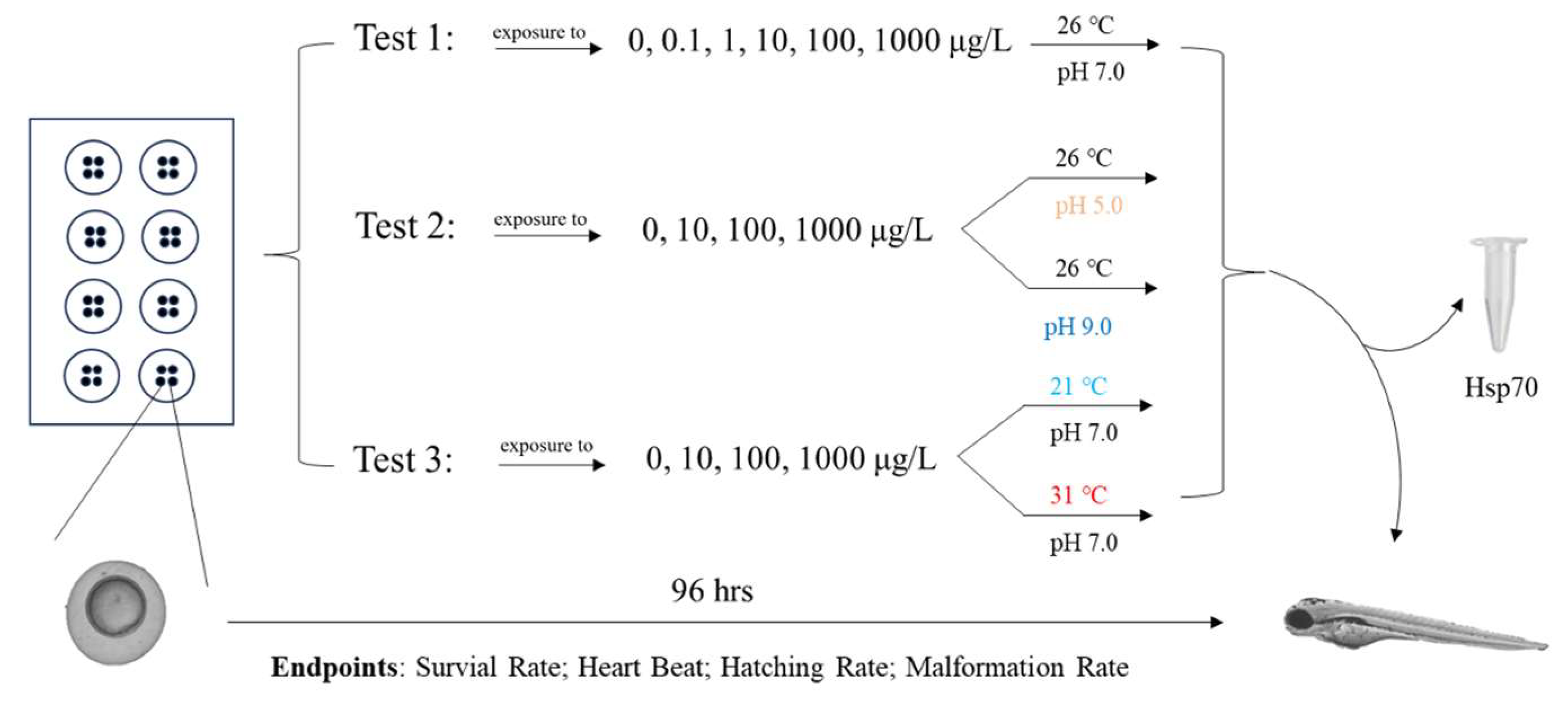
Figure 1. Flowchart of the experimental design.
Environments 12 00334 g001
Figure 2. Survival of zebrafish embryos exposed to azoxystrobin at different pH values. Values are means ± SE. Different letters indicate a significant difference (p < 0.05).
Environments 12 00334 g002
Figure 3. Survival of zebrafish embryos exposed to azoxystrobin at different temperatures. Values are means ± SE. Different letters indicate a significant difference (p < 0.05).
Environments 12 00334 g003
Figure 4. Heart rates of zebrafish embryos exposed to azoxystrobin at different pH values. Values are means ± SE. Different letters indicate a significant difference (p < 0.05).
Environments 12 00334 g004
Figure 5. Heart rates of zebrafish embryos exposed to azoxystrobin at different temperatures. Values are means ± SE. Different letters indicate a significant difference (p < 0.05).
Environments 12 00334 g005
Figure 6. Malformation rate of zebrafish embryos exposed to azoxystrobin at different pH and temperatures. Values are means ± SE. Different letters indicate a significant difference (p < 0.05).
Environments 12 00334 g006
Figure 7. Embryonic development in the 0 and 1000 ug/L azoxystrobin treatment groups at 96 hpf at different pH. Red arrows mark edemas, red triangles eye defects, black arrows spinal curvature, and black triangles mark tail defects.
Environments 12 00334 g007
Figure 8. Embryonic development in the 0 and 1000 ug/L azoxystrobin treatment groups at 96 hpf at different temperatures. Red arrows mark edemas, red triangles eye defects, and black triangles mark tail defects.
Environments 12 00334 g008
Figure 9. Hsp70 level of zebrafish embryos exposed to azoxystrobin at 26 °C, pH 7. Values are means ± SE. Different letters indicate a significant difference (p < 0.05).
Environments 12 00334 g009
Figure 10. Hsp70 level of zebrafish embryos exposed to azoxystrobin at different pH and temperatures. Values are means ± SE. Different letters indicate a significant difference (p < 0.05).
Environments 12 00334 g010
Table 1. Endpoints observed at various periods during the 96 h of the fish embryo test.
Endpoints12 hpf24 hpf48 hpf60 hpf72 hpf96 hpf
Survival rate
Developmental
delays 1
Heart rates
Hatching success
Malformations 2
☑ = endpoint measured at the indicated time point; ☐ = not measured. 1 No somites, non-detachment of the tail, and no development of the eyes; 2 Edema, eye defects, tail defects, deformation of the spine, and light pigmentation.
Disclaimer/Publisher’s Note: The statements, opinions and data contained in all publications are solely those of the individual author(s) and contributor(s) and not of MDPI and/or the editor(s). MDPI and/or the editor(s) disclaim responsibility for any injury to people or property resulting from any ideas, methods, instructions or products referred to in the content.