Article

Manganese Removal from the Seo-Gok Reservoir Water Using DNA Aptamers

1
Lab of Functional Aptamer, Department of Bioindustry and Bioresource Engineering, College of Life Sciences, Sejong University, Seoul 05006, Republic of Korea
2
Department of Biomedical Laboratory Science, College of Software and Digital Healthcare Convergence, Yonsei University MIRAE Campus, Wonju 26493, Republic of Korea
3
Medical Science Research Center, Korea University College of Medicine, Seoul 02841, Republic of Korea
4
Faptamer Inc., Seoul 05006, Republic of Korea
*
Authors to whom correspondence should be addressed.
Environments 2025, 12(4), 99; https://doi.org/10.3390/environments12040099
Submission received: 28 October 2024 / Revised: 7 March 2025 / Accepted: 14 March 2025 / Published: 25 March 2025
(This article belongs to the Special Issue Advanced Technologies in Water Purification)

Abstract

:
Manganese (Mn) is widely used in many industries but is also biologically harmful when abundant in the environment. While there are several commercially available methods for manganese removal from water, efficient and cost-effective solutions for addressing manganese contamination in diverse environmental matrices remain limited. In this study, we developed a new method for removing Mn from contaminated lakes using an aptamer. The Seo-Gok Reservoir was selected as the study area due to its significant levels of Mn contamination. We first screened aptamers that bind to Mn through systematic evolution of ligands by exponential enrichment (SELEX). Among 6 aptamers (from FA-M1 to FA-M6), the FA-M1 aptamer exhibited the highest binding affinity to Mn with the lowest Kd value of 4.56 × 10⁻⁹ M. Potential Mn-binding sites in aptamers were predicted by analyzing the secondary structures. To confirm the binding of Mn to the proposed region, we evaluated the sequence homology of the screened aptamers. Aptamer specificity was evaluated against diverse metals. We demonstrated that FA-M1 could remove more than 95% of Mn from an aqueous sample; 99.9% of this Mn could then be recovered. FA-M1 removed more than 90% of Mn from a sample of the Mn-contaminated Seo-Gok Reservoir, indicating that aptamers can be utilized to remove Mn ions from the environment.

1. Introduction

The increasing worldwide discharge of industrial and domestic waste has substantially affected the environment. These wastewaters are commonly laden with toxic metals, such as manganese (Mn), significant amounts of which are deposited in natural aquatic and terrestrial ecosystems. Manganese (Mn) is a transition metal with a multifaceted array of industrial alloys. It typically occurs in nature as a component of other minerals. The most common Mn-bearing mineral is pyrolusite (MnO2) [1], which is currently mined in Australia, South Africa, and Gabon. Mn is an essential cofactor for many enzymatic processes that drive biological functions; however, it is also a source of neurotoxicity and can lead to movement disorders [1,2].
Waterborne Mn is particularly hazardous owing to its toxicity and tendency to bioaccumulate. According to the results of the CINBIOSE research team, higher levels of Mn exposure through drinking water are associated with increased intellectual impairment and reduced intelligence quotients in school-aged children [3]. In particular, in the field of movement disorders, Mn is notorious for causing parkinsonism [1]. The United States Environmental Protection Agency (EPA) has set a health advisory for lifetime exposure to manganese in drinking water of 0.3 mg/L (300 μg/L). Efforts to remove manganese from water have been extensively explored through various approaches. Traditional methods include precipitation, adsorption, filtration, and ion exchange [4]. More recent advancements emphasize biological reduction techniques, such as biological oxidation, biosorption, bioaccumulation, and synergistic methodologies [5]. However, these methods can only remove waste Mn and do not facilitate its reuse or recovery, resulting in resource wastage. Moreover, current methods comprise a series of complex processes, such as dissolution, filtration, concentration, precipitation, and drying [6].
Recently, considerable research has been conducted on the use of aptamers for advanced diagnostics and applied industries, including the biomedical/pharmaceutical [7], food safety [8], and environmental monitoring sectors [9,10]. Aptamers are short sequences of single-stranded DNA or RNA molecules that can selectively bind to a specific target, including proteins, peptides, carbohydrates, small molecules, and even live cells. Aptamers are extremely versatile and bind to targets with high selectivity and specificity. Owing to this unique property, aptamers have recently been utilized in metal binding applications and have been integrated into industrial processes for heavy metal detection [11]. Aptamers with affinity for a desired target are selected from a large oligonucleotide library through a process called systematic evolution of ligands by exponential enrichment (SELEX). Aptamers can specifically bind to a target with high affinity and be reused following a process of heat denaturation [12]. For this reason, various studies are being conducted into the properties of aptamers.
In this study, we aimed to develop an aptamer-based strategy for the efficient removal and recovery of Mn from the environment. To achieve this, we screened a library of aptamers and identified six candidates with high specificity for Mn binding. Among them, we selected the aptamer with the strongest binding affinity. Finally, we evaluated the potential application of this aptamer in purifying lake water heavily contaminated with Mn.

2. Materials and Methods

2.1. SELEX for the Selection of Mn Aptamers

The DNA library used in this experiment (chemical synthesis, purified by PAGE) was produced by Bioneer Korea, and an aptamer with high target specificity and affinity was experimentally generated using SELEX. The forward primer (Fp, 5′-GGTAATACGACTCACTATAGGGAGATACCAGCTTATTCAATT-3′) and the biotin reverse primer (bRp, 5′-biotin-AGATTTGCACTTACTATCT-3′) were used for SELEX. We used a single-stranded DNA pool with 40 random sequence intermediates (5′-GGTAATACGACTCACTATAGGGAGATACCAGCTTATTCAATT-N40-AGATAGTAA-GTGCAATCT-3′). Asymmetric polymerase chain reaction (PCR) was originally used to preferentially amplify one strand of DNA over the other, and streptavidin was used to isolate the asymmetric PCR product and prepare single-stranded DNA (ssDNA). The amplified DNA was verified using 2% agarose gel electrophoresis and purified with a PCR purification kit, resulting in a final volume of 50 µL. The double-stranded DNA (dsDNA) library obtained from PCR was denatured by heating at 85 °C for 5 min to generate single-stranded DNA (ssDNA). After denaturation, the solution was rapidly cooled at 4 °C. To remove biotinylated ssDNA and primers, the ssDNA was incubated with streptavidin agarose resin, which selectively binds biotinylated molecules. The purified forward-strand ssDNA was further cleaned using the PCI (phenol–chloroform–isoamyl alcohol) method, and the final ssDNA was dried and dissolved in distilled water. Epoxy-sepharose 6B resin was modified by coupling with iminodiacetic acid (IDA) to create a binding environment for manganese. The resin was then incubated with 0.1 M MnSO4 solution for 16 h at 37 °C to bind manganese to the IDA group. After activation, the Mn–IDA resin was subjected to repeated washes with 0.1 M acetate buffer (pH 4.0) and 0.1 M Tris–HCl (pH 8.0) to remove unbound materials. This Mn–IDA resin was used in subsequent aptamer selection steps. The ssDNA aptamers obtained from the above experiment (100 µL) were mixed with an equal volume (100 µL) of 2× binding buffer (100 mM phosphate buffer, 100 mM NaCl, 2 mM MgCl2, 2 mM imidazole, pH 7.4). The mixture was then heated to 85 °C for 5 min to denature the aptamers, followed by gradual cooling at room temperature for more than 1 h to facilitate the formation of stable three-dimensional structures of the DNA aptamers. The structured DNA aptamers were then incubated with Mn–IDA resin to selectively bind manganese-specific aptamers. Following binding, the resin was washed five times with 1× binding buffer, and the bound aptamers were eluted using an elution buffer at 85 °C. After two rounds of elution, the eluate was purified by PCI extraction and then precipitated with 100% ethanol to recover the DNA aptamers. The final aptamers were dissolved in distilled water. To remove nonspecific DNA aptamers, negative selection was performed using IDA resin without manganese. The ssDNA aptamer pool was incubated with Mn-free IDA resin, and the nonbinding aptamers were collected for use in subsequent SELEX rounds. This process was repeated for a total of 10 SELEX rounds, ensuring the selection of only manganese-specific aptamers. During the SELEX process, the concentration and affinity of the eluted DNA aptamers were quantitatively analyzed using a NanoDrop spectrophotometer. This allowed for the assessment of aptamer binding characteristics and the monitoring of the SELEX progression. Through this process, DNA aptamers that specifically bind to manganese were successfully selected.

2.2. Measuring Aptamer Affinity and Specificity Using Surface Plasmon Resonance (SPR)

A Series S sensor chip (NTA, Cytiva, Uppsala, Sweden) was used to measure the affinity between Mn and the aptamer. The NTA chip surface was activated by flowing 350 mM EDTA (ethylenediaminetetraacetic acid) solution at a rate of 30 μL/min for 1 min, and aptamers were immobilized on the chip surface by flowing 0.5 mM MnCl2 (Sigma-Aldrich, St. Louis, MO, USA) at a rate of 10 μL/min for 10 min. The obtained aptamer candidates were dissolved in the HBS-EP buffer (GE Healthcare, Chicago, IL, USA) at concentrations of 200, 400, 600, and 800 nM. Mn-bound DNA aptamers were injected into the prepared sensor chip at different concentrations (200, 400, 600, and 800 nM) to quantify their specific affinity for Mn. Surface plasmon resonance (SPR) experiments were performed using a Biacore T200 (Biacore, Uppsala, Sweden), and the velocity variables were acquired and quantified using the BIAevaluation program (Biacore). After each experiment, the sensor chip was regenerated using 350 mM EDTA.

2.3. Predicted Secondary Structures

An aptamer forms a unique secondary structure based on its sequence, which strongly affects its function and characteristics. Therefore, predicting and analyzing the secondary structure of an aptamer is a prerequisite for understanding aptamer and target combinations. Mfold (version 3.1, online) with Zuker’s algorithm was used to model the secondary structure of the ssDNA. Salt concentrations were set to 0.5 mM MgCl2 and 150 mM NaCl. The folding temperature was set to 25 °C [13].

2.4. Sequence Cluster Analysis

Sequence analysis was performed to determine the similarities between the Mn aptamer sequences. ClustalW (version 2.1) is a series of computer programs widely used in bioinformatics for multiple sequence alignment, and it is used to align multiple nucleotide or protein sequences efficiently [14]. It uses progressive alignment methods that first align the most similar sequences and work down to the least similar sequences until a global alignment is created. Sequence similarities between the Mn aptamers were analyzed and aligned. Jalview (version 2.11.2.0) is a free cross-platform program used for multiple sequence alignment, editing, visualization, and analysis [15]. In this study, we used Jalview to align, view, and edit sequence alignments, analyze them based on phylogenetic trees and principal component analysis plots, and explore molecular structures and annotations.

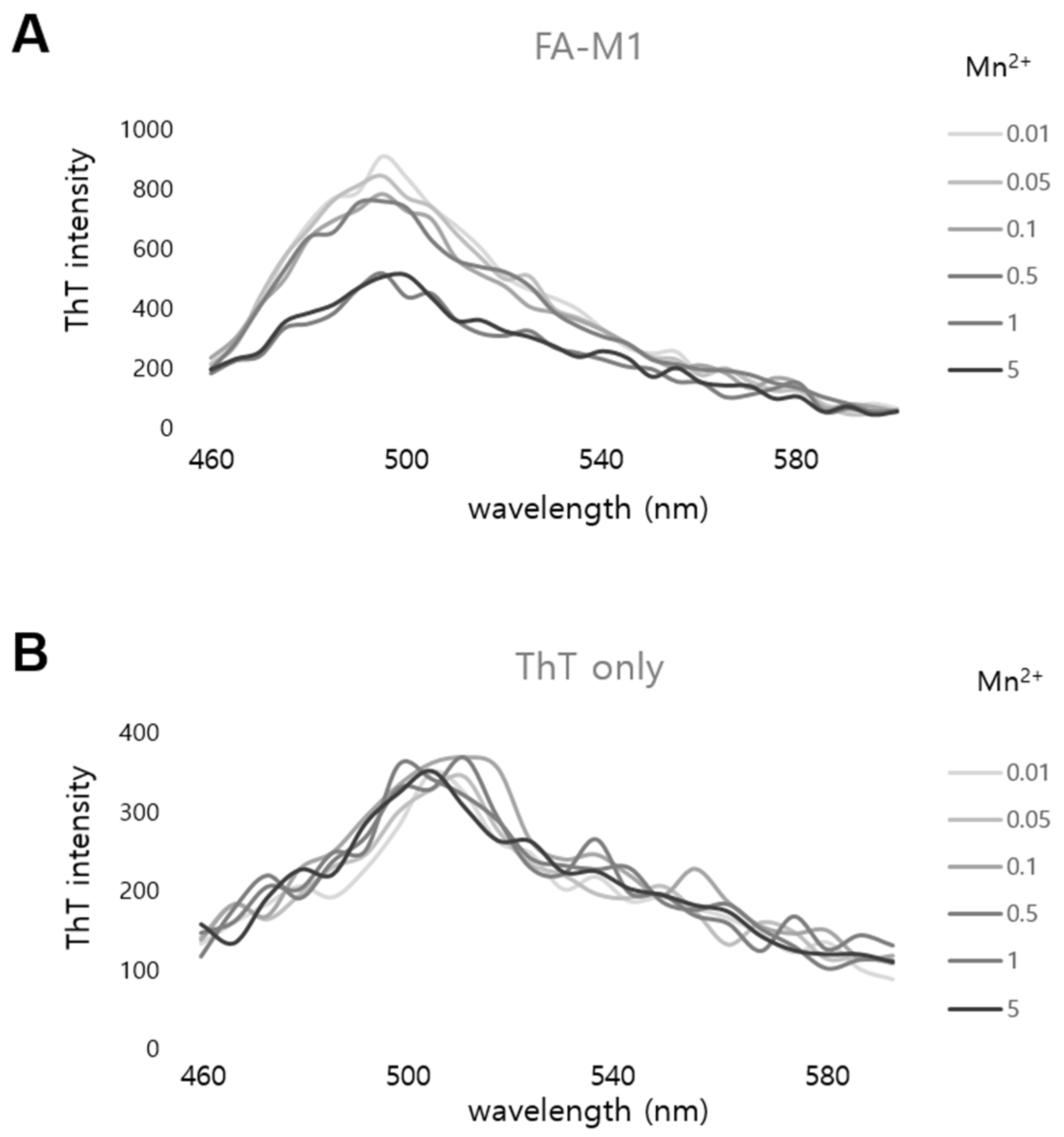
2.5. Fluorescent Spectrum Analysis Between an Aptamer and Mn2+ Using Thioflavin T

Thioflavin T (ThT, 3,6-dimethyl-2-(4-dimethylaminophenyl)-benzothiazolium cation) was purchased from Sigma-Aldrich. Fluorescence measurements were conducted using a 1 µM ThT solution in the presence of 0.1 mM aptamer and varying concentrations of Mn2+. The binding buffer used in the aptamer and ThT experiments was composed of 5 mM Tris–HCl (pH 7.5), 0.5 mM EDTA, and 1 M NaCl. Measurements were performed at room temperature using an EnSpire multimode plate reader (PerkinElmer Inc., Waltham, MA, USA). Fluorescence emission spectra were recorded between 460 nm and 600 nm, with an excitation wavelength of 425 nm.

2.6. Mn Removal from Synthetic Solutions

After determining the affinity and specificity of the selected aptamers for binding Mn ions, we determined whether these aptamers could remove Mn ions from an artificially produced solution containing Mn. To achieve this, we first designed a complex containing biotinylated aptamers bound to a streptavidin–agarose resin using a strong combination of streptavidin and biotin. The beads used were Pierce™ Streptavidin Agarose (Thermo Scientific™, Waltham, MA, USA), with a diameter ranging from 45 to 165 μm, and the microtubes used were Micro Bio-Spin™ Chromatography Columns (Bio-Rad, Hercules, CA, USA), with a size of 0.8 mL. For the experiment, 0.8 nmol of aptamers were used per 400 μL of beads in each chromatography column. We measured the amount of Mn removed by each aptamer–resin complex from the artificially produced solution. The manganese removal rate was determined as a percentage by dividing the manganese concentration in the solution that passed through the column by the initial manganese concentration in the solution (removal rate = pass [Mn]/initial [Mn] × 100). Manganese(II) chloride was purchased from Sigma-Aldrich (244589-10G, St. Louis, MO, USA). After aptamer–bead coupling, 300 µL of 10 µM Mn2+ ions (302 μg/L) were transferred into a microtube containing the aptamer–bead complex and incubated for 10 min at room temperature with a rotator (Roto-Bot, Benchmark Scientific, Sayreville, NJ, USA). After incubation, the microtubes were centrifuged for 4 min at 13,000× g. The supernatants were collected as the sample. Inductively coupled plasma optical emission spectroscopy (ICP-OES) was used to measure the concentration of Mn2+ in each sample. A PlasmaQuant PQ 9000 (Analytik Jena, Erfurt, Germany) equipped with a standard kit was used to analyze the concentration of Mn2+ in each sample. The measured data were sampled and processed using the Aspect PQ 1.2.4.0 program. The machine was operated according to the manufacturer’s instructions.

2.7. Mn Removal from the Reservoir Sample

One sample was obtained from the Seo-Gok Reservoir, which is close to an abandoned Mn mine in Wonju City, South Korea (37°17′28.2″ N, 127°56′36.6″ E). Additional water samples were collected from the nearby Yongsugol Valley (37°16′50.3″ N, 127°56′27.8″ E) and Seom River (37°14′31.9″ N, 127°44′51.0″ E) for comparison with the reservoir water. The Mn removal procedure was identical to that used to remove Mn from laboratory-prepared aqueous solutions, as described above. A total of 300 µL of the reservoir sample was introduced into the aptamer–bead complex and incubated for 10 min at room temperature with a rotator (Roto-Bot, Benchmark Scientific, Sayreville, NJ, USA). After incubation, the microtube was centrifuged for 4 min at 13,000× g. The concentrations of Mn and other constituents present in the reservoir sample were measured in the supernatant solutions using ICP-OES. Calibration was performed using ICP-OES standards adjusted to the sample matrix. For determination, we used the ICP multielement standard solution VI (Merck, Darmstadt, Germany).

2.8. Measurement of the Recovery Rate of Mn

The aptamer–bead complex with bound Mn ions in a column was heated at 95 °C for 3 min to measure the recovery of Mn from the aptamer. Next, the Mn concentration was determined using the same method as that mentioned in Section 2.5.

2.9. Statistical Analysis

All statistical analyses were conducted using SPSS software (IBM® SPSS® Statistics, ver. 23). For multiple comparisons, the data were analyzed using one-way analysis of variance (ANOVA). Tukey’s post-hoc test was used to identify significant differences between the groups.

3. Results

3.1. Screening and Selection of Mn-Specific DNA Aptamers by SELEX

Mn ion-specific DNA aptamers were screened from a random library of chemically synthesized DNA using in vitro selection and the SELEX amplification method [16] (Figure 1). Mn was immobilized on epoxy-activated Sepharose 6B resin, which allowed for the typical SELEX process (Figure 2A). The aptamer was amplified after reacting and washing with the aptamer library to identify the aptamer that binds to fixed Mn. This process was repeated 10 times for the selected aptamers that showed high specificity and binding activity. All the SELEX rounds were monitored by quantifying the concentration of the eluted aptamer (Figure 2B). The concentration of the amplified aptamers in each round was directly measured using real-time PCR. The concentration of aptamers selected from the pool was proportional to the number of rounds. After the 10th round, the concentrations of the selected aptamers were relatively high (Figure 2B). Therefore, their sequences were analyzed after obtaining six vector clones (from FA-M1 to FA-M6) containing each aptamer in the 10th round (Table 1).

3.2. Determination of the Most Effective Aptamer for Binding Mn

The secondary structures of the 6 selected aptamers were predicted using Mfold and are shown in Figure 3A. All 6 aptamers (from FA-M1 to FA-M6) exhibited a unique hairpin structure, but contained a similar random sequence (Table 1). To analyze the binding affinity of the 6 selected aptamers for Mn2+, we examined the Kd value of each aptamer using an SPR assay on a chip platform fixed with Mn ions (Figure 3B and Table 2). Of the 6 aptamers (from FA-M1 to FA-M6), the FA-M1 aptamer exhibited the highest binding affinity to Mn with the lowest Kd value of 4.56 × 10⁻⁹ M.

3.3. Specific Sequence Analysis of the Aptamers That Bind to Mn

Sequence analysis was performed to compare the similarities between the aptamers that bind to Mn. Using the program ClustalW, we performed multiple alignments of the Mn aptamer sequences and found a consensus: repeated multi-guanine (RMG: GGTGGGXGGGXGGGTGGA) occurred in all the random sequences (Figure 4A). Figure 4B shows that 4 of the 6 aptamers (FA-M1, FA-M2, FA-M4, and FA-M6) are very similar. Based on the 2D structure of each aptamer sequence, the RMG sequence seems to be responsible for the hairpin stem and loop structure (Figure 3A). To confirm this hypothesis, we sequentially substituted each of the two guanines of FA-M1 with thymines and then analyzed the 2D structure of the altered aptamers using Mfold (Supplementary Figure S1). While the FA-M1 sequence forms a hairpin stem containing 3–4 guanines, the aptamer sequences substituted with thymine either do not form a hairpin structure or form a much longer hairpin stem. These results suggest that an aptamer capable of binding to Mn is composed of a specific sequence containing an RMG capable of forming a hairpin stem and loop structure. Furthermore, the presence of a G-rich region in an aptamer is generally indicative of G-quadruplex structure formation [17,18]. To investigate whether the aptamers identified in our study exhibit this characteristic structural feature, we performed an experiment using Thioflavin T (ThT). Our findings revealed that the fluorescence intensity of ThT incorporated into FA-M1 decreased in a dose-dependent manner upon the addition of Mn, strongly suggesting the formation of a G-quadruplex structure within the identified aptamer sequence (Figure 5).
Because the aptamers FA-M1, FA-M2, and FA-M3 showed high affinity and specificity for Mn, we evaluated the ability of these aptamers to remove Mn from an artificially produced solution. Compared with the aptamer containing a random sequence as a control, FA-M1, FA-M2, and FA-M3 removed significantly higher amounts of Mn from the solution, with removal rates of more than 95% (Figure 6).
To further demonstrate the Mn-binding specificity of the FA-M1 aptamer, binding experiments were performed with a variety of other metal ions. The 8 metal ions (Co2+, Cr2+, Cu2+, Fe2+, Zn2+, Ni2+, Al3+, and Eu3+) chosen as the test panel are commonly found in the environment. We determined the degree to which the FA-M1–bead complex removed each ion from a solution containing a mixture of Mn and 8 metal ions; we observed that more than 70% of the Mn was removed, while less than 10% of the other ions were removed (Supplementary Figure S2). These results suggest that the FA-M1 aptamer interacts specifically with Mn and could provide highly efficient removal.

3.4. Mn Removal from the Seo-Gok Reservoir Using the Mn-Binding Aptamer FA-M1

Previous studies found that excess Mn (over 1.5 mM MnCl2) prohibits the efficiency of photosynthesis in long-term hydroponic cultivation [19]. Therefore, we evaluated the ability of the FA-M1 aptamer to specifically interact with Mn in environmental samples from the Seo-Gok Reservoir, which showed a high Mn concentration compared with that of the artificially made solution (Table 3). The Seo-Gok Reservoir, located near Wonju City, Korea, was established to store water from the Seo-Gok Stream for agricultural use. It has high Mn concentrations due to the proximity of abandoned Mn mines around it. The standard Mn concentration in drinking water is 50 μg/L, and the Mn concentration in the Seo-Gok Reservoir is very high at 442.2 μg/L, compared to the nearby Yongsugol Valley and the Seom River in Wonju City (Table 4). We measured the rate of Mn removal by the aptamer FA-M1 from the Seo-Gok Reservoir sample. The manganese removal rate was determined as a percentage by dividing the manganese concentration in the solution that passed through the column containing the FA-M1 aptamer–bead complex by the initial manganese concentration. As shown in Figure 7, FA-M1 showed a high Mn removal rate of 90% or more. These results indicate that the aptamer can effectively remove Mn ions even from actual Mn-contaminated water environments.

3.5. Recovery of Mn from a Mn-Containing Aptamer–Bead Complex

The ability to recover Mn from the column or resin that binds to the ion is important for two reasons: the column or resin can be used repeatedly, while the recovered material can also be reused or repurposed. In previous studies, we developed columns to remove radioactive cobalt and nickel using aptamers with recovery rates of more than 90% [20,21]. We evaluated whether Mn could be recovered from the Mn removal column developed in this study and found that when elution was conducted twice at 95 °C with an isolation buffer (the same volume as the volume of the beads), more than 99.9% of the Mn captured in the aptamer could be collected (Figure 8).

4. Discussion

Mn is essential for the physiological activity and metabolism of plants as it is actively involved in enzymatic activity. However, excess Mn limits the growth and yield of plants [22]. Water from the Seo-Gok Reservoir is used as agricultural water in the neighboring area; however, because of its high Mn concentration, its use poses a problem. In this study, we confirmed that in the Seo-Gok Reservoir water, the Mn content was at least 100 times higher than that in the agricultural water in the neighboring area (Table 3). We assessed whether Mn could be removed from the Seo-Gok Reservoir water using a new metal ion removal technology based on aptamers. First, an aptamer that strongly binds to Mn was screened; we confirmed that the binding affinity of the discovered aptamers was similar to or higher than that of the reported binding affinities between other metal ions and their specific aptamers (Figure 3 and Table 2). We also confirmed that >95% of Mn was removed under laboratory conditions using the screened aptamers. Based on this, we conducted a Mn removal experiment using water from the Seo-Gok Reservoir and confirmed that about 90% of the Mn was removed, similar to the laboratory conditions. This aptamer-based technology for removing metal ions has a great economic advantage because it can be recycled more than 20 times compared to existing ion exchange resins. In the future, the effects of aptamer-based Mn removal will be analyzed by comparing the yield and crop growth of the nearby farms. If an aptamer-based technology that can remove certain metal ions, including heavy metal ions, can be commercialized, it is believed that the concentrations of various heavy metals exposed to the environment and the agricultural water quality can be controlled.

5. Conclusions

Because of its harmful effects on ecosystems and human health, Mn exposure and intake have been studied for quite some time. However, to date, technologies for purifying Mn-contaminated environments have not been applied. Therefore, in this article, we proposed a novel method for removing Mn from contaminated water. We used SELEX to identify 6 types of aptamers that bind to Mn. We then performed an Mn removal and recovery test using the aptamers FA-M1, FA-M2, and FA-M3, with binding affinities in the range of 4.56~4.89 × 10⁻⁹ M. These high-specificity aptamers were able to remove 95% or more of Mn from a solution, and subsequently, more than 99.9% of the Mn complexed with the aptamer could be recovered. In addition, the FA-M1 aptamer removed approximately 90% of the Mn from a sample of water from the Seo-Gok Reservoir. The results of this study suggest an alternative method for protecting ecosystems and humans from heavy metal exposure by utilizing aptamers, an eco-friendly biomaterial.

Supplementary Materials

The following supporting information can be downloaded at: https://www.mdpi.com/article/10.3390/environments12040099/s1, Figure S1: Prediction of the Mn-binding region on an aptamer using the Mfold program; Figure S2: Specificity of the manganese aptamer FA-M1.

Author Contributions

Conceptualization, M.Y. and E.-O.K.; methodology, D.J. and S.Y.L.; software, S.Y.L.; validation, S.Y.L., D.J. and W.-S.K.; formal analysis, D.J.; investigation, S.Y.L. and W.-S.K.; resources, M.Y. and K.-J.R.; data curation, M.Y. and S.Y.L.; writing—original draft preparation, S.Y.L.; writing—review and editing, M.Y.; visualization, S.Y.L.; funding acquisition, M.Y. and E.-O.K. All authors have read and agreed to the published version of the manuscript.

Funding

This research was supported the Ministry of SMEs and Startups (MSS, Korea), which was granted financial resources by the Korea Institute of Technology and Information (TIPA), Republic of Korea [No. RS-2023-00269405] and the National Research Foundation of Korea (NRF), which is funded by the Korean government (MSIT) [No. 2023R1A2C1006127].

Data Availability Statement

Raw data that support the outcomes of this study are available from the corresponding authors upon reasonable request.

Conflicts of Interest

Author Miyong Yun was employed by the company Faptamer Inc. The remaining authors declare that the research was conducted in the absence of any commercial or financial relationships that could be construed as a potential conflict of interest.

References

  1. Bowman, A.B.; Kwakye, G.F.; Hernández, E.H.; Aschner, M. Role of manganese in neurodegenerative diseases. J. Trace Elem. Med. Bio 2011, 25, 191–203. [Google Scholar] [CrossRef]
  2. Santamaria, A.B. Manganese exposure, essentiality & toxicity. Indian J. Med. Res. 2008, 128, 484–500. [Google Scholar]
  3. Bouchard, M.F.; Sauvé, S.; Barbeau, B.; Legrand, M.; Brodeur, M.E.; Bouffard, T.; Limoges, E.; Bellinger, D.C.; Mergler, D. Intellectual Impairment in School-Age Children Exposed to Manganese from Drinking Water. Environ. Health Perspect. 2011, 119, 138–143. [Google Scholar] [CrossRef] [PubMed]
  4. Nkele, K.; Mpenyana-Monyatsi, L.; Masindi, V. Challenges, advances and sustainabilities on the removal and recovery of manganese from wastewater: A review. J. Clean. Prod. 2022, 377, 134152. [Google Scholar] [CrossRef]
  5. Wu, R.; Yao, F.; Li, X.; Shi, C.; Zang, X.; Shu, X.; Liu, H.; Zhang, W. Manganese Pollution and Its Remediation: A Review of Biological Removal and Promising Combination Strategies. Microorganisms 2022, 10, 2411. [Google Scholar] [CrossRef]
  6. Tobiason, J.E.; Bazilio, A.; Goodwill, J.; Mai, X.Y.; Nguyen, C. Manganese Removal from Drinking Water Sources. Curr. Pollut. Rep. 2016, 2, 168–177. [Google Scholar] [CrossRef]
  7. Zhou, W.; Huang, P.J.; Ding, J.; Liu, J. Aptamer-based biosensors for biomedical diagnostics. Analyst 2014, 139, 2627–2640. [Google Scholar] [CrossRef]
  8. Shoaib, M.; Li, H.; Zareef, M.; Khan, I.M.; Iqbal, M.W.; Niazi, S.; Raza, H.; Yan, Y.; Chen, Q. Recent Advances in Food Safety Detection: Split Aptamer-Based Biosensors Development and Potential Applications. J. Agric. Food Chem. 2025, 73, 4397–4424. [Google Scholar] [CrossRef]
  9. McConnell, E.M.; Nguyen, J.; Li, Y. Aptamer-Based Biosensors for Environmental Monitoring. Front. Chem. 2020, 8, 434. [Google Scholar] [CrossRef]
  10. Kulabhusan, P.K.; Hussain, B.; Yüce, M. Current Perspectives on Aptamers as Diagnostic Tools and Therapeutic Agents. Pharmaceutics 2020, 12, 646. [Google Scholar] [CrossRef]
  11. Guo, W.; Zhang, C.; Ma, T.; Liu, X.; Chen, Z.; Li, S.; Deng, Y. Advances in aptamer screening and aptasensors’ detection of heavy metal ions. J. Nanobiotechnol. 2021, 19, 166. [Google Scholar] [CrossRef] [PubMed]
  12. Sekhon, S.S.; Lee, S.H.; Lee, K.A.; Min, J.H.; Lee, B.T.; Kim, K.W.; Ahn, J.Y.; Kim, Y.H. Defining the copper binding aptamotif and aptamer integrated recovery platform (AIRP). Nanoscale 2017, 9, 2883–2894. [Google Scholar] [CrossRef]
  13. Zuker, M. Mfold web server for nucleic acid folding and hybridization prediction. Nucleic Acids Res. 2003, 31, 3406–3415. [Google Scholar] [CrossRef] [PubMed]
  14. Li, K.B. ClustalW-MPI: ClustalW analysis using distributed and parallel computing. Bioinformatics 2003, 19, 1585–1586. [Google Scholar] [CrossRef]
  15. Waterhouse, A.M.; Procter, J.B.; Martin, D.M.A.; Clamp, M.; Barton, G.J. Jalview Version 2-a multiple sequence alignment editor and analysis workbench. Bioinformatics 2009, 25, 1189–1191. [Google Scholar] [CrossRef]
  16. Tuerk, C.; Gold, L. Systematic Evolution of Ligands by Exponential Enrichment—Rna Ligands to Bacteriophage-T4 DNA-Polymerase. Science 1990, 249, 505–510. [Google Scholar] [CrossRef] [PubMed]
  17. Liu, Y.B.; Wang, X.Q.; Liu, J.W. Unexpected enrichment of DNA aptamers for Zn ions from an insulin selection. Chem. Commun. 2024, 60, 6280–6283. [Google Scholar] [CrossRef]
  18. Xia, T.; Yuan, J.H.; Fang, X.H. Conformational Dynamics of an ATP-Binding DNA Aptamer: A Single-Molecule Study. J. Phys. Chem. B 2013, 117, 14994–15003. [Google Scholar] [CrossRef]
  19. Millaleo, R.; Reyes-Díaz, M.; Ivanov, A.G.; Mora, M.L.; Alberdi, M. Manganese as Essential and Toxic Element for Plants: Transport, Accumulation and Resistance Mechanisms. J. Soil Sci. Plant Nutr. 2010, 10, 476–494. [Google Scholar] [CrossRef]
  20. Jang, D.H.; Lee, S.Y.; Kim, H.; Yun, M.Y. Selection of a nickel-specific DNA aptamer and development of a protocol for radioactive nickel removal. Ann. Nucl. Energy 2024, 195, 110138. [Google Scholar] [CrossRef]
  21. Lee, S.Y.; Jang, D.H.; Kim, H.; Yun, M. Removal and isolation of radioactive cobalt using DNA aptamers. Radiochim. Acta 2023, 111, 357–365. [Google Scholar] [CrossRef]
  22. Li, J.F.; Jia, Y.D.; Dong, R.S.; Huang, R.; Liu, P.D.; Li, X.Y.; Wang, Z.Y.; Liu, G.D.; Chen, Z.J. Advances in the Mechanisms of Plant Tolerance to Manganese Toxicity. Int. J. Mol. Sci. 2019, 20, 5096. [Google Scholar] [CrossRef] [PubMed]
Figure 1. Schematic illustration of the SELEX process. A schematic representation of the SELEX (systematic evolution of ligands by exponential enrichment) process used to generate aptamers specific to manganese ions. The process involves iterative cycles of binding, washing, and amplification to enrich for high-affinity aptamers.
Environments 12 00099 g001
Figure 2. Screening and selection of Mn-specific DNA aptamers. (A) Coupling of Mn to an epoxy-activated Sepharose 6B resin for aptamer selection. (B) Quantitative monitoring of Mn-binding aptamers during successive SELEX rounds, illustrating the enrichment of aptamers with high specificity.
Environments 12 00099 g002
Figure 3. Secondary structures and binding affinities of the selected Mn aptamers. (A) Predicted secondary structures of the six selected Mn aptamers (from FA-M1 to FA-M6) modeled using Zuker’s algorithm. (a) FA-M1, (b) FA-M2, (c) FA-M, (d) FA-M4, (e) FA-M5, and (f) FA-M6 aptamer. (B) Surface plasmon resonance (SPR) sensorgrams showing the binding kinetics and dissociation constants (Kd) of the aptamers to Mn ions. X-axis: RU (response unit: the change in the signal detected during a binding interaction), Y-axis: time (seconds).
Environments 12 00099 g003aEnvironments 12 00099 g003b
Figure 4. Sequence homology analysis of Mn aptamers. (A) Multiple sequence alignment of Mn-binding aptamers highlighting conserved regions. (B) Phylogenetic tree of the selected aptamers indicating similarities in the sequence and potential functional motifs.
Environments 12 00099 g004
Figure 5. Fluorescent spectrum analysis for FA-M1 using Thioflavin T with various concentrations of Mn2+. Schematic representation of ThT fluorescence spectra of (A) FA-M1 and (B) without FA-M1 in the presence of Mn2+ at concentrations of 0.01, 0.05, 0.1, 0.5, 1, and 5 µM.
Environments 12 00099 g005
Figure 6. Mn removal efficiency using aptamer–bead complexes. Removal rates of manganese ions from synthetic solutions using aptamer–bead complexes. Results indicate high removal efficiency by the selected aptamers (FA-M1, FA-M2, and FA-M3). All the experiments were repeated three times. X-axis: control and aptamer; Y-axis: removal rate percentage (resulting/initial ion concentration). Error bars represent the standard deviations for the replicates (n = 3). Note: *** statistically significant difference in the removal rate compared to the control (p-value < 0.001).
Environments 12 00099 g006
Figure 7. Application of the FA-M1 aptamer in the Mn-contaminated reservoir water. Capacity of the FA-M1 aptamer–bead complex to remove manganese and other metal ions from the Seo-Gok Reservoir water sample. Results demonstrate selective and efficient manganese removal. All the experiments were repeated three times. X-axis: metal ions, Y-axis: removal rate percentage (passing through/initial ion concentration). Error bars represent the standard deviations for the replicates (n = 3). Note: *** statistically significant difference in the removal rate compared to the control (other metal ions excluding manganese) (p-value < 0.001).
Environments 12 00099 g007
Figure 8. Recovery of manganese from aptamer–bead complexes. Recovery rates of manganese from the FA-M1 aptamer–bead complexes following thermal denaturation. The results confirm the potential for repeated use and recovery of manganese with minimal loss. X-axis: the control and the aptamers; Y-axis: recovery rate percentage (recovery/removal ion concentration). Error bars represent the the standard deviation (n = 3). Note: *** statistically significant difference in the removal rate compared to the control (p-value < 0.001).
Environments 12 00099 g008
Table 1. Sequences of the selected aptamers that bind to Mn ions.
Aptamer NameAptamer Sequence
FA-M1GGTAATACGACTCACTATAGGGAGATACCAGCTTATTCAATTGGTGGGTGGGTGGGTGGAAGATGAAAAGGGGATCCGGGTGAGATTGCACTTACTATCT
FA-M2GGTAATACGACTCACTATAGGGAGATACCAGCTTATTCAATTCCATCGGTGGGTGGGCGGGTGGAGAGGGCTTTGTTAAAACAGATTGCACTTACTATCT
FA-M3GGTAATACGACTCACTATAGGGAGATACCAGCTTATTCAATTAGAGGTCAGGGTAGGGAGGGGGGAATAAGGTGTCACACGGAGATTGCACTTACTATCT
FA-M4GGTAATACGACTCACTATAGGGAGATACCAGCTTATTCAATTCGGTGGGTGGGTGGGTGGAGAAGAGTAAGTGGCGATAAGGAGATTGCACTTACTATCT
FA-M5GGTAATACGACTCACTATAGGGAGATACCAGCTTATTCAATTTTGATGCACAGTACGGATGAGTAGGCAGGGTAGGAGTGGAAGATTGCACTTACTATCT
FA-M6GGTAATACGACTCACTATAGGGAGATACCAGCTTATTCAATTCGGTGGGAGGGCGGGTGGAGAATTATAATACGATTTGGAAAGATTGCACTTACTATCT
Table 2. Mn aptamer dissociation constants (Kd) and Gibbs free energy (ΔG) values. The Kd values were measured using an SPR instrument as a change in the refractive index over time, ΔG—as a change in free energy; Since all reactions naturally progress toward a state of lower energy, a negative change is favored. The experiments were carried out at 25 °C.
Aptamer NameKD ValueΔG Value (kcal/mol)
FA-M14.56 × 10−9−9.40
FA-M24.86 × 10−9−10.38
FA-M34.88 × 10−9−10.54
FA-M45.11 × 10−9−14.96
FA-M55.39 × 10−9−10.82
FA-M65.81 × 10−9 −7.63
Table 3. Water condition in the Seo-Gok Reservoir. The initial Mn content measured by ICP-OES was 442.2 μg/L. Conc.: concentrations are shown as μg/L.
IonsCoNiMnCuCrAgAlFeCd
Conc.1.30.8442.21.40.10.210.776.70.0
IonsLiMoPbBaZnBKMg
Conc.1.30.30.126.33.015.9195.2398.3
Table 4. Comparison of the water conditions in the Seo-Gok Reservoir and other places in Wonju City. Concentrations are shown as μg/L.
CoNiMnCuCr
Seo-Gok Reservoir1.30.8442.21.40.1
Seom River0.40.13.41.30.1
Yongsugol Valley0.20.42.80.40.0
Disclaimer/Publisher’s Note: The statements, opinions and data contained in all publications are solely those of the individual author(s) and contributor(s) and not of MDPI and/or the editor(s). MDPI and/or the editor(s) disclaim responsibility for any injury to people or property resulting from any ideas, methods, instructions or products referred to in the content.

