Abstract
This study examines the blood-oxygen level dependent (BOLD) activation of the brain associated with the four distinctive thinking styles associated with the four personality orientations of the Gountas Personality Orientations (GPO) survey: Emotion/Feeling-Action, Material/Pragmatic, Intuitive/Imaginative, and Thinking/Logical. The theoretical postulation is that each of the four personality orientations has a dominant (primary) thinking style and a shadow (secondary) thinking style/trait. The participants (N = 40) were initially surveyed to determine their dominant (primary) and secondary thinking styles. Based on participant responses, equal numbers of each dominant thinking style were selected for neuroimaging using a unique fMRI cognitive activation paradigm. The neuroimaging data support the general theoretical hypothesis of the existence of four different BOLD activation patterns, associated with each of the four thinking styles. The fMRI data analysis suggests that each thinking style may have its own cognitive activation system, involving the frontal ventromedial, posterior medial, parietal, motor, and orbitofrontal cortex. The data also suggest that there is a left hemisphere relationship for the Material/Pragmatic and Thinking/Logical styles and a right activation relationship for Emotional/Feeling and Intuitive/Imaginative styles. Additionally, the unique self-reflection paradigm demonstrated that perception of self or self-image, may be influenced by personality type; a finding of potentially far-reaching implications.
1. Introduction
Understanding the neural basis of human personality is a core imperative of cognitive neuroscience [1,2] and social neuroscience [3,4]. Researchers have employed a variety of techniques including functional magnetic resonance imaging (fMRI) [5], electroencephalography (EEG) [6], in addition to behavioral and psychometric tests [7,8]. One of the many important research questions is to further investigate the neurobiology of behavioral relationships and personality traits/orientations. Several studies have explored these associations between individual personality traits/orientations and decision making [9,10,11]. Personality traits and disorders are found to correlate with poor or maladaptive decision making (e.g., dissociative, compulsive and affective disorders). Maladaptive personality traits and disorders have been associated with deficits in distinct neural systems [12,13,14]. The focus of this study is on the adaptive and ‘normal’ subjects’ personality orientations and putative four thinking styles (GPOs). The aim was to explore the neural basis of the new GPO model using fMRI, and test whether the hypothesized four distinct brain systems’ activations are associated with each of the four personality orientations (GPO). This was achieved with the use of a unique self-reflection fMRI paradigm.
Neuroimaging techniques are well suited to studying how personality traits are coded in the brain because it is non-invasive and affords several relatively simple analytic approaches to reveal spatial localization of discrete functional architectures [15,16]. Personality trait differences activate different brain networks, established through ‘functional connectivity’ of resting-state fMRI acquisitions and inter-regional network connectivities. Using fMRI, Adelstein et al. [17] found that each of the five NEO dimensions predicted distinct brain activations and resting state networks. Haas and colleagues [18] reported that agreeableness, a measure of sympathy and social cooperation, predicted blood-oxygen level dependent (BOLD) changes in the temporoparietal cortex during the testing of an emotional attribution task. After controlling for the other Big 5 dimensions, (and educational levels), participants who were highly agreeable made faster emotionally valenced decision attributions and exhibited greater BOLD signal intensity in the right temporoparietal junction. The medial frontal cortex (mFC) myelination and activation have also been implicated in other personality studies, respectively [19,20]. In addition, photic driving EEG responses were correlated with the NEOPI-R personality dimensions of openness, agreeableness, and consciousness with multiple EEG bands. The results suggested individual activations in the occipital-parietal and temporal cortices for these three dimensions [21], which have been independently corroborated [22,23]. Several personality related studies have reported that distinct electrical activation characteristics reflect differences in behavioral and decision-making processes associated with specific personality orientations/traits [24,25].
The current neuroimaging study used a Jungian-based personality model, developed by Gountas and colleagues [9,10,26], which identifies four ontogenetically different personality orientations that correspond to different social behavioral preferences and thinking styles. According to Gountas et al. [10], each personality orientation (PO) focuses on different types of information input (e.g., empirical/physical, abstract/pictorial, ideational/conceptual, experiential/ feelings), and consequently, use different cognitive processing systems that affect preferences and decision-making choices. Notably, according to the GPO model, each individual has access to two thinking styles, but one thinking style is the dominant PO (primary) and the second is the secondary PO (e.g., Material dominant PO and Thinking secondary PO). By developing the GPO measure into a word-based self-reflection computer driven experimental task for an EEG, and subsequent fMRI studies, Gountas et al. [27] were able to identify distinctive EEG networks of brain electrical activity, using EEG alpha coherence associated with each of the four personality orientations.
The theoretical model for each of the four GPO thinking styles is based on extensive qualitative research (depth interviews and focus groups), as well as quantitative surveys to establish their validity and reliability [9,10]. The four personality orientations are an alternative model to the dual processing systems [9,10,28], because the four GPO model more clearly identifies the nature of the logical slow deliberate rational processing (system 2) and adds more in-depth and specific explanation about the system 1, hypothesized to be fast, heuristic, or intuitive cognitive processing [28].
According to Gountas et al. [9,10,11], the four GPOs identify four distinctive traits based on fundamentally different modes of information processing. The broad traits of each of the four GPOs are as follows:
- Thinking/Logical PO (L): They primarily focus on semantic ideational thinking and rational conceptual thinking style; they tend to focus on generating new ideas, making sense of the why and how things work, and emphasizing the need to know rational explanations of phenomena. Logical ideational individuals focus on making sense of life based on the coherence of existing valid knowledge, and scientific findings. They are able to evaluate information objectively, based on factual criteria and logically analyzing, thinking through (often without emotional influences) the merits of concepts and events based on rational assessment [29,30].
- Material/Pragmatic PO (P): They focus on physical empirical evidence and knowledge structures. They are characterized by a strong emphasis on physical attributes, characteristics, and phenomena, which are measurable and tangible. They prefer to deal with the physically objective realities, amenable to objective investigation. They aim for material success, material possessions, and enjoying the physical advantages of the world. Materialists get immense pleasure from physical pleasures and the tangible attributes of products (often without concerns for social or emotional impacts). They rely heavily on the input of their somatosensory faculties to make decisions. Embodied type of learning and sense making is the dominant cognitive processing style.
- Emotion/Feeling-Action PO (E): They engage in experiential and action-based learning, understanding, and thinking processes. They tend to be action oriented, observing and planning actions, and have high levels of energy. High sensorimotor arousal, influences goal-motivated actions to satisfy their needs. They are usually social intelligent, competitively oriented toward social goal achievements. They are driven by a high sense of self-efficacy, and a ‘can do’ self-reliant attitude. They are able to makes sense and find solutions to various challenges through action-based strategies. Social learning, role playing, and trial error experiential learning generate ideas and thinking to make sense of their world. Experiential information underpins conceptual thinking and understanding of what decisions to make.
- Intuitive/Imaginative PO (I): They are creative types of thinkers, able to visualize information and able to think abstract constructs. Imaginative visualizers are characterized by a heightened level of sensitivity and awareness of unconscious perceptions regarding world phenomena. They tend to rely on visual, graphic, or animated types of learning stimuli. They are more likely to use imagination habitually to process information and problem solving. Abstract thinkers can develop complete solutions rather than following a sequential inductive step by step analytical approach. Information is mentally and visually processed with ease and counterfactual concepts emerge to solve problems. They can produce imaginative, novel creative ideas about world experiences and phenomena that are not empirically obviously detectable.
Research Hypotheses
Previous research suggests that each orientation has a distinct frontal cortex organization with Logical thinking associated with left hemisphere and the Intuitive/Imaginative thinking style demonstrating a right hemisphere cortical organization and cognitive activation patterns [31]. Several fMRI studies suggest that ventromedial prefrontal cortex (vmPFC), orbitofrontal cortex (OFC), and posterior medial frontal cortex (pMFC) are associated with self-reflection types of responses and decision-making processes [20,32,33].
The current study was designed to expand our previous EEG research findings of the neural basis of the Gountas PO model, by using the blocked fMRI design [27]. The general hypothesis of this fMRI study was that the ventromedial prefrontal cortex, orbitofrontal cortex, and posterior medial cortex would be activated differentially by the four GPOs during a unique self–reflection paradigm, and in addition, would support the bilateral hemisphere and frontal and temporal-parietal relationships identified in the previous EEG study [27]. Based on previous literature, we also hypothesized that regions associated with various cognitive and emotional processes would be specifically related to each of the orientations. For example, the Emotional PO would be associated with regional emotional processing [34,35,36,37,38,39]. The Logical PO (the ideational logical reasoning) are hypothesized to produce stronger activation in prefrontal regions associated with executive function, [40,41]. The Imaginative PO (the creative visualizing thinkers), will demonstrate higher activations in the medial prefrontal cortex, inferior parietal cortex (including the temporoparietal junction), together with visual sensory brain regions [42,43,44,45]. Finally, the Material/Pragmatic type (the material/pragmatic rational thinkers) will produce stronger activations in the inferior parietal lobe (IPL) and association brain regions [19,41,42,46].
2. Materials and Methods
2.1. Participants
Informed and written consent was obtained from all participants (university students and community sample), according to the research protocols as approved by the University Human Ethics Committee. (SUHREC Project 2009/096) Initially, forty-eight (n = 48) participants were pre-tested with the Gountas Personality Orientation (GPO) instrument to determine their personality orientations and ensure equal numbers of at least ten participants of each of the four GPOs. A final sample of forty participants (n = 40, 10 for each PO) were selected for the fMRI study; with a mean age of 27.5 years and SD 7.6 years. The sample consisted of 22 females (20 right handed), and 18 males (16 right handed). The forty participants comprised four equal groups representing each of the four dominant personality orientations. All participants were self-screened for neurological conditions, ensuring that there was no history of head trauma, or any psychiatric disorders, or substance abuse, and appropriate current medication status to participate in the fMRI study.
2.2. Experimental Materials
The GPO Survey Assessment is a 39 question inventory identifying the four primary and secondary orientations of thinking styles. The GPO survey quantifies four personality orientation or broad traits that are associated with how people think (thinking style preferences), what types of information they naturally prefer to focus on (cognitive processing systems used), and what broad types of cognitive processes they go through [26,47]. The four GPOs are: Emotion/Feeling-Action (E), Material/Pragmatic (P) Intuitive/Imaginative (I), and Thinking/Logical (L). Each participant completed the 39-question survey to group together the four phylogenetic groups of cognitive processing personality orientations.
For all fMRI experimental test recordings and analysis, participants were grouped based on their a priori dominant and secondary personality orientations as scored by the GPO instrument results. Based on the original paper by Gountas et al. [26], the survey results of the factor analysis for the four personality orientations produced a very robust Cronbach’s alpha for each orientation: Thinking/Logical α = 0.85, Material/Pragmatic α = 0.80, Emotion/Feeling-Action α = 0.83, and Imaginative/Intuitive, α = 0.85. Each of the orientations was also found to be independent from each other. To test our hypotheses, the fMRI results for each hypothesized PO (dominant and secondary orientation) were analyzed as four separate groups and therefore tested separately for differences from each of the other POs (e.g., Emotion/Feeling-Action PO vs. Non-E/F PO). This was done to improve and verify each PO group’s statistical significance for the fMRI results and each PO group was compared with the other three groups without the same PO traits. The final numbers in each grouping (with primary and secondary orientation) were: Thinking/Logical PO: (n = 19, non-T n = 21); Intuitive/Imaginative PO: (n = 19, Non-I n = 21); Material/Pragmatic PO: (n = 22, Non-M n = 18); Emotion/Feeling-Action PO: (n = 17, Non-E n = 23).
2.3. Neuroimaging fMRI Methodology
Participants were scanned using a Siemens 3T Tim Trio equipped with a 12-channel head coil at the Brain Research Institute (Melbourne, Australia). Each scanning session involved acquisition of a high-resolution T1-weighted magnetization-prepared gradient echo (MPRAGE) scan (192 sagittal slices; 0.9 mm isometric voxels; TR = 1900 ms, TE = 2.6 ms), that was used to register functional image activation into the standard stereotactic space defined by the Montreal Neurological Institute (MNI space). During each functional run, 139 gradient-echo, echo-planar T2*-weighted images (EPIs: TR = 3000 ms, TE = 30 ms, FoV = 216 × 216 mm, 72 × 72 imaging matrix, 44 interleaved axial slices, 3 mm isometric voxels) were acquired. The cognitive activation task was triggered by the onset of the sixth volume (volumes 1–5 were discarded), hence each experimental run was associated with 134 EPI images.
2.4. Neuroimaging Tasks
Stimulus delivery and recording of behavioral responses were controlled with E-Prime software (Psychology Software Tools Inc., Sharpsburg, PA, USA). The cognitive activation paradigm was presented in a typical block design comprising three tasks: a Fixation task, a Word task, and a Sentence task (or self-reflection task). The Word and Sentence tasks were constructed from words and questions based directly on the GPO psychometric instrument (see Figure 1). Participants had to make a ranking order choice between four alternative options corresponding to each of the four POs. Block onsets were locked to scan separate onsets, and each block lasted 21 s (i.e., seven scans or volumes). Each block was repeated 12 times (one trial), with a rest period mid-way through the protocol to secure against potential participant fatigue. Trials were repeated twice.
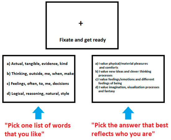
Figure 1.
Three components of the block design protocol. The tasks were sectioned into three components of varying demand: Fixation-Rest (top), Words task (left) and Sentences self-reflection task (right). Examples of the words and sentences used are illustrated.
The Word and Sentence tasks were visually and physically similar (luminosity and spatial frequency) and based on the GPO survey to minimize any visual novelty effects. The participants were required to press (make a choice) one of four buttons for both tasks; each button corresponded to a separate choice of screen text (labelled as a, b, c, d, respectively). The requirement for the Word task was to read four lines of text and select the line of words they preferred most-least in a hierarchic sequence: a, b, c, or d. The requirement for the Sentence task was to read four lines of short sentences and choose which sentence best represented their own thinking style or cognitive processing (self-reflection). These trials and tasks were repeated as part of the scanning sequence (block design).
2.5. Neuroimaging and Statistical Analysis
All aspects of image pre-processing and statistical analysis were performed using SPM8 (Wellcome Trust Centre for Neuroimaging) and associated toolboxes.
a. Pre-processing
Initially, slice-artifacts present in Echo-planar imaging (EPI) were repaired using Artrepair tools [48]. Functional images were then realigned to the first image acquired, and the T1 scan was co-registered to a mean realigned EPI computed during realignment. T1 images were subsequently normalized to the MNI template supplied with SPM8, and the parameters of this transformation were applied to realigned EPIs. Normalized EPIs were then smoothed using an 8 mm FWHM Gaussian filter.
b. Modeling of neuroimaging data
Participant level modeling of pre-processed fMRI data was performed on the image data acquired in the first run as a ‘localizer task’ and the remaining two runs (‘task’) separately, using the general linear model (GLM) approach in SPM8. Initially, separate box-car functions defining the onsets and durations of the three conditions (fixation, word, and sentence tasks) were convolved with the canonical hemodynamic response function (HRF) supplied in SPM8 for each analysis. Motion realignment parameters estimated during pre-processing were modeled as covariates of no interest, and after parameter estimation, contrasts were computed. A single subject multi-trial was done to estimate statistical power required for whether we needed 1, 2 or 3 trials. Based on these analyses, it was deemed that two trials were sufficient (7 × 12 = 84 scans). The SPM Statistical Design for Rest (Fixate), Words, and Sentences tasks was implemented accordingly (note threshold T = 3.11).
c. Group level modeling
Our analytic approach involved using the first run as a ‘localizer task’ from which regions of interest (ROIs) were determined via one-sample t-tests. Significant clusters of activation (thresholding was p < 0.001 uncorrected at the voxel level and p < 0.05 family-wise error (FWE) corrected at the cluster level) observed in these tests were set as ROIs. This was performed instead of doing non-independent analyses with voxel by psychometric correlations as suggested by Vul et al. [49]. Cluster corrections with family-wise error (FWE) and false discovery rate (FDR) were applied to cluster level and peak level data.
All consequent ROI processing (ROI building, signal extraction) was performed using MarsBar region of interest toolbox (Version 0.43) for SPM8 [50]. The mean signal intensity from each ‘scan’ and each ‘task’ run was extracted for each ROI. For each ROI, the signal time-course from the scan preceding each block (x0) until three scans following each block (i.e., 10 TRs) were averaged then baselined by subtracting the BOLD signal intensity from the first scan in each block (x1) from each TR (i.e., x0 − x1, x1 − x1, x2 − x1, …, x10 − x1) [51]. These time-courses were then averaged across groups and plotted as a function of TR. Brain activity associated with preponderant GPOs were examined in separate but complimentary analyses: (1) signal time-courses for each ROI were extracted from the remaining two runs and plotted for each group by condition, and (2) two-sample t-tests were computed to assess brain activation linked to GPOs.
3. Results
3.1. Personality Orientations and fMRI Activity
Self-reflection during the GPO experimental testing was localized using the block design for contrasting Words and Sentences. A group fMRI analysis was performed to examine each personality orientation: Emotion/Feeling-Action (E), Material/Pragmatic (P), Intuitive/Imaginative (I), and Thinking/Logical (L). Using a two-sample t-test for contrasting groups, SPM analysis produced the fMRI images illustrated in Figure 2A–D. These demonstrate significant activation (p < 0.05) in different brain areas for each of the four orientations. Those with the specific PO orientation as either their dominant or secondary thinking style (Trait group) were contrasted against those without the PO, (Non-trait group), (see Table 1a,b). The effects of age, handedness, and gender were not found to be significant.
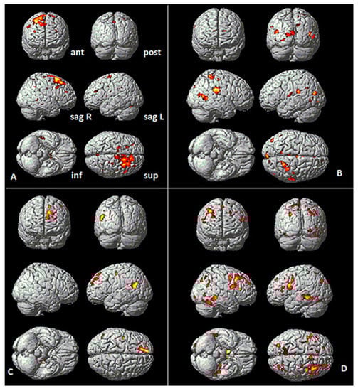
Figure 2.
fMRI two-sample t-test Sentences versus Word Overall activation (threshold p < 0.05). Talairach coordinates for each significant regional difference are summarized in Table 1a,b. Note anatomical views are anterior (ant), posterior (pos), left sagittal (sag L), right sagittal (sag R), inferior (inf), and superior (sup). (A) (Top Left), Emotion Type (17 E group vs. 23 Non-E group). (B) (Top Right) Imaginative Type (n = 19 I group vs. 21 Non-I group). (C) (Bottom Left) Logical Type (19 L group vs. 21 Non-L group). (D) (Bottom Right) Material Type (22 M group vs. 18 Non-M group).

Table 1.
(a) Group analysis (trait versus non-trait) contrast of Emotion/Feeling-Action PO and Imaginative/Intuitive PO; region by Talairach coordinates, Brodmann area (BA), hemisphere (H), and function (significant activations p < 0.05). Note definitions of common functions based on previous work [52]. (b) Group analysis (trait versus Non-trait) contrast of Material/Pragmatic PO and Logical/Thinking PO; region by Talairach coordinates, Brodmann area (BA), hemisphere (H), and function (significant activations p < 0.05). Note definitions of common functions, see [52].
Additionally, the fMRI two sample t-test, for the Words vs. Sentences contrasts, demonstrate several other relationships. For example, the Emotion/Feeling-Action types have right frontal activity while the Logical/Thinking types have statistically more left frontal brain activity (see Figure 2A vs. Figure 2C). Interestingly, the stronger brain activation in the left hemisphere for the Logical/Thinking types contrasted with the Emotion/Feeling-Action and Imaginative/Intuitive types with more significant activation of the right hemisphere. The group analysis data (Trait PO vs. non-Trait PO) summarized in Table 1a,b, further highlight these different relationships.
3.2. Emotion/Feeling-Action Personality Orientation
The following Brodmann Areas 6, 8, 20, 30, 44, were significantly active (threshold p < 0.05, Family-Wise Error-FWE corrected) during the self-reflection phase of the reading tasks (Sentences vs. Words) for the participants with E PO (see Figure 2A and Table 1a). Figure 2A illustrates right frontal activation consistent with the hypothesis that E types use the right pFMC, which is involved in processing emotions [34]. Interestingly other regions associated with memory (hippocampus), planning complex movements, and representation of complex object features, and face perception were also activated, consistent with previous findings in emotion perception [53,54,55]. Compared to the other POs, this collection of brain regions for introspection is very specific to the Emotion/Feeling-Action PO. However, one area (BA30) was also active for the Imaginative/Intuitive PO but was restricted to the right hemisphere.
3.3. Intuitive/Imaginative Personality Orientation
For the Intuitive/Imaginative type, the following Brodmann Areas 2, 5, 11, 30, 37, 39, 41, 48 were significantly active (threshold p < 0.05), (see Figure 2B and Table 1a). This PO demonstrates an imaginative approach to decision-making, utilizing strategic thinking, intuition, emotions, and memory. This can be clearly seen by the fact that the orbitofrontal areas (BA39) and (BA11) were active (consistent with Yasuno et al. [19], supporting the hypothesis for the Imaginative/Intuitive PO. The various association regions involved in sensory and somatosensory were also involved (BA2, BA5 and BA41) together with face identification (BA37) and memory (BA48).
3.4. Logical/Thinking Personality Orientation
The key regions activated for this PO were those associated with logical, planning processes: the dorsolateral prefrontal cortex (BA9) and a region associated with integration of sensory and mnemonic information, the regulation of intellectual function, action, and working memory (BA46), (see Figure 2C and Table 1b). Other regions associated with decision-making, risk evaluation, social context, beliefs, and visual memory were also activated (BA18, 23, 25, 36, 37, and BA48). This is consistent with the hypothesis that the Logical trait would exhibit activation in the vmPFC and Dorsolateral Prefrontal Cortex, consistent with Saito et al. [40].
3.5. Material/Pragmatic Personality Orientation
This orientation primarily demonstrated regions of activation associated with empirical (mathematical) thinking and physical reward preferences, learning through factual information and analysis; and using empathy and emotion-based decision-making (anterior cingulate gyrus—BA32), (see Figure 2D and Table 1b). However, other regions associated with risk, fear, executive decision making (BA25), and identifying social context (BA36) were also significant for this type. This is consistent with the hypothesis that Material/Pragmatic traits are associated with vmPFC (BA25) activation [19,40]. Other regions activated were associated with complex physical object visual sensory processing (BA20) and sensory memory (BA48). This is consistent with the descriptors for the Material/Pragmatic PO, and that sensory information associated with decision-making and self-reward are important factors for this orientation.
The fMRI data analysis (Figure 2 and Table 1a, b) suggests that all hypotheses were supported; however, several additional brain regions were also activated for each PO. We observed that the Emotion/Feeling and Intuitive/Imaginative POs had their activations distributed to the right hemisphere, while Thinking/Logical types tended to have more left hemisphere distribution of activity. The Material/Pragmatic tended to have processes associated with both the left and right hemisphere.
3.6. Personality Orientations and ROI Activity-Localizer Scan
To understand the time-course characteristics of the task-related activation associated with each PO, regions of interest (ROIs) were identified using another approach, namely, MR time-course during localizer scans [51]. By analyzing all participant data together (n = 40) for Sentences versus Words, cortical and subcortical activations were identified as regions of interest (ROIs). This is illustrated in Figure 3, where a one-sample t-test was performed (note that T = 3.312788 for p < 0.001). Additional regions including subcortical were identified. Twenty-two ROIs were examined for further analysis. These are listed in Table 2.
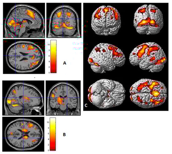
Figure 3.
Localizer scan, region of interest analysis (ROIs). One sample t-test for “Sentences versus Baseline” overall activation. Note for n = 40, t = 3.312788 (p < 0.001). Twenty-two ROIs were identified by this approach (see Table 2). (A) One sample t-test Sentences vs. Baseline overall activation sectioned at the precuneus (sagittal, coronal, and transverse). (B) One sample t-test Sentences vs. Baseline overall activation sectioned (sagittal, coronal, transverse) at the level of the thalamus. (C) One sample t-test Sentences versus Baseline overall activation.
Table 2.
Summary of regions of interest (ROI): 22 ROIs were identified from data associated with Figure 3. Note (p < 0.05) (Talairach coordinates Brodmann area (BA), hemisphere (H)).
The more significant frontal ROIs (from Table 2) for each PO were selected and produced as MR time-course images (see Figure 4, Figure 5, Figure 6 and Figure 7). Additionally, significant examples were also included for Imaginative and Material POs. Significant group effects were found with ROI (higher percentage signal change) versus PO (Table 3). Figure 4 illustrates the key ROI time-course associated with the Thinking/Logical types; the left mid-frontal cortex (BA10), which were significantly different (p = 0.011, 0.046, 0.044) for the average time course for Scans 3, 4 and 5, respectively, distinguished this Logical group from a non-Logical group with a higher signal change of this region.
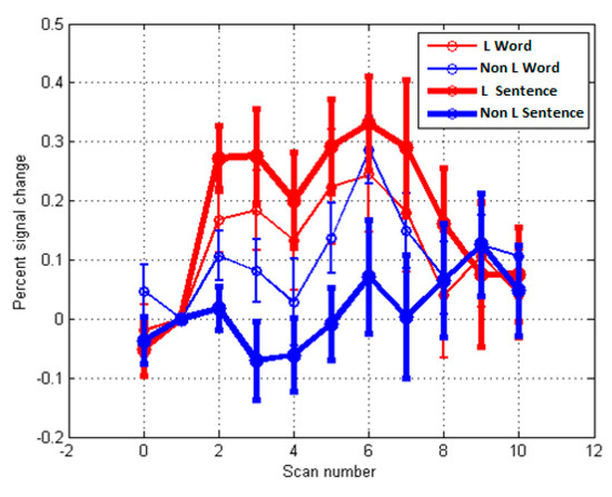
Figure 4.
MR time-course during localizer activity averaged across participants for Sentences versus Word overall activation. (p < 0.05) for Thinking/Logical PO. The ROI highlighted is the left mid frontal cortex or Brodmann area 10; comparing the Logical group (red; n = 19) with the Non-Logical group (blue; n = 21). Note for Scans 3, 4, 5, (p = 0.011, 0.046, 0.044, respectively), F (1.38) = 7.137.
Figure 5.
MR time-course during localizer activity averaged across participants for Sentences versus Word overall activation. (p < 0.05) for Intuitive/ Imaginative PO—ROI analysis, comparing Intuitive (red; n = 19) with Non-Intuitive (blue; n = 21). (A) Brodmann area 9 right dorsolateral prefrontal cortex (RDLPFC); for Scans 7, 8, 9, (p = 0.007, 0.001, 0.007), F (1.38) = 13.497, respectively. (B) Left cuneus or Brodmann area 17; for scan 6, 7, 8 (p = 0.032, 0.018, 0.013), F (1.38) = 6.760, respectively.
Figure 6.
MR time-course during localizer activity averaged across participants for Sentences versus Word overall activation. (p < 0.05) for Material/Pragmatic PO—ROI time course analysis (22 M-group vs. 18 non-M group). (A) BA9 right DLPFC (Scan 3, where p = 0.038, F (1.38) = 4.290). (B) Superior parietal lobule, somatosensory association cortex (BA7), (Scan 6, where p = 0.033, F (1.38) = 4.890).
Figure 7.
MR time-course during localizer activity averaged across participants for Sentences versus Word overall activation. (p < 0.05) for Emotion/Feeling-Action PO—ROI analysis (n = 17 E group versus n = 23 Non E group) for Brodmann area 9 (right hemisphere) where p = 0.032, F (1.38) = 4.955 for Scans 9 and 10.
Table 3.
PO ROI Time course analysis: significant PO trait group effects—ROI (Higher activity—% Signal Change) Note: for between group statistics—* p < 0.05, ** p < 0.001).
Figure 5 again illustrates the fMRI two sample t-test Sentences versus Word overall activation and the average time course activation for the Intuitive/Imaginative PO. Sections A and B illustrate the time course (scans) for two significant regions; Brodmann area 9 right dorsolateral prefrontal cortex (RDLPFC); for Scans 7, 8, 9, (p = 0.007, 0.001, 0.007), F (1.38) = 13.497, respectively, and left cuneus or Brodmann areas 17; for Scans 6, 7, 8 (p = 0.032, 0.018, 0.013) F (1.38) = 6.760, respectively. During self-reflection regions associated with visualization, association, somatosensory, and emotional processes; left inferior frontal gyrus, left inferior parietal lobule, cuneus, and insula were significantly active (see Table 3). This is consistent with the PO description for this trait’s characteristics.
Similarly, in Figure 6, data for the Material/Pragmatic PO are illustrated for ROI BA7 (right hemisphere). Note that higher activity in Brodmann area 7 on right hemisphere where p = 0.033, F (1.38) = 4.890. This is consistent with the reliance on physical senses and somatosensory systems and a reliance on limbic systems (see Table 3, LMBG p = 0.004) for this PO. Figure 7 illustrates the fMRI activation and ROI time course for the right BA 9, where p = 0.032, F (1.38) = 4.955. Note that this is only significant for Scans 9 and 10, suggesting a difficulty in achieving activation during self-reflection.
4. Discussion
The aim of the current study was to explore the potential neural basis of personality related thinking styles using a self-reflection activation protocol. We hypothesized that this would elicit distinct significant activation in frontal regions such as the ventromedial prefrontal cortex, orbitofrontal cortex, and posterior medial cortex for each of the four traits. Primarily, this hypothesis was supported by a significant correlation between BOLD activation (ROI) and PO type; the Emotional type and the pMFC (BA6), the Logical types with vmPFC and dorsolateral prefrontal cortex (BA9), the Imaginative with OFC (BA11), and finally Material/Pragmatic with vmPFC (BA25).
Second, we also observed support for the bilateral hemisphere relationships identified in our previous EEG study [27,56]. Our current results suggest that different personality orientations with the corresponding thinking styles, during the fMRI experimental test introspection, are reflected in different neural organizations or preferred brain regional relationships. Each of the four PO groups demonstrated clear hemispherical lateralization differences and/or distinct regional recruitment during introspective decision-making. The Imaginative had significant activation of the visual sensory brain regions as expected, and the Material/Pragmatic with association in the somatosensory brain regions (Figure 5B and Figure 6B), consistent with GPO characteristics.
This work adds to the previous personality literature [15,17,32,33,57,58] and task design [57] by highlighting the specific roles and recruitment of specific brain regions associated with the characteristics/traits associated with each personality orientation during the self-reflection paradigm. Even though the study was constructed to investigate the specific role of the ventromedial prefrontal cortex, orbitofrontal cortex, and posterior medial cortex [20,32,33], regions associated with executive function—planning, strategy, emotions, memory, language and sensation association—were activated during self-reflection for each PO (Table 1a,b, Figure 2). We tentatively speculate that many of these additional regions may be a preferred or efficient process associated with each PO as part of their environmental and educational life experiences [26,29]. From these data, each orientation may have its own activation for processing self-awareness and decision making. Taken together, the specificity of the ROI for each Personality Orientation reflects specific introspection trait activation and perhaps the default mode networks as highlighted by Buckner et al. [16], with each PO demonstrating the most efficient network associated with their PO.
The Emotional/Feeling-Action PO, recruits regions associated with action observation, inferential thinking and planning, social learning, and emotion memory (i.e., BA 6, 8, 20, 30, 44). The cingulate gyrus (BA30) has been known to be involved in emotion formation and processing [52] and intense emotional reflection [59]. Visual association may also be a preferred mode of cognitive processing and it may be an important outcome of the interpretation of motor information for the Emotion/Feeling-Action types, allowing them to visualize action–emotion relationships during self-reflection (BA8, BA20).
The Imaginative/Intuitive PO demonstrated activation of regions known to be involved with creative thinking and counterfactual inventive thoughts (BA11, BA39). These regions are also involved in executive decision-making, understanding metaphors and abstract concepts [42,52]. This PO also had more regions activated in the right hemisphere (BA 2, 5, 11, 30, 37, 41, and 48), regions shown to be associated with higher executive decision making, memory, sensory associative processes [52], and imagined sensations [60]. During the self-reflection activation process, the Imaginative/Intuitive PO also activated regions associated with the fusiform and cingulate gyrus, consistent with previous imagination studies [42,60,61]. The Material/Pragmatic PO produced stronger activations with vMFC (BA25) brain regions, which have been reported to also process risk and semantic meaning, and activation in (BA32) related to physical reward-based learning (see Table 1b). This is consistent with the behavioral characteristics associated with the Material/Pragmatic PO type. Similarly, the Logical/Thinking PO activated brain regions associated with executive decision-making, consolidation of ideas and organizing information (BA 46, 9, 10, and 23), (see Table 1b).
To further identify these distinct PO regional relationships, localizer scans associated with ROIs (see Figure 3 and Table 2) calculated from the whole cohort were applied [36]. Moreover, by comparing the associated fMRI localizer scans, twenty-two regions of interest (ROI) were identified. The scanning time or average time-course of activation was calculated to illustrate the ROI activation for each dominant PO’s frontal and associated regions significant activations by scan (see Figure 4, Figure 5, Figure 6 and Figure 7). This ROI data analysis using Brodmann’s functional localization summary [52] adds further weight to the specific regions activated, which support the hypothesized behavioral characteristics of each PO.
According to these ROI analyses, the Logical/Thinking PO was represented by specific activity in the left mid frontal gyrus (see Figure 4) or Brodmann area 9 during the self-reflection task analyses (i.e., Sentences > Words-Introspection). The data suggest that those with Logical PO dominant personality orientation tend to rely on networks normally associated with strategic planning and higher executive functions, located in the left dorsolateral prefrontal cortex [62]. As listed in Table 3, this specifically occurs during the average time course activation during Scans 3 to 5, demonstrating the value of investigating time course information associated with cognitive processes. Adelstein et al. [17] also demonstrated similar activation for specific personality traits such as Openness to Experience, specifically the intellect facet of the NEO-PI [63]. Interestingly, the Logical PO may have similar neural processes to the intellect facet of the Openness to Experience.
The Emotion/Feeling-Action PO, however, is best represented with significant activation in the left and right dorsal lateral prefrontal cortex, frontal regions (BA6, BA47), and the left caudate nucleus (Table 3). Those who do not have this PO had significantly different regions activated in the frontal, parietal, occipital, and limbic regions, consistent with the findings associated with low emotional intelligence [64], suggesting a low efficiency in processing emotional information. The Emotion/Feeling-Action PO has more right activity than the left, consistent with previous research associated with emotional processing [65]. However, this was further supported by a recent study by Haas et al. [18] who reported increased BOLD activity in the right temporo-parietal cortex during performance of an emotion attribution task. Interestingly, these participants were also able to engage the emotion networks more quickly than others who scored low in the agreeableness trait. These two traits may be associated by activating similar brain region processes.
Similarly, this study revealed a number of regions activated for the Material/Pragmatic PO. This PO demonstrated brain activations associated with both the left and right hemispheres and amygdala. This may be consistent with the findings from previous fMRI studies associated with the NEO-PI’s Extraversion facet as reported by Omura et al. [66] whereby similar regions were activated as for the Material PO. This conjecture is supported by the reported similarities in personality characteristics associated with Material PO and Extraversion and Neuroticism [67]. Similar findings were also reported with respect to low social skills [3]. The Imaginative/Intuitive PO produced distinct ROI activities distributed at the right hemisphere and involving more networks in the frontal, parietal, occipital, and limbic areas than any other PO (see Table 3). The Imaginative PO related findings suggest that many resources are available for efficient processing of different types of information (e.g., logical, emotional and imagery/pictorial). This is consistent with studies in sensation and perception [61], memory and intelligence [68], and trait and identity coupled networks, as reported by Hassabis et al. [69]. Regions associated with visual processing (BA 16, 17, cuneus) tend to be more significantly active in the Imaginatives, which is consistent with the literature supporting the role of imagination and visual imagery [61]. However, unlike Imaginative PO, the Logical PO tends to have more left hemisphere distribution of activity and centered in the left prefrontal cortex; the dorsolateral prefrontal cortex. This region is known for higher executive functions such as integrating complex relations [70].
The fMRI data highlighted distinctive characteristics across all four orientations in the ventromedial prefrontal cortex (VMPFC) and orbitofrontal cortex (OFC). In a review by Wagner et al. [71], they suggested that the VMPFC is more active during periods of self-evaluation, while the OFC becomes more active during unrealistic or overconfident judgments of self-evaluation. The Emotion/Feeling-Action PO also demonstrated higher OFC activity. The M and L PO did demonstrate VMPFC activity consistent with self-perception and self-representation neural systems highlighted by Wagner et al. [71]. This study demonstrates the value of using a self r-reflection paradigm, which is able to “tease” out the preferred networks associated with each orientation. However, it has also produced findings that give further insight into self-image and the influence of personality orientations. Neuroimaging results were also consistent with Johnson et al. [32,33], where medial prefrontal and posterior medial cortex activation occurred in association with self-reflection, instrumental, or experiential self-reflection, respectively. Our data suggest that the narrative or idea of one’s self-image seems related or influenced by their personality orientation or thinking style. This has implications with respect to self-insight [72], social interaction, and meta-consciousness [73]. The paradigm could be useful in future investigation of the nature of self-image and inward and outward directed focus of one’s sense of self. According to Hixon and Swann [72], self-introspection is useful in improving insight into oneself, and may be useful for improving the outcomes of various psychological interventions. We have designed a paradigm that is able to identify and measure these processes and potential influences.
5. Conclusions
In the current study, we observed distinctive brain region activation associated with self-reflection, corroborating previous research [27]. However, the current study further revealed more spatial and functional detail by use of the statistical parametric mapping fMRI techniques, illustrating the specificity of the ROIs for each Personality Orientation. These findings clearly add to the current personality networks literature [20,71] and highlight the activation reflecting the cognitive substrates used to make introspective decisions.
Nonetheless, caution needs to be applied in interpreting the data because of the use of reverse inferences [74,75]. Given the exploratory nature of this study, the reported interpretation of the data should be considered as a hypothesis generated for both neurotypical and clinical cohorts; for personality disorders as well as for understanding how individuals can react to emotional stimuli [76] based on their personality orientations and traits.
Author Contributions
Conceptualization, J.C. and J.G.; Methodology, J.C., P.J., M.H. and D.C.; Software, P.J. and M.H.; Formal analysis, P.J., M.H. and J.C.; Investigation, J.C. and J.G.; Data curation, J.C.; Writing—original draft preparation, J.C., M.H. and J.G.; Writing—review and editing, J.C., J.G., D.C. and M.H.; Project administration, J.C.; Funding acquisition, J.G. and D.C.
Funding
This research was funded by LaTrobe and Swinburne University, Professional Development grants.
Acknowledgments
The authors acknowledge the facilities and scientific and technical assistance of the National Imaging Facility, a National Collaborative Research Infrastructure Strategy (NCRIS) capability, at the Swinburne Node, Swinburne University of Technology.
Conflicts of Interest
The authors declare no conflict of interest. The funders had no role in the design of the study; in the collection, analyses, or interpretation of data; in the writing of the manuscript, or in the decision to publish the results.
References
- DeYoung, C.G.; Gray, J.R. Personality neuroscience: Explaining individual differences in affect, behaviour and cognition. In The Cambridge Handbook of Personality Psychology; Corr, P.J., Matthews, G., Eds.; Cambridge University Press: Cambridge, UK, 2009; pp. 323–346. [Google Scholar]
- Read, S.J.; Monroe, B.M.; Brownstein, A.L.; Yany, Y.; Chopra, G.; Miller, L.C. A neural network model of the structure and dynamics of human personality. Psychol. Rev. 2010, 117, 61–92. [Google Scholar] [CrossRef] [PubMed]
- Cheung, C.H.M.; Rutherford, H.J.V.; Mayes, L.C.; McPartland, J.C. Neural responses to faces reflect social personality traits. Soc. Neurosci. 2010, 5, 351–359. [Google Scholar] [CrossRef] [PubMed] [Green Version]
- Wang, Y.; Wei, D.; Li, W.; Qiu, J. Individual differences in brain structure and resting-state functional connectivity associated with type A behaviour patterns. Neuroscience 2014, 272, 217–228. [Google Scholar] [CrossRef] [PubMed]
- Kennis, M.; Rademaker, A.R.; Geuze, E. Neurral Correlates of personality: An integrative review. Neurosci. Biobehav. Methods 2013, 37, 73–95. [Google Scholar] [CrossRef] [PubMed]
- De Pascalis, V.; Speranza, O. Personality Effects on Attentional Shifts to Emotional Charged Cues: ERP, Behavioural and HR Data. Personal. Ind. Differ. 2000, 29, 217–238. [Google Scholar] [CrossRef]
- Knutson, B.; Cooper, J.C. Functional magnetic resonance imaging of reward prediction. Curr. Opin. Neurol. 2005, 18, 411–417. [Google Scholar] [CrossRef]
- Knutson, B.; Taylor, J.; Kaufman, M.; Peterson, R.; Glover, G. Distributed neural representation of expected value. J. Neurosci. 2005, 25, 4806–4812. [Google Scholar] [CrossRef]
- Brancaleone, V.; Gountas, J. Personality characteristics of the market mavens. In Advances in Consumer Research; Association of Consumer Research: Orlando, FL, USA, 2007; Volume 34, pp. 522–527. [Google Scholar]
- Gountas, J.; Gountas, S. Personality Orientations, emotional states, customer satisfaction and intention to repurchase. J. Bus. Res. 2007, 60, 72–75. [Google Scholar] [CrossRef]
- Gountas, J.; Gountas, S. Personality types and tourist segmentation. Tour. Anal. 2000, 5, 151–156. [Google Scholar]
- Hopper, A.; Ciorciari, J.; Johnson, G.; Spensley, J.; Sergejew, A.; Stough, C. EEG Coherence and Dissociative Identity Disorder. J. Dissociation Trauma. 2002, 3, 75–88. [Google Scholar] [CrossRef]
- Lawrence, L.M.; Ciorciari, J.; Kyrios, M. Cognitive processes associated with compulsive buying behaviours and related EEG coherence. Psychiatry Res. 2014, 221, 97–103. [Google Scholar] [CrossRef] [PubMed]
- Barrash, J.; Tranel, D.; Anderson, S.W. Acquired personality disturbances associated with bilateral damage to the ventromedial prefrontal region. Dev. Neuropsychol. 2000, 18, 355–381. [Google Scholar] [CrossRef] [PubMed]
- Sampaio, A.; Soares, J.M.; Coutinho, J.; Sousa, N.; Goncalves, O.F. The Big Five default brain: Functional evidence. Brain Struct. Funct. 2014, 219, 1913–1922. [Google Scholar] [CrossRef] [PubMed]
- Buckner, R.L.; Andrews-Hanna, J.R.; Schacter, D.L. The brain’s default network; anatomy, function and relevance to disease. Ann. N. Y. Acad. Sci. 2008, 1124, 1–38. [Google Scholar] [CrossRef] [PubMed]
- Adelstein, J.S.; Shehzad, Z.; Mennes, M.; DeYoung, C.G.; Zuo, X.; Kelly, C.; Margulies, D.S.; Bloomfield, A.; Gray, J.R.; Castellanos, F.X.; et al. Personality Is Reflected in the Brain’s Intrinsic Functional Architecture. PLoS ONE 2011, 6, e27633. [Google Scholar] [CrossRef] [PubMed]
- Haas, B.W.; Ishak, A.; Denison, L.; Anderson, I.; Filkowski, M.M. Agreeableness and brain activity during emotion attribution decisions. J. Personal. 2015, 57, 26–31. [Google Scholar] [CrossRef]
- Yasuno, F.; Kudo, T.; Yamamoto, A.; Matsuoka, K.; Takahashi, M.; Iida, H.; Ihara, M.; Nagatsuka, K.; Kishimoto, T. Significant correlation between openness personality in normal subjects and brain myelin mapping with T1/T2-weighted MR imaging. Heliyon 2017, 3, e00411. [Google Scholar] [CrossRef]
- Beer, J.S.; Lombardo, M.V.; Bhanji, J.P. Roles of medial prefrontal cortex and orbitofrontal cortex in self-evaluation. J. Cogn. Neurosci. 2010, 22, 2108–2119. [Google Scholar] [CrossRef]
- Stough, C.; Donaldson, C.; Scarlata, B.; Ciorciari, J. Psychophysiological correlates of the NEO PI-R openness, agreeableness and conscientiousness: Preliminary Results. Int. J. Psychophysiol. 2001, 41, 87–91. [Google Scholar] [CrossRef]
- Kovalenko, A.A.; Pavlenko, V.B.; Chernyi, S.V. Correlations of the Characteristics of Evoked EEG Potentials with the Neuroticism Level in Healthy Adults. Neurophysiology 2010, 42, 162–168. [Google Scholar] [CrossRef]
- Brühl, A.B.; Viebke, M.C.; Baaumgartner, T.; Kaffenberger, T.; Herwig, U. Neural correlates of personality diensionsand affective measures during the anticipation of emotional stimuli. Brain Imaging Behav. 2011, 5, 86–96. [Google Scholar] [CrossRef] [PubMed]
- Glimcher, P.W.; Rustichini, A. Neuroeconomics: The Consilience of Brain and Decision. Science 2004, 306, 447. [Google Scholar] [CrossRef] [PubMed]
- Chi, S.E.; Park, C.B.; Lim, S.L.; Park, E.H.; Lee, Y.H.; Lee, K.H.; Kim, E.J.; Kim, H.T. EEG and personality orientations: A consideration based on the brain oscillatory systems. Personal. Ind. Differ. 2005, 39, 669–681. [Google Scholar] [CrossRef]
- Gountas, J.; Gountas, S. A new psychographic segmentation method using Jungian MBTI variables in the tourism industry. In Consumer Psychology of Tourism, Hospitality and Leisure; Mazanec, J., Crouch, G., Ritchie, B.J., Woodside, A., Eds.; CABI: Oxford, UK, 2001; Volume 2, pp. 215–230. [Google Scholar]
- Gountas, J.; Ciorciari, J. Inside the Minds of the Trendsetters. In Australasian Science; Nolch, G., Ed.; Control Publications Pty Ltd: Malvern, Victoria, Australia, 2010; Volume 31, pp. 14–17. [Google Scholar]
- Evans, J.; St, B.T. Dual-processing accounts of reasoning, judgment and social cognition. Annu. Rev. Psychol. 2008, 59, 255–278. [Google Scholar] [CrossRef] [PubMed]
- Gountas, J. Personality Types and Tourism Holiday Preferences; University of Reading: Reading, UK, 2003. [Google Scholar]
- Moss, S. Jungian Typology: Myers-Briggs and Personality; Collins Dove Publications: Melbourne, Australia, 1989. [Google Scholar]
- Cope, L.; Schaich Borg, J.; Harenski, C.; Sinnott-Armstrong, W.; Lieberman, D.; Nyalakanti, P.; Kiehl, K. Hemispheric Asymmetries during Processing of Immoral Stimuli. Front. Evolut. Neurosci. 2010, 2, 110. [Google Scholar] [CrossRef] [PubMed] [Green Version]
- Johnson, S.C.; Baxter, L.C.; Wilder, L.S.; Pipe, J.G.; Heiserman, J.E.; Prigatano, G.P. Neural correlates of self-reflection. Brain 2002, 125, 1808–1814. [Google Scholar] [CrossRef] [Green Version]
- Johnson, M.K.; Raye, C.L.; Mitchell, K.J.; Touryan, S.R.; Greene, E.J.; Nolen-Hoeksema, S. Dissociating medial frontal and posterior cingulate activity during self-reflection. Soc. Cogn. Affect. Neurosci. 2006, 1, 56–64. [Google Scholar] [CrossRef]
- Diener, C.; Kuehner, C.; Brusniak, W.; Ubl, B.; Wessa, M.; Flor, H. A meta-analysis of neurofunctional imaging studies of emotion and cognition in major depression. NeuroImage 2012, 61, 677–685. [Google Scholar] [CrossRef]
- Morales, S.; Bowman, L.C.; Velnoskey, K.R.; Fox, N.A.; Redcay, E. An fMRI study of action observation and action execution in childhood. Dev. Cogn. Neurosci. 2019, 37, 1–10. [Google Scholar] [CrossRef]
- Biagi, L.; Cioni, G.; Fogassi, L.; Guzzetta, A.; Sgandurra, G.; Tosetti, M. Action observation network in childhood: A comparative fMRI study with adults. Dev. Sci. 2016, 19, 1075–1086. [Google Scholar] [CrossRef]
- Di Pellegrino, G.; Fadiga, L.; Fogassi, L.; Gallese, V.; Rizzolatti, G. Understanding motor events: A neurophysiological study. Exp. Brain Res. 1992, 91, 176–180. [Google Scholar] [CrossRef] [PubMed]
- Southgate, V.; Johnson, M.H.; Karoui, I.E.; Csibra, G. Motor system activation reveals infants’ on-line prediction of others’ goals. Psychol. Sci. 2010, 21, 355–359. [Google Scholar] [CrossRef] [PubMed]
- Van Elk, M.; Van Schie, H.T.; Hunnius, S.; Vesper, C.; Bekkering, H. You’ll never crawl alone: Neurophysiological evidence for experience-dependent motor resonance in infancy. NeuroImage 2008, 43, 808–814. [Google Scholar] [CrossRef] [PubMed]
- Saito, K.; Mukawa, N.; Saito, M. Brain Activity Comparison of Different-Genre Video Game Players. In Proceedings of the Paper Presented at the Second International Conference on Innovative Computing, Kumamoto, Japan, 5–7 September 2007. [Google Scholar] [CrossRef]
- Prado, J.; Chadha, A.; Booth, J.R. The Brain Network for Deductive Reasoning: A Quantitative Meta-analysis of 28 Neuroimaging Studies. J. Cogn. Neurosci. 2011, 23, 3483–3497. [Google Scholar] [CrossRef] [PubMed] [Green Version]
- Wang, C.; Wai, Y.; Weng, Y.; Yu, J.; Wang, J. The cortical modulation from the external cues during gait observation and imagination. Neurosci. Lett. 2008, 443, 232–235. [Google Scholar] [CrossRef]
- Kringelbach, M.L.; Rolls, E.T. The functional neuroanatomy of the human orbitofrontal cortex: Evidence from neuroimaging and neuropsychology. Prog. Neurobiol. 2004, 72, 341–372. [Google Scholar] [CrossRef]
- Andrews-Hanna, J.R.; Smallwood, J.; Spreng, R.N. The default network and self-generated thought: Component processes, dynamic control, and clinical relevance. Ann. N. Y. Acad. Sci. 2014, 1316, 29–52. [Google Scholar] [CrossRef]
- Schacter, D.L.; Addis, D.R.; Hassabis, D.; Martin, V.C.; Spreng, R.N.; Szpunar, K.K. The Future of Memory: Remembering, Imagining, and the Brain. Neuron 2012, 76, 677–694. [Google Scholar] [CrossRef] [Green Version]
- Bar, M. A cognitive neuroscience hypothesis of mood and depression. Trends Cogn. Sci. 2009, 13, 456–463. [Google Scholar] [CrossRef] [Green Version]
- Gountas, S.; Gountas, J. The Influence of Emotions on Service Product Evaluation. Tour. Anal. 2004, 8, 125–129. [Google Scholar] [CrossRef]
- Mazaika, P.; Whitfield, S.; Cooper, J.C. Detection and repair of transient artifacts in fMRI data. Neuroimage 2005, 26 (Suppl. 1), S36. [Google Scholar]
- Vul, E.; Harris, C.; Winkielman, P.; Pashler, H. Puzzingly high correlations in fMRI studies of emotion, personality and social cognition. Perspect. Psychol. Sci. 2009, 4, 274–290. [Google Scholar] [CrossRef] [PubMed]
- Brett, M.; Anton, J.L.; Valabregue, R.; Poline, J.B. Region of interest analysis using an SPM toolbox. In Proceedings of the Presented at the 8th International Conference on Functional Mapping of the Human Brain, Sendai, Japan, 2–6 June 2002. [Google Scholar]
- Andrews, T.J.; Ewbank, M.P. Distinct representations for facial identity and changeable aspects of faces in the human temporal lobe. NeuroImage 2004, 23, 905–913. [Google Scholar] [CrossRef] [PubMed]
- Garey, L.J. Brodmann’s Localisation in the Cerebral Cortex; Springer: New York, NY, USA, 2006; ISBN1 0-387-26917-7. ISBN2 978-0387-26917-7. [Google Scholar]
- Karle, K.N.; Ethofer, T.; Jacob, H.; Brück, C.; Erb, M.; Lotze, M.; Kreifelts, B. Neurobiological correlates of emotional intelligence in voice and face perception networks. Soc. Cogn. Affect. Neurosci. 2018, 13, 233–244. [Google Scholar] [CrossRef] [PubMed] [Green Version]
- Tarrasuik, J.; Ciorciari, J.; Stough, C. Understanding the Neurobiology of Emotional Intelligence. In Assessing Emotional Intelligence: Theory, Research, and Applications; Stough, C., Saklofske, D.H., Parker, J.D.A., Eds.; The Springer Series on Human Exceptionality; Springer: Boston, MA, USA, 2009; ISBN1 10 038788369X. ISBN2 13 978-0387883694. [Google Scholar]
- Ciorciari, J. Biological Basis of Personality: EEG Studies and Behaviour. In Case Studies in Psychology; Whelan, T., Ed.; Thomson Learning: Melbourne, Australia, 2006; ISBN 0170130002. [Google Scholar]
- Ciorciari, J.; Gountas, J. Psychophysiological investigations of individual differences (personality orientations) using EEG & fMRI. Symposium B4: The Oscillations of Biological Systems: Methods and Perspectives, Part 2. In Proceedings of the 17th World Congress of Psychophysiology, Hiroshima, Japan, 23–27 September 2014. [Google Scholar]
- Kelley, W.M.; Macrae, C.N.; Wyland, C.L.; Caglar, S.; Inati, S.; Heatherton, T.F. Finding the Self? An event-related fMRI study. J. Cogn. Neurosci. 2002, 14, 785–794. [Google Scholar] [CrossRef]
- Bjørnebekk, A.; Fjell, A.M.; Walhovd, K.B.; Grydeland, H.; Torgersen, S. Neuronal correlates of the five-factor model (FFM) of human personality: Multimodal imaging in a large healthy sample. NeuroImage 2013, 65, 194–208. [Google Scholar] [CrossRef] [PubMed]
- Aron, A.; Fisher, H.; Mashek, D.J.; Strong, G.; Li, H.; Brown, L.L. Reward, Motivation, and Emotion Systems Associated With Early-Stage Intense Romantic Love. J. Neurophysiol. 2005, 94, 327–337. [Google Scholar] [CrossRef] [Green Version]
- Jackson, P.L.; Brunet, E.; Meltzoff, A.N.; Decety, J. Empathy examined through the neural mechanisms involved in imagining how I feel versus how you feel pain. Neuropsychologia 2006, 44, 752–761. [Google Scholar] [CrossRef] [Green Version]
- McNorgan, C. A meta-analytic review of multisensory imagery identifies the neural correlates of modality-specific and modality-general imagery. Front. Neurosci. 2012, 6, 285. [Google Scholar] [CrossRef] [Green Version]
- MacDonald, A.W., III; Cohen, J.D.; Stenger, V.A.; Carter, C.S. Dissociating the role of the dorsolateral prefrontal and anterior cingulate cortex in cognitive control. Science 2000, 288, 1835. [Google Scholar] [CrossRef]
- Costa, P.T.; McCrae, R.R. NEO PI-R Professional Manual; Psychological Assessment. Resources: Odessa, FL, USA, 1992. [Google Scholar]
- Jausovec, N.; Jausovec, K. Differences in induced gamma and alpha oscillation in the human brain related to verbal/performance and emotional intelligence. Int. J. Psychophysiol. 2005, 56, 223–235. [Google Scholar] [CrossRef] [PubMed]
- Borod, J.C.; Andelman, F.; Obler, L.K.; Tweedy, J.R.; Welkowitz, J. Right hemisphere specialization for the identification of emotional words and sentences: Evidence from stroke patients. Neuropsychologia 1992, 30, 827–844. [Google Scholar] [CrossRef]
- Omura, K.; Todd Constable, R.; Canli, T. Amygdala gray matter concentration is associated with extraversion and neuroticism. Neuroreport 2005, 16, 1905–1908. [Google Scholar] [CrossRef] [PubMed]
- Furnham, A.; Moutafi, J.; Crump, J. The relationship between the revised NEO-personality inventory and the Myers_Briggs type indicator. Soc. Behav. Personal. 2003, 31, 577–584. [Google Scholar] [CrossRef]
- Zhu, L.; Guo, X.; Li, J.; Zheng, L.; Wang, Q.; Yang, Z. Hippocampal activity is associated with self-descriptiveness effect in memory, whereas self-reference effect in memory depends on Medial prefrontal activity. Hippocampus 2012, 22, 1540–1552. [Google Scholar] [CrossRef]
- Hassabis, D.; Spreng, R.N.; Rusu, A.A.; Robbins, C.A.; Mar, R.A.; Schacter, D.L. Imagine all the people: How the brain creates and uses personality models to predict behaviour. Cereb. Cortex 2013, 24, 1979–1987. [Google Scholar] [CrossRef]
- Kroger, J.K.; Sabb, F.W.; Fales, C.L.; Bookheimer, S.Y.; Cohen, M.S.; Holyoak, K.J. Recruitment of Anterior Dorsolateral Prefrontal Cortex in Human Reasoning: A Parametric Study of Relational Complexity. Cereb. Cortex 2002, 12, 477–485. [Google Scholar] [CrossRef]
- Wagner, D.D.; Haxby, J.V.; Heatherton, T.F. The representation of self and person knowledge in the medial prefrontal cortex. Wiley Interdiscip. Rev. 2012, 3, 451–470. [Google Scholar] [CrossRef] [Green Version]
- Hixon, J.G.; Swann, W.B. When does introspection bear fruit? Self-reflection, self-insight, and interpersonal choices. J. Personal. Soc. Psychol. 1993, 64, 35–43. [Google Scholar] [CrossRef]
- Smallwood, J.; Schooler, J.W.; Turk, D.J.; Cunningham, S.J.; Burns, P.; Macrae, C.N. Self-reflection and the temporal focus of the wandering mind. Conscious. Cogn. 2011, 20, 1120–1126. [Google Scholar] [CrossRef]
- Eklund, A.; Nichols, T.E.; Knutsson, H. Cluster failure: Why fMRI inferences for spatial extent have inflated false-positive rates. Proc. Natl. Acad. Sci. USA 2016, 113, 7900–7905. [Google Scholar] [CrossRef] [PubMed] [Green Version]
- Plassmann, H.; Venkatraman, V.; Huettel, S.; Yoon, C. Consumer Neuroscience: Applications, Challenges, and Possible Solutions. J. Mark. Res. 2015, 52, 427–435. [Google Scholar] [CrossRef]
- Canli, T.; Zhao, Z.; Desmond, J.E.; Kang, E.; Gross, J.; Gabrieli, J.D.E. An fMRI study of personality influences on brain reactivity to emotional stimuli. Behav. Neurosci. 2001, 115, 33–42. [Google Scholar] [CrossRef] [PubMed]
© 2019 by the authors. Licensee MDPI, Basel, Switzerland. This article is an open access article distributed under the terms and conditions of the Creative Commons Attribution (CC BY) license (http://creativecommons.org/licenses/by/4.0/).