Article

Oocyte Morphometric Assessment and Gene Expression Profiling of Oocytes and Cumulus Cells as Biomarkers of Oocyte Competence in Sheep

by
Carolina Maside
1,2,3,*,
Irene Sánchez-Ajofrín
1,
Daniela Medina-Chávez
1,
Benner Alves
4,
José Julián Garde
1 and
Ana Josefa Soler
1
1
SaBio IREC (CSIC-UCLM-JCCM), ETSIAM, Campus Universitario, s/n, 02071 Albacete, Spain
2
Biotechnology of Animal and Human Reproduction (TechnoSperm), Institute of Food and Agricultural Technology, University of Girona, 17003 Girona, Spain
3
Unit of Cell Biology, Department of Biology, Faculty of Sciences, University of Girona, 17003 Girona, Spain
4
Ovid Research Company, Berkeley, CA 94710, USA
*
Author to whom correspondence should be addressed.
Animals 2021, 11(10), 2818; https://doi.org/10.3390/ani11102818
Submission received: 20 August 2021 / Revised: 19 September 2021 / Accepted: 24 September 2021 / Published: 27 September 2021
(This article belongs to the Special Issue In Vitro Embryo Production in Domestic Animals)

Abstract

:

Simple Summary

In vitro production (IVP) of embryos is an essential element of many reproductive biotechnologies. Although some remarkable achievements have been obtained for ovine IVP, the overall efficiency is still low. A limiting factor of the technique is the great variability of the oocyte quality used in IVP. The current selection criterion of oocytes is based on a morphological factor, which is insufficient to determine oocytes’ competence. Therefore, the identification of biomarkers for the selection of oocytes of high developmental competence is a major research goal. The purpose of this study was to evaluate the morphometric parameters of oocytes and the relative gene expression of oocytes and cumulus cells (CCs) as biomarkers of oocyte quality after individually culturing them in order to increase the final efficiency of the IVP technique in sheep. We observed that higher blastocyst rates were obtained from oocytes classified as intermediate and large according to total diameter, oocyte diameter, and zona pellucida thickness. Moreover, the expression of genes related to oocyte quality was higher in oocytes classified as large and in CCs from oocytes classified as large and thus able to reach the blastocyst stage. Oocyte morphometric assessments and gene expression in CCs may be used as biomarkers of oocyte quality.

Abstract

Oocyte quality is crucial for subsequent embryo development and so it is a major challenge in assisted reproductive technologies. The aim of the present work was to evaluate the morphometric parameters of oocytes (experiment 1) and the relative gene expression of oocytes and cumulus cells (CCs) (experiment 2) as biomarkers of oocyte quality after individually culturing them (one oocyte or embryo/drop). In experiment 1, individually matured oocytes were measured and classified into small, intermediate, and large oocytes after a cluster analysis, based on total diameter (with zona pellucida, ZP), oocyte diameter (without ZP), and ZP thickness. These oocytes were individually fertilized in vitro and cultured. The embryo development was evaluated up to the blastocyst stage. According to the total diameter, oocyte diameter, and ZP thickness, the blastocyst rate decreased in the small oocytes group (3.1 ± 3.1, 14.1 ± 9.4, and 26.7 ± 3.9, respectively) compared to the intermediate (29.4 ± 5.2, 30.5 ± 10.1, and 28.6 ± 9.6, respectively) and large oocytes groups (54.2 ± 13.5, 44.4 ± 3.9, and 67.6 ± 12.4, respectively). In addition, the probability of reaching the blastocyst stage was positively related to the total diameter (p < 0.001), oocyte diameter (p < 0.05), and ZP thickness (p < 0.001). Furthermore, the relative gene expression of BAX, BCL2, GDF9, and GJA1 was lower in oocytes classified as large. In experiment 2, the mRNA transcript relative abundance pattern of genes in CCs was evaluated according to oocyte total diameter and developmental stage reached. CCs from oocytes classified as large and oocytes capable of developing to the blastocyst stage had a lower relative expression of BAX, STAR, and PTGS2, while a higher expression of HAS2 and SDC2 transcript was observed for those oocytes. In conclusion, oocyte morphometric parameters and gene expression analysis in oocytes and CCs provide methods for the identification of the most competent oocytes for assisted reproductive technologies in sheep.

1. Introduction

In vitro production (IVP) of embryos is an essential instrument for many reproductive biotechnologies such as nuclear transfer, cloning, gene editing, and stem cell research. Moreover, IVP in sheep is of great interest to accelerate breeding and improve the productivity and profitability of livestock. Despite some remarkable achievements obtained for the IVP of ovine embryos, its overall efficiency is still low, with only 20–50% of oocytes reaching the blastocyst stage [1]. One of the main reasons for the low blastocyst rates is the high variability in the quality of oocytes used for in vitro maturation (IVM). Oocyte quality is crucial for subsequent embryo development [2,3,4,5]. Developmental competence is acquired during folliculogenesis as the oocyte grows and during oocyte maturation (from the primordial follicle stage to ovulation) while acquiring the cytoplasmic organelles, proteins, and RNAs required for postfertilization development [5,6]. The IVP technique includes three successive procedures: IVM, a crucial step to provide high-quality oocytes, in vitro fertilization (IVF), and in vitro culture (IVC) of embryos. Oocytes selected for IVM are retrieved from follicles at different stages of folliculogenesis; most, although meiotically competent, have not undertaken all essential cytoplasmic changes to support further development. Moreover, the selection criteria currently used are based on subjective morphological parameters such as the homogeneity of the cytoplasm or the number of cumulus cell layers, and these may be insufficient to accurately differentiate oocytes of high or low quality [6,7,8,9]. Therefore, it is not surprising that methods for the selection of oocytes with the best developmental potential have been the focus of intense research for the last few decades. Previous studies in cattle and goats have shown that the size of oocytes is correlated with competence and development [10,11,12,13]. In bovine and caprine livestock, a minimum oocyte diameter of 115 μm is necessary for full meiotic competence, and full developmental competence is acquired at 120 μm [12,14,15].
During folliculogenesis, the oocyte is surrounded by follicular cells that differentiate at the antral stage into mural granulosa cells and cumulus cells (CCs), establishing a bidirectional communication between the oocyte and surrounding CCs. This communication is critical for follicular compartment development, oocyte maturation, and competence acquisition [16,17,18]. During IVP procedures, CCs are separated from the oocytes and discarded, making them a source of material for analysis with the aim of defining the competence of oocytes. Several studies in humans suggest that CCs may be used to gain insight into the viability and reproductive potential of the oocytes [19,20]. Moreover, previous studies showed that the relative mRNA expression of gremlin 1 (GREM1), hyaluronan synthase 2 (HAS2), prostaglandin-endoperoxide synthase 2 (PTGS2), growth differentiation factor 9 (GDF-9), pentraxin 3 (PTX3), epidermal growth factor receptor (EGFR), and steroidogenic acute regulatory protein (STAR) in CCs was related to oocyte development capacity and embryo morphology [20,21]. Furthermore, the mRNA transcript relative abundance patterns in CCs were identified as possible markers of bovine oocyte competence [22] or blastocyst formation [23].
With this background, the aim of the work was to evaluate whether the morphometric parameters of oocytes and the relative genes expression of oocytes and CCs subjected to individual IVP systems could be used as possible biomarkers of oocyte quality in IVP embryos.

2. Materials and Methods

2.1. Chemicals, Culture Media and Culture Conditions

Unless otherwise indicated, all chemicals used in the present study were acquired from Merck Life Sciences (Madrid, Spain). The ovaries were transported in 0.9% (w/v) NaCl supplemented with 0.1% gr/L penicillin and 0.1% gr/L streptomycin. Cumulus–oocyte complexes (COCs) were recovered in tissue culture medium 199 (TCM199) supplemented with 2.38 mg/mL N-(2-Hydroxyethyl)piperazine-N′-(2-ethanesulfonic acid), 4-(2-Hydroxyethyl)piperazine-1-ethanesulfonic acid (HEPES), 2 μL/mL heparin, and 4 μL/mL gentamicin (collected medium), and then washed in TCM199 supplemented with 4 μL/mL gentamycin (washing medium). The maturation medium used was TCM199 supplemented with 4 μL/mL gentamycin, 100 μM cysteamine, 10 mg/mL follicle-stimulating hormone, 10 mg/mL luteinizing hormone, and 10% fotal calf serum [24]. The medium used for in vitro fertilization was synthetic oviductal fluid (SOF) enriched with 10% estrous sheep serum [24]. A SOF media supplemented with 2.35 mg/mL HEPES (SOF–HEPES) was used for washing spermatozoa after thawing and before capacitation. The embryo culture medium used was SOF, supplemented with 3 mg/mL of bovine serum albumin (SOF–BSA), as described by Takahashi and First [25]. In vitro maturation and fertilization were performed under mineral oil at 38.5 °C in 5% CO2 in air and 98% relative humidity. In vitro embryo culture was performed under mineral oil at 38.5 °C in 5% CO2, 5% O2, and 90% N2 in air with high humidity.

2.2. Experimental Design

Oocytes (n = 199) were submitted to individual IVM, IVF, and IVC (one oocyte or embryo per drop). Routine IVM, IVF, and IVC protocols were performed where oocytes (n = 167) were cultured in groups (45–50 COCs or embryos per drop) as a control for the IVP procedure. In experiment 1, after IVM, images of individual matured oocytes (n = 133) were captured for later measurement. The measurements were: total diameter (with zona pellucida–ZP), oocyte diameter (without ZP), and ZP thickness. Then, individual IVF and IVC were performed and evaluated for each oocyte development to obtain the following classifications: (i) undeveloped: oocytes that were unable to be fertilized or to develop to the 2–8-cell stage; (ii) total embryos at 2–8-cell stage: all oocytes that were able to develop to the 2–8-cell stage; (iii) embryos arrested at the 2–8-cell stage: oocytes that reached the 2–8-cell stage but were unable to develop into blastocysts; (iv) blastocysts: oocytes that were able to develop to the blastocyst stage. In addition, gene expression was analyzed in oocytes (n = 45), classified according to the total diameter of oocyte. In experiment 2, the relative gene expression levels of CCs from COCs (n = 117) matured in vitro individually. Cumulus cells were classified according to the total diameter (n = 45) and developmental stage reached (n = 72) (described above) by the native oocyte. All experiments were conducted four times.

2.3. Oocyte Collection and In Vitro Maturation (IVM)

Ovaries were obtained from adult ewes around six years old (from January to May) at a slaughterhouse transported to the laboratory at 30 °C. Cumulus–oocyte complexes were recovered by slicing the ovaries with scalpels and placing in collected TCM199 medium. Only those COCs whose oocytes were surrounded by a complete, compact cumulus mass and that had evenly granulated cytoplasm were selected and washed in a washing medium. Then, COCs were randomly distributed in two groups: (i) group IVM (control): groups of 45–50 COCs were placed into each well of a four-well multidish (NUNC) containing 500 µL of pre-equilibrated maturation media; (ii) individual IVM: individual COCs were placed in 10 µL drops of IVM medium (1 COC/drop). The COCs were then incubated for 24 h. One part of the in vitro matured oocytes (10–15 oocytes for replicate) was analyzed to determine maturation rates.

2.4. In Vitro Fertilization (IVF)

After maturation, the COCs were partially denuded by gentle pipetting. Cumulus cells obtained from individual COCs were washed in PBS-BSA and stored at –80 °C until use. Denuded oocytes were divided into: (i) group IVF (control): groups of 40–45 oocytes were placed into four-well plates containing 450 μL of fertilization medium; (ii) individual IVF: oocytes were placed individually in 25 µL drops of fertilization medium (1 oocyte/drop). Frozen–thawed ram semen was centrifuged on a 45/90 discontinuous Percoll gradient for 10 min at 2900 rpm. The pellet was resuspended in 0.5 mL SOF–HEPES and washed for 5 min at 700 rpm. The final pellet concentration was determined using a Bürker chamber and adjusted to a final concentration at 10 × 106 spermatozoa per milliliter. Sperm was capacitated for 15 min at 38 °C in 5% CO2 with fertilization media. Oocytes and sperm were co-incubated at a final concentration of 106 spermatozoa per millilitre for 18 h at 38.5 °C in 5% CO2. One part of the in vitro fertilized oocytes (10–15 presumptive zygotes for replicate) was analyzed to determine the fertilization efficiency at 24 h postinsemination (hpi).

2.5. In Vitro Embryo Culture (IVC)

Twenty-four hours postinsemination, presumptive zygotes were washed of attached sperm cells and divided into: (i) group IVC (control): groups of 25 presumptive zygotes were placed into four-well plates containing 25 μL of culture medium (25 presumptive zygotes/drop); (ii) individual IVC (30 oocytes for replicate): individual presumptive zygotes were placed in 10 µL drops of culture medium (1 presumptive zygotes/drop). All zygotes were cultured for eight days. The cleavage rate was assessed at approximately 48 hpi and the blastocyst yield was recorded on days 6, 7, and 8.

2.6. Assessment of Maturation, Fertilization Efficiency, and Blastocyst Quality

To evaluate maturation rates, fertilization efficiency, and blastocyst quality, mature oocytes, presumptive zygotes, and expanded blastocysts were fixed in 0.5% glutaraldehyde in PBS for 20 min at room temperature and placed on a glass slide. After the addition of 1 µL SlowFace and 10 µg/mL Hoechst 33342, they were covered with a coverslip. The samples were analyzed with 20× augmentation by fluorescence microscopy (Eclipse 80i, Nikon Instruments Europe, Amsterdam, the Netherlands) using an excitation filter of 330–380 nm wavelength. The oocytes were considered mature when their chromosomes appeared organized at metaphase and showed an extruded first polar body (maturation rates). The fertilization efficiency was the number of monospermic zygotes (oocytes containing only one male pronucleus) divided by the total oocytes inseminated. Blastocyst quality was observed by nuclei cell counting of expanded blastocysts on days 6, 7, and 8.

2.7. Measure the Oocyte Morphometrics

After IVM, COCs were denuded by pipetting, washed, placed in SOF–BSA, and then individually measured to determine the total diameter, oocyte diameter (without ZP), and ZP thickness with a microscope equipped with a Nikon Camera and precalibrated software (ImageJ, National Institutes of Health, Bethesda, MA, USA). The values recorded were the mean of two measurements made perpendicular to each other.

2.8. Real-Time Quantitative PCR Analysis

After individual IVM, COCs were mechanically denuded by gentle pipetting. Denuded oocytes and CCs for gene expression analyses were collected and stored at −80 °C until being used for the PCR analysis. For RNA isolation, three pools of five oocytes classified according to the total diameter of oocytes (Experiment 1) and individual samples of CCs classified according to the total diameter and developmental stage reached by the native oocytes (Experiment 2) were collected for each experimental group. Total RNA from oocytes and CCs was isolated with a Trizol Plus Purification kit (Invitrogen, Barcelona, Spain). The RNA preparations were treated with RNeasy Micro Kit (Qiagen, Barcelona, Spain). After extraction, complementary DNA was synthesized from the isolated RNA using iScript cDNA Synthesis Kit (Bio-Rad Laboratories, Barcelona, Spain) according to the manufacturer’s protocol. The qPCR reaction was performed in a final volume of 20 µL, containing 2 µL of each complementary DNA, 1× Power SYBR Green PCR Master Mix (10 µL) (PE Applied Biosystems, Foster City, CA, USA), 6 µL of ultrapure water, and 0.5 mM of both sense and antisense primers. The gene-specific primers used for the amplification of different transcripts are shown in Table 1. The relative mRNA levels of apoptosis (BAX; BCL2), oocyte maturation (bone morphogenetic protein 15- BMP15; GDF9), growth factor (insulin-like factor receptor 2-IGF2R), and GAP junction genes (gap junction protein alpha 1-GJA1) were evaluated in oocytes and apoptosis (BAX, BCL2) and the acquisition of oocyte competence (STAR, HAS2, PTGS2 and syndecan 2—SDC2) genes was measured in CCs. Primer specificity and amplification efficiency were verified for each gene. The real-time PCR cycling conditions consisted of 50 °C for 2 min for UDG activation, 95 °C for 2 min followed by 40 cycles of 95 °C for 15 s, and 60 °C for 1 min. All amplifications were carried out in a LightCycler 480 II (Roche, Barcelona, Spain). Two candidate reference genes, glyceraldehyde-3-phosphate-dehydrogenaseand peptidylprolyl Isomerase A (PPIA) and glyceraldehyde-3-phosphate-dehydrogenase (GAPDH), were selected as endogenous controls to study the expression, stability, and normalization of gene expression in all samples. The expression stability of these genes was analyzed using BestKeeper software. BestKeeper highlighted PPIA as the reference gene with the least overall variation. Two replicates were carried out for all genes and transcript levels in oocytes and CCs were normalized to the content of PPIA. The delta-CT method was used to transform threshold cycle values into normalized relative expression levels [26].

2.9. Statistical Analysis

Statistical analysis was performed using the IBM SPSS 24.0 Statistics package (SPSS, Chicago, IL, USA). The mean ± SEM of binary variables (maturation, IVF efficiency, embryos with 2–8 cells, and blastocyst rates) was obtained by calculating the variable percentage in each replicate. All variables (maturation rates, fertilization efficiency, blastocyst rate, number of cells/blastocysts, and relative gene expression) were analyzed to evaluate normality by the Kolmogorov–Smirnov test and using a mixed-model ANOVA followed by the Bonferroni post hoc test. In vitro mature oocytes were clustered by total oocyte diameter, oocyte diameter, and thickness of ZP using iterative k-means cluster analysis to classify the oocytes of the dataset into a reduced number of subpopulations according to their oocyte dimensions. The effect of clusters, as fixed variable, on developmental stage (undeveloped, total embryos at the 2–8-cell stage, embryos arrested at the 2–8-cell stage, and blastocyst rates) were calculated and compared using a mixed-model ANOVA followed by a Bonferroni post hoc test. A logistic regression was performed to analyze the relationship between morphometric parameters (total oocyte diameter, oocyte diameter, and thickness of ZP) and blastocyst formation. Differences were considered significant when p < 0.05.

3. Results

Individual IVM, IVF, and IVC did not affect the developmental competence of oocytes (Table 2). There were no differences in the percentage of maturation, IVF efficiency, cleavage, and blastocysts as well as number of cells/blastocysts between individuals and groups (control).

3.1. Experiment 1: Assessment of Oocyte Morphometric Parameters as a Biomarker of Oocyte Competence

After the cluster analysis of oocyte morphometric parameters (total diameter, oocyte diameter, and ZP thickness), three groups of oocytes were clearly identified: (i) cluster 1 or small, (ii) cluster 2 or intermediate, (iii) cluster 3 or large (Table 3).
There were significant differences in the distribution of these three subpopulations between different developmental stages by total oocyte diameter (Table 4). The percentage of undeveloped oocytes increased in small oocytes as compared with intermediate and large oocytes (p < 0.05). Moreover, the total number of embryos at the 2–8-cell stage and blastocyst rates were decreased in the small oocytes group compared with the intermediate and large groups (p < 0.05).
Likewise, after classification by oocyte diameter (Table 5), the undeveloped rate was lower in intermediate and large oocytes than in small oocytes (p < 0.05). However, the intermediate and large oocytes obtained higher production rates (total number of embryos at the 2–8-cell stage and blastocyst rates) than small oocytes (p < 0.05).
In the same way, oocytes with small ZP thickness recorded a higher percentage of undeveloped oocytes compared to intermediate and large groups (p < 0.05). Moreover, the total number of embryos at the 2–8-cell stage decreased in the small oocytes group compared to the intermediate and large groups (p < 0.05). In addition, the large group recorded a higher blastocyst percentage than the other groups (p < 0.05) (Table 6).
Based on the data from individual IVP, we observed by linear regression that the probability of having a blastocyst was positively related to the total diameter (p < 0.001), oocyte diameter (p < 0.05), and ZP thickness (p < 0.001) (Figure 1).
The relative mRNA levels of apoptosis (BAX, BCL2), oocyte maturation (BMP15, GDF9), growth factor (IGF2R) and GAP junction genes (GJA1) were quantified in oocytes classified according to cluster analysis by total diameter (small, intermediate, or large) (Figure 2). After individual IVM, BAX (Figure 2A) and BCL2 (Figure 2B) was significantly less expressed in intermediate and large oocytes than in small oocytes (p < 0.05). In the same way, the mRNA expression of GJA1(Figure 2E) was lower in small oocytes as compared to intermediate and large oocytes (p < 0.02). In large oocytes, GDF9 (Figure 2D) was expressed significantly less than in small and intermediate oocytes (p < 0.02). However, the mRNA levels for BMP15 (Figure 2C) and IGFR2 (Figure 2F) did not differ between treatments.

3.2. Experiment 2: Evaluation of Gene Expression of CCs as Biomarker of Oocyte Competence

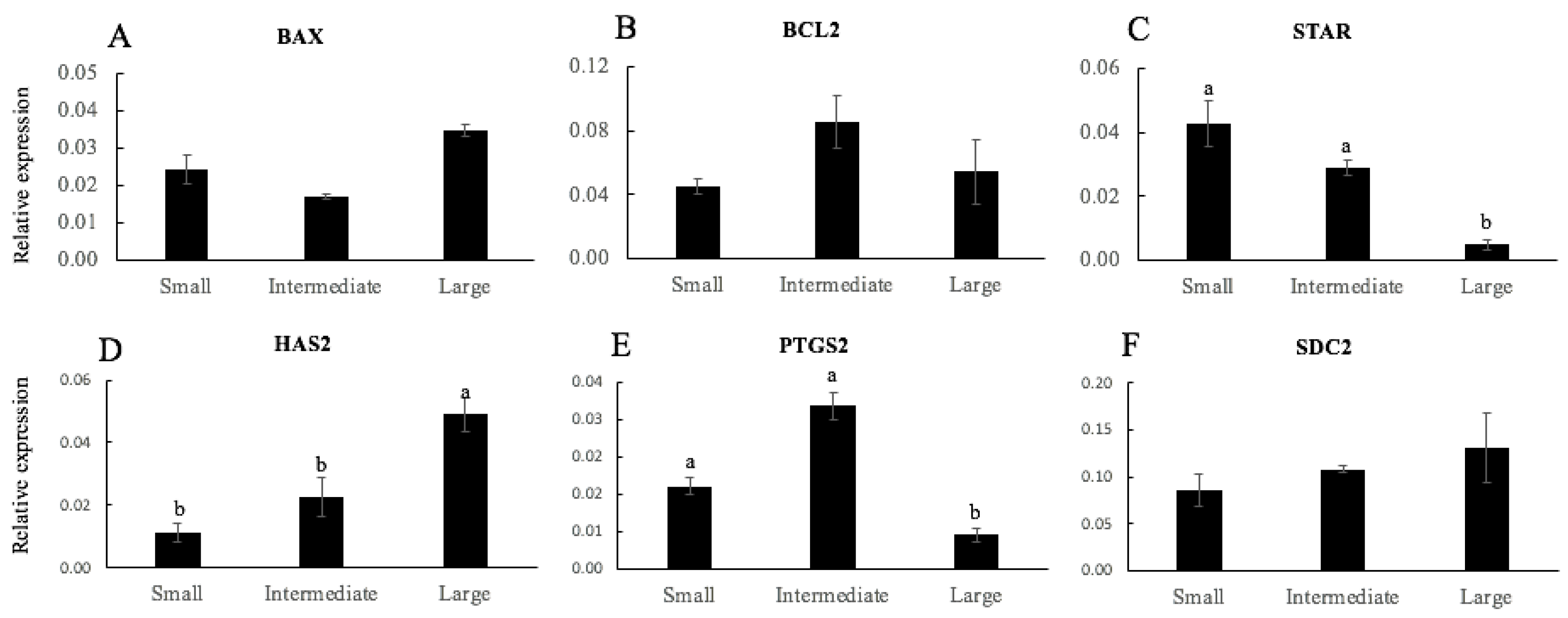
The mRNA transcript relative abundance pattern of apoptosis genes (BAX and BCL2) and acquisition of oocyte competence (STAR, HAS2, PTGS2, and SDC2) were measured in CCs from oocytes classified according to a clustering analysis by total diameter (small, intermediate, or large) (Figure 3) and oocytes classified according to the developmental stage reached (not developed, embryos arrested at 2–8-cell stage, and blastocysts) (Figure 4). As shown in Figure 3, there were no differences in the relative mRNA transcript levels of apoptosis genes (BAX and BCL2) (Figure 3A,B, respectively) and SDC2 (Figure 3F) between the groups. In large oocytes, the relative mRNA gene expression of HAS2 (Figure 3D) was higher compared to small and intermediate oocytes (p < 0.05). However, the relative mRNA levels of STAR (Figure 3C) and PTGS2 (Figure 3E) were lower in large oocytes when compared to small and intermediate oocytes (p < 0.05).
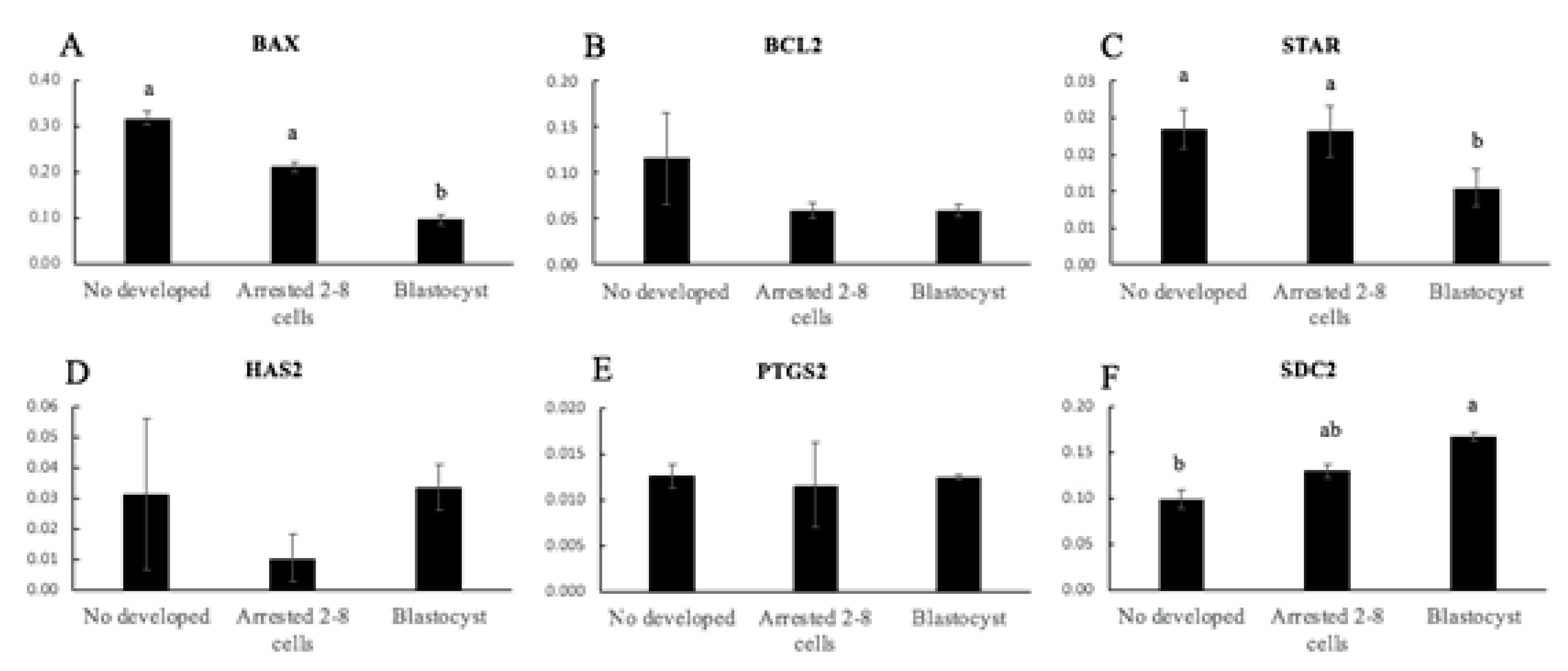
In CCs from oocytes classified according to the developmental stage reached, there were no differences in the relative mRNA transcripts levels of BCL2 (Figure 4B), HAS2 (Figure 4D), and PTGS2 (Figure 4E) between the groups. In the blastocyst group, the relative mRNA gene expression of BAX (Figure 4A) and STAR (Figure 4C) was lower than in the undeveloped group and in embryos arrested at the 2–8-cell stage (p < 0.02). In addition, the relative mRNA abundance expression of SDC2 (Figure 4F) was lower in the undeveloped and blastocyst groups (p < 0.05).

4. Discussion

Oocyte quality is crucial for the subsequent embryo development; therefore, the priority is the determination of individual biomarkers to select competent oocytes. In this context, the present work was conducted to improve IVP outcome in ovine by determining biomarkers of oocyte quality. To our knowledge, this research is the first to evaluate the morphometric parameters of oocytes and the relative gene expression of CCs as biomarkers by individual IVP in ovine. Although previous studies have reported that in vitro culture is more successful when the embryos are in large groups, due to reciprocal stimulation [27], some authors have shown that embryos produced in vitro individually had blastocyst rates similar to those cultured in groups [28], and the individual culture procedure had no effect on the embryo quality [29]. The present study utilized individual IVM, IVF, and IVC, which allowed for the monitoring of a single oocyte from the moment of maturation until the blastocyst stage. In addition, the individual IVP system allowed for the measurement of oocytes and relating their morphometric characteristics to their ability to produce embryos or not. We classified oocyte size into three categories, small, intermediate, and large, and a better outcome was obtained with the use of oocytes with 127.5 µm total diameter or 104.4 µm oocyte diameter (intermediate and large). Oocyte diameter as a criterion for oocyte selection has been extensively investigated in different species. In bovine species, oocytes acquired full meiotic competence at a diameter of 115 µm, and full developmental competence at a diameter of 120 µm [12,14]. In buffalo, developmental competence of oocytes may be achieved at a diameter of 145 μm [13]. In goats, higher rates of in vitro maturation were obtained in oocytes greater than 120 µm [15]. The difference in mean diameter of ovine oocytes in different studies may be due to different species or even different methodologies of oocyte diameter measurement.
In the present study, according to total diameter, we observed that 20%, 55%, and 25% of the oocytes were classified as small, intermediate, and large, respectively. Although about 50% of small oocytes were capable of cleavage, only 3% were able to produce blastocysts (Table 4). However, intermediate and large oocytes yielded greater percentages of cleavage (80% and 90%, respectively) and were capable of producing 30–54% of blastocysts, respectively.
Additionally, ZP thickness was measured to determine whether the ZP thickness could also be an indicator of oocyte quality. Interestingly, ZP thickness had a stronger correlation with in vitro embryo production, as oocytes with ZP ≥ 18.8 µm were able to obtain 100% cleavage and almost 68% blastocyst formation (Table 6). There are controversial reports regarding the effect of ZP thickness on oocyte quality. A previous study reported a negative correlation between ZP thickness and the ability of oocytes to resume meiosis and progress through the different stages of chromatin configuration in goat oocytes from early and antral follicles [30]. Another study reported that oocytes with high developmental competence had a significantly thicker ZP in equine oocytes [31]. In addition, ZP thickness in humans was associated with optimal folliculogenesis and/or oocyte maturation as well as blastocyst formation, with a slightly thinner ZP corresponding to significantly lower rates of conception or blastocyst development [32,33]. Regarding our results, we report that there was a relationship between the size of the oocyte and the thickness of the ZP, which together had an effect on the quality of the oocyte and subsequent embryonic development.
Furthermore, a statistical association between the total diameter, oocyte diameter, and ZP thickness with the formation of blastocysts was verified (Figure 1). The results of our study confirmed the findings of previous studies that reported that oocyte competence tended to increase with increasing oocyte size [12,13,14,15]. Therefore, the use of total diameter, oocyte diameter, and/or ZP thickness measurement as a noninvasive biomarker of oocyte quality would eliminate the subpopulation of small oocytes with poor quality, thereby improving IVP outcomes.
In addition, we investigated the mRNA expression patterns of six genes related to apoptosis and oocyte quality in oocytes categorized by total diameter. BCL2 and BAX are anti- and pro-apoptotic proteins, respectively, that participate in the mitochondria-dependent apoptosis pathway. Moreover, Van Soom et al. [34] reported that the different balance in the expression of pro- and anti-apoptotic genes may be a crucial factor determining the quality of the oocyte. In this study, greater expression of BAX transcript was observed in small oocytes, while lesser expression of BCL2 transcript was observed for intermediate and large oocytes. These results are similar to those previously described by Opiela et al. [35], who reported that bovine immature oocytes that were brilliant cresyl blue (BCB)-negative (poor quality) had a higher expression of BAX, while mature BCB+ (high quality) showed a lower expression of BCL2. Another study reported that the upregulation of apoptotic genes in vitrified bovine oocytes may be an early indicator of reduced developmental competence following vitrification [36]. Oocyte-secreted factors, BMP15 and GDF9, play crucial roles in the regulation of follicular development and oocyte maturation. In humans, high levels of GDF9 and BMP15 mRNAs in CCs are closely associated with oocyte competence and embryo quality [37]. In the same way, in goats, low levels of GDF-9 were expressed in immature oocytes at increasing levels at the time of cumulus cell expansion, after 12 h of IVM. On the contrary, in porcine oocytes, GDF9 mRNA was highly expressed during the IVM of COCs and slowly decreased during the IVM process. In addition, canine oocytes expressed high levels of GDF-9 in oocytes at the earliest state of development but decreased during the maturation process until oocyte competence was reached [38]. Similar results were obtained in our investigation; the mRNA expression of GDF9 was lower in oocytes with a greater capacity for embryonic development. The different results may be due to the maturation stage of the oocytes analyzed and/or the difference between species. In relation to cumulus expansion, gene expression of GJA1 or connexin 43 was lower in large oocytes. GJA1 has been proposed as a major mediator of cell–cell communication via gap junctions [39], being identified as a biomarker for the fertilization potential of human oocytes [40,41]. The reduced expression of GJA1 in CCs after oocyte maturation was beneficial [42] because it was related to cumulus expansion and the decreased diffusion of cAMP and cGMP from the CCs to the oocyte, which occur in the final process of oocyte maturation [43].
The expression profile of six selected genes was examined in CCs from oocytes classified according to the total diameter or embryo stage reached (Experiment 2). The selected genes (BAX, BCL2, STAR, HAS2, PTGS2, and SDC2) were representative of previously identified CCs markers of oocyte quality and development. Several studies have demonstrated that the increased incidence of CCs’ apoptosis was negatively correlated to oocyte maturation and quality, fertilization, embryo development, and pregnancy outcomes [44,45,46]. Cumulus cells’ apoptosis in oocytes that reached the blastocyst stage was low, and this has been reported to have negative effects on oocyte quality and subsequent embryo development [44,45,46]. Furthermore, BAX expression was correlated with a higher apoptosis level and lower oocyte developmental competence in vitro [47]. We observed that BAX mRNA expression was significantly lower in CCs associated with blastocyst production than those associated with embryos arrested at the 2–8-cell stage and undeveloped oocytes, while BCL2 mRNA concentrations did not vary in CCs, demonstrating that more competent oocytes cause less incidence of apoptosis. STAR is a critical enzyme in the steroidogenic activity of the ovary and is expressed in granulosa and CCs after the LH surge [48]. PTGS2 is involved in prostaglandin E2 synthesis and has been reported to induce cumulus expansion [49]. Although mRNA levels of PTGS2 in CCs are positively correlated with oocyte competence in other species such as cows [19], pigs [50], and humans [20], PTGS2 mRNA expression was significantly lower in CCs from in vivo-matured rabbit COCs, which exhibited better CCs expansion and higher developmental competence after IVF [51]. The results from our study showed that the expression of STAR and PTGS2 was lower in CCs of oocytes of high quality (from oocytes classified as large and oocytes that produced blastocysts). These findings are correlated with the lower expression for STAR and PTGS2 that was associated with the further developmental potential from embryos to blastocysts in human cumulus cells [40]. On the other hand, the expression of HAS2 and SDC2 was high in CCs of oocytes of high quality (from oocytes classified as large and oocytes that produced blastocysts). HAS2 are involved in final oocyte maturation [52]. Enhanced expression of HAS2 in CCs has been reported to be associated with better oocyte and embryo development in humans and sheep [20,21]. The SDC2 gene is involved in cell proliferation and cytoskeletal organization [47]. Previous studies associated the high quality of oocytes with cumulus expression of SDC2, which was reduced in poor-quality oocytes [47,53]. These results suggest that BAX, STAR, HAS2, PTGS2, and SDC2 expression is strongly associated with oocyte quality and the ability of oocytes to produce blastocysts.

5. Conclusions

In conclusion, oocyte morphometric assessments (total diameter, oocyte diameter, and ZP thickness) may be used as noninvasive biomarkers of oocyte quality, being a more efficient and objective method to select high-quality oocytes compared to morphological criteria. Moreover, the relative gene expression in CCs of BAX, STAR, HAS2, PTGS2, and SDC2 is also a good biomarker to select for high oocyte developmental competence, thereby improving IVP protocols in sheep. These findings could be applied in reproductive biotechnology to other species.

Author Contributions

Conceptualization, C.M. and A.J.S.; methodology, C.M., I.S.-A., and D.M.-C.; formal analysis, C.M. and B.A.; investigation, C.M. and A.J.S.; resources, A.J.S. and J.J.G.; writing—original draft preparation, C.M.; writing—review and editing, A.J.S. and J.J.G.; funding acquisition, A.J.S. and J.J.G. All authors have read and agreed to the published version of the manuscript.

Funding

This research was funded by the Spanish Ministry of Economy and Competitiveness (AGL2017-89017-R); The Junta de Comunidades de Castilla-La Mancha, Spain (PRT program; SBPLY/17/180501/000500), which provided co-funding support to C.M.

Institutional Review Board Statement

Our study was performed with ovaries from a slaughterhouse and semen from the germplasm bank of the Biology of Reproduction Group of the Universidad de Castilla-La Mancha (UCLM), which is authorized for the collection and storage of sheep semen (ES07RS02OC). Therefore, this article does not need ethics committee approval.

Informed Consent Statement

Not applicable.

Data Availability Statement

Data are available from the corresponding upon request.

Conflicts of Interest

The authors declare no conflict of interest.

References

  1. Zhu, J.; Moawad, A.R.; Wang, C.-Y.; Li, H.-F.; Ren, J.-Y.; Dai, Y.-F. Advances in In Vitro production of sheep embryos. Int. J. Veter. Sci. Med. 2018, 6, S15–S26. [Google Scholar] [CrossRef] [Green Version]
  2. Ebner, T.; Moser, M.; Sommergruber, M.; Tews, G. Selection based on morphological assessment of oocytes and embryos at different stages of preimplantation development: A review. Hum. Reprod. Updat. 2003, 9, 251–262. [Google Scholar] [CrossRef]
  3. Rizos, D.; Ward, F.; Duffy, P.; Boland, M.P.; Lonergan, P. Consequences of bovine oocyte maturation, fertilization or early embryo development In Vitro versus In Vivo: Implications for blastocyst yield and blastocyst quality. Mol. Reprod. Dev. 2002, 61, 234–248. [Google Scholar] [CrossRef]
  4. Gandolfi, F.; Brevini, T.A.L. RFD award lecture 2009. In Vitro maturation of farm animal oocytes: A useful tool for investigating the mechanisms leading to full-term development. Reprod. Fertil. Dev. 2010, 22, 495–507. [Google Scholar] [CrossRef]
  5. Sirard, M.-A.; Richard, F.; Blondin, P.; Robert, C. Contribution of the oocyte to embryo quality. Theriogenology 2006, 65, 126–136. [Google Scholar] [CrossRef]
  6. Krisher, R.L. The effect of oocyte quality on development. J. Anim. Sci. 2004, 82, E14–E23. [Google Scholar]
  7. Lonergan, P.; Rizos, D.; Gutierrez-Adan, A.; Fair, T.; Boland, M.P. Oocyte and embryo quality: Effect of origin, culture conditions and gene expression patterns. Reprod. Domest. Anim. 2003, 38, 259–267. [Google Scholar] [CrossRef]
  8. Coticchio, G.; Sereni, E.; Serrao, L.; Mazzone, S.; Iadarola, I.; Borini, A. What criteria for the definition of oocyte quality? Ann. N. Y. Acad. Sci. 2004, 1034, 132–144. [Google Scholar] [CrossRef]
  9. Mohammadi-Sangcheshmeh, A.; Soleimani, M.; Deldar, H.; Salehi, M.; Soudi, S.; Hashemi, S.M.; Schellander, K.; Hoelker, M. Prediction of oocyte developmental competence in ovine using glucose-6-phosphate dehydrogenase (G6PDH) activity determined at retrieval time. J. Assist. Reprod. Genet. 2012, 29, 153–158. [Google Scholar] [CrossRef] [Green Version]
  10. Hyttel, P.; Fair, T.; Callesen, H.; Greve, T. Oocyte growth, capacitation and final maturation in cattle. Theriogenology 1997, 47, 23–32. [Google Scholar] [CrossRef]
  11. Anguita, B.; Jimenez-Macedo, A.R.; Izquierdo, D.; Mogas, T.; Paramio, M.-T. Effect of oocyte diameter on meiotic competence, embryo development, p34 (cdc2) expression and MPF activity in prepubertal goat oocytes. Theriogenology 2007, 67, 526–536. [Google Scholar] [CrossRef]
  12. Otoi, T.; Yamamoto, K.; Koyama, N.; Tachikawa, S.; Suzuki, T. Bovine oocyte diameter in relation to developmental competence. Theriogenology 1997, 48, 769–774. [Google Scholar] [CrossRef]
  13. Raghu, H.M.; Nandi, S.; Reddy, S.M. Follicle size and oocyte diameter in relation to developmental competence of buffalo oocytes In Vitro. Reprod. Fertil. Dev. 2002, 14, 55–61. [Google Scholar] [CrossRef]
  14. Fair, T.; Hyttel, P.; Greve, T. Bovine oocyte diameter in relation to maturational competence and transcriptional activity. Mol. Reprod. Dev. 1995, 42, 437–442. [Google Scholar] [CrossRef]
  15. Cadenas, J.; Maside, C.; Ferreira, A.; Vieira, L.; Leiva-Revilla, J.; Paes, V.; Alves, B.; Brandão, F.; Rodrigues, A.; Wheeler, M.; et al. Relationship between follicular dynamics and oocyte maturation during In Vitro culture as a non-invasive sign of caprine oocyte meiotic competence. Theriogenology 2018, 107, 95–103. [Google Scholar] [CrossRef]
  16. Dumesic, D.A.; Meldrum, D.R.; Katz-Jaffe, M.G.; Krisher, R.L.; Schoolcraft, W.B. Oocyte environment: Follicular fluid and cumulus cells are critical for oocyte health. Fertil. Steril. 2015, 103, 303–316. [Google Scholar] [CrossRef]
  17. Kidder, G.M.; Vanderhyden, B.C. Bidirectional communication between oocytes and follicle cells: Ensuring oocyte developmental competence. Can. J. Physiol. Pharmacol. 2010, 88, 399–413. [Google Scholar] [CrossRef] [PubMed] [Green Version]
  18. Monniaux, D. Driving folliculogenesis by the oocyte-somatic cell dialog: Lessons from genetic models. Theriogenology 2016, 86, 41–53. [Google Scholar] [CrossRef] [PubMed]
  19. Assidi, M.; Dufort, I.; Ali, A.; Hamel, M.; Algriany, O.; Dielemann, S.; Sirard, M.-A. Identification of potential markers of oocyte competence expressed in bovine cumulus cells matured with follicle-stimulating hormone and/or phorbol myristate acetate In Vitro. Biol. Reprod. 2008, 79, 209–222. [Google Scholar] [CrossRef] [Green Version]
  20. McKenzie, L.J.; Pangas, S.A.; Carson, S.A.; Kovanci, E.; Cisneros, P.; Buster, J.E.; Amato, P.; Matzuk, M.M. Human cumulus granulosa cell gene expression: A predictor of fertilization and embryo selection in women undergoing IVF. Hum. Reprod. 2004, 19, 2869–2874. [Google Scholar] [CrossRef] [PubMed] [Green Version]
  21. Amiri, M.V.; Deldar, H.; Pirsaraei, Z.A. Impact of supplementary royal jelly on In Vitro maturation of sheep oocytes: Genes involved in apoptosis and embryonic development. Syst. Biol. Reprod. Med. 2016, 62, 31–38. [Google Scholar] [CrossRef] [Green Version]
  22. Melo, E.O.; Cordeiro, D.M.; Pellegrino, R.; Wei, Z.; Daye, Z.J.; Nishimura, R.C.; Dode, M.A.N. Identification of molecular markers for oocyte competence in bovine cumulus cells. Anim. Genet. 2016, 48, 19–29. [Google Scholar] [CrossRef] [Green Version]
  23. Devjak, R.; Tacer, K.F.; Juvan, P.; Klun, I.V.; Rozman, D.; Bokal, E.V. Cumulus cells gene expression profiling in terms of oocyte maturity in controlled ovarian hyperstimulation using GnRH agonist or GnRH antagonist. PLoS ONE 2012, 7, e47106. [Google Scholar] [CrossRef]
  24. García-Álvarez, O.; Maroto-Morales, A.; Berlinguer, F.; Fernández-Santos, M.; Esteso, M.; Mermillod, P.; Ortiz, J.; Ramon, M.; Pérez-Guzmán, M.; Garde, J.; et al. Effect of storage temperature during transport of ovaries on In Vitro embryo production in Iberian red deer (Cervus elaphus hispanicus). Theriogenology 2011, 75, 65–72. [Google Scholar] [CrossRef] [PubMed]
  25. Takahashi, Y.; First, N. In Vitro development of bovine ane-cell embryos: Influence of glucose, lactate, pyruvate, amino acids and vitamins. Theriogenology 1992, 37, 963–978. [Google Scholar] [CrossRef]
  26. Livak, K.J.; Schmittgen, T.D. Analysis of relative gene expression data using real-time quantitative PCR and the 2(-Delta Delta C(T)) Method. Methods 2001, 25, 402–408. [Google Scholar] [CrossRef]
  27. Goovaerts, I.; Leroy, J.; Jorssen, E.; Bols, P. Noninvasive bovine oocyte quality assessment: Possibilities of a single oocyte culture. Theriogenology 2010, 74, 1509–1520. [Google Scholar] [CrossRef]
  28. Bunel, A.; Jorssen, E.; Merckx, E.; Leroy, J.; Bols, P.; Sirard, M. Individual bovine In Vitro embryo production and cumulus cell transcriptomic analysis to distinguish cumulus-oocyte complexes with high or low developmental potential. Theriogenology 2015, 83, 228–237. [Google Scholar] [CrossRef]
  29. Goovaerts, I.; Leroy, J.; Rizos, D.; Bermejo-Álvarez, P.; Gutierrez-Adan, A.; Jorssen, E.; Bols, P. Single In Vitro bovine embryo production: Coculture with autologous cumulus cells, developmental competence, embryo quality and gene expression profiles. Theriogenology 2011, 76, 1293–1303. [Google Scholar] [CrossRef]
  30. Correia, H.; Vieira, L.; Maside, C.; Paes, V.; Silva, R.; Alves, B.; Santos, F.; Apgar, G.; Rodrigues, A.; Figueiredo, J. Ovarian transport temperature (4 vs. 33 °C) impacts differently the In Vitro development of isolated goat preantral and antral follicles. Small Rumin. Res. 2017, 155, 16–23. [Google Scholar] [CrossRef]
  31. Mohammadi-Sangcheshmeh, A.; Held, E.; Rings, F.; Ghanem, N.; Salilew-Wondim, D.; Tesfaye, D.; Sieme, H.; Schellander, K.; Hoelker, M. Developmental competence of equine oocytes: Impacts of zona pellucida birefringence and maternally derived transcript expression. Reprod. Fertil. Dev. 2014, 26, 441–452. [Google Scholar] [CrossRef]
  32. Ebner, T.; Balaban, B.; Moser, M.; Shebl, O.; Urman, B.; Ata, B.; Tews, G. Automatic user-independent zona pellucida imaging at the oocyte stage allows for the prediction of preimplantation development. Fertil. Steril. 2010, 94, 913–920. [Google Scholar] [CrossRef] [PubMed]
  33. Shen, Y.; Stalf, T.; Mehnert, C.; Eichenlaub-Ritter, U.; Tinneberg, H.-R. High magnitude of light retardation by the zona pellucida is associated with conception cycles. Hum. Reprod. 2005, 20, 1596–1606. [Google Scholar] [CrossRef] [PubMed]
  34. Van Soom, A.; Vandaele, L.; Goossens, K.; de Kruif, A.; Peelman, L. Gamete origin in relation to early embryo development. Theriogenology 2007, 68, S131–S137. [Google Scholar] [CrossRef]
  35. Opiela, J.; Kątska-Książkiewicz, L.; Lipiński, D.; Słomski, R.; Bzowska, M.; Ryńska, B. Interactions among activity of glucose-6-phosphate dehydrogenase in immature oocytes, expression of apoptosis-related genes Bcl-2 and Bax, and developmental competence following IVP in cattle. Theriogenology 2008, 69, 546–555. [Google Scholar] [CrossRef]
  36. Anchamparuthy, V.M.; Pearson, R.E.; Gwazdauskas, F.C. Expression pattern of apoptotic genes in vitrified-thawed bovine oocytes. Reprod. Domest. Anim. 2009, 45, e83–e90. [Google Scholar] [CrossRef]
  37. Li, Y.; Li, R.Q.; Ou, S.B.; Zhang, N.F.; Ren, L.; Wei, L.N.; Zhang, Q.X.; Yang, D.Z. Increased GDF9 and BMP15 mRNA levels in cumulus granulosa cells correlate with oocyte maturation, fertilization, and embryo quality in humans. Reprod. Biol. Endocrinol. 2014, 12, 81. [Google Scholar] [CrossRef] [Green Version]
  38. Reyes, M.D.L.; Rojas, C.; Parraguez, V.; Palomino, J. Expression of growth differentiation factor 9 (GDF-9) during In Vitro maturation in canine oocytes. Theriogenology 2013, 80, 587–596. [Google Scholar] [CrossRef]
  39. Gittens, J.E.I.; Kidder, G.M. Differential contributions of connexin37 and connexin43 to oogenesis revealed in chimeric reaggregated mouse ovaries. J. Cell Sci. 2005, 118, 5071–5078. [Google Scholar] [CrossRef] [Green Version]
  40. Feuerstein, P.; Cadoret, V.; Dalbies-Tran, R.; Guerif, F.; Bidault, R.; Royere, D. Gene expression in human cumulus cells: One approach to oocyte competence. Hum. Reprod. 2007, 22, 3069–3077. [Google Scholar] [CrossRef] [PubMed] [Green Version]
  41. Hasegawa, J.; Yanaihara, A.; Iwasaki, S.; Mitsukawa, K.; Negishi, M.; Okai, T. Reduction of connexin 43 in human cumulus cells yields good embryo competence during ICSI. J. Assist. Reprod. Genet. 2007, 24, 463–466. [Google Scholar] [CrossRef] [PubMed] [Green Version]
  42. Edry, I.; Sela-Abramovich, S.; Dekel, N. Meiotic arrest of oocytes depends on cell-to-cell communication in the ovarian follicle. Mol. Cell. Endocrinol. 2006, 252, 102–106. [Google Scholar] [CrossRef] [PubMed]
  43. Shao, L.; Chian, R.-C.; Xu, Y.; Yan, Z.; Zhang, Y.; Gao, C.; Gao, L.; Liu, J.; Cui, Y. Genomic expression profiles in cumulus cells derived from germinal vesicle and MII mouse oocytes. Reprod. Fertil. Dev. 2016, 28, 1798–1809. [Google Scholar] [CrossRef] [PubMed]
  44. Lee, K.S.; Joo, B.S.; Na, Y.J.; Yoon, M.S.; Choi, O.H.; Kim, W.W. Cumulus cells apoptosis as an indicator to predict the quality of oocytes and the outcome of IVF-ET. J. Assist. Reprod. Genet. 2001, 18, 490–498. [Google Scholar] [CrossRef] [PubMed]
  45. Ruvolo, G.; Bosco, L.; Pane, A.; Morici, G.; Cittadini, E.; Roccheri, M.C. Lower apoptosis rate in human cumulus cells after administration of recombinant luteinizing hormone to women undergoing ovarian stimulation for In Vitro fertilization procedures. Fertil. Steril. 2007, 87, 542–546. [Google Scholar] [CrossRef] [Green Version]
  46. Corn, C.M.; Hauser-Kronberger, C.; Moser, M.; Tews, G.; Ebner, T. Predictive value of cumulus cell apoptosis with regard to blastocyst development of corresponding gametes. Fertil. Steril. 2005, 84, 627–633. [Google Scholar] [CrossRef]
  47. Salhab, M.; Dhorne-Pollet, S.; Auclair, S.; Guyader-Joly, C.; Brisard, D.; Dalbiès-Tran, R.; Dupont, J.; Ponsart, C.; Mermillod, P.; Uzbekova, S. In Vitro maturation of oocytes alters gene expression and signaling pathways in bovine cumulus cells. Mol. Reprod. Dev. 2012, 80, 166–182. [Google Scholar] [CrossRef]
  48. Sasson, R.; Rimon, E.; Dantes, A.; Cohen, T.; Shinder, V.; Land-Bracha, A.; Amsterdam, A. Gonadotrophin-induced gene regulation in human granulosa cells obtained from IVF patients. Modulation of steroidogenic genes, cytoskeletal genes and genes coding for apoptotic signalling and protein kinases. Mol. Hum. Reprod. 2004, 10, 299–311. [Google Scholar] [CrossRef]
  49. Davis, B.J.; Lennard, D.E.; Lee, C.A.; Tiano, H.F.; Morham, S.G.; Wetsel, W.C.; Langenbach, R. Anovulation in cyclooxygenase-2-deficient mice is restored by prostaglandin E2 and interleukin-1beta. Endocrinology 1999, 140, 2685–2695. [Google Scholar] [CrossRef]
  50. Kang, J.-T.; Atikuzzaman, M.; Kwon, D.-K.; Park, S.-J.; Kim, S.-J.; Moon, J.-H.; Koo, O.-J.; Jang, G.; Lee, B.-C. Developmental competence of porcine oocytes after In Vitro maturation and In Vitro culture under different oxygen concentrations. Zygote 2011, 20, 1–8. [Google Scholar] [CrossRef]
  51. Arias-Álvarez, M.; Garcia, R.; López-Tello, J.; Rebollar, P.G.; Gutierrez-Adan, A.; Lorenzo, P.L. In Vivo and In Vitro maturation of rabbit oocytes differently affects the gene expression profile, mitochondrial distribution, apoptosis and early embryo development. Reprod. Fertil. Dev. 2017, 29, 1667–1679. [Google Scholar] [CrossRef] [PubMed] [Green Version]
  52. Schoenfelder, M.; Einspanier, R. Expression of hyaluronan synthases and corresponding hyaluronan receptors is differentially regulated during oocyte maturation in cattle. Biol. Reprod. 2003, 69, 269–277. [Google Scholar] [CrossRef] [PubMed]
  53. Dhali, A.; Javvaji, P.K.; Kolte, A.P.; Francis, J.R.; Roy, S.C.; Sejian, V. Temporal expression of cumulus cell marker genes during In Vitro maturation and oocyte developmental competence. J. Assist. Reprod. Genet. 2017, 34, 1493–1500. [Google Scholar] [CrossRef] [PubMed]
Figure 1. Relationship between blastocyst formation and morphometric measures: (A) total diameter, (B) oocyte diameter, and (C) zona pellucida thickness. Each circle on the graph represents an oocyte that was evaluated. The oocytes analyzed were defined by binary values (0 = no; 1 = yes). A logistic regression is represented by the following equations: Logit P = –13,462 + (0.0935 × total diameter), p < 0.001; Logit P = –6562 + (0.0530 × oocyte diameter), p < 0.05; Logit P = –4577 + (0.252 × total diameter), p < 0.001.
Animals 11 02818 g001
Figure 2. Relative gene expression of mRNA of BAX (A), BCL2 (B), BMP15 (C), GDF9 (D), GJA3 (E), and IGF2R (F) in ovine oocytes according to the clustering of total diameter (small, intermediate, or large). Different letters denote significant differences (p < 0.05, at least).
Animals 11 02818 g002
Figure 3. Relative gene expression of mRNA of BAX (A), BCL2 (B), STAR (C), HAS2 (D), PTGS2 (E), and SDC2 (F) of cumulus cells from sheep oocytes according to a clustering analysis of the total diameter (small, intermediate, and large). Different letters denote significant differences (p < 0.05, at least).
Animals 11 02818 g003
Figure 4. Relative gene expression of mRNA of BAX (A), BCL2 (B), STAR (C), HAS2 (D), PTGS2 (E), and SDC2 (F) and of cumulus cells from sheep oocytes according to the developmental stage reached (undeveloped, embryos arrested at the 2–8-cells stage, and blastocysts). Different letters denote significant differences (p < 0.05, at least).
Animals 11 02818 g004
Table 1. The gene-specific primers used for qPCR in oocytes and cumulus cells.
Target GeneGene FunctionPrimer Sequence (5′→3′)Product SizeGenBank Accession No.
PPIAReference geneF-TCAACCCCACCGTGTTCTTC
R-GTCACCACCCTGGCACATAA
194NM_001308578.1
BAXApoptosisF-GTTGTCGCCCTTTTCTACTTTGC
R-CAGCCCATGATGGTCCTGATC
89NM_173894.1
BCL2ApoptosisF-GGAGCTGGTGGTTGACTTTC
R-CTAGGTGGTCATTCAGGTAAG
518NM_001077486.2
BMP15Oocyte maturationF-CTACGACTCCGCTTCGTGTGT
R-AGTGCCATGCCACCAGAAC
69NM_001031752.1
GDF9Oocyte maturationF-GAAGTGGGACAACTGGATTGTG
R-CCCTGGGACAGTCCCCTTTA
71NM_174681.2
GJA3Gap junctionsF-TGCCTTTCGTTGTAACACTCA
R-AGAACACATGAGCCAGGTACA
143NM_174068.2
HAS2Oocyte competenceF-CCTCATCATCCAAAGCCTG
R-ACATTTCCGCAAATAGTCTG
138NM_174079.2
IGF2RGrowth factorFGCTGCGGTGTGCCAAGTGAAAAAG
R-AGCCCCTCTGCCGTTGTTACCT
201NM_174352.2
STAROocyte competenceF-GCCAGGTGTTGAAGGCCA
R-TCTTTAACAGACTTGGAGGCTTCC
104NM_001009243.1
PTGS2Oocyte competenceF-AGGAGGTCTTTGGTCTGGTG
R-TCTGGAACAACTGCTCATCG
126NM_001009432.1
Table 2. Mean values of individual and group (control) IVC parameters.
TotalMaturation %Fertilization Efficiency %Cleavage %Blastocyst %Number of Cells/Blastocysts
Individual19981.0 ± 5.256.4 ± 1.278.2 ± 6.729.9 ± 1.3108.9 ± 3.5
Control16781.9 ± 7.152.5 ± 3.177.9 ± 6.930.5 ± 1.388.9 ± 16.2
Data are expressed as mean ± SEM.
Table 3. Mean values of morphometric parameters of oocyte size in each generated cluster for total diameter, oocyte diameter, and ZP thickness.
ClusterTotal Diameter µmOocyte Diameter µmZP Thickness µm
1 (small)88.478.39.7
2 (intermediate)127.5104.414.9
3 (large)141.9116.818.8
Table 4. Mean values of different developmental stages by clustering of morphometric parameters of oocyte by total diameter.
Cluster Total Diameter Developmental Stages
Undeveloped %Total Embryos at 2–8-Cell Stage %Embryos Arrested at 2–8-Cell Stage %Blastocysts %
154.8 ± 11.4 a45.1 ± 11.4 a42.0 ± 8.93.1 ± 3.1 a
(small)(13/24)(11/24)(10/24)(1/24)
220.9 ± 9.3 b79.0 ± 9.3 b49.6 ± 5.329.4 ± 5.2 b
(intermediate)(12/76)(64/76)(40/76)(24/76)
34.5 ± 4.5 b95.4 ± 4.5 b41.2 ± 13.354.2 ± 13.5 b
(arge)(2/33)(31/33)(12/33)(19/33)
Data are expressed as mean ± SEM. Values within columns are significantly different (a,b p < 0.05).
Table 5. Mean values of different developmental stages by clustering of morphometric parameters of oocytes by oocyte diameter.
Cluster Oocyte Diameter Developmental Stages
Undeveloped %Total Embryos at 2–8-Cell Stage %Embryos Arrested at 2–8-Cell Stage %Blastocysts %
150.8 ± 11.0 a49.2 ± 11.0 a35.0 ± 1.614.1 ± 9.4 a
(small)(12/28)(16/28)(10/28)(6/28)
217.2 ± 7.8 b82.8 ± 7.8 b52.3 ± 10.130.5 ± 10.1 ab
(intermediate)(9/71)(62/71)(41/71)(21/71)
317.1 ± 5.7 b82.8 ± 5.7 b38.4 ± 9.644.4 ± 3.9 b
(large)(7/34)(27/34)(11/34)(16/34)
Data are expressed as mean ± SEM. Values within columns are significantly different (a,b p < 0.05).
Table 6. Mean values of different developmental stages by clustering of morphometric parameters of oocytes by ZP thickness.
Cluster ZP ThicknessDevelopmental Stages
Undeveloped %Total Embryos at 2–8-Cell Stage %Embryos Arrested at 2–8-Cell Stage %Blastocysts %
141.8 ± 10.3 a58.2 ± 10.3 a31.5 ± 12.426.7 ± 3.9 a
(small)(24/60)(45/60)(21/60)(15/60)
26.6 ± 4.1 b93.3 ± 4.1 b64.7 ± 6.328.6 ± 9.6 a
(intermediate)(3/56)(53/56)(34/56)(19/56)
30.0 ± 0.0 b100 ± 0.0 b32.4 ± 12.467.6 ± 12.4 b
(large)(0/17)(17/17)(6/17)(11/17)
Data are expressed as mean ± SEM. Values within columns are significantly different (a,b p < 0.05).
Publisher’s Note: MDPI stays neutral with regard to jurisdictional claims in published maps and institutional affiliations.