Article

Bacterial Strains from Soybean Nodules in the Lower Volga Region Belong to a New Subspecies Bradyrhizobium japonicum subsp. saratovii subsp. nov.


by
Aleksandr S. Sidorin
1,2,
Gennady L. Burygin
1,3,4,*,
Andrey V. Fedorov
2,
Aleksandr D. Katyshev
2,
Yaroslav M. Krasnov
2 and
Oksana V. Tkachenko
1
1
Department of Plant Breeding, Selection, and Genetics, Institute of Genetics and Agronomy, Saratov State University of Genetics, Biotechnology and Engineering Named After N.I. Vavilov, Saratov 410012, Russia
2
Russian Research Anti-Plague Institute “Microbe”, Saratov 410005, Russia
3
Institute of Biochemistry and Physiology of Plants and Microorganisms, Saratov Scientific Centre of the Russian Academy of Sciences, Saratov 410049, Russia
4
Department of Organic and Bioorganic Chemistry, Institute of Chemistry, Saratov State University, Saratov 410012, Russia
*
Author to whom correspondence should be addressed.
Microorganisms 2026, 14(3), 684; https://doi.org/10.3390/microorganisms14030684
Submission received: 8 February 2026 / Revised: 9 March 2026 / Accepted: 13 March 2026 / Published: 18 March 2026
(This article belongs to the Special Issue Plant Growth-Promoting Bacteria)

Abstract

The isolation of locally adapted rhizobial strains with high symbiotic activity represents an effective strategy for increasing soybean yield under extreme environmental conditions. In this study, seven novel strains were isolated from nodules of soybeans grown in a greenhouse using field soil from the Lower Volga region. Five genomes were assembled into complete circular chromosomes, whereas two strains yielded near-complete chromosomes containing single repeat-mediated junctions. All strains had putative plasmids that were independently validated as circular by long-read mapping and confirmed by the presence of characteristic replication and conjugation-associated genes. Genome sequences of strains were about 11 Mb, and GC contents were 63.1–63.3%. Comparative genome analyses demonstrated that all strains had average nucleotide identity values of 95.4% with Bradyrhizobium japonicum USDA 6T and 96.3% with Bradyrhizobium barranii 144S4T, forming a distinct cluster in phylogenetic trees. No significant differences were detected between B. japonicum and B. barranii that would explain the species boundary. Therefore, it is proposed to unite all novel strains into the subspecies Bradyrhizobium japonicum subsp. saratovii subsp. nov., and all other strains of B. japonicum and B. barranii we suggest dividing into four subspecies: Bradyrhizobium japonicum subsp. japonicum subsp. nov., Bradyrhizobium japonicum subsp. barranii comb. nov., Bradyrhizobium japonicum subsp. apii comb. nov., and Bradyrhizobium japonicum subsp. saratovii subsp. nov. The proposed taxonomic framework expands current knowledge of the biodiversity of soybean symbiotic bacteria and contributes to a better understanding of the distribution and the evolution of bacteria Bradyrhizobium spp. in previously unexplored regions.

1. Introduction

In modern agrobiotechnology, microbial biologics have gained increasing popularity as a means of rational soil resource management [1,2,3,4,5]. Biological fertilizers typically contain symbiotic nitrogen-fixing bacteria [6,7]. However, not all strains included in currently used bioproducts are capable of exerting an effective plant growth-promoting effect. This is due to their high host specificity, low persistence on plant roots, and poor adaptation to the soil and climatic conditions of the target region [8]. An effective solution to this problem is the isolation and selection of efficient indigenous bacterial strains from the territory where crop cultivation is planned. Strains obtained in this way are already adapted to the local edaphic and climatic conditions.
Leguminous crops are characterized by high economic and ecological importance for agricultural production [9]. Soybean (Glycine max (L.) Merr.), as one of the most important representatives of this plant family, is of particular interest due to its high protein and oil content in the grain. In 2024, soybeans were cultivated worldwide on approximately 143 million ha, with a total production of 398 million tons [10]. The most important soybean producers are Brazil and the United States, which together account for about 66% of global production. However, annual production exceeds 1 million tons in at least 12 other countries. Over the past decade, global soybean production has increased by 23%, which is associated with improved production efficiency and an 18.5% expansion of the cultivated area. Russia ranks eighth among the world’s soybean producers, with a production of 7.0 million tons harvested from 4.13 million ha. Moreover, the area under soybean cultivation in Russia has been steadily increasing and has expanded by 98% since 2015. Consequently, soybean cultivation is spreading into large areas where this crop was previously not grown.
The productivity of soybeans, as well as other leguminous crops, largely depends on the formation of symbioses with root nodule bacteria capable of fixing atmospheric nitrogen [11]. When isolating bacteria from soybean nodules, strains belonging to the genera Bradyrhizobium, Mesorhizobium, Agrobacterium, Sinorhizobium, and others are most frequently obtained [12,13,14]. Among these, bacteria of the genus Bradyrhizobium are the most widely used as inoculants [15,16,17,18,19,20]. Since the use of rhizobial strains as biofertilizers underlies modern soybean cultivation technologies, it is essential to evaluate the ability of potential inoculants to form effective symbioses with plants under new ecological conditions.
The Lower Volga region (Russia), including the Saratov region, is a non-traditional region for soybean cultivation. Its soil and climatic conditions differ markedly from those of conventional soybean-growing regions, primarily due to low rainfall. As a result, soybeans are cultivated mainly under irrigation in this region, and the cultivated area has been increasing rapidly in recent years. Because soybean is an introduced crop in the Volga region, local soils lack soybean-specific symbiotic bacteria, while commercial Bradyrhizobium spp. strains used in biofertilizers are exposed to non-optimal environmental conditions.
Bacteria of the genus Bradyrhizobium belong to the class Alphaproteobacteria, family Nitrobacteraceae (order Hyphomicrobiales) [21]. The genus currently comprises 75 validly published and 19 non-validly published species [22]. The large number of species within the genus makes it difficult to assign new strains of Bradyrhizobium spp. to a particular species. Standard 16S rRNA testing is not applicable to this genus due to the high percentage of sequence identity between representatives of different species [23]. For species identification of strains, whole-genome sequence analysis is used. The average nucleotide identity (ANI), for which it is proposed to use 94–96.5% and 97–98% as the levels of species and subspecies boundries, respectively: ANI values of less than 94% for strains of different species, 95–97% for strains of the same species but different subspecies, and more than 98% for strains of the same subspecies [24,25,26].
Many strains of Bradyrhizobium spp. are of considerable economic importance due to their ability to fix atmospheric nitrogen and form effective symbioses with leguminous crops [27,28,29]. There are no published reports describing the isolation of Bradyrhizobium root nodule bacteria from the Lower Volga region, a newly explored area for soybean cultivation. The aim of the present study was to isolate bacteria from nodules of soybeans grown in a greenhouse using field soil from the Lower Volga region, to characterize them, and to perform their taxonomic identification.

2. Materials and Methods

2.1. Bacterial Strains and Culture Conditions

Root nodules of natural origin were collected from soybean plants (Glycine max (L.) Merr.), cultivar Natalie, grown in a greenhouse in soil sampled from the educational and experimental farm “Povolzhye” of Vavilov University in 2023 (Saratov region, Russia; 51.126105146540645, 45.999733721150704). The soil cover of the farm is represented by typical dark chestnut soils (Kastanozem, WRB). The soil texture corresponds to medium and heavy loams. The humus content in the 0–30 cm soil layer was 2.9%. The soil was slightly alkaline (pH 7.2) and characterized by low nitrate nitrogen availability (6 mg/kg), a moderate content of available phosphorus (16.3 mg/kg), and a high level of exchangeable potassium (300 mg/kg). Maize (Zea mays L.) was used as the preceding crop.
Soybean plants were cultivated in the greenhouse for 30 days in 3-L vessels with field soil from the farm “Povolzhye” and, at the time of bacterial isolation, were at the early flowering stage. Nodules were formed on the main root and on first-order lateral roots. The nodules selected for the study were thoroughly washed with sterile distilled water to remove adhering soil particles. To eliminate epiphytic microorganisms, soybean nodules were sequentially treated with a 1% sodium hypochlorite solution for 2 min and 70% ethanol for 30 s. Surface-sterilized nodules were homogenized in a sterile mortar with the addition of 1–2 mL of physiological saline. A series of tenfold dilutions was prepared from the resulting homogenate, and 10 µL of each dilution was plated onto solid Yeast Mannitol Agar (YMA) medium of the following composition (g/L): yeast extract, 0.5; K2HPO4, 0.2; MgSO4·7H2O, 0.2; mannitol, 7.0; agar, 15 (pH 7.0–7.2) [30]. Plates were incubated for 7–10 days at 28 °C until colonies appeared. Pure cultures were obtained by repeated culturing of single colonies. Cultures were maintained on solid YMA at 4 °C and at −20 °C in liquid YMA supplemented with 15% glycerol. A total of ten strains of root nodule bacteria were isolated.
The ability of the strains to form functional nodules on plants was evaluated by reinoculation of soybean seeds (cv. Natalie). Seeds were sterilized in 0.2% diacid solution for 2 min, then washed with sterile distilled water on a magnetic stirrer five times for 10 min each. The strains were grown in YMA liquid nutrient medium at 30 °C and 110 rpm for 24–48 h. For inoculation, a 50 mL bacterial suspension with a concentration of 1 × 10⁸ cells/mL was used. Sterile soybean seeds (25 seeds for each variant) were inoculated by immersion in the strain suspension for 2 h. For the control, sterile seeds were soaked in an equal volume of sterile water. Seeds were planted in a 500 mL container with sand in five replicates. Sand was pre-sterilized by heating in a dry-heat oven at 250 °C for 12 h. The plants were grown at a temperature of 22–24 °C during the day and 18–20 °C at night, with a relative humidity of 60–70% and a daylight duration of 14–16 h. Lighting was maintained at a level consistent with the biological requirements of the crop. Watering was provided as needed, maintaining substrate moisture at 60–70% of its full capacity. Watering was carried out with distilled water, and fertilizing was carried out on the 15th and 25th days of cultivation with nitrogen-free Knop solution containing (g/L): K2HPO4—0.25; MgSO4 × 7H2O—1; KCl—0.125; FeSO4 × 7H2O—0.0125. For 30-day-old plants, an analysis of morphometric characteristics and formed nodules was carried out. The obtained data were compared based on the analysis of variance and Duncan’s test (p ˂ 0.05) in the Statistica v10 program.
Only seven strains were selected for further analysis. Among the selected strains were those with clear growth-promoting properties, as well as those that had a negative impact on plants. The strains were deposited in the Collection of Rhizosphere Microorganisms of the Institute of Biochemistry and Physiology of Plants and Microorganisms, Russian Academy of Sciences (IBPPM RAS) (Table 1) [31].

2.2. Microbiological Characterization

Cultural and morphological characteristics were determined on solid and liquid YMA media. Carbon source utilization was assessed on a minimal agar medium (g/L): tryptone, 1.0; yeast extract, 0.1; carbon source (glucose, arabinose, sucrose, rhamnose, lactose, galactose, maltose, as well as sorbitol, malic acid, and citric acid), 10.0; agar, 15.0. Catalase activity was tested on YMA by adding 3% hydrogen peroxide to bacterial cultures. Denitrification ability was evaluated by growing the bacteria in YMA broth supplemented with 0.1% potassium nitrate for 4 days, followed by the addition of 0.1 mL of Griess reagent. Each isolate was characterized for cultural characteristics of bacterial growth in liquid YMA medium under different pH values (pH range 4–10) and NaCl concentrations (0.5% and 1.0%).

2.3. Complete Genome Sequencing and Assembly

Genomic DNA was extracted using the EasyPure Genomic DNA Kit (TransGen Biotech, Beijing, China). Complete genome sequencing was performed using two platforms: DNBSEQ-G50 (MGI, Shenzhen, China) and GridION (Oxford Nanopore Technologies, Oxford, UK). Library preparation was carried out using the MGIEasy FS DNA Library Prep Set, DNBSEQ-G50 High-throughput Sequencing Set (PE150), and SQK-RBK114-96 kits, respectively. Basecalling of Oxford Nanopore reads was performed using dorado v0.8.3 (SUP model). The dna_r10.4.1_e8.2_400bps_sup@v4.3.0 model was used for basecalling. Subsequently, filtlong v0.2.1 [32] was used to remove 5% of reads with the lowest mean Qscore and reads shorter than 2000 nucleotides. Read quality control was performed using fastp and fastplong [33,34]. Metrics of the filtered Oxford Nanopore Technology (ONT, Oxford, UK) reads are presented in Table S1.
Oxford Nanopore reads were assembled using Flye v2.9.2 (options: --nano-hq --read-error 0.03) [35]. Minor assembly errors were corrected by mapping the corresponding MGI reads using Polypolish v0.6.0 [36]. Assembly quality was assessed using BUSCO v6.0.0 with the bradyrhizobium_odb12 dataset. Assembly metrics are provided in Table S2.
Replicon visualization was performed using Galaxy [37,38]. Complete genome sequences of the strains were annotated using the open-source software package Prokka v1.15.2 [39] under the Linux operating system (Ubuntu 22.04).

2.4. Replicon Classification and Circularization Validation

Replicons were classified as chromosomal or plasmid based on gene content and assembly structure. Chromosomal sequences were identified by the presence of core housekeeping genes and/or rRNA operons (e.g., dnaA, gyrA/gyrB, rpoB, recA, 16S/23S rRNA, and parA/parB), which are widely used as markers of primary bacterial chromosomes. Contigs carrying these markers were interpreted as chromosomal segments, even when present as separate fragments due to unresolved repeat junctions. Putative plasmid replicons were identified by the presence of conjugation and type IV secretion system genes (e.g., virB/type IV secretion), consistent with established plasmid replication and mobilization systems, in the absence of a complete chromosomal marker set.
Circular topology was validated by mapping raw Oxford Nanopore reads to the final assemblies using minimap2 with the map-ont preset optimized for nanopore data [40]. Resulting alignments were processed using SAMtools [41]. For each contig, mean sequencing coverage was calculated, and the fraction of secondary alignments (SAM flag 256) was determined following the SAM/BAM specification [42]. Circularization was confirmed by detecting junction-spanning reads, defined as long reads aligning within 5 kb of both the start and end coordinates of a contig.
De novo assembly of strains I-2 and I-5 was performed using Flye --nano-raw mode with an expected genome size of ~9–10 Mb and an assembly coverage limit (--asm-coverage 40) to prioritize long reads for repeat resolution. Additional assemblies using read subsets ≥ 20 kb were generated to assess the impact of read length on repeat resolution. Assembly graphs were visually inspected using Bandage [43] to identify structural ambiguities and repeat-mediated junctions.

2.5. Phylogenetic and Comparative Genomic Analyses

Average nucleotide identity (ANI) was calculated using the OrthoANI algorithm implemented in ANI Calculator (https://www.ezbiocloud.net/tools/ani, accessed on 7 August 2025) [44]. ANI values recalculated using fastANI v1.34 were highly consistent with those obtained via the EZBioCloud ANI calculator, with differences typically below 0.3% for closely related taxa and below 1% for more distant comparisons. These minor differences did not affect taxonomic conclusions. The digital DNA-DNA hybridization (dDDH) was calculated using the online resource Genome-to-Genome Distance Calculator (GGDC) v3.0 [45]. Pairwise comparison matrices were generated using online resources [46]. Genome BLAST distance phylogeny (GBDP) v2.1 trees were constructed using the Type (Strain) Genome Server (TYGS) platform [45]. For single nucleotide polymorphism (SNP) analysis, contigs of the studied strains were aligned against the genome of the B. japonicum type strain USDA 6 (accession number ASM28437v1) using Snippy v4.6 [47]. The search and alignment of genes were performed using MEGA 7 [48]. Phylogenetic trees based on SNP data were constructed in SeaView v5.0.4 [49] using the maximum likelihood method with the GTR model. Tree visualization was performed using FigTree v1.4.3 [50], resulting in rooted phylogenetic trees. To account for the potential impact of homologous recombination on phylogenetic inference, recombinant regions were detected and removed using Gubbins [51]. Gubbins iteratively reconstructs a maximum likelihood phylogeny, identifies clusters of elevated SNP density indicative of recombination events, masks recombinant regions, and rebuilds the tree until convergence. Sequences with excessive missing data were excluded using a maximum missing data threshold of 31%. The resulting recombination-filtered alignment of polymorphic sites was used for final phylogenetic reconstruction.
Maximum likelihood phylogenetic inference was performed using IQ-TREE2 [52] under the GTR + G nucleotide substitution model. Branch support was assessed using 1000 ultrafast bootstrap replicates and 1000 SH-like approximate likelihood ratio test (SH-aLRT) replicates [53].
Alignment of the nucleotide sequences of the chromosomes was performed using the Mauve program version 2.4.0 with default settings [54].

3. Results

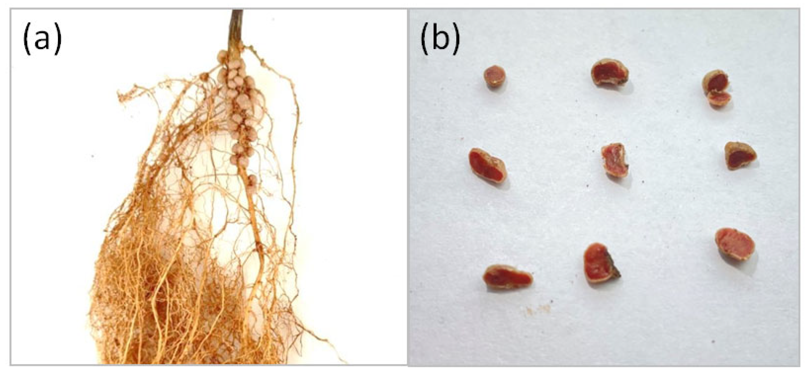
To select efficient bacterial strains for use as inoculants, ten pure isolates were obtained from washed and surface-sterilized root nodules of soybean plants (Glycine max (L.) Merr.), cultivar Natalie (Figure 1a): I-1, I-2, I-3, I-4, I-5, I-6, II-1, II-2, III-1, and III-2. To select the most promising strains for further analysis, a preliminary assessment of the symbiotic activity of the isolated strains was carried out using the reinoculation method.

3.1. Evaluation of Inoculation Effectiveness

Inoculation with strains significantly affected soybean morphometric traits after 30 days of growth (Table 2). The highest dry shoot weight was observed in plants inoculated with strains II-2 (561.6 mg), I-1 (397.6 mg), III-1 (378.4 mg), I-2 (379.0 mg), and II-1 (390.7 mg), all significantly exceeding or equaling the control (283.1 mg). The greatest increase in root weight was recorded for strains I-1 (648.3 mg) and II-2 (488.0 mg), compared with 295.2 mg in the control.
All inoculated variants formed nodules, whereas nodulation was absent in the control. Nodules exhibited an intense red coloration in cross section (Figure 1b), indicating the presence of leghemoglobin in the nodule tissues, which is essential for biological nitrogen fixation.
The highest nodule numbers were detected in plants inoculated with II-2 (25.0 nodules) and III-1 (24.8 nodules). The greatest nodule dry weight was recorded for I-2 (42.9 mg), III-1 (34.6 mg), and II-2 (33.1 mg). Strains III-2 and I-4 demonstrated comparatively low symbiotic efficiency, showing reduced plant weight and lower nodulation parameters.
Overall, strains II-2, III-1, I-2, and I-1 exhibited the strongest positive effects on soybean growth and nodulation. For the next stage of research, not only the strains that demonstrated the greatest effectiveness in promoting plant growth but also those that exerted a growth-inhibiting effect on plants were selected. The inclusion of strains with opposing biological effects in the analysis was necessitated by the need for a comprehensive assessment of the diversity of bacterial associations interacting with soybean plants. This will allow for the examination of growth-promoting and growth-inhibiting effects as manifestations of different strategies of interaction between microorganisms and the host plant.

3.2. Phenotypic Characterization of the Strains

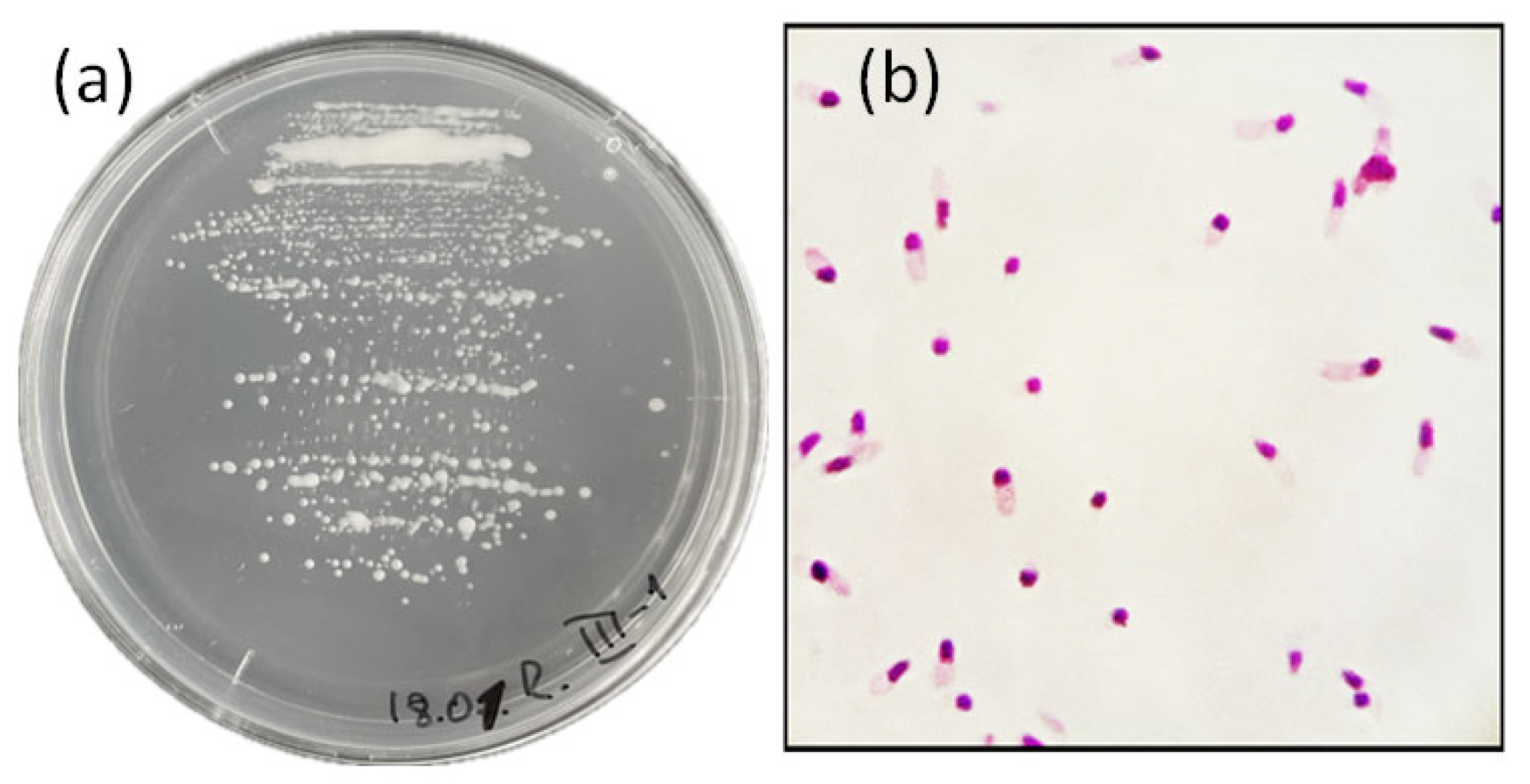
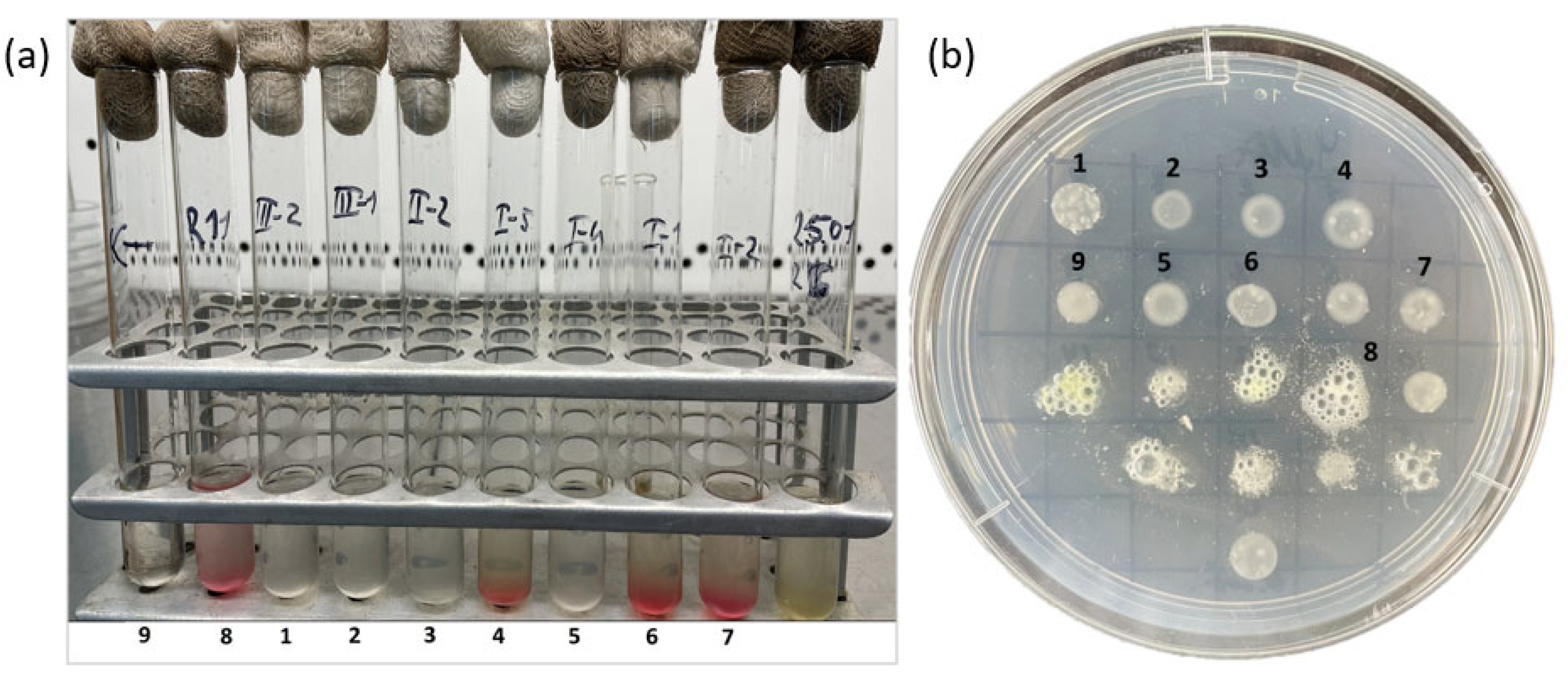
Various cultural–morphological and physiological–biochemical characteristics of the selected strains were examined (Table 3). All isolates were slow-growing, with visible colony growth observed after 5–7 days of incubation. The optimal growth conditions for all strains were 28 °C and pH 7.0. All strains were aerobic, Gram-negative rods and exhibited monopolar staining in Gram-stained preparations (Figure 2). Colonies formed by the isolates on solid YMA medium were mucoid, circular, and convex with smooth margins, reaching a diameter of 1–2 mm after 7–9 days of incubation at 28 °C (Figure 2). The strains were able to grow over a pH range of 5–9, forming a homogeneous suspension in liquid YMA medium; at pH 4 and 10, only bottom growth with the formation of flocculent precipitates was observed. Bacterial growth was inhibited at an NaCl concentration of 1% in the medium. The strains actively utilized glucose, arabinose, sucrose, maltose, sorbitol, and galactose as carbon sources; weak growth was observed on rhamnose and lactose, whereas malic and citric acids were not fermented. All strains were catalase-positive, and only three strains (I-1, I-2, and I-5) exhibited denitrifying activity (Figure 3).

3.3. Genome Sequencing of the Isolated Strains, Replicon Classification, and Circularization Validation

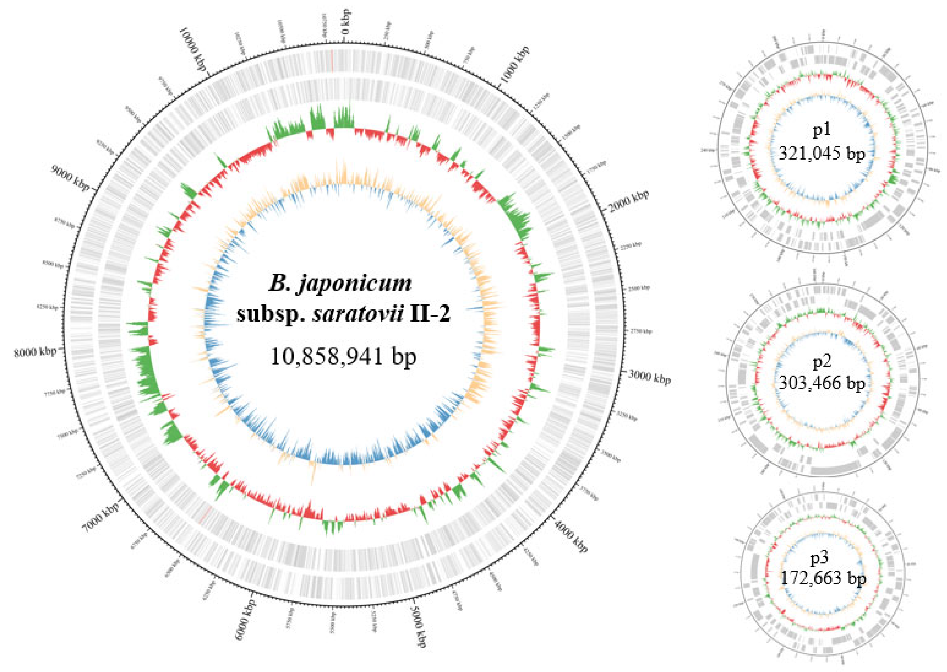
The genomic characteristics of the isolated strains are summarized in Table 4. Genome sizes showed only minor variation among the strains, ranging from 10.9 Mb (strain III-2) to 11.7 Mb (strain II-2). The number of coding DNA sequences (CDSs) also varied slightly, from 10,807 (strain III-2) to 11,447 (strain II-2). The GC content of all genomes ranged from 63.1% to 63.3%.
Assembly validation metrics are summarized in Table S3. Mean coverage (×) represents the average sequencing depth calculated across each contig after mapping raw ONT reads to the final assembly. The secondary alignment fraction corresponds to the proportion of alignments flagged as secondary (SAM flag 256) and was used as an indicator of repeat-associated or homologous genomic regions. Elevated values suggest structural ambiguity or collapsed repeat sequences. Junction-spanning reads (n) indicate the number of long reads aligning within 5 kb of both the start and end coordinates of a contig. Detection of such reads was considered direct evidence of circular topology.
Five strains (I-1, I-4, II-2, III-1, and III-2) yielded fully circularized chromosomes and putative plasmids without structural ambiguities (Figure 4). In contrast, strains I-2 and I-5 each contained a single repeat-mediated chromosomal junction preventing complete circularization. All putative plasmid replicons across all strains were independently validated as circular. The putative plasmids in all strains exhibited GC contents substantially lower than those of the corresponding chromosomes. This observation suggests that these plasmids were likely acquired by the ancestral strains via horizontal gene transfer [57].
Comparison of OrthoANI values among all genomes revealed a high degree of similarity, exceeding 99% (Table 5). However, the alignment fraction (AF) used for genome comparisons was below 70% in most cases. Only the comparisons between strains I-1 vs. I-2 and I-2 vs. I-5 showed AF values above 70% (71.6% and 89.8%, respectively). Alignment of the nucleotide sequences of the chromosomes of the novel strains (Figure S1) did not reveal extended insertions or deletions but showed significant rearrangements of fragments within the chromosomes, which could be reflected in a decrease in AF values. These results indicate that, although the strains are highly similar according to most genomic metrics, the relatively low alignment coverage reflects pronounced genomic individuality among the analyzed strains.

3.4. Phylogenetic Analysis Based on SNP Data

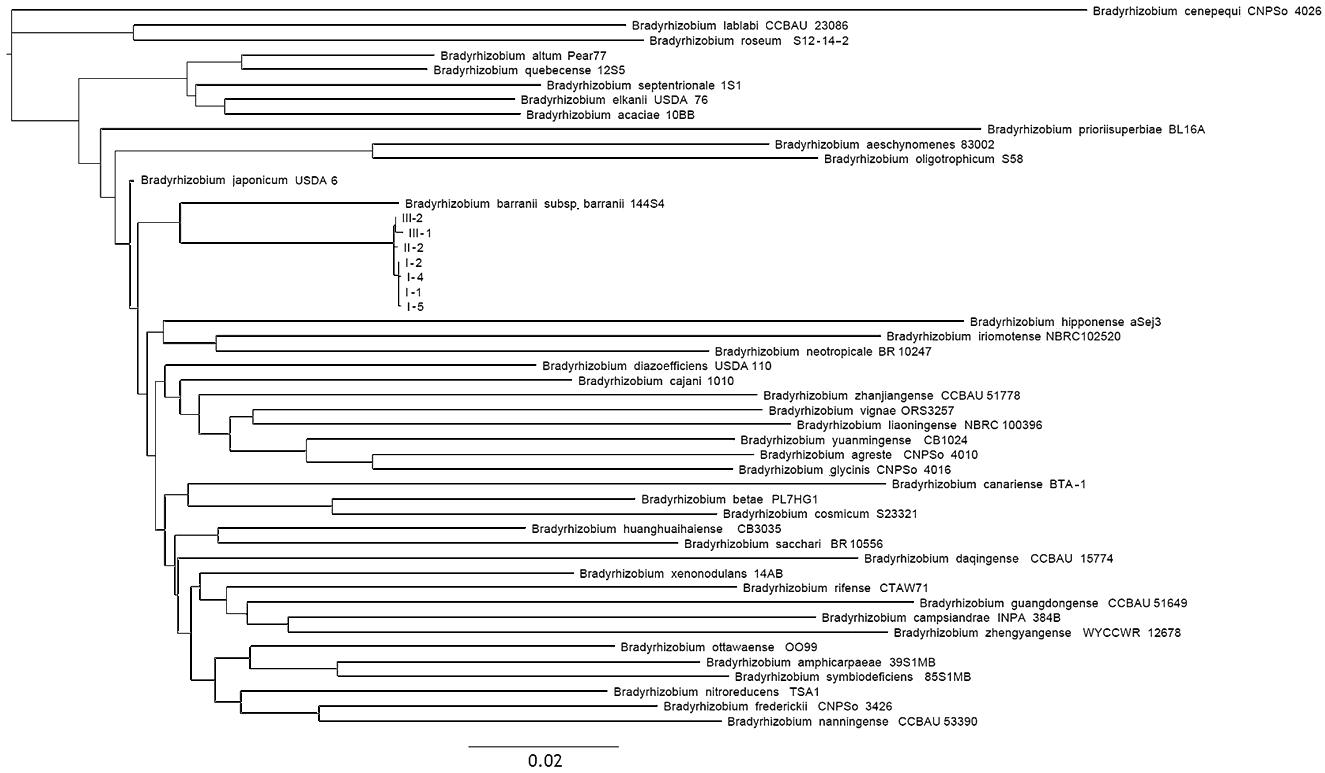
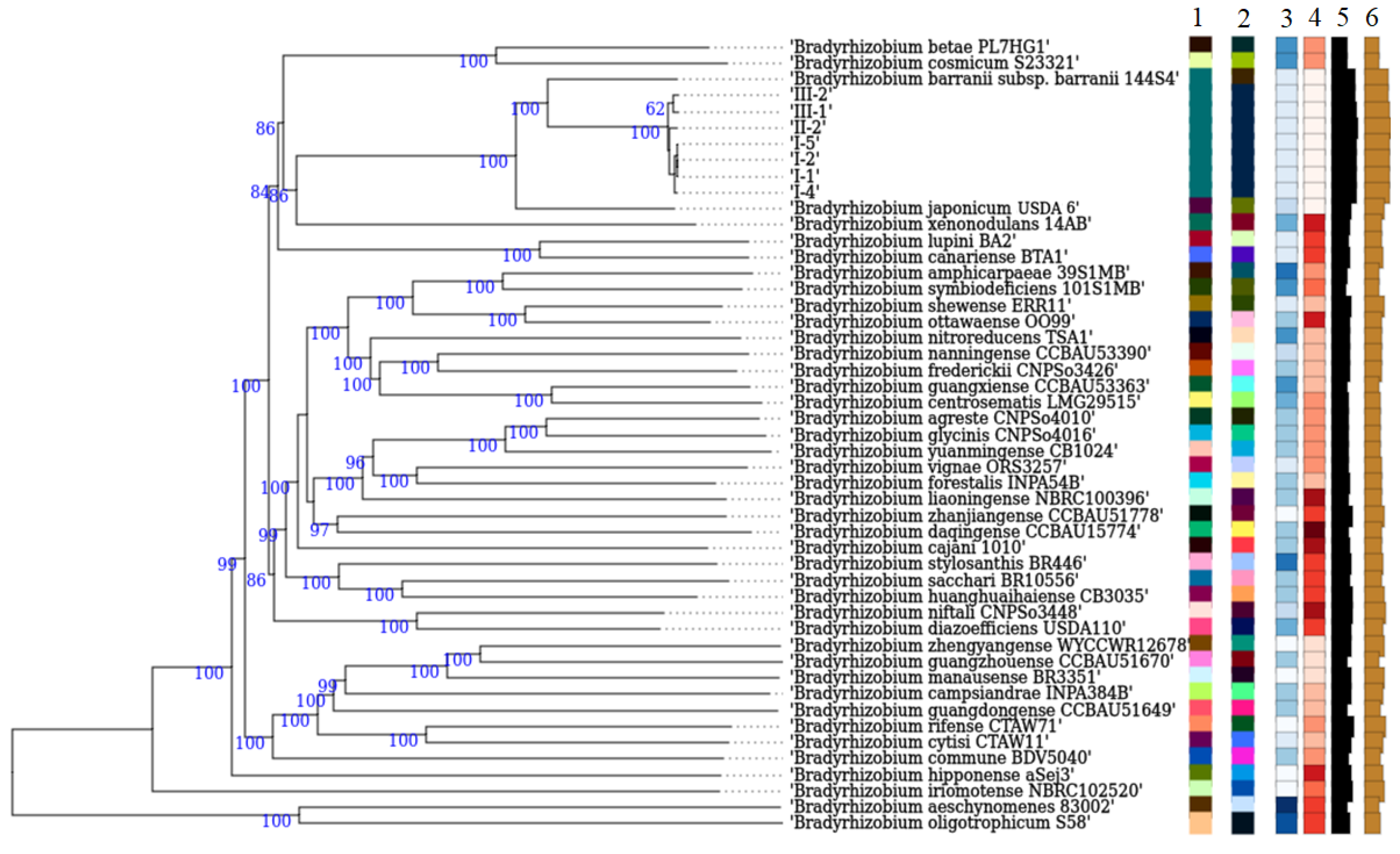
To further analyze the phylogenetic affiliation of the seven representative Bradyrhizobium sp. isolates, a phylogenetic tree was constructed based on single nucleotide polymorphism (SNP) analysis. For this purpose, reference strains of the genus Bradyrhizobium were retrieved from the NCBI international nucleotide sequence database. The SNP-based approach enables genome-wide comparisons of strains based on core single nucleotide polymorphisms. In total, 5351 single nucleotide substitutions were identified. The results of the analysis showed that all of the novel strains clustered into a distinct group (Figure 5), consistent with the results obtained from rrs gene analysis, and confirmed their affiliation with the genus Bradyrhizobium. However, based on branch lengths in the SNP-based phylogenetic tree, the two closest related species to novel strains were identified as B. japonicum and B. barranii.

3.5. Average Nucleotide Identity (ANI) Analysis

At present, Average Nucleotide Identity (ANI) is considered one of the most important criteria for determining the taxonomic position of microorganisms. The generally accepted threshold value for species boundries based on ANI is 95%. All novel strains and the type strains of species within the genus Bradyrhizobium were analyzed using the OrthoANI test (Table 6). The 95% threshold was exceeded in comparisons with the B. japonicum type strain USDA 6T and with the type strains of the subspecies B. barranii subsp. barranii and B. barranii subsp. apii.
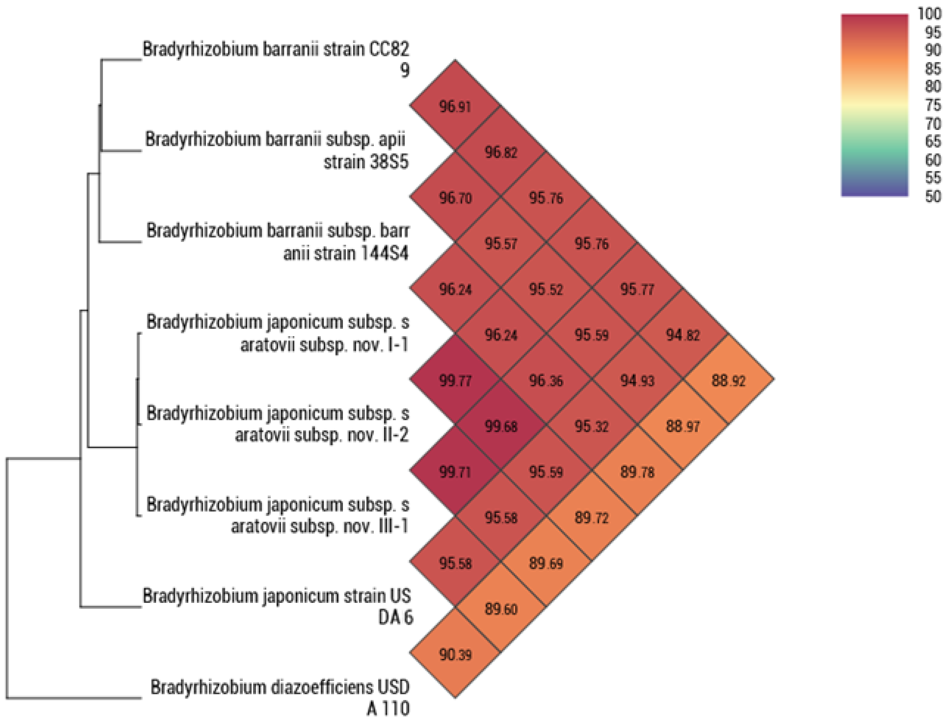
Figure 6 clearly demonstrates the separation of Bradyrhizobium strains into distinct groups, in which representative reference strains with the highest ANI values were selected. For clarity, strains with ANI values > 95% were included: three strains of B. barranii (isolated in Canada and Australia) and the B. japonicum type strain USDA 6T. B. diazoefficiens USDA 110T was used as the strain with the highest ANI value among those with ANI ≤ 95%. The positioning of the strains on the dendrogram is consistent with the results obtained using other obtained methods. All B. barranii strains form a separate clade in the dendrogram, novel strains occupy an intermediate position between B. barranii and B. japonicum, and B. diazoefficiens USDA 110 is located as the root of the tree.
In agreement with the previous results, the analysis of representative strains using the TYGS platform yielded similar findings (Figure 7). All isolated strains occupy an intermediate phylogenetic position between the two species B. barranii and B. japonicum. Strains I-1, I-2, I-4, I-5, II-2, III-1, and III-2, together with B. barranii 144S4T, form a distinct species-level group, within which novel strains are assigned, according to the TYGS results, to a different (novel) subspecies.
Based on comparative genome analyses using ANI, GBDP, and SNP approaches, the strains isolated in this study occupy an intermediate (nearly equidistant) phylogenetic position between the previously described species B. japonicum and B. barranii, forming a distinct cluster in phylogenetic trees. However, because ANI values between the new strains and the type strains of B. japonicum and B. barranii exceed 95%, the proposal of a novel species is not justified. At the same time, assignment of the new strains to the species B. barranii is also questionable, as this would result in an excessive phylogenetic proximity between the species B. japonicum and B. barranii.
Given that the ANI value between the type strains of B. japonicum and B. barranii is 95.2%, a fact that was not considered during the description of B. barranii in 2022 [55], despite ANI analysis already being recommended for the description of new bacterial species at that time, we propose that B. barranii should be regarded as a later heterotypic synonym of B. japonicum. Nevertheless, due to the observed differences between these bacterial groups, we propose to subdivide the species B. japonicum into the following subspecies: Bradyrhizobium japonicum subsp. japonicum subsp. nov., Bradyrhizobium japonicum subsp. barranii comb. nov., and Bradyrhizobium japonicum subsp. apii comb. nov. The seven strains isolated in this study, which form a distinct lineage within Bradyrhizobium japonicum, are proposed to represent a new subspecies, Bradyrhizobium japonicum subsp. saratovii subsp. nov.
The genome of strain CC829 is positioned on a separate branch relative to the other strains proposed as subspecies of B. japonicum, with ANI values below 97% (Figure 7). This indicates that strain CC829, which is used in commercial inoculants for legumes, belongs to a distinct genomic group within the species B. japonicum [58].

3.6. Phylogenetic Analysis of B. japonicum Strains

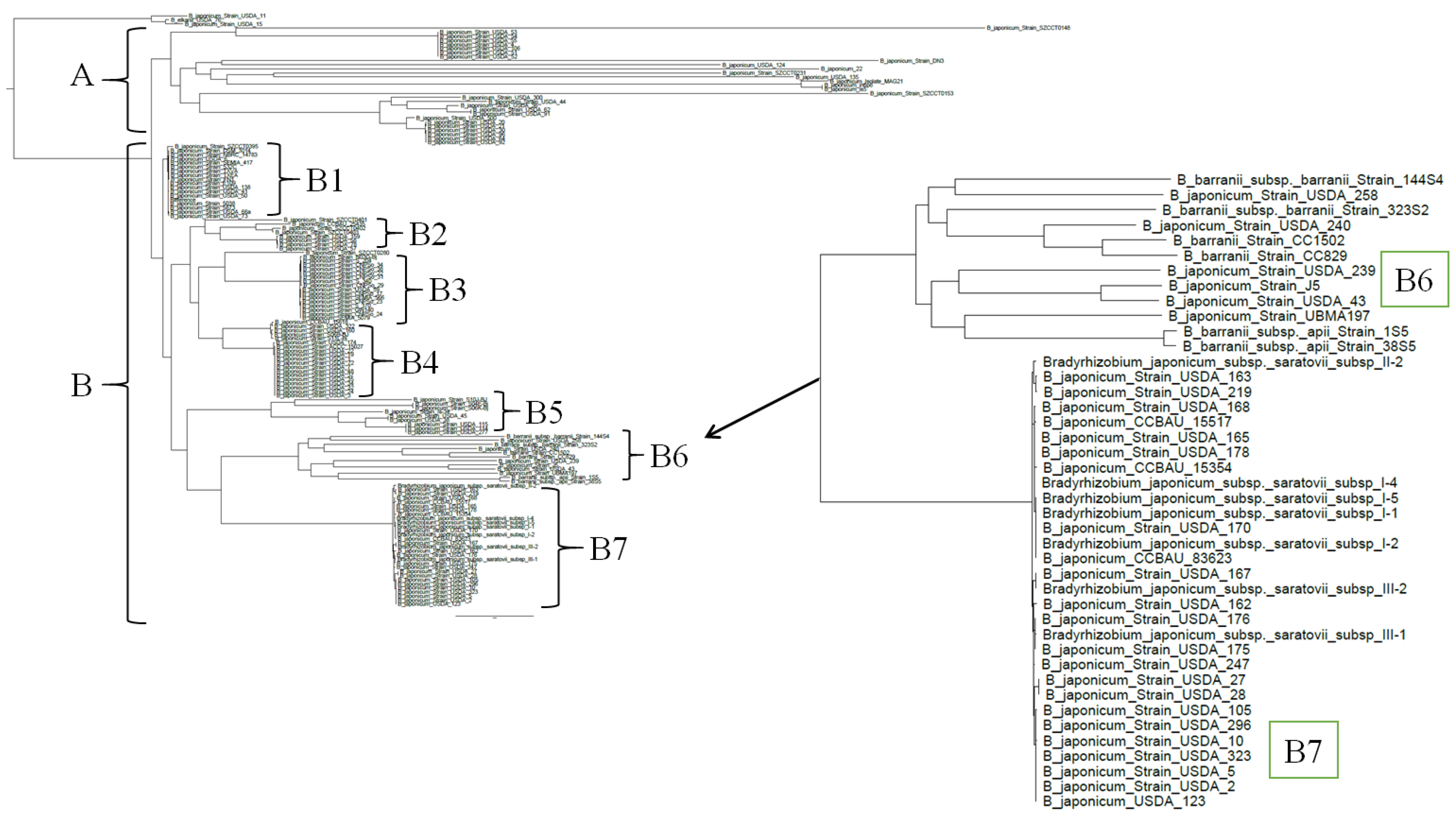
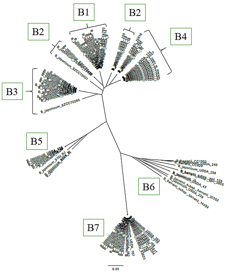
Figure 6, Figure 7 and Figure 8 demonstrate that all of the novel strains occupy an intermediate position between the two species B. japonicum and B. barranii, as confirmed by ANI analysis. To further clarify the taxonomic placement of the isolated strains, all genomes of B. japonicum and B. barranii strains available in the NCBI database were compared. As in the previous analyses, a core SNP-based phylogenetic analysis was conducted (Figure 9). The resulting dendrogram clearly illustrates the clustering of strains into phylogenetic lineages. For clarity, the tree was rooted using the genome of the type strain of another species, Bradyrhizobium elkanii USDA 76. Nearly all B. japonicum strains were divided into two main branches, designated A and B. The B. japonicum type strain USDA 6T is positioned in the group B1 at the base of branch B (Figure S2). All seven of the novel strains and twenty-three other strains of B. japonicum are located in group B7 (Bradyrhizobium japonicum subsp. saratovii subsp. nov.) and are confirmed to belong to the species B. japonicum, whereas B. barranii strains cluster separately from novel strains within a distinct group, B6, together with other B. japonicum strains (Figure S3). Branch A contains 29 strains that are clearly separated from all other lineages on the dendrogram (Figure S4). Two strains, B. japonicum USDA 15 and B. japonicum USDA 11, are located alongside B. elkanii USDA 76T.
Strains from groups B2, B3, B4, and B5 (Figures S2 and S5) exhibited ANI values above 98% when compared with the B. japonicum type strain USDA 6T, despite the SNP-based differences observed, confirming their affiliation with the species B. japonicum. The results of the ANI analysis are summarized in pairwise comparison matrices (Figure S6). Of the 144 strains analyzed, the majority were identified as B. japonicum, including both novel isolates and strains previously classified as B. barranii. Thirty-one strains exhibited ANI values below 95% relative to the type strain, consistent with the results of the SNP analysis (Figure 8, Figure 9 and Figures S7 and S8).
Because homologous recombination may bias SNP-based phylogenetic inference in rhizobia, we subsequently performed recombination detection and masking using Gubbins. The recombination-filtered alignment was used to reconstruct a second maximum likelihood tree with branch support assessment (Figure 9). Strains showing ANI values below 95% relative to the B. japonicum type strain in the initial SNP tree (branch A) were excluded by Gubbins at the alignment filtering stage due to elevated levels of missing data. The exclusion of these genomes did not alter the phylogenetic placement of the remaining strains. Comparison of the unfiltered and recombination-filtered phylogenies demonstrated overall topological congruence. Our strains formed a distinct and well-supported clade in both trees.
Based on the ANI, dDDH, and SNP test results, the reclassification was proposed for 23 of the 31 analyzed strains of B. japonicum (Table 7). Consequently, we propose a reconsideration of the taxonomic status of these 31 strains and their reassignment according to the obtained results. For the remaining eight strains, for which no closely related species could be identified among other Bradyrhizobium species, we propose classification as Bradyrhizobium sp., without specifying a species-level affiliation.

4. Discussion

It is well established that the key bacterial inoculants used for soybean cultivation predominantly belong to the genera Bradyrhizobium and Sinorhizobium [59,60]. Bradyrhizobium represents a highly diverse bacterial genus that includes species of considerable economic importance due to their ability to fix atmospheric nitrogen and to form effective symbiotic associations with leguminous crops used in agriculture [27,29,55]. However, the efficiency of biological nitrogen fixation is closely linked to the selection of highly effective strains, preferably originating from local populations. Therefore, expanding the known diversity of rhizobia through the identification and characterization of naturally occurring strains is essential.
In the present study, ten pure-culture strains of root nodule bacteria were isolated from surface-sterilized nodules of soybean (Glycine max (L.) Merr.) cultivar Natalie, grown in a greenhouse using field soil from the Lower Volga region. In the Saratov region, soybean is an introduced crop and thus requires the implementation of effective cultivation strategies adapted to local conditions. At this time, no data have been reported in the scientific literature regarding the isolation of rhizobial strains from the Lower Volga region, highlighting the novelty and regional relevance of the present study.
At the initial stage of the study, the symbiotic activity of ten isolated strains was evaluated using a reinoculation assay on soybean plants. Based on the morphometric parameters of inoculated plants, seven strains were selected for further analysis. The selection included strains that promoted plant growth as well as those showing neutral or negative effects. The inclusion of strains with contrasting symbiotic performance was considered important for a comprehensive analysis of plant–microbe interactions and for identifying factors determining effective or ineffective symbiosis. The observed variation in plant weight and nodulation parameters indicates substantial differences in symbiotic efficiency among the tested strains.
Strains II-2 and III-1 combined high nodule number with increased plant weight, suggesting effective nitrogen fixation and well-established symbiotic compatibility. Notably, strain I-2 formed fewer nodules than II-2 but produced the highest nodule dry mass, which may indicate larger or more physiologically active nodules. The high root weight observed for strain I-1 may reflect enhanced root system development associated with effective rhizobial signaling and early symbiotic establishment. In contrast, strains III-2 and I-4 showed limited promotion of plant growth and lower nodulation intensity, suggesting reduced symbiotic performance under the experimental conditions. Overall, the results demonstrate that symbiotic effectiveness among the studied strains is strongly strain-dependent and highlight the importance of selecting highly efficient strains for further functional and genomic analyses. Although long-read sequencing enabled near-complete assemblies for all strains, strains I-2 and I-5 each contained a single unresolved chromosomal junction associated with repetitive genomic regions.
In strain I-2, the unresolved region corresponded to an ~87 kb sequence exhibiting approximately twofold higher coverage relative to the chromosomal average. This coverage pattern, together with the assembly graph structure, indicates the collapse of a near-identical chromosomal duplication. Only one assembled copy of this repeat was present in the final assembly, preventing unambiguous circularization of the chromosome. In strain I-5, the chromosomal ambiguity was caused by two large homologous regions (~80–90 kb) forming a bifurcation in the assembly graph. Unlike strain I-2, no coverage doubling was observed, suggesting the presence of diverged but highly homologous segments rather than identical duplicated copies. Such repeat-mediated junctions are known limitations even in long-read assemblies when repeat length approaches or exceeds effective read length distribution. Importantly, plasmid replicons in both strains were fully circularized and independently validated by junction-spanning long reads. Gene content analysis confirmed the chromosomal identity of the primary replicons. Therefore, the unresolved structures represent repeat-mediated chromosomal configurations rather than additional replicons. Taken together, these findings demonstrate that five genomes are complete and fully circularized, whereas two represent high-quality near-complete assemblies with a single unresolved repeat-mediated chromosomal junction.
Previous studies on Bradyrhizobium have demonstrated that the high level of conservation of the 16S rRNA gene does not allow reliable discrimination of strains at the species level [61,62,63,64]. In contrast, SNP-based analyses provide high resolution for both intra- and interspecific differentiation, as well as for phylogenetic inference in bacterial taxa [65]. For example, researchers in Brazil have applied SNP analysis using the Snippy pipeline to detect genetic differences within closely related groups of Bradyrhizobium strains, while other researchers have employed SNP-based approaches to identify genetic variation and potential associations with symbiotic phenotypes among Bradyrhizobium strains [66,67].
To determine the species and phylogenetic relationships of the Bradyrhizobium strains, whole genome data for the same strains were retrieved from the NCBI GenBank database. Based on 5351 identified core SNPs, a non-ultrametric phylogenetic tree was constructed using the maximum likelihood method with the GTR model (Figure 5). On the resulting dendrogram, each reference strain, with the exception of novel isolates, was represented by an individual branch. All strains investigated in the present study formed a tightly clustered group located between the clades corresponding to B. japonicum and B. barranii. Since each species was represented by a distinct branch on the dendrogram, the intermediate position of novel strains suggests that they either belong to a novel species within the genus Bradyrhizobium or represent a transitional group between these two species.
Analysis of average nucleotide identity (ANI) based on whole genome comparisons enables the assessment of genetic and evolutionary distances between strains and provides species and subspecies boundaries, with a widely accepted threshold of 95–96% and 98%, respectively [62,68,69,70,71]. The application of ANI analysis to Bradyrhizobium strains has proven effective for identifying novel taxonomic units, particularly among isolates obtained from root nodules of various leguminous plants or from soils. For example, researchers in Morocco employed ANI analysis to compare genomes of indigenous Bradyrhizobium species isolated from nodules of Retama dasycarpa (L.) Boiss., whereas studies in Brazil applied this approach to investigate Bradyrhizobium strains associated with two leguminous hosts, Vigna unguiculata (L.) Walp. and Glycine max (L.) Merr. [72,73].
Taxonomic identification of the novel strains was further assessed by comparing them with genomes of the type strains retrieved from NCBI. As SNP analysis clustered novel strains together with representatives of both B. japonicum and B. barranii, the dataset was expanded to include strains belonging to these two species. The results of ANI calculations were consistent with the SNP-based phylogenetic analysis and are summarized in Table 6. Among all analyzed strains, the 95% ANI threshold was exceeded for all strains of B. japonicum and B. barranii. Given that ANI is widely used for species boundaries and that the highest nucleotide identity values relative to novel strains were observed for B. barranii strains, an initial assignment of novel isolates to this species could be considered. However, when a reduced set of strains with ANI values above 95% was analyzed, all the novel strains consistently occupied an intermediate position between B. japonicum and B. barranii on the phylogenetic dendrograms (Figure 6 and Figure 7).
To corroborate the results obtained from SNP-based phylogeny and ANI analyses, a genome-scale phylogenetic tree was constructed using the genome BLAST distance phylogeny (GBDP) approach implemented in the TYGS platform. According to the results, all isolated strains occupy an intermediate position between the two species B. barranii and B. japonicum (Figure 7). However, the TYGS analysis revealed a highly supported clade corresponding to the species cluster representing B. barranii, which included all seven of the novel strains. Within this clade, strains I-1, I-2, I-4, I-5, II-2, III-1, and III-2 formed a distinct sub-branch separated from the type strain B. barranii 144S4. According to TYGS, this sub-branch corresponds to a different (novel) subspecies.
Because novel strains consistently occupied an intermediate phylogenetic position between B. japonicum and B. barranii, pairwise comparisons were performed for all available strains of these two species together with novel strains. For this purpose, 130 genomes of B. japonicum and six genomes of B. barranii were retrieved from the NCBI database. A non-ultrametric phylogenetic tree was constructed using a maximum likelihood SNP-based approach based on 18,976 identified core SNPs (Figure 8). The genome of B. elkanii USDA 76 was used as the outgroup to root the tree.
SNP-based analysis enables the detection and alignment of shared core SNPs across large strain datasets and represents a promising approach for microbial differentiation and phylogenetic inference [65,74,75]. The resulting dendrogram clearly demonstrates the separation of strains into distinct phylogenetic lineages. If novel strains belonged to a novel species, they would be expected to cluster near the root of the tree alongside B. elkanii USDA 76 or to occupy a position intermediate between the root and B. japonicum strains. However, all of the novel strains are deeply nested within the tree, forming group B7 among other B. japonicum strains. Similarly, all six B. barranii strains are also located within the tree, clustering in the group B6 together with additional B. japonicum strains.
The branching pattern of the dendrogram reflects a hierarchical structure based on shared SNPs, with more closely related strains forming tighter clusters. The B. japonicum type strain USDA 6T is positioned at the base of branch B within group B1. Based on the overall topology of the tree, it can be inferred that all strains located below group B1, corresponding to the entire B branch, belong to the species B. japonicum, including both novel strains and all strains previously classified as B. barranii. Accordingly, B. barranii should be regarded as a later heterotypic synonym of B. japonicum. In addition, 29 strains assigned to B. japonicum formed a separate branch (A). If this interpretation is accepted, strains belonging to branch A do not, in fact, represent B. japonicum. Two other strains, B. japonicum USDA 15 and B. japonicum USDA 11, occupied a basal position together with B. elkanii USDA 76 at the root of the tree. Overall, these results suggest that not all strains currently assigned to B. japonicum genuinely belong to this species and that a comprehensive taxonomic revision is required. The same conclusion applies to strains currently classified as B. barranii.
Because homologous recombination can influence SNP-based phylogenetic inference, particularly in rhizobia, recombinant regions were subsequently identified and removed using Gubbins prior to final tree reconstruction. This approach reduces the impact of horizontally acquired genomic segments and allows phylogenetic relationships to be inferred primarily from vertically inherited variation.
Importantly, the overall topology of the phylogeny remained consistent after recombination filtering (Figure 8 and Figure 9). The newly sequenced strains continued to form a distinct and well-supported monophyletic group, indicating that their phylogenetic placement is not attributable to recombinant regions. This topological stability supports the robustness of the observed clustering pattern.
Genomes comprising branch A in the initial tree (Figure 8)—characterized by lower ANI values relative to the type strain—were excluded by Gubbins during the alignment filtering stage due to elevated levels of missing data (>48%). The exclusion of these genomes did not alter the structure of the remaining phylogeny or the position of the focal clade (Figure 9). Their removal is therefore interpreted as a consequence of alignment incompleteness rather than as a biologically driven reassignment. Overall, the concordance between ANI analysis and recombination-filtered SNP phylogeny supports the presence of a genomically coherent lineage among the newly sequenced strains.
Taxonomic identification of strains is mainly performed by whole-genome analysis using the ANI and GGDC tests, for which numerical values for species boundaries are available [24,62,71,76]. However, for strains with which the ANI and GGDC values that lie right on the species boundary, there are difficulties in assigning them to one or another species. Such difficulties were also encountered in our work. The novel isolated strains, based on the ANI and GGDC test results, can be assigned to either species: B. barranii or B. japonicum. We carefully examined the arguments in the work of Bromfield et al. (2022) [55] regarding the description of the B. barranii species. ANI values for B. barranii strains with B. japonicum type strain USDA 6T can be assessed ambiguously: both for strains of a new species (ANI values < 95–96% [69]) and for strains of the same species (ANI values > 94.5% [24,26]). The results of digital DDH are also ambiguous. On the one hand, the DDH value is 63.60% [60.7–66.4%], which is below 70% for the species boundary. On the other hand, the probability that DDH > 70% (i.e., same species) is 63.6% (via logistic regression). Furthermore, phylogenetic trees constructed based on the analysis of the genomes of all strains of the B. barranii and B. japonicum species available in GenBank (Figure 8) show the absence of distinctness of B. barranii strains from B. japonicum strains. Assigning strains of clade B7 to either the B. barranii or B. japonicum species would, in our opinion, hinder the use of ANI and GGDC tests for the taxonomic identification of strains of this group of bacteria. Thus, based on bioinformatical analysis, we suggest that B. barranii is a later heterotypic synonym of B. japonicum, which should take precedence. ANI value analysis supports the reassignment of B. barranii subsp. barranii and B. barranii subsp. apii to B. japonicum subsp. barranii and B. japonicum subsp. apii, respectively.
We propose to assign all strains of B. japonicum that have an ANI value with B. japonicum type strain USDA 6T of more than 98% to B. japonicum subsp. japonicum. We propose to name clade B7, containing 23 strains of B. japonicum and novel strains isolated from soybean nodules in the Lower Volga region, as B. japonicum subsp. saratovii subsp. nov. in honor of the Saratov region.
Based on the core SNP analysis, it can be inferred that 31 strains currently named as “B. japonicum” but not clustering within branch B likely belong to other species. To test this hypothesis, pairwise ANI comparisons were performed for all 31 strains. The ANI results were fully consistent with the SNP-based phylogeny. All strains forming the separate A branch (Figure S4) exhibited ANI values below 95% when compared with the B. japonicum type strain USDA 6T, supporting their exclusion from B. japonicum sensu stricto.
For all 31 strains, a comparative analysis of ANI, dDDH, and core SNPs was performed against type strains of other species of the genus Bradyrhizobium (Table 7; Figures S7 and S8). Based on the obtained results, a revised taxonomic assignment was proposed for 23 strains previously classified as B. japonicum. The strain B. japonicum USDA 135 clustered together with the type strain B. liaoningense NBRC 100396 on the phylogenetic dendrogram and showed a 100% ANI value, allowing its reassignment to the species B. liaoningense. The strain B. japonicum USDA 124 clustered with B. huanghuaihaiense CB3035 and exhibited an ANI value of 98% relative to this strain, indicating its affiliation with the species B. huanghuaihaiense. Seven B. japonicum strains (USDA 4, USDA 51, USDA 52, USDA 53, USDA 54, USDA 55, and USDA 106) clustered together with the B. ottawaense type strain OO99 and demonstrated ANI values of approximately 99%, supporting their reassignment to the species B. ottawaense. Twelve B. japonicum strains (USDA 20, USDA 21, USDA 30, USDA 36, USDA 44, USDA 62, USDA 64, USDA 91, USDA 92, USDA 96, USDA 300, and USDA 500) formed a single phylogenetic clade with the B. diazoefficiens type strain USDA 110 and showed ANI values ≥ 99%, confirming their assignment to the species B. diazoefficiens. Two strains, USDA 15 and USDA 11, clustered with B. elkanii USDA 76 and exhibited ANI values ≥ 99%, indicating that they belong to the species B. elkanii. For eight strains (22, in8p8, is5, MAG21, SZCCT0148, SZCCT0153, DN3, and SZCCT0231), no type strains of other Bradyrhizobium species with ANI values ≥ 95% were identified. Moreover, these strains formed distinct, isolated branches on the phylogenetic dendrogram, separate from other type strains of the genus. Therefore, these strains are recommended to be renamed as Bradyrhizobium sp. without assignment to a specific species.

4.1. Description of Bradyrhizobium japonicum subsp. japonicum subsp. nov.

B. japonicum subsp. japonicum (ja.po’ni.cum. N.L. neut. adj. japonicum, pertaining to Japan).
The species description is unchanged from its description as Bradyrhizobium japonicum [56]. The type strain is strain ATCC 10324T (=CCUG 27876T=CIP 106093T=DSM 30131T=HAMBI 2314T=IFO 14783T=JCM 20679T=LMG 6138T=NBRC 14783T=NRRL B-4507T=NRRL L-241T=USDA 6T=VKM B-1967T), isolated from soybean nodules in Japan.
The GenBank accessions for the complete genome sequence of B. japonicum subsp. japonicum are PRJDA67463, SAMD00060992, ASM28437v1, and AP012206.

4.2. Description of Bradyrhizobium japonicum subsp. barranii comb. nov.

B. japonicum subsp. barranii (bar.ra’ni.i. N.L. gen. masc. n. barranii, of Barran. Named after the late microbiologist Dr. Leslie R. Barran, whose research at Agriculture and Agri-Food Canada led to significant advances in the knowledge of the ecology and genetics of symbiotic nitrogen-fixing bacteria).
The subspecies description is unchanged from its description as Bradyrhizobium barranii subsp. barranii [55]. The type strain is strain 144S4T (=HAMBI 3722T=LMG 31552T), isolated from a root nodule of a soybean plant that was inoculated with root-zone soil of Amphicarpaea bracteata (L.) Fernald plants growing in Quebec, Canada.
The GenBank accessions for the complete genome sequence of B. japonicum subsp. barranii are PRJNA714589, SAMN18312838, ASM1756564v3, and CP086136–CP086139.

4.3. Description of Bradyrhizobium japonicum subsp. apii comb. nov.

B. japonicum subsp. apii (a’pi.i. N.L. gen. neut. n. apii, of the plant genus Apios Fabr.).
The subspecies description is unchanged from its description as Bradyrhizobium barranii subsp. apii [55]. The type strain is strain 38S5T (=HAMBI 3721T=LMG 31556T), isolated from a root nodule of a soybean plant that was inoculated with root-zone soil of Apios americana Medik. plants growing in Quebec, Canada.
The GenBank accessions for the complete genome sequence of B. japonicum subsp. apii are PRJNA714594, SAMN18312895, ASM1756568v3, and CP096251–CP096254.

4.4. Description of Bradyrhizobium japonicum subsp. saratovii subsp. nov.

B. japonicum subsp. saratovii (sa.ra.to’vi.i. N.L. gen. neut. n. saratovii, from Saratov. Named after the city in which the type strain was isolated).
Strains conform to the species description and exhibit the following characteristics. Strains are slow-growing. On solid YMA medium, colonies appear after 7–9 days at 28 °C, forming sticky, circular, convex colonies with smooth edges, 1–2 mm in diameter. Optimal growth occurs at 28 °C and pH 7. All strains are aerobic, Gram-negative rods, capable of growing in the pH range 4–9. Strains can survive in media containing up to 1% NaCl. Carbon sources utilized include glucose, arabinose, sucrose, maltose, sorbitol, and galactose; in the absence of other carbon sources, strains can also utilize rhamnose and lactose. Malic and citric acids are not used. Strains are catalase-positive; denitrification ability varies among strains. Strains induce nodule formation on the roots of soybean (Glycine max (L.) Merr.).
The type strain is strain GmNp2m2T (=II-2T=IBPPM 764T), isolated from a root nodule of a non-inoculated soybean (Glycine max (L.) Merr.) cultivar Natalie that was grown in typical dark chestnut soil in the Saratov region, Russia. The NCBI BioProject accession for the complete genome sequence of B. japonicum subsp. saratovii is PRJNA1258613.

5. Conclusions

As a result of the present study, seven novel strains of root nodule bacteria of the genus Bradyrhizobium were isolated and identified from the Lower Volga region, where such strains had not previously been reported. Comparative analyses based on 16S rRNA gene sequences, average nucleotide identity (ANI), genome BLAST distance phylogeny (GBDP), and single nucleotide polymorphism (SNP) analysis demonstrated that the isolated strains occupy an intermediate position between Bradyrhizobium japonicum and Bradyrhizobium barranii, forming a distinct phylogenetic cluster. At the same time, the obtained genomic data indicate that B. barranii is a later heterotypic synonym of B. japonicum. Based on the identified genetic differences, subdivision of the species into several subspecies is proposed: Bradyrhizobium japonicum subsp. japonicum subsp. nov. comb. nov., Bradyrhizobium japonicum subsp. barranii subsp. nov. comb. nov., Bradyrhizobium japonicum subsp. apii subsp. nov. comb. nov., and Bradyrhizobium japonicum subsp. saratovii subsp. nov. The proposed taxonomic revision expands current knowledge of the biodiversity of soybean symbiotic bacteria and contributes to a more accurate understanding of their distribution in previously unexplored regions.

Supplementary Materials

The following supporting information can be downloaded at: https://www.mdpi.com/article/10.3390/microorganisms14030684/s1, Figure S1: Alignment of the nucleotide sequences of the chromosomes of the novel strains I-1, I-2, I-4, I-5, II-2, III-1, and III-2 using the Mauve program; Figure S2: Phylogenetic relationships of B. japonicum and B. barranii strains based on whole genome analysis of 18,976 identified core SNPs (phylogenetic groups B1, B2, B3); Figure S3: Phylogenetic relationships of B. japonicum and B. barranii strains based on whole genome analysis of 18,976 identified core SNPs (phylogenetic groups B6 and B7); Figure S4: Phylogenetic relationships of B. japonicum and B. barranii strains based on whole genome analysis of 18,976 identified core SNPs (phylogenetic group A); Figure S5: Phylogenetic relationships of B. japonicum and B. barranii strains based on whole genome analysis of 18,976 identified core SNPs (phylogenetic groups B4 and B5); Figure S6: Matrices for comparison of average nucleotide identity of B. japonicum strains; Figure S7: Phylogenetic tree of Bradyrhizobium japonicum strains based on whole genome analysis of 3559 identified core SNPs; Figure S8: Matrices for comparison of the average nucleotide identity of 31 B. japonicum strains together with type strains of other Bradyrhizobium species; Table S1: ONT read metrics after fastplong preprocessing; Table S2: Metrics of assembled genomes; Table S3: Validation of genome assembly circularization and repeat structure across seven Bradyrhizobium strains; Table S4: Distribution of strains from the GenBank database in genomic groups of the species Bradyrhizobium japonicum.

Author Contributions

Conceptualization, A.S.S., G.L.B. and O.V.T.; methodology, A.S.S. and G.L.B.; software, A.S.S., A.V.F. and A.D.K.; validation, A.S.S., G.L.B. and O.V.T.; formal analysis, A.S.S., G.L.B. and O.V.T.; investigation, A.S.S. and G.L.B.; resources, G.L.B., O.V.T. and Y.M.K.; data curation, A.S.S., G.L.B., O.V.T. and Y.M.K.; writing—original draft preparation, A.S.S.; writing—review and editing, A.S.S., G.L.B. and O.V.T.; visualization, A.S.S.; supervision, G.L.B. and O.V.T.; project administration, G.L.B. and O.V.T. All authors have read and agreed to the published version of the manuscript.

Funding

The research was carried out within the state assignment of the Ministry of Science and Higher Education of the Russian Federation (project No. 1025120800043-6-4.1.6).

Institutional Review Board Statement

Not applicable.

Informed Consent Statement

Not applicable.

Data Availability Statement

Data are contained within the article and its Supplementary Materials. Additional data supporting the findings of this study are available from the corresponding author upon reasonable request.

Acknowledgments

The authors gratefully acknowledge the support of colleagues at the Russian Research Anti-Plague Institute “Microbe” Natalia Osina, Natalia Sharapova, and Ekaterina Naryshkina, for assistance with genome sequencing and data analysis.

Conflicts of Interest

The authors declare no conflicts of interest.

Abbreviations

The following abbreviations are used in this manuscript:
ANIAverage Nucleotide Identity
dDDHdigital DNA-DNA hybridization
GBDPGenome BLAST Distance Phylogeny
GGDCGenome-to-Genome Distance Calculator
ONTOxford Nanopore Technology
SNPSingle Nucleotide Polymorphism
TYGSThe Type (Strain) Genome Server
YMAYeast Mannitol Agar

References

  1. Kumar, R.; Kumawat, N.; Sahu, Y.K. Role of biofertilizers in agriculture. Pop. Kheti 2017, 5, 63–66. [Google Scholar]
  2. Serafin-Andrzejewska, M.; Jama-Rodzeńska, A.; Helios, W.; Kozak, M.; Lewandowska, S.; Zalewski, D.; Kotecki, A. Influence of nitrogen fertilization, seed inoculation and the synergistic effect of these treatments on soybean yields under conditions in south-western Poland. Sci. Rep. 2024, 14, 6672. [Google Scholar] [CrossRef] [Scilit]
  3. Sarhadi, W.A.; Sarhadi, W.A.; Yosofzai, W.; Osmani, M.H.; Masomi, M.; Tungate, K.; Kwon, S. Fertilizers and Inoculants Effects on Soybean Yield and Yield Components. In Proceedings of the 3rd International Congress of the Turkish Journal of Agriculture—Food Science and Technology, Malatya, Türkiye, 13–16 September 2023; pp. 90–95. [Google Scholar]
  4. Das, H.K. Azotobacters as biofertilizer. Adv. Appl. Microbiol. 2019, 108, 1–43. [Google Scholar] [CrossRef] [Scilit]
  5. Daniel, A.I.; Fadaka, A.O.; Gokul, A.; Bakare, O.O.; Aina, O.; Fisher, S.; Burt, A.F.; Mavumengwana, V.; Keyster, M.; Klein, A. Biofertilizer: The future of food security and food safety. Microorganisms 2022, 10, 1220. [Google Scholar] [CrossRef] [Scilit] [PubMed]
  6. Bashan, Y.; de-Bashan, L.E.; Prabhu, S.R.; Hernandez, J.P. Advances in plant growth-promoting bacterial inoculant technology: Formulations and practical perspectives (1998–2013). Plant Soil. 2014, 378, 1–33. [Google Scholar] [CrossRef] [Scilit]
  7. Mus, F.; Crook, M.B.; Garcia, K.; Garcia Costas, A.; Geddes, B.A.; Kouri, E.D.; Paramasivan, P.; Ryu, M.; Oldroyd, G.E.D.; Poole, P.S.; et al. Symbiotic nitrogen fixation and the challenges to its extension to nonlegumes. Appl. Environ. Microbiol. 2016, 82, 3698–3710. [Google Scholar] [CrossRef] [Scilit]
  8. Zhou, Y.; Liu, D.; Li, F.; Dong, Y.; Jin, Z.; Liao, Y.; Li, X.; Peng, S.; Delgado-Baquerizo, M.; Li, X. Superiority of native soil core microbiomes in supporting plant growth. Nat. Commun. 2024, 15, 6599. [Google Scholar] [CrossRef] [Scilit] [PubMed]
  9. Jacob, C.; Carrasco, B.; Schwember, A.R. Advances in breeding and biotechnology of legume crops. Plant Cell Tissue Organ. Cult. 2016, 127, 561–584. [Google Scholar] [CrossRef] [Scilit]
  10. FAOSTAT. Available online: http://www.fao.org/faostat/ (accessed on 8 August 2025).
  11. Helios, W.; Serafin-Andrzejewska, M.; Kozak, M.; Lewandowska, S. Impact of nitrogen fertilisation and inoculation on soybean nodulation, nitrogen status, and yield in a Central European climate. Agriculture 2025, 15, 1654. [Google Scholar] [CrossRef] [Scilit]
  12. Li, Q.Q.; Wang, E.T.; Chang, Y.L.; Zhang, Y.Z.; Zhang, Y.M.; Sui, X.H.; Chen, W.F.; Chen, W.X. Ensifer sojae sp. nov., isolated from root nodules of Glycine max grown in saline-alkaline soils. Int. J. Syst. Evol. Microbiol. 2011, 61, 1981–1988. [Google Scholar] [CrossRef] [Scilit]
  13. Bender, F.R.; Alves, L.C.; da Silva, J.F.M.; Ribeiro, R.A.; Pauli, G.; Nogueira, M.A.; Hungria, M. Microbiome of nodules and roots of soybean and common bean: Searching for differences associated with contrasting performances in symbiotic nitrogen fixation. Int. J. Mol. Sci. 2022, 23, 12035. [Google Scholar] [CrossRef] [Scilit]
  14. Temprano-Vera, F.; Rodríguez-Navarro, D.N.; Acosta-Jurado, S.; Perret, X.; Fossou, R.K.; Navarro-Gómez, P.; Zhen, T.; Yu, D.; An, Q.; Buendía-Clavería, A.M.; et al. Sinorhizobium fredii strains HH103 and NGR234 form nitrogen fixing nodules with diverse wild soybeans (Glycine soja) from Central China but are ineffective on Northern China accessions. Front. Microbiol. 2018, 9, 2843. [Google Scholar] [CrossRef] [Scilit] [PubMed]
  15. Wang, J.Y.; Wang, R.; Zhang, Y.M.; Liu, H.C.; Chen, W.F.; Wang, E.T.; Sui, X.H.; Chen, W.X. Bradyrhizobium daqingense sp. nov., isolated from soybean nodules. Int. J. Syst. Evol. Microbiol. 2013, 63, 616–624. [Google Scholar] [CrossRef] [Scilit] [PubMed]
  16. Szczerba, A.; Płażek, A.; Kopeć, P.; Wójcik-Jagła, M.; Dubert, F. Effect of different Bradyrhizobium japonicum inoculants on physiological and agronomic traits of soybean (Glycine max) associated with different expression of nodulation genes. BMC Plant Biol. 2024, 24, 1201. [Google Scholar] [CrossRef] [Scilit]
  17. Silva, L.R.; Pereira, M.J.; Azevedo, J.; Mulas, R.; Velazquez, E.; González-Andrés, F.; Valentão, P.; Andrade, P.B. Inoculation with Bradyrhizobium japonicum enhances the organic and fatty acids content of soybean (Glycine max (L.) Merrill) seeds. Food Chem. 2013, 141, 3636–3648. [Google Scholar] [CrossRef] [Scilit]
  18. Prusiński, J.; Baturo-Cieśniewska, A.; Borowska, M. Response of soybean (Glycine max (L.) Merrill) to mineral nitrogen fertilization and Bradyrhizobium japonicum seed inoculation. Agronomy 2020, 10, 1300. [Google Scholar] [CrossRef] [Scilit]
  19. Księżak, J.; Bojarszczuk, J. The Effect of mineral N fertilization and Bradyrhizobium japonicum seed inoculation on productivity of soybean (Glycine max (L.) Merrill). Agriculture 2022, 12, 110. [Google Scholar] [CrossRef] [Scilit]
  20. Zveushe, O.K.; de Dios, V.R.; Zhang, H.; Zeng, F.; Liu, S.; Shen, S.; Kang, Q.; Zhang, Y.; Huang, M.; Sarfaraz, A.; et al. Effects of co-inoculating Saccharomyces spp. with Bradyrhizobium japonicum on atmospheric nitrogen fixation in soybeans (Glycine max (L.)). Plants 2023, 12, 681. [Google Scholar] [CrossRef] [Scilit]
  21. Göker, M.; Christensen, H.; Fingerle, V.; Kostovski, M.; Margos, G.; Moore, E.R.B.; Oren, A.; Patrick, S.; Reischl, U.; Vázquez-Boland, J.A. List of Recommended Names for bacteria of medical importance: Report of the Ad Hoc Committee on Mitigating Changes in Prokaryotic Nomenclature. Int. J. Syst. Evol. Microbiol. 2025, 75, 6943. [Google Scholar] [CrossRef] [Scilit]
  22. Available online: https://lpsn.dsmz.de/genus/bradyrhizobium (accessed on 3 January 2026).
  23. Morel Revetria, M.A.; Sanjuan, J.; Berriel, V.; Velázquez, E. Bradyrhizobium monzae sp. nov. isolated from a root nodule of the introduced legume Crotalaria ochroleuca in Uruguay. Int. J. Syst. Evol. Microbiol. 2025, 75, 006973. [Google Scholar] [CrossRef] [Scilit]
  24. Palmer, M.; Steenkamp, E.T.; Blom, J.; Hedlund, B.P.; Venter, S.N. All ANIs are not created equal: Implications for prokaryotic species boundaries and integration of ANIs into polyphasic taxonomy. Int. J. Syst. Evol. Microbiol. 2020, 70, 2937–2948. [Google Scholar] [CrossRef] [Scilit]
  25. Alenezi, F.N.; Slama, H.B.; Bouket, A.C.; Cherif-Silini, H.; Silini, A.; Luptakova, L.; Nowakowska, J.A.; Oszako, T.; Belbahri, L. Bacillus velezensis: A treasure house of bioactive compounds of medicinal, biocontrol and environmental importance. Forests 2021, 12, 1714. [Google Scholar] [CrossRef] [Scilit]
  26. Qiu, X.; Ye, K.; Ma, Y.; Zhao, Q.; Wang, L.; Yang, J. Genome sequence-based species classification of Enterobacter cloacae complex: A study among clinical isolates. Microbiol. Spectr. 2024, 12, e0431223. [Google Scholar] [CrossRef] [Scilit] [PubMed]
  27. Klepa, M.S.; Ferraz Helene, L.C.; O’Hara, G.; Hungria, M. Bradyrhizobium agreste sp. nov., Bradyrhizobium glycinis sp. nov. and Bradyrhizobium diversitatis sp. nov., isolated from a biodiversity hotspot of the genus Glycine in Western Australia. Int. J. Syst. Evol. Microbiol. 2021, 71, 004742. [Google Scholar] [CrossRef] [Scilit] [PubMed]
  28. Bromfield, E.S.; Cloutier, S. Bradyrhizobium septentrionale sp. nov. (sv. septentrionale) and Bradyrhizobium quebecense sp. nov. (sv. septentrionale) associated with legumes native to Canada possess rearranged symbiosis genes and numerous insertion sequences. Int. J. Syst. Evol. Microbiol. 2021, 71, 004831. [Google Scholar] [CrossRef] [Scilit]
  29. Cabral Michel, D.; Martins da Costa, E.; Azarias Guimarães, A.; Soares de Carvalho, T.; Santos de Castro Caputo, P.; Willems, A.; de Souza Moreira, F.M. Bradyrhizobium campsiandrae sp. nov., a nitrogen-fixing bacterial strain isolated from a native leguminous tree from the Amazon adapted to flooded conditions. Arch. Microbiol. 2021, 203, 233–240. [Google Scholar] [CrossRef] [Scilit]
  30. Beringer, J.E.; Ruiz Sainz, J.E.; Johnston, A.W.B. Methods for the genetic manipulation of Rhizobium. In Microbial Methods for Environmental Biotechnology (Society for Applied Bacteriology Technical Series 19); Grainger, J.M., Lynch, J.M., Eds.; Academic Press: London, UK, 1984; pp. 79–94. [Google Scholar]
  31. Available online: https://collection.ibppm.ru/ (accessed on 20 January 2026).
  32. Wick, R.R. Filtlong [Software]. GitHub. Available online: https://github.com/rrwick/Filtlong (accessed on 1 January 2025).
  33. Chen, S.; Zhou, Y.; Chen, Y.; Gu, J. fastp: An ultra-fast all-in-one FASTQ preprocessor. Bioinformatics 2018, 34, i884–i890. [Google Scholar] [CrossRef] [Scilit]
  34. Chen, S. Ultrafast one-pass FASTQ data preprocessing, quality control, and deduplication using fastp. Imeta 2023, 2, e107. [Google Scholar] [CrossRef] [Scilit] [PubMed]
  35. Kolmogorov, M.; Yuan, J.; Lin, Y.; Pevzner, P. Assembly of long error-prone reads using repeat graphs. Nat. Biotechnol. 2019, 37, 540–546. [Google Scholar] [CrossRef] [Scilit]
  36. Wick, R.R.; Holt, K.E. Polypolish: Short-read polishing of long-read bacterial genome assemblies. PLoS Comput. Biol. 2022, 18, e1009802. [Google Scholar] [CrossRef] [Scilit]
  37. Batut, B.; Hiltemann, S.; Bagnacani, A.; Baker, D.; Bhardwaj, V.; Blank, C.; Bretaudeau, A.; Brillet-Guéguen, L.; Čech, M.; Chilton, J.; et al. Community-driven data analysis training for biology. Cell Syst. 2018, 6, 752–758. [Google Scholar] [CrossRef] [Scilit]
  38. Hiltemann, S.; Rasche, H.; Gladman, S.; Hotz, H.R.; Larivière, D.; Blankenberg, D.; Jagtap, P.D.; Wollmann, T.; Bretaudeau, A.; Goué, N.; et al. Galaxy Training: A powerful framework for teaching. PLoS Comput. Biol. 2023, 19, e1010752. [Google Scholar] [CrossRef] [Scilit] [PubMed]
  39. Seemann, T. Prokka: Rapid prokaryotic genome annotation. Bioinformatics 2014, 30, 2068–2069. [Google Scholar] [CrossRef] [Scilit]
  40. Li, H. Minimap2: Pairwise alignment for nucleotide sequences. Bioinformatics 2018, 34, 3094–3100. [Google Scholar] [CrossRef] [Scilit]
  41. Danecek, P.; Bonfield, J.K.; Liddle, J.; Marshall, J.; Ohan, V.; Pollard, M.O.; Whitwham, A.; Keane, T.; McCarthy, S.A.; Davies, R.M.; et al. Twelve years of SAMtools and BCFtools. GigaScience 2021, 10, giab008. [Google Scholar] [CrossRef] [Scilit] [PubMed]
  42. Li, H.; Handsaker, B.; Wysoker, A.; Fennell, T.; Ruan, J.; Homer, N.; Marth, G.; Abecasis, G.; Durbin, R. The Sequence Alignment/Map format and SAMtools. Bioinformatics 2009, 25, 2078–2079. [Google Scholar] [CrossRef] [Scilit]
  43. Wick, R.R.; Schultz, M.B.; Zobel, J.; Holt, K.E. Bandage: Interactive visualization of de novo genome assemblies. Bioinformatics 2015, 31, 3350–3352. [Google Scholar] [CrossRef] [Scilit]
  44. Yoon, S.H.; Ha, S.M.; Lim, J.M.; Kwon, S.J.; Chun, J. A large-scale evaluation of algorithms to calculate average nucleotide identity. Antonie Van Leeuwenhoek 2017, 110, 1281–1286. [Google Scholar] [CrossRef] [Scilit]
  45. Meier-Kolthoff, J.P.; Sardà Carbasse, J.; Peinado-Olarte, R.L.; Göker, M. TYGS and LPSN: A database tandem for fast and reliable genome-based classification and nomenclature of prokaryotes. Nucleic Acids Res. 2022, 50, D801–D807. [Google Scholar] [CrossRef] [Scilit] [PubMed]
  46. Rodriguez-R, L.M.; Konstantinidis, K.T. The enveomics collection: A toolbox for specialized analyses of microbial genomes and metagenomes. PeerJ Prepr. 2016, 4, e1900v1. [Google Scholar]
  47. Seemann, T. Snippy [Software]. GitHub. Available online: https://github.com/tseemann/snippy (accessed on 1 July 2025).
  48. Kumar, S.; Stecher, G.; Tamura, K. MEGA7: Molecular Evolutionary Genetics Analysis Version 7.0 for Bigger Datasets. Mol. Biol. Evol. 2016, 33, 1870–1874. [Google Scholar] [CrossRef] [Scilit]
  49. Gouy, M.; Guindon, S.; Gascuel, O. SeaView version 4: A multiplatform graphical user interface for sequence alignment and phylogenetic tree building. Mol. Biol. Evol. 2010, 27, 221–224. [Google Scholar] [CrossRef] [Scilit]
  50. Rambaut, A. FigTree [Software]. GitHub. Available online: https://github.com/rambaut/figtree (accessed on 1 July 2025).
  51. Croucher, N.J.; Page, A.J.; Connor, T.R.; Delaney, A.J.; Keane, J.A.; Bentley, S.D.; Parkhill, J.; Harris, S.R. Rapid phylogenetic analysis of large samples of recombinant bacterial whole genome sequences using Gubbins. Nucleic Acids Res. 2015, 43, e15. [Google Scholar] [CrossRef] [Scilit]
  52. Minh, B.Q.; Schmidt, H.A.; Chernomor, O.; Schrempf, D.; Woodhams, M.D.; von Haeseler, A.; Lanfear, R. IQ-TREE 2: New models and efficient methods for phylogenetic inference in the genomic era. Mol. Biol. Evol. 2020, 37, 1530–1534, Erratum in: Mol. Biol. Evol. 2020, 37, 2461. https://doi.org/10.1093/molbev/msaa131. [Google Scholar] [CrossRef] [Scilit] [PubMed]
  53. Guindon, S.; Dufayard, J.F.; Lefort, V.; Anisimova, M.; Hordijk, W.; Gascuel, O. New algorithms and methods to estimate maximum-likelihood phylogenies: Assessing the performance of PhyML 3.0. Syst. Biol. 2010, 59, 307–321. [Google Scholar] [CrossRef] [Scilit]
  54. Darling, A.C.; Mau, B.; Blattner, F.R.; Perna, N.T. Mauve: Multiple alignment of conserved genomic sequence with rearrangements. Genome Res. 2004, 14, 1394–1403. [Google Scholar] [CrossRef] [Scilit] [PubMed]
  55. Bromfield, E.S.; Cloutier, S.; Wasai-Hara, S.; Minamisawa, K. Strains of Bradyrhizobium barranii sp. nov. associated with legumes native to Canada are symbionts of soybeans and belong to different subspecies (subsp. barranii subsp. nov. and subsp. apii subsp. nov.) and symbiovars (sv. glycinearum and sv. septentrionale). Int. J. Syst. Evol. Microbiol. 2022, 72, 005549. [Google Scholar] [CrossRef] [Scilit]
  56. Jordan, D.C. Transfer of Rhizobium japonicum Buchanan 1980 to Bradyrhizobium gen. nov., a genus of slow-growing, root nodule bacteria from leguminous plants. Int. J. Syst. Bacteriol. 1982, 32, 136–139. [Google Scholar] [CrossRef] [Scilit]
  57. Garcia-Vallvé, S.; Romeu, A.; Palau, J. Horizontal gene transfer in bacterial and archaeal complete genomes. Genome Res. 2000, 10, 1719–1725. [Google Scholar] [CrossRef] [Scilit] [PubMed]
  58. Kim, M.; Oh, H.S.; Park, S.C.; Chun, J. Towards a taxonomic coherence between average nucleotide identity and 16S rRNA gene sequence similarity for species demarcation of prokaryotes. Int. J. Syst. Evol. Microbiol. 2014, 64, 346–351. [Google Scholar] [CrossRef] [Scilit]
  59. Man, C.X.; Wang, H.; Chen, W.F.; Sui, X.H.; Wang, E.T.; Chen, W.X. Diverse rhizobia associated with soybean grown in the subtropical and tropical regions of China. Plant Soil. 2008, 310, 77–87. [Google Scholar] [CrossRef] [Scilit]
  60. Tian, C.F.; Zhou, Y.J.; Zhang, Y.M.; Li, Q.Q.; Zhang, Y.Z.; Li, D.F.; Wang, S.; Wang, J.; Gilbert, L.B.; Li, Y.R.; et al. Comparative genomics of rhizobia nodulating soybean suggests extensive recruitment of lineage-specific genes in adaptations. Proc. Natl. Acad. Sci. USA 2012, 109, 8629–8634. [Google Scholar] [CrossRef] [Scilit]
  61. Menna, P.; Barcellos, F.G.; Hungria, M. Phylogeny and taxonomy of a diverse collection of Bradyrhizobium strains based on multilocus sequence analysis of the 16S rRNA gene, ITS region and glnII, recA, atpD and dnaK genes. Int. J. Syst. Evol. Microbiol. 2009, 59, 2934–2950. [Google Scholar] [CrossRef] [Scilit] [PubMed]
  62. Richter, M.; Rosselló-Móra, R. Shifting the genomic gold standard for the prokaryotic species definition. Proc. Natl. Acad. Sci. USA 2009, 106, 19126–19131. [Google Scholar] [CrossRef] [Scilit]
  63. Duran, D.; Rey, L.; Mayo, J.; Zuniga-Davila, D.; Imperial, J.; Ruiz-Argüeso, T.; Martínez-Romero, E.; Ormeño-Orrillo, E. Bradyrhizobium paxllaeri sp. nov. and Bradyrhizobium icense sp. nov., nitrogen-fixing rhizobial symbionts of Lima bean (Phaseolus lunatus L.) in Peru. Int. J. Syst. Evol. Microbiol. 2014, 64, 2072–2078. [Google Scholar] [CrossRef] [Scilit]
  64. de Lajudie, P.M.; Andrews, M.; Ardley, J.; Eardly, B.; Jumas-Bilak, E.; Kuzmanović, N.; Lassalle, F.; Lindström, K.; Mhamdi, R.; Martínez-Romero, E.; et al. Minimal standards for the description of new genera and species of rhizobia and agrobacteria. Int. J. Syst. Evol. Microbiol. 2019, 69, 1852–1863. [Google Scholar] [CrossRef] [Scilit] [PubMed]
  65. Leaché, A.D.; Oaks, J.R. The utility of single nucleotide polymorphism (SNP) data in phylogenetics. Annu. Rev. Ecol. Evol. Syst. 2017, 48, 69–84. [Google Scholar] [CrossRef] [Scilit]
  66. Klepa, M.S.; diCenzo, G.C.; Hungria, M. Comparative genomic analysis of Bradyrhizobium strains with natural variability in the efficiency of nitrogen fixation, competitiveness, and adaptation to stressful edaphoclimatic conditions. Microbiol. Spectr. 2024, 12, e0026024. [Google Scholar] [CrossRef] [Scilit]
  67. Bender, F.R.; Nagamatsu, S.T.; Delamuta, J.R.M.; Ribeiro, R.A.; Nogueira, M.A.; Hungria, M. Genetic variation in symbiotic islands of natural variant strains of soybean Bradyrhizobium japonicum and Bradyrhizobium diazoefficiens differing in competitiveness and in the efficiency of nitrogen fixation. Microb. Genom. 2022, 8, e000795. [Google Scholar] [CrossRef] [Scilit]
  68. Goris, J.; Konstantinidis, K.T.; Klappenbach, J.A.; Coenye, T.; Vandamme, P.; Tiedje, J.M. DNA–DNA hybridization values and their relationship to whole-genome sequence similarities. Int. J. Syst. Evol. Microbiol. 2007, 57, 81–91. [Google Scholar] [CrossRef] [Scilit]
  69. Chun, J.; Oren, A.; Ventosa, A.; Christensen, H.; Arahal, D.R.; da Costa, M.S.; Rooney, A.P.; Yi, H.; Xu, X.W.; De Meyer, S.; et al. Proposed minimal standards for the use of genome data for the taxonomy of prokaryotes. Int. J. Syst. Evol. Microbiol. 2018, 68, 461–466. [Google Scholar] [CrossRef] [Scilit]
  70. Hördt, A.; López, M.G.; Meier-Kolthoff, J.P.; Schleuning, M.; Weinhold, L.M.; Tindall, B.J.; Gronow, S.; Kyrpides, N.C.; Woyke, T.; Göker, M. Analysis of 1,000+ type-strain genomes substantially improves taxonomic classification of Alphaproteobacteria. Front. Microbiol. 2020, 11, 468. [Google Scholar] [CrossRef] [Scilit] [PubMed]
  71. Sutton, G.G.; Brinkac, L.M.; Clarke, T.H.; Fouts, D.E. Enterobacter hormaechei subsp. hoffmannii subsp. nov., Enterobacter hormaechei subsp. xiangfangensis comb. nov., Enterobacter roggenkampii sp. nov., and Enterobacter muelleri is a later heterotypic synonym of Enterobacter asburiae based on computational analysis of sequenced Enterobacter genomes. F1000Research 2018, 7, 521. [Google Scholar] [CrossRef] [Scilit] [PubMed]
  72. Lamrabet, M.; Missbah El Idrissi, M. Comparative genomic analysis of native Bradyrhizobium spp. nodulating Retama dasycarpa in Moroccan semi-arid ecosystems: Insights into symbiotic diversity and environmental adaptation. BMC Genom. 2025, 26, 984. [Google Scholar] [CrossRef] [Scilit]
  73. Pereira de Moraes Carvalho, C.; Oliveira, A.F., Jr.; Rouws, L.F.M.; dos Santos Dourado, F.; Reed Rodrigues Coelho, M.; Alves, B.J.R.; Zilli, J.É. Genetic diversity and nodulation potential of Bradyrhizobium strains in cowpea and soybean. Plants 2025, 14, 3857. [Google Scholar] [CrossRef] [Scilit] [PubMed]
  74. Anisimova, E.A.; Fakhrutdinov, N.A.; Mirgazov, D.A.; Dodonova, E.A.; Elizarova, I.A.; Gorbunova, M.E.; Khammadov, N.I.; Zainullin, L.I.; Osyanin, K.A. Differentiation of Bacillus anthracis strains based on SNP and VNTR genome polymorphism. Vavilov J. Genet. Sel. 2022, 26, 560–567. [Google Scholar] [CrossRef] [Scilit]
  75. Eroshenko, G.A.; Balykova, A.N.; Nikiforov, K.A.; Krasnov, Y.M.; Kukleva, L.M.; Naryshkina, E.A.; Kuznetsov, A.A.; Popov, N.V.; Kutyrev, V.V. Retrospective analysis of dissemination of the 2.MED1 phylogenetic branch of Yersinia pestis in the Caucasus. PLoS ONE 2023, 18, e0283670. [Google Scholar] [CrossRef] [Scilit]
  76. Meier-Kolthoff, J.P.; Auch, A.F.; Klenk, H.P.; Göker, M. Genome sequence-based species delimitation with confidence intervals and improved distance functions. BMC Bioinform. 2013, 14, 60. [Google Scholar] [CrossRef] [Scilit]
Figure 1. Nodules of soybean plants inoculated with strain II-2. (a) Nodules on soybean roots. (b) Cross-section of nodules.
Microorganisms 14 00684 g001
Figure 2. Cultural and morphological features of nodule bacteria on YMA agar medium. (a) Bacterial growth on the medium: strain III-1. (b) Gram staining.
Microorganisms 14 00684 g002
Figure 3. Biochemical characteristics of the strains. (a) Evaluation of denitrifying activity. (b) Evaluation of catalase activity. 1—strain III-2, 2—III-1, 3—II-2, 4—I-5, 5—I-4, 6—I-1, 7—I-2, 8—positive control, 9—negative control.
Microorganisms 14 00684 g003
Figure 4. Circular representation of the chromosome and plasmids (strain II-2, NCBI BioProject number PRJNA1258613). The outer ring indicates the genomic coordinates in kilobase pairs. The second ring represents gene density along the chromosome, with coding sequences located on the forward strand shown in green and those on the reverse strand shown in red. The inner ring displays GC skew [(G-C)/(G + C)], where positive values are shown in orange and negative values in blue.
Microorganisms 14 00684 g004
Figure 5. Phylogenetic relationships of the studied strains to the type strains of Bradyrhizobium spp. based on whole genome analysis of 5351 identified core SNPs. The dendrogram was constructed using SeaView v5.0.4 with 500 bootstrap support, the maximum likelihood method, and the GTR model. The scale bar indicates 0.02 substitutions per nucleotide position.
Microorganisms 14 00684 g005
Figure 6. Heat map and dendrogram showing the distribution of strains based on average nucleotide identity values. The genome of B. diazoefficiens strain USDA 110 was used as the root.
Microorganisms 14 00684 g006
Figure 7. Phylogenetic tree of GBDP intergenic distances based on the whole genome sequences of novel strains and 42 type strains of the genus Bradyrhizobium. Numbers above the branches represent pseudo-bootstrap GBDP support values > 60% out of 100 replicates, with an average branch support of 89.9%. Colors from left to right indicate membership in species clusters (1), subspecies clusters (2), G + C percentage (3), delta statistics (4), genome size (in bp, 5), and protein abundance (6).
Microorganisms 14 00684 g007
Figure 8. Phylogenetic relationships of B. japonicum and B. barranii strains based on whole genome analysis of 18,976 identified core SNPs. The maximum likelihood method with the GTR model was used using SeaView 5.0.4, with 500 bootstrap support. Letters indicate phylogenetic groups of strains that have ANI values with B. japonicum USDA 6 < 94-95% (group A) and > 94-95% (group B), respectively. Subgroups B1-B7 indicate different genomic clusters within species B. japonicum. All genomic groups and subgroups are presented in detail in the Supplementary Materials (Figures S2–S5, Table S4). Rooting was performed using the B. elkanii strain USDA 76.
Microorganisms 14 00684 g008
Figure 9. Maximum likelihood phylogeny reconstructed from recombination-filtered core genome SNPs. Recombinant regions were removed using Gubbins, and the tree was inferred in IQ-TREE2 under the GTR + G model with 1000 SH-aLRT and 1000 ultrafast bootstrap replicates. Letters indicate phylogenetic groups of strains that have ANI values with B. japonicum USDA 6 < 94-95% (group A) and > 94-95% (group B), respectively. Subgroups B1-B7 indicate different genomic clusters within species B. japonicum. All genomic groups and subgroups are presented in detail in the Supplementary Materials (Figures S2–S5, Table S4). The scale bar indicates 0.05 substitutions per nucleotide position.
Microorganisms 14 00684 g009
Table 1. Strains isolated in this work.
StrainCollection AcronymNCBI AcronymNCBI BioProject Accession
I-1IBPPM 730GmNp1m1PRJNA1253145
I-2IBPPM 731GmNp1m2PRJNA1253146
I-4IBPPM 732GmNp1m4PRJNA1253149
I-5IBPPM 733GmNp1m5PRJNA1253150
II-2IBPPM 734GmNp2m2PRJNA1258613
III-1IBPPM 735GmNp3m1PRJNA1258614
III-2IBPPM 736GmNp3m2PRJNA1258615
Table 2. The influence of strains on morphometric traits of soybean plants in a pot experiment.
VariantDry Shoot Weight, mgDry Root Weight, mgNumber of Tubers, pcsDry Weight of Nodules, mg
Control283.1 ab295.2 bc0 a0 a
III-2195.8 a71.2 a10.40 bc4.3 ab
I-5330.5 ab142.6 ab11.4 abc14.7 abc
II-2561.6 c488 de25.0 c33.1 bc
I-6350.0 ab211.9 abc15.0 abc16.6 abc
III-1378.4 abc326.9 c24.8 c34.6 c
I-3339.4 ab356.1 cd18.8 abc16.6 abc
I-1397.6 c648.3 de17.0 abc27.7 abc
I-2379.0 abc344.1 cd21.8 bc42.9 c
I-4257.0 ab134.6 ab5.8 ab3.7 ab
II-1390.7 c137.7 ab14.2 abc17.3 abc
Note: variants marked with different letter indices differ significantly according to the results of the analysis of variance (p ˂ 0.05).
Table 3. Physiological and biochemical characteristics of the studied strains.
CharacteristicStrain
I-1I-2I-4I-5II-2III-1III-2B. barranii 144S4T [55]B. japonicum USDA 6T [55,56]
pH 4–9+++++++++
NaCl (0.5%)+++++++++
NaCl (1%)--------±
Catalase+++++++ND *ND
Denitrification++-+---NDND
Glucose+++++++-±
Arabinose+++++++ND+
Galactose+++++++--
Sucrose+++++++--
Malic acid--------+
Citric acid---------
Rhamnose±±±±±±±--
Lactose±±±±±±±--
Maltose+++++++--
Sorbitol++++++++±
* ND—no data.
Table 4. Characteristics of the genomes of the studied strains.
ParameterI-1I-2I-4I-5II-2III-1III-2
Total length (bp)11,200,04011,349,54311,517,98811,396,05311,656,11511,458,15410,981,944
Putative plasmids3343333
Number of contigs4657444
Number of CDS11,09411,33411,33711,36711,44711,37110,807
Number of tRNAs61625962616357
Number of rRNAs6666666
GC content total (%)63.2163.2263.1463.2163.1363.1363.29
GC content chromosome (%)63.3263.57 *63.3563.58 *63.2863.3263.40
GC content plasmids (%)60.4–61.860.4–61.860.3–61.660.4–61.960.9–61.260.1–61.660.9–61.2
* average value of contigs because the chromosome is not complete.
Table 5. Comparison of genomes of isolated strains according to the average nucleotide identity and the alignment fraction.
AF
StrainI-1I-2I-4I-5II-2III-1III-2
I-1- 71.6 68.868.767.763.165.6
I-299.97-66.689.866.162.464.5
I-499.9399.94-67.164.463.463.4
I-599.9699.9899.93-65.162.464.7
II-299.7899.8099.7799.81-65.066.9
III-199.7299.7399.7799.7499.74-64.8
III-299.7999.7099.7799.7499.7099.80-
   OrthoANI    
Table 6. Average nucleotide identity (OrthoANI, %) values for isolated strains and type strains of closely related species of the genus Bradyrhizobium.
Reference StrainI-1I-2I-4I-5II-2III-1III-2
B. barranii ssp. barranii 144S4T96.3196.3896.3296.3196.3096.3696.31
B. barranii ssp. apii 38S5T95.4995.5495.5395.5195.4695.5495.48
B. japonicum USDA 6T95.4295.4095.3695.4595.4195.4395.40
B. diazoefficiens USDA 110T89.5889.5189.4989.5689.6489.4789.46
B. ottawaense OO99T89.0289.0289.0489.0889.1589.0489.08
B. xenonodulans 14ABT89.1589.0889.0489.1489.0389.0589.08
B. niftali CNPSo 3448T88.7488.7988.7688.7788.7788.7588.78
B. betae PL7HG1T88.7488.7688.7888.7988.7688.8088.66
B. liaoningense NBRC 100396T88.4088.4288.4988.3888.5888.4188.46
B. elkanii USDA 76T80.7181.1680.7880.8681.3380.7981.09
Table 7. List of B. japonicum strains proposed for the reclassification.
Current Name (Genome Accession Number)ANI Value with
B. japonicum USDA 6T, %
Proposed NamedDDH (Identities/HSP Length) Estimate (GLM-Based)Justification
B. japonicum USDA 135
(GCF_000472945.1)
88B. liaoningense
USDA 135
99.30% [98.9–99.5%]ANI value with B. liaoningense NBRC 100396 (GCA_030160735.1) = 100%
B. japonicum USDA 124
(GCA_000374205.1)
90B. huanghuaihaiense
USDA 124
78.10% [75.2–80.8%]ANI value with B. huanghuaihaiense CB3035 (GCA_025200885.1) = 98%
B. japonicum USDA 4
(GCA_024170865.1)
90B. ottawaense
USDA 4
92.80% [90.8–94.3%]ANI value with B. ottawaense OO99 (GCA_002278135.3) = 99%
B. japonicum USDA 51
(GCA_025962335.1)
90B. ottawaense
USDA 51
92.80% [90.8–94.3%]ANI value with B. ottawaense OO99 (GCA_002278135.3) = 99%
B. japonicum USDA 52
(GCA_025962315.1)
90B. ottawaense
USDA 52
92.80% [90.8–94.3%]ANI value with B. ottawaense OO99 (GCA_002278135.3) = 99%
B. japonicum USDA 53
(GCA_025962325.1)
90B. ottawaense
USDA 53
92.80% [90.8–94.3%]ANI value with B. ottawaense OO99 (GCA_002278135.3) = 99%
B. japonicum USDA 54
(GCA_025962375.1)
90B. ottawaense
USDA 54
92.80% [90.8–94.3%]ANI value with B. ottawaense OO99 (GCA_002278135.3) = 99%
B. japonicum USDA 55
(GCA_025962395.1)
90B. ottawaense
USDA 55
92.80% [90.8–94.3%]ANI value with B. ottawaense OO99 (GCA_002278135.3) = 99%
B. japonicum USDA 106
(GCA_025961105.1)
90B. ottawaense
USDA 106
92.80% [90.8–94.3%]ANI value with B. ottawaense OO99 (GCA_002278135.3) = 99%
B. japonicum USDA 20
(GCA_024171125.1)
90B. diazoefficiens
USDA 20
100.00% [100–100%]ANI value with B. diazoefficiens USDA 110 (GCA_001642675.1) = 100%
B. japonicum USDA 21
(GCA_024171105.1)
90B. diazoefficiens
USDA 21
99.90% [99.9–100%]ANI value with B. diazoefficiens USDA 110 (GCA_001642675.1) = 100%
B. japonicum USDA 30
(GCA_024171185.1)
90B. diazoefficiens
USDA 30
99.80% [99.7–99.9%]ANI value with B. diazoefficiens USDA 110 (GCA_001642675.1) = 100%
B. japonicum USDA 36
(GCA_025962255.1)
90B. diazoefficiens
USDA 36
89.40% [87.1–91.4%]ANI value with B. diazoefficiens USDA 110 (GCA_001642675.1) = 99%
B. japonicum USDA 44
(GCA_025962295.1)
90B. diazoefficiens
USDA 44
89.40% [87.1–91.4%]ANI value with B. diazoefficiens USDA 110 (GCA_001642675.1) = 99%
B. japonicum USDA 62
(GCA_025962455.1)
90B. diazoefficiens
USDA 62
89.00% [86.5–91%]ANI value with B. diazoefficiens USDA 110 (GCA_001642675.1) = 99%
B. japonicum USDA 64
(GCA_025962445.1)
90B. diazoefficiens
USDA 64
99.80% [99.7–99.9%]ANI value with B. diazoefficiens USDA 110 (GCA_001642675.1) = 100%
B. japonicum USDA 91
(GCA_025962555.1)
90B. diazoefficiens
USDA 91
88.90% [86.5–91%]ANI value with B. diazoefficiens USDA 110 (GCA_001642675.1) = 99%
B. japonicum USDA 92
(GCA_025960915.1)
90B. diazoefficiens
USDA 92
99.80% [99.6–99.9%]ANI value with B. diazoefficiens USDA 110 (GCA_001642675.1) = 100%
B. japonicum USDA 96
(GCA_025962515.1)
90B. diazoefficiens
USDA 96
99.80% [99.6–99.9%]ANI value with B. diazoefficiens USDA 110 (GCA_001642675.1) = 100%
B. japonicum USDA 300
(GCA_017831985.1)
91B. diazoefficiens
USDA 300
91.10% [88.9–92.9%]ANI value with B. diazoefficiens USDA 110 (GCA_001642675.1) = 99%
B. japonicum USDA 500
(GCA_017831945.1)
90B. diazoefficiens
USDA 500
97.30% [96.2–98.1%]ANI value with B. diazoefficiens USDA 110 (GCA_001642675.1) = 100%
B. japonicum USDA 11
(GCA_024171285.1)
84B. elkanii USDA 1190.40% [88.2–92.3%]ANI value with B. elkanii USDA 76 (GCA_023278185.1) = 99%
B. japonicum USDA 15
(GCA_024170535.1)
84B. elkanii USDA 1594.60% [92.9–95.9%]ANI value with B. elkanii USDA 76 (GCA_023278185.1) = 99%
B. japonicum DN3
(GCA_041357655.1)
88Bradyrhizobium sp.
DN3
-Not found species with
value ANI > 95%
B. japonicum 22
(GCA_024171145.1)
87Bradyrhizobium sp. 22-Not found species with
value ANI > 95%
B. japonicum in8p8
(GCA_000426845.1)
87Bradyrhizobium sp. in8p8-Not found species with
value ANI > 95%
B. japonicum is5
(GCA_000421305.1)
87Bradyrhizobium sp. is5-Not found species with
value ANI > 95%
B. japonicum MAG21
(GCA_041987565.1)
87Bradyrhizobium sp. MAG21-Not found species with
value ANI > 95%
B. japonicum SZCCT0148
(GCA_018129985.1)
87Bradyrhizobium sp. SZCCT0148-Not found species with
value ANI > 95%
B. japonicum SZCCT0153
(GCA_018129995.1)
89Bradyrhizobium sp. SZCCT0153-Not found species with
value ANI > 95%
B. japonicum SZCCT0231
(GCA_018130245.1)
88Bradyrhizobium sp. SZCCT0231-Not found species with
value ANI > 95%
Disclaimer/Publisher’s Note: The statements, opinions and data contained in all publications are solely those of the individual author(s) and contributor(s) and not of MDPI and/or the editor(s). MDPI and/or the editor(s) disclaim responsibility for any injury to people or property resulting from any ideas, methods, instructions or products referred to in the content.