Case Report

Sino-Cranial Infection Caused by Curvularia spicifera in a Young Adult Without Overt Immunosuppression: A Case Report and Review of the Literature

by
Arianna Emiliozzi
1,
Alessandro Capone
1,*,
Marina Selleri
2,
Carlo Giacomo Palazzo
2,
Antonella Vulcano
2,
Chiara Temperoni
1,
Alessandra Lodi
1,
Davide Luglietto
3,
Assunta Gallo
4,
Riccardo Antonio Ricciuti
3,
Carla Fontana
2,† and
Stefania Cicalini
1,†
1
Systemic and Immune Depression-Associated Infections Unit, National Institute for Infectious Diseases “Lazzaro Spallanzani”, IRCCS, 00149 Rome, Italy
2
Laboratory of Microbiology and Biobank, National Institute for Infectious Diseases “Lazzaro Spallanzani”, IRCCS, 00149 Rome, Italy
3
Department of Neurosurgery, Azienda Ospedaliera San Camillo Forlanini, 00152 Rome, Italy
4
Laboratory of Microbiology, Azienda Ospedaliera San Camillo Forlanini, 00152 Rome, Italy
*
Author to whom correspondence should be addressed.
†
These authors equally contributed to the work.
Microorganisms 2026, 14(10), 2245; https://doi.org/10.3390/microorganisms14102245
Submission received: 12 August 2026 / Revised: 25 September 2026 / Accepted: 28 September 2026 / Published: 3 October 2026
(This article belongs to the Section Medical Microbiology)

Abstract

Curvularia species are melanized dematiaceous molds widely distributed in soil, plants, and decaying organic matter. Human infections are uncommon and typically involve the paranasal sinuses, skin, cornea, or respiratory tract, whereas central nervous system (CNS) involvement is rare. We report a locally invasive sino-cranial infection caused by Curvularia spicifera in a 20-year-old immunocompetent man with chronic sinusitis and features consistent with allergic fungal rhinosinusitis (AFRS). Brain magnetic resonance imaging revealed acute-on-chronic sinusitis complicated by a frontal sinus mucocele with orbital and intracranial extension. Management involved urgent neurosurgical source control followed by endoscopic sinonasal surgery. C. spicifera was repeatedly isolated from independent deep surgical specimens. The isolate was identified as Curvularia spicifera using a polyphasic approach integrating morphological, proteomic (MALDI-TOF MS), and molecular evidence. Histopathological examination demonstrated mixed neutrophilic and eosinophilic inflammation consistent with AFRS. Antifungal treatment was initiated with liposomal amphotericin B plus voriconazole; amphotericin B was subsequently discontinued because of severe hypokalemia, whereas voriconazole was continued under therapeutic drug monitoring. At the time of writing, the patient had completed three months of voriconazole therapy, with progressive clinical improvement and good tolerability, and was awaiting further radiological follow-up to guide the decision on treatment duration. Review of published cases indicates that contiguous spread from chronic sinonasal disease represents a frequent pathogenic mechanism of CNS Curvularia infection and that severe disease may occur even in the absence of overt immunosuppression. This case highlights the importance of early multidisciplinary management, aggressive surgical source control, accurate microbiological identification, and prolonged mold-active antifungal therapy with careful monitoring for treatment-related toxicity.

1. Introduction

Curvularia is a filamentous dematiaceous fungus found in soil and plant material, especially in tropical and subtropical environments [1]. Human infections caused by Curvularia species are uncommon and are generally classified within the spectrum of phaeohyphomycosis [2], a group of infections caused by melanized fungi. Reported clinical manifestations include allergic and invasive fungal rhinosinusitis [3], keratitis, cutaneous and subcutaneous infections, respiratory disease, disseminated infection [4], and rare central nervous system (CNS) involvement.
Unlike many invasive mold infections that predominantly affect severely immunocompromised hosts, Curvularia infections have frequently been reported in immunocompetent individuals [5]. In CNS involvement, morbidity and mortality remain high. Most reported cases appear to arise either from contiguous extension of chronic or invasive sinonasal disease or, less commonly, from hematogenous spread after inhalational or cutaneous acquisition. Curvularia spicifera, formerly classified within Bipolaris spicifera, has been implicated in invasive and non-invasive human disease [3,6], including sino-orbital and CNS presentation [7].
The taxonomy of Curvularia has undergone substantial revision over the last decade [8]. Multilocus phylogenetic analyses have shown that several species that are clinically significant and were previously classified in the genus Bipolaris, such as B. spicifera and Bipolaris hawaiiensis, are part of the Curvularia complex [9]. This taxonomic reorganization has significant clinical implications, as historical reports of CNS and sinus infections attributed to B. spicifera should now be interpreted as C. spicifera infections. Additionally, morphological identification alone may be unreliable due to morphological overlap among dematiaceous fungi. Thus, molecular characterization is increasingly important for species-level identification [3,10].
Predisposing factors described in the literature include chronic sinusitis, allergic fungal rhinosinusitis (AFRS) [11], previous sinonasal surgery, immunosuppression, corticosteroid exposure, diabetes, exposure to contaminated organic material, and inhaled marijuana [12]. These conditions represent distinct clinicopathological entities. Fungal colonization refers to recovery of a fungus without evidence of associated tissue disease, whereas a fungal ball is characterized by a non-invasive accumulation of fungal elements within a sinus cavity. AFRS is an eosinophil-dominant inflammatory disorder associated with allergic mucin and fungal material, without histological evidence of tissue invasion. Invasive fungal rhinosinusitis, by contrast, involves extension into the sinonasal mucosa, bone, orbit, vascular structures, or intracranial compartment. Distinguishing among these entities requires integration of microbiological, histopathological, radiological, and operative findings.
However, the factors that allow progression from localized sinonasal disease to invasive sino-cranial infection in apparently immunocompetent hosts remain poorly understood.
Due to the rarity of CNS involvement, optimal management remains poorly standardized and generally relies on aggressive surgical debridement combined with prolonged systemic antifungal therapy [13]. Here, we report a case of an intracranial infection caused by C. spicifera in a young immunocompetent man and review the available literature on CNS and intracranial Curvularia infections, including cases historically reported under the Bipolaris nomenclature, identified through a narrative review of the literature in PubMed and Embase. Host-related, local sinonasal, and environmental factors that may have contributed to the development of invasive sino-cranial infection in an apparently immunocompetent patient were also discussed.

2. Case Report

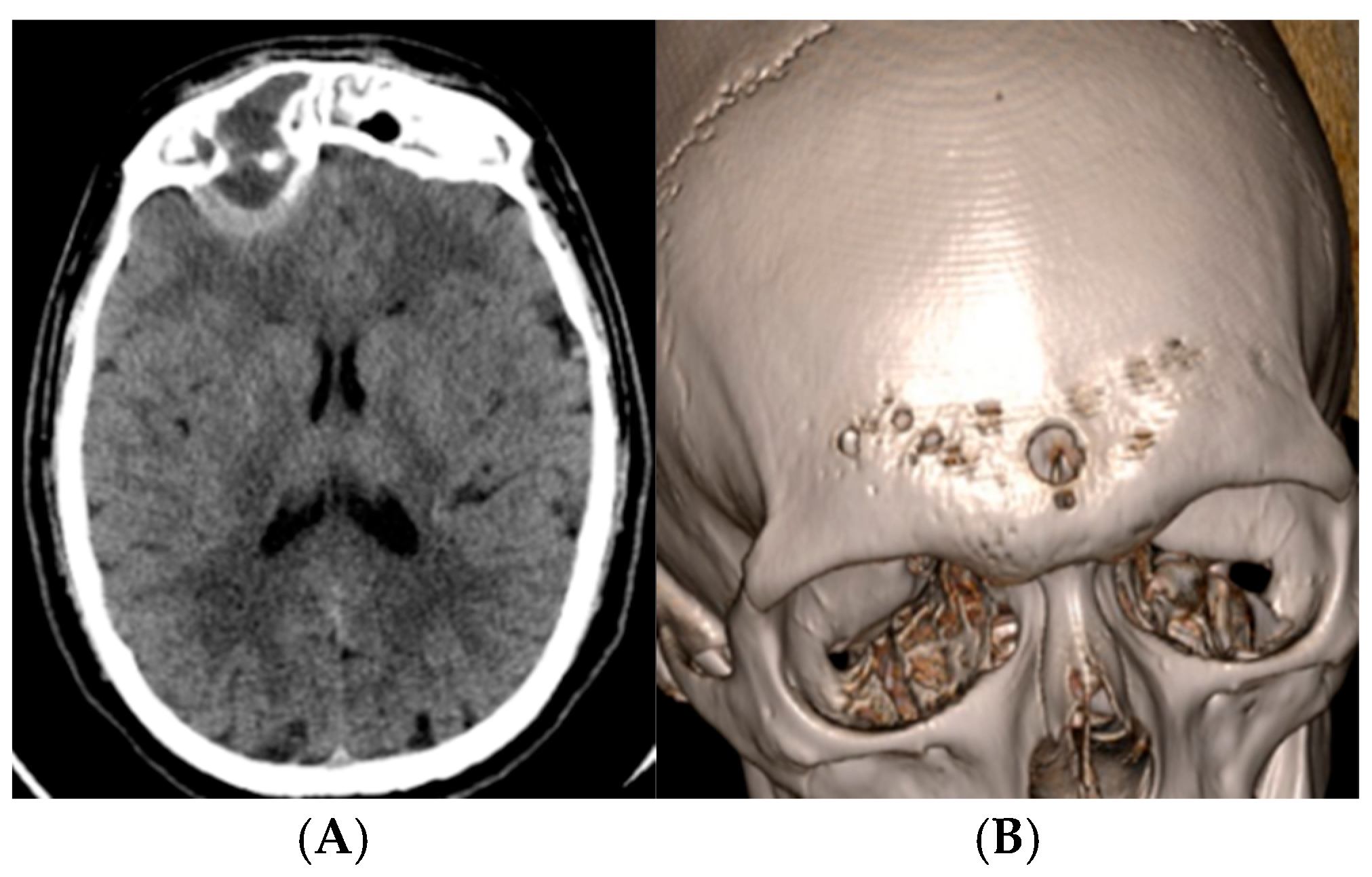
A 20-year-old immunocompetent man from Central Italy whose medical history was notable only for chronic sinusitis and who was not taking any regular medications was referred to the emergency department of a tertiary care hospital after an outpatient non-contrast brain computed tomography (CT) scan revealed acute-on-chronic frontal and ethmoidal sinusitis complicated by a 26 mm frontal sinus mucocele. The lesion was associated with erosion of the posterior sinus wall and contiguous extension into the right frontal lobe and superior orbit (Figure 1A,B).
The patient reported a 6-month history of anosmia and ageusia, followed by progressive headache, right orbital pain, and periorbital swelling. He denied intranasal drug use but reported occasional inhaled marijuana use. Occupational history was notable for outdoor work as a dog trainer, with frequent exposure to soil, grass, dust, and rural environments. He also reported consuming approximately one alcoholic drink per day.
No underlying immunosuppressive conditions or other recognized risk factors for invasive fungal infection were identified.
On admission, the patient was in good general condition, afebrile, alert, cooperative, and fully oriented, with no focal neurological deficits. Laboratory tests were unremarkable except for leukocytosis (15,000 cells/mm3) with neutrophilia, while C-reactive protein was within the normal range.
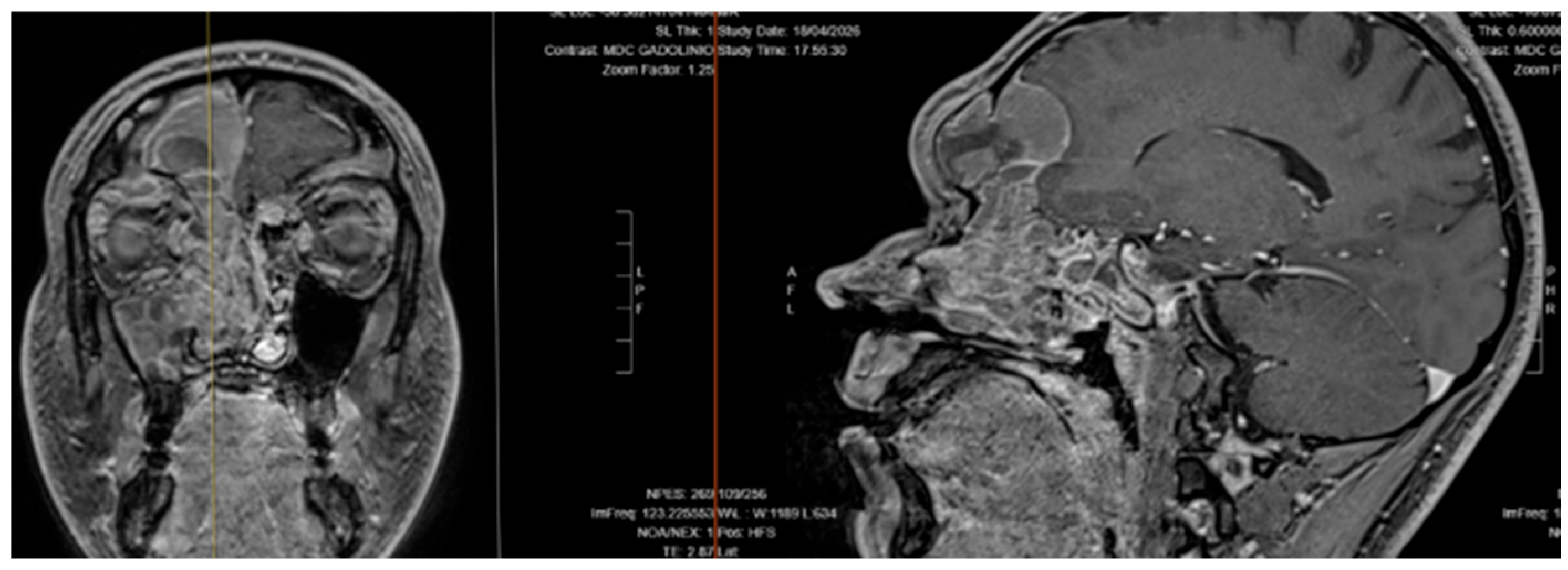
Contrast-enhanced magnetic resonance imaging (MRI) confirmed acute-on-chronic frontal and ethmoidal sinusitis complicated by a 26 mm frontal sinus mucocele with erosion of the posterior sinus wall and contiguous extension into the right frontal lobe and superior orbit. The intracranial lesion demonstrated imaging characteristics consistent with an epidural empyema associated with surrounding vasogenic edema (Figure 2). No additional intracranial lesions were identified.
Because of the extent of intracranial involvement and the risk of neurological deterioration, urgent neurosurgical intervention was undertaken. Intraoperative findings demonstrated extensive frontal sinus destruction with erosion of the posterior sinus wall, skull base osteolysis involving the ethmoid and orbital roofs, and inflammatory tissue densely adherent to the dura overlying the frontal lobe. A bifrontal craniotomy with frontal sinus cranialization and extensive debridement was performed. Additional purulent material and dense mucoid contents consistent with a frontal sinus mucocele were identified and removed. Multiple deep specimens obtained during surgery were submitted for microbiological and histopathological analyses. The extensive inflammatory and osteolytic process confirmed the locally invasive nature of the disease and allowed adequate surgical source control.
One week later, after stabilization of the neurological condition, endoscopic sinonasal surgery was performed to achieve complete debridement and further source control, including ethmoidectomy and excision of polypoid inflammatory tissue involving the frontal recess and ethmoidal cells. Additional tissue specimens were submitted independently for microbiological and histopathological analyses.
Laboratory investigations excluded major causes of immunodeficiency. HIV serology and chronic viral hepatitis screening were negative. As part of the immunological work-up performed to investigate possible underlying predisposing conditions, serum immunoglobulins were assessed. Total serum IgE was elevated (1002 kU/L), while a mild reduction in IgG was detected (652 mg/dL; reference range 700–1600 mg/dL). This isolated mild IgG reduction was not considered indicative of a clinically significant immunodeficiency. Chest radiography and abdominal ultrasonography did not reveal additional infectious foci. Serum β-D-glucan and galactomannan tests were negative.
Pending microbiological results, empirical broad-spectrum antibacterial therapy with meropenem at CNS dosage and linezolid was initiated by the neurosurgical team. Linezolid, however, was discontinued early because of severe gastrointestinal intolerance.
Microbiological cultures from the first intraoperative specimen yielded Curvularia species and Bacillus spp. Cultures from the second surgical specimen again isolated Curvularia species, subsequently identified as C. spicifera, together with coagulase-negative staphylococci. The repeated isolation of Curvularia from deep surgical specimens supported its etiologic role.
Species identification was initially obtained by matrix-assisted laser desorption ionization time-of-flight mass spectrometry (MALDI-TOF MS) using the MALDI Biotyper Sirius system (Bruker Daltonics GmbH & Co. KG, Bremen, Germany), which identified the isolate as Curvularia spicifera with a secure species-level score of 2.0. Given the rarity of the isolate and the invasive nature of the infection, identification was subsequently further evaluated by PCR amplification and Sanger sequencing of the D1/D2 domain of the 28S ribosomal RNA gene using primers P1 (5′-ATCAATAAGCGGAGGAAAAG-3′) and P2 (5′-CTCTGGCTTCACCCTATTC-3′). PCR reactions were carried out in a final volume of 25 μL containing 1× PCR buffer, dNTPs (1.25 mM each), 10 pmol of each primer, Taq DNA polymerase (Takara Bio Inc., Shiga, Japan), and nuclease-free water. Amplification consisted of 40 cycles of denaturation at 94 °C for 45 s, annealing at 50 °C for 45 s, and extension at 72 °C for 1 min. PCR products were purified using ExoSAP-IT™ PCR Product Cleanup Reagent (Applied Biosystems, Foster City, CA, USA) and subsequently sequenced in both directions on an Applied Biosystems 3500xL Genetic Analyzer (Thermo Fisher Scientific, Waltham, MA, USA). Forward and reverse sequences were assembled into a consensus sequence using BioEdit version 7.7.1.0. The obtained partial D1/D2 LSU sequence (359 nucleotides) was analyzed using both the NCBI BLAST tool version BLASTN 2.17.0 and the MycoBank pairwise alignment platform against multiple curated fungal databases, including FunCBS, UNITE, SILVA_LSU, RefSeq, ITS/EF1 MycologyLab, PasteurYeasts/MOLDS, fusarioidID, and MIRRI. The sequence showed 100% identity over 359 nucleotides and 99% query coverage with Curvularia spicifera reference sequences. Acknowledging the recognized taxonomic complexity of the Curvularia/Bipolaris complex and the limitations of relying on a single relatively short genetic marker, species-level assignment was not based on sequence data alone. Instead, a polyphasic approach was adopted, integrating phenotypic, proteomic, and molecular evidence. The isolate displayed macroscopic and microscopic features consistent with Curvularia spp. MALDI-TOF MS independently identified the isolate as C. spicifera, and molecular analysis supported the same identification. Therefore, the identification of the isolate as Curvularia spicifera was based on the convergence of independent phenotypic, proteomic, and molecular findings rather than on D1/D2 sequencing alone.
Antifungal susceptibility testing was performed using the Sensititre™ YeastOne™ colorimetric broth microdilution system (Thermo Fisher Scientific, Waltham, MA, USA) according to the manufacturer’s instructions for mold susceptibility testing. Plates were incubated at 35 °C and examined after 24, 48, and 72 h. MIC values were determined by visual assessment of the colorimetric endpoint and were defined as the lowest antifungal concentration preventing the color change in the growth indicator associated with fungal growth. Because no CLSI or EUCAST clinical breakpoints or species-specific epidemiological cutoff values are currently available for Curvularia spicifera, MIC values were reported without categorical interpretation. Previous studies have shown good agreement between the Sensititre YeastOne system and CLSI broth microdilution methodologies for antifungal susceptibility testing of filamentous fungi [14,15].
The isolate exhibited low MIC values for amphotericin B, itraconazole, and posaconazole, whereas higher MIC values were observed for fluconazole and echinocandins.
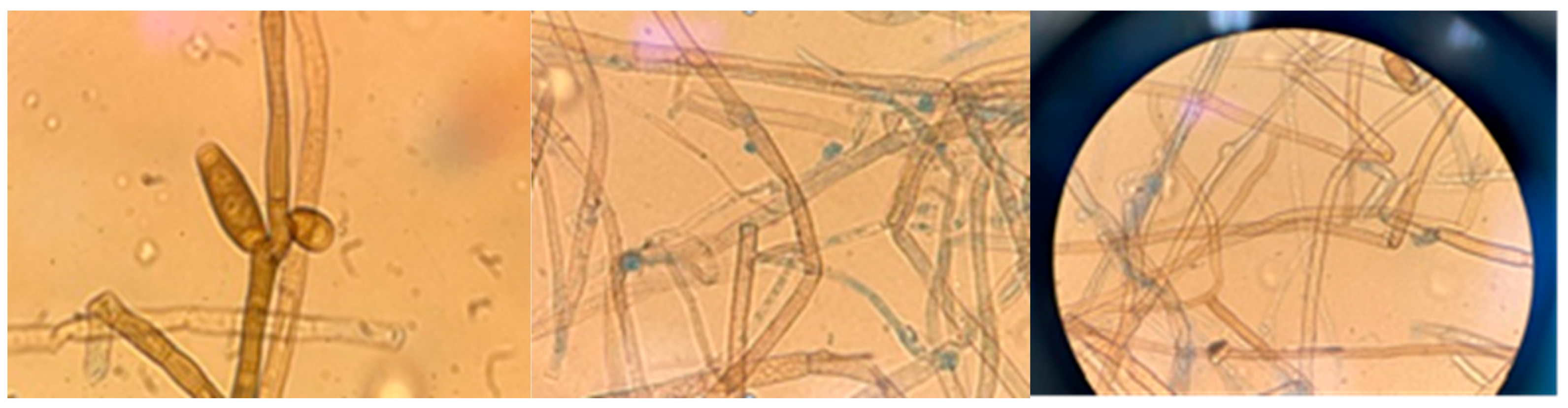
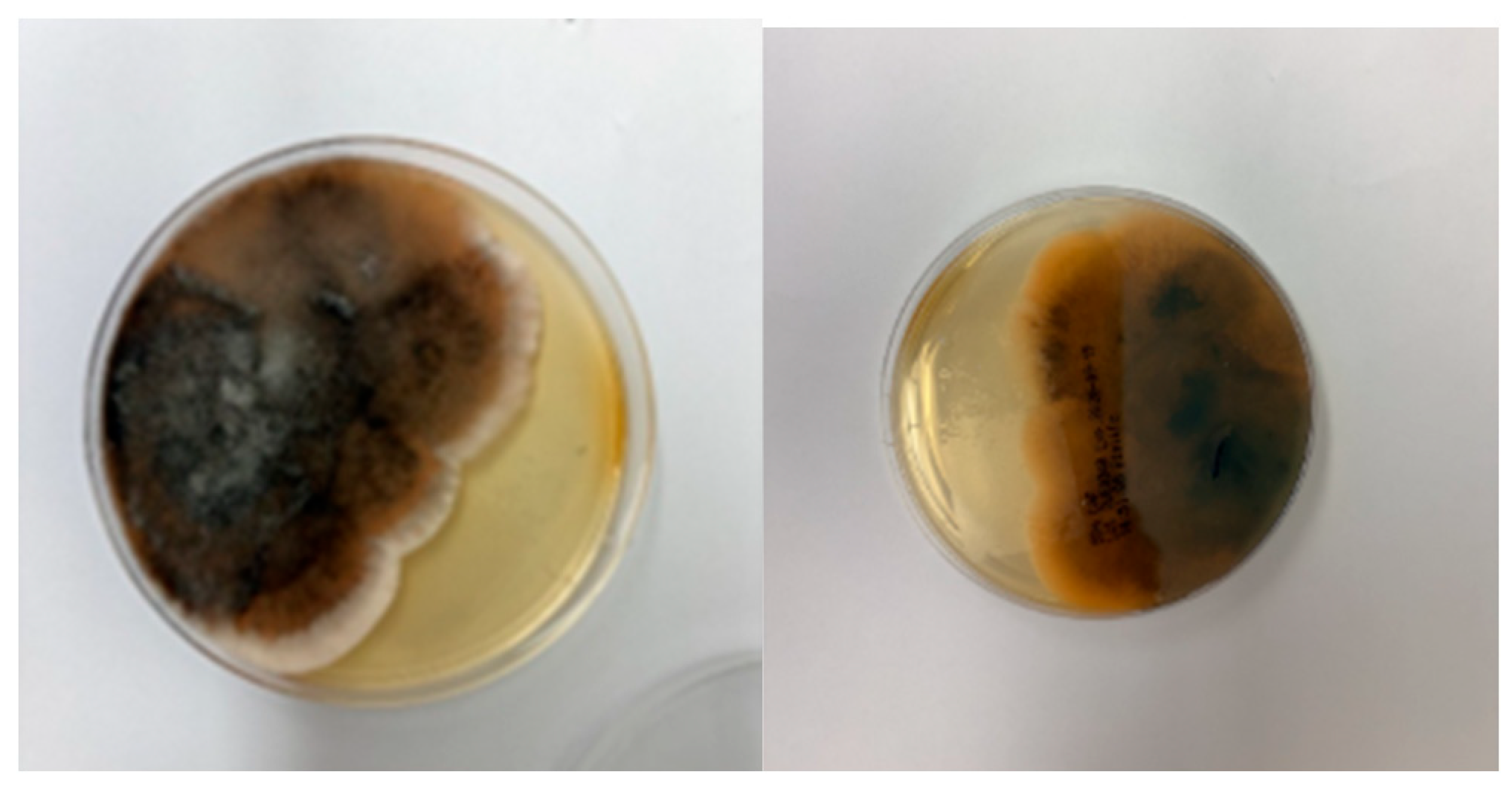
On Sabouraud dextrose agar, the isolate produced rapidly growing colonies that were initially grey-white and cottony. Over time, the colonies became dark olive-brown to black and developed a darkly pigmented reverse (Figure 3 and Figure 4). Microscopic examination of a lactophenol cotton blue preparation revealed septate dematiaceous hyphae and brown multicellular conidia with morphological features consistent with Curvularia species. These combined macroscopic and microscopic findings supported the identification of a dematiaceous mold belonging to the genus Curvularia. However, given the recognized morphological overlap within the Curvularia-Bipolaris complex, species-level identification was supported by the combined results of colony morphology, MALDI-TOF MS analysis, and D1/D2 sequencing.
Table 1 summarizes the MIC values obtained by antifungal susceptibility testing of the Curvularia isolates recovered from the first and second clinical samples.
Antifungal susceptibility testing was performed using the Sensititre™ YeastOne™ colorimetric broth microdilution system according to the manufacturer’s instructions for mold susceptibility testing. As no CLSI or EUCAST clinical breakpoints or species-specific epidemiological cutoff values (ECVs) are currently available for C. spicifera, MICs are reported without categorical interpretation. The discrepancy between the initial and reference laboratory voriconazole MICs highlights the challenges of antifungal susceptibility testing for uncommon dematiaceous fungi. Notably, susceptibility testing was repeated twice at the reference laboratory and yielded the same voriconazole MIC (2 µg/mL) in both determinations. While itraconazole, posaconazole, and amphotericin B showed closely comparable MIC values between the two laboratories, some variability was also observed for fluconazole and the echinocandins. Similar variability in azole susceptibility has been reported among clinically relevant Curvularia isolates, including members of the C. spicifera clade. Consequently, antifungal selection was based on the integration of available in vitro susceptibility data, pharmacokinetic/pharmacodynamic (PK/PD) principles, therapeutic drug monitoring (TDM), expected CNS drug exposure, and the patient’s clinical and radiological response [3,14,16,17].
Histological examination of neurosurgical and otorhinolaryngological specimens documented mixed neutrophilic and eosinophilic inflammation. In particular, nasal tissue showed polypoid sinonasal mucosa with a dense mixed inflammatory infiltrate rich in eosinophilic granulocytes, findings compatible with AFRS, although formal diagnostic criteria could not be fully documented.
Four days after the first surgical intervention, following the microbiological finding, an Infectious Diseases consultation was requested, and combined antifungal therapy with intravenous liposomal amphotericin B and oral voriconazole was initiated, while susceptibility testing was still pending. Meropenem was discontinued after 10 days because of progressive liver transaminase elevation and because repeated Curvularia isolation supported fungal infection as the primary process. After two weeks of combined antifungal therapy, the patient developed severe hypokalemia (1.8 mmol/L) without electrocardiographic abnormalities or clinical manifestations. Liposomal amphotericin B was discontinued, and aggressive electrolyte replacement resulted in progressive normalization. Voriconazole TDM was performed, and dose escalation to 600 mg/day was required to achieve therapeutic concentrations. The patient remained afebrile and clinically stable, with progressive improvement, and was discharged 22 days after neurosurgical intervention on oral voriconazole.
Follow-up brain and maxillofacial MRI performed one month after neurosurgery showed heterogeneous frontal sinus material and meningeal enhancement, interpreted as post-inflammatory changes in the context of a favorable clinical evolution. Despite the favorable radiological findings, given the type of infection and the extent of CNS involvement, we decided to continue voriconazole therapy. At the time of writing, the patient had completed more than three months of treatment, with progressive clinical improvement and good clinical and laboratory tolerability of voriconazole. A further follow-up brain MRI is scheduled at four months after surgery and will guide the decision on treatment discontinuation, together with the overall clinical course, drug exposure, and tolerability, in the absence of established guidelines defining the optimal duration of antifungal therapy for CNS Curvularia infections.

3. Discussion

The present case describes a locally invasive CNS fungal infection caused by C. spicifera in an immunocompetent host. Its main features of interest include the probable contiguous spread from chronic sinonasal disease, the coexistence of histopathological findings suggestive of AFRS phenotype, and the repeated isolation of C. spicifera from deep surgical specimens supported by molecular evidence. Together, these findings support a true invasive infection rather than environmental contamination and further demonstrate that Curvularia species may cause severe CNS disease even in the absence of overt immunosuppression.
CNS and intracranial infections caused by Curvularia species represent a rare manifestation within the broader spectrum of dematiaceous fungal infections of the central nervous system, which are more commonly associated with neurotropic pathogens such as Cladophialophora bantiana [2].
Since the first intracerebral infection attributed to a species currently classified within the genus Curvularia in 1973 [18], an increasing number of cases have been described, likely reflecting both improved recognition of Curvularia as a human pathogen and advances in microbiological identification techniques.
As shown in Table 2, the clinical spectrum of CNS Curvularia infection is remarkably broad, including brain abscess, cerebritis, meningitis, cranial-base mass-like disease, and intracranial extension of invasive sinusitis.
In parallel, the taxonomy of the genus has undergone substantial revision. Multilocus phylogenetic studies have shown that several clinically relevant fungi, including B. spicifera and B. hawaiiensis, are part of the Curvularia complex. Consequently, a significant proportion of historical reports of CNS and sinus infections attributed to Bipolaris species would currently be classified as Curvularia infections. This taxonomic reclassification is important to consider when reviewing older clinical series and interpreting published outcome data [9].
Both immunocompromised and immunocompetent hosts have been affected by CNS Curvularia infections. Reported risk groups include patients with solid or hematological malignancies, transplant recipients, individuals receiving immunosuppressive therapy, and premature neonates. However, numerous cases of intracerebral and rhinosinus-related infection have also occurred in otherwise healthy individuals, supporting a relevant role for local predisposing factors and environmental exposure in disease pathogenesis.
The literature identifies three main clinical patterns of CNS Curvularia infection: (i) contiguous extension from chronic or invasive sinonasal disease, often involving the frontal sinus, skull base, or orbit [37]; (ii) infection occurring in the setting of significant immunosuppression, such as solid-organ transplantation, hematological malignancies, or prolonged corticosteroid therapy [31]; and (iii) disease potentially related to inhalational exposure to contaminated organic material, including marijuana [33], particularly in the presence of local or systemic predisposing factors. The present case shares features with both the first and third patterns, given the longstanding sinonasal disease and the presence of potential environmental exposures.
Notably, the clinical course was slowly progressive, with months of anosmia, ageusia, headache, orbital pain, and periorbital swelling before diagnosis, mirroring several previously reported cases in which sinonasal manifestations preceded CNS involvement by weeks or months.
These observations support a pathogenic model in which inhaled Curvularia conidia colonize chronically inflamed sinonasal mucosa, particularly in the presence of an allergic inflammatory environment [38]. Elevated serum IgE levels and eosinophil-rich polypoid mucosa are consistent with an AFRS-like phenotype. Recent evidence has further strengthened the association between C. spicifera and allergic fungal rhinosinusitis in immunocompetent individuals. Ferraro et al. described two cases of non-invasive fungal rhinosinusitis in which diagnosis relied on the integration of radiological findings, eosinophil-rich inflammatory mucosa, fungal culture, and molecular identification. Their observations support the concept that C. spicifera may persist within a chronic allergic inflammatory environment and contribute to progressive sinonasal disease even in the absence of an overt immunosuppression phenotype [3]. Chronic obstruction together with persistent fungal-allergic inflammation may have promoted mucocele formation, progressive local expansion, bone erosion [39], orbital involvement, and ultimately contiguous spread to the frontal lobe [40]. Although the patient was not overtly immunocompromised, subtle host-related factors, including mild IgG reduction and pronounced type 2/eosinophilic inflammation, may have increased susceptibility to locally invasive disease.
Environmental exposure deserves consideration in the present case. Curvularia species are ubiquitous environmental fungi that are widely distributed in soil, grass, decaying vegetation, and plant debris. Although this widespread distribution may complicate distinguishing infection from contamination, the repeated isolation of C. spicifera from multiple deep surgical specimens obtained during separate procedures, together with consistent radiological findings and histopathological evidence of fungus-associated inflammation, strongly supports its role as the causative pathogen rather than an incidental colonizer.
Based on the patient’s occupational and recreational history, we hypothesized that two kinds of exposures may have increased his predisposition to developing the infection: inhalational marijuana use and repeated outdoor exposure.
Recreational marijuana smoking represents a plausible source of inhalational fungal exposure, given the well-documented contamination of cannabis products by viable fungal spores, including Aspergillus, Curvularia, and other environmental molds [41].
Marijuana use has been associated with invasive fungal infections, particularly aspergillosis in susceptible hosts, presumably through inhalation of contaminated plant material and subsequent respiratory or sinonasal colonization. Notably, CNS Curvularia infections have been reported in individuals with heavy marijuana use, including a patient receiving chronic corticosteroid therapy for neurosarcoidosis who developed a brainstem abscess [12,28]. Although these observations do not establish a causal relationship, they highlight the importance of carefully assessing environmental and recreational exposures in patients presenting with uncommon mold infections.
In addition, the patient’s occupation as an outdoor dog trainer may have increased exposure to environmental reservoirs of Curvularia through repeated contact with soil, grass, dust, and organic material. While Curvularia infections have been described in dogs [42,43], most commonly as post-traumatic cutaneous infections and keratitis [44], no epidemiologic evidence currently links dog-training activities to human Curvularia sinusitis or CNS infection. Nevertheless, repeated exposure to outdoor environments may have increased opportunities for inhalational acquisition and should be regarded as a potential contributing factor rather than an established risk factor.
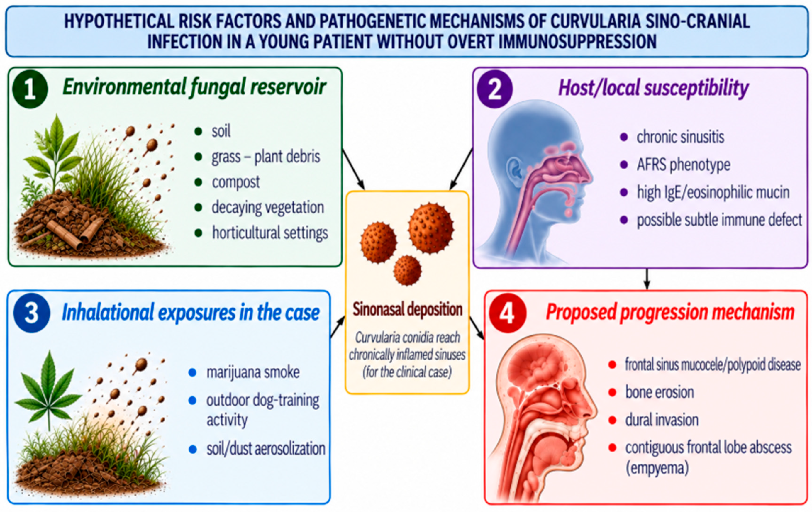
Figure 5 illustrates the hypothetical pathogenetic model for the sino-cranial Curvularia infection observed in the present case. The model hypothesizes inhalational acquisition from contaminated organic material and environmental exposure to soil and grass, followed by sino-nasal colonization in the setting of chronic AFRS. Subsequent local progression may have led to mucocele formation, erosion, intraorbital spread, and ultimately contiguous extension to the frontal lobe with empyema development.
Accurate microbiological identification is essential for the management of infections caused by dematiaceous fungi. Morphological differentiation among members of the Curvularia–Bipolaris–Cochliobolus complex can be challenging because of the frequent overlap in colony morphology and microscopic features. In the present case, species identification obtained by MALDI-TOF MS was further supported by sequencing of the D1/D2 domain of the 28S rRNA gene, providing complementary taxonomic information in support of the MALDI-TOF MS result. The combination of proteomic and molecular approaches is particularly valuable for uncommon molds such as Curvularia, for which historical taxonomic revisions and phenotypic overlap may complicate conventional identification. Molecular sequencing has therefore become increasingly important for accurate species-level identification and for clarifying epidemiological associations. In the present case, species assignment was based on the integration of morphological, proteomic, and molecular findings rather than on D1/D2 sequencing alone. In recent years, these approaches have substantially improved recognition of C. spicifera as an emerging opportunistic human pathogen [3].
An additional finding of interest was the marked difference between the voriconazole MIC obtained in the local laboratory and that reported by the reference laboratory (0.12 vs. 2 μg/mL). Although the reasons for this discrepancy cannot be determined retrospectively, susceptibility testing of uncommon dematiaceous fungi remains challenging, and inter-laboratory variation has been described. Moreover, antifungal susceptibility profiles within the genus Curvularia are heterogeneous. In a large study of molecularly identified clinical isolates, voriconazole and itraconazole showed limited overall activity against several Curvularia species [45], while earlier investigations reported elevated azole fungicidal endpoints in some isolates belonging to the B. spicifera/C. spicifera group [46]. As no CLSI or EUCAST clinical breakpoints are currently available for C. spicifera, MIC values should be interpreted cautiously and integrated with clinical evolution, drug exposure, and site of infection. In our patient, the decision to continue voriconazole therapy was supported by its favorable CNS penetration, TDM results, and the progressive clinical and radiological improvement observed during follow-up.
CNS infection caused by Curvularia is associated with substantial morbidity and mortality, even among immunocompetent individuals. Reported cases include both therapeutic failures and late relapses, highlighting the aggressive nature of the disease and the risk of unfavorable outcomes.
Management of CNS Curvularia infections remains particularly challenging because of the limited number of effective antifungal drugs, the absence of organism-specific clinical breakpoints, and the lack of randomized studies or evidence-based recommendations regarding the duration of therapy.
Consequently, current practice relies largely on published case experience and expert guidance on phaeohyphomycosis, which emphasize the importance of aggressive source control [32], repeated debridement when required, and prolonged systemic antifungal therapy; amphotericin B, itraconazole, voriconazole, and combination regimens have all been used in published Curvularia infections (Table 2).
Given the severity of CNS Curvularia infections and the risk of treatment failure or late relapse, selection of an effective and tolerable antifungal regimen is critical. Historically, amphotericin B, either as monotherapy or in combination with a triazole, has represented the cornerstone of treatment [1,14], while itraconazole was widely used in earlier years when alternative azoles were not available. Voriconazole has subsequently emerged as an attractive option because of its oral formulation and favorable CNS penetration; however, its use requires TDM and careful surveillance for hepatotoxicity, neurotoxicity, visual disturbances, photosensitivity, and drug–drug interactions.
In our patient, amphotericin B had to be discontinued because of severe hypokalemia, underscoring the practical challenges and toxicity-related limitations of prolonged combination therapy.
Isavuconazole may represent a promising alternative for long-term treatment owing to its favorable pharmacokinetic profile, good tolerability, and demonstrated in vitro activity against dematiaceous fungi [47]. Clinical experience with Curvularia infections remains limited, although sporadic cases have been reported. Notably, Cornely et al. [48] described successful treatment of disseminated pulmonary and cutaneous Curvularia lunata infection in a patient with hematological malignancy, with a favorable response after 52 days of isavuconazole therapy.
Despite advances in antifungal therapy, timely and aggressive surgical source control, including complete excision or debridement whenever anatomically feasible, remains a cornerstone of treatment and should be combined with prolonged systemic antifungal therapy to optimize clinical outcomes. In our patient, urgent neurosurgical intervention was considered necessary because of the site and extent of intracranial involvement and the risk of neurological deterioration; however, the timing and extent of surgery should be individualized according to the clinical and radiological features of each case.
Regarding the optimal duration of antifungal treatment for CNS Curvularia infections, no standardized recommendations are currently available. Treatment courses reported in the literature vary considerably, reflecting the heterogeneity of clinical presentation, the extent of intracranial involvement, the immune status of the host, and the completeness of surgical debridement. Among the published case reports reviewed, treatment duration ranged from a few weeks to 12 months; however, the exact timing of treatment discontinuation and long-term follow-up were inconsistently reported. Consequently, the available evidence remains insufficient to define the optimal duration of antifungal treatment or to support evidence-based recommendations [49].
Nevertheless, prolonged therapy is generally considered necessary because of the severity of CNS infection and the potential risk of persistence or relapse. Treatment duration should therefore be individualized and guided by clinical evolution, serial radiological findings, the adequacy of surgical source control, and drug tolerability [49].
Given the extent of CNS involvement, in our patient, voriconazole treatment was continued beyond three months. The favorable safety profile observed in this patient allowed prolonged therapy while serial imaging was used to monitor disease evolution and guide future treatment discontinuation.
Overall, awareness of potential risk factors for Curvularia infection and their careful investigation during the diagnostic workup may facilitate earlier recognition of the disease and prompt initiation of appropriate management.
This case reinforces the importance of multidisciplinary care involving infectious disease specialists, neurosurgeons, otolaryngologists, microbiologists, radiologists, and pathologists. Repeated isolation from deep specimens, histopathologic correlation, species-level identification, antifungal susceptibility testing, and imaging follow-up are all essential for establishing the diagnosis and guiding therapy.

4. Conclusions

This case highlights that C. spicifera can cause severe, locally invasive sino-cranial infection even in young individuals without overt immunosuppression. Chronic sinusitis/AFRS, inhalational marijuana use, and repeated occupational outdoor exposure were identified as potential local or exposure-related factors in this patient; however, their possible role in the development of invasive infection should be considered hypothetical and cannot be inferred from a single case. The available evidence underscores the importance of maintaining a high index of suspicion, obtaining a detailed environmental and recreational exposure history, and achieving prompt surgical source control combined with microbiological identification supported by phenotypic and molecular methods. Given the potential for treatment failure, toxicity, and late relapses, prolonged antifungal therapy and careful long-term clinical and radiological follow-up remain essential components of management.

Author Contributions

Conceptualization, A.E. and A.C.; clinical data collection, C.T., A.L., D.L. and R.A.R.; microbiology, M.S., C.G.P., A.V., A.G. and C.F.; radiology, D.L. and R.A.R.; writing—original draft, A.E., A.C., C.F. and S.C.; writing—review and editing, M.S., C.G.P., A.V., C.T., A.L., D.L., A.G. and R.A.R.; supervision: C.F. and S.C. All authors have read and agreed to the published version of the manuscript.

Funding

This work was supported by grants from the Italian Ministry of Health (Grant 2801210, Ricerca Corrente, Linea 3, IRCCS INMI L. Spallanzani, 2025–2027).

Institutional Review Board Statement

According to applicable national regulations and institutional policy, formal ethics committee approval was not required for this single-patient case report.

Informed Consent Statement

Written informed consent was obtained from the patient for publication of the clinical data and images included in the manuscript.

Data Availability Statement

All relevant data are included in the manuscript. Additional anonymized clinical details may be available from the corresponding author upon reasonable request, subject to institutional policy.

Acknowledgments

We acknowledge the contribution and support from the Italian national Node (MIRRI-IT) of the European Research Infrastructure MIRRI-ERIC. During the preparation of this manuscript, the authors used ChatGPT tools to assist with the language editing, including grammar, spelling, punctuation and style refinement. ChatGPT was also used to generate and refine Figure 5 and the graphical abstract. The authors have reviewed and edited the output and take full responsibility for the content of this publication.

Conflicts of Interest

The authors declare no conflicts of interest.

Abbreviations

The following abbreviations are used in this manuscript:
AFRSallergic fungal rhinosinusitis
CLSIClinical and Laboratory Standards Institute
CNScentral nervous system
CTcomputed tomography
ECVepidemiological cutoff value
MALDI-TOF MSmatrix-assisted laser desorption ionization time-of-flight mass spectrometry
MICminimum inhibitory concentration
MRImagnetic resonance imaging
PK/PDpharmacokinetic/pharmacodynamic
TDMtherapeutic drug monitoring

References

  1. Goldman, M.; Reddy, R.; Lucke-Wold, B.; Barpujari, A.; Cameron, M.; Porche, K.; Laurent, D.; Duarte, E.; Lobo, B.C.; Koch, M. Curvularia and the Brain: Case Demonstration of Optimal Management. Arch. Emerg. Med. Crit. Care 2022, 6, 1051. [Google Scholar] [CrossRef] [PubMed]
  2. Revankar, S.G.; Sutton, D.A.; Rinaldi, M.G. Primary Central Nervous System Phaeohyphomycosis: A Review of 101 Cases. Clin. Infect. Dis. 2004, 38, 206–216. [Google Scholar] [CrossRef] [Scilit] [PubMed]
  3. Ferraro, N.; Iskandar, E.; Pitrolo, A.M.G.; Ramus, M.; Pagella, F.; Introini, S.; Baldanti, F.; Cavanna, C. Curvularia Spicifera in Non-Invasive Fungal Rhinosinusitis: Case Reports and Diagnostic Insights. Pathogens 2026, 15, 523. [Google Scholar] [CrossRef] [Scilit] [PubMed]
  4. Rohwedder, J.J.; Simmons, J.L.; Colfer, H.; Gatmaitan, B. Disseminated Curvularia Lunata Infection in a Football Player. Arch. Intern. Med. 1979, 139, 940–941. [Google Scholar] [CrossRef] [Scilit]
  5. Velasco, J.; Revankar, S. CNS Infections Caused by Brown-Black Fungi. J. Fungi 2019, 5, 60. [Google Scholar] [CrossRef] [Scilit] [PubMed]
  6. Kobayashi, H.; Sano, A.; Aragane, N.; Fukuoka, M.; Tanaka, M.; Kawaura, F.; Fukuno, Y.; Matsuishi, E.; Hayashi, S. Disseminated Infection by Bipolaris Spicifera in an Immunocompetent Subject. Med. Mycol. 2008, 46, 361–365. [Google Scholar] [CrossRef] [Scilit] [PubMed]
  7. McGinnis, M.R.; Campbell, G.; Gourley, W.K.; Lucia, H.L. Phaeohyphomycosis Caused by Bipolaris Spicifera: An Informative Case. Eur. J. Epidemiol. 1992, 8, 383–386. [Google Scholar] [CrossRef] [Scilit] [PubMed]
  8. Krizsán, K.; Papp, T.; Manikandan, P.; Shobana, C.S.; Chandrasekaran, M.; Vágvölgyi, C.; Kredics, L. Clinical Importance of the Genus Curvularia. In Medical Mycology; CRC Press: Boca Raton, FL, USA, 2015. [Google Scholar]
  9. Manamgoda, D.S.; Cai, L.; McKenzie, E.H.C.; Crous, P.W.; Madrid, H.; Chukeatirote, E.; Shivas, R.G.; Tan, Y.P.; Hyde, K.D. A Phylogenetic and Taxonomic Re-Evaluation of the Bipolaris-Cochliobolus-Curvularia Complex. Fungal Divers. 2013, 56, 131–144. [Google Scholar] [CrossRef] [Scilit]
  10. Revankar, S.G.; Sutton, D.A. Melanized Fungi in Human Disease. Clin. Microbiol. Rev. 2010, 23, 884–928. [Google Scholar] [CrossRef] [Scilit] [PubMed]
  11. Cavanna, C.; Seminari, E.; Pusateri, A.; Mangione, F.; Lallitto, F.; Esposto, M.C.; Pagella, F. Allergic Fungal Rhinosinusitis Due to Curvularia Lunata. New Microbiol. 2014, 37, 241–245. [Google Scholar] [PubMed]
  12. Gongidi, P.; Sarkar, D.; Behling, E.; Brody, J. Cerebral Phaeohyphomycosis in a Patient with Neurosarcoidosis on Chronic Steroid Therapy Secondary to Recreational Marijuana Usage. Case Rep. Radiol. 2013, 2013, 191375. [Google Scholar] [CrossRef] [Scilit] [PubMed]
  13. Chowdhary, A.; Meis, J.F.; Guarro, J.; de Hoog, G.S.; Kathuria, S.; Arendrup, M.C.; Arikan-Akdagli, S.; Akova, M.; Boekhout, T.; Caira, M.; et al. ESCMID and ECMM Joint Clinical Guidelines for the Diagnosis and Management of Systemic Phaeohyphomycosis: Diseases Caused by Black Fungi. Clin. Microbiol. Infect. 2014, 20, 47–75. [Google Scholar] [CrossRef] [Scilit] [PubMed]
  14. CLSI M61-Performance Standards for Antifungal Susceptibility Testing of Filamentous Fungi|GlobalSpec. Available online: https://standards.globalspec.com/std/14297791/clsi-m61 (accessed on 3 August 2026).
  15. Patel, R.; Mendrick, C.; Knapp, C.C.; Grist, R.; McNicholas, P.M. Clinical Evaluation of the Sensititre YeastOne Plate for Testing Susceptibility of Filamentous Fungi to Posaconazole. J. Clin. Microbiol. 2007, 45, 2000–2001. [Google Scholar] [CrossRef] [Scilit] [PubMed]
  16. Kidd, S.E.; Westblade, L.F. Bipolaris or Curvularia? Resolving the Spicy Issue of How Clinical Isolates Should Be Reported. PLoS Pathog. 2024, 20, e1012678. [Google Scholar] [CrossRef] [Scilit] [PubMed]
  17. EUCAST: Clinical Breakpoint Tables. Available online: https://www.eucast.org/bacteria/clinical-breakpoints-and-interpretation/clinical-breakpoint-tables/ (accessed on 3 August 2026).
  18. Fuste, F.J.; Ajello, L.; Threlkeld, R.; Henry, J.E. Drechslera Hawaiiensis: Causative Agent of a Fatal Fungal Meningo-Encephalitis. Med. Mycol. 1973, 11, 59–63. [Google Scholar] [CrossRef] [Scilit] [PubMed]
  19. Lampert, R.P.; Hutto, J.H.; Donnelly, W.H.; Shulman, S.T. Pulmonary and Cerebral Mycetoma Caused by Curvularia Pallescens. J. Pediatr. 1977, 91, 603–605. [Google Scholar] [CrossRef] [Scilit] [PubMed]
  20. Friedman, A.D.; Campos, J.M.; Rorke, L.B.; Bruce, D.A.; Arbeter, A.M. Fatal Recurrent Curvularia Brain Abscess. J. Pediatr. 1981, 99, 413–415. [Google Scholar] [CrossRef] [Scilit] [PubMed]
  21. Yoshimori, R.N.; Moore, R.A.; Itabashi, H.H.; Fujikawa, D.G. Phaeohyphomycosis of Brain: Granulomatous Encephalitis Caused by Drechslera Spicifera. Am. J. Clin. Pathol. 1982, 77, 363–370. [Google Scholar] [CrossRef] [Scilit] [PubMed]
  22. Biggs, P.J.; Allen, R.L.; Powers, J.M.; Holley, H.P. Phaeohyphomycosis Complicating Compound Skull Fracture. Surg. Neurol. 1986, 25, 393–396. [Google Scholar] [CrossRef] [Scilit] [PubMed]
  23. Adam, R.D.; Paquin, M.L.; Petersen, E.A.; Saubolle, M.A.; Rinaldi, M.G.; Corcoran, J.G.; Galgiani, J.N.; Sobonya, R.E. Phaeohyphomycosis Caused by the Fungal Genera Bipolaris and Exserohilum. A Report of 9 Cases and Review of the Literature. Medicine 1986, 65, 203–217. [Google Scholar] [CrossRef] [Scilit] [PubMed]
  24. Ismail, Y.; Johnson, R.H.; Wells, M.V.; Pusavat, J.; Douglas, K.; Arsura, E.L. Invasive Sinusitis with Intracranial Extension Caused by Curvularia Lunata. Arch. Intern. Med. 1993, 153, 1604–1606. [Google Scholar] [CrossRef] [Scilit]
  25. Klapper, S.R.; Lee, A.G.; Patrinely, J.R.; Stewart, M.; Alford, E.L. Orbital Involvement in Allergic Fungal Sinusitis. Ophthalmology 1997, 104, 2094–2100. [Google Scholar] [CrossRef] [Scilit] [PubMed]
  26. Ebright, J.R.; Chandrasekar, P.H.; Marks, S.; Fairfax, M.R.; Aneziokoro, A.; McGinnis, M.R. Invasive Sinusitis and Cerebritis Due to Curvularia Clavata in an Immunocompetent Adult. Clin. Infect. Dis. 1999, 28, 687–689. [Google Scholar] [CrossRef] [Scilit] [PubMed]
  27. Latham, R.H. Bipolaris Spicifera Meningitis Complicating a Neurosurgerical Procedure. Scand. J. Infect. Dis. 2000, 32, 102–103. [Google Scholar] [CrossRef] [Scilit] [PubMed]
  28. Carter, E.; Boudreaux, C. Fatal Cerebral Phaeohyphomycosis Due to Curvularia Lunata in an Immunocompetent Patient. J. Clin. Microbiol. 2004, 42, 5419–5423. [Google Scholar] [CrossRef] [Scilit] [PubMed]
  29. Smith, T.; Goldschlager, T.; Mott, N.; Robertson, T.; Campbell, S. Optic Atrophy Due to Curvularia Lunata Mucocoele. Pituitary 2007, 10, 295–297. [Google Scholar] [CrossRef] [Scilit] [PubMed]
  30. Singh, H.; Irwin, S.; Falowski, S.; Rosen, M.; Kenyon, L.; Jungkind, D.; Evans, J. Curvularia Fungi Presenting as a Large Cranial Base Meningioma: Case Report. Neurosurgery 2008, 63, E177; Discussion E177. [Google Scholar] [CrossRef] [Scilit] [PubMed]
  31. Rosow, L.; Jiang, J.X.; Deuel, T.; Lechpammer, M.; Zamani, A.A.; Milner, D.A.; Folkerth, R.; Marty, F.M.; Kesari, S. Cerebral Phaeohyphomycosis Caused by Bipolaris Spicifera after Heart Transplantation. Transpl. Infect. Dis. 2011, 13, 419–423. [Google Scholar] [CrossRef] [Scilit] [PubMed]
  32. Gadgil, N.; Kupferman, M.; Smitherman, S.; Fuller, G.N.; Rao, G. Curvularia Brain Abscess. J. Clin. Neurosci. 2013, 20, 173–175. [Google Scholar] [CrossRef] [Scilit] [PubMed]
  33. Skovrlj, B.; Haghighi, M.; Smethurst, M.E.; Caridi, J.; Bederson, J.B. Curvularia Abscess of the Brainstem. World Neurosurg. 2014, 82, 241.e9–241.e13. [Google Scholar] [CrossRef] [Scilit] [PubMed]
  34. Bova, C.; Vigna, E.; Gentile, M.; Fiaschi, E. Cerebral Pheohyphomycosis Due to Curvularia Species. IDCases 2022, 27, e01391. [Google Scholar] [CrossRef] [Scilit] [PubMed]
  35. Pham, J.; Kulla, B.; Johnson, M. Invasive Fungal Infection Caused by Curvularia Species in a Patient with Intranasal Drug Use: A Case Report. Med. Mycol. Case Rep. 2022, 37, 1–3. [Google Scholar] [CrossRef] [Scilit] [PubMed]
  36. Bono, B.C.; Olei, S.; Rossini, Z.; Fernandes, B.; Valeri, M.; Pessina, F. Primary Brain Necrotizing Granulomas Caused by Curvularia Spicifera. Infect. Dis. Now. 2022, 52, 381–383. [Google Scholar] [CrossRef] [Scilit] [PubMed]
  37. Samaddar, A.; Shrimali, T.; Tiwari, S.; Sharma, A. First Report of Human Infection Caused by Curvularia Warraberensis, Manifesting as Invasive Sinusitis with Intracranial Involvement. J. Mycol. Med. 2023, 33, 101337. [Google Scholar] [CrossRef] [Scilit] [PubMed]
  38. Posteraro, B.; Scarano, E.; La Sorda, M.; Torelli, R.; De Corso, E.; Mulé, A.; Paludetti, G.; Fadda, G.; Sanguinetti, M. Eosinophilic Fungal Rhinosinusitis Due to the Unusual Pathogen Curvularia Inaequalis. Mycoses 2010, 53, 84–88. [Google Scholar] [CrossRef] [Scilit] [PubMed]
  39. Riegler, A.N.; Grayson, J.W.; Greenway, J.W.; Woodworth, B.A.; McCarty, T.P.; Pappas, P.G.; Leal, S.M. Histopathologic Insights Into Curvularia-Induced Chronic Granulomatous Fungal Sinusitis: A Guide for Surgical Pathologists. Mod. Pathol. 2025, 38, 100898. [Google Scholar] [CrossRef] [Scilit] [PubMed]
  40. Marinucci, V.; Chahine, E.B.; Bush, L.M. Invasive Fungal Rhinosinusitis Caused by Curvularia in a Patient With Type 1 Diabetes Mellitus: A Case Report. J. Pharm. Pr. 2022, 35, 311–316. [Google Scholar] [CrossRef] [Scilit] [PubMed]
  41. Gwinn, K.D.; Leung, M.C.K.; Stephens, A.B.; Punja, Z.K. Fungal and Mycotoxin Contaminants in Cannabis and Hemp Flowers: Implications for Consumer Health and Directions for Further Research. Front. Microbiol. 2023, 14, 1278189. [Google Scholar] [CrossRef] [Scilit] [PubMed]
  42. Rothenburg, L.S.; Snider, T.A.; Wilson, A.; Confer, A.W.; Ramachandran, A.; Mani, R.; Rizzi, T.; Nafe, L. Disseminated Phaeohyphomycosis in a Dog. Med. Mycol. Case Rep. 2017, 15, 28–32. [Google Scholar] [CrossRef] [Scilit] [PubMed]
  43. Jaffey, J.A.; Cañete-Gibas, C.F.; Wiederhold, N.P.; Sanders, C.J.; Struthers, J.D.; Black, A.; Wu, B.; Thomas, K.S.; Bennett, P.; Watt, J. Novel Curvularia Species Causing Disseminated Phaeohyphomycosis in a Dog. Top. Companion Anim. Med. 2025, 64, 100939. [Google Scholar] [CrossRef] [Scilit] [PubMed]
  44. Herráez, P.; Rees, C.; Dunstan, R. Invasive Phaeohyphomycosis Caused by Curvularia Species in a Dog. Vet. Pathol. 2001, 38, 456–459. [Google Scholar] [CrossRef] [Scilit] [PubMed]
  45. da Cunha, K.C.; Sutton, D.A.; Fothergill, A.W.; Gené, J.; Cano, J.; Madrid, H.; de Hoog, S.; Crous, P.W.; Guarro, J. In vitro antifungal susceptibility and molecular identity of 99 clinical isolates of the opportunistic fungal genus Curvularia. Diagn. Microbiol. Infect. Dis. 2013, 76, 168–174. [Google Scholar] [CrossRef] [Scilit] [PubMed]
  46. Espinel-Ingroff, A. In Vitro Fungicidal Activities of Voriconazole, Itraconazole, and Amphotericin B against Opportunistic Moniliaceous and Dematiaceous Fungi. J. Clin. Microbiol. 2001, 39, 954–958. [Google Scholar] [CrossRef] [Scilit] [PubMed]
  47. Zheng, H.; Song, N.; Mei, H.; Dong, J.; Li, D.; Li, X.; Liu, W. In Vitro Activities of Ravuconazole and Isavuconazole against Dematiaceous Fungi. Antimicrob. Agents Chemother. 2020, 64, e00643-20. [Google Scholar] [CrossRef] [Scilit] [PubMed]
  48. Cornely, O.A.; Mullane, K.M.; Ostrosky-Zeichner, L.; Maher, R.M.; Croos-Dabrera, R.; Lu, Q.; Lademacher, C.; Perfect, J.R.; Oren, I.; Schmitt-Hoffmann, A.-H.; et al. Isavuconazole for Treatment of Rare Invasive Fungal Diseases. Mycoses 2018, 61, 518–533. [Google Scholar] [CrossRef] [Scilit] [PubMed]
  49. Bengyella, L.; Yekwa, L.E.; Waikhom, S.D.; Nawaz, K.; Iftikhar, S.; Motloi, T.S.; Tambo, E.; Roy, P. Upsurge in Curvularia Infections and Global Emerging Antifungal Drug Resistance. Asian J. Sci. Res. 2017, 10, 299–307. [Google Scholar] [CrossRef] [Scilit]
Figure 1. (A) Axial brain CT scan showing a frontal sinus mucocele with erosion of the posterior sinus wall and direct extension into the right frontal lobe. (B) Three-dimensional CT reconstruction confirming erosion of the outer table of the frontal bone.
Microorganisms 14 02245 g001
Figure 2. Coronal and sagittal contrast-enhanced T1-weighted MRI demonstrating ethmoidal, maxillary, and frontal sinusitis associated with a right frontal epidural empyema.
Microorganisms 14 02245 g002
Figure 3. Microscopic morphology of Curvularia spicifera after lactophenol cotton blue staining. The preparation shows septate dematiaceous hyphae and brown multicellular conidia that are cylindrical to slightly curved, consistent with the morphology of Curvularia species.
Microorganisms 14 02245 g003
Figure 4. Macroscopic appearance of Curvularia spicifera. Colonies grown on Sabouraud dextrose agar showed rapid growth with a cottony to floccose pale grey/olivaceous-grey surface that progressively became olivaceous-brown to blackish brown, with a dark brown to black reverse.
Microorganisms 14 02245 g004
Figure 5. Hypothetical risk factors and proposed mechanisms of Curvularia sino-cranial infection in the present clinical case.
Microorganisms 14 02245 g005
Table 1. In vitro antifungal susceptibility of the Curvularia spicifera isolate.
Antifungal AgentInitial Laboratory MIC (Microg/mL)Reference Laboratory MIC (Microg/mL)
Amphotericin B0.12<0.12
Anidulafungin0.250.03
Caspofungin44
Micafungin28
Fluconazole1664
Itraconazole0.060.03
Voriconazole0.122
Posaconazole0.010.015
IsavuconazoleND1
Flucytosine64ND
Table 2. Published cases of CNS and intracranial Curvularia infections.
Patient Age/SexImmune Status/Predisposing FactorsCNS and Intracranial/Adjacent SiteExtra CNS InfectionPathogenSurgeryAntifungal TherapyOutcome and Follow-upRef.
31/FUndiagnosed disseminated lymphocytic lymphosarcoma, identified at autopsyMeningoencephalitis with severe granulomatous vasculitis and a right frontal tumor-like lesionNoDrechslera hawaiiensis
(currently classified as Curvularia hawaiiensis)
NoNoneDied 11 days after surgery[18]
13/MImmunocompetent; mild mental disability; eosinophilia/elevated IgE; repeated soil/dirt inhalationRight frontoparietal cerebral mass; recurrence 3 years laterPulmonary micetomaCurvularia pallescensComplete excision; cranial decompression at recurrenceGriseofulvin; amphotericin B stopped for azotemia; IV miconazole for 6 weeks
Recurrence treated with IV and intrathecal miconazole
Initial improvement; fatal relapse 3 years later[19,20]
25/MImmunocompetent; football player with chronic skin ulcers after football abrasionsLeft parietal cerebral abscessDisseminated to soft-tissue, pulmonary, paravertebral diseaseCurvularia lunataMultiple drainage proceduresMiconazole for 5 weeks; later added amphotericin BCerebral abscess resolved; systemic relapse[4]
21/FImmunocompetent; no clear risk factorBilateral frontal cerebral lesions; granulomatous fungal encephalitisNoDrechslera spicifera (currently classified as Curvularia spicifera)Right frontal brain biopsyAmphotericin B, total dose 2 g, plus flucytosine for 4 monthsSurvived; persistent hemiparesis and emotional lability[21]
21/FImmunocompetent; skull fracture with environmental contamination, persistent CSF leakage and corticosteroid therapyPost-traumatic meningoencephalitis with fungal vasculitisNoDrechslera spicifera (currently classified as Curvularia spicifera)Wound debridement and craniectomy with dural graft; right frontal lobectomy and evacuation of intracerebral hematomaNo antifungal therapyDied 28 days after admission[22]
49/FCarcinomatous meningitis in previous metastatic breast cancer; Ommaya reservoir for intrathecal chemotherapyFungal meningitis and ventriculitisNoBipolaris spicifera
(currently classified as Curvularia spicifera)
NoNoneDied after 14 days[23]
26/FImmunocompetent; chronic sinus infectionRight occipital lesion/meningitis; left temporal and sphenoid wing mass 6 years laterChronic sinusitisB. spicifera
(currently classified as C. spicifera)
Craniotomy and biopsy; sinus surgeryAmphotericin B plus ketoconazole for 3 monthsSurvived with persistent severe visual deficit[7]
46/FImmunocompetent; chronic sinusitisSphenoidal mass extended intracraniallyChronic sinusitisC. lunataTrans-septal sphenoidectomy with removal of a fungal ballAmphotericin B for 4 months, followed by 8 months of oral ketoconazoleNo recurrence 2 months after stopping therapy[24]
26/MImmunocompetent; nasal polyposis; eosinophiliaEthmoid/sphenoid allergic fungal sinusitis with orbital extension; Radiological involvement of the cavernous sinusNoninvasive allergic fungal sinusitisBipolaris spicifera (currently classified as Curvularia spicifera)Frontoethmoidectomy, orbitotomy; repeat sinus surgeryNo antifungal therapyAsymptomatic at 1 year[25]
46/FImmunocompetent; asthma, hay fever, chronic sinusitis; multiple sinus surgeriesBifrontal cerebritis contiguous with cribriform plate erosionInvasive sinusitisCurvularia clavataBilateral sphenoethmoidectomy and frontal sinusotomy; no brain surgeryAmphotericin B for 7 months, then itraconazole for 1 monthMarked improvement at 24 months[26]
18/MPreviously healthy; postoperative neurosurgical setting, steroids; recent diving in lake waterPostoperative meningitisNoB. spicifera
(currently classified as C. spicifera)
No surgery; ventriculostomy for intrathecal therapyIV and intrathecal amphotericin B plus itraconazole, stopped for liver toxicity: duration 5 monthsSurvived; residual deficits at 1 year[27]
21/MImmunocompetent; asthma; occasional marijuana/cocaine useRight basal ganglia/anterior horn lesion; later ventricular spreadNoC. lunataStereotactic needle aspiration and biopsyAmphotericin B, liposomal amphotericin B; flucytosine added laterDied 1 month after admission[28]
57/MImmunocompetent; no clear risk factorsSphenoid mucocele extending into pituitary fossa/suprasellar region with optic nerve compressionnoC. lunataTranssphenoidal resectionAmphotericin B/liposomal amphotericin B for 4 weeks, then oral voriconazole for 4 weeksDisease-free at month 8[29]
35/MPlasma cell dyscrasiaLarge cranial base lesion with brainstem compression and hydrocephalusNoCurvularia geniculataTransnasal/transclival biopsy and limited resectionVoriconazole; amphotericin B added laterDied 4 months after biopsy[30]
55/MHeart transplant 6 weeks earlier; on therapy with mycophenolate, prednisone, cyclosporineLeft basal ganglia abscessNoB. spicifera
(currently classified as C. spicifera)
Brain biopsyLiposomal amphotericin B, then voriconazole for 12 monthsMRI resolution at 12 months; no relapse after stopping therapy[31]
50/FImmunocompetent; chronic sinusitis; previous sinus surgeryRight inferior frontal lobe extending into the ethmoid, right orbit, and cranial baseSinusitisCurvularia speciesCombined CNS and sinus surgeryVoriconazoleDisease-free after 3 years[32]
37/MNeurosarcoidosis on chronic steroids; heavy marijuana useMultiple supratentorial brain abscessesPulmonary abscessCurvularia speciesNeurosurgical debridement and resection; repeated resectionEmpirical IV antifungal therapy, agents not specifiedNo new lesions at 2-month follow-up[12]
33/MImmunocompetent; heavy marijuana use and alcohol abuseRight medulla/brainstem abscessLeft lung granulomaCurvularia speciesFirst open biopsy and drainage; repeat drainage and subtotal debulkingVoriconazole, flucytosine, liposomal amphotericin B for 10 weeks; prolonged voriconazole (at least 13 months)Alive and improved at 13 months[33]
68/MPrevious non-Hodgkin lymphoma treated with chemo-radiotherapyMultiple supratentorial and subtentorial lesionsNoCurvularia speciesBrain lesion biopsyItraconazole, IV voriconazole, liposomal amphotericin BDied 10 days after treatment initiation[34]
43/FImmunocompetent; intranasal cocaine abuse and perfume inhalant useMultifocal cerebral infarctionsKidney abscess, invasive sphenoid sinusitisCurvularia buchloesNasal endoscopy and biopsyIV amphotericin B for 30 days, then switch to oral voriconazoleNeurologic deficits persisted at 30-day follow-up[35]
28/MImmunocompetent; chronic rhinosinusitis, nasal polyps, prior sinus surgery, marijuana useRight anterior cranial fossa/frontal abscess with skull-base extensionSinusitisCurvularia speciesCombined endonasal and bifrontal craniotomy with gross total resectionAmphotericin B 4 weeks, then voriconazole 12 monthsStable at 12-month follow-up[1]
68/MGastric DLBCL treated with chemoradiotherapy and autologous bone marrow transplant 3 years earlierMultiple bilateral supratentorial necrotizing granulomas C. spiciferaRemoval of the most superficial right frontal lesionNot specified dual IV antifungal therapyDied 2 months after surgery[36]
33/FImmunocompetent; previously treated pulmonary tuberculosisChronic invasive sphenoid/ethmoid sinusitis with pituitary, midbrain, facial-vestibulocochlear complex and vascular involvementSinusitisCurvularia warraberensisEndoscopic sinus surgery and bilateral sphenoethmoidectomyLiposomal amphotericin B plus itraconazoleDied 32 days after admission[37]
Historical fungal names are reported as originally published. Current nomenclature was updated according to the taxonomic revision of the Bipolaris–Cochliobolus–Curvularia complex proposed by Manamgoda et al. and subsequent updates [9,16].
Disclaimer/Publisher’s Note: The statements, opinions and data contained in all publications are solely those of the individual author(s) and contributor(s) and not of MDPI and/or the editor(s). MDPI and/or the editor(s) disclaim responsibility for any injury to people or property resulting from any ideas, methods, instructions or products referred to in the content.