Article

Periodontal Health in Individuals Living with HIV: An Exploratory and Descriptive Molecular Approach of Microbial Interspecific and Intraspecific Diversity in Brazilian Patients

by
Patricia N. Olivares Ponce
1,2,
Lana Bitencourt Chaves
1,
Daiana de Souza Perce-da-Silva
3,4,
Ana Luiza Carneiro-Alencar
1,
Cinthia Magalhães Rodolphi
1,
Isabela Ferreira Soares
1,
Rodrigo Nunes Rodrigues-da-Silva
5,
Ana Caroline Alves-da-Silva
2,
Fabio Vidal Marques
2,6,
Rafael Vidal Peres
2,
Dennis de Carvalho Ferreira
2,
Rodrigo Carvalho de Souza
2,
Cristiane Gonçalves
2,
Lucio Souza Gonçalves
2,* and
Josué da Costa Lima-Junior
1,2,*
1
Immunoparasitology Laboratory, Oswaldo Cruz Institute, Fiocruz, Rio de Janeiro 21040-360, RJ, Brazil
2
Postgraduate Program in Dentistry, Faculty of Dentistry, Estácio de Sá University-IDOMED, Rio de Janeiro 22640-100, RJ, Brazil
3
Clinical Immunology Laboratory, Oswaldo Cruz Institute, Fiocruz, Rio de Janeiro 21040-360, RJ, Brazil
4
Basic and Applied Immunology Laboratory, Petrópolis Medical School, Arthur Sá Earp Neto University Center, Petrópolis 25680-120, RJ, Brazil
5
Laboratory of Hantaviroses and Rickettsioses, Oswaldo Cruz Institute, Fiocruz, Rio de Janeiro 21040-360, RJ, Brazil
6
Faculty of Dentistry, Rio de Janeiro State University, Rio de Janeiro 20551-030, RJ, Brazil
*
Authors to whom correspondence should be addressed.
Microorganisms 2025, 13(4), 867; https://doi.org/10.3390/microorganisms13040867
Submission received: 12 March 2025 / Revised: 2 April 2025 / Accepted: 3 April 2025 / Published: 10 April 2025

Abstract

:
Oral manifestations of HIV infection can be an early sign of the disease and may indicate progression to AIDS. Although antiretroviral therapies, especially highly active antiretroviral therapy (HAART), have reduced the prevalence of HIV-related oral lesions, ongoing updates in diagnosis and treatment are essential due to the extended life expectancy of individuals living with HIV. Periodontal disease is a significant concern in these patients, influenced by altered immune responses and microbial dynamics, though the mechanisms are not fully understood. This exploratory study aimed to investigate the oral microbiota and periodontal disease prevalence in HIV-positive individuals by analyzing subgingival plaque samples from 24 patients. We identified 12 bacterial species using Polymerase Chain Reaction (PCR) and amplicon sequencing. Seven species were detected, with Filifactor alocis, Tannerella forsythia, and Porphyromonas endodontalis being the most common. Porphyromonas gingivalis was present in only 13.6% of samples, while T. forsythia was found in 58.3%. Genetic diversity was also observed in P. endodontalis and Selenomonas sputigena amplicons, with specific single nucleotide polymorphisms (SNPs) identified in both species. These results highlight the complex microbial interactions in the oral environments of people living with HIV, emphasizing the need for personalized diagnostic and therapeutic strategies for managing oral health in this population.

1. Introduction

Oral health in individuals living with HIV (PLHIV) poses unique challenges and complexities. While HAART has significantly improved the overall health outcomes of PLHIV, oral manifestations remain a significant concern, impacting both diagnosis and management strategies [1]. Candidiasis, particularly oral candidiasis, stands as a hallmark of HIV-related oral lesions, with various clinical presentations prevalent among PLHIV. Additionally, periodontal disease presents a significant burden, characterized by altered microbial profiles and immune responses in PLHIV [2].
The oral microbiota plays a crucial role in the health and disease of individuals infected with HIV, with alterations observed in both diversity and composition, especially following HAART. While studies suggest a reduced microbial diversity in HIV-infected individuals, particularly post-HAART, further research with larger sample sizes is warranted to elucidate the potential impact of HIV infection and treatment on the oral microbiome. Moreover, understanding these microbial alterations may shed light on the emergence of opportunistic pathogens in the oral cavity.
In this scenario, periodontal disease in PLHIV presents a complex interplay of microbial colonization and host immune responses. While classic periodontal pathogens are prevalent in both HIV-positive and -negative individuals, PLHIV may exhibit a predisposition to additional infections by atypical microorganisms. Notably, the immunosuppressive state in PLHIV may favor the colonization of such pathogens, contributing to the severity of periodontal disease. In addition to considering the classic pathogens, the intraspecific diversity of periodontal bacteria within PLHIV warrants attention. Variations in the genotypic and phenotypic characteristics of periodontal pathogens may influence disease progression and treatment outcomes in this population [3]. Understanding the intraspecific diversity of these bacteria can provide observations into the pathogenicity and virulence factors associated with periodontal disease in PLHIV, ultimately guiding more effective therapeutic interventions.
Advancements in molecular methods, particularly Polymerase Chain Reaction (PCR) and sequencing techniques, have revolutionized microbial identification, allowing for the detection of unculturable or difficult-to-culture species. These methods have significantly contributed to our understanding of microbial dynamics in oral diseases, offering more specificity into exploring previously unknown pathogens and enhancing diagnostic accuracy. One of the key tools in this process is the analysis of the 16S ribosomal RNA (rRNA) gene, which provides a powerful means of characterizing the bacterial composition of the oral microbiome [4]. Through the sequencing of the 16S rRNA gene, clinical and researchers have been able to gain deeper access to the global genetic, metabolic, and ecological changes associated with periodontitis [4]. This approach has revealed that the disease microbiome is enriched in virulence factors and adapted to a parasitic lifestyle that takes advantage of the disrupted host homeostasis [4]. Moreover, the disease state has been found to occupy a narrow region within the space of possible configurations of the oral microbiome, suggesting a common structure that is distinct from completely healthy samples [4]. Ultimately, the 16S bacterial analysis has emerged as a crucial tool in the diagnosis and identification of periodontal and peri-implant bacterial species, enabling a deeper understanding of these complex microbial communities and their role in oral health and disease [4,5]. In patients with HIV, 16S analysis has been used to identify shifts in the oral microbiome associated with disease progression and immune status [6]. This highlights the broad applicability of this approach in understanding the oral microbiome and its clinical implications.
Studies utilizing the 16S rRNA gene as a molecular marker have highlighted the significance of intraspecific diversity within bacterial communities, including those in the oral cavity [7]. This diversity has been implicated in influencing the clinical presentation and progression of periodontal diseases, with specific bacterial variants or strains potentially correlating with disease severity and treatment responses. Critically, the impact of periodontal disease may be exacerbated in immunocompromised individuals such as PLHIV [8]. In these cases, dysbiotic shifts in the oral microbiome could intensify clinical manifestations and compromise treatment outcomes.
Understanding intraspecific diversity within the 16S rRNA gene of oral bacteria and its implications for the pathogenesis and clinical course of periodontal diseases holds significant promise for personalized diagnosis and targeted therapeutic interventions, particularly in immunocompromised patients [4,9]. Moreover, addressing the knowledge gap in intraspecific microbial diversity among clinical samples from South American countries is crucial. Exploring such diversity could unveil regional variations in the oral microbiome and their associations with periodontal health outcomes, thus informing the development of more effective prevention and treatment strategies tailored to individual patient needs. Therefore, we aimed to investigate the microbial diversity present in Brazilian patients with HIV by employing molecular techniques, focusing on both the interspecific (between different microbial species) and intraspecific (within the same microbial species) variations observed in microbial communities. Ultimately, this research will enhance our understanding of microbial diversity’s role in health and disease, with implications for developing targeted therapeutic strategies and improving patient outcomes.

2. Materials and Methods

2.1. Study Population and Sample Collection

The study involved 24 patients enrolled in an ongoing HIV infection research project at the Department of Oral Pathology and Diagnosis, School of Dentistry, Universidade Estácio de Sá. After providing informed consent and receiving comprehensive information about the study’s objectives, risks, and benefits, participants were included if they were patients with HIV/AIDS aged over 13 years. Patients with incomplete HIV status data in their medical records were excluded.
Participants’ socio-demographic information, including smoking status, age, and gender, as well as their medical history—such as diabetes, hypertension, heart disease, and use of ARV drugs—was gathered through a self-administered questionnaire. HIV-related data, including details on antiretroviral therapy, T-CD4 + lymphocyte counts, and viral load, were extracted from medical charts.
Subgingival plaque samples were collected using a sterile curette, which was scraped along the tooth at the gingival sulcus. The sampling was conducted under aseptic conditions, with samples collected into 0.5 mL of sterile Tris/EDTA buffer. No bleeding was observed at the sampling sites during collection. The collected plaque samples were subsequently used for molecular analysis to evaluate the diversity of 12 bacterial species.

2.2. Clinical Record

Periodontal measurements were assessed during a complete periodontal examination performed at the School of Dentistry, at the Estácio de Sá University. They were recorded at six sites per tooth (distobuccal, buccal, mesiobuccal, distolingual, lingual, and mesiolingual) in all teeth, excluding third molars. The examination was performed by the same experienced periodontist observer (intraclass correlation coefficient of 0.86 for probing depth and 0.80 for clinical attachment loss). Screening of the clinical assessments was evaluated (periodontal probing depth (PPD) (mm), clinical attachment loss (CAL) (mm), bleeding on probing (BOP) [10] and visible supragingival biofilm (VSB) [11]) using a periodontal probe with a diameter of 0.5 mm (PCV12, HuFriedy, Chicago, IL, USA).

2.3. DNA Extraction

DNA extraction followed the protocol of the QIAamp® DNA Blood Mini Kit (Qiagen, Hilden, Germany). Initially, lyophilized Protease K was reconstituted and all kit components were prepared. Subsequently, 20 µL of Protease K and 200 µL of the sample were combined in a 1.5 mL Eppendorf tube with 200 µL of Buffer AL. After vortexing for 15 s, the tube was incubated at 56 °C for 10 min. Next, 200 µL of 96–100% ethanol was added, vortexed for 15 s, and the lysate was transferred to a column without touching the column walls, using an attached Eppendorf tube. The column was centrifuged at 8000 rpm for 3 min, and the flow-through was discarded. Following washes with 500 µL of Buffer AW1 (centrifuged at 8000 rpm for 1 min) and Buffer AW2 (centrifuged at 13,000 rpm for 3 min), the column was air-dried for 3 min at room temperature. DNA was eluted with 100 µL of Buffer AE onto the column membrane, incubated for 5 min at room temperature, and centrifuged at 10,000 rpm for 1 min. DNA was stored at 4 °C. All procedures adhered to laboratory standards and manufacturer’s guidelines, including biosafety measures and proper reagent handling.

2.4. DNA Quantification

After DNA extraction, 2 µL of each sample was used for quantification using a Nanodrop 2000 spectrophotometer (Thermo Scientific) at the Immunoparasitology Laboratory of Instituto Oswaldo Cruz. DNA quantification was performed at a wavelength of 260 nm. Before readings, samples were gently mixed, and the Nanodrop analysis cell was cleaned and equilibrated. The equipment was calibrated using ultrapure water as a blank, and the baseline was adjusted. Three independent readings were taken for each sample, and the DNA concentration was calculated using the Beer–Lambert law. Additionally, DNA purity was assessed by the 260 nm/280 nm absorbance ratio. The results were recorded, and the DNA concentration was standardized for subsequent experiments.

2.5. Primer Selection

Taxon-specific oligonucleotide primers were used to detect target microorganisms. A pair of universal bacterial primers targeting nearly all bacterial 16S rRNA genes at the same position served as a positive control for PCR, indicating bacterial presence in clinical samples (Table 1).

2.6. Polymerase Chain Reaction (PCR)

The methodology for DNA amplification by Polymerase Chain Reaction (PCR) utilized the PCR Master Mix from Promega, specifically optimized for routine PCR. Each amplification reaction was conducted in a final volume of 50 µL, comprising the following components: 25 µL of PCR Master Mix, 1.2 µL of Forward primer (FOW), 1.2 µL of Reverse primer (REV), 5 µL of DNA template, and 17.6 µL of water. The PCR Master Mix contains essential components for DNA amplification. After preparing the reaction mix, samples were subjected to a thermal cycler programmed to perform denaturation, annealing, and extension steps tailored to the specific DNA template (Table 1). Positive and negative controls were included to monitor amplification specificity and efficiency. Following PCRs, amplified products were analyzed by agarose gel electrophoresis to verify successful amplification and the presence of the expected DNA fragment size. Amplification was carried out using the Veriti Thermal Cycler (Applied Biosystems, Foster City, CA, USA). Post-PCR, products were separated on a 3% Agarose gel (Sigma Aldrich, St. Louis, MI, USA) at 4 V/cm in 1× TAE buffer (0.04 M TRIS-acetate, 1 mM EDTA). Sample loading on the gel involved preparing a mixture for the 100 bp marker, comprising 5 µL of marker and 3 µL of GelRed, and another for our sample, consisting of 2 µL of loading dye, 2 µL of GelRed, and 10 µL of amplified product. Samples were carefully loaded into the gel wells, and electrophoresis was conducted at 110 V for 50 min. Following the electrophoresis run, pre-amplified products were visualized under ultraviolet (UV) illumination to identify DNA bands resulting from amplification. The GeneRuler 100 bp Plus DNA Ladder (Thermo Scientific, Waltham, MA, USA) served as a molecular size standard to estimate the size of amplified products. Comparing bands from our sample with the ladder allowed for the approximate determination of amplified fragment sizes. The visualization of DNA bands under UV light enabled assessment of the amplification efficiency and confirmation of the expected DNA fragment size. Electrophoresis results were recorded for subsequent data analysis and interpretation (Figure 1).

2.7. DNA Sequencing

DNA samples, purified accordingly, were distributed into a 96-well plate (Applied Biosystems-PN. c/10 plates). For each well, a final volume of 7.5 μL was added, containing the necessary primers and Milli-Q water, as applicable. The DNA sequencing reaction mixture was calculated based on the number of samples to be analyzed per plate. For each plate, the required amount consisted of 100 μL of BigDye (Applied Biosystems-PN.:4336917-100-reaction kit) and 150 μL of 5× sequencing buffer (Applied Biosystems-PN:4336699). The plate was sealed with a PCR plate adhesive or Axygen rubber mat (AxyMat-Code CM-96-RD). Next, 2.5 μL of the reaction mixture was added to each well of the plate (1 μL of BigDye + 1.5 μL of 5× buffer). The plate was briefly centrifuged to ensure proper mixing. The following thermal cycler protocol was used: 94 °C for 10 s, 50 °C for 5 s, and 60 °C for 4 min, repeated for 40 cycles. After the sequencing reaction, samples underwent precipitation to remove free ddNTPs, which could interfere with DNA sequence reading on the DNA analyzer. The plate was briefly centrifuged, and then 30 μL (3× volume) of 75% isopropanol (MERCK) was added to each well. Samples were vortexed for 10 s, followed by a 15 min incubation period, protected from light, and centrifuged again at 4000 rpm for 45 min. The supernatant was discarded by pouring the plate over paper towels and gently tapping it on the bench. Then, 50 μL (5× volume) of 75% ethanol (MERCK) was added to each well of the plate, which was centrifuged again for 15 min at 4000 rpm. The supernatant was discarded using paper towels, and the plate was inverted and centrifuged at 600 rpm for ethanol absorption, stopping when reaching 600 rpm. Subsequently, the plate was placed on a 60 °C heat block for 10 min, protected from light. The resulting dry plate could be stored, protected from light, at −20 °C for a maximum of approximately 30 days. For the final step, 10 μL of Hi-Di formamide (Applied Biosystems P/N:4311320) was added to each well of the dry plate, which was briefly centrifuged. Then, the plate was placed on a 95 °C heat block for 3 min, protected from light, and immediately transferred to ice for 5 to 10 min. After brief centrifugation to allow the products adhered to well walls to concentrate at the bottom, the plate was subjected to capillary electrophoresis using an ABI 3730xl instrument.

2.8. Sequence Analysis

The genetic diversity of the 16S gene fragments was analyzed using DnaSP v6 software to estimate the diversity within the population based on genetic diversity parameters such as number of segregating sites (S), number of haplotypes (h), haplotype diversity (Hd), and nucleotide diversity (π). Natural selection on the 16S gene fragments was assessed using Tajima’s D test and the Z test. Tajima’s D values were calculated using the total number of mutations estimated with DnaSP v6 software to test the neutral theory of evolution. Positive values may suggest positive or balancing selection, maintaining alleles at balanced frequencies, while negative values indicate purifying selection or recent population expansion. The Z-test was performed with MEGA7 v6.0; non-synonymous to synonymous substitution rates (dN/dS) (1000 bootstrap replicates) were estimated using the Nei-Gojobori method with Jukes correction, where p < 0.05 was considered significant.

3. Results

3.1. General Epidemiological Data

A total of 24 patients participated in the study, out of which 21 completed the epidemiological data form. The mean age of the participants was 38 years, ranging from 25 to 60 years. Regarding gender distribution, 7 participants were female, 10 were male, and 4 did not provide this information.
The duration since HIV diagnosis ranged from 2 to 23 years. Participants’ educational levels varied, with two having incomplete elementary education, seven completing elementary school, two having incomplete high school education, and six completing high school. Information on educational attainment was not available for four participants.
The clinical and epidemiological characteristics of the 21 participants are summarized in Table 2. The mean age of the cohort was 42.5 years (±7.9), and the average duration since diagnosis was 8.2 years (±6.2). Hematological assessments revealed a mean leukocyte count of 4783 cells/mm3 (±2096), with lymphocyte and neutrophil counts averaging 1758 cells/mm3 (±696) and 2319 cells/mm3 (±1158), respectively. The immunological profile included a mean T-CD4 count of 661.4 cells/mm3 (±354.1) and a mean T-CD8 count of 792.9 cells/mm3 (±397.1), resulting in a CD4/CD8 ratio of 0.865 (±0.286).
In terms of clinical comorbidities, 17% of participants were diagnosed with diabetes, and 22% had hypertension. Additionally, 28% of the cohort experienced herpes simplex virus infections, and another 28% were diagnosed with shingles. These findings indicate a notable prevalence of both metabolic and infectious conditions within the study population, emphasizing the need for comprehensive management strategies to address these associated health issues.
Clinical and epidemiological data were collected and summarized in the following Table 2.

3.2. Clinical Measurements

Clinical measurements [PPD, CAL, BOP, plaque accumulation (PL), and visible supragingival biofilm] were recorded at six sites per tooth in all teeth of all patients by two trained examiners, using a conventional manual periodontal probe (Hu-Friedy, Chicago, IL, USA).
Table 3 presents a summary of the clinical parameters of all the patients studied. VSB percentages ranged from 6.8% to 90.5%, with the highest biofilm presence in patient 5 (90.5%) and the lowest in patient 14 (6.8%). BOP values varied from 2.8% to 77.7%, with the highest value found in patient 12 (77.7%) and the lowest in patient 4 (2.8%). PPD measurements ranged from 1.29 mm to 3.59 mm, with patient 24 exhibiting the deepest probing depth (3.59 mm) and patient 21 the shallowest (1.29 mm). CAL values ranged from 0.20 mm to 4.47 mm, with the highest attachment loss observed in patients 10 (4.47 mm) and 12 (3.88 mm), while patient 13 showed the lowest CAL (0.0 mm). These findings indicate considerable variability in periodontal conditions across the patients, with significant differences in biofilm accumulation, gingival inflammation, probing depth, and attachment loss.

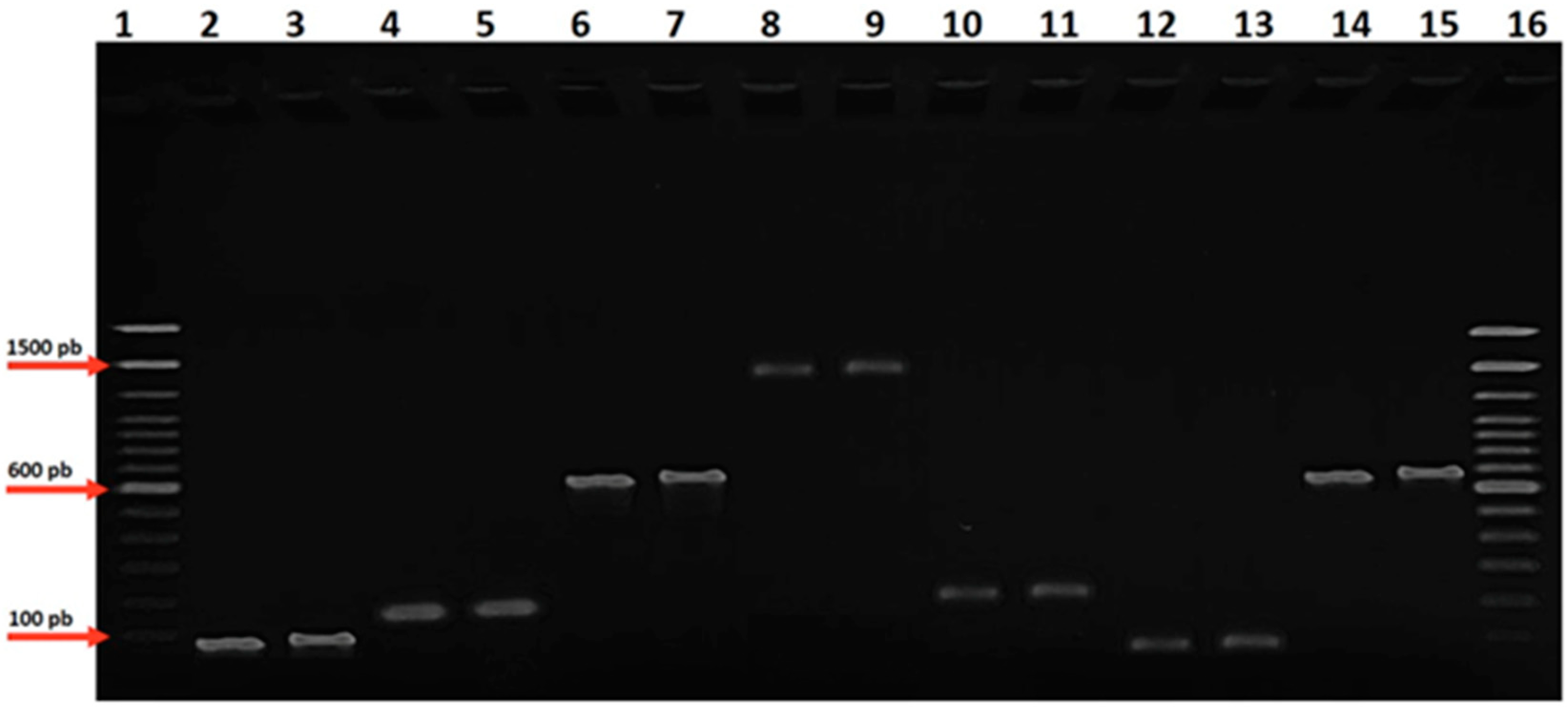
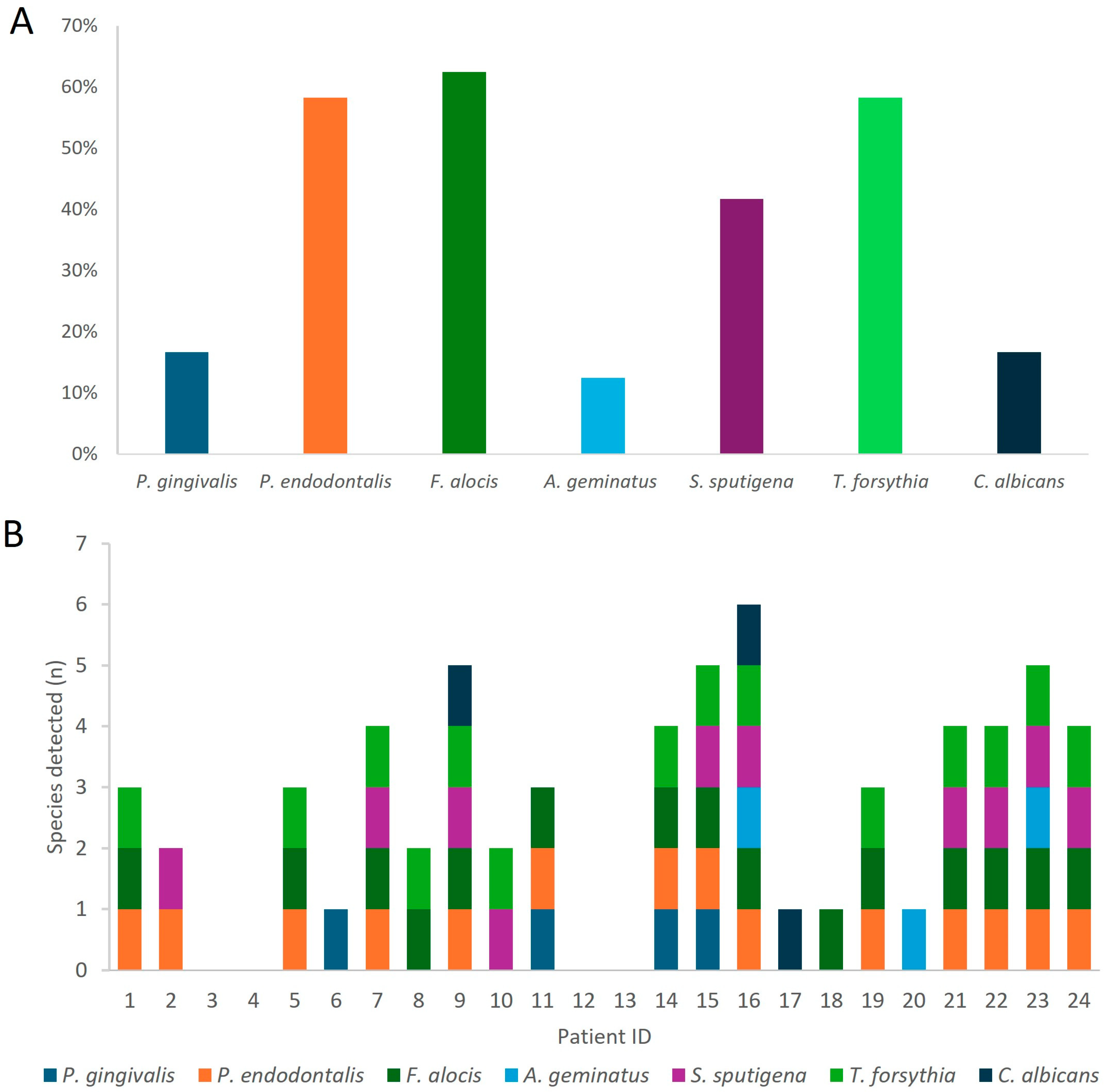
3.3. Molecular Identification of Targeted Oral Pathogens

In our study on the molecular identification of oral pathogens, we successfully identified the presence 7 out of the 12 targeted bacterial genes in our collected samples. These include P. gingivalis, P. endodontalis, F. alocis, A. geminatus, C. albicans, T. forsythia, and S. sputigena (Figure 2). Among the 24 patients from whom subgingival plaque was collected, 20 tested positive for bacterial presence in their samples (Figure 2). Specifically, four patients had single bacterial infections, three had infections with two bacteria, four had infections with three bacteria, four had infections with four bacteria, four had infections with five bacteria, and only one patient had infections with six bacteria. Combinations of F. alocis, T. forsythia, and P. endodontalis were the most frequently observed bacterial combinations.
The most frequently detected bacterium was F. alocis, found in 15 samples, representing 62.5% of the total. This was followed by T. forsythia and P. endodontalis, each identified in 14 samples (58.3%). S. sputigena was detected in 10 samples (41.6%), while both P. gingivalis and C. albicans were found in 4 samples each, accounting for 13.6%. A. geminatus was least common, found in three samples, representing 12.5%.
Our descriptive findings highlight the prevalence of putative periodontal pathogens in our study cohort, albeit below normal levels and/or undetected for some pathogens. P. gingivalis was detected in only 13.6% of samples, while Tanerella forsythia was more prevalent at 58.3%. Treponema denticola was not detected. F. alocis emerged as the most frequently detected bacterium, followed by Tanerella forsythia, a well-known periodontal pathogen, and P. endodontalis, an endodontic pathogen, which was found at a high frequency in the subgingival plaque of our HIV-positive patients.
Last, to contextualize our findings with the Brazilian clinical and epidemiological scenario, we also examined published studies on the same periodontal pathogens in non-HIV individuals (Table 4). Our results indicate small variations in the prevalence of microbial species in PLHIV compared to HIV-negative individuals from other Brazilian studies (Table 4). P. gingivalis was detected in 16.7% of PLHIV, a lower prevalence than HIV-negative individuals (29.4%) but still within the reported range (7.5–58.4%). P. endodontalis showed a high prevalence (58.3%) in HIV-positive individuals, similar to the 50.0–77.0% range in HIV-negative individuals. F. alocis was found in 62.5% of PLHIV, higher than the average in HIV-negative studies but within the reported range (20.0–67.0%). Conversely, A. geminatus was present in 12.5% of PLHIV, lower than the 33.0% reported in HIV-negative individuals. S. sputigena (41.7%) and T. forsythia (58.3%) had frequencies comparable to or slightly higher than those in HIV-negative individuals (15.4–67.0% and 12.0–100%, respectively). Finally, C. albicans was present in 16.7% of PLHIV, below the average for HIV-negative studies but within the reported range (14.5–50.0%). Notably, all values found in our study fell within the ranges reported by other Brazilian studies [23,24,25,26,27,28,29,30,31,32], suggesting that the oral microbiota of PLHIV follows previously described patterns. However, the wide variability in microbial frequencies across studies highlights the need for further research to clarify these comparisons and better understand the underlying factors driving these differences.

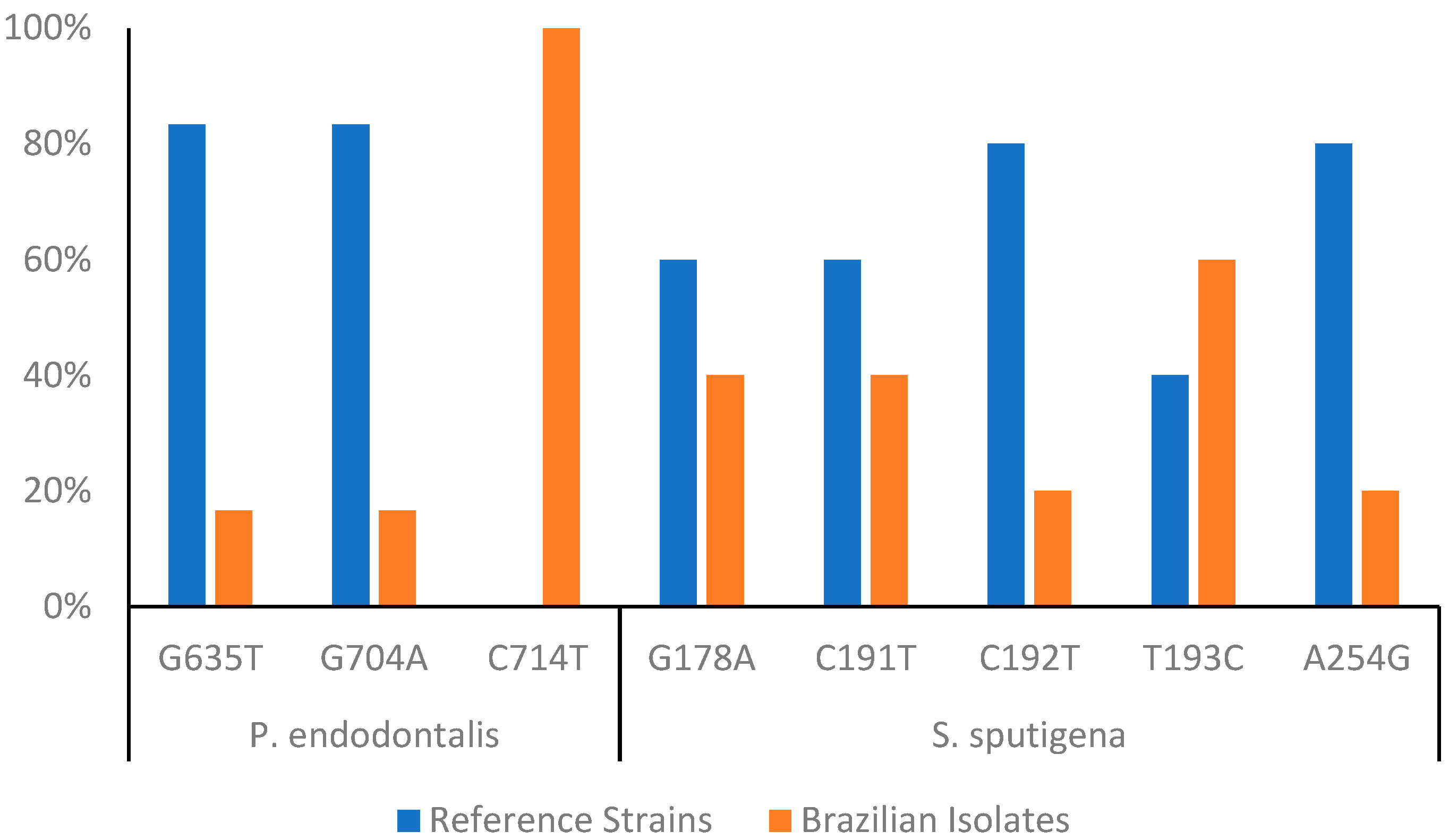
3.4. Intraspecific Diversity Analysis

Sequencing the PCR-amplified gene fragments of all participants revealed that the amplicon sequences of Brazilian isolates studied were 100% conserved in P. gingivalis, F. alocis, A. geminatus, C. albicans, and T. forsythia. However, notable intraspecific variability was observed in amplicons of P. endodontalis and S. sputigena (Supplementary File S1).
The polymorphisms in the P. endodontalis gene were investigated, focusing particularly on single nucleotide polymorphisms (SNPs). The strain ATCC 35406 was used as the wild-type reference sequence, and a collection of Brazilian isolates was analyzed for the presence of SNP polymorphisms. Three key SNPs were identified: G635T, G704A, and C714T. G635T and G704A showed genetic variation in Brazilian isolates, with frequencies of 17%, while C714T was present in all Brazilian isolates (100%) but absent in the ATCC 35406 reference sequence.
S. sputigena demonstrated notable polymorphism compared to all the bacteria identified in our study, including our previous analyses. Five key SNPs were identified in the S. sputigena gene, highlighting genetic variability in Brazilian isolates: G178A (40%), C191T (40%), C192T (20%), T193C (60%), and A254G (20%) (Figure 3).

4. Discussion

Understanding microbial diversity in individuals living with HIV is essential for assessing potential impacts on oral and systemic health. This study aimed to characterize the interspecific and intraspecific variations in microbial communities among 24 Brazilian HIV-positive individuals using molecular techniques. The decision to include this number of individuals, while it may limit certain analyses, aligns with numerous studies investigating the oral microbiota in Brazilian patients with HIV. In this scenario, we successfully identified the presence of 7 out of the 12 targeted genes, including P. gingivalis, P. endodontalis, F. alocis, A. geminatus, C. albicans, T. forsythia, and S. sputigena [33].
The most prevalent bacterium in our cohort was F. alocis, found in 15 samples (62.5%), followed by T. forsythia and P. endodontalis, each identified in 14 samples. In contrast, P. gingivalis and C. albicans were detected in only four samples each, while A. geminatus was the least common, present in three samples [4,34,35]. These findings suggest that the oral microbial profile of HIV-positive individuals may differ from that of the general population, potentially due to the immunocompromised state and altered host–microbe interactions in this patient population [36,37].
Interestingly, while T. forsythia is a well-known periodontal pathogen, P. gingivalis was detected at a relatively low frequency (13.6%) in our cohort, which is in contrast to its established role in the pathogenesis of periodontal disease [38]. Treponema denticola, another key periodontal pathogen, was not detected in any of the samples. These interesting findings may be a result of the use of HAART in our patient population, which has been shown to influence the oral microbiome [39].
The high prevalence of F. alocis, an emerging oral pathogen, in our study cohort is particularly noteworthy. F. alocis has been associated with periodontal disease and has been reported to exhibit increased abundance in HIV-positive individuals [40]. The prevalence of this bacterium, along with the high frequency of T. forsythia and P. endodontalis, suggests that the oral microbiome of HIV-positive individuals may be dominated by a distinct set of pathogens compared to the general population. However, further research is needed to fully understand the role of these bacteria in the oral health of immunocompromised individuals [41].
These findings suggest that the oral microbial profiles of HIV-positive individuals may differ significantly from those typically associated with periodontal diseases in the general population. The high prevalence of F. alocis, a less well-studied oral pathogen, underscores the need for further research into its potential role as an opportunistic pathogen and its impact on the oral health of immunocompromised patients [34,37].
Additionally, the relatively low detection rates of classic periodontal pathogens, such as P. gingivalis and T. denticola, highlight the complex and diverse nature of the oral microbiome in HIV-positive individuals. Moreover, the high frequency of P. endodontalis, an endodontic pathogen, is particularly notable, as it suggests that HIV-positive individuals may be at increased risk of endodontic infections and complications. These findings underscore the need for comprehensive oral health management strategies that address the diverse and potentially opportunistic nature of the oral microbiome in HIV-positive individuals [38]. This may be due to the unique immunological and environmental factors that shape the oral microbial ecology in this patient population, which can differ from the general population [40].
In this context, the inability to determine when patients first acquired HIV represents a limitation of our study, as this information could have provided valuable context around the time elapsed since the infection; however, the nature of our sample collection did not allow for this analysis, as none of the individuals studied could inform or estimate the date of initial infection. Moreover, a limitation of the design of our study is the absence of a group comprising HIV-negative individuals. While this study provides valuable foundational data on microbial diversity and pathogen prevalence among HIV-positive individuals, future research incorporating a control group will be critical for distinguishing the effects of HIV infection from other confounding factors, thereby advancing our understanding of oral health challenges in this population. To overcome this limitation, we accessed the frequency of the detected species in Brazilian patients without HIV infection from other studies [23,24,25,26,27,28,29,30,31,32]. This additional approach suggests that the microbial composition in PLHIV aligns with previously reported frequencies in the Brazilian population, with some species showing small variations in prevalence. While P. gingivalis was less frequent in PLHIV compared to HIV-negative individuals [23,24,25,26,27,28], P. endodontalis and F. alocis exhibited similar or higher detection rates. Other species, such as A. geminatus and C. albicans, appeared less prevalent in PLHIV, whereas S. sputigena and T. forsythia showed comparable or slightly increased frequencies. Despite these variations, all observed values remained within the ranges reported in other Brazilian studies. The broad variability in microbial prevalence across studies reproduce the complexity of oral microbiota dynamics and increase the need for further research to explore potential influencing factors, such as host immune status, environmental conditions, and methodological differences in microbial detection.
Regarding the interspecific diversity found in our study, the high prevalence of various putative periodontal pathogens, including F. alocis, T. forsythia, and P. endodontalis, emphasizes the complex nature of the oral microbiome in HIV-positive individuals. These findings have important implications for the oral health management of this patient population, as they suggest the need for targeted interventions and close monitoring to prevent the development of periodontal and endodontic complications [42]. The identification of polymorphisms that impact molecular diagnostics is a crucial and relevant aspect of sequencing 16S gene segments in oral bacterial samples. These polymorphisms represent genetic variations in DNA sequences that can influence the structure and function of molecules, including 16S segments, which are widely used as targets for taxonomic analysis and bacterial identification.
Detecting polymorphisms in 16S sequences can provide data about the evolution and phylogeny of oral bacteria. Such information is essential for understanding the relationships between different bacterial species and how they have adapted to the oral environment over time. Therefore, in the final phase of our study, we focus on sequencing the amplified DNA fragments to identify single nucleotide polymorphisms (SNPs) in the amplicons of detected species. By identifying polymorphisms that can be associated with specific diseases or relevant clinical characteristics, we aim to develop more sensitive and specific molecular diagnostic techniques. This approach could lead to more effective and personalized diagnostic tests tailored to individual patient needs. Due to a limitation in our sample and amplicon size, we did not find any significant SNPs in the majority of amplicons studied. However, in P. endodontalis and S. sputigena, we did observe some interesting SNPs. Using the P. endodontalis strain ATCC 35406 as a reference, Brazilian isolates were screened for SNPs. Three notable SNPs were identified in G635T and G704A. These SNPs were present in 17% of the Brazilian isolates. C714T: This SNP was present in 100% of the Brazilian isolates but absent in the ATCC 35406 reference strain, suggesting a possible distinction between the Brazilian isolates and the reference strain. Despite the limited sample size and the low number of substitution sites, these SNP findings warrant further investigation, as they may provide data into the genomic diversity and potential virulence factors of P. endodontalis in the context of HIV infection. Interestingly, S. sputigena exhibited a higher degree of polymorphism compared to other bacteria in the study. Five SNPs were identified in the S. sputigena gene (G178A and C191T, present in 40% of the Brazilian isolates; T193C, observed in 60% of the Brazilian isolates; and C192T and A254G, found in 20% of the Brazilian isolates). These findings corroborate the genomic diversity of a key target region for the molecular identification of S. sputigena, an opportunistic pathogen that has been associated with periodontal diseases. Last, while our study employed a targeted molecular approach to efficiently detect specific oral pathogens associated with periodontal and endodontic infections in HIV-positive patients, advancing to whole-genome sequencing or larger amplicon analyses in future research could provide a more comprehensive understanding of the microbiota and genetic variability. Despite the limitations of our approach, it effectively described the prevalence of key pathogens in microbial dynamics and methods for improving diagnostic strategies in this population.

5. Conclusions

In conclusion, our study successfully identified several oral pathogens in subgingival plaque samples from HIV-positive patients, with F. alocis, T. forsythia, and P. endodontalis being the most frequently detected. The molecular approach revealed a diverse range of bacterial infections, with most patients harboring multiple pathogens. Notably, F. alocis emerged as the predominant pathogen, and we observed a significant prevalence of T. forsythia, a well-established periodontal pathogen. Our findings underscore the complex microbial landscape in the oral cavity of patients with HIV, highlighting the need for further research into the interplay between these pathogens and HIV. Additionally, intraspecific diversity was evident in P. endodontalis and S. sputigena, suggesting that genetic variability in Brazilian isolates is a field to be explored with full 16S sequencing. Lastly, this study provides a reference for future studies aimed at improving molecular diagnostic and therapeutic strategies for managing oral infections in this population.

Supplementary Materials

The following supporting information can be downloaded at: https://www.mdpi.com/article/10.3390/microorganisms13040867/s1, Supplementary File S1.

Author Contributions

Conceptualization, J.d.C.L.-J., L.S.G., P.N.O.P. and L.B.C.; methodology P.N.O.P., L.B.C., R.N.R.-d.-S., D.d.C.F., A.L.C.-A., I.F.S., C.M.R. and D.d.S.P.-d.-S.; formal analysis J.d.C.L.-J., L.S.G., P.N.O.P., L.B.C.; R.C.d.S. and C.G.; data curation, A.C.A.-d.-S., L.S.G., P.N.O.P. and R.C.d.S., writing—original draft preparation, J.d.C.L.-J., L.S.G., P.N.O.P., L.B.C., F.V.M., R.V.P., C.G. and D.d.C.F.; writing—review and editing, J.d.C.L.-J. and L.S.G.; funding acquisition, J.d.C.L.-J. and L.S.G. All authors have read and agreed to the published version of the manuscript.

Funding

This study was supported by the Fundação Carlos Chagas Filho de Amparo à Pesquisa do Estado do Rio de Janeiro (FAPERJ)—Apoio a Projetos Temáticos: E-26/210.068/2023, E26/204.328/2024—CNE to J.d.C.L.-J. and E-26/200.991/2022 to L.S.G., J.d.C.L.-J. and L.S.G. received a Productivity Research Fellowship from CNPq. FIOCRUZ (PAEF II, IOC-008-FIO-22-2-49). The APC was funded by Fiocruz.

Institutional Review Board Statement

Institutional Review Board Statement: The study was conducted in accordance with the Declaration of Helsinki, and approved by Ethics Committee of Universidade Estácio de Sá/UNESA/RJ. Approval Code: CAAE 27853119.4.000.5284; Approval Date: 18/09/2020.

Informed Consent Statement

Informed consent was obtained from all subjects involved in the study.

Data Availability Statement

The datasets supporting the conclusions of this article are included within the article.

Acknowledgments

The cooperation and generous donation of samples from all individuals who agreed to participate in this study.

Conflicts of Interest

The authors declare no conflicts of interest.

References

  1. Nittayananta, W.; Talungchit, S.; Jaruratanasirikul, S.; Silpapojakul, K.; Chayakul, P.; Nilmanat, A.; Pruphetkaew, N. Effects of long-term use of HAART on oral health status of HIV-infected subjects. J. Oral Pathol. Med. 2010, 39, 397–406. [Google Scholar] [CrossRef]
  2. Glick, M.; Muzyca, B.C.; Lurie, D.; Salkin, L.M. Oral manifestations associated with HIV-related disease as markers for immune suppression and AIDS. Oral Surg. Oral Med. Oral Pathol. 1994, 77, 344–349. [Google Scholar] [CrossRef] [PubMed]
  3. Chatzopoulos, G.; Doufexi, A.; Kalogirou, F. Association of susceptible genotypes to periodontal disease with the clinical outcome and tooth survival after non-surgical periodontal therapy: A systematic review and meta-analysis. Med. Oral 2016, 21, e14–e29. [Google Scholar] [CrossRef]
  4. Liu, B.; Faller, L.L.; Klitgord, N.; Mazumdar, V.; Ghodsi, M.; Sommer, D.D.; Gibbons, T.R.; Treangen, T.J.; Chang, Y.-C.; Li, S.; et al. Deep Sequencing of the Oral Microbiome Reveals Signatures of Periodontal Disease. PLoS ONE 2012, 7, e37919. [Google Scholar] [CrossRef] [PubMed]
  5. Ghensi, P.; Manghi, P.; Zolfo, M.; Armanini, F.; Pasolli, E.; Bolzan, M.; Bertelle, A.; Dell’Acqua, F.; Dellasega, E.; Waldner, R.; et al. Strong oral plaque microbiome signatures for dental implant diseases identified by strain-resolution metagenomics. NPJ Biofilms Microbiomes 2020, 6, 47. [Google Scholar] [CrossRef]
  6. Coker, M.O.; Cairo, C.; Garzino-Demo, A. HIV-Associated Interactions Between Oral Microbiota and Mucosal Immune Cells: Knowledge Gaps and Future Directions. Front. Immunol. 2021, 12, 676669. [Google Scholar] [CrossRef] [PubMed]
  7. Inaba, H.; Amano, A. Roles of Oral Bacteria in Cardiovascular Diseases—From Molecular Mechanisms to Clinical Cases: Implication of Periodontal Diseases in Development of Systemic Diseases. J. Pharmacol. Sci. 2010, 113, 103–109. [Google Scholar] [CrossRef]
  8. Ryder, M.I.; Nittayananta, W.; Coogan, M.; Greenspan, D.; Greenspan, J.S. Periodontal disease in HIV/AIDS. Periodontol. 2000 2012, 60, 78–97. [Google Scholar] [CrossRef]
  9. Tribble, G.D.; Kerr, J.E.; Wang, B.-Y. Genetic Diversity in the Oral Pathogen Porphyromonas Gingivalis: Molecular Mechanisms and Biological Consequences. Future Microbiol. 2013, 8, 607–620. [Google Scholar] [CrossRef]
  10. Westfelt, E.; Rylander, H.; Dahlén, G.; Lindhe, J. The effect of supragingival plaque control on the progression of advanced periodontal disease. J. Clin. Periodontol. 1998, 25, 536–541. [Google Scholar] [CrossRef]
  11. Ainamo, J.; Bay, I. Problems and proposals for recording gingivitis and plaque. Int. Dent. J 1975, 4, 229–235. [Google Scholar]
  12. Lombardo Bedran, T.B.; Marcantonio, R.A.C.; Spin Neto, R.; Alves Mayer, M.P.; Grenier, D.; Spolidorio, L.C.; Spolidorio, D.P. Porphyromonas endodontalis in chronic periodontitis: A clinical and microbiological cross-sectional study. J. Oral Microbiol. 2012, 4, 10123. [Google Scholar] [CrossRef] [PubMed]
  13. Siqueira, J.F.; Rôças, I.N. Detection of Filifactor alocis in endodontic infections associated with different forms of periradicular diseases. Oral Microbiol. Immunol. 2003, 18, 263–265. [Google Scholar] [CrossRef]
  14. Carlier, J.-P.; Marchandin, H.; Jumas-Bilak, E.; Lorin, V.; Henry, C.; Carrière, C.; Jean-Pierre, H. Anaeroglobus geminatus gen. nov., sp. nov., a novel member of the family Veillonellaceae. Int. J. Syst. Evol. Microbiol. 2002, 52, 983–986. [Google Scholar] [CrossRef] [PubMed]
  15. Medikeri, R.S. Quantification of Selenomonas sputigena in Chronic Periodontitis in Smokers Using 16S rDNA Based PCR Analysis. J. Clin. Diagn. Res. 2015, 9, ZC13-7. [Google Scholar] [CrossRef]
  16. Siqueira, J.F.; Rôças, I.N. Polymerase chain reaction-based analysis of microorganisms associated with failed endodontic treatment. Oral Surg. Oral Med. Oral Pathol. Oral Radiol. Endod. 2004, 97, 85–94. [Google Scholar] [CrossRef] [PubMed]
  17. Siqueira, J.F.; Rôças, I.N. PCR-based identification of Treponema maltophilum, T. amylovorum, T. medium, and T. lecithinolyticum in primary root canal infections. Arch. Oral Biol. 2003, 48, 495–502. [Google Scholar] [CrossRef]
  18. Rôças, I.N.; Neves, M.A.S.; Provenzano, J.C.; Siqueira, J.F. Susceptibility of As-yet-uncultivated and Difficult-to-culture Bacteria to Chemomechanical Procedures. J. Endod. 2014, 40, 33–37. [Google Scholar] [CrossRef]
  19. Ashimoto, A.; Chen, C.; Bakker, I.; Slots, J. Polymerase chain reaction detection of 8 putative periodontal pathogens in subgingival plaque of gingivitis and advanced periodontitis lesions. Oral Microbiol. Immunol. 1996, 11, 266–273. [Google Scholar] [CrossRef]
  20. Siqueira, J.F.; Rôças, I.N.; Favieri, A.; Santos, K.R.N. Detection of Treponema denticola in endodontic infections by 16S rRNA gene-directed polymerase chain reaction. Oral Microbiol. Immunol. 2000, 15, 335–337. [Google Scholar] [CrossRef]
  21. Kan, V.L. Polymerase Chain Reaction for the Diagnosis of Candidemia. J. Infect. Dis. 1993, 168, 779–783. [Google Scholar] [CrossRef] [PubMed]
  22. Dumani, A.; Yoldas, O.; Yilmaz, S.; Koksal, F.; Kayar, B.; Akcimen, B.; Seydaoglu, G. Polymerase chain reaction of enterococcus faecalis and candida albicans in apical periodontitis from Turkish patients. J. Clin. Exp. Dent. 2012, 4, e34–e39. [Google Scholar] [CrossRef] [PubMed]
  23. Araújo, L.L.; Lourenço, T.G.B.; Colombo, A.P.V. Periodontal Disease Severity Is Associated to Pathogenic Consortia Comprising Putative and Candidate Periodontal Pathogens. J. Appl. Oral Sci. 2023, 31, e20220359. [Google Scholar] [CrossRef]
  24. Gonçalves, L.S.; Soares Ferreira, S.M.; Souza, C.O.; Souto, R.; Colombo, A.P. Clinical and Microbiological Profiles of Human Immunodeficiency Virus (HIV)–Seropositive Brazilians Undergoing Highly Active Antiretroviral Therapy and HIV-Seronegative Brazilians With Chronic Periodontitis. J. Periodontol. 2007, 78, 87–96. [Google Scholar] [CrossRef]
  25. Colombo, A.P.V.; Teles, R.P.; Torres, M.C.; Souto, R.; Rosalém, W., Jr.; Mendes, M.C.S.; Uzeda, M. Subgingival Microbiota of Brazilian Subjects With Untreated Chronic Periodontitis. J. Periodontol. 2002, 73, 360–369. [Google Scholar] [CrossRef]
  26. Szwarcwald, C.L.; Malta, D.C.; Barros, M.B.A.; de Souza Júnior, P.R.B.; Romero, D.; de Almeida, W.D.S.; Damacena, G.N.; Werneck, A.O.; da Silva, D.R.P.; Lima, M.G.; et al. Associations of Sociodemographic Factors and Health Behaviors with the Emotional Well-Being of Adolescents during the COVID-19 Pandemic in Brazil. Int. J. Environ. Res. Public Health 2021, 18, 6160. [Google Scholar] [CrossRef] [PubMed]
  27. Gonçalves, L.F.H.; Fermiano, D.; Feres, M.; Figueiredo, L.C.; Teles, F.R.P.; Mayer, M.P.A.; Faveri, M. Levels of Selenomonas Species in Generalized Aggressive Periodontitis. J. Periodontal Res. 2012, 47, 711–718. [Google Scholar] [CrossRef]
  28. de Oliveira, M.C.; Gomes-Filho, I.S.; Stöcker, A.; Neto, L.O.B.; do Nascimento Santos, A.; da Cruz, S.S.; Passos-Soares, J.S.; Falcão, M.M.L.; Meireles, J.R.C.; Seymour, G.J.; et al. Microbiological Findings of the Maternal Periodontitis Associated to Low Birthweight. Einstein 2020, 18, eAO4209. [Google Scholar] [CrossRef]
  29. Gonçalves, C.; Soares, G.M.S.; Faveri, M.; Pérez-Chaparro, P.J.; Lobão, E.; Figueiredo, L.C.; Baccelli, G.T.; Feres, M. Association of Three Putative Periodontal Pathogens with Chronic Periodontitis in Brazilian Subjects. J. Appl. Oral Sci. 2016, 24, 181–185. [Google Scholar] [CrossRef]
  30. Shaikh, H.F.M.; Oswal, P.U.; Kugaji, M.S.; Katti, S.S.; Bhat, K.G.; Kandaswamy, E.; Joshi, V.M. Association of F. Alocis and D. Pneumosintes with Periodontitis Disease Severity and Red Complex Bacteria. Dent J (Basel) 2024, 12, 105. [Google Scholar] [CrossRef]
  31. Serra e Silva Filho, W.; Casarin, R.C.; Nicolela Junior, E.L.; Passos, H.M.; Sallum, A.W.; Gonçalves, R.B. Microbial Diversity Similarities in Periodontal Pockets and Atheromatous Plaques of Cardiovascular Disease Patients. PLoS ONE 2014, 9, e109761. [Google Scholar] [CrossRef] [PubMed]
  32. de Carvalho, F.G.; Silva, D.S.; Hebling, J.; Spolidorio, L.C.; Spolidorio, D.M.P. Presence of Mutans Streptococci and Candida Spp. in Dental Plaque/Dentine of Carious Teeth and Early Childhood Caries. Arch Oral Biol 2006, 51, 1024–1028. [Google Scholar] [CrossRef]
  33. Belibasakis, G.N. Grand Challenges in Oral Infections and Microbes. Front. Oral. Health 2020, 1, 2. [Google Scholar] [CrossRef] [PubMed]
  34. Džunková, M.; Martinez-Martinez, D.; Gardlík, R.; Behuliak, M.; Janšáková, K.; Jiménez, N.; Vázquez-Castellanos, J.F.; Martí, J.M.; D’Auria, G.; Bandara, H.M.H.N.; et al. Oxidative stress in the oral cavity is driven by individual-specific bacterial communities. NPJ Biofilms Microbiomes 2018, 4, 29. [Google Scholar] [CrossRef] [PubMed]
  35. Wolff, D.; Frese, C.; Schoilew, K.; Dalpke, A.; Wolff, B.; Boutin, S. Amplicon-based microbiome study highlights the loss of diversity and the establishment of a set of species in patients with dentin caries. PLoS ONE 2019, 14, e0219714. [Google Scholar] [CrossRef]
  36. Jarosz, L.M.; Deng, D.M.; Van Der Mei, H.C.; Crielaard, W.; Krom, B.P. Streptococcus mutans Competence-Stimulating Peptide Inhibits Candida albicans Hypha Formation. Eukaryot. Cell 2009, 8, 1658–1664. [Google Scholar] [CrossRef]
  37. Li, X.; Kolltveit, K.M.; Tronstad, L.; Olsen, I. Systemic Diseases Caused by Oral Infection. Clin. Microbiol. Rev. 2000, 13, 547–558. [Google Scholar] [CrossRef]
  38. Atanasova, K.R.; Yilmaz, Ö. Looking in the Porphyromonas gingivalis cabinet of curiosities: The microbium, the host and cancer association. Mol. Oral Microbiol. 2014, 29, 55–66. [Google Scholar] [CrossRef]
  39. Griffen, A.L.; Thompson, Z.A.; Beall, C.J.; Lilly, E.A.; Granada, C.; Treas, K.D.; DuBois, K.R.; Hashmi, S.B.; Mukherjee, C.; Gilliland, A.E.; et al. Significant effect of HIV/HAART on oral microbiota using multivariate analysis. Sci. Rep. 2019, 9, 19946. [Google Scholar] [CrossRef]
  40. Abranches, J.; Zeng, L.; Kajfasz, J.K.; Palmer, S.R.; Chakraborty, B.; Wen, Z.T.; Richards, V.P.; Brady, L.J.; Lemos, J.A. Biology of Oral Streptococci. Microbiol. Spectr. 2018, 6, 5. [Google Scholar] [CrossRef]
  41. Whitmore, S.E.; Lamont, R.J. Oral Bacteria and Cancer. PLoS Pathog 2014, 10, e1003933. [Google Scholar] [CrossRef] [PubMed]
  42. Téllez-Corral, M.A.; Herrera-Daza, E.; Cuervo-Jimenez, H.K.; Arango-Jimenez, N.; Morales-Vera, D.Z.; Velosa-Porras, J.; Latorre-Uriza, C.; Escobar-Arregoces, F.M.; Hidalgo-Martinez, P.; Cortés, M.E.; et al. Patients with obstructive sleep apnea can favor the predisposing factors of periodontitis by the presence of P. melaninogenica and C. albicans, increasing the severity of the periodontal disease. Front. Cell. Infect. Microbiol. 2022, 12, 934298. [Google Scholar] [CrossRef] [PubMed]
Figure 1. Gel electrophoresis of subgingival plaque samples, amplified by PCR and run on an agarose gel, displaying the molecular markers for bacterial species identified in our study. Lane 1 displays the molecular weight marker (100 bp). Lanes 2 and 3: bands for P. gingivalis. Lanes 4 and 5: P. endodontalis. Lanes 6 and 7 display bands for F. alocis. Lanes 8 and 9 display bands for A. geminatus. Lanes 10 and 11 display bands for C. albicans. Lanes 12 and 13 display bands for T. forsythia. Lanes 14 and 15 display bands for S. sputigena. Each band represents the positive amplification of the respective bacterial pathogen found in our study.
Microorganisms 13 00867 g001
Figure 2. Molecular identification of oral pathogens in dental plaque samples of patients with HIV. (A) Frequency of PCR-detected pathogenw among all studied samples from patients with HIV (n = 24). (B) Interspecific diversity detected by PCR in plaque samples from each patient with HIV enrolled in the study.
Microorganisms 13 00867 g002
Figure 3. Single nucleotide polymorphism frequency in P. endodontalis and S. sputigena relative to undocumented sequences from our Brazilian cohort and the reference strains ATCC 35406 and ATCC 35185, respectively.
Microorganisms 13 00867 g003
Table 1. Primers and the required amplicon lengths for the analysis.
TargetsPrimer Pairs (5′–3′)Amplicon Length (bp)Reference
Porphyromonas gingivalis ACC TTA CCC GGG ATT GAA ATG83[12]
CAA CCA TGC AGC ACCT AC ATA GAA
Porphyromonas endodontalis GCT GCA GCT CAA CTG TAG TCT TG110[12]
TCA GTG TCA GAC GGA GCC TAG TAC
Filifactor alocis CAG GTG GTT TAA CAA GTT AGT GG594[13]
CTA AGT TGT CCT TAG CTG TCT CG
Anaeroglobus geminatus GTGCTGCAGAGAGTTTGATCCTGGCTCAG1408[14]
CACGGATCCTACGGGTACCTTGTTACGACTT
Selenomonas sputigena AGAGTTTGATCCTGGCTCAG700[15]
CTCAATATTCTCAA GCTCGGTT
Enterococcus faecalis GTT TAT GCC GCA TGG CAT AAG AG310[16]
CCG TCA GGG GAC GTT CAG
Treponema lecithinolyticum CTT GCT CCT TTC TGA GAG TGG CGG950[17]
ACG CAT CCG TAT CTC TAC GAA CTT
Treponema médium AGA GTT TGA TCC TGG CTC AG156[17]
CCT TAT GAA GCA CTG AGT GTA TTC
Fretibacterium fastidiuosum AGA GTT TGA TCC TGG CTC AG969[18]
GAA AGT ACG TCG TCG CCC TTT CAG
Treponema denticola TAA TAC CGA ATG TGC TCA TTT ACA T316[19,20]
TCA AAG AAG CAT TCC CTC TTC TTC TTA
Tannerella forsythia AGC GAT GGT AGC AAT ACC TGT C88[12]
TTC GCC GGG TTA TCC CTC
Candida albicans GCC GGT GAC GAC GCT CCA AGA GCT G158[21,22]
CCG TGT TCA ATT GGG TAT CTC AAG GTC
Table 2. Clinical and epidemiological data from all studied patients enrolled in this study.
Summarized Data from Studied Patients (n = 24)
Clinical and epidemiologicalMean ± SD
Age42.5 ± 7.9
Time elapsed since the diagnosis (years)8.2 ± 6.2
Leukocytes4783 ± 2096
Linfocytes1758 ± 696
Neutrophils2319 ± 1158
T-CD4 cells count661.4 ± 354.1
T-CD8 cells count792.9 ± 397.1
CD4/CD8 ratio0.865 ± 0.286
Co-morbitiesFrequency (%)
Diabetes17%
Hipertension22%
Herpes28%
Zoster28%
Table 3. Summary of oral clinical measurements in studied patients. VSB: visible supragingival biofilm. BOP: bleeding on probing. PPD: periodontal probing depth. CAL: clinical attachment loss. SD: standard deviation.
Patient% VSB% BOPPPD (mm)CAL (mm)
MeanSDMedianRangeMeanSDMedianRange
118.826.82.580.743.01.0–5.02.660.733.01.0–5.0
238.212.01.870.652.01.0–3.01.870.682.00.0–3.0
3----------
442.72.82.100.322.01.0–3.02.220.502.01.0–4.0
590.519.32.610.743.01.0–6.02.680.713.01.0–6.0
6----------
748.228.62.400.702.01.0–6.01.291.940.00.0–6.0
8--2.500.722.01.0–5.00.00.000.00.0–0.0
937.542.12.390.752.01.0–4.00.200.730.0 0.0–3.0
1065.326.03.591.733.02.0–9.04.472.004.02.0–10
1169.449.52.890.733.02.0–6.03.012.224.00.0–7.0
1284.477.73.401.303.02.0–8.03.882.435.00.0–9.0
1315.149.21.911.992.01.0–3.00.590.850.00.0–4.0
146.83.41.690.702.01.0–4.00.941.120.00.0–4.0
1552.237.32.000.842.01.0–6.02.141.322.00.0–7.0
16----------
1767.523.32.480.712.01.0–5.00.751.350.00.0–5.0
1838.014.72.651.252.01.0–7.03.051.423.01.0–8.0
1936.613.92.230.692.01.0–4.02.780.823.01.0–5.0
2025.840.21.790.462.01.0–3.02.050.872.01.0–8.0
21--1.290.801.00.0–5.01.681.471.00.0–8.0
2246.412.73.261.523.01.0–9.03.381.533.01.0–10.0
23----------
2470.837.52.710.923.01.0–5.02.911.113.01.0–7.0
Table 4. Prevalence of microbial species in studied PLHIV and non-HIV studied individuals from Brazilian studies.
SpeciesHIV (+) Studied IndividualsHIV (−) Individuals from Other Brazilian StudiesRefs.
FrequencyMean Frequency (Range)
P. gingivalis16.7%29.4% (7.5–58.4%)[23,24,25,26,27,28]
P. endodontalis58.3%63.5% (50.0–77.0%)[29]
F. alocis62.5%43.0% (20.0–67.0%)[23,29,30,31]
A. geminatus12.5%33.0% (33.0%)[27,31]
S. sputigena41.7%44.6% (15.4–67.0%)[31]
T. forsythia58.3%46.2% (12.0–100%)[24,26,28]
C. albicans16.7%27.5% (14.5–50.0%)[32]
Disclaimer/Publisher’s Note: The statements, opinions and data contained in all publications are solely those of the individual author(s) and contributor(s) and not of MDPI and/or the editor(s). MDPI and/or the editor(s) disclaim responsibility for any injury to people or property resulting from any ideas, methods, instructions or products referred to in the content.

