1. Introduction
Otitis media (OM) is one of the most common pediatric diseases worldwide, accounting for millions of healthcare visits and antibiotic prescriptions annually. By the age of three, nearly 80% of children will experience at least one episode of acute otitis media (AOM), while around 40% will have three or more recurrent episodes [
1,
2]. Chronic otitis media with effusion (COME) affects up to 20% of children and can lead to conductive hearing loss, speech delay, impaired language development, and reduced quality of life [
3]. The incidence of OM peaks between 6 months and 2 years of age, a period that coincides with both anatomical and immunological vulnerabilities specifically, the presence of short, more horizontal Eustachian tubes, and an immature immune system that is still developing its full protective capacity [
4]. Although the prevalence decreases with age, older children with comorbidities such as allergic rhinitis or craniofacial abnormalities remain at risk [
5]. The overall socioeconomic impact of OM is substantial, with an estimated global incidence of more than 700 million new cases annually, including approximately 50% occurring in children under five years of age. The disease contributes to over 30 million disability-adjusted life years (DALYs) lost each year, accounting for considerable healthcare costs, parental work absenteeism, and its role in driving antimicrobial resistance [
6].
Epidemiology varies by region. In high-income countries, widespread use of
Streptococcus pneumoniae conjugate vaccines and
Haemophilus influenzae type b vaccines have reduced severe OM and related complications. However, the serotype replacement and persistence of nontypeable
H. influenzae (NTHi) and
Moraxella catarrhalis continue to maintain a significant disease burden [
7,
8]. In low- and middle-income countries, OM remains a major cause of morbidity and mortality due to limited vaccination coverage, poor healthcare access, overcrowding, and higher prevalence of respiratory infections. Chronic suppurative otitis media (CSOM) and hearing loss remain major challenges in these regions [
9].
Risk factors for OM include prematurity, low birth weight, genetic predisposition, immunodeficiency, exposure to tobacco smoke, daycare attendance, lack of breastfeeding, and socioeconomic disadvantage [
10]. Microbiological factors are also central, as colonization by
S. pneumoniae, NTHi, and
M. catarrhalis is strongly linked to recurrence and chronicity [
11,
12].
Traditionally, the middle ear was considered sterile, with pathogens invading only during episodes of acute infection. However, molecular approaches have demonstrated that the middle ear harbors a resident microbiome with diverse bacterial communities [
13]. This paradigm shift has redefined OM as a disease resulting from complex interactions among microbes, host immunity, and environmental factors, rather than solely from pathogen invasion [
14]. These insights underscore the importance of studying the microbiome to gain a deeper understanding of OM pathogenesis and to develop novel preventive and therapeutic strategies.
The aim of this review was to examine the interaction between biofilm formation and the middle ear microbiome in pediatric otitis media. By exploring how microbiome dysbiosis promotes biofilm development, and how biofilms reshape microbiota composition, we can highlight their mutual significance in the pathogenesis and persistence of disease.
2. Materials and Methods
This review was conducted as a targeted narrative literature analysis aimed at synthesizing current and historical evidence on the relationship between the middle ear microbiome, biofilm formation, and therapeutic approaches in pediatric OM. A systematic search was performed exclusively in the PubMed/MEDLINE database using the following Boolean string: (“otitis media” [MeSH Terms] OR “middle ear infection” OR “chronic otitis media” OR “otitis media with effusion”) AND (“microbiome” OR “microbiota” OR “dysbiosis”) AND (“biofilm” OR “biofilm formation” OR “quorum sensing”) AND (“child” OR “pediatric” OR “infant”) AND (“therapy” OR “antibiotic” OR “probiotic” OR “antibiofilm” OR “microbiome modulation”). Filters were applied to include human studies, English-language publications, and pediatric populations aged 18 years or younger. The last search was conducted on 25 October 2025, covering the time window from 2000 to 2025, which corresponds to the era of molecular and sequencing-based microbiome research in OM. Earlier seminal publications predating 2000 were also included when considered methodologically or conceptually relevant, such as foundational studies on middle ear anatomy, Eustachian tube physiology, or early biofilm biology.
Titles and abstracts were manually screened for eligibility, followed by full-text evaluation. Studies were included if they reported microbiological, molecular, immunological, or clinical data related to the microbiome, dysbiosis, or biofilm formation in pediatric OM, including acute, recurrent, chronic, or effusion forms. Both clinical and experimental designs were considered, encompassing sequencing-based microbiome analyses (16S rRNA, metagenomics, metatranscriptomics), in vitro and translational biofilm models, and systematic or narrative reviews relevant to the topic. Publications exploring therapeutic or preventive interventions, including antibiotics, probiotics, live biotherapeutics, or antibiofilm agents, were also included. Studies were excluded if they were conducted exclusively in adults, relied solely on animal or computational models without pediatric or translational relevance, or were unrelated to the middle ear or nasopharyngeal microbiota. Non-peer-reviewed materials, preprints, and conference abstracts without full text were not considered.
Given the heterogeneity of study designs, data synthesis followed a narrative evidence-weighting approach. Greater interpretive weight was assigned to studies employing high-resolution molecular techniques and clinical pediatric populations. The overall consistency and reliability of the data were assessed qualitatively using modified GRADE principles, with emphasis on methodological rigor, reproducibility, and direct applicability to pediatric otitis media.
3. Anatomy and Immunology of the Pediatric Middle Ear
3.1. Anatomical Features of the Pediatric Eustachian Tube
The Eustachian tube (ET) is a complex fibrocartilaginous organ that plays a crucial homeostatic role in the middle ear by providing pressure equalization, draining secretions, and serving as a mechanical barrier against nasopharyngeal pathogens. In infants and young children, anatomical immaturities combined with underdeveloped immune and mucociliary defense systems result in less effective ET function. Insufficient ventilation and secretion clearance from the middle ear contribute significantly to the high incidence of recurrent acute otitis media (RAOM) and COME.
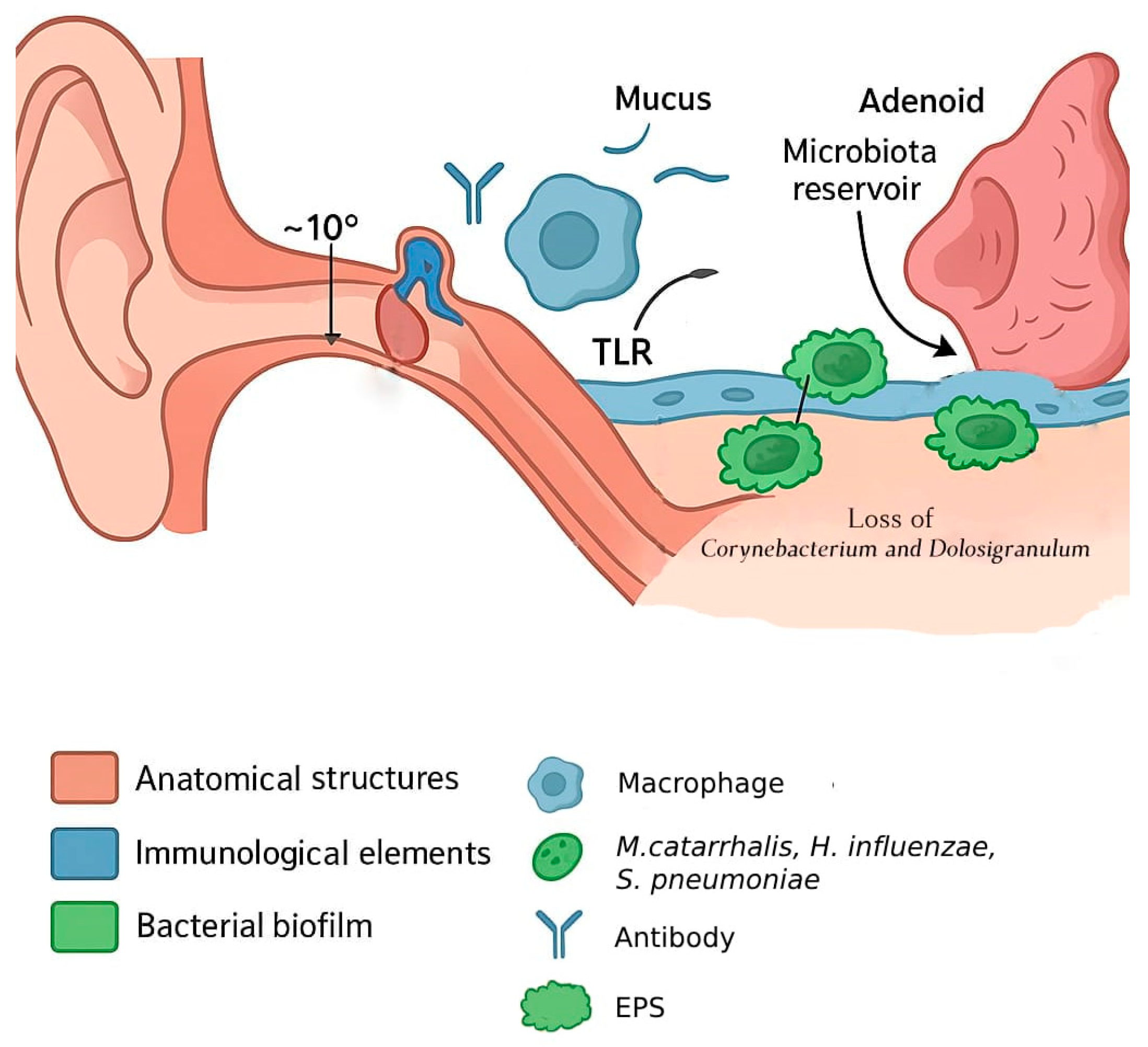
At birth and during early childhood, the ET is shorter and more horizontal than in adults, measuring around 16–18 mm (vs. ~30–38 mm in adults), with an angle of ~10° relative to the skull base (~35–45° in adults) [
15,
16]. The pediatric ET is also wider at the distal end, more flaccid, and lacks full cartilage support and elastin in the hinge/recoil regions (its cartilaginous portion is relatively more compliant and distensible in children) [
17]. Furthermore, the tensor veli palatini muscle, which is the main active opener of the cartilaginous ET, has a smaller cross-section, a less favorable vector of pull, and shorter attachment to the ET cartilage in infants [
18].
These anatomical differences lead to increased reflux of nasopharyngeal secretions due to the low angle and immature valve mechanisms at the nasopharyngeal opening, impaired drainage and poor aeration of the middle ear, and easier entry of pathogens from the nasopharynx. Because the ET cannot open reliably or fully, negative middle ear pressure develops, effusion accumulates, and infections more easily establish. Anatomical immaturity correlates with epidemiologic data: the peak incidence of OM (both AOT and COME) occurs between 6 and 24 months of age, when ET anatomy and associated musculature are still immature.
3.2. Local Immune Defense Mechanisms of the Middle Ear
The immune defenses of the middle ear are underdeveloped in early childhood and gradually mature over the first few years of life. Newborns and infants have lower levels of immunoglobulins (especially IgA), reduced neutrophil function (e.g., oxidative burst), and less effective epithelial immune responses. Adaptive immune components build up with antigen exposure over time (while specific quantitative data for all these items are sparse in the literature, the trend is well documented) [
19]. The mucosal epithelial cells of the ET and middle ear express pattern recognition receptors (like Toll-like receptors, TLRs) and secrete cytokines/chemokines (IL-1β, TNF-α, IL-8), which recruit neutrophils and monocytes. In infants, TLR responsiveness is lessened, delaying or reducing expression [
20]. Over time, middle ear mucosa and associated lymphoid tissue (MALT, including the adenoids) contribute to the production of IgA in secretions, which helps prevent bacterial adherence. Secretory IgA has been found in middle ear effusions (MEE) and shares antigen specificities with nasopharyngeal secretions, reflecting similar mucosal immunity in the upper airway [
21]. Secondary adaptive immune response is mediated by memory T and B cells, which produce high-affinity IgG into serum and local tissues. Comparisons between AOT and COME show differing immune profiles. Acute cases tend to have stronger pro-inflammatory cytokine responses and immune cell recruitment, whereas chronic effusions are more characterized by persistent lower-grade inflammation and sometimes suppressive features, such as increased levels of regulatory cytokines (e.g., IL-10, TGF-β) and the presence of regulatory T cells that dampen local immune activity [
20,
22].
The middle ear and ET are lined with a ciliated pseudostratified epithelium similar to respiratory mucosa. Goblet cells secrete mucus that traps microbes, and ciliary beating transports that mucus out toward the nasopharynx while tight junctions between epithelial cells maintain barrier integrity, preventing paracellular pathogen entry. However, in children, ciliary ultrastructure and function may not yet be fully mature, and viral infections (e.g., RSV) can damage cilia, impairing mucociliary transport. In addition, inflammatory conditions (infection, allergy) further disrupt the airway epithelial barrier by impairing ciliary function and breaking down tight junctions. Cytokines, like IL-4, IL-5, and IL-13, along with allergen-activated proteases, decrease the expression of tight junction proteins, leading to increased permeability and loss of ciliated cells. This damage, combined with reduced ciliary beat frequency and abnormal ciliary distribution, prevents effective mucus clearance, allowing pathogens and allergens to penetrate deeper into the airway [
23]. Children with congenital or genetic conditions such as cleft palate, Down syndrome, or primary ciliary dyskinesia have particularly compromised mucociliary function and a higher risk of COME/RAOM.
3.3. Clinical Implications
The combination of anatomical immaturity (shorter, more horizontal ET with weak or inefficient active opening), underdeveloped innate and adaptive immunity, and less efficient mucociliary clearance predisposes infants and toddlers to negative middle ear pressure, effusions, and recurrent infections. RAOM and COME epidemiologic peak incidence at 6–24 months coincides with maximal anatomical and immunological immaturity of ET. As the tube elongates, the angle becomes more vertical, the cartilage stiffens, and the tensor veli palatini muscle becomes more functional, allowing for more effective ventilation and clearance [
24]. The maturation of mucosal and systemic immune defenses, including enhanced secretory IgA production and improved innate signaling and mucociliary function, contributes to greater resistance to infection [
25]. Together, these developmental changes reduce the likelihood of middle ear effusion, pathogen entry, and inflammation, explaining the sharp decline in otitis media incidence after early childhood (
Figure 1).
Alterations in these host defense mechanisms are reflected in shifts within the microbial communities colonizing the nasopharynx and middle ear, linking the developmental anatomy and immunity of early childhood with subsequent microbiome composition and disease susceptibility.
4. The Middle Ear Microbiome: Commensals and Pathogens
The human middle ear is increasingly recognized as a dynamic ecological niche with a diverse and adaptable microbial community. This microbiome plays a dual role: certain bacteria function as commensals that contribute to mucosal homeostasis, while classical otopathogens can disrupt this balance and initiate disease processes such as AOM, COME, and CSOM [
26,
27].
Recent advances in molecular methods, particularly 16S rRNA amplicon sequencing, have revealed that the middle ear has a richer and more diverse bacterial community than traditional culture techniques had suggested [
28,
29]. Studies demonstrate that healthy ears or less disease-prone individuals are often colonized by commensal bacteria such as
Corynebacterium spp. and
Dolosigranulum spp., whereas diseased ears are dominated by known otopathogens (
H. influenzae,
S. pneumoniae,
M. catarrhalis) or by less common bacteria such as
Alloiococcus otitidis and
Turicella otitidis [
30,
31,
32].
Importantly, the balance between commensals and pathogens influences not only the onset of infection but the structure of biofilms, the resilience of the microbial ecosystem, and clinical outcomes. Understanding this interplay is central to developing microbiome-targeted interventions and alternatives to traditional antibiotic strategies [
33].
4.1. Characterization of the Healthy Middle Ear Microbiota
Subsequent sequencing-based investigations have provided detailed characterization of the bacterial taxa present in the middle ear mucosa and effusions. Minami et al. profiled 155 middle ear samples and found that normal middle ears, particularly in children, were dominated by
Proteobacteria, with contributions from
Actinobacteria,
Firmicutes, and
Bacteroidetes [
28]. Microbial composition differed significantly between children and adults, suggesting an age-dependent development of the middle ear microbiome.
Liu et al. used 16S rRNA pyrosequencing to analyze a pediatric case with chronic serous OM and compared the middle ear with adenoid and tonsil tissue. They detected
Pseudomonadaceae as the dominant family in the middle ear, while the adenoid had a more diverse community overlapping with both tonsillar and middle ear microbiota [
26]. This finding supports the concept of the adenoids as a microbial reservoir, with potential to induce both middle ear and tonsillar infections.
More recent comparative studies, such as those by Chan et al. and Jervis-Bardy et al., have reinforced that the “healthy” or less-diseased middle ear tends to exhibit greater microbial diversity and often includes bacteria traditionally regarded as commensals or skin-associated microbiota, such as
Corynebacterium spp., coagulase-negative
Staphylococcus, and
Cutibacterium spp. [
29,
30]. These microorganisms are thought to play neutral or even beneficial roles in maintaining microbial balance, contrasting with the dominance of
H. influenzae,
S. pneumoniae, or
M. catarrhalis observed in acute or recurrent infections.
The current evidence suggests that the healthy middle ear microbiota is a low-biomass, age-dependent community enriched with commensal bacteria, which may contribute to immune tolerance and barrier maintenance. Understanding its baseline composition is essential for distinguishing health-associated communities from those influencing pathogeneses in OM.
4.2. Microbiome Profiling Methods (Cultures vs. Sequencing Techniques)
The study of the middle ear microbiome has evolved considerably over the past six decades. Initially, investigations relied exclusively on culture-based techniques, which led to the classical model of OM pathogenesis dominated by
H. influenzae,
S. pneumoniae, and
M. catarrhalis. While these methods remain clinically relevant for antimicrobial susceptibility testing, they underrepresent the true microbial diversity present in the middle ear [
34,
35].
Molecular approaches such as polymerase chain reaction (PCR) and quantitative PCR (qPCR) introduced higher sensitivity and allowed detection of fastidious organisms but remained limited to targeted bacteria. The real paradigm shift came with the adoption of culture-independent, community-wide methods such as 16S rRNA amplicon sequencing, which revealed the presence of previously underappreciated bacteria including
A. otitidis and
T. otitidis, as well as broader community shifts associated with health and disease conditions [
29,
30]. More recently, shotgun metagenomics and metatranscriptomics have provided functional insights, allowing strain-level resolution and identification of genes related to virulence, antimicrobial resistance, and biofilm formation [
36].
Evidence from comparative studies highlights the gaps between methods. Culture-negative middle ear effusions frequently contain bacterial DNA and even RNA transcripts when analyzed with sequencing or RT-PCR, indicating the presence of metabolically active microbes despite apparent sterility on culture [
26]. Similarly, sequencing has repeatedly revealed bacteria such as
Alloiococcus and
Turicella, virtually absent in culture-based studies but often abundant in chronic OM [
29,
30].
Thus, culture and sequencing should be seen as complementary: culture provides actionable clinical information, while sequencing broadens our ecological and mechanistic understanding of the middle ear microbiome.
Comparative overview of profiling methods is presented in
Table 1.
4.3. Protective Role of Commensal Organisms
The composition of the middle ear and upper respiratory tract microbiome is not only determined by the presence of pathogens but also by the protective role of commensal species. Increasing evidence suggests that certain commensal bacteria act as ecological stabilizers, preventing colonization or overgrowth of otopathogens.
Several case–control studies have demonstrated that children less prone to RAOM or COME harbor higher abundances of
Corynebacterium spp. and
Dolosigranulum pigrum in the nasopharynx as well as
Fusobacterium nucleatum,
Gemella sanguinis, and
Eubacterium sulci [
33,
37,
38]. These organisms are considered keystone commensals: their presence is associated with a reduced risk of pathogen dominance. Mechanistically, they may protect via competitive exclusion for epithelial binding sites, production of lactic acid and other inhibitory metabolites, or modulation of mucosal immune responses [
39].
For example, Lappan et al. showed that children resistant to RAOM had significantly more
Corynebacterium spp. and
Dolosigranulum spp. in their nasopharyngeal microbiota, whereas those with recurrent disease were enriched in
Moraxella spp. and
Haemophilus spp. [
33]. Similarly, Xu et al. observed that a higher abundance of these commensals correlated with reduced detection of
S. pneumoniae and
H. influenzae in the middle ear fluid [
40]. Jo et al. defined
F. nucleatum,
G. sanguinis, and
E. sulci as age-specific species, whose patterns change consistently with age and can be used to predict the child’s age from the microbiome alone.
Oribacterium parvum,
Cardiobacterium homini,
Streptococcus salivarius,
Veillonella parvula, and
Neisseria mucosa were identified as bacteria that may modulate transitions between commensal-rich structures towards a pathogen-dominated COME (high
S. pneumoniae,
H. influenzae) [
38].
Other organisms such as
Streptococcus salivarius have also emerged as potential protective pathobiont-inhibiting species. In vitro studies demonstrate that
S. salivarius isolates from children can inhibit the growth of
H. influenzae,
M. catarrhalis, and
A. otitidis, raising the possibility of probiotic interventions [
41].
The middle ear is usually a low-biomass site in health, yet its microbial composition is shaped by the nasopharynx and adenoids, which act as reservoirs for organisms that may ascend through the ET. A diverse, commensal-rich nasopharyngeal microbiota functions as the first line of defense against OM. Although direct evidence for protective effects within the middle ear itself is scarce, maintaining this balance, by limiting antibiotic disruption, promoting breastfeeding, or even exploring probiotic supplementation, emerges as a promising strategy for preventing disease.
4.4. How Colonization by H. influenzae, S. pneumoniae, and M. catarrhalis Influence the Structure of the Microbiome
The microbial ecology of the middle ear is profoundly influenced by colonization with the three classical otopathogens:
H. influenzae,
S. pneumoniae, and
M. catarrhalis. These bacteria usually colonize the nasopharynx during early childhood, often in the wake of viral upper respiratory tract infections that compromise mucosal integrity and alter the commensal community. The dysbiosis of the adenoid microbiome was significantly association with the COME microbiome [
38]. These pathogens can then ascend via the ET into the middle ear cavity, where the immature anatomy of children and their developing immunity provide an environment susceptible to persistence [
35,
42].
The presence of otopathogens does more than initiate acute infection. It actively restructures the microbial community. Effusions dominated by
H. influenzae,
S. pneumoniae, or
M. catarrhalis consistently show reduced bacterial diversity, reflecting the ability of these species to suppress beneficial commensals such as
Corynebacterium and
Dolosigranulum [
29,
33]. This loss of diversity undermines ecological resilience and predisposes the middle ear to recurrent or chronic disease. Furthermore, the microbial landscape changes over time: acute infections are typically characterized by high burdens of classical otopathogens, while chronic effusions often contain less well-studied bacteria such as
A. otitidis or
T. otitidis, which seem to flourish in the altered environment established by the initial infection [
30].
Furthermore, the microbial landscape changes over time. In preschool children (2–6 years old), anatomical and functional immaturity of the ET is likely the causative agent of OM. In older children, ET length and angle approach adult configuration, ventilation improves, and adenoidal tissue gradually decreases. While a sharp decline in OM can be attributed to maturation of EF, some children remain susceptible despite structurally mature anatomy. Jo et al. showed that in children who continue to have COME beyond the preschool years (6–12-years old), disease is strongly associated with a highly abnormal, age-independent adenoid microbiota. COME samples no longer followed the age-dependent maturation seen in healthy controls but instead showed low-diversity communities rich in
S. pneumoniae and
H. influenzae. This study implied that a pathogen-rich adenoid microbiome may play a greater role in promoting chronic inflammation rather than ET dysfunction alone [
38].
5. Biofilm Formation in the Middle Ear
Biofilm biology is now recognized as a cornerstone of OM research. Rather than representing an acute, self-limited infection, pediatric OM frequently evolves into a chronic condition sustained by surface-attached microbial communities that exhibit marked tolerance to antibiotics and immune-mediated clearance [
43,
44]. These biofilms play a central role in recurrent and persistent disease in children and are shaped by the anatomical, immunological, and ecological features of the pediatric middle ear.
A biofilm consists of microbial cells encased in a self-produced extracellular polymeric substance (EPS) composed of polysaccharides, proteins, lipids, and extracellular DNA (eDNA), which provides structural stability, enhances nutrient retention, promotes horizontal gene transfer, and protects bacteria from antimicrobial and immune pressures [
43,
44]. Although the classical stages of biofilm formation, attachment, microcolony formation, maturation, and dispersal, have been extensively described [
45,
46,
47,
48,
49,
50,
51], the relevance of these stages in pediatric OM depends strongly on host factors such as ET dysfunction, viral injury to the epithelium, and immature mucociliary and immune responses [
15,
20,
23].
In the pediatric middle ear, initial bacterial adhesion is facilitated by inflammation-induced alterations of the mucosa, including upregulation of adhesion molecules and impaired mucociliary clearance. Otopathogens such as
H. influenzae,
S. pneumoniae, and
M. catarrhalis exploit these conditions to form stable EPS-embedded communities that demonstrate substantially increased tolerance to antibiotics and oxidative stress [
52,
53,
54,
55]. Biofilms in this niche also contain metabolically heterogeneous subpopulations, including dormant persister cells that survive lethal antibiotic concentrations [
29,
34,
56,
57].
A growing body of evidence indicates that biofilms in pediatric OM are rarely monospecies. Instead, polymicrobial biofilms represent the dominant in vivo state and introduce additional layers of complexity relevant to chronicity and treatment failure. Cooperative interactions between
H. influenzae and
M. catarrhalis enhance biofilm biomass, structural integrity, and antibiotic tolerance compared with single-species communities [
43,
44,
52,
53,
55].
M. catarrhalis can protect
H. influenzae from pneumococcal antagonism [
58,
59], while
H. influenzae modulates pneumococcal autolysis and oxidative stress responses, stabilizing mixed biofilm survival [
57]. These cross-species interactions reshape quorum sensing (QS) dynamics, alter metabolic gradients, and create protective micro-niches that promote persistence and reseeding of the middle ear even after therapy [
58,
60].
6. Influence of the Middle Ear Microbiota on Biofilm Formation and Disease Persistence
6.1. Interactions Between Microbiota and Pathogen Bacteria
The microbiota can be both protective against and contribute to OM by modulating inflammation and immune responses. A healthy microbiota in the upper respiratory tract competes with pathogens and activates antimicrobial and anti-inflammatory components [
59,
61]. Frequent commensal bacteria,
Corynebacterium spp. and
D. pigrum, can resist colonization of otopathogens by using several mechanisms. By direct antibiosis,
D. pigrum alone or with
C. pseudodiphtheriticum inhibits
Staphylococcus aureus and
S. pneumoniae using synthesized antagonists and/or combined metabolites that mediate killing or growth suppression [
62,
63]. Also, a
Corynebacterium-mediated habitat can be modified by release of antipneumococcal free fatty acids which cause direct chemical antagonism. Nutrient competition is also a mechanism of pathogen interference.
D. pigrum relies on host or co-colonizers for essential micronutrients, which creates a complex metabolic interdependence and competitive environment. Synergistic growth with nasal
Corynebacterium spp. can secure or redirect resources toward a community composition that is more restrictive to
S. pneumoniae. This results in reduced pneumococcal growth across diverse serotypes in the presence of
D. pigrum and
C. pseudodiphtheriticum [
63]. Signaling-level interference is reflected in disrupting QS or the reception of environmental cues that trigger adhesion/biofilm genes [
64]. Commensals enzymatically inactivate autoinducers, degrading N-acyl-homoserine lactones (AHLs) with lactonases, acylases, or oxidoreductases so signals never reach the activation threshold of biofilm and virulence regulons [
65]. This consequently impedes coordinated bacterial group behaviors like matrix production and adhesion. Gram-positive commensals secrete non-cognate autoinducing peptides (AIPs) and the autoinducer-2 (AI-2) [
66], which competitively block signaling pathogen peptide-QS, demising the expression of accessory gene regulator (
agr) system virulence factors in vivo [
67]. By disrupting the QS systems of pathogens, commensals can repress pathogen colonization and further inhibit the formation of biofilms [
68].
6.2. Microbiota Dysbiosis in the Middle Ear
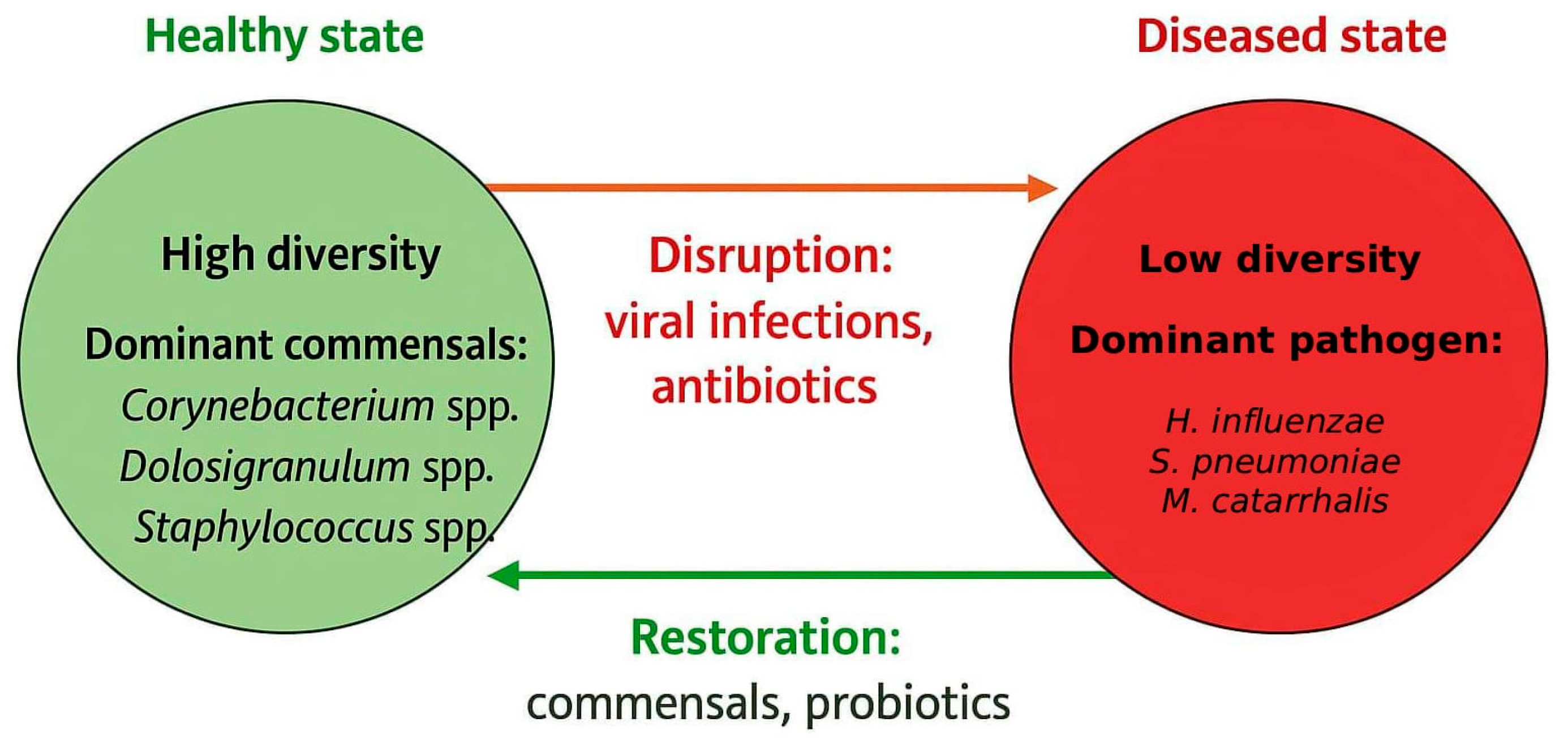
Dysbiosis of the middle ear microbiota is a result of a shift in the components of bacterial community compared to their levels in a healthy state, and it is linked to OM [
69]. This microbial imbalance is characterized by a loss of beneficial bacteria, an overgrowth of pathogen bacteria, or a decrease in overall microbial diversity, linked to changes in host–microbe interactions which promote infections and chronic inflammatory conditions [
70] (
Figure 2).
Multiple factors can cause dysbiosis of the middle ear microbiota, predisposing children to OM. Antibiotic exposure is a well-documented factor contributing to middle ear microbiota dysbiosis. Broad-spectrum antibiotics reduce the protective bacteria such as
Corynebacterium spp. and
Dolosigranulum spp. while sparing biofilm-embedded otopathogens [
69]. Recurrent viral infections (RSV, influenza, adenovirus) also play an important role by impairing mucociliary clearance, altering epithelial receptor expression, and providing ground for secondary bacterial colonization. Host-related predispositions such as immature mucosal immunity, lower secretory IgA levels, and genetic variations in innate receptors can further increase this effect and impair early pathogen recognition [
71]. Finally, environmental pressures including daycare attendance, passive smoke exposure, air pollution, and close sibling transmission alter microbial ecology, often reducing diversity of microbiota and enriching an otopathogen-dominant environment [
72]. Pneumococcal conjugate vaccines (PCVs) vaccination could also disrupt the microbiome by decreasing vaccine-specific serotype carriage [
73,
74]. Higher diversity and stability of microbiota was observed in PCV13-vaccinated infants, which might be consistent with a lower susceptibility to otitis media [
73,
75]. The use of conjugate
H. influenzae serotype b (Hib) vaccines have led to a significant reduction in pharyngeal carriage, decreasing invasive disease. Non-typeable
H. influenzae (NTHi), which was not targeted by standard Hib vaccines, NTHi carriage, and NTHi-associated diseases such as otitis media, was mostly unaffected and consequently may even become relatively more prominent [
76]. More recently, protein D-containing pneumococcal conjugate formulations (PHiD-CV) have been shown to induce anti-protein D responses and to reduce NTHi carriage and NTHi-positive middle ear infections [
77]. By actively restructuring the nasopharyngeal microbiome, vaccination against
S. pneumoniae and
H. influenzae may change competitive and cooperative interactions among airway bacteria, with potential consequences for the balance between otopathogens and protective commensals.
6.3. Microbiota Dysbiosis and Immune Response Modulation
Microbiota colonize the mucosal barrier and settle in the respiratory tract, playing integral roles in initiating an immune response, from maintaining homeostasis to responding to infectious challenges [
78]. Commensal bacteria exert protective functions against respiratory pathogens through host-mediated immunity and direct action by inhibiting/killing pathogens and competing for colonization. Mucosal defense is stimulated by activating epithelial pattern-recognition receptors and low-level immune activation, which sustains antimicrobial peptide secretion (defensins, cathelicidins), supports secretory IgA–mediated exclusion, and preserves integrity of tight junction and ciliary function [
20,
79]. These actions maintain a stable barrier against otopathogens. Experimental dysbiosis of airway microbiota weakens innate immunity responses in myeloid and innate-like lymphocytes (macrophage interferon responses, NK cell activity). When pathogen bacterial communities prevail, the host responds typically by shifting cytokine expression: elevated IL-1β, IL-6, TNF-α, and IL-8 sustain neutrophil recruitment, inflammation, and mucus hypersecretion [
20,
80]. Other innate immunity changes include altered Toll-like receptor (TLR) signaling that can be either an overly aggressive or suppressed immune reaction, depending on which microbes dominate [
81].
During inflammation driven by pathogens, polymeric immunoglobulin receptor (pIgR), a key protein for mucosal immunity, can be downregulated, trapping polymeric IgA in the tissue and reducing luminal sIgA availability [
82]. Reduced sIgA weakens immune exclusion and the protective commensal barrier. Also, commensal-specific T cells can become drivers of low-grade, biofilm-enabling inflammation in the middle ear by shifting from −10 secretion that suppresses pro-inflammatory responses to IL-17 secretion that promotes neutrophil-recruiting inflammation [
83]. This inflammatory state disrupts epithelial junctions and impairs ciliary beating, thickens mucus, and generates eDNA and other polymers that facilitate bacterial surface attachment and biofilm formation [
70]. These changes can explain how dysbiosis of the middle ear microbiota can cause persistence of biofilm-forming pathogens despite therapy and host clearance attempts.
6.4. The Relationship Between Microbiome Dysbiosis and Biofilm Formation
Microbiome dysbiosis in the upper airway and middle ear represents favoring opportunistic otopathogens and serves as one of the facilitating factors of biofilm formation in otitis media. Episodes of repeated upper respiratory infections, along with other environmental factors causing microbiome dysbiosis, can disrupt mucociliary clearance, damage epithelial cilia, and disrupt expression of adhesion receptors (such as ICAM-1) on epithelial cells, increasing the likelihood of bacterial attachment [
84]. The middle ear experiences increased oxygen absorption across the inflamed mucosa, leading to a significant reduction in total middle ear pressure and impaired ventilation [
85]. Some of the otopathogens, especially facultative anaerobes like
H. influenzae and
M. catarrhalis, utilize these conditions for increased mucosal adherence as an initial step in biofilm formation [
86,
87]. These host inflammatory responses reinforce the biofilm niche, allowing these bacteria to transition into biofilm growth modes characterized by adherence, extracellular matrix production, and metabolic adaptation.
After initial attachment to the mucosal surface, bacteria begin clustering into microcolonies. eDNA released through autolysis or bacteriophage induction provides the initial matrix scaffold [
88]. Biofilm maturation involves developing a three-dimensional architecture with thick EPS which consists of extracellular DNA, proteins, lipids, and polysaccharides. In
S. pneumoniae biofilm, capsule downregulation occurs during maturation. Non-encapsulated or low-capsule phenotypes dominate biofilm biomass which enhances aggregation and nutrient diffusion [
89]. This stage is strongly influenced by QS systems, like LuxS/AI-2, which are specific for
S. pneumoniae,
H. influenzae, and
M. catarrhalis and coordinate biofilm thickness, virulence factor production, and resistance phenotypes [
90,
91,
92].
Within mature biofilms, bacteria undergo phenotypic changes caused by stress responses, QS signals, and nutrient limitation within the biofilm microenvironment. These changes involve entering a slow-growth metabolic state, accompanied by increased genetic competence and horizontal gene transfer that enhance bacterial adaptation and antibiotic resistance [
93]. Within mature pneumococcal biofilms, some bacteria differentiate to dormant persister cells, which are highly tolerant to antibiotics, oxidative stress, and the host’s immune response [
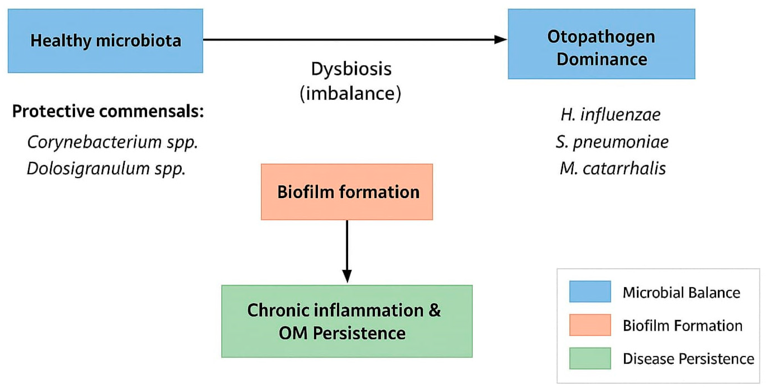
94]. Dysbiosis and biofilm maturation create a self-reinforcing cycle, where microbial imbalance facilitates biofilm formation and biofilms maintain dysbiosis by protecting pathogens and preventing commensal recovery.
There is strong evidence that some otopathogens cooperate and interact with each other within the biofilm formation, causing more severe and persistent infections and higher burden of antibiotic use. Dysbiotic states in which
H. influenzae and
S. pneumoniae are dominant together favor cooperative biofilm formation for both species.
H. influenzae may inhibit pneumococcal autolysis and stabilize mixed communities [
95]. Also, changes in pH, oxygen, and nutrient availability can influence pneumococcus to reduce its competitive pressure, which allows
H. influenzae to persist [
96]. Change in virulence-associated gene expression (adhesins, immune modulators) can happen in both species.
H. influenzae adapts to the presence of pneumococcus by upregulating oxidative stress defenses and biofilm-associated genes (catalases, peroxidases, DNA repair enzymes that neutralize hydrogen peroxide), reducing susceptibility to pneumococcal antagonism and stabilizing its persistence within mixed biofilms [
97].
Colonization with
M. catarrhalis plays a key role in stabilizing mixed-species communities in the nasopharynx and middle ear by promoting the persistence of other otopathogens. Experimental models have shown that
M. catarrhalis protects
H. influenzae from the bactericidal activity of
S. pneumoniae in a polymicrobial biofilm [
49]. This stabilizing effect is not fully explained by catalase activity alone, suggesting additional protective mechanisms such as metabolic cooperation, quorum signaling modulation, and physical incorporation into the biofilm matrix. The presence of
M. catarrhalis enhances both biofilm biomass and antibiotic tolerance, as well as long-term colonization by
H. influenzae and
S. pneumoniae. It enables cooperative dynamics among otopathogens, which could explain why carriers of
M. catarrhalis are more prone to recurrent or chronic otitis media [
98]. These findings suggest that OM should be viewed less as an infection but as a microbiome dysbiosis-based disorder, in which the balance between commensals and otopathogens determines the clinical outcome of the disease (
Figure 3).
7. Current Research and Future Directions in Microbiome- and Biofilm-Targeted Therapy
7.1. Microbiome-Targeted Therapy
Microbiome-targeted therapies for OM are increasingly under investigation; many of the available strategies are not adapted for treatment of this disease (
Table 2). One promising direction involves live biotherapeutic products (LBPs), pharmaceutical-grade probiotics created to modulate the respiratory microbiome by targeted competitive inhibition of otopathogens or immune modulation. For example, intranasal application of
Haemophilus haemolyticus has shown potential in inhibiting NTHi colonization, while nasal sprays containing
Streptococcus salivarius and
Streptococcus strains have demonstrated reductions in acute and secretory OM, even reducing the need for surgical intervention [
41,
99].
The development of next-generation probiotics is rapidly evolving beyond traditional, broad-spectrum strains toward live, strain-specific engineered microorganisms designed for a precise therapeutic effect [
41]. These genetically modified microbes can be engineered to produce anti-inflammatory molecules, antimicrobial peptides, or quorum sensing inhibitors, directly targeting pathogenic bacteria or modulating host immune responses to reduce inflammation and biofilm formation. Advances in site-specific delivery systems (nasal sprays, hydrogels, and resorbable films) are improving the precision of probiotic administration by enabling targeted colonization and prolonged retention in the nasopharynx or middle ear [
100]. These delivery modalities enhance the survival and activity of probiotics in anatomically challenging sites, optimizing their therapeutic impact [
101]. The co-administration of synbiotics, combining selected probiotic strains with prebiotics that selectively sustain beneficial bacteria, represents a promising strategy to restore and maintain a healthy microbiome [
102]. By promoting synergistic interactions within the microbiome, synbiotics help maintain the resilience and functional stability of microbial communities, providing protection against otopathogen overgrowth and the development of chronic middle ear disease.
Postbiotics, or non-living microbial derivatives, remain an attractive concept for modulating local immunity or microbial networks, though data in the OM context are currently sparse [
103].
Using bacteriophages, as phage therapy, to target specific pathogens demonstrates theoretical benefits in penetrating biofilms, and preliminary studies suggest that trans-tympanic delivery could overcome the tympanic membrane barrier, though no clinical applications in acute OM have yet been conducted [
104].
Microbiota transplants, such as fecal microbiota transplantation (FMT), have been previously established in the gastrointestinal environment, but the concept remains experimental for otitis media. Their applicability is limited by delivery routes and specific microbiome matching [
105]. Two recent studies have published data on nasal microbiota transplantation (NMT) in treatment of chronic rhinosinusitis, with possible implications for countering antimicrobial resistance in respiratory infections [
106].
Use of engineered microbes, genetically programmed to produce therapeutic molecules or inhibit pathogens, is still a theoretical strategy. There are is vitro research proving that this engineered strain of
Mycoplasma pneumoniae was very efficient against an acute
Pseudomonas aeruginosa lung infection [
107]. Given that
P. aeruginosa is one of the frequently isolated pathogens in CSOM, research in this direction could be promising. Across all these approaches, they face common challenges: establishing stringent regulatory pathways for living therapeutics, ensuring safety and effective delivery modalities, managing microbial strain specificity and colonization stability, and potential immune reactions or unintended ecological disruptions [
108].
The future of treating OM is shifting toward precision microbiome modulation, individualized microbiome-specific interventions that aim to restore microbial balance rather than eradicate bacteria without discrimination. The goal is to create treatments based on a patient’s unique nasopharyngeal or middle ear microbiome composition, which is now accessible through advanced microbial profiling technologies such as 16S rRNA sequencing and whole metagenomic shotgun sequencing [
13,
109,
110]. These tools enable the identification of diagnostic microbiome signatures, allowing for patient stratification and prediction of disease risk or treatment responsiveness [
111]. This proved especially relevant in pediatric OM, where microbial heterogeneity influences both pathogenesis and therapeutic outcomes.
Table 2.
Microbiome-targeted therapy.
Table 2.
Microbiome-targeted therapy.
| Therapeutic Strategy | Key Findings | Challenges |
|---|
| LBPs (nasal probiotics) | H. haemolyticus and S. salivarius/S. oralis strains reduce OM episodes and surgical need (intranasal use) | Complex host–microbe interactions, variability in patient responses, delivering issues, regulatory guidelines [112] |
| Next-generation Probiotics | Can be designed with the ability to produce specific beneficial molecules, modulating immune responses; more targeted and effective therapeutic approach | Need for expensive, anaerobic culture media for industrial-scale production; need for effective delivery systems to ensure viability, safety concerns, especially in vulnerable populations [113] |
| Microbiota Transplants | Widely used in gut disorders; limited, experimental application in OM | Delivery routes, specific microbiome matching, donor selection, screening to prevent pathogen transmission, lack of standardized protocols and regulatory guidelines [114] |
| Postbiotics | Conceptually promising; little OM-specific data currently | Lack of a standardized definition, regulatory guidelines, delivery issues and absorption, more data on safety and dosing [115] |
| Phage Therapy | Potential for biofilm penetration; trans-tympanic delivery explored; no clinical OM use yet | Bacterial resistance to phages, limited host specificity of phages, delivery issues, unpredictable pharmacokinetics and pharmacodynamics, potential immune system interactions, and regulatory guidelines [116] |
| Engineered Microbes | Conceptual therapeutic potential; largely preclinical | Limited survival in targeted environment, potential off-target effects on the respiratory microbiota, delivering issues, high genetic diversity and rapid evolution of pathogens, safety issues [117] |
| Precision microbiome modulation | Alteration of molecular signaling, immune modulation, and drug metabolism of microbial communities for personalized health outcomes | Microbiome’s complexity, difficulties in achieving stable and predictable modulation, limitations of tools for clinical application [118] |
7.2. Antibiofilm Agents
Biofilm-targeted therapeutics aim to inhibit or eradicate biofilm communities responsible for chronic infections (
Table 3). Among the most promising candidates are biofilm-disrupting agents like N-acetylcysteine (NAC), DNase, and dispersin B, which degrade major biofilm matrix components and weaken the EPS [
119,
120,
121,
122]. Another interesting approach involves QS inhibitors (QSIs). These agents, both natural and synthetic molecules, have shown efficacy in reducing biofilm formation and enhancing antibiotic susceptibility across various pathogens [
123,
124].
Recent advances in biofilm therapeutics focus on overcoming the intrinsic resistance of biofilm communities through smart drug delivery systems. Nanoparticles and liposomes engineered to penetrate the dense EPS can deliver antibiotics or biofilm-degrading enzymes directly [
125]. These delivery systems are often designed to respond to specific environmental conditions, triggering a controlled release and minimizing off-target effects [
126]. Antibiotics in biofilm-specific formulations use carriers like liposomes, hydrogels, or nanoparticles to overcome restricted antibiotic penetration, altered bacterial metabolism, and increased resistance. These specialized formulations aim to improve antibiotic delivery, protect antibiotics from degrading enzymes in the biofilm, or utilize compounds that disrupt the biofilm matrix [
127].
Bacteriophages are increasingly explored for their ability to target bacteria embedded within biofilms, offering adjunctive or alternative therapeutic pathways, but their clinical use remains limited [
128].
Anti-adhesives are a novel strategy for preventing bacterial infections and biofilm formation by blocking initial bacterial attachment. Compounds that show promise for future research in treatment of otitis media biofilm include natural substances like probiotics (Lactobacillus), mannosides (C-mannosides), and hyaluronic acid [
129,
130,
131,
132,
133].
Pilicides and curlicides are anti-biofilm agents that inhibit the formation of bacterial surface protein fibers, pili and curli, crucial for attachment to host surfaces and to each other as a first step in biofilm formation. They have been studied in the context of urinary tract infections (UTIs) caused by
Escherichia coli and other general biofilm infections, but their specific efficacy in the treatment of otitis media (ear infection) biofilms is still in the research phase [
134].
Table 3.
Anti-biofilm therapy.
Table 3.
Anti-biofilm therapy.
| Therapeutic Strategy | Effect | Challenges |
|---|
| Biofilm-disrupting agents | Dispersin B degrades poly-β-1,6-N-acetyl-D-glucosamine (PNAG), disrupting biofilm matrix; DNase degrades extracellular DNA (eDNA); NAC disrupt the extracellular polymeric substance (EPS) matrix by breaking disulfide bonds; direct antimicrobial effect, increased oxidative stress | Variable effect on mature biofilms, potential shielding of target matrix components |
| Quorum sensing inhibitors | Natural and synthetic QSIs suppress biofilm formation, reduce virulence, and improve antibiotic response | Direct resistance, lack of specificity, limited monotherapy efficacy, wash-away effect |
| Smart drug delivery systems (nanoparticles) | Mesoporous silica NPs (±lectin or gold layer) enable targeted antibiotic release, photothermal disruption, and enhanced biofilm penetration | Health and environmental toxicity, complex manufacturing, safety concerns |
| Bacteriophages | Bacterial lysis and the destruction of the biofilm structure, production of depolymerase enzymes that degrade the biofilm’s EPS | Narrow host range, bacterial resistance, storage and stability issues [135] |
| Antiadhesives | Creation of an overlayer on mucosal surface and prevention of bacterial adhesion | Biocompatibility, routes of application and fixation, degradation |
| Pilicides and curlicides | Binding to the periplasmic chaperone protein and/or chaperone-like proteins, blocking the transfer of the pilin subunit and halting the assembly of functional pili on the bacterial surface which leads to loss of ability to adhere to surfaces, colonize host tissues, and form biofilms | Questionable success metrics, need for combination therapy due to multiple bacterial virulence factors, potential bacterial resistance or evasion [136] |
8. Conclusions and Future Perspectives
Pediatric OM remains among the most prevalent diseases of early childhood, reflecting a convergence of developmental anatomy, immunological immaturity, and the complex microbial ecology of the upper airway. The contemporary view moves beyond the outdated model of a sterile middle ear invaded episodically by pathogens. Instead, the middle ear constitutes an ecological niche shaped by dynamic interactions between the nasopharyngeal microbial reservoir, local commensals, and host defenses. Within this niche, biofilm formation has emerged as a central pathogenic mechanism, enabling microorganisms to persist, evade immune clearance, and resist conventional antimicrobial treatment. By transforming acute into recurrent and chronic inflammatory infections, biofilms account for much of the morbidity, treatment failure, and long-term sequelae associated with otitis media in children.
This reconceptualization has direct therapeutic implications. Conventional antibiotic regimens, although clinically indispensable, are insufficient against biofilm-embedded bacteria and may disrupt protective commensal populations that contribute to colonization resistance. Future interventions will increasingly rely on microbiome-based and antibiofilm strategies that restore ecological balance rather than simply eliminate pathogens.
In summary, pediatric otitis media represents a multifactorial condition shaped by host developmental factors, immune maturation, and microbiome dynamics. Understanding these complex interactions provides the foundation for more effective prevention and treatment strategies.
Viewed through this ecological and mechanistic lens, pediatric otitis media emerges not simply as an infectious disease but as the consequence of disrupted interactions between host and microbiota within a biofilm-dominated environment. Future progress in prevention and treatment will depend on translating these insights into clinically actionable strategies capable of reducing both the immediate burden of recurrent infections and the long-term risk of auditory and developmental sequelae in children worldwide.
Author Contributions
Conceptualization, A.J. and I.C.; writing—original draft preparation, A.J., I.C., N.J., B.B. and K.S.V.; writing—review and editing, A.J., I.C. and K.S.V.; visualization, N.K.; supervision, A.J. and I.C.; funding acquisition, I.C. All authors have read and agreed to the published version of the manuscript.
Funding
This research was funded by the Ministry of Science, Technological Development and Innovation, Republic of Serbia, grant number 451-03-137/2025-03/200110.
Institutional Review Board Statement
Not applicable.
Informed Consent Statement
Not applicable.
Data Availability Statement
No new data were created or analyzed in this study. Data sharing is not applicable.
Conflicts of Interest
The authors declare no conflicts of interest.
Abbreviations
The following abbreviations are used in this manuscript:
| ET | Eustachian tube |
| RAOM | Recurrent acute otitis media |
| MEE | Middle ear effusions |
| RSV | Respiratory syncytial virus |
| COME | Chronic otitis media effusion |
| QS | Quorum sensing |
| NTHi | Nontypeable H. influenzae |
| OT | Otits media |
| TLR | Toll-like receptor |
| eDNA | Extracellular DNA |
| EPS | Extracellular polymeric substance |
| LBPs | Live biotherapeutic products |
References
- Monasta, L.; Ronfani, L.; Marchetti, F.; Montico, M.; Vecchi Brumatti, L.; Bavcar, A.; Grasso, D.; Barbiero, C.; Tamburlini, G. Burden of Disease Caused by Otitis Media: Systematic Review and Global Estimates. PLoS ONE 2012, 7, e36226. [Google Scholar] [CrossRef] [PubMed]
- Jin, Y.; Yang, X.; Sun, H.; Zhang, J.; Yang, S.; Jiang, S.; Song, Q.; Zhang, G.; Ma, B.; Yang, K.; et al. Global, Regional, and National Burdens of Otitis Media from 1990 to 2019: A Population Based Study. Ear Hear. 2024, 45, 658–665. [Google Scholar] [CrossRef] [PubMed]
- Dong, L.; Jin, Y.; Dong, W.; Jiang, Y.; Li, Z.; Su, K.; Yu, D. Trends in the Incidence and Burden of Otitis Media in Children: A Global Analysis from 1990 to 2021. Eur. Arch. Otorhinolaryngol. 2025, 282, 2959–2970. [Google Scholar] [CrossRef]
- Alshehri, S.; Musleh, A. The Role of Eustachian Tube Dysfunction in Recurrent Chronic Otitis Media: A Cross-Sectional Study of Anatomical and Functional Variations. Healthcare 2025, 13, 77. [Google Scholar] [CrossRef]
- Is Allergic Rhinitis Related to Otitis Media with Effusion in Adults and Children? Applying Epidemiological Guidelines for Causation—PubMed. Available online: https://pubmed.ncbi.nlm.nih.gov/40497981/ (accessed on 28 September 2025).
- Huang, G.-J.; Lin, B.-R.; Li, P.-S.; Tang, N.; Fan, Z.-J.; Lu, B.-Q. The Global Burden of Otitis Media in 204 Countries and Territories from 1992 to 2021: A Systematic Analysis for the Global Burden of Disease Study 2021. Front. Public. Health 2024, 12, 1519623. [Google Scholar] [CrossRef]
- Kaur, R.; Schulz, S.; Sherman, A.; Andrejko, K.; Kobayashi, M.; Pichichero, M. Anticipated Effects of Higher-valency Pneumococcal Conjugate Vaccines on Colonization and Acute Otitis Media. Pediatr. Infect. Dis. J. 2024, 43, 1004–1010. [Google Scholar] [CrossRef]
- Hanquet, G.; Krizova, P.; Dalby, T.; Ladhani, S.N.; Nuorti, J.P.; Danis, K.; Mereckiene, J.; Knol, M.J.; Winje, B.A.; Ciruela, P.; et al. SpIDnet group1. Serotype Replacement after Introduction of 10-Valent and 13-Valent Pneumococcal Conjugate Vaccines in 10 Countries, Europe. Emerg. Infect. Dis. 2022, 28, 137–138. [Google Scholar] [CrossRef]
- Onifade, A.; Katolo, H.W.; Mookerjee, S.; Bhutta, M.F. Epidemiology of Chronic Suppurative Otitis Media: Systematic Review To Estimate Global Prevalence. J. Epidemiol. Glob. Health 2025, 15, 55. [Google Scholar] [CrossRef]
- Paing, A.; Elliff-O’Shea, L.; Day, J.; Joshi, D.; Arnold, S.; Holland Brown, T.; Kennedy, V. Modifiable Risk Factors for Developing Otitis Media with Effusion in Children under 12 Years in High-Income Countries: A Systematic Review. Arch. Dis. Child. 2024, 110, 45–51. [Google Scholar] [CrossRef]
- Mather, M.W.; Drinnan, M.; Perry, J.D.; Powell, S.; Wilson, J.A.; Powell, J. A systematic review and meta-analysis of antimicrobial resistance in paediatric acute otitis media. Int. J. Pediatr. Otorhinolaryngol. 2019, 123, 102–109. [Google Scholar] [CrossRef] [PubMed]
- Bowman-Derrick, S.; Harris, T.M.; Beissbarth, J.; Kleinecke, M.; Lawrence, K.; Wozniak, T.M.; Bleakley, A.; Rumaseb, A.; Binks, M.J.; Marsh, R.L.; et al. Can non-typeable Haemophilus influenzae carriage surveillance data infer antimicrobial resistance associated with otitis media? Pediatr Investig. 2023, 7, 13–22. [Google Scholar] [CrossRef]
- Folino, F.; Ruggiero, L.; Capaccio, P.; Coro, I.; Aliberti, S.; Drago, L.; Marchisio, P.; Torretta, S. Upper Respiratory Tract Microbiome and Otitis Media Intertalk: Lessons from the Literature. J. Clin. Med. 2020, 9, 2845. [Google Scholar] [CrossRef]
- Jamal, A.; Alsabea, A.; Tarakmeh, M.; Safar, A. Etiology, Diagnosis, Complications, and Management of Acute Otitis Media in Children. Cureus 2022, 14, e28019. [Google Scholar] [CrossRef]
- Licameli, G.R. The Eustachian Tube. Update on Anatomy, Development, and Function. Otolaryngol. Clin. N. Am. 2002, 35, 803–809. [Google Scholar] [CrossRef] [PubMed]
- Yu, Y.; Geffen, B.; McCrary, H.; Dunya, G.; Rampton, J.; Zhang, C.; Presson, A.P.; Poe, D.; Park, A.H. Measurements of The Pediatric Cartilaginous Eustachian Tube: Implications for Balloon Dilation. Laryngoscope 2023, 133, 396–402. [Google Scholar] [CrossRef]
- Suzuki, C.; Sando, I.; Balaban, C.D.; Kitagawa, M.; Takasaki, K. Difference in Attachment of the Tensor Veli Palatini Muscle to the Eustachian Tube Cartilage with Age. Ann. Otol. Rhinol. Laryngol. 2003, 112, 439–443. [Google Scholar] [CrossRef] [PubMed]
- Doyle, W.J.; Swarts, J.D. Eustachian Tube-Tensor Veli Palatini Muscle-Cranial Base Relationships in Children and Adults: An Osteological Study. Int. J. Pediatr. Otorhinolaryngol. 2010, 74, 986–990. [Google Scholar] [CrossRef]
- Stenfors, L.E. Non-Specific and Specific Immunity to Bacterial Invasion of the Middle Ear Cavity. Int. J. Pediatr. Otorhinolaryngol. 1999, 49 (Suppl. 1), S223–S226. [Google Scholar] [CrossRef] [PubMed]
- Massa, H.M.; Spann, K.M.; Cripps, A.W. Innate Immunity in the Middle Ear Mucosa. Front. Cell Infect. Microbiol. 2021, 11, 764772. [Google Scholar] [CrossRef]
- Kaur, R.; Kim, T.; Casey, J.R.; Pichichero, M.E. Antibody in Middle Ear Fluid of Children Originates Predominantly from Sera and Nasopharyngeal Secretions. Clin. Vaccine Immunol. 2012, 19, 1593–1596. [Google Scholar] [CrossRef]
- Zielnik-Jurkiewicz, B.; Stankiewicz-Szymczak, W. Pro-Inflammatory Interleukins in Middle Ear Effusions from Atopic and Non-Atopic Children with Chronic Otitis Media with Effusion. Eur. Arch. Otorhinolaryngol. 2016, 273, 1369–1378. [Google Scholar] [CrossRef]
- Frey, A.; Lunding, L.P.; Ehlers, J.C.; Weckmann, M.; Zissler, U.M.; Wegmann, M. More Than Just a Barrier: The Immune Functions of the Airway Epithelium in Asthma Pathogenesis. Front. Immunol. 2020, 11, 761. [Google Scholar] [CrossRef]
- Thornton, R.B.; Hakansson, A.; Hood, D.W.; Nokso-Koivisto, J.; Preciado, D.; Riesbeck, K.; Richmond, P.C.; Su, Y.C.; Swords, W.E.; Brockman, K.L. Panel 7—Pathogenesis of Otitis Media—A Review of the Literature between 2015 and 2019. Int. J. Pediatr. Otorhinolaryngol. 2020, 130 (Suppl. 1), 109838. [Google Scholar] [CrossRef]
- Mather, M.W.; Powell, S.; Talks, B.; Ward, C.; Bingle, C.D.; Haniffa, M.; Powell, J. Dysregulation of Immune Response in Otitis Media. Expert. Rev. Mol. Med. 2021, 23, e10. [Google Scholar] [CrossRef]
- Liu, C.M.; Cosetti, M.K.; Aziz, M.; Buchhagen, J.L.; Contente-Cuomo, T.L.; Price, L.B.; Keim, P.S.; Lalwani, A.K. The Otologic Microbiome: A Study of the Bacterial Microbiota in a Pediatric Patient with Chronic Serous Otitis Media Using 16SrRNA Gene-Based Pyrosequencing. Arch. Otolaryngol. Head. Neck Surg. 2011, 137, 664–668. [Google Scholar] [CrossRef] [PubMed]
- Nogues, J.C.; Pérez-Losada, M.; Preciado, D. Review of Otitis Media Microbiome Studies: What Do They Tell Us? Laryngoscope Investig. Otolaryngol. 2020, 5, 936–940. [Google Scholar] [CrossRef] [PubMed]
- Minami, S.B.; Mutai, H.; Suzuki, T.; Horii, A.; Oishi, N.; Wasano, K.; Katsura, M.; Tanaka, F.; Takiguchi, T.; Fujii, M.; et al. Microbiomes of the Normal Middle Ear and Ears with Chronic Otitis Media. Laryngoscope 2017, 127, E371–E377. [Google Scholar] [CrossRef] [PubMed]
- Chan, C.L.; Wabnitz, D.; Bardy, J.J.; Bassiouni, A.; Wormald, P.-J.; Vreugde, S.; Psaltis, A.J. The Microbiome of Otitis Media with Effusion. Laryngoscope 2016, 126, 2844–2851. [Google Scholar] [CrossRef]
- Jervis-Bardy, J.; Rogers, G.B.; Morris, P.S.; Smith-Vaughan, H.C.; Nosworthy, E.; Leong, L.E.X.; Smith, R.J.; Weyrich, L.S.; De Haan, J.; Carney, A.S.; et al. The Microbiome of Otitis Media with Effusion in Indigenous Australian Children. Int. J. Pediatr. Otorhinolaryngol. 2015, 79, 1548–1555. [Google Scholar] [CrossRef]
- Santos-Cortez, R.L.P.; Hutchinson, D.S.; Ajami, N.J.; Reyes-Quintos, M.R.T.; Tantoco, M.L.C.; Labra, P.J.; Lagrana, S.M.; Pedro, M.; Llanes, E.G.D.V.; Gloria-Cruz, T.L.; et al. Middle Ear Microbiome Differences in Indigenous Filipinos with Chronic Otitis Media Due to a Duplication in the A2ML1 Gene. Infect. Dis. Poverty 2016, 5, 97. [Google Scholar] [CrossRef]
- Boers, S.A.; de Zeeuw, M.; Jansen, R.; van der Schroeff, M.P.; van Rossum, A.M.C.; Hays, J.P.; Verhaegh, S.J.C. Characterization of the Nasopharyngeal and Middle Ear Microbiota in Gastroesophageal Reflux-Prone versus Gastroesophageal Reflux Non-Prone Children. Eur. J. Clin. Microbiol. Infect. Dis. 2018, 37, 851–857. [Google Scholar] [CrossRef]
- Lappan, R.; Imbrogno, K.; Sikazwe, C.; Anderson, D.; Mok, D.; Coates, H.; Vijayasekaran, S.; Bumbak, P.; Blyth, C.C.; Jamieson, S.E.; et al. A Microbiome Case-Control Study of Recurrent Acute Otitis Media Identified Potentially Protective Bacterial Genera. BMC Microbiol. 2018, 18, 13. [Google Scholar] [CrossRef]
- Rayner, M.G.; Zhang, Y.; Gorry, M.C.; Chen, Y.; Post, J.C.; Ehrlich, G.D. Evidence of Bacterial Metabolic Activity in Culture-Negative Otitis Media with Effusion. JAMA 1998, 279, 296–299. [Google Scholar] [CrossRef]
- Bluestone, C.D.; Klein, J.O. Otitis Media in Infants and Children, 4th ed.; WB Saunders: Philadelpia, PA, USA, 2007; ISBN 978-1-55009-335-3. [Google Scholar]
- Doxey, A.C.; Abu Mazen, N.; Homm, M.; Chu, V.; Hunjan, M.; Lobb, B.; Lee, S.; Kurs-Lasky, M.; Williams, J.V.; MacDonald, W.; et al. Metatranscriptomic Profiling Reveals Pathogen and Host Response Signatures of Pediatric Acute Sinusitis and Upper Respiratory Infection. Genome Med. 2025, 17, 22. [Google Scholar] [CrossRef]
- Laufer, A.S.; Metlay, J.P.; Gent, J.F.; Fennie, K.P.; Kong, Y.; Pettigrew, M.M. Microbial Communities of the Upper Respiratory Tract and Otitis Media in Children. mBio 2011, 2, e00245-00210. [Google Scholar] [CrossRef] [PubMed]
- Jo, J.-W.; Kim, S.K.; Byun, J.Y.; Hong, S.M.; Kim, B.-S. The Association between the Adenoid Microbiome and Chronic Otitis Media with Effusion in Children Differs According to Age. Front. Cell Infect. Microbiol. 2025, 15, 1660939. [Google Scholar] [CrossRef]
- de Steenhuijsen Piters, W.A.A.; Bogaert, D. Unraveling the Molecular Mechanisms Underlying the Nasopharyngeal Bacterial Community Structure. mBio 2016, 7, e00009-16. [Google Scholar] [CrossRef] [PubMed]
- Xu, Q.; Almudervar, A.; Casey, J.R.; Pichichero, M.E. Nasopharyngeal Bacterial Interactions in Children. Emerg. Infect. Dis. 2012, 18, 1738–1745. [Google Scholar] [CrossRef] [PubMed]
- Jörissen, J.; van den Broek, M.F.L.; De Boeck, I.; Van Beeck, W.; Wittouck, S.; Boudewyns, A.; Van de Heyning, P.; Topsakal, V.; Van Rompaey, V.; Wouters, I.; et al. Case-Control Microbiome Study of Chronic Otitis Media with Effusion in Children Points at Streptococcus Salivarius as a Pathobiont-Inhibiting Species. mSystems 2021, 6, e00056-21. [Google Scholar] [CrossRef]
- Nokso-Koivisto, J.; Hovi, T.; Pitkäranta, A. Viral Upper Respiratory Tract Infections in Young Children with Emphasis on Acute Otitis Media. Int. J. Pediatr. Otorhinolaryngol. 2006, 70, 1333–1342. [Google Scholar] [CrossRef]
- Hall-Stoodley, L.; Costerton, J.W.; Stoodley, P. Bacterial Biofilms: From the Natural Environment to Infectious Diseases. Nat. Rev. Microbiol. 2004, 2, 95–108. [Google Scholar] [CrossRef]
- Donlan, R.M.; Costerton, J.W. Biofilms: Survival Mechanisms of Clinically Relevant Microorganisms. Clin. Microbiol. Rev. 2002, 15, 167–193. [Google Scholar] [CrossRef]
- Stoodley, P.; Sauer, K.; Davies, D.G.; Costerton, J.W. Biofilms as Complex Differentiated Communities. Annu. Rev. Microbiol. 2002, 56, 187–209. [Google Scholar] [CrossRef] [PubMed]
- Jurcisek, J.A.; Bakaletz, L.O. Biofilms Formed by Nontypeable Haemophilus Influenzae in Vivo Contain Both Double-Stranded DNA and Type IV Pilin Protein. J. Bacteriol. 2007, 189, 3868–3875. [Google Scholar] [CrossRef] [PubMed]
- Mizrahi, A.; Cohen, R.; Varon, E.; Bonacorsi, S.; Bechet, S.; Poyart, C.; Levy, C.; Raymond, J. Non Typable-Haemophilus Influenzae Biofilm Formation and Acute Otitis Media. BMC Infect. Dis. 2014, 14, 400. [Google Scholar] [CrossRef]
- Leid, J.G.; Shirtliff, M.E.; Costerton, J.W.; Stoodley, P. Human Leukocytes Adhere to, Penetrate, and Respond to Staphylococcus Aureus Biofilms. Infect. Immun. 2002, 70, 6339–6345. [Google Scholar] [CrossRef]
- Bair, K.L.; Campagnari, A.A. Moraxella Catarrhalis Promotes Stable Polymicrobial Biofilms With the Major Otopathogens. Front. Microbiol. 2019, 10, 3006. [Google Scholar] [CrossRef] [PubMed]
- Perez, A.C.; Pang, B.; King, L.B.; Tan, L.; Murrah, K.A.; Reimche, J.L.; Wren, J.T.; Richardson, S.H.; Ghandi, U.; Swords, W.E. Residence of Streptococcus Pneumoniae and Moraxella Catarrhalis within Polymicrobial Biofilm Promotes Antibiotic Resistance and Bacterial Persistence in Vivo. Pathog. Dis. 2014, 70, 280–288. [Google Scholar] [CrossRef]
- Kaplan, J.B. Biofilm Dispersal: Mechanisms, Clinical Implications, and Potential Therapeutic Uses. J. Dent. Res. 2010, 89, 205–218. [Google Scholar] [CrossRef]
- Finder, J.D. Airway Clearance Modalities in Neuromuscular Disease. Paediatr. Respir. Rev. 2010, 11, 31–34. [Google Scholar] [CrossRef]
- Verhaegh, S.J.C.; Snippe, M.L.; Levy, F.; Verbrugh, H.A.; Jaddoe, V.W.V.; Hofman, A.; Moll, H.A.; van Belkum, A.; Hays, J.P. Colonization of Healthy Children by Moraxella Catarrhalis Is Characterized by Genotype Heterogeneity, Virulence Gene Diversity and Co-Colonization with Haemophilus Influenzae. Microbiology 2011, 157, 169–178. [Google Scholar] [CrossRef]
- Pettigrew, M.M.; Laufer, A.S.; Gent, J.F.; Kong, Y.; Fennie, K.P.; Metlay, J.P. Upper Respiratory Tract Microbial Communities, Acute Otitis Media Pathogens, and Antibiotic Use in Healthy and Sick Children. Appl. Environ. Microbiol. 2012, 78, 6262–6270. [Google Scholar] [CrossRef]
- Swords, W.E. Quorum Signaling and Sensing by Nontypeable Haemophilus Influenzae. Front. Cell Infect. Microbiol. 2012, 2, 100. [Google Scholar] [CrossRef] [PubMed]
- Hall-Stoodley, L.; Hu, F.Z.; Gieseke, A.; Nistico, L.; Nguyen, D.; Hayes, J.; Forbes, M.; Greenberg, D.P.; Dice, B.; Burrows, A.; et al. Direct Detection of Bacterial Biofilms on the Middle-Ear Mucosa of Children with Chronic Otitis Media. JAMA 2006, 296, 202–211. [Google Scholar] [CrossRef]
- Krueger, A.; Val, S.; Pérez-Losada, M.; Panchapakesan, K.; Devaney, J.; Duah, V.; DeMason, C.; Poley, M.; Rose, M.; Preciado, D. Relationship of the Middle Ear Effusion Microbiome to Secretory Mucin Production in Pediatric Patients with Chronic Otitis Media. Pediatr. Infect. Dis. J. 2017, 36, 635–640. [Google Scholar] [CrossRef] [PubMed]
- Li, Y.-H.; Tian, X. Quorum Sensing and Bacterial Social Interactions in Biofilms. Sensors 2012, 12, 2519–2538. [Google Scholar] [CrossRef] [PubMed]
- Khan, R.; Petersen, F.C.; Shekhar, S. Commensal Bacteria: An Emerging Player in Defense Against Respiratory Pathogens. Front. Immunol. 2019, 10, 1203. [Google Scholar] [CrossRef]
- Miller, M.B.; Bassler, B.L. Quorum Sensing in Bacteria. Annu. Rev. Microbiol. 2001, 55, 165–199. [Google Scholar] [CrossRef] [PubMed]
- Szekely, D.; Zara, F.; Patrascu, R.; Dumitru, C.S.; Novacescu, D.; Manole, A.; Mogoanta, C.A.; Iovanescu, D.; Iovanescu, G. Histopathological and Molecular Insights into Chronic Nasopharyngeal and Otic Disorders in Children: Structural and Immune Mechanisms Underlying Disease Chronicity. Life 2025, 15, 1228. [Google Scholar] [CrossRef]
- Brugger, S.D.; Eslami, S.M.; Pettigrew, M.M.; Escapa, I.F.; Henke, M.T.; Kong, Y.; Lemon, K.P. Dolosigranulum Pigrum Cooperation and Competition in Human Nasal Microbiota. mSphere 2020, 5, e00852-20. [Google Scholar] [CrossRef]
- Cisneros, M.; Blanco-Fuertes, M.; Lluansí, A.; Brotons, P.; Henares, D.; Pérez-Argüello, A.; González-Comino, G.; Ciruela, P.; Mira, A.; Muñoz-Almagro, C. Synergistic Inhibition of Pneumococcal Growth by Dolosigranulum Pigrum and Corynebacterium Pseudodiphtheriticum: Insights into Nasopharyngeal Microbial Interactions. Microbiol. Spectr. 2025, 13, e0013825. [Google Scholar] [CrossRef]
- Su, Y.; Ding, T. Targeting Microbial Quorum Sensing: The next Frontier to Hinder Bacterial Driven Gastrointestinal Infections. Gut Microbes 2023, 15, 2252780. [Google Scholar] [CrossRef]
- Ziegert, Z.; Dietz, M.; Hill, M.; McBride, M.; Painter, E.; Elias, M.H.; Staley, C. Targeting Quorum Sensing for Manipulation of Commensal Microbiota. BMC Biotechnol. 2024, 24, 106, Correction in BMC Biotechnol. 2025, 25, 14. https://doi.org/10.1186/s12896-025-00949-7. [Google Scholar] [CrossRef] [PubMed]
- Verbeke, F.; De Craemer, S.; Debunne, N.; Janssens, Y.; Wynendaele, E.; Van de Wiele, C.; De Spiegeleer, B. Peptides as Quorum Sensing Molecules: Measurement Techniques and Obtained Levels In Vitro and In Vivo. Front. Neurosci. 2017, 11, 183. [Google Scholar] [CrossRef]
- Inagaki, R.; Koshiba, A.; Nasuno, E.; Kato, N. Eliminating Extracellular Autoinducing Peptide Signals Inhibits the Staphylococcus Aureus Quorum Sensing Agr System. Biochem. Biophys. Res. Commun. 2024, 711, 149912. [Google Scholar] [CrossRef] [PubMed]
- Glatthardt, T.; Campos, J.C.M.; Chamon, R.C.; de Sá Coimbra, T.F.; de Almeida Rocha, G.; de Melo, M.A.F.; Parente, T.E.; Lobo, L.A.; Antunes, L.C.M.; Dos Santos, K.R.N.; et al. Small Molecules Produced by Commensal Staphylococcus Epidermidis Disrupt Formation of Biofilms by Staphylococcus aureus. Appl. Environ. Microbiol. 2020, 86, e02539-19. [Google Scholar] [CrossRef]
- Marsh, R.L.; Aho, C.; Beissbarth, J.; Bialasiewicz, S.; Binks, M.; Cervin, A.; Kirkham, L.-A.S.; Lemon, K.P.; Slack, M.P.E.; Smith-Vaughan, H.C. Panel 4: Recent Advances in Understanding the Natural History of the Otitis Media Microbiome and Its Response to Environmental Pressures. Int. J. Pediatr. Otorhinolaryngol. 2020, 130 (Suppl. 1), 109836. [Google Scholar] [CrossRef] [PubMed]
- Yuan, V.G.; Xia, A.; Santa Maria, P.L. Chronic Suppurative Otitis Media: Disrupted Host-Microbial Interactions and Immune Dysregulation. Front. Immunol. 2025, 16, 1547206. [Google Scholar] [CrossRef]
- Xu, L.; Earl, J.; Bajorski, P.; Gonzalez, E.; Pichichero, M.E. Nasopharyngeal Microbiome Analyses in Otitis-Prone and Otitis-Free Children. Int. J. Pediatr. Otorhinolaryngol. 2021, 143, 110629. [Google Scholar] [CrossRef]
- Elgamal, Z.; Singh, P.; Geraghty, P. The Upper Airway Microbiota, Environmental Exposures, Inflammation, and Disease. Medicina 2021, 57, 823. [Google Scholar] [CrossRef]
- Ben-Shimol, S.; Givon-Lavi, N.; Leibovitz, E.; Raiz, S.; Greenberg, D.; Dagan, R. Impact of Widespread Introduction of Pneumococcal Conjugate Vaccines on Pneumococcal and Nonpneumococcal Otitis Media. Clin. Infect. Dis. 2016, 63, 611–618. [Google Scholar] [CrossRef] [PubMed]
- Devine, V.T.; Jefferies, J.M.; Clarke, S.C.; Faust, S.N. Nasopharyngeal Bacterial Carriage in the Conjugate Vaccine Era with a Focus on Pneumococci. J. Immunol. Res. 2015, 2015, 394368. [Google Scholar] [CrossRef]
- Mika, M.; Maurer, J.; Korten, I.; Allemann, A.; Aebi, S.; Brugger, S.D.; Qi, W.; Frey, U.; Latzin, P.; Hilty, M. Influence of the Pneumococcal Conjugate Vaccines on the Temporal Variation of Pneumococcal Carriage and the Nasal Microbiota in Healthy Infants: A Longitudinal Analysis of a Case-Control Study. Microbiome 2017, 5, 85. [Google Scholar] [CrossRef]
- Gilsdorf, J.R. Hib Vaccines: Their Impact on Haemophilus Influenzae Type b Disease. J. Infect. Dis. 2021, 224, S321–S330. [Google Scholar] [CrossRef]
- Clarke, C.; Bakaletz, L.O.; Ruiz-Guiñazú, J.; Borys, D.; Mrkvan, T. Impact of Protein D-Containing Pneumococcal Conjugate Vaccines on Non-Typeable Haemophilus Influenzae Acute Otitis Media and Carriage. Expert. Rev. Vaccines 2017, 16, 751–764. [Google Scholar] [CrossRef]
- Li, X.; Chen, M.; Chen, T.; Xie, L.; Luo, Q.; Fan, X.; Yin, Y.; Meng, S.; Jin, Z.; He, Y.; et al. The Intricate Interplay among Microbiota, Mucosal Immunity, and Viral Infection in the Respiratory Tract. J. Transl. Med. 2025, 23, 488. [Google Scholar] [CrossRef]
- Parrish, J.M.; Soni, M.; Mittal, R. Subversion of Host Immune Responses by Otopathogens during Otitis Media. J. Leukoc. Biol. 2019, 106, 943–956. [Google Scholar] [CrossRef]
- Walker, R.E.; Walker, C.G.; Camargo, C.A.; Bartley, J.; Flint, D.; Thompson, J.M.D.; Mitchell, E.A. Nasal Microbial Composition and Chronic Otitis Media with Effusion: A Case-Control Study. PLoS ONE 2019, 14, e0212473. [Google Scholar] [CrossRef]
- Ciaston, I.; Dobosz, E.; Potempa, J.; Koziel, J. The Subversion of Toll-like Receptor Signaling by Bacterial and Viral Proteases during the Development of Infectious Diseases. Mol. Aspects Med. 2022, 88, 101143. [Google Scholar] [CrossRef] [PubMed]
- Sánchez Montalvo, A.; Gohy, S.; Rombaux, P.; Pilette, C.; Hox, V. The Role of IgA in Chronic Upper Airway Disease: Friend or Foe? Front. Allergy 2022, 3, 852546. [Google Scholar] [CrossRef] [PubMed]
- Ivanov, I.I.; Tuganbaev, T.; Skelly, A.N.; Honda, K. T Cell Responses to the Microbiota. Annu. Rev. Immunol. 2022, 40, 559–587. [Google Scholar] [CrossRef]
- Liu, K.; Casey, J.; Pichichero, M. Serum Intercellular Adhesion Molecule 1 Variations in Young Children with Acute Otitis Media. Clin. Vaccine Immunol. 2010, 17, 1909–1916. [Google Scholar] [CrossRef]
- Ar, A.; Herman, P.; Lecain, E.; Wassef, M.; Huy, P.T.B.; Kania, R.E. Middle Ear Gas Loss in Inflammatory Conditions: The Role of Mucosa Thickness and Blood Flow. Respir. Physiol. Neurobiol. 2007, 155, 167–176. [Google Scholar] [CrossRef]
- Avadhanula, V.; Rodriguez, C.A.; Ulett, G.C.; Bakaletz, L.O.; Adderson, E.E. Nontypeable Haemophilus Influenzae Adheres to Intercellular Adhesion Molecule 1 (ICAM-1) on Respiratory Epithelial Cells and Upregulates ICAM-1 Expression. Infect. Immun. 2006, 74, 830–838. [Google Scholar] [CrossRef] [PubMed]
- Conway, I.O.; Kurs-Lasky, M.; Shaikh, N. Moraxella Catarrhalis Infrequently Cultured from Middle Ear Fluid of Children with Acute Otitis Media. Pediatr. Infect. Dis. J. 2023, 42, e416–e417. [Google Scholar] [CrossRef]
- Sharma, D.K.; Rajpurohit, Y.S. Multitasking Functions of Bacterial Extracellular DNA in Biofilms. J. Bacteriol. 2024, 206, e0000624. [Google Scholar] [CrossRef]
- Keller, L.E.; Bradshaw, J.L.; Pipkins, H.; McDaniel, L.S. Surface Proteins and Pneumolysin of Encapsulated and Nonencapsulated Streptococcus Pneumoniae Mediate Virulence in a Chinchilla Model of Otitis Media. Front. Cell Infect. Microbiol. 2016, 6, 55. [Google Scholar] [CrossRef]
- Armbruster, C.E.; Hong, W.; Pang, B.; Weimer, K.E.D.; Juneau, R.A.; Turner, J.; Swords, W.E. Indirect Pathogenicity of Haemophilus Influenzae and Moraxella Catarrhalis in Polymicrobial Otitis Media Occurs via Interspecies Quorum Signaling. mBio 2010, 1, e00102-10. [Google Scholar] [CrossRef] [PubMed]
- Yadav, M.K.; Vidal, J.E.; Go, Y.Y.; Kim, S.H.; Chae, S.-W.; Song, J.-J. The LuxS/AI-2 Quorum-Sensing System of Streptococcus Pneumoniae Is Required to Cause Disease, and to Regulate Virulence- and Metabolism-Related Genes in a Rat Model of Middle Ear Infection. Front. Cell Infect. Microbiol. 2018, 8, 138. [Google Scholar] [CrossRef] [PubMed]
- Ioannidis, A.; Chatzipanagiotou, S.; Vassilaki, N.; Giannakopoulos, P.; Hatzaki, D.; Magana, M.; Sachlas, A.; Mpekoulis, G.; Radiotis, A.; Tsakanikos, M.; et al. Biofilm-Forming Bacteria Implicated in Complex Otitis Media in Children in the Post-Heptavalent Pneumococcal Conjugate Vaccine (PCV7) Era. Microorganisms 2023, 11, 545. [Google Scholar] [CrossRef]
- Ghosh Moulic, A.; Deshmukh, P.; Gaurkar, S.S. A Comprehensive Review on Biofilms in Otorhinolaryngology: Understanding the Pathogenesis, Diagnosis, and Treatment Strategies. Cureus 2024, 16, e57634. [Google Scholar] [CrossRef]
- Lannes-Costa, P.S.; Fernandes, I.R.; Pena, J.M.S.; Costa, B.R.F.V.; da Cunha, M.M.L.; Ferreira-Carvalho, B.T.; Nagao, P.E. Antibiotic Resistance and Presence of Persister Cells in the Biofilm-like Environments in Streptococcus Agalactiae. Antibiotics 2024, 13, 1014. [Google Scholar] [CrossRef] [PubMed]
- Hong, W.; Khampang, P.; Erbe, C.; Kumar, S.; Taylor, S.R.; Kerschner, J.E. Nontypeable Haemophilus Influenzae Inhibits Autolysis and Fratricide of Streptococcus Pneumoniae In Vitro. Microbes Infect. 2014, 16, 203–213. [Google Scholar] [CrossRef] [PubMed]
- Tikhomirova, A.; Trappetti, C.; Paton, J.C.; Kidd, S.P. The Outcome of H. Influenzae and S. Pneumoniae Inter-Species Interactions Depends on pH, Nutrient Availability and Growth Phase. Int. J. Med. Microbiol. 2015, 305, 881–892. [Google Scholar] [CrossRef]
- McMahon, F.; Ware, R.S.; Grimwood, K.; Atack, J.M. Haemophilus Influenzae and Pneumococci: Co-Colonization, Interactions, Cooperation and Competition. Pediatr. Pulmonol. 2025, 60, e27318. [Google Scholar] [CrossRef] [PubMed]
- Broides, A.; Dagan, R.; Greenberg, D.; Givon-Lavi, N.; Leibovitz, E. Acute Otitis Media Caused by Moraxella Catarrhalis: Epidemiologic and Clinical Characteristics. Clin. Infect. Dis. 2009, 49, 1641–1647. [Google Scholar] [CrossRef]
- Neeff, M.; Kimita, W.; Waldvogel-Thurlow, S.; Douglas, R.G.; Biswas, K. Host-Microbe Interactions in Healthy and CSOM-Affected Middle Ears. Microorganisms 2025, 13, 339. [Google Scholar] [CrossRef]
- Gupta, P.; Sumit Dutta, S.; Chatterjee, D.; Das, A.; Nandi, A. Nasal, Transdermal Delivery of Probiotics. In Advances in Probiotic Delivery Systems; Academic Press: Cambridge, MA, USA, 2025; pp. 371–390. ISBN 978-0-443-27594-4. [Google Scholar]
- Coleman, A.; Cervin, A. Probiotics in the Treatment of Otitis Media. The Past, the Present and the Future. Int. J. Pediatr. Otorhinolaryngol. 2019, 116, 135–140. [Google Scholar] [CrossRef]
- Mosquera, F.E.C.; de la Rosa Caldas, M.; Naranjo Rojas, A.; Perlaza, C.L.; Liscano, Y. Probiotic, Prebiotic, and Synbiotic Supplementation for the Prevention and Treatment of Acute Otitis Media: A Systematic Review and Meta-Analysis. Children 2025, 12, 591. [Google Scholar] [CrossRef]
- Malagón-Rojas, J.N.; Mantziari, A.; Salminen, S.; Szajewska, H. Postbiotics for Preventing and Treating Common Infectious Diseases in Children: A Systematic Review. Nutrients 2020, 12, 389. [Google Scholar] [CrossRef]
- Kono, M.; Umar, N.K.; Takeda, S.; Ohtani, M.; Murakami, D.; Sakatani, H.; Kaneko, F.; Nanushaj, D.; Hotomi, M. Novel Antimicrobial Treatment Strategy Based on Drug Delivery Systems for Acute Otitis Media. Front. Pharmacol. 2021, 12, 640514. [Google Scholar] [CrossRef]
- Wang, J.-W.; Kuo, C.-H.; Kuo, F.-C.; Wang, Y.-K.; Hsu, W.-H.; Yu, F.-J.; Hu, H.-M.; Hsu, P.-I.; Wang, J.-Y.; Wu, D.-C. Fecal Microbiota Transplantation: Review and Update. J. Formos. Med. Assoc. 2019, 118 (Suppl. 1), S23–S31. [Google Scholar] [CrossRef]
- Shekhar, S.; Schwarzer, M.; Dhariwal, A.; Petersen, F.C. Nasal Microbiota Transplantation: A Gateway to Novel Treatments. Trends Microbiol. 2025, 33, 264–267. [Google Scholar] [CrossRef] [PubMed]
- Mazzolini, R.; Rodríguez-Arce, I.; Fernández-Barat, L.; Piñero-Lambea, C.; Garrido, V.; Rebollada-Merino, A.; Motos, A.; Torres, A.; Grilló, M.J.; Serrano, L.; et al. Engineered Live Bacteria Suppress Pseudomonas Aeruginosa Infection in Mouse Lung and Dissolve Endotracheal-Tube Biofilms. Nat. Biotechnol. 2023, 41, 1089–1098. [Google Scholar] [CrossRef] [PubMed]
- Kelly, V.W.; Liang, B.K.; Sirk, S.J. Living Therapeutics: The Next Frontier of Precision Medicine. ACS Synth. Biol. 2020, 9, 3184–3201. [Google Scholar] [CrossRef]
- Santos-Cortez, R.L.P.; Ehrlich, G.D.; Ryan, A.F. Editorial: Otitis Media Genomics and the Middle Ear Microbiome. Front. Genet. 2021, 12, 763688. [Google Scholar] [CrossRef] [PubMed]
- Taha, A.B. Bacteriological Profile, Antibiotic Susceptibility, and Biofilm Formation in Children with Chronic Suppurative Otitis Media. Int. J. Pediatr. Otorhinolaryngol. 2025, 188, 112208. [Google Scholar] [CrossRef]
- Fujikawa, T.; Tanimoto, K.; Kawashima, Y.; Ito, T.; Honda, K.; Takeda, T.; Sonobe, A.; Aoki, N.; Bai, J.; Tsutsumi, T. Cholesteatoma Has an Altered Microbiota with a Higher Abundance of Staphylococcus Species. Laryngoscope Investig. Otolaryngol. 2022, 7, 2011–2019. [Google Scholar] [CrossRef]
- Microbiome Therapeutics Innovation Group; Barberio, D. Navigating Regulatory and Analytical Challenges in Live Biotherapeutic Product Development and Manufacturing. Front. Microbiomes 2024, 3, 1441290. [Google Scholar] [CrossRef]
- Al-Fakhrany, O.M.; Elekhnawy, E. Next-Generation Probiotics: The Upcoming Biotherapeutics. Mol. Biol. Rep. 2024, 51, 505. [Google Scholar] [CrossRef]
- Chotirmall, S.H.; Bogaert, D.; Chalmers, J.D.; Cox, M.J.; Hansbro, P.M.; Huang, Y.J.; Molyneaux, P.L.; O’Dwyer, D.N.; Pragman, A.A.; Rogers, G.B.; et al. Therapeutic Targeting of the Respiratory Microbiome. Am. J. Respir. Crit. Care Med. 2022, 206, 535–544. [Google Scholar] [CrossRef]
- Lan, J.; Wu, S.; Li, H.; Wang, J.; Li, M. Postbiotics in Respiratory Health: Functional Components, Innovative Application, and Emerging Challenges. J. Nutr. 2025, 155, 3676–3692. [Google Scholar] [CrossRef] [PubMed]
- Khosravi, A.; Chen, Q.; Echterhof, A.; Koff, J.L.; Bollyky, P.L. Phage Therapy for Respiratory Infections: Opportunities and Challenges. Lung 2024, 202, 223–232. [Google Scholar] [CrossRef]
- Charbonneau, M.R.; Isabella, V.M.; Li, N.; Kurtz, C.B. Developing a New Class of Engineered Live Bacterial Therapeutics to Treat Human Diseases. Nat. Commun. 2020, 11, 1738. [Google Scholar] [CrossRef]
- Shahid, U. Microbiome-Guided Precision Medicine: Mechanistic Insights, Multi-Omics Integration, and Translational Horizons. J. Precis. Med. Health Dis. 2025, 3, 100018. [Google Scholar] [CrossRef]
- Jotic, A.; Savic Vujovic, K.; Cirkovic, A.; Božić, D.D.; Brkic, S.; Subotic, N.; Bukurov, B.; Korugic, A.; Cirkovic, I. Antibiofilm Effects of Novel Compounds in Otitis Media Treatment: Systematic Review. Int. J. Mol. Sci. 2024, 25, 12841. [Google Scholar] [CrossRef] [PubMed]
- Božić, D.D.; Ćirković, I.; Milovanović, J.; Bufan, B.; Folić, M.; Savić Vujović, K.; Pavlović, B.; Jotić, A. In Vitro Antibiofilm Effect of N-Acetyl-L-Cysteine/Dry Propolis Extract Combination on Bacterial Pathogens Isolated from Upper Respiratory Tract Infections. Pharmaceuticals 2023, 16, 1604. [Google Scholar] [CrossRef] [PubMed]
- Pakkulnan, R.; Thonglao, N.; Chareonsudjai, S. DNase I and Chitosan Enhance Efficacy of Ceftazidime to Eradicate Burkholderia Pseudomallei Biofilm Cells. Sci. Rep. 2023, 13, 1059. [Google Scholar] [CrossRef]
- Principi, N.; Esposito, S. Biofilm Production and Its Implications in Pediatrics. Microorganisms 2024, 12, 1522. [Google Scholar] [CrossRef]
- Zeng, X.; Zou, Y.; Zheng, J.; Qiu, S.; Liu, L.; Wei, C. Quorum Sensing-Mediated Microbial Interactions: Mechanisms, Applications, Challenges and Perspectives. Microbiol. Res. 2023, 273, 127414. [Google Scholar] [CrossRef]
- Vashistha, A.; Sharma, N.; Nanaji, Y.; Kumar, D.; Singh, G.; Barnwal, R.P.; Yadav, A.K. Quorum Sensing Inhibitors as Therapeutics: Bacterial Biofilm Inhibition. Bioorg Chem. 2023, 136, 106551. [Google Scholar] [CrossRef]
- Birk, S.E.; Boisen, A.; Nielsen, L.H. Polymeric Nano- and Microparticulate Drug Delivery Systems for Treatment of Biofilms. Adv. Drug Deliv. Rev. 2021, 174, 30–52. [Google Scholar] [CrossRef]
- Veider, F.; Sanchez Armengol, E.; Bernkop-Schnürch, A. Charge-Reversible Nanoparticles: Advanced Delivery Systems for Therapy and Diagnosis. Small 2024, 20, e2304713. [Google Scholar] [CrossRef] [PubMed]
- Zafer, M.M.; Mohamed, G.A.; Ibrahim, S.R.M.; Ghosh, S.; Bornman, C.; Elfaky, M.A. Biofilm-Mediated Infections by Multidrug-Resistant Microbes: A Comprehensive Exploration and Forward Perspectives. Arch. Microbiol. 2024, 206, 101. [Google Scholar] [CrossRef] [PubMed]
- Raouf, M.; Essa, S.; El Achy, S.; Essawy, M.; Rafik, S.; Baddour, M. Evaluation of Combined Ciprofloxacin and Azithromycin Free and Nano Formulations to Control Biofilm Producing Pseudomonas Aeruginosa Isolated from Burn Wounds. Indian. J. Med. Microbiol. 2021, 39, 81–87. [Google Scholar] [CrossRef] [PubMed]
- Asadi, A.; Razavi, S.; Talebi, M.; Gholami, M. A Review on Anti-Adhesion Therapies of Bacterial Diseases. Infection 2019, 47, 13–23, Correction in Infection 2019, 47, 25–26. https://doi.org/10.1007/s15010-018-1240-3. [Google Scholar] [CrossRef]
- Algburi, A.R.; Jassim, S.M.; Popov, I.V.; Weeks, R.; Chikindas, M.L. Lactobacillus Acidophilus VB1 Co-Aggregates and Inhibits Biofilm Formation of Chronic Otitis Media-Associated Pathogens. Braz. J. Microbiol. 2024, 55, 2581–2592. [Google Scholar] [CrossRef]
- Drago, L.; Cappelletti, L.; De Vecchi, E.; Pignataro, L.; Torretta, S.; Mattina, R. Antiadhesive and Antibiofilm Activity of Hyaluronic Acid against Bacteria Responsible for Respiratory Tract Infections. APMIS 2014, 122, 1013–1019. [Google Scholar] [CrossRef]
- Romanò, C.L.; De Vecchi, E.; Bortolin, M.; Morelli, I.; Drago, L. Hyaluronic Acid and Its Composites as a Local Antimicrobial/Antiadhesive Barrier. J. Bone Jt. Infect. 2017, 2, 63–72. [Google Scholar] [CrossRef]
- Ribić, R. Mannose Derivatives as Anti-Infective Agents. Int. J. Mol. Sci. 2025, 26, 10230. [Google Scholar] [CrossRef]
- Qvortrup, K.; Hultqvist, L.D.; Nilsson, M.; Jakobsen, T.H.; Jansen, C.U.; Uhd, J.; Andersen, J.B.; Nielsen, T.E.; Givskov, M.; Tolker-Nielsen, T. Small Molecule Anti-Biofilm Agents Developed on the Basis of Mechanistic Understanding of Biofilm Formation. Front. Chem. 2019, 7, 742. [Google Scholar] [CrossRef] [PubMed]
- Łusiak-Szelachowska, M.; Weber-Dąbrowska, B.; Górski, A. Bacteriophages and Lysins in Biofilm Control. Virol. Sin. 2020, 35, 125–133. [Google Scholar] [CrossRef] [PubMed]
- Maura, D.; Ballok, A.E.; Rahme, L.G. Considerations and Caveats in Anti-Virulence Drug Development. Curr. Opin. Microbiol. 2016, 33, 41–46. [Google Scholar] [CrossRef] [PubMed]
| Disclaimer/Publisher’s Note: The statements, opinions and data contained in all publications are solely those of the individual author(s) and contributor(s) and not of MDPI and/or the editor(s). MDPI and/or the editor(s) disclaim responsibility for any injury to people or property resulting from any ideas, methods, instructions or products referred to in the content. |
© 2025 by the authors. Licensee MDPI, Basel, Switzerland. This article is an open access article distributed under the terms and conditions of the Creative Commons Attribution (CC BY) license (https://creativecommons.org/licenses/by/4.0/).





 ,
,