Article

Intracellular Behaviour of Three Legionella pneumophila Strains within Three Amoeba Strains, Including Willaertia magna C2c Maky

1
Microbes, Evolution, Phylogeny and Infection Department, Institut de Recherche pour le Développement IRD 198, Institut Hospitalo-Universitaire (IHU), Aix-Marseille Université, 13007 Marseille, France
2
R&D Department, Amoéba, 38 Avenue des Frères Montgolfier, 69680 Chassieu, France
*
Author to whom correspondence should be addressed.
Pathogens 2020, 9(2), 105; https://doi.org/10.3390/pathogens9020105
Submission received: 10 January 2020 / Revised: 4 February 2020 / Accepted: 5 February 2020 / Published: 6 February 2020
(This article belongs to the Special Issue Legionella Contamination in Water Environment)

Abstract

:
Legionella pneumophila is a facultative intracellular pathogen found in aquatic environments as planktonic cells within biofilms and as intracellular parasites of free-living amoebae such as Acanthamoeba castellanii. This pathogen bypasses the elimination mechanism to replicate within amoebae; however, not all amoeba species support the growth of L. pneumophila. Willaertia magna C2c Maky, a non-pathogenic amoeba, was previously demonstrated to possess the ability to eliminate the L. pneumophila strain Paris. Here, we study the intracellular behaviour of three L. pneumophila strains (Paris, Philadelphia, and Lens) within W. magna C2c Maky and compare this strain to A. castellanii and W. magna Z503, which are used as controls. We observe the intracellular growth of strain Lens within W. magna Z503 and A. castellanii at 22 °C and 37 °C. Strain Paris grows within A. castellanii at any temperature, while it only grows at 22 °C within W. magna Z503. Strain Philadelphia proliferates only within A. castellanii at 37 °C. Within W. magna C2c Maky, none of the three legionella strains exhibit intracellular growth. Additionally, the ability of W. magna C2c Maky to decrease the number of internalized L. pneumophila is confirmed. These results support the idea that W. magna C2c Maky possesses unique behaviour in regard to L. pneumophila strains.

1. Introduction

Legionella pneumophila is an aerobic, Gram-negative bacterium that causes Legionellosis, a severe form of pneumonia, following inoculation with contaminated aerosol [1]. This bacterial infection manifests as two clinical forms that include Legionnaires’ disease, which is a life-threatening respiratory disease, and Pontiac fever, a milder self-limiting illness [2,3]. Among the sixteen currently identified serogroups of L. pneumophila, serogroup 1 is involved in the majority of infections [4,5]. This microorganism is ubiquitous throughout natural and artificial aquatic environments [6]. Legionellosis outbreaks are frequently related to contaminated water systems that produce aerosols, which occurs primarily within cooling towers [7]. Indeed, cooling towers provide ideal conditions for pathogen growth, as they frequently possess temperatures above 20 °C, at which L. pneumophila can proliferate [8,9,10].
Free-living amoebae (FLA) are ubiquitous protozoa that inhabit common aquatic environments and are frequently co-isolated with L. pneumophila in water cooling towers [11,12]. FLA are predatory and consume bacteria to facilitate their growth [13,14]; however, some bacteria such as L. pneumophila have evolved to avoid the phagolysosome fusion and can multiply within FLA, ultimately killing these amoebae before disseminating into the environment [9,15,16,17]. Furthermore, amoeba cysts can provide L. pneumophila with protection against unfavourable conditions and chemical treatments. Therefore, the association between FLA and this pathogen makes the control and monitoring of water-cooling towers difficult and makes eradication of L. pneumophila almost impossible [18,19].
Previous studies, however, have demonstrated that all FLAs do not exhibit the same behaviours when they come into contact with L. pneumophila strains. While Acanthamoeba sp. and Vermamoeba (formerly Hartmannella) vermiformis support the intracellular growth of L. pneumophila, the Willaertia magna strain C2c Maky has been demonstrated to eliminate the L. pneumophila serogroup 1 strain Paris ATCC 33152 [20], which is a virulent pathogen strain responsible for severe legionellosis epidemics in France [21]. W. magna C2c Maky is a free-living amoeba that is a member of the Vahlkampfiidae family [22]. This amoeba is a thermophilic FLA that is isolated from the water of thermal swimming pools (http://www.amoeba-biocide.com/en/page/learn-more-about-willaertia-magna-c2c-maky), and it has the capacity to grow at high temperatures (up to 44 °C) in xenic or axenic media. The living forms of this amoeba include a large trophozoite (50–100 µm) and a cyst (18–21 µm) form, and it can produce temporary flagella [22,23]. The lack of pathogenicity of this amoeba was demonstrated by cytotoxicity testing on human cells and was confirmed by genomic analysis [24]. According to these findings, the Amoéba company developed a natural biocide using W. magna C2c Maky to eliminate L. pneumophila as an alternative to chemical biocides (http://www.amoeba-biocide.com/en/page/revolutionary-biocide). The present study is performed to verify the elimination and the absence of the reservoir effect. Specifically, the decrease in the number of internal L. pneumophila and the absence of internal L. pneumophila multiplication within W. magna C2c Maky, when both microorganisms are co-cultured, is confirmed. The assay is performed by examining adhesion (the usual way of life for free-living amoeba) with three strains of L. pneumophila to assess the consistency of amoeba behaviour toward legionella strains. The assay lasts for one week and includes a daily count of intracellular L. pneumophila and amoebas by culture and Trypan blue staining, respectively. The behaviour of W. magna C2c Maky is compared to that of W. magna Z503 to determine if two amoeba strains of the same species have the same behavior. Moreover, it is compared to A. castellanii, an amoeba known to multiply amoeba-resistant bacteria such as the three L. pneumophila strains studied.

2. Results

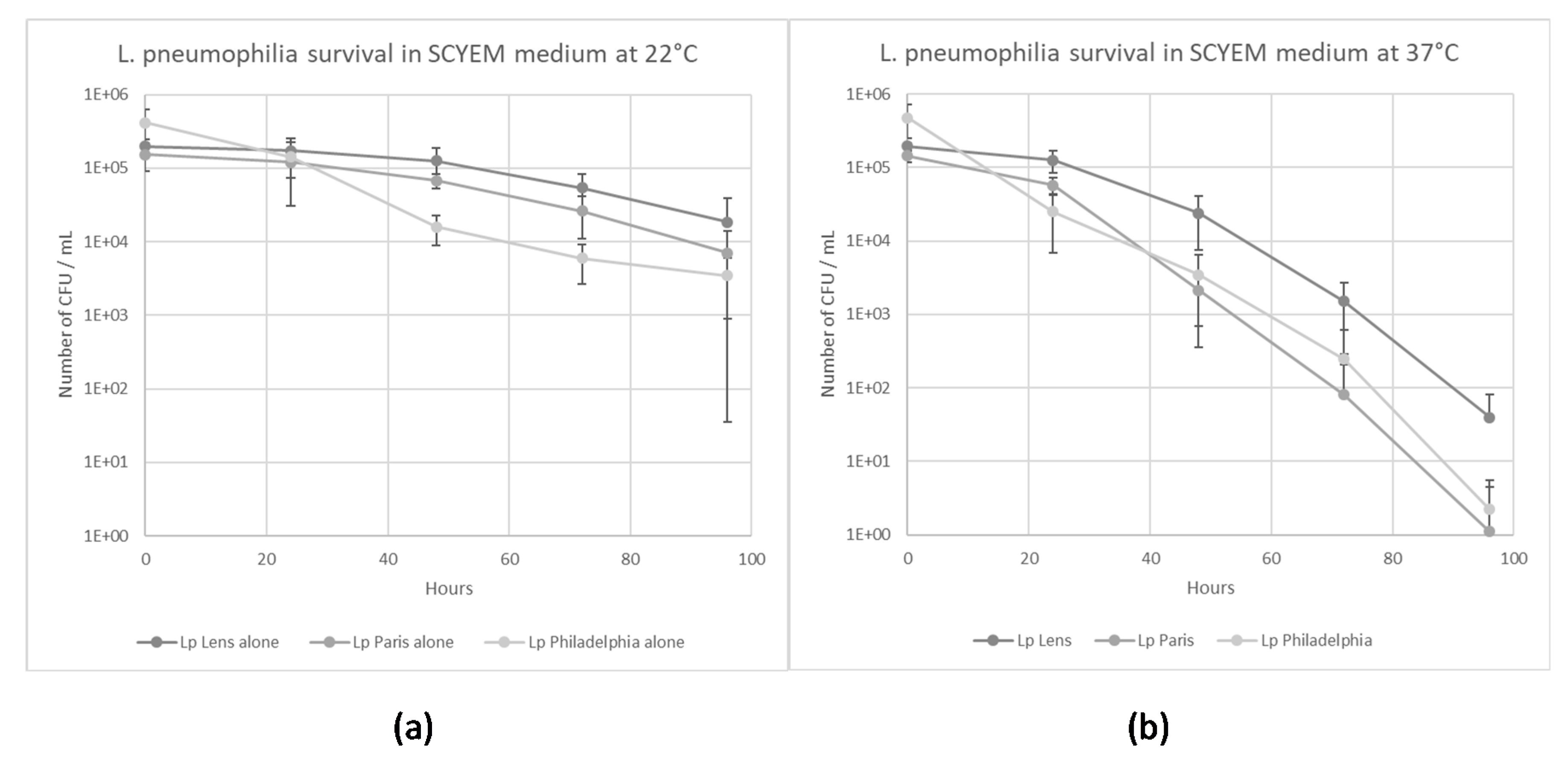
2.1. L. pneumophila Survival in Coculture Medium

The survival of the three L. pneumophila strains in the calf serum-casein-yeast extract medium (SCYEM) was evaluated at 22 °C and 37 °C (Figure 1a,b). The survival of L. pneumophila Lens decreased to 2 × 10⁴ CFU/mL and to 40 CFU/mL in SCYEM medium within 96 h at 22 °C and 37 °C, respectively. The survival of L. pneumophila Paris decreased to 7 × 10³ CFU/mL and to 1 CFU/mL in SCYEM medium within 96 h at 22 °C and 37 °C, respectively. Finally, the survival of L. pneumophila Philadelphia decreased to 3 × 10³ CFU/mL and to 2 CFU/mL in SCYEM medium within 96 h at 22 °C and 37 °C, respectively.

2.2. Amoeba Survival in coculture Medium

Survival of the three amoebas in the presence or in absence of bacteria was evaluated over 96 h at 22 °C and 37 °C in coculture medium (Figure 2a,b). The three amoeba strains could be maintained in SCYEM medium for 96 h in the presence or absence of bacteria at 22 °C and 37 °C with the exception of A. castellanii when co-cultivated with L. pneumophila strains. Found at the end of the experiment, the control condition of A. castellanii in the absence of bacteria was maintained at 2 × 10⁵ cells/mL, while in the presence of L. pneumophila Lens, Paris, and Philadelphia, the amoeba number decreased to 556, 444 and 2333 cells/mL, respectively (Figure 2b). A. castellanii could not survive in the presence of the three L. pneumophila strains at 37 °C.

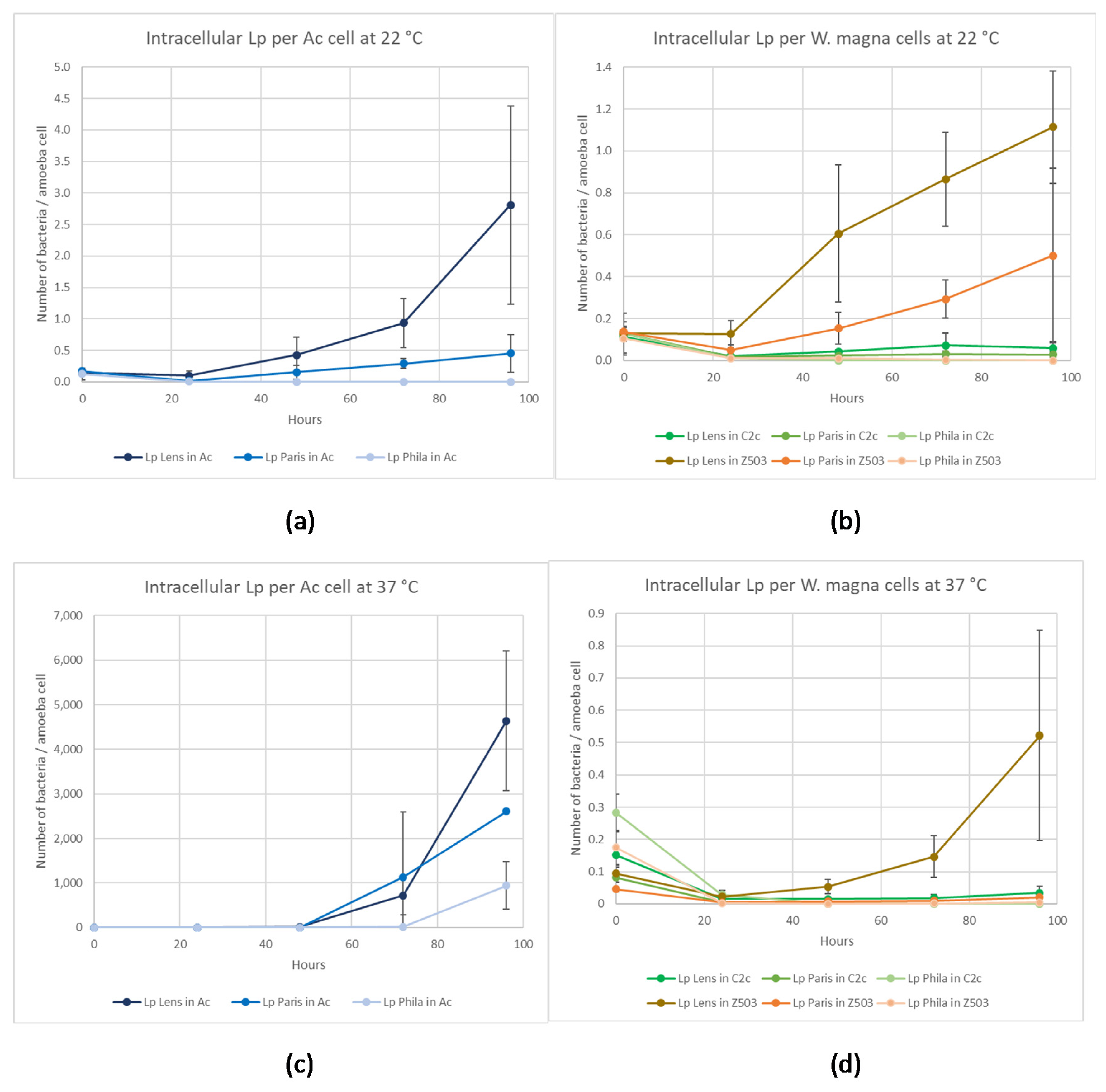
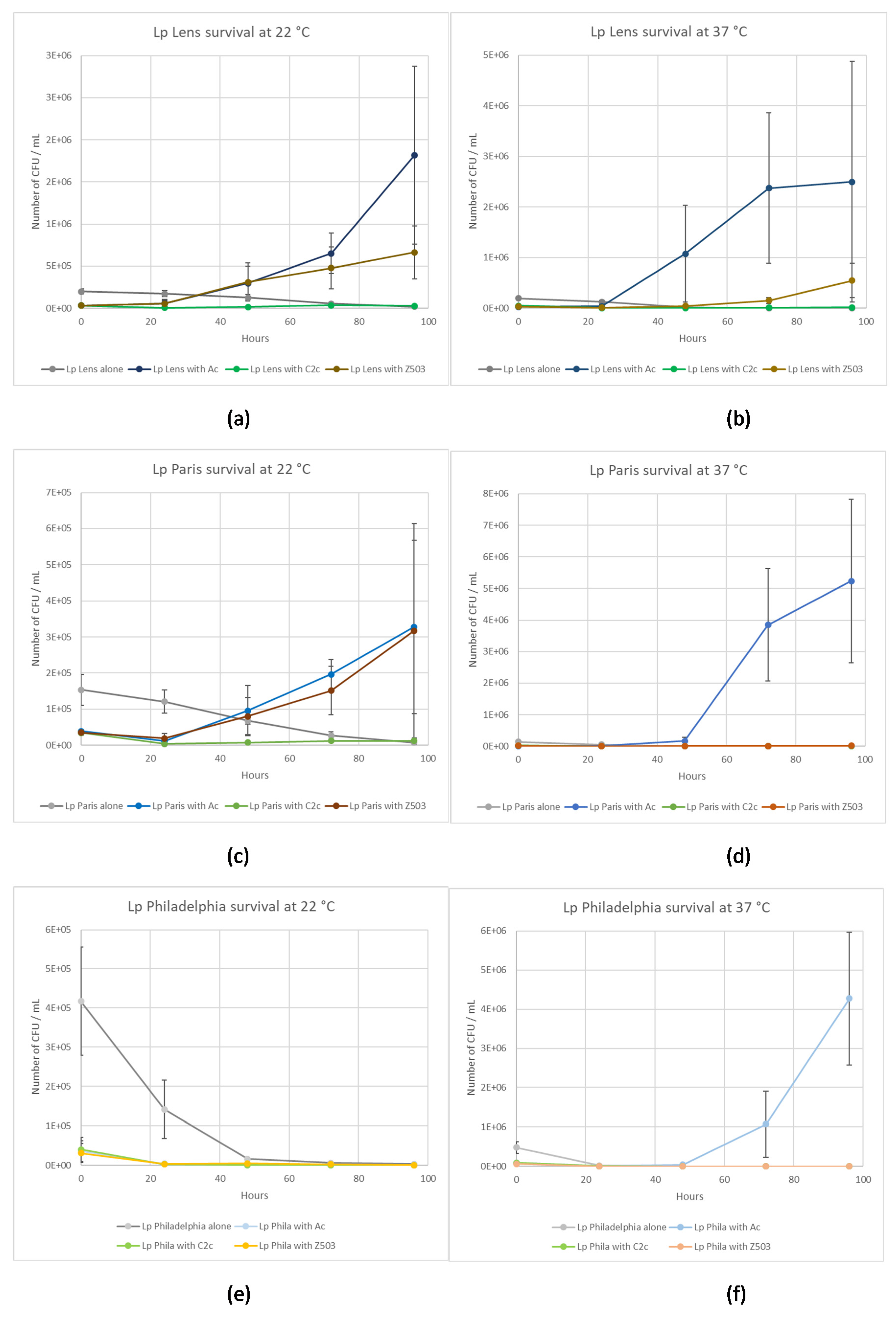
2.3. Co-Culture Experiments

2.3.1. L. pneumophila Lens co-cultivated with Amoeba Strains

The mean initial amount of amoeba-internalized bacteria at 22 °C was 16 ± 0.5% (16% in A. castellanii, 15% in W. magna C2c Maky, and 16% in W. magna Z503). Seen at 37 °C, a mean bacterial uptake of 20 ± 5.5% was observed (15% in A. castellanii, 26% in W. magna C2c Maky, and 18% in W. magna Z503).
A significant decrease (p < 0.05) in the number of intracellular L. pneumophila Lens per W. magna C2c Maky cell was observed after 24 h (5-fold and 10-fold reduction at 22 °C and 37 °C, respectively), while the level remained nearly constant for A. castellanii at 22 °C and 37 °C and for W. magna Z503 at 22 °C with no significant difference between T0 and T0 + 24 h (p > 0.05) (Figure 3). Occurring at T0 + 96 h (Figure 3), the percentage of intracellular L. pneumophila Lens per W. magna C2c Maky cell was reduced by 48 ± 0.3% at 22 °C and 77 ± 1.2% at 37 °C, and an increase was observed for W. magna Z503 (9-fold at 22 °C and 5-fold at 37 °C) and A. castellanii (19-fold at 22 °C and 50,000-fold at 37 °C). Observed at 37 °C, a small number of A. castellanii cells were still alive (5.6 × 10² ± 5.9 × 10² amoebas/mL), demonstrating that amoeba cell lysis occurred following the intracellular multiplication of L. pneumophila Lens.
Considering the number of L. pneumophila Lens at 22 °C and 37 °C, a significant increase (p < 0.05) was obtained when the bacterium was co-cultivated with W. magna Z503 and A. castellanii, and this was not observed when L. pneumophila Lens was cultivated alone or in the presence of W. magna C2c Maky (Figure 4a,b), demonstrating an intracellular multiplication of L. pneumophila Lens in W. magna Z503 and A. castellanii as the bacterium was unable to multiply by itself in the coculture medium (Figure 1a,b).

2.3.2. L. pneumophila Paris Co-Cultivated with Amoeba Strains

Occurring at 22 °C, we reported a mean L. pneumophila Paris uptake by amoebas of 24 ± 1.5% (25% in A. castellanii, 23% in W. magna C2c Maky, and 23% in W. magna Z503). The initial mean amount of cells internalized by amoebas decreased to 14 ± 5.0% at 37 °C (9% in A. castellanii, 19% in W. magna C2c Maky and 13% in W. magna Z503).
A significant decrease of the number of intracellular L. pneumophila Paris per amoeba cell (p < 0.05) first was observed in the three amoebas after 24 h, with the exception of A. castellanii at 37 °C (8-fold for W. magna C2c Maky, 3-fold for W. magna Z503, and 9-fold for A. castellanii at 22 °C and 19-fold for W. magna C2c Maky, 11-fold for W. magna Z503, and 2-fold for A. castellanii at 37 °C) (Figure 3). This decrease was maintained until the end of the experiment (T0 + 96 h) only by W. magna C2c Maky, and the percentage of intracellular L. pneumophila Paris per amoeba cell was reduced by 79 ± 2% at 22 °C and 98 ± 0.1% at 37 °C (p < 0.05). The opposite was observed for W. magna Z503 and A. castellanii at 22 °C and 37 °C, as the decrease measured after 24 h was not maintained. Seen at 48 h, the level of intracellular L. pneumophila Paris per amoeba cell began to increase until it reached 4-fold and 3-fold more bacteria per amoeba cell than that observed at T0 for W. magna Z503 and A. castellanii, respectively at 22 °C. Observed at 37 °C for W. magna Z503, the number of intracellular L. pneumophila Paris per amoeba cell at T0 + 96 h was 5-fold the ratio observed at 24 h, but it did not reach the initial ratio. Regarding A. castellanii, a strong increase was observed at both temperatures, and the initial ratio was slightly increased by 3-fold at 22 °C (p > 0.05) and strongly increased by 60,000-fold at 37 °C (p < 0.05). Furthermore, the correlation between the increase in L. pneumophila Paris and the low concentration of viable A. castellanii (5.6 × 10² ± 5.9 × 10² cells/mL) after 96 h indicated that a high intracellular multiplication of L. pneumophila Paris occurred that was followed by a release of bacteria in the medium after A. castellanii death.
Considering the number of L. pneumophila Paris at 22 °C, a significant increase (p < 0.05) was obtained when the bacterium was co-cultured with W. magna Z503 and A. castellanii, and this was not observed when L. pneumophila Paris was cultured alone or in the presence of W. magna Z503 at 37 °C and W. magna C2c Maky at both 22 °C and 37 °C (Figure 4c,d), demonstrating an intracellular multiplication of L. pneumophila Paris in W. magna Z503 and A. castellanii at 22 °C and only in A. castellanii at 37 °C as the bacterium was unable to multiply by itself in the coculture medium (Figure 1a,b).

2.3.3. L. pneumophila Philadelphia Co-Cultivated with Amoeba Strains

The mean bacterial internalization by amoebas was 9 ± 1.1% (9% in A. castellanii, 10% in W. magna C2c Maky, and 7% in W. magna Z503) at 22 °C, and the initial amount of internalized cells by amoebas increased to 17 ± 3.8% (19% in A. castellanii, 20% in W. magna C2c Maky, and 13% in W. magna Z503).
Occurring at 22 °C, a rapid and significant (p < 0.05) decrease in the number of intracellular L. pneumophila per amoeba cell was observed within 24 h (20-fold for A. castellanii, 11-fold for W. magna C2c Maky, and 10-fold for W. magna Z503) in the three amoebas (Figure 3). Then, a slow but significant (p < 0.05) decrease continued until the death of more than 99% of intracellular L. pneumophila Philadelphia in all cases. Even if this decrease could be attributed to the bacterial death in the coculture medium, the experiment demonstrated the absence of intra-amoeba multiplication of L. pneumophila Philadelphia necessary for survival at 22 °C.
Occurring at 37 °C, a similar rapid decrease in the number of intracellular L. pneumophila per amoeba was observed within 24 h for all three amoebas (20-fold for A. castellanii, 10-fold for W. magna C2c Maky, and 92-fold for W. magna Z503). Then, differential behaviours were observed depending on the amoeba strains. Regarding W. magna C2c Maky, the significant decrease (p < 0.05) continued until the death of more than 99.99% of the intracellular L. pneumophila Philadelphia per amoeba cell (Figure 3d). Concerning W. magna Z503, a decrease also was observed up to 97% elimination of intracellular L. pneumophila Philadelphia per amoeba cell after 96 h (p < 0.05) (Figure 3d). To contrast, for A. castellanii, a significant increase (p < 0.05) in intracellular L. pneumophila Philadelphia per amoeba cell appeared after 48 h, demonstrating an intra-amoeba multiplication up to 2600-fold at the end point (Figure 3c).
Considering the number of L. pneumophila Philadelphia at 22 °C, a significant decrease (p < 0.05) was obtained in all cases (Figure 4e), while at 37 °C, a significant increase (p < 0.05) was observed when L. pneumophila Philadelphia was cultured in the presence of A. castellanii (Figure 4f). This demonstrated an intracellular multiplication of L. pneumophila Philadelphia A. castellanii at 37 °C, as the bacterium was unable to multiply by itself in SCYEM medium (Figure 1a,b).

2.4. Microscopic Observations of Intracellular L. pneumophila Philadelphia at 37 °C

Microscopic observations were performed at T0, T0 + 48 h, and T0 + 96 h. Occurring at T0, excess intracellular L. pneumophila Philadelphia bacteria were observed in the presence of the three amoebas (Figure 5A,D,G). Regarding A. castellanii at 48 h, a strong bacterial multiplication was observed (Figure 5B) which was not observed for both W. magna strains (Figure 5E,H). Occurring at 96 h, lysis of A. castellanii after intracellular bacterial multiplication was clearly evident (Figure 5C), and only a small amount of amoeba lysis could be observed for both W. magna strains (Figure 5F,I).

2.5. Statistical Comparison of Amoeba Behavior

Analysis of variance tests (ANOVA) were performed to determine if W. magna C2c Maky interacted with L. pneumophila in a significantly different manner compared to interactions with the two other amoebas.
Concerning the three bacterial strains, T0 data obtained in the presence of the three amoebas were not statistically different at 22 °C (p > 0.05); however, at 37 °C, a significant difference in behaviour (p < 0.05) was detected at T0.
Pairwise comparisons (Dunn test) established that at 72 h and 96 h at both temperatures and with the three legionella strains, W. magna C2c Maky behaviour was statistically different from that of the two other amoeba strains (Table 1). This significant difference was observed even after 24 h with strain Paris at both temperatures, and at 22 °C for strain Lens. Statistical tests provided evidence that W. magna C2c Maky behaved differently compared to W. magna Z503 and A. castellanii cells in the presence of Legionella strains.

3. Discussion

This work explores the permissiveness of three amoeba strains regarding the intracellular multiplication of three pathogenic L. pneumophila strains under two temperature conditions (22 °C and 37 °C) that correspond to temperatures found in cooling towers in which L. pneumophila are known to replicate within certain strains of amoebae [10,25]. It is important to demonstrate that W. magna C2c Maky does not multiply L. pneumophila as we aim to propose it as a natural biocide to treat cooling towers.
The three L. pneumophila strains are a representative set of L. pneumophila serogroup 1 that is responsible for 95% of the legionellosis disease world-wide [5]. Strain Philadelphia is a clinical isolate that is historically responsible for the very first outbreak. It possesses gene traits that allow for multiplication in a number of hosts such as peripheral blood mononuclear cells, peritoneal macrophages, and A. castellanii, A. polyphaga, or A. lenticulate [26,27,28,29]. The Philadelphia strain is, according to the EN 13623 European standard, the only strain for which testing is required to validate a disinfectant against Legionella in Europe. L. pneumophila Lens was chosen because it was responsible for an outbreak in the north of France between November 2003 and January 2004 where 86 confirmed cases resulted in 17 deaths [30]. L. pneumophila Paris was chosen because, among the endemic strains of L. pneumophila serogroup 1, sequence type 1 (ST1) strains are among the most prevalent, particularly the ST1/Paris pulsotype. This endemic type was responsible for 8.2% of French culture-proven cases of Legionnaire’s disease from 1995 through 2006. ST1/Paris pulsotype isolates also have been detected in clinical and environmental samples taken from several other countries around the world, including Switzerland, Italy, Spain, Sweden, the United States, Japan, Senegal, and Canada [21,30].
Our experiments demonstrate differential behaviours among amoeba species infected by the pathogenic bacteria. Compared to A. castellanii and W. magna Z503, the intracellular L. pneumophila are efficiently eliminated by W. magna C2c Maky at 22 °C and 37 °C. Indeed, the experiments report not only a non-replication, but also an elimination of the intracellular strains Lens, Paris and Philadelphia within W. magna C2c Maky. Furthermore, the coculture medium used in the survey is not adapted to the survival of the legionella bacteria, and they, therefore, must parasitize the amoebae to facilitate their own growth. Indeed, the experiments demonstrate that the three legionella strains were unable to remain at the inoculation level and began to die after 24 h (Figure 1). Although the medium is not adapted to L. pneumophila strains, it was chosen for the co-culture study because an increase of the bacterial number during the co-culture experiment necessarily indicates that the multiplication occurred within amoeba. The bacterial multiplication is observed both in A. castellanii and W. magna Z503, and it is not observed in W. magna C2c Maky. The assays reveal a multiplication of all legionella strains within A. castellanii at 37 °C and the intracellular multiplication of strain Lens and Paris at 22 °C. Indeed, the strain Philadelphia grows at 37 °C (Figure 3c) and does not multiply at 22 °C (Figure 3a) within A. castellanii. Based on this, these results suggest a behaviour that is influenced by the temperature conditions. Several previous studies revealed the effect of temperature on the relationship between L. pneumophila and free-living amoeba (FLA) [9,31,32]. L. pneumophila serogroup 1, for example, replicated in A. castellanii at 25 °C but were digested at temperatures below 20 °C [25]. Dupuy et al. assessed the ability of 12 amoeba strains of Naegleria sp., Acanthamoeba sp., and Vermamoeba sp. to support the multiplication of L. pneumophila Lens at various temperatures (25 °C, 30 °C and 40 °C), and they revealed a more efficient intracellular proliferation with increasing temperatures [33]. Additionally, we did not observe the same behaviour according to the different bacteria and amoeba strains used during our experiments. Indeed, the strain Lens replicates at 37 °C within W. magna strain Z503, but not in W. magna C2c Maky (Figure 3d). The co-culture at 22 °C of W. magna Z503 with L. pneumophila strain Paris and strain Lens reveals a multiplication of the bacteria; however, no replication is observed during co-culture with strain Philadelphia (Figure 3b). The difference in amoeba permissiveness has been highlighted previously, especially in regard to Naegleria, Acanthamoeba, Vermamoeba and Micriamoeba tesseris [9,34]. The non-replication of legionella within W. magna C2c Maky was previously observed with strain Paris [20]. Our study confirms this result, as the resistance of W. magna C2c Maky towards L. pneumophila Paris is illustrated by the observed significant decrease in the bacterial concentration after 4 days of co-culture at 22 °C and 37 °C (Figure 4c,d). Dey et al. [20], however, reported a moderate increase in strains Philadelphia and Lens within W. magna C2c at 37 °C while in our study the intracellular bacterial concentration significantly decreased in culture with W. magna C2c Maky at 22 °C and 37 °C. These differences can be explained by the protocol parameters used in the former study, particularly regarding the culture medium and elimination of extracellular bacteria. The authors used serum casein glucose yeast extract medium (SCGYEM) that was favourable to L. pneumophila survival, so bacteria were not forced to multiply into amoeba to survive. Additionally, Dey and co-workers did not eliminate extracellular bacteria by centrifugation, and the observed increase could be due to extracellular bacterial replication, such as that resulting from necrotrophic growth as previously demonstrated [35].
W. magna C2c Maky is demonstrated to possess a high efficiency for digesting the intracellular L. pneumophila cells in all strains used in this survey. The growth of L. pneumophila within amoebas is known to enhance the pathogenicity and invasion of L. pneumophila [15,36]; however, no intracellular bacterial replication is observed when we infect W. magna C2c Maky with L. pneumophila strains derived from a first co-culture that was thought to be more virulent (unpublished data).
The action on different L. pneumophila strains and the absence of internal proliferation support the fact that W. magna C2c Maky could be used as a biocide to combat L. pneumophila proliferation in cooling tower water. This observation is consistent with the control of legionella by W. magna C2c Maky observed in real conditions during field trials in functioning cooling towers (http://www.amoeba-biocide.com/sites/default/files/180711_cp_amoeba_us_positive_efficacy_field_test_en_vedf_0.pdf). The traditional method to control bacterial growth in cooling tower water is primarily based on the use of chemical biocides [37,38]. Indeed, the oxidizing agent chlorine is the most used product for cooling tower treatment [39]. The chemical biocide is efficient to prevent L. pneumophila proliferation, although some previous studies reported incomplete eradication of legionella from installations and progressive re-colonization within these systems within weeks or months [40,41]. Moreover, these chemical biocides are dangerous to the environment, they degrade the installation systems, and they require the application of other products such as anti-corrosive agents [42,43]. Described by Iervolino, treatment with another oxidizing agent (H2O2/Ag) was inadequate for legionella control, and, instead, it caused a rapid increase of one logarithmic unit [44]. Chemical biocide action also is not completely efficient against biofilms and amoeba cysts that can provide protection against disinfection treatment [16,17,45]. Finally, chemical biocides used in cooling towers can select L. pneumophila populations, and chemical biocides can promote resistance to biocides and to human health antibiotics [46,47].
To conclude, W. magna C2c Maky is not associated with any human or animal infection, and this is in agreement with the lack of pathogenicity demonstrated in vivo and suggested by genomic analysis [24,48]. This organism is likely a safe and efficient candidate for legionella control in cooling towers and could provide an alternative solution to chemical biocides.

4. Materials and Methods

4.1. Free-Living Amoebae Culture

Willaertia magna C2c Maky (ATCC® PTA-7824), Willaertia magna Z503 (ATCC® 50035), and Acanthamoeba castellanii (ATCC® 30010) were purchased from ATCC and cultivated according to their recommendation into 10 mL of modified PYNFH medium (ATCC medium 1034) in a T-25 tissue culture flask. Amoebae were then grown in cell factories in serum casein yeast extract medium (SCYEM) at 30 °C. SCYEM medium is derived from serum casein glucose yeast extract medium (SCGYEM) medium [49] and contained 10 g·L−1 casein, 5 g·L−1 yeast extract, 10% foetal calf Serum, 1.325 g·L−1 Na2HPO4, and 0.8 g·L−1 KH2PO4. After 72 h (during exponential phase), the cell factories were gently shaken, and the amoeba suspensions were transferred to 50 mL Falcon® tubes. Amoeba populations were then quantified using a Malassez haemocytometer cell counting chamber method (Thermo Fisher Scientific, France) with Trypan blue by mixing 100 µL of Trypan blue with 100 µL of amoeba sample. According to the results, the amoebae concentration in Falcon® tubes was then adjusted to 3 × 10⁵ cells/mL by the addition of SCYEM. The amoebas were then washed twice in SCYEM using centrifugation at 3000× g for 10 min, and the supernatants were then discarded. Amoeba populations were then re-quantified, and the amoeba suspensions were finally adjusted to 3 × 10⁵ cells/mL in 100 mL of SCYEM. A final quantification was performed to verify the concentration.
Each final solution of W. magna C2c Maky, W. magna Z503, and A. castellanii corresponded to working suspensions that were named AWSC2C, AWSZ503, and AWSAC, respectively (Table 2).

4.2. Legionella Pneumophila Cultures

L. pneumophila strain Philadelphia (ATCC 33152), L. pneumophila strain Lens (CIP 108280), and L. pneumophila strain Paris (CIP 107629) were grown on buffered charcoal yeast extract (BCYE) agar plates (Thermo Fisher Scientific, Dardilly, France) at 36 °C for 72 hours and then harvested by scraping, suspended in phosphate-buffered saline (PBS), centrifuged at 9500 xg for 10 min, and washed once in PBS. The supernatants were then discarded. The L. pneumophila suspensions were then diluted in PBS to obtain 3 × 10⁷ bacteria/mL.
The legionella final suspensions represented the bacterial stock working suspensions, and they were identified as BWSPhila, BWSParis, and BWSLens (Table 2).

4.3. Bacterial Survival in the Coculture Medium (Control)

The three control bacterial conditions were prepared as described in Table 2 by adding 10 mL of SCYEM to the 0.1 mL bacteria working solutions (BWSPhila, BWSParis, or BWSLens) in 25 cm3 flasks (Dutscher, Brumath, France) and incubated at 22 °C or 37 °C. This operation corresponded to the T0 time point of the bacterial controls. Occurring at T0, T0 + 24 h, T0 + 48 h, T0 + 72 h, and T0 + 96 h, 1 mL was sampled in each flask and then serially 10-fold diluted in SCYEM and plated on buffered charcoal yeast extract plates (BCYE) in triplicate. BCYE plates were incubated at 36 °C, and colony forming units (CFU) were counted after 5 days. Each condition was performed for three independent replicates and repeated three times (n = 9).

4.4. Amoeba Survival in the coculture Medium (Control)

The three amoeba working solutions (AWSC2C, AWSZ503, or AWSAC) were prepared as described in Table 2 (10 mL of working solutions) and incubated at 22 °C or 37 °C in 25 cm3 flasks. Occurring at T0, T0 + 24 h, T0 + 48 h, T0 + 72 h, and T0 + 96 h, the flasks were gently shaken, and the numbers of amoeba cells were quantified using a haemocytometer cell counting chamber method with Trypan blue. Each condition was performed for three independent replicates and repeated three times (n = 9).

4.5. Co-Culture Assays

Amoeba and bacterial working solutions were mixed in 25 cm3 flasks by adding the required volume according to Table 1. To provide an example, 10 mL of W. magna C2c Maky at 3 × 10⁵ cells/mL was mixed with 0.1 mL of L. pneumophila Lens at 3 × 10⁷ CFU / mL. All flasks were left to stand for 2 h at 22 °C ± 2 °C or at 37 °C ± 2 °C to allow for amoebae/bacteria contact and the internalization of L. pneumophila into amoebae. After the 2-h contact process, each flask was gently shaken 10 times, and the suspension was transferred into a 15 mL Flacon® tube and centrifuged at 3000× g for 5 min. This step allowed for the removal of non-internalized (i.e., extracellular) L. pneumophila from the co-culture suspensions. The pellet was resuspended in 10 mL of sterile SCYEM, and the suspension was poured into a new 25 cm3 flask and incubated at 22 °C ± 2 °C or at 37 °C ± 2 °C. This time point corresponded to the T0 time point of the assay. Each condition was performed for three independent replicates and repeated three times (n = 9), with the exception of the co-culture with strain Philadelphia that was repeated four times at 22 °C (n = 15).

4.6. L. pneumophila and Amoeba Quantifications in Co-Culture Assays from T0 to T0 + 96 h

Occurring at T0, T0 + 24 h, T0 + 48 h, T0 + 72 h, and T0 + 96 h, a washing step was performed. The culture supernatant was removed from each flask and replaced by 10 mL of sterile SCYEM. This step was intended to remove extracellular L. pneumophila to allow for the detection of only intracellular bacteria. Each flask was gently shaken 10 times and an aliquot of 1 mL was sampled. Quantification of amoeba populations was performed using 0.1 mL of each aliquot utilizing a haemocytometer cell counting chamber method with Trypan blue. The remaining 0.9 mL were treated with Triton™ X-100 [31] at 0.02% v/v (final concentration) for 2 min to lyse amoebas and to recover the internal L. pneumophila. The sample was then serially 10-fold diluted in SCYEM and plated on BCYE plates in triplicate, with the exception of the undiluted conditions that were spread onto five plates when the number of L. pneumophila was intended to decrease below the detection limit. BCYE plates were incubated at 36 °C, and CFU were counted after 5 days.

4.7. Microscopic Observations in Co-Culture with L. pneumophila Philadelphia at 37 °C

Co-cultures of L. pneumophila Philadelphia using the three amoeba strains at 37 °C were sampled from running experiments and stained by the Gimenez technique [50,51] at T0, T0 + 48 h, and T0 + 96 h. Co-cultures (0.1 mL) were deposited onto glass slides by using a Shandon Cytospin 4 cytocentrifuge (Thermo Scientific, Illkirch-France) at 800× g for 10 min and then stained using the Gimenez technique. Briefly, each of the glass slides were stained with fuchsin solution for 3 min and washed with water. Then, the glass slides were stained with malachite green for 5–10 s and washed, and this step was repeated twice. Finally, the glass slides were allowed to dry at room temperature.
The observations were performed using a LEICA DM 2500 LED microscope (Leica Microsystemes SAS, Nanterre-France) under an ×100 oil immersion objective.

4.8. Statistical Analyses

Statistical significance of co-culture studies was determined for 22 °C and 37 °C conditions through the use of analysis of variance (ANOVA) (Kruskal–Wallis test and multiple pair-wise comparison Dunn test).

Author Contributions

Conceptualization, S.D. and J.-B.E.; methodology, I.H., A.J., A.C., B.Q.; formal analysis, I.H. and S.D.; writing—original draft preparation; I.H., S.D. and J.-B.E.; writing—review and editing, S.D.; supervision, S.D. and O.A. All authors have read and agreed to the published version of the manuscript.

Funding

This research was funded by the French Government under the “Investissements d’avenir” (Investments for the Future) program managed by the Agence Nationale de la Recherche (ANR, French National Agency for Research), (reference: Méditerranée Infection 10-IAHU-03), by Région Provence-Alpes-Côte d’Azur and European funding FEDER PRIMI.

Acknowledgments

We are very grateful to B. La Scola for his advice throughout the completion of this study and for reviewing this article prior to publication.

Conflicts of Interest

The authors declare no conflict of interest.

References

  1. Stout, J.E.; Yu, V.L. Legionellosis. N. Engl. J. Med. 1997, 337, 682–687. [Google Scholar] [CrossRef] [PubMed]
  2. Fraser, D.W.; Tsai, T.R.; Orenstein, W.; Parkin, W.E.; Beecham, H.J.; Sharrar, R.G.; Harris, J.; Mallison, G.F.; Martin, S.M.; McDade, J.E.; et al. Legionnaires’ Disease. N. Engl. J. Med. 1977, 297, 1189–1197. [Google Scholar] [CrossRef] [PubMed]
  3. Kaufmann, A.F.; McDade, J.E.; Patton, C.M.; Bennett, J.V.; Skaliy, P.; Feeley, J.C.; Anderson, D.C.; Potter, M.E.; Newhouse, V.F.; Gregg, M.B.; et al. Pontiac Fever: Isolation of the etiologic agent (legionella pneumophila) and demonstration of its mode of transmission. Am. J. Epidemiol. 1981, 114, 337–347. [Google Scholar] [CrossRef] [PubMed]
  4. Benson, R.F.; Fields, B.S. Classification of the genus Legionella. Semin. Respir. Infect. 1998, 13, 90–99. [Google Scholar]
  5. Phin, N.; Parry-Ford, F.; Harrison, T.; Stagg, H.R.; Zhang, N.; Kumar, K.; Lortholary, O.; ZumLa, A.; Abubakar, I. Epidemiology and clinical management of Legionnaires’ disease. Lancet Infect. Dis. 2014, 14, 1011–1021. [Google Scholar] [CrossRef]
  6. Borella, P.; Guerrieri, E.; Marchesi, I.; Bondi, M.; Messi, P. Water ecology of Legionella and protozoan: Environmental and public health perspectives. Biotechnol. Annu. Rev. 2005, 11, 355–380. [Google Scholar]
  7. Hamilton, K.A.; Prussin, A.J.; Ahmed, W.; Haas, C.N. Outbreaks of Legionnaires’ Disease and Pontiac Fever 2006–2017. Curr. Environ. Health Rep. 2018, 5, 263–271. [Google Scholar] [CrossRef]
  8. Pine, L.; George, J.R.; Reeves, M.W.; Harrell, W.K. Development of a chemically defined liquid medium for growth of Legionella pneumophila. J. Clin. Microbiol. 1979, 9, 615–626. [Google Scholar]
  9. Rowbotham, T.J. Preliminary report on the pathogenicity of Legionella pneumophila for freshwater and soil amoebae. J. Clin. Pathol. 1980, 33, 1179–1183. [Google Scholar] [CrossRef] [Green Version]
  10. Yamamoto, H.; Sugiura, M.; Kusunoki, S.; Ezaki, T.; Ikedo, M.; Yabuuchi, E. Factors stimulating propagation of legionellae in cooling tower water. Appl. Environ. Microbiol. 1992, 58, 1394–1397. [Google Scholar] [CrossRef] [Green Version]
  11. Barbaree, J.M.; Fields, B.S.; Feeley, J.C.; Gorman, G.W.; Martin, W.T. Isolation of protozoa from water associated with a legionellosis outbreak and demonstration of intracellular multiplication of Legionella pneumophila. Appl. Environ. Microbiol. 1986, 51, 422–424. [Google Scholar] [CrossRef] [PubMed] [Green Version]
  12. Scheikl, U.; Sommer, R.; Kirschner, A.; Rameder, A.; Schrammel, B.; Zweimüller, I.; Wesner, W.; Hinker, M.; Walochnik, J. Free-living amoebae (FLA) co-occurring with legionellae in industrial waters. Eur. J. Protistol. 2014, 50, 422–429. [Google Scholar] [CrossRef] [PubMed] [Green Version]
  13. Clarholm, M. Protozoan grazing of bacteria in soil—impact and importance. Microb. Ecol. 1981, 7, 343–350. [Google Scholar] [CrossRef] [PubMed]
  14. Rodríguez-Zaragoza, S. Ecology of Free-Living Amoebae. Crit. Rev. Microbiol. 1994, 20, 225–241. [Google Scholar] [CrossRef]
  15. Cirillo, J.D.; Falkow, S.; Tompkins, L.S. Growth of Legionella pneumophila in Acanthamoeba castellanii enhances invasion. Infect. Immun. 1994, 62, 3254–3261. [Google Scholar] [CrossRef] [Green Version]
  16. Greub, G.; Raoult, D. Microorganisms Resistant to Free-Living Amoebae. Clin. Microbiol. Rev. 2004, 17, 413–433. [Google Scholar] [CrossRef] [Green Version]
  17. Rowbotham, T.J. Isolation of Legionella pneumophila from clinical specimens via amoebae, and the interaction of those and other isolates with amoebae. J. Clin. Pathol. 1983, 36, 978–986. [Google Scholar] [CrossRef] [Green Version]
  18. Kilvington, S.; Price, J. Survival of Legionella pneumophila within cysts of Acanthamoeba polyphaga following chlorine exposure. J. Appl. Bacteriol. 1990, 68, 519–525. [Google Scholar] [CrossRef]
  19. Linder, J.W.-K. Ewert Free-living Amoebae Protecting Legionella in Water: The Tip of an Iceberg? Scand. J. Infect. Dis. 1999, 31, 383–385. [Google Scholar] [CrossRef]
  20. Dey, R.; Bodennec, J.; Mameri, M.O.; Pernin, P. Free-living freshwater amoebae differ in their susceptibility to the pathogenic bacterium Legionella pneumophila. FEMS Microbiol. Lett. 2009, 290, 10–17. [Google Scholar] [CrossRef] [Green Version]
  21. Aurell, H.; Etienne, J.; Forey, F.; Reyrolle, M.; Girardo, P.; Farge, P.; Decludt, B.; Campese, C.; Vandenesch, F.; Jarraud, S. Legionella pneumophila Serogroup 1 Strain Paris: Endemic Distribution throughout France. J. Clin. Microbiol. 2003, 41, 3320–3322. [Google Scholar] [CrossRef] [PubMed] [Green Version]
  22. De Jonckheere, J.F.; Dive, D.G.; Pussard, M.; Vickerman, K. Willaertia Magna gen. nov. sp. nov. (Vahlkampfiidae), a Thermophilic Amoeba Found in Different Habitats. Available online: https://eurekamag.com/research/001/281/001281223.php (accessed on 6 February 2019).
  23. Robinson, B.S.; Christy, P.E.; De Jonckheere, J.F. A temporary flagellate (mastigote) stage in the vahlkampfiid amoeba Willaertia magna and its possible evolutionary significance. Biosystems 1989, 23, 75–86. [Google Scholar] [CrossRef]
  24. Hasni, I.; Chelkha, N.; Baptiste, E.; Mameri, M.R.; Lachuer, J.; Plasson, F.; Colson, P.; La Scola, B. Investigation of potential pathogenicity of Willaertia magna by investigating the transfer of bacteria pathogenicity genes into its genome. Sci. Rep. 2019, 9, 18318. [Google Scholar] [CrossRef] [PubMed]
  25. Ohno, A.; Kato, N.; Sakamoto, R.; Kimura, S.; Yamaguchi, K. Temperature-Dependent Parasitic Relationship between Legionella pneumophila and a Free-Living Amoeba (Acanthamoeba castellanii). Appl. Environ. Microbiol. 2008, 74, 4585–4588. [Google Scholar] [CrossRef] [Green Version]
  26. Casini, B.; Baggiani, A.; Totaro, M.; Mansi, A.; Costa, A.L.; Aquino, F.; Miccoli, M.; Valentini, P.; Bruschi, F.; Lopalco, P.L.; et al. Detection of viable but non-culturable legionella in hospital water network following monochloramine disinfection. J. Hosp. Infect. 2018, 98, 46–52. [Google Scholar] [CrossRef]
  27. Arslan-Aydoğdu, E.Ö.; Kimiran, A. An investigation of virulence factors of Legionella pneumophila environmental isolates. Braz. J. Microbiol. Publ. Braz. Soc. Microbiol. 2018, 49, 189–199. [Google Scholar] [CrossRef]
  28. García, M.T.; Jones, S.; Pelaz, C.; Millar, R.D.; Abu Kwaik, Y. Acanthamoeba polyphaga resuscitates viable non-culturable Legionella pneumophila after disinfection. Environ. Microbiol. 2007, 9, 1267–1277. [Google Scholar] [CrossRef]
  29. Molmeret, M.; Jarraud, S.; Mori, J.P.; Pernin, P.; Forey, F.; Reyrolle, M.; Vandenesch, F.; Etienne, J.; Farge, P. Different growth rates in amoeba of genotypically related environmental and clinical Legionella pneumophila strains isolated from a thermal spa. Epidemiol. Infect. 2001, 126, 231–239. [Google Scholar] [CrossRef] [Green Version]
  30. Cazalet, C.; Rusniok, C.; Brüggemann, H.; Zidane, N.; Magnier, A.; Ma, L.; Tichit, M.; Jarraud, S.; Bouchier, C.; Vandenesch, F.; et al. Evidence in the Legionella pneumophila genome for exploitation of host cell functions and high genome plasticity. Nat. Genet. 2004, 36, 1165–1173. [Google Scholar] [CrossRef] [Green Version]
  31. Buse, H.Y.; Ashbolt, N.J. Differential growth of Legionella pneumophila strains within a range of amoebae at various temperatures associated with in-premise plumbing. Lett. Appl. Microbiol. 2011, 53, 217–224. [Google Scholar] [CrossRef]
  32. Rowbotham, T.J. Pontiac fever, amoebae, and legionellae. Lancet Lond. Engl. 1981, 1, 40–41. [Google Scholar] [CrossRef]
  33. Dupuy, M.; Binet, M.; Bouteleux, C.; Herbelin, P.; Soreau, S.; Héchard, Y. Permissiveness of freshly isolated environmental strains of amoebae for growth of Legionella pneumophila. FEMS Microbiol. Lett. 2016, 363, fnw022. [Google Scholar] [CrossRef] [PubMed]
  34. Atlan, D.; Coupat-Goutaland, B.; Risler, A.; Reyrolle, M.; Souchon, M.; Briolay, J.; Jarraud, S.; Doublet, P.; Pélandakis, M. Micriamoeba tesseris nov. gen. nov. sp.: A new taxon of free-living small-sized Amoebae non-permissive to virulent Legionellae. Protist 2012, 163, 888–902. [Google Scholar] [CrossRef]
  35. Temmerman, R.; Vervaeren, H.; Noseda, B.; Boon, N.; Verstraete, W. Necrotrophic growth of Legionella pneumophila. Appl. Environ. Microbiol. 2006, 72, 4323–4328. [Google Scholar] [CrossRef] [Green Version]
  36. Richards, A.M.; Von Dwingelo, J.E.; Price, C.T.; Abu Kwaik, Y. Cellular microbiology and molecular ecology of Legionella-amoeba interaction. Virulence 2013, 4, 307–314. [Google Scholar] [CrossRef] [Green Version]
  37. McDade, J.E. Legionella and the Prevention of Legionellosis; Bartram, J., Chartier, Y., Lee, J.V., Bond, K., Surman-Lee, S., Eds.; World Health Organization: Geneva, Switzerland, 2007; ISBN 978-92-4-156297-3. [Google Scholar]
  38. Carducci, A.; Verani, M.; Battistini, R. Legionella in industrial cooling towers: Monitoring and control strategies. Lett. Appl. Microbiol. 2010, 50, 24–29. [Google Scholar] [CrossRef]
  39. Canals, O.; Serrano-Suárez, A.; Salvadó, H.; Méndez, J.; Cervero-Aragó, S.; Ruiz de Porras, V.; Dellundé, J.; Araujo, R. Effect of chlorine and temperature on free-living protozoa in operational man-made water systems (cooling towers and hot sanitary water systems) in Catalonia. Environ. Sci. Pollut. Res. Int. 2015, 22, 6610–6618. [Google Scholar] [CrossRef]
  40. Heimberger, T.; Birkhead, G.; Bornstein, D.; Same, K.; Morse, D. Control of nosocomial Legionnaires’ disease through hot water flushing and supplemental chlorination of potable water. J. Infect. Dis. 1991, 163, 413. [Google Scholar] [CrossRef]
  41. Thomas, V.; Bouchez, T.; Nicolas, V.; Robert, S.; Loret, J.F.; Lévi, Y. Amoebae in domestic water systems: Resistance to disinfection treatments and implication in Legionella persistence. J. Appl. Microbiol. 2004, 97, 950–963. [Google Scholar] [CrossRef]
  42. Abdel-Wahab, A.; Batchelor, B. Chloride Removal from Recycled Cooling Water Using Ultra-High Lime with Aluminum Process. Water Environ. Res. 2002, 74, 256–263. [Google Scholar] [CrossRef]
  43. Pagnier, I.; Merchat, M.; La Scola, B. Potentially pathogenic amoeba-associated microorganisms in cooling towers and their control. Future Microbiol. 2009, 4, 615–629. [Google Scholar] [CrossRef]
  44. Iervolino, M.; Mancini, B.; Cristino, S. Industrial Cooling Tower Disinfection Treatment to Prevent Legionella spp. Int. J. Environ. Res. Public. Health 2017, 14, 1125. [Google Scholar] [CrossRef] [Green Version]
  45. Farhat, M.; Moletta-Denat, M.; Frère, J.; Onillon, S.; Trouilhé, M.-C.; Robine, E. Effects of disinfection on Legionella spp., eukarya, and biofilms in a hot water system. Appl. Environ. Microbiol. 2012, 78, 6850–6858. [Google Scholar] [CrossRef] [Green Version]
  46. Maillard, J.-Y. Resistance of Bacteria to Biocides. Microbiol. Spectr. 2018, 6. [Google Scholar] [CrossRef]
  47. Wéry, N.; Bru-Adan, V.; Minervini, C.; Delgénes, J.-P.; Garrelly, L.; Godon, J.-J. Dynamics of Legionella spp. and bacterial populations during the proliferation of L. pneumophila in a cooling tower facility. Appl. Environ. Microbiol. 2008, 74, 3030–3037. [Google Scholar] [CrossRef] [Green Version]
  48. Rivera, F.; Lares, F.; Gallegos, E.; Ramirez, E.; Bonilla, P.; Calderon, A.; Martinez, J.J.; Rodriguez, S.; Alcocer, J. Pathogenic amoebae in natural thermal waters of three resorts of Hidalgo, Mexico. Environ. Res. 1989, 50, 289–295. [Google Scholar] [CrossRef]
  49. De Jonckheere, J. Use of an axenic medium for differentiation between pathogenic and nonpathogenic Naegleria fowleri isolates. Appl. Environ. Microbiol. 1977, 33, 751–757. [Google Scholar] [CrossRef] [Green Version]
  50. Gimenez, D.F. Staining rickettsiae in yolk-sac cultures. Stain Technol. 1964, 39, 135–140. [Google Scholar] [CrossRef]
  51. Greer, P.W.; Chandler, F.W.; Hicklin, M.D. Rapid demonstration of Legionella pneumophila in unembedded tissue. An adaptation of the Giménez stain. Am. J. Clin. Pathol. 1980, 73, 788–790. [Google Scholar] [CrossRef]
Figure 1. L. pneumophila survival in coculture medium at 22 °C (a) and 37 °C (b). Results are expressed as the mean +/− 95% CI (Confidence Interval based on the standard error of the mean).
Pathogens 09 00105 g001
Figure 2. Amoeba survival at 22 °C (a) and 37 °C (b) in coculture medium in the presence or absence of the three L. pneumophila strains (Lens, Paris, and Philadelphia). The red bar is the detection limit of the Malassez cell counting. Results are expressed as the mean +/− 95% CI (Confidence Interval based on the standard error of the mean).
Pathogens 09 00105 g002
Figure 3. Comparison of the evolution of the number of intracellular L. pneumophila cells (Lens, Paris, and Philadelphia) per amoeba cell (A. castellanii, W. magna C2c Maky, and W. magna Z503). Results are expressed as the mean +/− 95% CI (Confidence Interval based on the standard error of the mean). (a) L. pneumophila number per A. castellanii cell at 22 °C (n = 9 for Lp Lens and Paris, n = 15 for Lp Philadelphia); (b) L. pneumophila number per A. castellanii cell at 37 °C (n = 9); (c) L. pneumophila number per W. magna cell (C2c and Z503) at 22 °C (n = 9 for Lp Lens and Paris, n = 15 for Lp Philadelphia); (d) L. pneumophila number per W. magna cell (C2c and Z503) at 37 °C (n = 9).
Pathogens 09 00105 g003
Figure 4. Comparison of the evolution of the number of L. pneumophila cells in the presence or absence of amoeba cells (alone, or in presence of A. castellanii, W. magna C2c Maky, or W. magna Z503). Results are expressed as the mean +/− 95% CI (Confidence Interval based on the standard error of the mean). (a) L. pneumophila Lens at 22 °C (n = 9); (b) L. pneumophila Lens at 37 °C (n = 9); (c) L. pneumophila Paris at 22 °C (n = 9); (d) L. pneumophila Paris at 37 °C (n = 9); (e) L. pneumophila Philadelphia at 22 °C (n = 15); (f) L. pneumophila Philadelphia at 37 °C (n = 9).
Pathogens 09 00105 g004
Figure 5. Optical microscopy observation using Gimenez staining of A. castellanii (AC), W. magna C2c Maky (DF), and W. magna Z503 (GI) infected with L. pneumophila Philadelphia at 37 °C. Photos of the co-cultures were acquired at T0 (A,D,G), T0 + 48 h (B,E,H), and T0 + 96 h (C,F,I).
Pathogens 09 00105 g005
Table 1. Statistical analysis of the behaviour of the three amoeba strains in the presence of the three Legionella strains at 22 °C and 37 °C. Significant differences for W. magna C2c Maky are highlighted in yellow.
22 °C37 °C
L. pneumophila LensT024 h48 h72 h96 hT024 h48 h72 h96 h
With A. castellaniiAAAAAAAAAA
With W. magna Z503AAAAAABABAAA
With W. magna C2c MakyABBBBABBCB
L. pneumophila ParisT024 h48 h72 h96 hT024 h48 h72 h96 h
With A. castellaniiAAAAACAAAA
With W. magna Z503AAAAABABBB
With W. magna C2c MakyABBBBABCCC
L. pneumophila PhiladelphiaT024 h48 h72 h96 hT024 h48 h72 h96 h
With A. castellaniiAAAABABABAAABAB
With W. magna Z503AAAAABBBAA
With W. magna C2c MakyAAABBAABCC
Table 2. Preparation of the co-cultures.
Co-CultureAWS 1 VolumeBWS 2 Volume
L.p. Philadelphia + W. magna C2c Maky10 mL AWSC2C0.1 mL BWSPhila
L.p. Philadelphia + W. magna Z50310 mL AWSZ5030.1 mL BWSPhila
L.p. Philadelphia + A. castellanii.10 mL AWSAC0.1 mL BWSPhila
L.p. Paris + W. magna C2c Maky10 mL AWSC2C0.1 mL BWSParis
L.p. Paris + W. magna Z50310 mL AWSZ5030.1 mL BWSParis
L.p. Paris + A. castellanii.10 mL AWSAC0.1 mL BWSParis
L.p. Lens + W. magna C2c Maky10 mL AWSC2C0.1 mL BWSLens
L.p. Lens + W. magna Z50310 mL AWSZ5030.1 mL BWSLens
L.p. Lens + A. castellanii.10 mL AWSAC0.1 mL BWSLens
Control L.p. Philadelphia10 mL SCYEM0.1 mL BWSPhila
Control L.p. Paris10 mL SCYEM0.1 mL BWSParis
Control L.p. Lens10 mL SCYEM0.1 mL BWSLens
Control W. magna C2c Maky10 mL AWSC2C0 mL
Control W. magna Z50310 mL AWSZ5030 mL
Control A. castellanii10 mL AWSAC0 mL
1 AWS: Amoeba Working Solution at 3 × 10⁵ cells / mL; 2 BWS: Bacteria Working Solution at 3 × 10⁷ CFU / mL.

