Article

Occurrence and Genetic Diversity of Trichomonas gallinae in Captive Synanthropic Birds in Southeastern Brazil

by
Amanda Garcia Pereira
1,
Sarah Raquel Jesus Santos Simões
1,
Maitê Cardoso Coelho da Silva
1,
Ana Cláudia Calchi
2,
Ricardo Bassini-Silva
3,
Ana Carolina Castro-Santiago
4,
Rosangela Zacarias Machado
2,
Marcos Rogério André
2 and
Karin Werther
1,*
1
Serviço de Patologia de Animais Silvestres—SEPAS, Departamento de Patologia, Reprodução e Saúde Única, Faculdade de Ciências Agrárias e Veterinárias, Universidade Estadual Paulista “Júlio de Mesquita Filho”—UNESP, Jaboticabal 14884-900, SP, Brazil
2
Vector-Borne Bioagents Laboratory—VBBL, Departamento de Patologia, Reprodução e Saúde Única, Faculdade de Ciências Agrárias e Veterinárias, Universidade Estadual Paulista “Júlio de Mesquita Filho”—UNESP, Jaboticabal 14884-900, SP, Brazil
3
Laboratório de Coleções Zoológicas, Instituto Butantan, São Paulo 05503-900, SP, Brazil
4
Departamento de Medicina Veterinária Preventiva e Saúde Animal, Faculdade de Medicina Veterinária e Zootecnia, Universidade de São Paulo—USP, São Paulo 05508-270, SP, Brazil
*
Author to whom correspondence should be addressed.
Pathogens 2026, 15(4), 428; https://doi.org/10.3390/pathogens15040428
Submission received: 4 March 2026 / Revised: 8 April 2026 / Accepted: 11 April 2026 / Published: 16 April 2026
(This article belongs to the Special Issue Biology, Epidemiology and Interactions of Parasitic Diseases)

Abstract

Avian trichomonosis is caused by protozoa of the genus Trichomonas, mainly Trichomonas gallinae, which infects the upper digestive tract of birds and is commonly associated with Columbiformes, the main reservoirs of the parasite. This study aimed to investigate the occurrence and genetic diversity of Trichomonas spp. in captive synanthropic birds from southeastern Brazil. Oropharyngeal swabs were collected from 281 birds belonging to 13 avian orders and analyzed using Diamond medium culture, Giemsa-stained smears, and molecular assays. Of the 262 samples submitted to culture analysis, 72 (27.48%) showed trophozoite-like structures under light microscopy. Molecular screening based on the ITS1–5.8S–ITS2 region detected Trichomonas DNA in 76 out of 267 samples with successful DNA extraction (28.46%), including 72 Columba livia domestica from Franca, one Coragyps atratus from Ribeirão Preto, and three rock doves from Jaboticabal. Among the ITS-positive samples, 67 (88.15%) amplified the Fe-hydrogenase gene, and 65 (85.5%) were also positive for the 18S rRNA gene. Only six samples (2.29%) exhibited structures compatible with Trichomonas spp. in Giemsa-stained smears. Phylogenetic analyses based on ITS sequences grouped the isolates into two clades within the Trichomonas gallinae complex. Greater genetic diversity was observed using Fe-hydrogenase and 18S rRNA markers, revealing multiple haplotypes and clades. Molecular assays, particularly PCR applied directly to oropharyngeal swabs, showed higher sensitivity for detecting and characterizing Trichomonas gallinae compared to culture and cytology. These findings highlight the high occurrence and genetic diversity of T. gallinae in captive synanthropic pigeons and reinforce the importance of molecular tools for epidemiological surveillance in wildlife facilities.

1. Introduction

Two main species of Trichomonas are known to infect birds: Trichomonas gallinae (Rivolta, 1878), which has major clinical significance among wild birds and typically colonizes the upper digestive tract (oropharynx, esophagus, and crop); and Tetratrichomonas gallinarum (Martin and Robertson, 1911), which primarily inhabits the lower digestive tract, especially the ceca of Galliformes and Anseriformes [1,2,3].
Although T. gallinae primarily infects the upper digestive tract, it may disseminate hematogenously to the lungs, liver, air sacs, pancreas, bones, and cranial sinuses [4]. Transmission occurs through several routes, including crop milk, courtship and mating behaviors, direct contact with infected individuals, and indirect exposure to contaminated food, water, or prey. Consequently, birds across a broad range of taxonomic orders may be infected, often asymptomatically [5,6]. In pigeons, which serve as principal reservoirs and disseminators of T. gallinae, early lesions manifest as small, white-to-yellowish plaques in the oral cavity. Severe infections may result in high mortality, particularly in young birds, with rates reaching 80–90% or higher [4].
Although direct diagnosis can be attempted using stained smears of oropharyngeal swabs, this method is often limited by the parasite’s fragility. Molecular techniques offer improved sensitivity and facilitate phylogenetic characterization of isolates. Polymerase chain reaction (PCR) is particularly effective at detecting low parasite loads. Several genetic targets, including the internal transcribed spacer (ITS) region, 18S rRNA, and Fe-hydrogenase genes, have proven valuable for species identification and haplotype discrimination [7]. Among these, ITS1 and ITS2 are widely used to confirm the presence of T. gallinae [8].
In Brazil, the earliest studies on avian Trichomonas infections were conducted in Rio Grande do Sul. De Carli et al. [9] and Tasca and De Carli [10] reported occurrence rates of 62.3% (104/167) and 26.47% (18/68), respectively, in domestic pigeons. Previous studies have used culture-based approaches for parasite detection and identification.
Arenales et al. [11] documented a case of trichomoniasis in a rock pigeon (Columba livia domestica) treated at the UNESP Veterinary Hospital in Araçatuba, São Paulo State. The bird died during clinical examination, and a white-yellowish, friable mass with a putrid odor was found on the hard palate. Cytological examination revealed protozoa consistent with T. gallinae. In another study, Joppert [12] reported characteristic oral lesions of trichomoniasis in raptors admitted to the Division of Veterinary Medicine and Wildlife Management in São Paulo. Lesions were observed in 10% (4/40) of caracaras (Caracara plancus), 10.8% (8/74) of American kestrels (Falco sparverius), and 4.2% (2/47) of Strigiformes (Tropical Screech-Owl—Megascops choliba and great horned owl—Bubo virginianus), with diagnosis based on saline wet-mount examinations. Additionally, Godoy et al. [13] detected compatible lesions in 1.66% (6/360) of Passeriformes submitted to the Wild Animal Rehabilitation Center at the Tietê Ecological Park.
In Minas Gerais State, Ecco et al. [14] examined paraffin-embedded tissues (tongue, oropharynx, esophagus, crop, proventriculus, and gizzard) and confirmed Trichomonas DNA in six birds by means of PCR targeting the intergenic and partial 5.8S rRNA regions. These included two Striped owls (Asio clamator), one American kestrel (F. sparverius), green-winged saltator (Saltator similis), and one toco toucan (Ramphastos toco). Phylogenetic analysis revealed two major clades. Sequences from a Striped owl and a kestrel grouped with T. gallinae, while one green-winged saltator sequence clustered with Simplomonas. The remaining two sequences formed separate clades. These findings suggest unresolved phylogenetic placements, later reassigned to T. vaginalis-like organisms based on further analysis of the 18S rRNA and α-tubulin genes [15]. In Rio Grande do Sul, Bruni et al. [16] confirmed T. gallinae in a chimango caracara (Milvago chimango) exhibiting characteristic lesions through culture and ITS1-based PCR.
Importantly, all birds included in this study were synanthropic species maintained in captivity in zoos, rehabilitation centers, or wildlife facilities and do not represent free-living urban populations.
Therefore, the present study aimed to (i) determine the occurrence of Trichomonas spp. in captive synanthropic birds from São Paulo State, Brazil, (ii) evaluate the genetic diversity of T. gallinae using ITS, Fe-hydrogenase, and 18S rRNA markers, and (iii) compare the performance of different diagnostic methods (culture, cytology, and PCR) for parasite detection.

2. Materials and Methods

2.1. Study Site and Samples

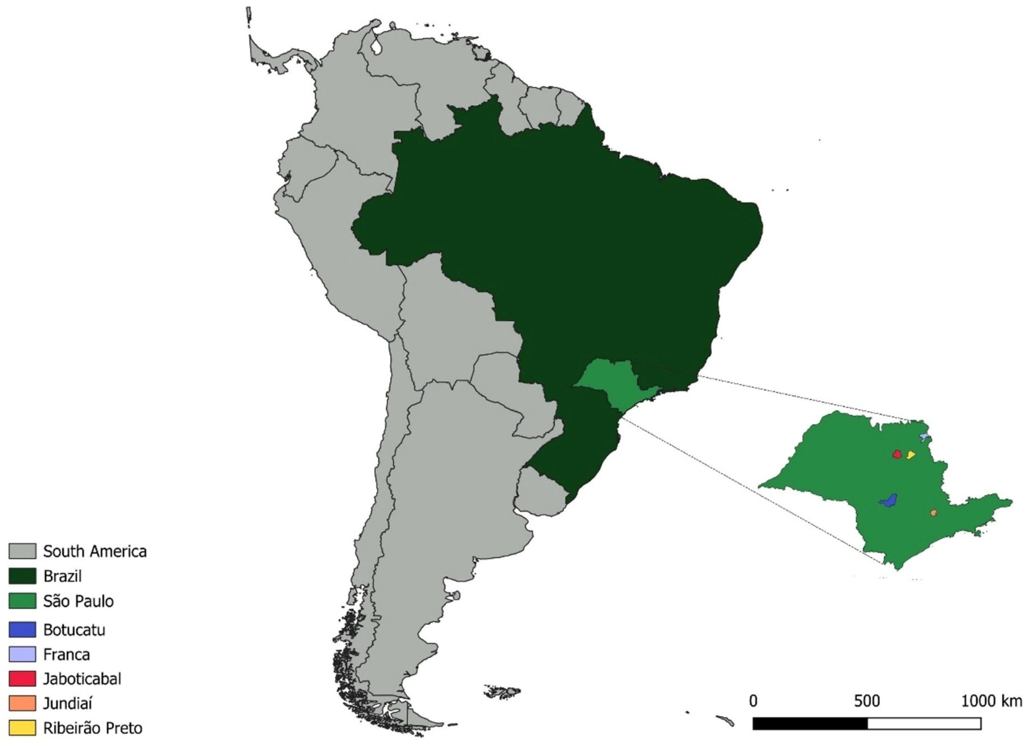
The oropharyngeal swab samples used (n = 281) in this study were collected between July 2022 and January 2023 from wild birds maintained in captivity in different cities (Table 1) in the state of São Paulo: Franca (Latitude: −20.5418, Longitude: −47.4197) (121 birds), Ribeirão Preto (Latitude: −21.170888, Longitude: −47.799273) (51 birds), Jundiaí (Latitude: −23.185705, Longitude: −46.897812) (58 birds), Botucatu (Latitude: −22.8904, Longitude: −48.4553) (32 birds), and Jaboticabal (Latitude: −21.2554, Longitude: −48.3224) (19 birds from the routine of SEPAS—Wild Animal Pathology Service) (Figure 1).
The birds included in this study originated from different captive conditions, including wildlife rehabilitation centers, zoos, and temporary holding facilities for rescued animals (Table 1). Most individuals were admitted due to injury or environmental displacement, and their duration of captivity varied from a few days to several months. These heterogeneous conditions may influence parasite exposure and transmission dynamics. From each of these birds, three oropharyngeal swabs were collected: (i) the first swab was stored in Diamond medium for the evaluation of trichomonad growth; (ii) the second was used to prepare Giemsa-stained smears on slides; and (iii) the third was used for DNA extraction and molecular assays.
For the remaining 19 birds that originated from the SEPAS routine, culture and Giemsa-stained cytology were not performed in Diamond medium because these birds were already dead and had been frozen.

2.2. Ethical Statement

All procedures were authorized by the Ethics Committees on Animal Use of the School of Agricultural and Veterinarian Sciences (FCAV/UNESP) (CEUA no. 732/21) and Institute Chico Mendes for Conservation of Biodiversity (SISBIO no. 77538-1).

2.3. Trichomonas spp. Culture

For the cultivation of Trichomonas spp., oropharyngeal swabs collected from captive birds were used.
Swabs intended for culture were immediately placed into Diamond medium, a modified version of the Trypticase–Yeast Extract–Maltose (TYM) medium. Each sample was resuspended in 9 mL of Diamond medium and incubated at 37 °C for 72 h. Cultures were examined daily for turbidity and for the presence of Trichomonas trophozoites using light microscopy. Prior to examination, samples were centrifuged at 8000 rpm for 15 min. A 10 µL aliquot of the pellet was placed on microscope slides and observed under a Eclipse E200 microscope (Nikon, Tokyo, Japan) at magnifications ranging from 40× to 1000×. Pellets from cultures showing growth were transferred to cryotubes and stored at −20 °C for subsequent DNA extraction and molecular analysis.
A total of 262 samples were subjected to culture in Diamond medium because some samples were obtained from birds that were already dead and frozen at the time of collection, which precluded culture-based analysis.
The term “trophozoite-like structures” refers to flagellated, motile organisms with morphology compatible with Trichomonas spp. under light microscopy.

2.4. Oropharyngeal Swab Smears

Oropharyngeal smears were prepared from birds sampled in zoos located in Ribeirão Preto (n = 51), Franca (n = 121), Jundiaí (n = 58), and Botucatu (n = 32). Smears were made on clean, degreased microscope slides by gently spreading the sample in a circular motion. After air drying, slides were fixed in absolute methanol for 3 min and stained with Giemsa solution (Interlab) for 40 min using approximately 5 mL per slide. Following staining, slides were rinsed with running water and allowed to dry vertically for 30 min before microscopic evaluation [17].

2.5. DNA Extraction

For DNA extraction, swabs were stored in 0.25 mL of autoclaved phosphate-buffered saline (PBS, pH 7.2) at −20 °C. Prior to extraction, samples were incubated at 40 °C for 15 min. No washing steps were performed before DNA extraction. Instead, excess PBS was removed, leaving ~300 µL of solution in the tube. Proteinase K and lysis buffer from the Biopur Mini Spin Plus kit (MoBius Life Science, Paraná, Brazil) were then added per the manufacturer’s instructions. The swab was removed before loading the lysate into the spin column, and the remainder of the protocol followed the kit guidelines.
Delays between sample collection and processing, as well as freezing of cultures, may have negatively affected DNA yield and PCR amplification efficiency.

2.6. Endogenous β-Actin PCR and DNA Electrophoresis in Agarose Gel

To assess the presence of PCR inhibitors and avoid false negatives, amplification of the endogenous avian β-actin gene was performed using conventional PCR with primers β-actin-F (5′-ATCTCGTCTTGTTTTATGCG-3′) and β-actin-R (5′-TATCCGTAAGGATCTGTATG-3′) [18]. Ultra-pure water was used as a negative control, and DNA from Pantanal birds [19] served as a positive control. PCR products were resolved via electrophoresis in a 1.0% agarose gel stained with ethidium bromide (0.5 µL/mL) in TBE buffer (pH 8.0), run at 100 V/150 mA for 50 min. A 100 bp molecular weight marker (Life Technologies®, Carlsbad, CA, USA) was used to estimate product size, visualized under a ChemiDoc MP Imaging System (Bio-Rad®, Hercules, CA, USA).

2.7. PCR Assay for Trichomonas spp. and Subsequent Sequencing and BLASTn Analysis

Samples that tested positive for the β-actin gene and those from positive cultures were screened for Trichomonas DNA using conventional PCR targeting the ITS1/5.8S/ITS2 region (369 bp) [20]. Positive samples were further characterized via PCR amplification of the near-full-length 18S rRNA gene (~1500 bp) [21] and the Fe-hydrogenase gene (~1000 bp) [22]. Table 1 lists the primers and cycling conditions. Ultra-pure water and DNA from T. gallinae isolated from a naturally infected Falco peregrinus were used as negative and positive controls, respectively. The primer sequences and thermal cycling conditions used for each PCR assay (ITS1/5.8S/ITS2, 18S rRNA and Fe-hydrogenase genes) are described in Table 2.
To obtain extended sequences of the 18S rRNA gene, additional primers were designed (F: 5′-CAAGGGCGAGAGTAGGAGTA-3′, R: 5′-ACCGAGTCATCCAATCGGTA-3′), which were used alongside standard primers for sequencing.
Consensus sequences were constructed using Phred-Phrap v23 [23] from bidirectional reads, with a minimum base quality score of 20. Sequence identity was assessed by BLASTn searches against GenBank (https://www.ncbi.nlm.nih.gov/genbank (accessed on 12 January 2026)).

2.8. Phylogenetic Analyses

Sequences were aligned with reference sequences retrieved from GenBank using ClustalW in BioEdit v7.0.5.3 [24]. Bayesian phylogenetic trees were generated using MrBayes v3.2.2 on XSEDE [25] via the CIPRES Science Gateway [26]. Analyses were run for 1,000,000 generations, with evolutionary models selected via jModelTest 2 [27]. Tree editing and rooting using outgroup sequences were conducted in TreeGraph v2.0.56-381 beta [28].

2.9. Diversity Analysis and Haplotype Network

New alignments were constructed using the ITS1/5.8S/ITS2, the near-full-length 18S rRNA and the Fe-hydrogenase nucleotide sequences of T. gallinae obtained in the present study, along with other sequences retrieved from GenBank and derived from birds sampled in different countries. For this purpose, only sequences longer than 321 bp for the ITS region, 1351 bp for the 18S rRNA gene, and 851 bp for the Fe-hydrogenase gene were selected. These three alignments were used in the genetic diversity analysis, conducted with DnaSP v5 software [29], to calculate nucleotide diversity (π), haplotype diversity (Hd), the number of haplotypes (h), and the average number of nucleotide differences (K). TCS networks were generated with popART (https://popart.maths.otago.ac.nz/ (accessed on 12 January 2026)) [30] to visualize haplotype relationships based on the different genetic markers.

3. Results

3.1. Trichomonas spp. Culture

A total of 262 samples were included in the culture analysis. Although 281 oropharyngeal swabs were collected in total, 19 samples originated from birds obtained through the SEPAS routine and had been previously frozen at the time of collection. As freezing compromises the viability of Trichomonas trophozoites, these samples were not suitable for culture-based analysis and were therefore excluded.
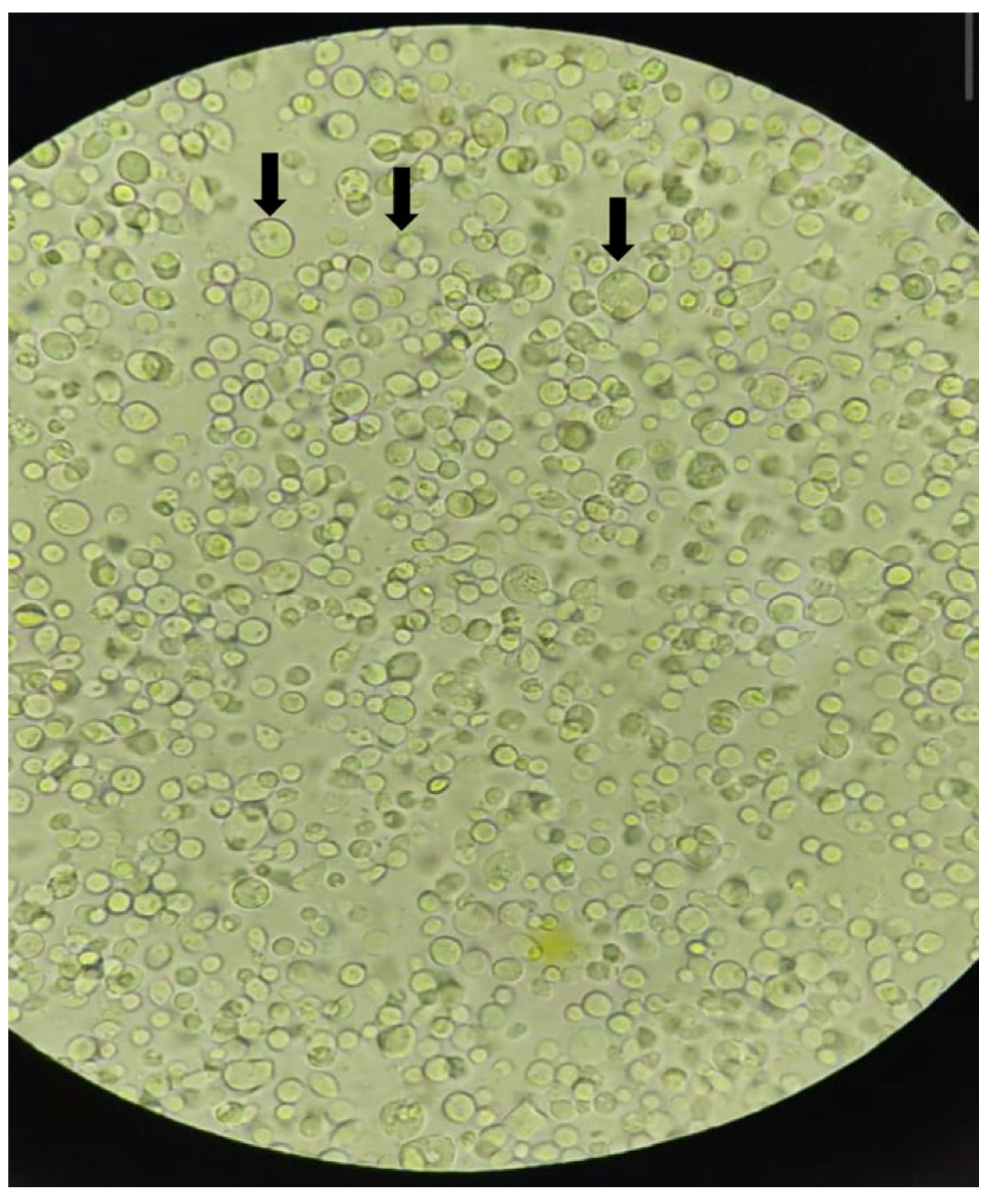
Of the 262 samples submitted to Diamond medium culture, 72 (27.48%) showed structures consistent with Trichomonas trophozoites under light microscopy, exhibiting characteristic motility (Figure 2) (see Supplementary Videos S1 and S2). DNA extraction was performed using the Tissue kit (Qiagen, Valencia, CA, USA) for subsequent Trichomonas spp.-specific PCR testing, which revealed that only cultured samples from C. livia were positive. Additionally, two samples from Columbiformes from the same location were positive for the Fe-hydrogenase gene, while no sample amplified the 18S rRNA gene.

3.2. Giemsa-Stained Oropharyngeal Swab Smears

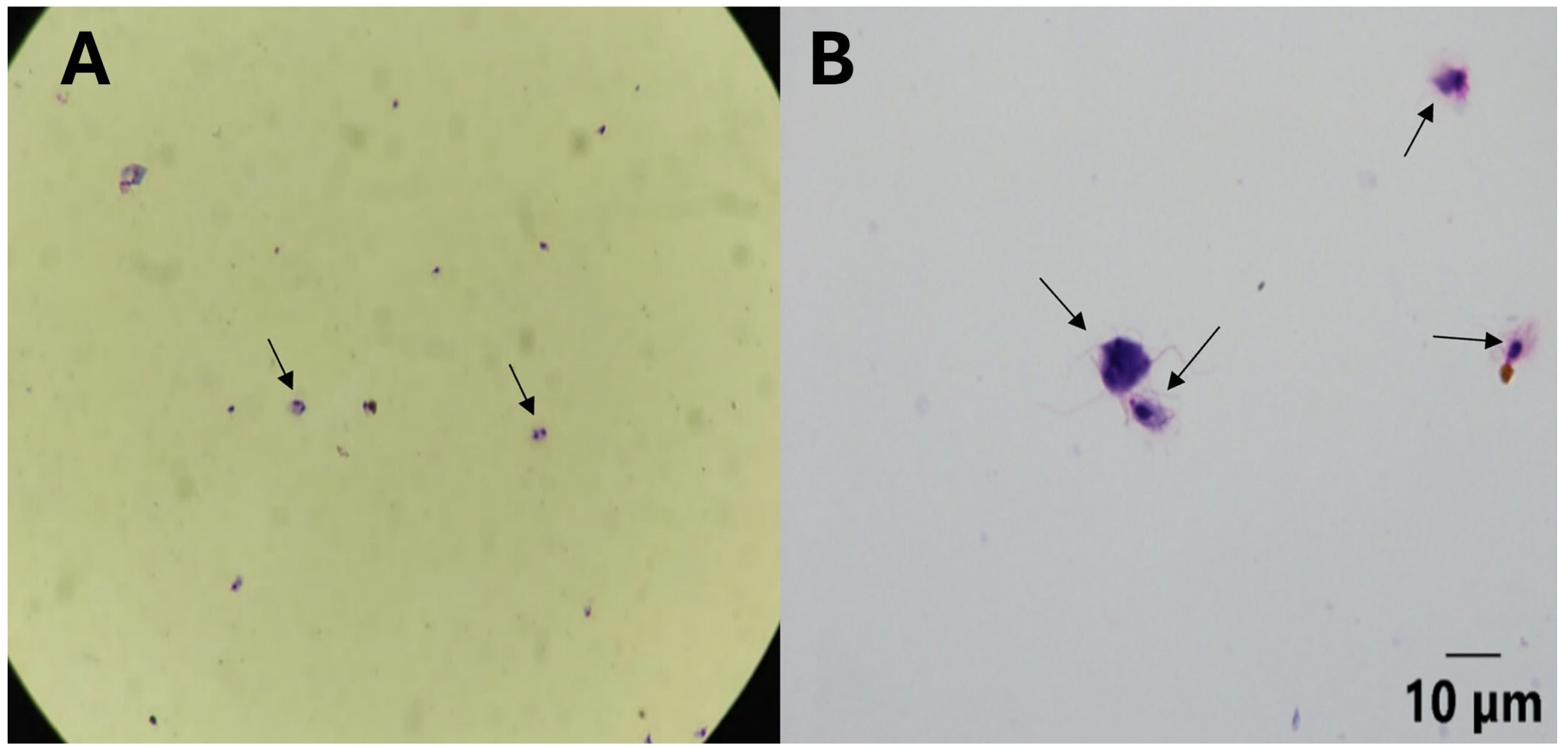
Among 262 Giemsa-stained oropharyngeal swab smears analyzed, six samples (2.29%) from Columbiformes in Franca revealed pseudocysts of Trichomonas spp. (Figure 3). These samples were negative in culture. Most smears showed varying quantities of anucleated and nucleated squamous cells, while seven samples (2.67%) also exhibited moderate numbers of red blood cells.
None of the samples positive by Giemsa-stained smears were exclusively detected by this method; all represented a minority subset compared to molecular and culture-based detection.

3.3. DNA Extraction and PCR for Endogenous Gene

DNA was extracted from 281 oropharyngeal swabs collected from captive birds. Of these, 267 (95.01%) tested positive for the endogenous avian β-actin gene.

3.4. PCR and Sequence Analysis of the ITS1/5.8S/ITS2 Regions of Trichomonas spp.

Out of the 267 samples positive for the endogenous avian β-actin gene, 76 (28.46%) tested positive in the PCR assay based on the ITS1/5.8S/ITS2 regions of Trichomonas, comprising: 72/121 rock doves from Franca, 1/5 C. atratus from Ribeirão Preto, and 3/12 rock doves from Jaboticabal. Detailed individual results for all samples are provided in Supplementary Table S1.
From the 76 positive samples, 34 amplicons with high intensity bands in agarose gel electrophoresis were sequenced. BLASTn analysis showed that the obtained ITS1/5.8S/ITS2 sequences had identities ranging from 93.73% to 100% with sequences of T. gallinae detected in C. livia in Spain (EU881912) and C. livia in China (MH733819) (Supplementary Table S2).
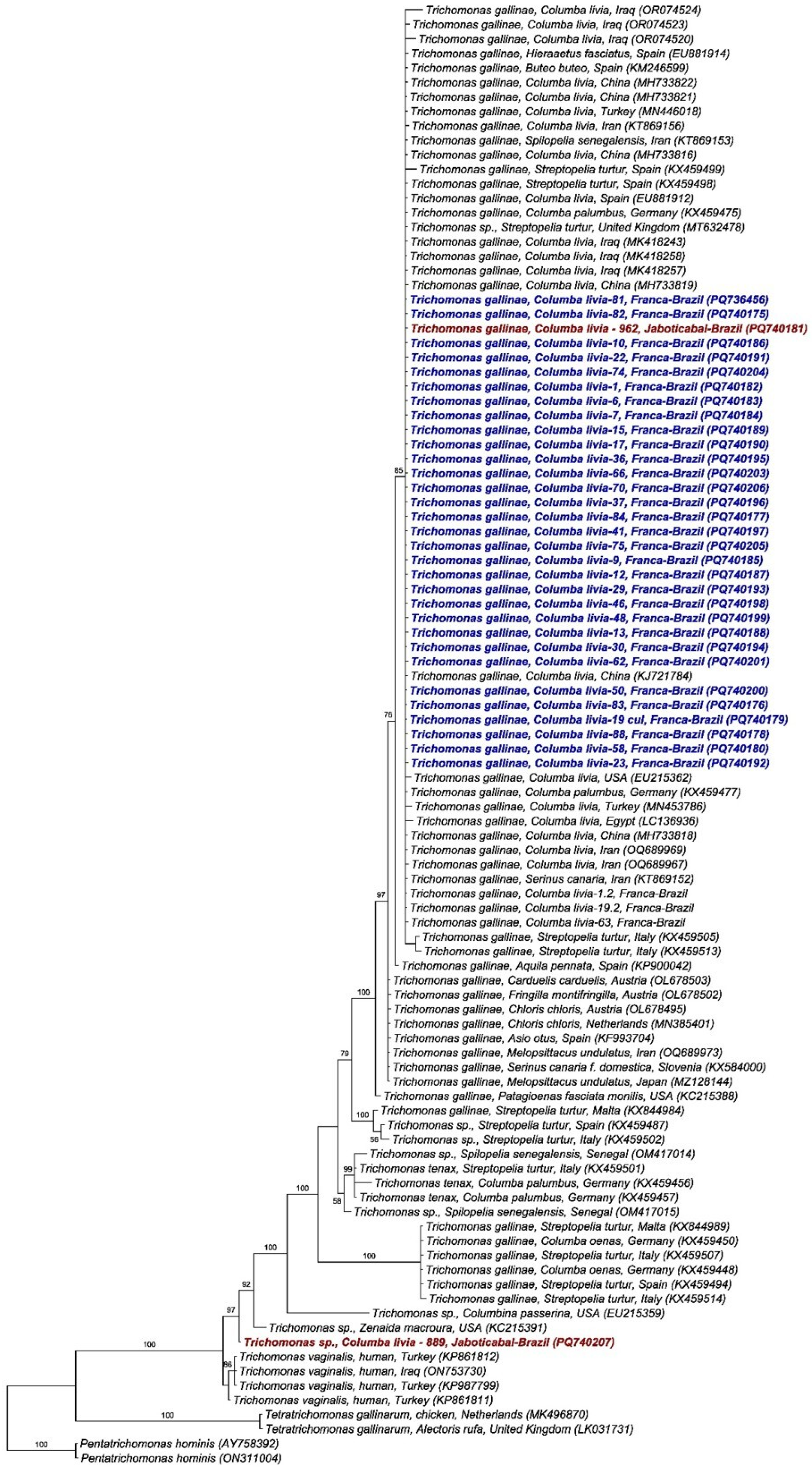
Phylogenetic analysis using the Bayesian method and the TIM2 + G evolutionary model, based on a 399 bp alignment of the ITS1/5.8S/ITS2 regions (Figure 4) of Trichomonas spp., positioned the 34 T. gallinae sequences obtained in this study into two distinct clades. All sequences obtained from rock doves in Franca and one from Jaboticabal clustered in a single clade with posterior probability of 85%, along with T. gallinae sequences detected in Columbiformes sampled in China, Turkey, Iraq, Egypt, USA, Spain, Italy, Germany, and Iran, as well as Accipitridae (H. fasciatus and B. buteo) sampled in Spain and a Passeriforme (S. canaria) from Iran. Meanwhile, one sequence obtained from a pigeon in Jaboticabal was positioned in a distinct clade, closely related to Trichomonas sp. Detected in Zenaida macroura from the USA.
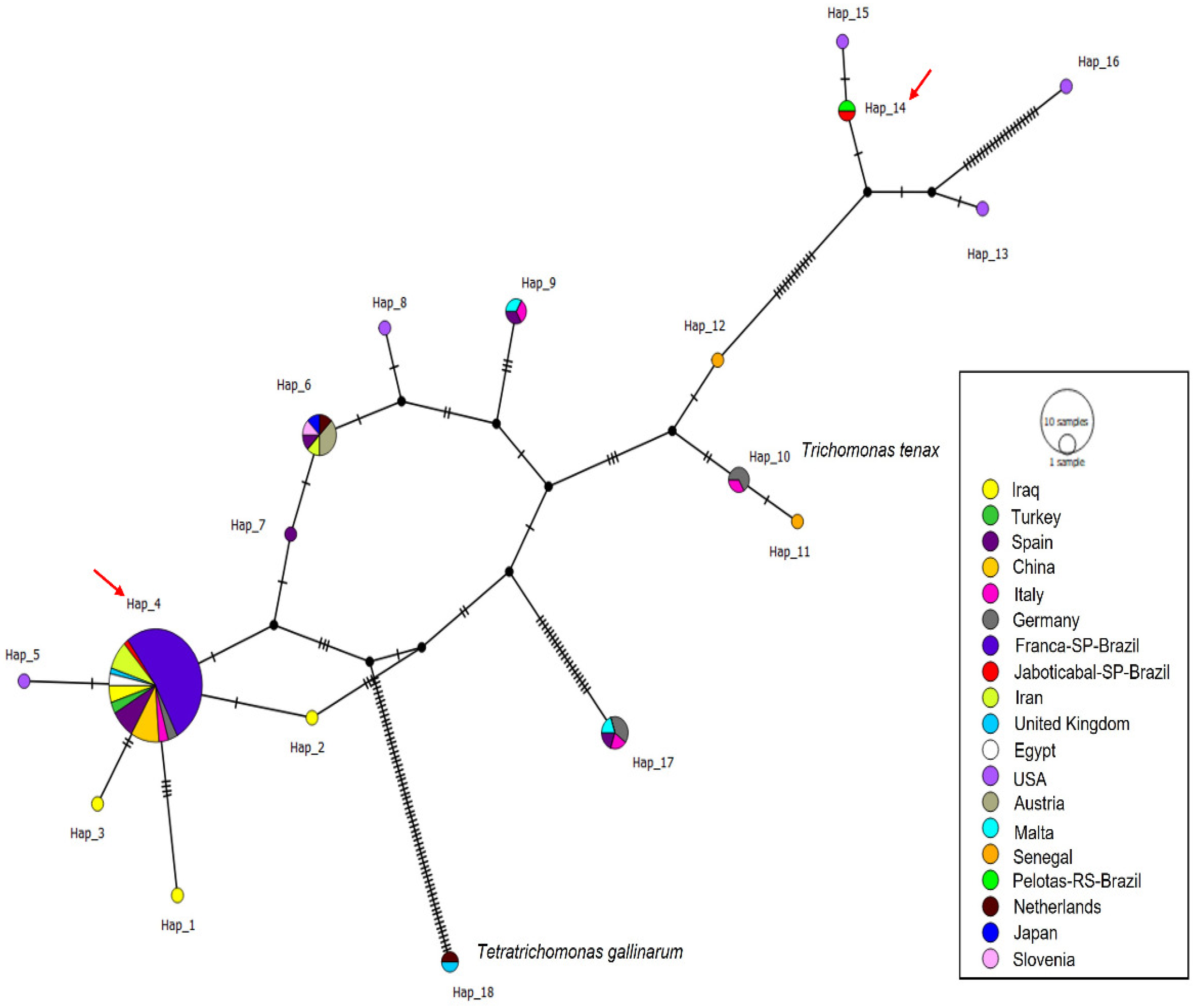
A total of 96 ITS1/5.8S/ITS2 sequences were analyzed (34 sequences obtained in this study and 62 Trichomonas gallinae sequences retrieved from GenBank), resulting in 18 distinct haplotypes. The sequences obtained in this study were distributed into only two haplotypes (haplotypes #4 and #14). Haplotype #4 was separated from haplotypes #2 and #5 by just one mutational event and comprised all sequences detected in Franca, along with one sequence from Jaboticabal and others detected in C. livia, S. turtur, C. palumbus, and H. fasciatus sampled in Iran, China, the UK, Spain, Turkey, and Egypt. Haplotype #14 was separated from haplotype #15 by just one mutational event and included one sequence from Jaboticabal and one detected in Ramphastos dicolorus from Pelotas, RS (Figure 5).
The diversity data are presented in Table 3. Haplotype numbering is locus-specific and does not imply correspondence among ITS1/5.8S/ITS2, Fe-hydrogenase, and 18S rRNA markers.

3.5. PCR and Analysis of the Fe-Hydrogenase Gene Sequences of Trichomonas spp.

For the Fe-hydrogenase gene, of the 76 samples analyzed, 67 (88.15%) were positive and sequenced, all from rock doves sampled in Franca. BLASTn analysis showed that the obtained sequences presented identities ranging from 98% to 100% with sequences of T. gallinae detected in Streptopelia decaocto in Spain (Supplementary Table S2).
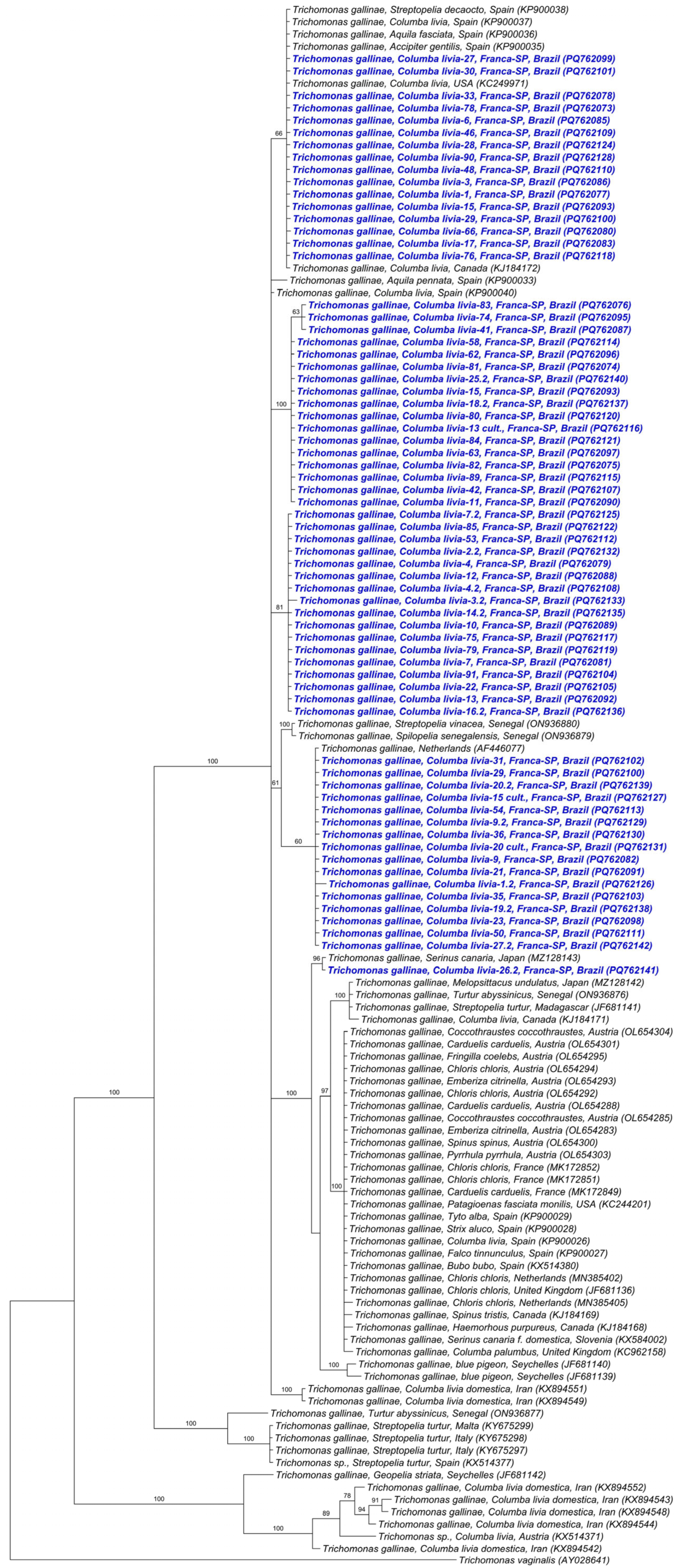
Phylogenetic analysis using the Bayesian method and the TIM2 + I + G evolutionary model from an 879 bp alignment of the Fe-hydrogenase gene (Figure 6) showed that the Fe-hydrogenase sequences obtained in the study were distributed among several clades, with support indices varying from 60% to 100%. Two clades were composed solely of sequences detected in Franca-SP. In one clade, the sequences clustered with sequences detected in Columbiformes (C. livia—USA, Canada, S. decaocto—Spain) and Accipitridae (A. fasciata—Spain, A. gentilis—Spain). Another clade was formed by sequences from the present study and one sequence of T. gallinae detected in the Netherlands. The last clade consisted of a sequence detected in Franca and another detected in a Passeriforme (S. canaria) in Japan.
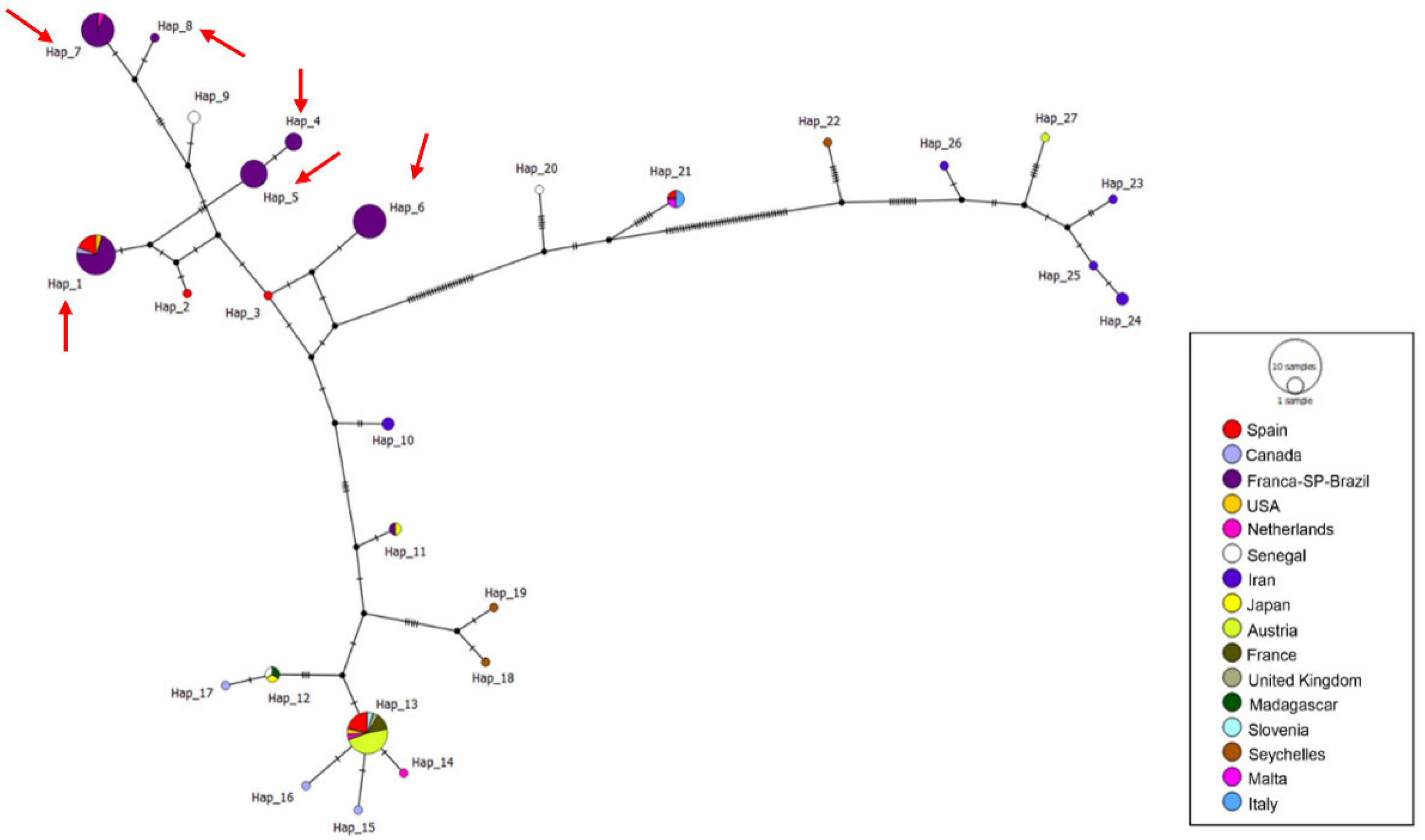
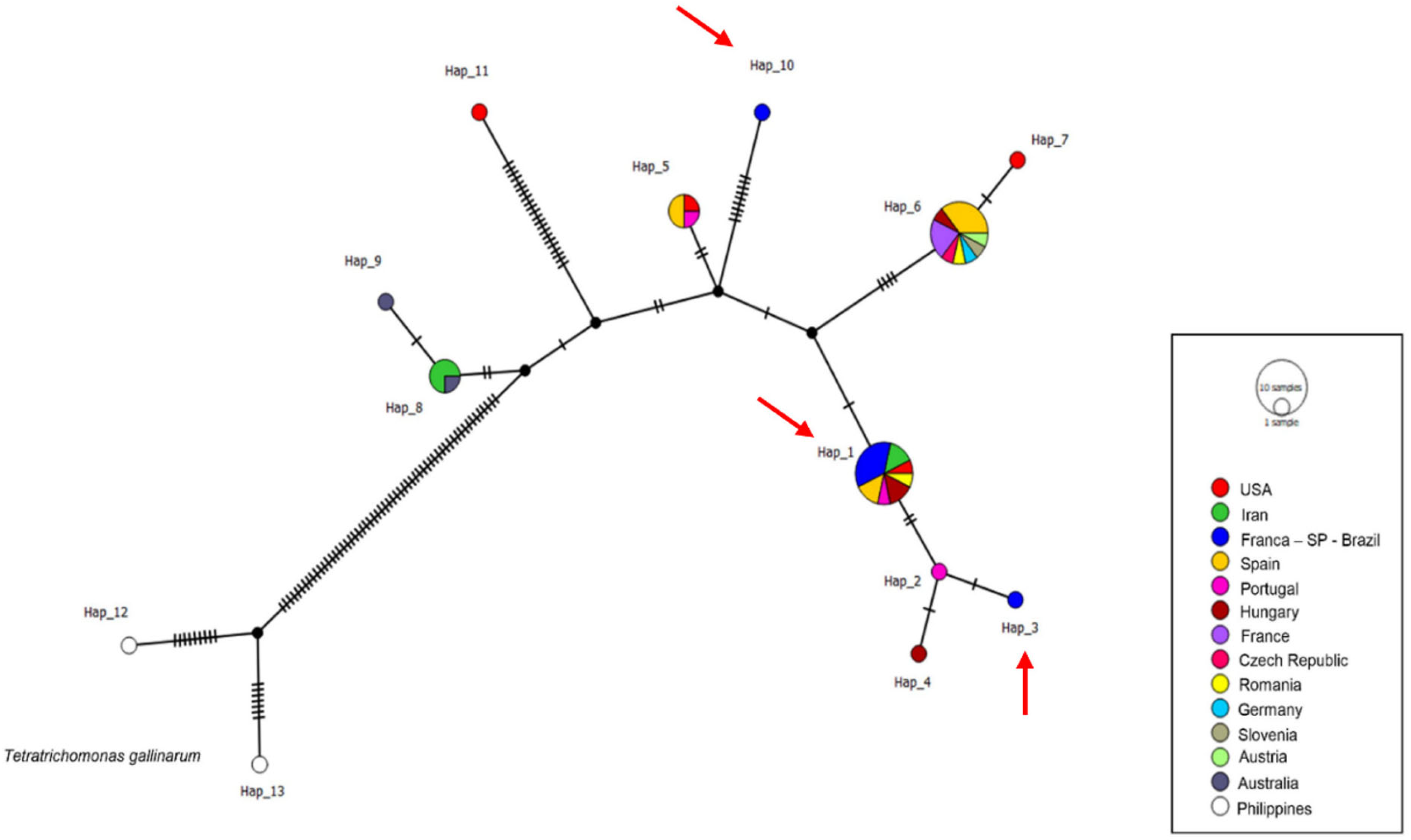
A total of 118 Fe-hydrogenase sequences were included in the haplotype analysis, comprising sequences obtained in this study (n = 60) and additional reference sequences retrieved from GenBank (n = 58). A total of 27 different haplotypes were obtained, and the sequences detected in this study were distributed among six distinct haplotypes: #1, #4, #5, #6, #7, and #8. Three haplotypes (#4, #5, and #6) were composed solely of samples from Franca, SP. Haplotype #1 was formed by 15 sequences from Franca, along with sequences detected in S. decaocto, C. livia, A. gentilis, and R. pigeon in Spain, Canada, and the USA, respectively. Haplotype #7 was formed by 14 sequences obtained from rock doves in Franca and sequences detected in C. chloris from Amsterdam (Figure 7). All haplotypes containing sequences from the present study, except for haplotype #4, which separated from haplotype #5 via a mutational event, were separated from median vectors by a mutational event. Haplotype numbering is locus-specific and does not imply correspondence among ITS1/5.8S/ITS2, Fe-hydrogenase, and 18S rRNA markers.

3.6. PCR and Sequence Analysis of the 18S rRNA Gene of Trichomonas spp.

Of the 76 analyzed samples, 65 (85.5%) were positive for the 18S rRNA gene, all from C. livia sampled in Franca. Of these, only seven samples were suitable for sequencing.
BLASTn analysis showed that the obtained sequences presented identities ranging from 99% to 100% with sequences of T. gallinae detected in E. dove from the USA and C. livia from Portugal (Supplementary Table S2).
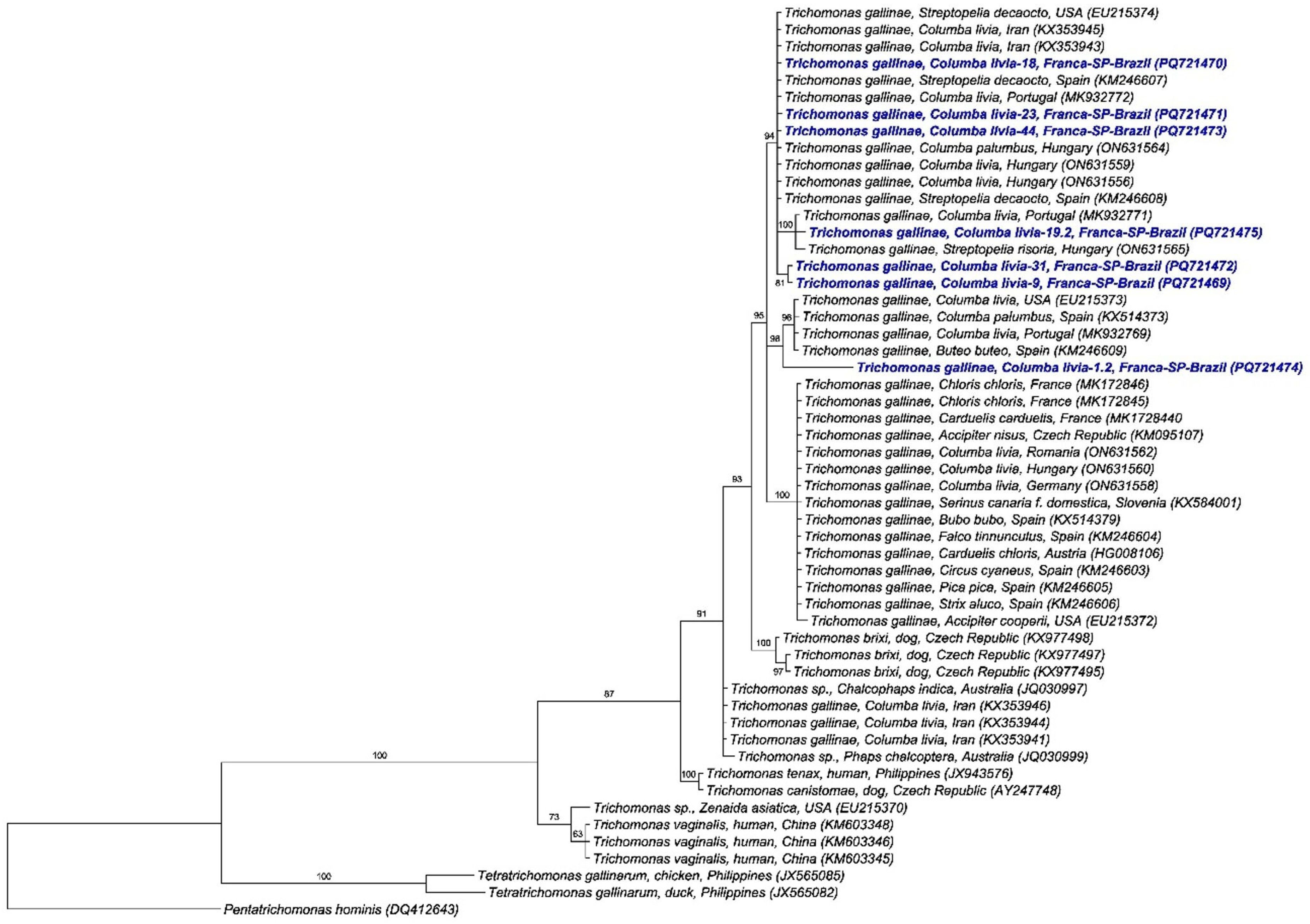
Phylogenetic analysis using the Bayesian method and the GTR + I + G evolutionary model based on a 1424 bp alignment of the 18S rRNA gene (Figure 8) positioned the sequences obtained in this study into four distinct clades, with posterior probability values ranging from 91% to 100%. Two clades were composed solely of sequences detected in rock doves from Franca, with one formed by a single sequence that clustered near a clade containing sequences of T. gallinae detected in C. livia, C. palumbus, and B. buteo in the USA, Spain, and Portugal. The other two clades were composed of sequences from this study alongside sequences detected in Columbiformes from Iran, Hungary, Portugal, Spain, and the USA.
In the haplotype analysis, 45 sequences of the 18S rRNA gene were used (seven obtained in the present study and 38 retrieved from GenBank), which were distributed across 13 different haplotypes. The sequences detected in this study comprised three distinct haplotypes (#1, #3, and #10). Haplotype #1 was composed of 5 sequences from Franca, along with sequences detected in C. livia, S. decaocto, and C. palumbus in Spain, Portugal, Hungary, Romania, the USA, and Iran. Haplotypes #3 and #10 were composed solely of sequences obtained from C. livia sampled in Franca. Haplotype #10 separated from a median vector by nine mutational events (Figure 9). Haplotype numbering is locus-specific and does not imply correspondence among ITS1/5.8S/ITS2, Fe-hydrogenase, and 18S rRNA markers.

4. Discussion

The primary hosts of T. gallinae are Columbiformes, particularly rock doves (C. livia), which likely explain the high number of positive swab samples obtained from the upper digestive tract in this study [31]. Avian trichomoniasis is a parasitic disease affecting a wide range of bird species, including both wild and domestic birds globally. Most of the positive birds in our study shared similar feeding habits, such as frugivory, granivory, or insectivory [32]. Shared feeding behaviors may facilitate food contamination and subsequent transmission of the parasite, especially when infected birds feed near others [33]. This scenario was observed in the present study, as rock doves from Franca, which tested positive, were housed together in a shared enclosure. Notably, these were carrier pigeons with scheduled departures and returns, during which they had direct contact with each other and shared food and water sources. Their transportation for racing events may also promote the spread of the parasite by exposing other birds to contaminated environments.
No clinical signs or visible lesions were observed in the birds sampled in this study. However, trichomoniasis-associated lesions in the upper respiratory tract have previously been documented in passerines such as Haemorhous mexicanus in Bakersfield, California [34,35]. Clinical signs described in infected birds include weight loss, ruffled feathers, vomiting, dyspnea, dysphagia, and diarrhea [36]. Asymptomatic infections are possible and may be associated with avirulent strains or host factors such as immune competence, particularly in adult birds [37].
Previous studies have reported variable prevalence of Trichomonas gallinae in Columbiformes, depending on host species, geographic region, and diagnostic approach. For example, prevalence values of 34.2% in Columba palumbus in Spain [38] and 52.7% in Columba livia domestica in Spain [39] have been described. Lower prevalence rates were reported in mourning doves (Zenaida macroura) in Florida (5.6%) [40] and in white-winged dove (Zenaida asiatica) in the USA (21.3%) [41], whereas localized outbreaks with higher occurrence, such as 33.3% in C. palumbus in southern Spain, have also been documented [42].
In the present study, considering molecular positivity by at least one PCR target (ITS1/5.8S/ITS2, Fe-hydrogenase or 18S rRNA), Trichomonas gallinae showed a high occurrence in C. livia domestica from Franca. This finding likely reflects intense parasite circulation within a captive population, where host density, shared enclosures, and management conditions differ substantially from those of free-living birds, limiting direct quantitative comparisons with field-based prevalence studies.
Most sequences clustered with haplotypes previously reported in Columbiformes, Falconiformes, and Passeriformes, reinforcing the role of Columbiformes as the main reservoirs and sources of infection for other avian orders. The identification of a distinct clade composed of a single sequence from Jaboticabal suggests the occurrence of local genetic variation and potential host-associated structuring.
Broader phylogenetic analyses based on the Fe-hydrogenase and 18S rRNA genes revealed a higher level of genetic diversification compared to ITS-based analysis, with six and four clades identified, respectively. This greater resolution highlights the importance of using multiple molecular markers for a more comprehensive assessment of genetic diversity within the T. gallinae complex. In particular, the higher haplotype variability observed for the Fe-hydrogenase gene may reflect its greater discriminatory power compared to ribosomal or intergenic regions [43].
The presence of multiple haplotypes circulating within the same host population suggests the coexistence of genetically distinct lineages, which may have implications for pathogenicity, transmission dynamics, and host susceptibility.
The presence of diverse haplotypes and clade distributions is well supported in the literature. Gerhold et al. [15] identified 12 different ITS1-5.8S-ITS2 sequence groups in Trichomonas spp., and Grabensteiner et al. [44] highlighted genetic heterogeneity within T. gallinae using Fe-hydrogenase and ITS markers. Further research is necessary to determine whether these genetic variants represent a single species with multiple lineages or distinct species.
Culture has traditionally been considered a reference method for detecting Trichomonas spp. due to its interpretability and historical use, even in asymptomatic hosts [45]. Culture-based detection is considered more sensitive than direct smear microscopy using saline [22,46,47].
According to Diamond [48], the inclusion of fetal bovine serum is essential for parasite growth due to its content of amino acids, fatty acids, and trace elements. In this study, culture was more sensitive than Giemsa-stained smears, consistent with previous reports, [48,49]. However, PCR-based methods demonstrated higher sensitivity than both culture and cytology when applied directly to oropharyngeal swabs. Real-time PCR assays may further improve sensitivity and allow quantification of parasite load, representing a promising approach for future epidemiological studies [50,51].
Incubation time is critical for culture efficacy [45]. In our study, parasite growth was not observed at 24 h but became evident after 48 to 60 h, with optimal detection at 72 h. Without nutrient replenishment, protozoa perished beyond this point. Early attempts at DNA extraction after 72 h culture incubation faced challenges due to freezing, which negatively impacted yield. Immediate DNA extraction after culture led to improved detection, though PCR positivity remained lower than the number of culture-positive samples (72/262; 27.48%). This discrepancy may be attributed to PCR inhibition caused by components in the culture medium.
Only 6 of 262 oropharyngeal smears (2.29%) revealed T. gallinae trophozoites, confirming the low sensitivity of this method compared to culture and molecular diagnostics. The trophozoites observed were morphologically consistent with those described by Mohamed et al. [45].
The higher sensitivity observed for PCR compared to culture and cytology is consistent with previous studies, reinforcing its value as the preferred diagnostic method for detecting Trichomonas gallinae, especially in samples with low parasite load. Additionally, the use of multiple molecular markers allowed a more comprehensive assessment of genetic diversity, revealing the presence of multiple haplotypes circulating within the same host population.
The absence of a washing step prior to DNA extraction may have influenced PCR performance, possibly due to the presence of inhibitors derived from the culture medium or biological material. This factor may partially explain discrepancies between culture positivity and PCR amplification observed in this study. In addition, real-time PCR (qPCR) has been increasingly explored in avian pathogen detection due to its higher sensitivity and ability to quantify nucleic acids. This quantitative capacity may provide important insights into infection dynamics, including the relationship between parasite burden and clinical manifestation, as well as transmission potential in captive populations. Although not employed in the present study, the incorporation of real-time PCR in future investigations could improve diagnostic accuracy and contribute to a better understanding of the epidemiology of T. gallinae.
Besides T. gallinae, other species such as T. vaginalis-like organisms have been reported in doves and owls [14,15], while T. tenax has been identified in European pigeons [52]. T. gypaetinii was found in vultures and eagles [53,54]. Emerging variants such as T. stlaberi and T. canistomae-like have also been detected in S. turtur and A. gentilis in Spain, suggesting even greater diversity among avian trichomonads.
Given the confirmed role of Columbiformes as primary hosts, pigeons serve as the main reservoir for T. gallinae in many regions of São Paulo State [11,12]. Zoos, rehabilitation centers, and wildlife facilities should evaluate the risks associated with releasing birds that have recovered from infection, as asymptomatic carriers can serve as long-term reservoirs, facilitating the dissemination of the parasite.

5. Conclusions

A high occurrence of T. gallinae was found in pigeons in the present study, confirming the role of these animals as primary hosts for the agent. PCR assays showed higher sensitivity in this study, particularly when applied directly to oropharyngeal swabs, proving useful for both detection and molecular characterization of Trichomonas gallinae. The analysis of oropharyngeal swab smears from birds, while quick and easy to perform, showed low sensitivity for diagnosing T. gallinae infection and should not be used as the sole laboratory method for diagnosing trichomoniasis. Although the culture of the parasite in Diamond medium shows good sensitivity for detection, the sample must be kept in this medium and incubated at 37 °C immediately after collection, which hampers the use of this technique in epidemiological studies in remote locations. Different haplotypes of T. gallinae occur in rock doves in the southeastern region of Brazil. The Fe-hydrogenase gene proved to be the molecular marker with the greatest capacity to differentiate T. gallinae haplotypes when compared to the ITS-1-5.8S-ITS2 and 18S rRNA regions.

Supplementary Materials

The following supporting information can be downloaded at https://www.mdpi.com/article/10.3390/pathogens15040428/s1. Video S1: Motile trophozoites of Trichomonas gallinae observed after 48 h of culture. Video S2: Motile trophozoites of Trichomonas gallinae observed after 72 h of culture; Supplementary Table S1. Detection results for β-actin, ITS-1-5.8S-ITS2, 18S rRNA, culture, and Fe-hydrogenase assays. Symbols indicate the following: (+) positive result; (−) negative result; (ND) not done; Supplementary Table S2. BLASTn results of sequences obtained in PCR assays for Trichomonas spp. from birds captured in Ribeirão Preto, Franca, Jundiaí, and Botucatu, Brazil.

Author Contributions

Conceptualization, A.G.P. and methodology, A.G.P., A.C.C.; Visualization, Methodology, Investigation, Conceptualization, A.G.P., A.C.C., S.R.J.S.S., M.C.C.d.S.; Formal analysis, Visualization, Validation, Methodology, Investigation. R.Z.M.; Methodology, Conceptualization, R.B.-S.: Formal analysis, Visualization, Validation, Methodology, Investigation, A.C.C., A.C.C.-S., writing—original draft preparation, A.G.P.; Supervision, Investigation, Formal analysis, Methodology, Conceptualization, M.R.A.; Supervision, Project administration, Investigation, Funding acquisition, Formal analysis, Conceptualization, K.W. All authors have read and agreed to the published version of the manuscript.

Funding

This study was financed, in part, by the São Paulo Research Foundation (FAPESP), Brazil. Process Numbers #2021/05961-5, M.R.A. is a researcher sponsored by CNPq (National Council for Scientific and Technological Development; Productivity Grant to MRA—Process #303701/2021-8) and the Coordenação de Aperfeiçoamento de Pessoal de Nível Superior—Brazil (CAPES)—Finance Code 001.

Institutional Review Board Statement

All animal procedures and management protocols were approved by Institute Chico Mendes for Conservation of Biodiversity (SISBIO number 77538-1, 28 May 2021) and the School of Agricultural and Veterinarian Sciences (FCAV/UNESP) (protocol number 732/21, 12 April 2021).

Informed Consent Statement

Not applicable.

Data Availability Statement

The datasets generated and analyzed during the current study are available on the NCBI Genbank Nucleotide platform (https://www.ncbi.nlm.nih.gov/genbank/ accessed on 12 January 2026) and can be accessed through the following accession numbers: PQ736456, PQ740175–PQ740209 for the ITS1-5.8S rRNA-ITS2 region; PQ762073–PQ762142 for the iron hydrogenase gene; PQ721469–PQ721475 for the 18S rRNA gene.

Acknowledgments

The authors would like to thank the staff of the zoological institutions, wildlife rehabilitation centers, and environmental authorities involved in this study for their cooperation and support during bird sampling. We also acknowledge the Serviço de Patologia de Animais Silvestres (SEPAS), Faculdade de Ciências Agrárias e Veterinárias, UNESP–Jaboticabal, for providing laboratory infrastructure and technical assistance. The authors are grateful to all professionals who contributed to sample collection, animal management, and logistical support throughout the study.

Conflicts of Interest

The authors declare no conflicts of interest.

References

  1. Chou, S.; Hadano, S.; Kojima, A.; Yorisaki, M.; Yasuda, M.; Ike, K.; Tokiwa, T. Genetic characterization of Trichomonas gallinae (Rivolta, 1878) in companion birds in Japan and the genotypical relationship in the Asia region. J. Microbiol. Immunol. Infect. 2022, 55, 527–534. [Google Scholar] [CrossRef] [Scilit] [PubMed]
  2. Mehlhorn, H.; Al-Quraishy, S.; Aziza, A.; Hess, M. Fine structure of the bird parasites Trichomonas gallinae and Tetratrichomonas gallinarum from cultures. Parasitol. Res. 2009, 105, 751–756. [Google Scholar] [CrossRef] [Scilit]
  3. Amin, A.; Bilic, I.; Liebhart, D.; Hess, M. Trichomonads in birds—A review. Parasitology 2014, 141, 733–747. [Google Scholar] [CrossRef] [Scilit] [PubMed]
  4. Gupta, S.D.; Islam, M.; Kamal, T.; Faruque, M.R.A.L.; Faruk, M.S. The confirmatory diagnosis and therapeutic management of canker in pigeons. Res. J. Vet. Pract. 2023, 11, 20–25. [Google Scholar]
  5. Grunenwald, C.; Sidor, I.; Mickley, R.; Dwyer, C.; Gerhold, R. Tetratrichomonas and trichomonas spp.-associated disease in free-ranging common Eiders (Somateria mollissima) from Wellfleet Bay, MA and description of ITS1 region genotypes. Avian Dis. 2018, 62, 117–123. [Google Scholar] [CrossRef] [Scilit]
  6. Forzán, M.J.; Vanderstichel, R.; Melekhovets, Y.F.; Mcburney, S. Trichomoniasis in finches from the Canadian Maritime provinces—An emerging disease. Can. Vet. J. 2010, 51, 391–396. [Google Scholar]
  7. Mcburney, S.; Kelly-Clark, W.K.; Forzan, M.J.; Lawson, B.; Tyler, K.M.; Greenwood, S.J. Molecular characterization of Trichomonas gallinae isolates recovered from the Canadian Maritime provinces’ wild avifauna reveals the presence of the genotype responsible for the European finch trichomonosis epidemic and additional strains. Parasitology 2015, 142, 1053–1062. [Google Scholar] [CrossRef] [Scilit]
  8. Da Silva, D.G.; Barton, E.; Bunbury, N.; Lunness, P.; Bell, D.J.; Tyler, K.M. Molecular identity and heterogeneity of trichomonad parasites in a closed avian population. Infect. Genet. Evol. 2007, 7, 433–440. [Google Scholar] [CrossRef] [Scilit] [PubMed]
  9. De Carli, G.A.; Pansera, M.C.G.; Guerrero, J. Trichomonas gallinae (Rivolta, 1878) Stabler, 1938, no trato digestivo superior de pombos domestics Columba livia no Rio Grande do Sul. Primeiro Registro. Acta Biol. Leopoldensia 1979, 1, 85–95. [Google Scholar]
  10. Tasca, T.; De Carli, G.A. Hemolytic activity of fresh isolates and clones of Trichomonas gallinae. Parasitol. Día 1999, 23, 69–73. [Google Scholar] [CrossRef] [Scilit]
  11. Arenales, A.; Almeida, A.C.O.; Prado, L.F.; Garcia, S.; Luvizotto, M.C.R. Cytopathology in the diagnosis of oral trichomoniasis in a rock pigeon (Columba livia). Braz. J. Vet. Pathol. 2014, 7, 98–99. [Google Scholar] [CrossRef] [Scilit]
  12. Joppert, A.M. Estudo Prospectivo das Causas de Morte de Falconiformes e Strigiformes de Vida Livre no Município de São Paulo. Doctoral Dissertation, Universidade de São Paulo, São Paulo, Brazil, 2007. [Google Scholar]
  13. Godoy, S.N.; Matushima, E.R. A survey of diseases in passeriform birds obtained from illegal wildlife trade in São Paulo City, Brazil. J. Avian Med. Surg. 2010, 24, 199–209. [Google Scholar] [CrossRef] [Scilit]
  14. Ecco, R.; Preis, I.S.; Vilela, D.A.; Luppi, M.M.; Malta, M.C.; Beckstead, R.B.; Stimmelmayr, R.; Gerhold, R.W. Molecular confirmation of Trichomonas gallinae and other parabasalids from Brazil using the 5.8S and ITS-1 rRNA regions. Vet. Parasitol. 2012, 190, 36–42. [Google Scholar] [CrossRef] [Scilit] [PubMed]
  15. Gerhold, R.W.; Yabsley, M.J.; Smith, A.J.; Ostergaard, E.; Mannan, W.; Cann, J.D.; Fischer, J.R. Molecular characterization of the Trichomonas gallinae morphologic complex in the United States. J. Parasitol. 2008, 94, 1335–1341. [Google Scholar] [CrossRef] [Scilit] [PubMed]
  16. Bruni, M.P.; Echenique, J.V.Z.; Dos Santos, C.C.; de Macedo, M.R.P.; Bandarra, P.M.; Timm, C.D.; Pereira Schild, A.L.; Lopes Ruas, J.; Pereira Soares, M.; da Rosa Farias, N.A. The raptor Chimango Caracara (Milvago chimango) (Aves: Falconiformes)—A new host for Trichomonas gallinae (Protozoa: Trichomonadidae). Int. J. Parasitol. Parasites Wildl. 2019, 10, 310–313. [Google Scholar] [CrossRef] [Scilit]
  17. Soulsby, R.L.; Smallman, J.V. A Direct Method of Calculating Bottom Orbital Velocity Under Waves; The Ministry of Agriculture, Fisheries and Food: London, UK, 1986. [Google Scholar]
  18. Van Borm, S.; Steensels, M.; Ferreira, H.L.; Boschmans, M.; DE Vriese, J.; Lambrecht, B.; Van Den Berg, T. Its Application to Avian Influenza Diagnosis and Quantification. Avian Dis. 2007, 50, 213–220. [Google Scholar] [CrossRef] [Scilit] [PubMed]
  19. Alabí Córdova, A.S.; Fecchio, A.; Calchi, A.C.; Dias, C.M.; Machado, R.Z.; André, M.R. Molecular evidence of Bartonella spp. in tropical wild birds from the Brazilian Pantanal, the largest wetland in South America. Vet. Res. Commun. 2024, 48, 1631–1640. [Google Scholar] [CrossRef] [Scilit]
  20. Felleisen, R.S.J. Comparative sequence analysis of 5·8S rRNA genes and internal transcribed spacer (ITS) regions of trichomonadid protozoa. Parasitology 1997, 115, 111–119. [Google Scholar] [CrossRef] [Scilit]
  21. Ganas, P.; Jaskulska, B.; Lawson, B.; Zadravec, M.; Hess, M.; Bilic, I. Multi-locus sequence typing confirms the clonality of Trichomonas gallinae isolates circulating in European finches. Parasitology 2014, 141, 652–661. [Google Scholar] [CrossRef] [Scilit]
  22. Lawson, B.; Cunningham, A.A.; Chantrey, J.; Hughes, L.A.; John, S.K.; Bunbury, N.; Bell, D.J.; Tyler, K.M. A clonal strain of Trichomonas gallinae is the aetiologic agent of an emerging avian epidemic disease. Infect. Genet. Evol. 2011, 11, 1638–1645. [Google Scholar] [CrossRef] [Scilit]
  23. Ewing, B.; Green, P. Base-calling of automated sequencer traces using phred. II. Error probabilities. Genome Res. 1998, 8, 186–194. [Google Scholar] [CrossRef] [Scilit] [PubMed]
  24. Hall, T.A. BioEdit: A user-friendly biological sequence alignment editor and analysis program for Windows 95/98/NT. Nucleic Acids Symp. Ser. 1999, 41, 95–98. [Google Scholar]
  25. Ronquist, F.; Huelsenbeck, J.P. MrBayes 3: Bayesian phylogenetic inference under mixed models. Bioinformatics 2003, 19, 1572–1574. [Google Scholar] [CrossRef] [Scilit]
  26. Miller, M.A.; Pfeiffer, W.; Schwartz, T. The CIPRES science gateway: A community resource for phylogenetic analyses. In Proceedings of the 2011 TeraGrid Conference: Extreme Digital Discovery, Salt Lake City, UT, USA, 18–21 July 2011; pp. 1–8. [Google Scholar]
  27. Darriba, D.; Taboada, G.L.; Doallo, R.; Posada, D. jModelTest 2: More models, new heuristics and parallel computing. Nat. Methods 2012, 9, 772. [Google Scholar] [CrossRef] [Scilit]
  28. Stover, B.C.; Muller, K.F. TreeGraph 2: Combining and visualizing evidence from different phylogenetic analyses. BMC Bioinform. 2010, 11, 7. [Google Scholar] [CrossRef] [Scilit]
  29. Librado, P.; Rozas, J. DnaSP v5: A software for comprehensive analysis of DNA polymorphism data. Bioinformatics 2009, 25, 1451–1452. [Google Scholar] [CrossRef] [Scilit] [PubMed]
  30. Leigh, J.W.; Bryant, D. Popart: Full-feature software for haplotype network construction. Methods Ecol. Evol. 2015, 6, 1110–1116. [Google Scholar] [CrossRef] [Scilit]
  31. McDougald, L.R. Diseases of Poultry, 10th ed.; Iowa State University Press: Ames, IA, USA, 1991. [Google Scholar]
  32. Girard, Y.A.; Rogers, K.H.; Woods, L.W.; Chouicha, N.; Miller, W.A.; Johnson, C.K. Dual-pathogen etiology of avian trichomonosis in a declining band-tailed pigeon population. Infect. Genet. Evol. 2014, 24, 146–156. [Google Scholar] [CrossRef] [Scilit]
  33. Timm, C.D.; Timm, V.F. Avifauna Gaúcha: Guia de Identificação; USEB: Pelotas, Brazil, 2021. [Google Scholar]
  34. St Leger, J.; Shivaprasad, H.L. Passerine protozoal sinusitis: An infection you should know about. In Proceedings of the Association of Avian Veterinarians (AAV) Annual Conference and Expo, St Paul, MN, USA, 26–28 August 1998; pp. 157–160. [Google Scholar]
  35. Dorrestein, G.M. Bacterial and parasitic diseases of passerines. Vet. Clin. N. Am. Exot. Anim. Pract. 2009, 12, 433–451. [Google Scholar] [CrossRef] [Scilit]
  36. Narcisi, E.M.; Sevoian, M.; Honigberg, B.M. Pathologic changes in pigeons infected with a virulent Trichomonas gallinae strain (Eiberg). Avian Dis. 1991, 35, 55–61. [Google Scholar] [CrossRef] [Scilit]
  37. Forrester, D.J.; Foster, D.W. Trichomonosis. In Parasitic Diseases in Wild Birds; Atkinson, C.T., Thomas, N.J., Hunter, D.B., Eds.; Blackwell Publishing: Hoboken, NJ, USA, 2008; pp. 120–153. [Google Scholar]
  38. Villanúa, D.; Höfle, U.; Pérez-Rodríguez, L.; Gortázar, C. Trichomonas gallinae in wintering common wood pigeons Columba palumbus in Spain. IBIS 2006, 148, 641–648. [Google Scholar] [CrossRef] [Scilit]
  39. Sansano-Maestre, J.; Garijo-Toledo, M.M.; Gómez-Muñoz, M.T. Prevalence and genotyping of Trichomonas gallinae in pigeons and birds of prey. Avian Pathol. 2009, 38, 201–207. [Google Scholar] [CrossRef] [Scilit]
  40. Schulz, J.H.; Bermudez, A.J.; Millspaugh, J.J. Monitoring presence and annual variation of trichomoniasis in mourning doves. Avian Dis. 2005, 49, 387–389. [Google Scholar] [CrossRef] [Scilit] [PubMed]
  41. Conti, J.A.; Forrester, D.J. Interrelationships of parasites of white-winged doves and mourning doves in Florida. J. Wildl. Dis. 1981, 17, 529–536. [Google Scholar] [CrossRef] [Scilit] [PubMed]
  42. Höfle, U.; Gortázar, C.; Ortiz, J.A.; Knispel, B.; Kaleta, E.F. Outbreak of trichomoniasis in a woodpigeon (Columba palumbus) wintering roost. Eur. J. Wildl. Res. 2004, 50, 73–77. [Google Scholar] [CrossRef] [Scilit]
  43. Alrefaei, A.F.; Gerhold, R.W.; Nader, J.L.; Bell, D.J.; Tyler, K.M. Improved subtyping affords better discrimination of Trichomonas gallinae strains and suggests hybrid lineages. Infect. Genet. Evol. 2019, 73, 234–241. [Google Scholar] [CrossRef] [Scilit]
  44. Grabensteiner, E.; Bilic, I.; Kolbe, T.; Hess, M. Molecular analysis of clonal trichomonad isolates indicate the existence of heterogenic species present in different birds and within the same host. Vet. Parasitol. 2010, 172, 53–64. [Google Scholar] [CrossRef] [Scilit]
  45. Mohamed, H.M.; Saad, A.S.; Khalifa, M.M.; Abdel-Maogood, S.Z.; Awadalla, S.M.; Mousa, W.M. Detection and molecular characterization of Trichomonas gallinae recovered from domestic pigeons in Egypt. Parasitol. Res. 2023, 122, 257–263. [Google Scholar] [CrossRef] [Scilit]
  46. Fouts, A.C.; Kraus, S.J. Trichomonas vaginalis: Reevaluation of its clinical presentation and laboratory diagnosis. J. Infect. Dis. 1980, 141, 137–143. [Google Scholar] [CrossRef] [Scilit]
  47. Cooper, J.E.; Petty, S.J. Trichomoniasis in free-living goshawks (Accipiter gentilis gentilis) from Great Britain. J. Wildl. Dis. 1988, 24, 80–87. [Google Scholar] [CrossRef] [Scilit] [PubMed]
  48. Raza, A.; Qamar, M.F.; Rubtsova, N.; Saneela, S. Pathogenicity and diagnostic sensitivity of culture media for identifcation of Trichomonas gallinae in Domestic Pigeons of Lahore, Pakistan. J. Protozool. Res. 2018, 28, 11–21. [Google Scholar]
  49. Saikia, M.; Bhattacharjee, K.; Sarmah, P.C.; Deka, D.K.; Upadhyaya, T.N.; Konch, P. Prevalence and pathology of Trichomonas gallinae in domestic pigeon (Columba livia domestica) of Assam, India. Indian J. Anim. Res. 2021, 55, 84–89. [Google Scholar] [CrossRef] [Scilit]
  50. Diamond, L.S. A comparative study of 28 culture media for Trichomonas gallinae. Exp. Parasitol. 1954, 3, 251–258. [Google Scholar] [CrossRef] [Scilit] [PubMed]
  51. Kocan, R.M.; Knisley, J.O. Challenge infection as a means of determining the rate of disease resistant Trichomonas gallinae-free birds in a population. J. Wildl. Dis. 1970, 6, 13–15. [Google Scholar] [CrossRef] [Scilit]
  52. Lennon, R.J.; Dunn, J.C.; Stockdale, J.E.; Goodman, S.J.; Morris, A.J.; Hamer, K.C. Trichomonad parasite infection in four species of Columbidae in the UK. Parasitology 2013, 140, 1368–1376. [Google Scholar] [CrossRef] [Scilit]
  53. Kelly-Clark, W.K.; McBurney, S.; Forzán, M.J.; Desmarchelier, M.; Greenwood, S.J. Detection and characterization of a Trichomonas isolate from a rehabilitated bald eagle (Haliaeetus leucocephalus). J. Zoo Wildl. Med. 2013, 44, 1123–1126. [Google Scholar] [CrossRef] [Scilit]
  54. Martínez-Díaz, R.A.; Ponce-Gordo, F.; Rodríguez-Arce, I.; del Martínez-Herrero, M.C.; González, F.G.; Molina-López, R.Á.; Gómez-Muñoz, M.T. Trichomonas gypaetinii n. sp., a new trichomonad from the upper gastrointestinal tract of scavenging birds of prey. Parasitol. Res. 2015, 114, 101–112. [Google Scholar] [CrossRef] [Scilit]
Figure 1. Collection sites highlighting Latin America, Brazil, and the state of São Paulo, with emphasis on the municipalities of Franca, Ribeirão Preto, Jaboticabal, Jundiaí, and Botucatu.
Pathogens 15 00428 g001
Figure 2. Microscopic examination of fresh culture of Trichomonas sp., (40×), at the arrow showing pseudocysts of Trichomonas sp.
Pathogens 15 00428 g002
Figure 3. Trophozoites of Trichomonas sp. (black arrow) in Giemsa stained preparations (40× (A) and 100× (B)).
Pathogens 15 00428 g003
Figure 4. Phylogenetic analysis inferred by the Bayesian method and TIM2 + G evolutionary model of a 399 bp alignment of ITS1/5.8S/ITS2 sequences of Trichomonas spp. The sequences obtained in the present study are highlighted in color, with red corresponding to sequences obtained from birds sampled in Jaboticabal-SP, and blue to those obtained in Franca-SP. Pentatrichomonas hominis was used as an outgroup.
Pathogens 15 00428 g004
Figure 5. Haplotype network based on the ITS1-5.8S-ITS2 regions of Trichomonas spp. The colors represent the locations from which the sequences used in the analyses were detected, the black dots represent median vectors (inferred ancestral nodes), and the vertical black lines represent the number of mutational events between each haplotype. Red arrows indicate the haplotypes (#4 and #14) that contain sequences obtained in this study.
Pathogens 15 00428 g005
Figure 6. Phylogenetic analysis inferred by the Bayesian method and TIM2 + I + G evolutionary model of an alignment of 879 bp of Fe-hydrogenase gene sequences from Trichomonas spp. The sequences obtained in the present study are highlighted in blue. Trichomonas vaginalis was used as an outgroup.
Pathogens 15 00428 g006
Figure 7. Haplotype network based on the Fe-hydrogenase gene of Trichomonas spp. The colors represent the locations from which the sequences used in the analyses were detected, the black dots represent median vectors (inferred ancestral nodes), and the vertical black lines represent the number of mutational events between one haplotype and another. Red arrows indicate the haplotypes (#1, #4 to #8) that contain sequences obtained in this study.
Pathogens 15 00428 g007
Figure 8. Phylogenetic analysis inferred by the Bayesian method and GTR + I + G evolutionary model of a 1424 bp alignment of 18S rRNA gene sequences of Trichomonas spp. The sequences obtained in the present study are highlighted in blue. Pentatrichomonas hominis was used as an outgroup.
Pathogens 15 00428 g008
Figure 9. Haplotype network based on the 18S rRNA gene of Trichomonas spp. The colors represent the locations from which the sequences used in the analyses were detected, the black dots represent median vectors (inferred ancestral nodes), and the vertical black lines represent the number of mutational events between one haplotype and another. Red arrows indicate the haplotypes (#1, #3 and #10) that contain sequences obtained in this study.
Pathogens 15 00428 g009
Table 1. Description of sampled birds from different orders that were sampled in Jaboticabal, Franca, Ribeirão Preto, Botucatu and Jundiaí (SP, Brazil) for detection of Trichomonas spp. in oropharyngeal swabs.
CityQuantityOrderQuantity/Species
Jaboticabal12Columbiformes12 Columba livia domestica
3Piciformes3 R. toco
1Strigiformes1 Tyto furcata
2Psittaciformes1 A. arauna,
1 F. xanthopterygius
1Charadriiformes1 Vanellus chilensis
Franca121Columbiformes121 Columba livia domestica
Ribeirão Preto5Accipitriformes4 R. magnirostris e
1 F. sparverius
5Cathartiformes5 C. atratus
10Columbiformes10 Columbina talpacoti
1Nyctibiiformes1 Nyctibius griseus
10Passeriformes3 Saltator similis, 1 Cacicus cela, 3 Turdus rufiventris, 1 Thraupis sayaca, 1 Mimus saturninus, 1 Pitangus sulphuratus
2Piciformes2 R. toco
8Psittaciformes1 A. arauna, 3 A. auricapillus, 1 Eupsittula aurea, 3 Pyrrhura frontalis
10Strigiformes5 T. furcata, 4 A. cunicularia, 1 Megascops choliba
Botucatu19Passeriformes1 Sicalis flaveola, 2 Oryzoborus maximiliani, 2 Cyanocompsa brissonii, 2 C. cucullatus,
1 O. angolensis, 1 S. similis, 1 Gnorimopsar chopi, 3 Sporophila caerulescens, 2 P. sulphuratus, 2 Tachornis squamata, 1 Thraupis sayaca and 1 Guira guira
2Columbiformes2 Zenaida auriculata
3Piciformes3 R. toco
1Psittaciformes1 Brotogeris chiriri
1Anseriformes1 Anas bahamensis
6Strigiformes4 Ciccaba virgata, 1 Tyto furcata 1 Athene cunicularia
Jundiaí11Piciformes11 R. toco
1Nyctibiiformes1 Nyctibius griseus
10Strigiformes6 C. virgata, 2 A. cunicularia, 1 Asio clamator, 1 P. perspicillata
11Columbiformes2 Zenaida auriculata, 3 C. livia, 6 Columbina talpacoti
12Falconiformes2 C. plancus, 7 Falco sparverius, 1 Ictinia plumbea, 2 Rupornis magnirostris
4Cathartiformes4 C. atratus
2Pelecaniformes1 Tigrisoma lineatum, 1 Ardea cocoi
1Tinamiformes1 Rhynchotus rufescens
6Psittaciformes4 Psittacara leucophthalmus,
1 Brotogeris tirica,
1 Amazona aestiva
Table 2. Description of the primers and thermal conditions used in the conventional PCR assays for the endogenous gene and Trichomonas spp.
Primer NamesPrimer SequencesThermal ConditionsReferencesFragment Size
Endogenous gene
β-actin-F5′-CCTCATGAAGATCCTGACAGA3′95 °C for 5 min, followed by 35 cycles at 95 °C for 30 s, 54 °C for 30 s and 72 °C for 1 min and final extension at
72 °C for 5 min
[18]700 bp
β-actin-R5′-TCTCCTGCTCYAAYTCCA-3′
ITS1/5.8S/ITS2
TRF1(5′-TGCTTCAGCTCAGCGGGTCTTCC-3′)94 °C for 2 min followed by 40 cycles at 94 °C for 20 s, 66 °C for 20 s and 72 °C for 30 s, final extension at 72 °C for 5 min[20]369 bp
TRF2(5′-CGGTAGGTGAACCTGCCGTTGG-3′)
18S rRNA
Hm-Long-f(5′-AGGAAGCACACTATGGTCATAG-3′)95 °C for 15 min, followed by 40 cycles of 94 °C for 30 s, 55 °C for 1 min and 72 °C for 2 min, final extension at 72 °C for 10 min[21]1500 bp
Hm-Long-r(5′-CGTTACCTTGTTACGACTTCTCCTT-3′)
Fe-hydrogenase
TrichhydFOR(5′-GTTTGGGATGGCCTCAGAAT-3′)95 °C for 10 min followed by 36 cycles at 96° for 5 s, 53° for 3 s and 68° for 15 s, and final extension at 72 °C for 10 s.[22]1000 bp
TrichhydREV(5′-AGCCGAAGATGTTGTCGAAT-3′)
Table 3. Genetic diversity and polymorphism of ITS1/5.8S/ITS2, Fe-hydrogenase and 18S rRNA sequences of Trichomonas sp.
Molecular MarkerbpNVSGC%Hhd (Mean ± SD)π (Mean ± SD)K
ITS1-5.8S-ITS2 intergenic region3219610034180.588 ± 0.0590.03375 ± 0.007059.75417
Fe-hydrogenase85111810753.9270.893 ± 0.0130.02159 ± 0.0030517.40417
18S rRNA1351459848.9130.804 ± 0.0400.00927 ± 0.0030211.80
N = number of sequences analyzed; VS = number of site variables; GC% = G + C content; h = number of haplotypes; hd = haplotype diversity; SD = standard deviation; π = nucleotide diversity (per site); K = number of nucleotide differences.
Disclaimer/Publisher’s Note: The statements, opinions and data contained in all publications are solely those of the individual author(s) and contributor(s) and not of MDPI and/or the editor(s). MDPI and/or the editor(s) disclaim responsibility for any injury to people or property resulting from any ideas, methods, instructions or products referred to in the content.