Dysregulated IL-7/IL-7R-CD132 Axis and Intestinal Microsporidiosis in Crohn’s Disease

 , , , , , ,
  and
and
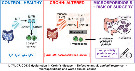
Abstract
1. Introduction
2. Materials and Methods
2.1. Study Population
2.2. Tissue Sampling and Processing
2.3. Encephalitozoon Cuniculi Antigen and Serology
2.4. Serum IL-7 Quantification
2.5. Cell Isolation and Flow Cytometry
2.6. Gene Expression Analysis (IL-7, CD127, CD132)
2.7. Protein Expression of IL-7 and Caspase-3
2.8. Statistical Analysis
3. Results
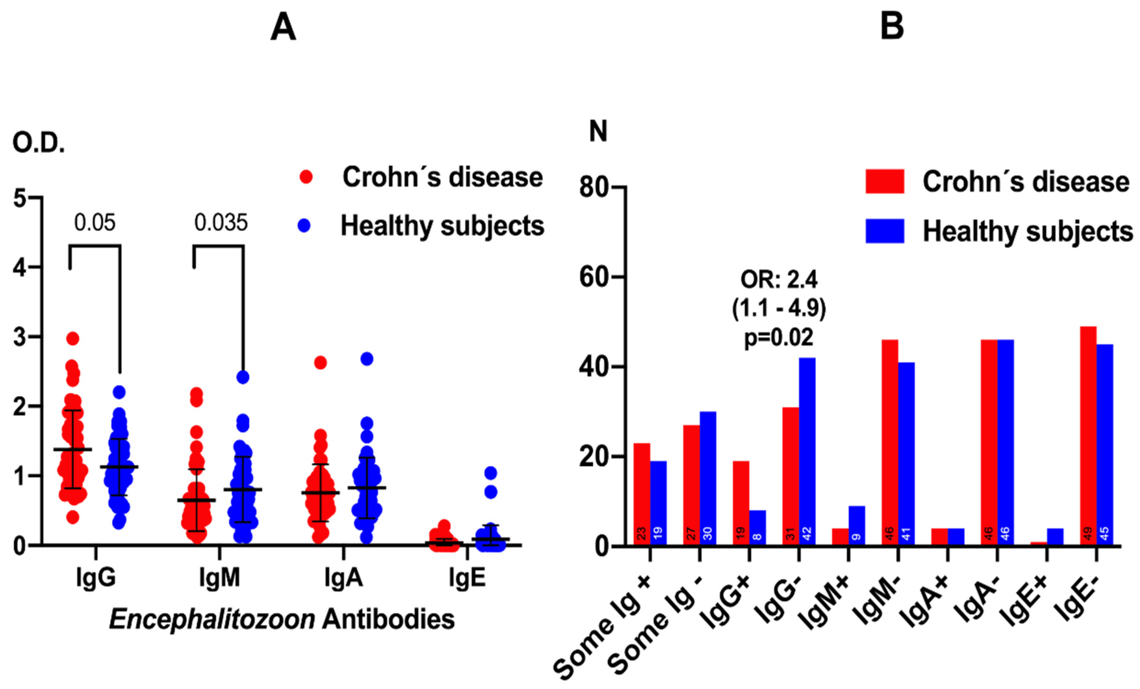
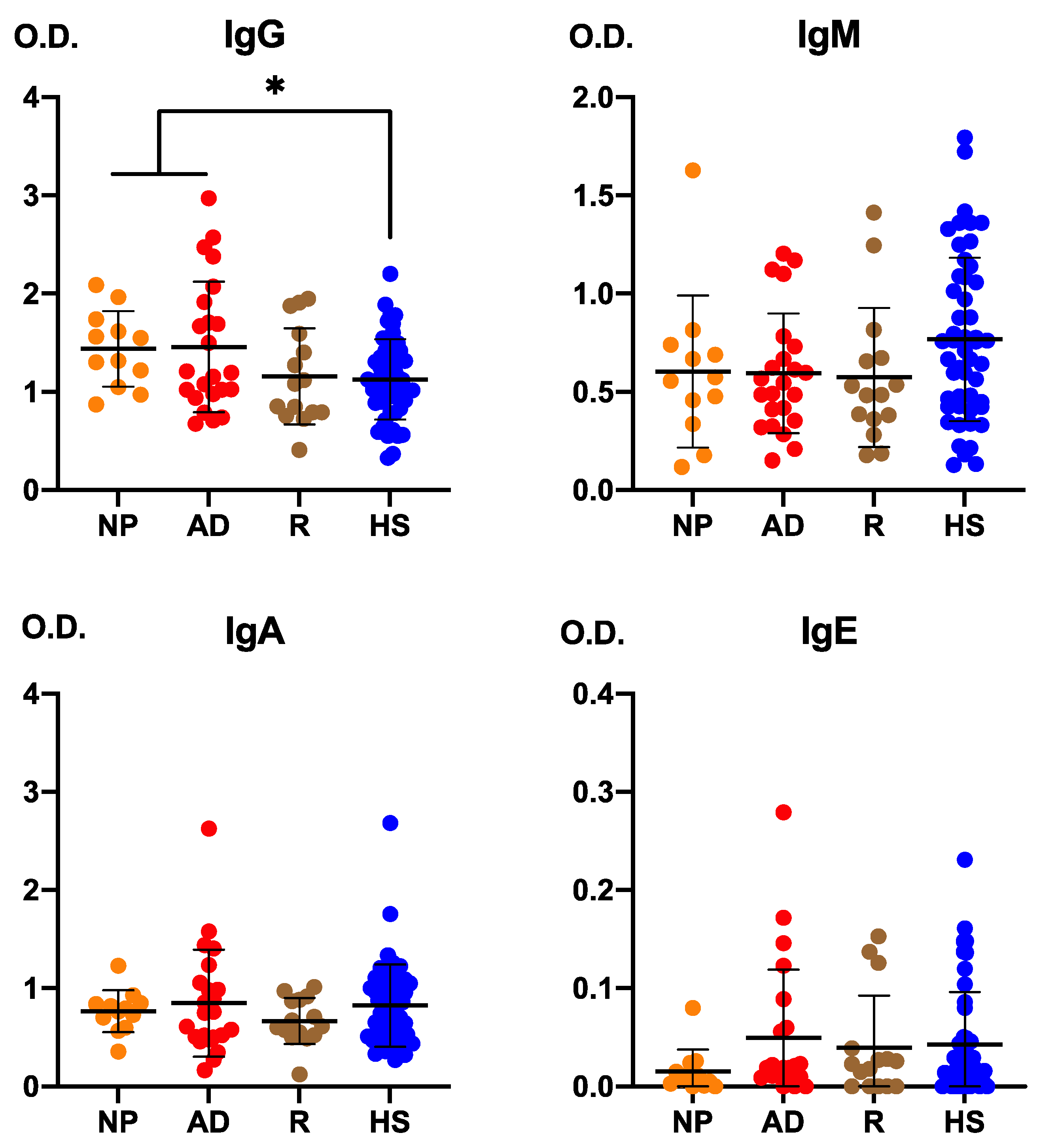
3.1. Anti-Encephalitozoon cuniculi Antibodies in Patients with Crohn Disease and Healthy Subjects
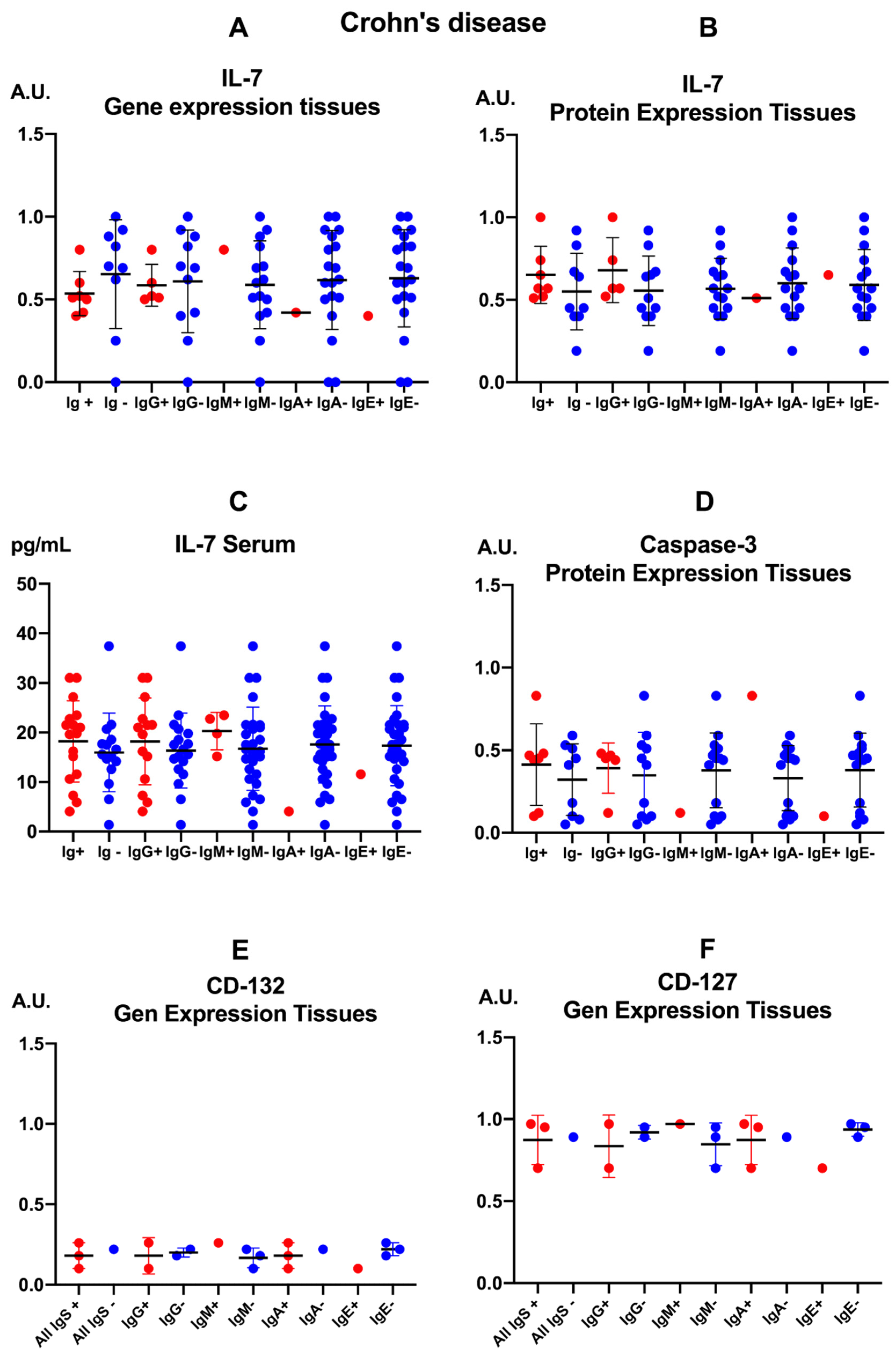
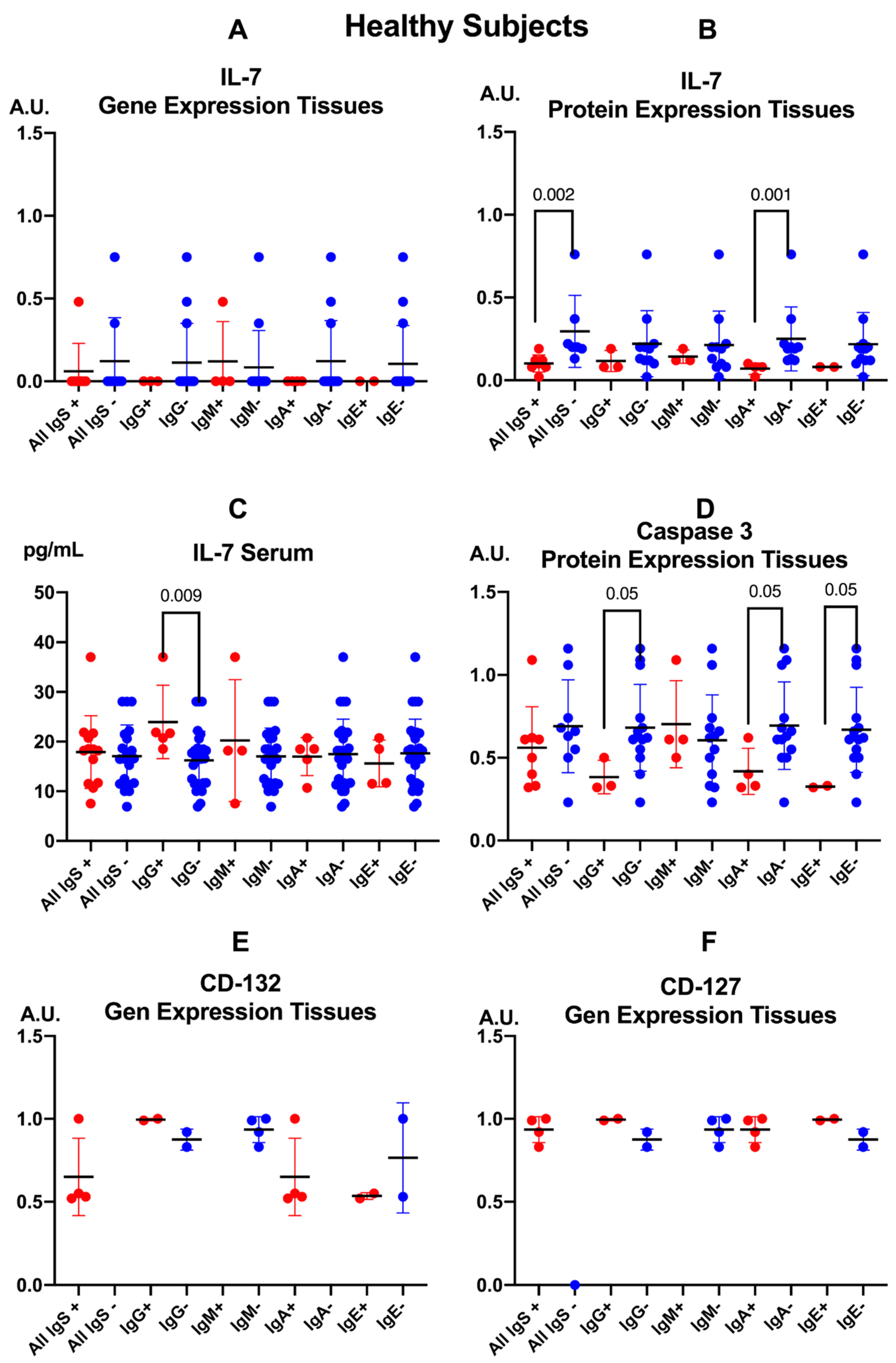
3.2. Assessment of IL-7, Its Receptor and Caspase-3 According to Positivity of Anti-Encephalitozoon cuniculi Antibodies
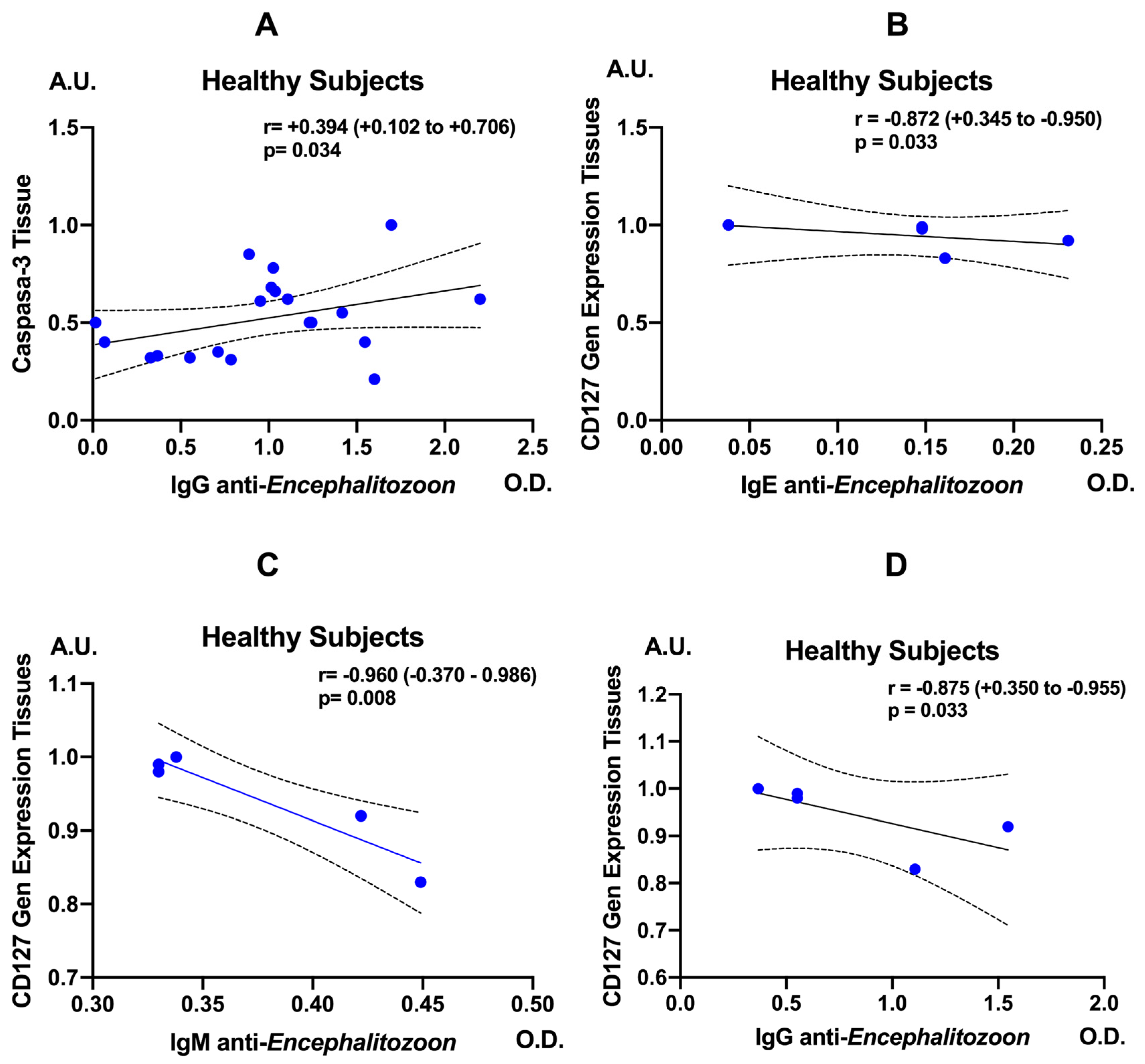
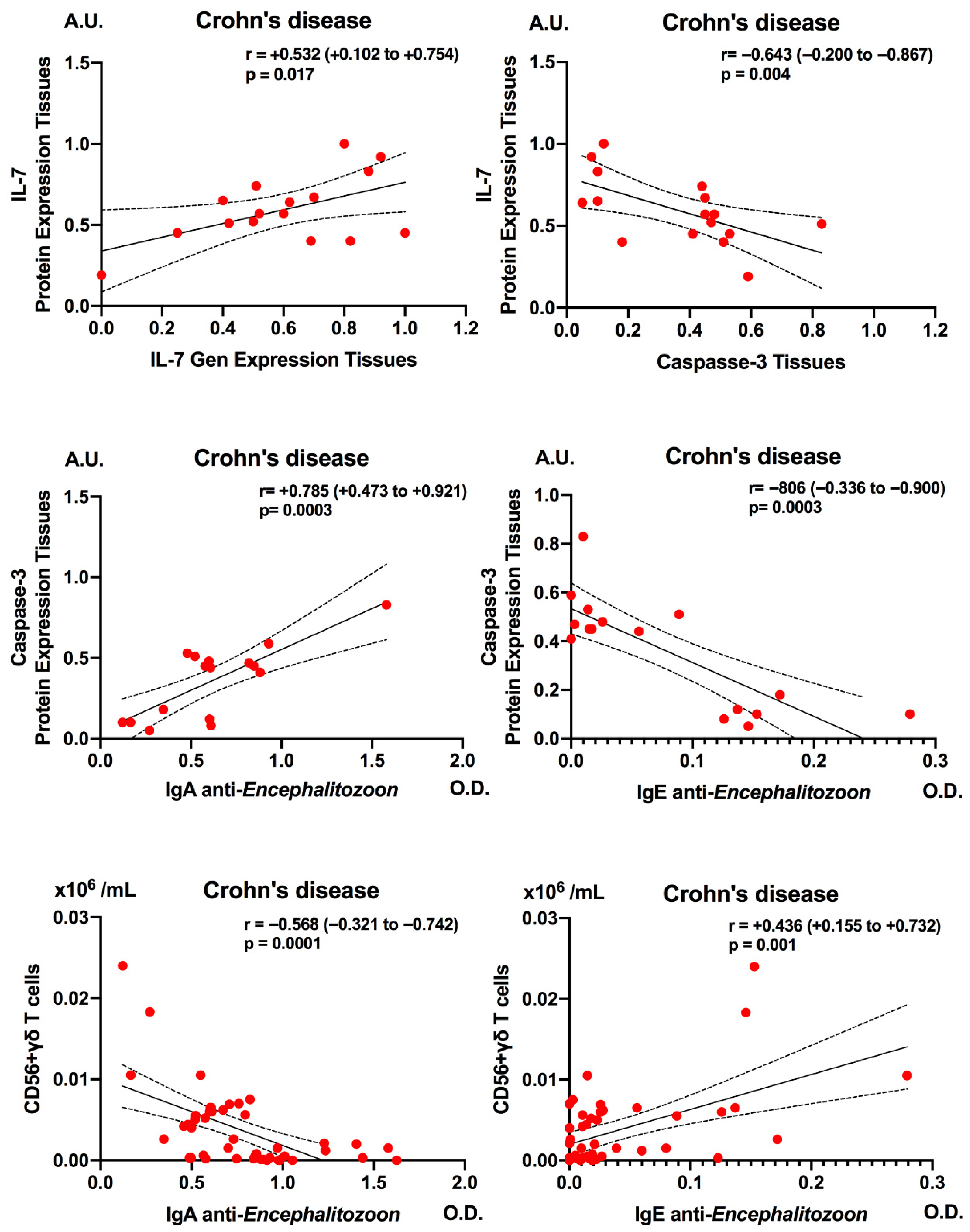
3.3. Correlations Caspase-3, IL-7 and CD3+ CD56+ γδ T-Cell Subsets with Anti-Encephalitozoon cuniculi Antibodies
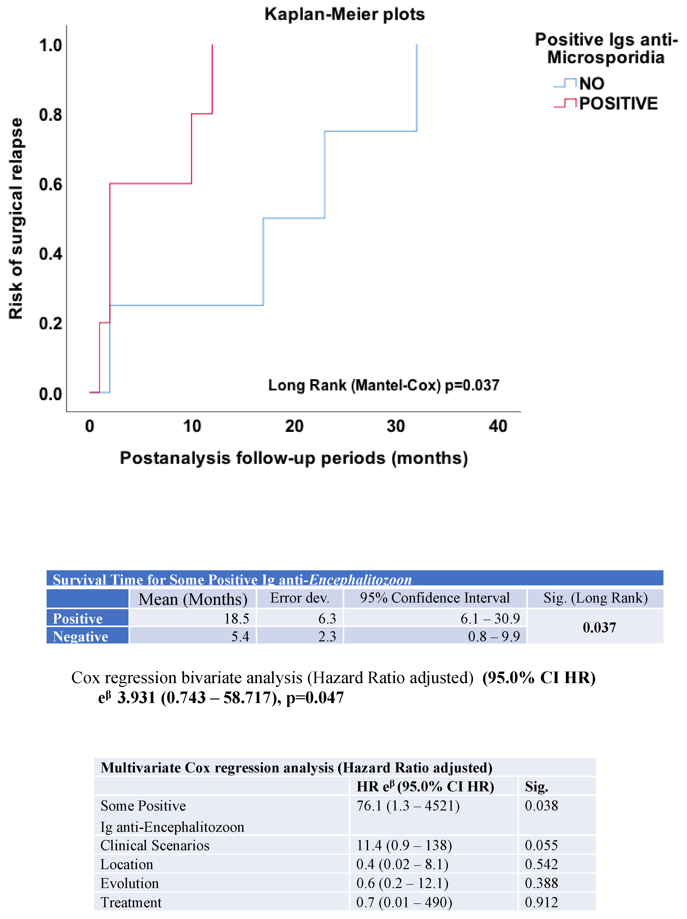
3.4. Positivity for Anti-Encephalitozoon cuniculi Immunoglobulins and Surgical Events in Patients with Crohn’s Disease
3.5. Anti-Encephalitozoon cuniculi Immunoglobulins According to Clinical Scenarios
3.6. Anti-Encephalitozoon cuniculi Immunoglobulins According to Treatment Status
4. Discussion
5. Conclusions
Supplementary Materials
Author Contributions
Funding
Institutional Review Board Statement
Informed Consent Statement
Data Availability Statement
Conflicts of Interest
Appendix A

| Group | Sample | IL-7 Gene Expression | Caspase-3 Protein Expression | IL-7 Protein Expression |
|---|---|---|---|---|
| Crohn’s Disease | 1 | 0.69 | 0.445 | 1.0 |
| Crohn’s Disease | 2 | 0.6 | 0.45 | 0.58 |
| Crohn’s Disease | 3 | 0.5 | 0.465 | 0.65 |
| Crohn’s Disease | 4 | 0.52 | 0.48 | 0.7 |
| Crohn’s Disease | 5 | 0.51 | 0.44 | 0.83 |
| Crohn’s Disease | 6 | 0.77 | 0.42 | 0.91 |
| Crohn’s Disease | 7 | 0.25 | 0.41 | 0.43 |
| Crohn’s Disease | 8 | 0.82 | 0.51 | 0.42 |
| Crohn’s Disease | 9 | 1.0 | 0.53 | 0.81 |
| Control | 10 | 0.0 | 0.59 | 0.15 |
| Control | 11 | 0.0 | 0.495 | 0.09 |
| Control | 12 | 0.0 | 0.55 | 0.13 |
| Control | 13 | 0.0 | 0.625 | 0.08 |
| Control | 14 | 0.0 | 0.655 | 0.08 |
| Control | 15 | 0.35 | 0.5 | 0.55 |
| Control | 16 | 0.0 | 0.61 | 0.14 |
| Control | 17 | 0.0 | 0.68 | 0.11 |
| Control | 18 | 0.0 | 0.74 | 0.12 |
| Control | 19 | 0.0 | 1.16 | 0.09 |
| Control | 20 | 0.48 | 1.09 | 0.45 |
| Control | 21 | 0.0 | 1.06 | NA |
| Group | Sample | CD127 (IL7Rα) Gene Expression | CD132 (IL7Rγ) Gene Expression |
|---|---|---|---|
| Crohn’s Disease | 1 | 2.8 | 0.4 |
| Crohn’s Disease | 2 | 3.56 | 0.88 |
| Crohn’s Disease | 3 | 3.88 | 1.04 |
| Crohn’s Disease | 4 | 3.84 | 0.72 |
| Control | 17 | 3.32 | 4.0 |
| Control | 18 | 4.0 | 4.4 |
| Control | 19 | 3.96 | 4.08 |
| Control | 20 | 3.68 | 4.06 |
| Variable | Crohn’s Disease (Mean ± SD, n) | Control (Mean ± SD, n) | p-Value |
|---|---|---|---|
| IL-7 Gene Expression (fold change vs. GAPDH) | 0.629 ± 0.219 (n = 9) | 0.069 ± 0.164 (n = 12) | <0.001 |
| Caspase-3 Protein Expression (A.U.) | 0.461 ± 0.040 (n = 9) | 0.730 ± 0.237 (n = 12) | 0.002 |
| IL-7 Protein Expression (A.U.) | 0.703 ± 0.203 (n = 9) | 0.181 ± 0.161 (n = 11) | <0.001 |
| Variable | Crohn’s Disease (Mean ± SD, n) | Control (Mean ± SD, n) | p-Value |
|---|---|---|---|
| CD127 (IL7Rα) Gene Expression | 3.520 ± 0.501 (n = 4) | 3.740 ± 0.314 (n = 4) | 0.490 |
| CD132 (IL7Rγ) Gene Expression | 0.760 ± 0.273 (n = 4) | 4.135 ± 0.180 (n = 4) | <0.001 |
| Variable | Treatment (Mean ± SD, n = 31) | NO (Mean ± SD, n = 19) | p-Value |
|---|---|---|---|
| IgG | 1.4 ± 0.6 | 1.3 ± 0.5 | 0.738 |
| IgM | 0.6 ± 0.3 | 0.6 ± 0.4 | 0.418 |
| IgA | 0.8 ± 0.4 | 0.8 ± 0.4 | 1.0 |
| IgE | 0.04 ± 0.4 | 0.04 ± 0.4 | 0.958 |
References
- Andreu-Ballester, J.C.; Pérez-Griera, J.; Garcia-Ballesteros, C.; Amigo, V.; Catalán-Serra, I.; Monforte-Albalat, A.; Bixquert-Jiménez, M.; Ballester, F. Deficit of interleukin-7 in serum of patients with Crohn’s disease. Inflamm. Bowel Dis. 2013, 19, E30–E31. [Google Scholar] [CrossRef] [Scilit] [PubMed]
- Catalan-Serra, I.; Sandvik, A.K.; Bruland, T.; Andreu-Ballester, J.C. Gammadelta T cells in Crohn’s disease: A new player in the disease pathogenesis? J. Crohn’s Colitis 2017, 11, 1135–1145. [Google Scholar] [CrossRef] [Scilit] [PubMed]
- Andreu-Ballester, J.C.; Garcia-Ballesteros, C.; Amigo, V.; Ballester, F.; Gil-Borrás, R.; Catalán-Serra, I.; Magnet, A.; Fenoy, S.; Del Aguila, C.; Ferrando-Marco, J.; et al. Microsporidia and its relation to Crohn’s disease: A retrospective study. PLoS ONE 2013, 8, e62107. [Google Scholar] [CrossRef] [Scilit] [PubMed]
- Andreu-Ballester, J.C.; Hurtado-Marcos, C.; García-Ballesteros, C.; Pérez-Griera, J.; Izquierdo, F.; Ollero, D.; Jiménez, A.; Gil-Borrás, R.; Llombart-Cussac, A.; López-Chuliá, F.; et al. Decreased gene expression of interleukin 2 receptor subunit γ (CD132) in tissues of patients with Crohn’s disease. World J. Gastroenterol. 2025, 31, 97120. [Google Scholar] [CrossRef] [Scilit]
- Hussain, M.S.; Bisht, A.S.; Gupta, G. Reduced interleukin-2 receptor subunit γ expression in Crohn’s disease: A potential mechanism for γδ T cell deficiency. World J. Gastroenterol. 2025, 31, 103180. [Google Scholar] [CrossRef] [Scilit]
- Cuéllar, C.; Hurtado-Marcos, C.; Valdivieso, E.; Vaccaro, L.; González-Fernández, J.; Jiménez, A.I.; Pérez-Griera, J.; Benlloch, S.; Amorós, C.; Gil-Borrás, R.; et al. Immune dysregulation, apoptosis impairment, and enhanced seroreactivity to Anisakis simplex in Crohn’s disease: Interplay of IL-7/IL-7R signalling and CD132 deficiency. Mem. Inst. Oswaldo Cruz 2026, 121, e250129. [Google Scholar] [CrossRef] [Scilit]
- Schmidt, E.C.; Shadduck, J.A. Mechanisms of resistance to the intracellular protozoan Encephalitozoon cuniculi in mice. J. Immunol. 1984, 133, 2712–2719. [Google Scholar] [CrossRef] [Scilit]
- Khan, I.A.; Schwartzman, J.D.; Kasper, L.H.; Moretto, M. CD8+ CTLs are essential for protective immunity against Encephalitozoon cuniculi infection. J. Immunol. 1999, 162, 6086–6091. [Google Scholar] [CrossRef] [Scilit]
- Khan, I.A.; Moretto, M.; Weiss, L.M. Immune response to Encephalitozoon cuniculi infection. Microbes Infect. 2001, 3, 401–405. [Google Scholar] [CrossRef] [Scilit]
- Moretto, M.; Durell, B.; Schwartzman, J.D.; Khan, I.A. Gamma delta T cell-deficient mice have a down-regulated CD8+ T cell immune response against Encephalitozoon cuniculi infection. J. Immunol. 2001, 166, 7389–7397. [Google Scholar] [CrossRef] [Scilit]
- Moretto, M.; Weiss, L.M.; Khan, I.A. Induction of a rapid and strong antigen-specific intraepithelial lymphocyte response during oral Encephalitozoon cuniculi infection. J. Immunol. 2004, 172, 4402–4409. [Google Scholar] [CrossRef] [Scilit]
- Moretto, M.M.; Harrow, D.I.; Khan, I.A. Effector CD8 T cell immunity in microsporidial infection: A lone defense mechanism. Semin. Immunopathol. 2015, 37, 281–287. [Google Scholar] [CrossRef] [Scilit]
- Braunfuchsová, P.; Salát, J.; Kopecký, J. CD8+ T lymphocytes protect SCID mice against Encephalitozoon cuniculi infection. Int. J. Parasitol. 2001, 31, 681–686. [Google Scholar] [CrossRef] [Scilit] [PubMed]
- Han, Y.; Gao, H.; Xu, J.; Luo, J.; Han, B.; Bao, J.; Pan, G.; Li, T.; Zhou, Z. Innate and adaptive immune responses against microsporidia infection in mammals. Front. Microbiol. 2020, 11, 1468. [Google Scholar] [CrossRef] [Scilit] [PubMed]
- Moretto, M.M.; Khan, I.A. Immune response to microsporidia. In Microsporidia; Weiss, L.M., Becnel, J.J., Eds.; Experientia Supplementum; Springer: Berlin/Heidelberg, Germany, 2022; Volume 114, pp. 373–388. [Google Scholar] [CrossRef] [Scilit]
- Goodman, T.; Lefrançois, L. Expression of the gamma-delta T-cell receptor on intestinal CD8+ intraepithelial lymphocytes. Nature 1988, 333, 855–858. [Google Scholar] [CrossRef] [Scilit] [PubMed]
- Bonneville, M.; Janeway, C.A., Jr.; Ito, K.; Haser, W.; Ishida, I.; Nakanishi, N.; Tonegawa, S. Intestinal intraepithelial lymphocytes are a distinct set of gamma delta T cells. Nature 1988, 336, 479–481. [Google Scholar] [CrossRef] [Scilit]
- Fischer, M.A.; Golovchenko, N.B.; Edelblum, K.L. γδ T cell migration: Separating trafficking from surveillance behaviors at barrier surfaces. Immunol. Rev. 2020, 298, 165–180. [Google Scholar] [CrossRef] [Scilit]
- Cuéllar, C.; Rodero, M.; Pérez-Griera, J.; Galindo-Regal, L.; Lopez-Chulia, F.; García-Ballesteros, C.; Andreu-Ballester, J.C. Association between anti-Anisakis simplex antibodies and interleukin-7 levels. Int. Immunopharmacol. 2022, 111, 109134. [Google Scholar] [CrossRef] [Scilit]
- Lennard-Jones, J.E. Classification of inflammatory bowel disease. Scand. J. Gastroenterol. 1989, 24, 2–6. [Google Scholar] [CrossRef] [Scilit]
- Papay, P.; Ignjatovic, A.; Karmiris, K.; Amarante, H.; Milheller, P.; Feagan, B.; D’HAens, G.; Marteau, P.; Reinisch, W.; Sturm, A.; et al. Optimising monitoring in the management of Crohn’s disease: A physician’s perspective. J. Crohn’s Colitis 2013, 7, 653–669. [Google Scholar] [CrossRef] [Scilit]
- del Aguila, C.; Rueda, C.; De la Camara, C.; Fenoy, S. Seroprevalence of anti-Encephalitozoon antibodies in Spanish immunocompetent subjects. J. Eukaryot. Microbiol. 2001, 48, 75S–78S. [Google Scholar] [CrossRef] [Scilit] [PubMed]
- Daschner, A.; Cuéllar, C.; Sánchez-Pastor, S.; Pascual, C.Y.; Martín-Esteban, M. Gastro-allergic anisakiasis as a consequence of simultaneous primary and secondary immune response. Parasite Immunol. 2002, 24, 243–251. [Google Scholar] [CrossRef] [Scilit] [PubMed]
- Gutiérrez, R.; Cuéllar, C. Immunoglobulins anti-Anisakis simplex in patients with gastrointestinal diseases. J. Helminthol. 2002, 76, 131–136. [Google Scholar] [CrossRef] [Scilit] [PubMed]
- Zeng, Z.; Mao, H.; Lei, Q.; He, Y. IL-7 in autoimmune diseases: Mechanisms and therapeutic potential. Front. Immunol. 2025, 16, 1545760. [Google Scholar] [CrossRef] [Scilit]
- Okada, E.; Yamazaki, M.; Tanabe, M.; Takeuchi, T.; Nanno, M.; Oshima, S.; Okamoto, R.; Tsuchiya, K.; Nakamura, T.; Kanai, T.; et al. IL-7 exacerbates chronic colitis with expansion of memory IL-7Rhigh CD4+ mucosal T cells in mice. Am. J. Physiol. Gastrointest. Liver Physiol. 2005, 288, G745–G754. [Google Scholar] [CrossRef] [Scilit]
- Willis, C.R.; Seamons, A.; Maxwell, J.; Treuting, P.M.; Nelson, L.; Chen, G.; Phelps, S.; Smith, C.L.; Brabb, T.; Iritani, B.M.; et al. Interleukin-7 receptor blockade suppresses adaptive and innate inflammatory responses in experimental colitis. J. Inflamm. 2012, 9, 39. [Google Scholar] [CrossRef] [Scilit]
- Hasani, Z.; Aghdaei, H.A.; Balaii, H.; Azimirad, M.; Mirsamadi, E.S.; Mirjalali, H.; Zali, M. The first study on opportunistic intestinal microsporidiosis in IBD patients receiving immunosuppressive medications in Iran. Epidemiol. Infect. 2017, 145, 2095–2099. [Google Scholar] [CrossRef] [Scilit]
- Jin, J.; Tang, Y.; Cao, L.; Wang, X.; Chen, Y.; An, G.; Zhang, H.; Pan, G.; Bao, J.; Zhou, Z. Microsporidia persistence in host impairs epithelial barriers and increases chances of inflammatory bowel disease. Microbiol. Spectr. 2024, 12, e0361023. [Google Scholar] [CrossRef] [Scilit]
- Capitini, C.M.; Chisti, A.A.; Mackall, C.L. Modulating T-cell homeostasis with IL-7: Preclinical and clinical studies. J. Intern. Med. 2009, 266, 141–153. [Google Scholar] [CrossRef] [Scilit]
- Jacobs, S.R.; Michalek, R.D.; Rathmell, J.C. IL-7 is essential for homeostatic control of T cell metabolism in vivo. J. Immunol. 2010, 184, 3461–3469. [Google Scholar] [CrossRef] [Scilit]
- Belarif, L.; Danger, R.; Kermarrec, L.; Nerrière-Daguin, V.; Pengam, S.; Durand, T.; Mary, C.; Kerdreux, E.; Gauttier, V.; Kucik, A.; et al. IL-7 receptor influences anti-TNF responsiveness and T cell gut homing in inflammatory bowel disease. J. Clin. Investig. 2019, 129, 1910–1925. [Google Scholar] [CrossRef] [Scilit]



| Encephalitozoon cuniculi | |||
|---|---|---|---|
| Positive | Negative | Significance | |
| Age | 37.3 ± 14.5 | 41.7 ± 14.3 | ns |
| HBI | 7.7 ± 4.1 | 6.4 ± 3.5 | ns |
| CDAI | 191.3 ± 108.7 | 138.1 ± 107.9 | ns |
| ESR | 45.6 ± 41.4 | 32.1 ± 32.1 | ns |
| CRP | 48.9 ± 50.5 | 33.5 ± 14.6 | ns |
| n (%) | n (%) | ||
| Gender | Odds Ratio | ||
| 13 (50.0) | 13 (50.0) | ns |
| 10 (43.5) | 14 (51.9) | |
| Clinical Scenarios | |||
| 11 (47.8) | 12 (44.4) | ns |
| 7 (30.4) | 5 (18.5) | |
| 5 (21.7) | 10 (37.0) | |
| Evolution | |||
| 12 (52.2) | 17 (63.0) | ns |
| 7 (30.4) | 7 (25.9) | |
| 4 (17.4) | 3 (11.1) | |
| Location | |||
| 12 (52.2) | 14 (51.9) | ns |
| 10 (43.5) | 10 (37.0) | |
| 1 (4.3) | 3 (11.1) | |
| Treatment | |||
| 14 (77.8) | 17 (70.8) | ns |
| 4 (22.2) | 7 (29.2) | |
Disclaimer/Publisher’s Note: The statements, opinions and data contained in all publications are solely those of the individual author(s) and contributor(s) and not of MDPI and/or the editor(s). MDPI and/or the editor(s) disclaim responsibility for any injury to people or property resulting from any ideas, methods, instructions or products referred to in the content. |
© 2026 by the authors. Licensee MDPI, Basel, Switzerland. This article is an open access article distributed under the terms and conditions of the Creative Commons Attribution (CC BY) license.
Share and Cite
Hurtado-Marcos, C.; Izquierdo, F.; Fenoy, S.; Águila, C.d.; Pérez-Griera, J.; Benlloch, S.; Amorós, C.; Ballesteros, C.G.; Chuliá, F.L.; Andreu-Ballester, J.C.; et al. Dysregulated IL-7/IL-7R-CD132 Axis and Intestinal Microsporidiosis in Crohn’s Disease. Pathogens 2026, 15, 429. https://doi.org/10.3390/pathogens15040429
Hurtado-Marcos C, Izquierdo F, Fenoy S, Águila Cd, Pérez-Griera J, Benlloch S, Amorós C, Ballesteros CG, Chuliá FL, Andreu-Ballester JC, et al. Dysregulated IL-7/IL-7R-CD132 Axis and Intestinal Microsporidiosis in Crohn’s Disease. Pathogens. 2026; 15(4):429. https://doi.org/10.3390/pathogens15040429
Chicago/Turabian StyleHurtado-Marcos, Carolina, Fernando Izquierdo, Soledad Fenoy, Carmen del Águila, Jaume Pérez-Griera, Salvador Benlloch, Cirilo Amorós, Carlos García Ballesteros, Francisca López Chuliá, Juan Carlos Andreu-Ballester, and et al. 2026. "Dysregulated IL-7/IL-7R-CD132 Axis and Intestinal Microsporidiosis in Crohn’s Disease" Pathogens 15, no. 4: 429. https://doi.org/10.3390/pathogens15040429
APA StyleHurtado-Marcos, C., Izquierdo, F., Fenoy, S., Águila, C. d., Pérez-Griera, J., Benlloch, S., Amorós, C., Ballesteros, C. G., Chuliá, F. L., Andreu-Ballester, J. C., & Cuéllar, C. (2026). Dysregulated IL-7/IL-7R-CD132 Axis and Intestinal Microsporidiosis in Crohn’s Disease. Pathogens, 15(4), 429. https://doi.org/10.3390/pathogens15040429