Article

Dancing with the Dust Devil: Examining the Lung Mycobiome of Sonoran Desert Wild Mammals and the Effect of Coccidioides Presence

Pathogen and Microbiome Institute, Northern Arizona University, Flagstaff, AZ 86011, USA
*
Author to whom correspondence should be addressed.
Pathogens 2025, 14(8), 807; https://doi.org/10.3390/pathogens14080807
Submission received: 31 May 2025 / Revised: 6 August 2025 / Accepted: 7 August 2025 / Published: 14 August 2025
(This article belongs to the Special Issue Epidemiology and Molecular Detection of Emerging Fungal Pathogens)

Abstract

Microbiome studies report a decrease in diversity associated with active infections. Under the endozoan hypothesis, Coccidioides can inhabit a host without causing disease. In this study, we describe and compare the lung mycobiome of Coccidioides-positive and -negative samples obtained from wildlife. If Coccidioides is not causing infection, we predict there will be no differences in the mycobiome between positive and negative samples. Lung samples were obtained from mammals previously trapped in Tucson, Arizona, USA (n = 26), and Mesa, Arizona, USA (n = 14). Samples were screened for Coccidioides with CocciDx, and mycobiome was characterized through Illumina-based amplicon sequencing of the internal transcribed spacer 2 (ITS2). We compared alpha and beta diversity of the mycobiome to assess the effects of Coccidioides’ presence and host taxonomy. A greater number of reads were captured from Tucson samples (114,706.4 ± 57,945.8) than from Mesa (384.9 ± 953.5); however, Mesa (16.8 ± 8.8) and Tucson (12 ± 7.8) had a similar number of fungal genera per sample. CocciDx detected Coccidioides in more samples than the ITS2 amplicon sequencing. All samples from Mesa and five from Tucson tested positive for Coccidioides. Therefore, Mesa samples were excluded from statistical analysis. No difference in alpha and beta diversity was associated with Coccidioides presence, which is consistent with the endozoan hypothesis. Host taxonomy had a significant effect on beta diversity. This effect is likely driven by host behavioral and physiological differences.

1. Introduction

While there is an emerging interest in lung mycobiome of wild animals [1,2,3,4], we still have a limited understanding of the complex host-mycobiome dynamics [3,4]. Additionally, our understanding of microbiome dynamics is primarily based on other tissue systems (e.g., gut, vagina, mouth, etc.). Those studies suggest a decrease in the diversity of microbial communities associated with active infection. However, the role of the microbiome in disease progression is not well characterized [5,6,7,8,9].
The lungs are a very dynamic environment. Organisms enter the airways through inhalation or contact with the gut and are eliminated through coughing, mucociliary transport, innate and adaptive immune system responses [10]. Therefore, microorganisms found in the lungs could be transient or part of the established microbial community, leading to complications in analyzing data and drawing conclusions [3,11,12]. Moreover, many of these studies have few healthy controls (or none) due to the invasive nature of collection of bronchoalveolar lavage fluid or biopsy specimens. Finally, in human and laboratory animal studies, differences in microbiome composition are associated with multiple factors such as age, mode of birth, health, diet, and other lifestyle factors [13,14,15,16,17].
Initially, studies described the diversity of fungi in mammalian lungs and their role as pathogens through culturing [18,19,20,21]. However, culture-based approaches have been shown to be ineffective in capturing total microbial diversity in the lung. In one study, 60% of the species detected with pyrosequencing were not recovered through culturing from clinical samples in patients with cystic fibrosis [22]. Community sequencing is a valuable tool to characterize microbial communities that more recently have been used to look at the mycobiome associated with wildlife [3,23,24,25,26,27,28,29,30,31].
Coccidioidomycosis is an environmentally acquired fungal disease caused by Coccidioides posadasii and Coccidioides immitis [32]. Most cases of the disease are asymptomatic and are often misdiagnosed [33,34,35,36]. Yet, 17,612 cases of coccidioidomycosis were reported in the United States in 2022, the majority in Arizona and California [37]. A study using data from 2019 estimated that the lifetime cost of coccidioidomycosis would be 736 million USD for the 10,539 cases diagnosed in Arizona in that year [38]. Coccidioides acts as a pathogen in humans and other mammals, such as dogs, horses, and even sea lions [39]. However, evidence also suggests that the fungus may behave as an “endozoan”, residing in the tissues of small mammals without causing coccidioidomycosis, similar to the relationship between endophytes and their plant hosts [40,41]. If this is the case, small mammals may serve as important reservoirs for Coccidioides in the environment, but additional evidence is required to resolve the role of small mammals on the Coccidioides life cycle [40,42,43,44,45].
Lung microbiome studies could provide valuable insights into the behavior of Coccidioides in small mammals. If Coccidioides behaves as an endozoan, there should be no significant difference in the microbiome of Coccidioides-positive and -negative lungs. To date, only one study has examined the impact of Coccidioides on the lung microbiome. This study found no significant differences associated with Coccidioides presence in the lungs of small mammals obtained from a museum collection [3]. Given the limited research on wildlife mycobiome and the effect of Coccidioides in the microbial communities, the overarching goal of this study is to characterize the mycobiome of small-mammal lung samples from Coccidioides-endemic areas and identify differences associated with the presence of Coccidioides.

2. Methods

2.1. Samples

All lung samples were obtained from specimens donated to this study postmortem (Scientific Collection License #SP650076). Animals were identified based on morphological characteristics. The Arizona Game and Fish Department donated lungs of Dipodomys (kangaroo rat, n = 1), Chaetodipus (pocket mouse, n = 12), Onychomys (grasshopper mouse, n = 1), Ammospermophilus (antelope ground squirrel, n = 1), Xerospermophilus (round-tailed ground squirrel, n = 1), Sylvilagus (desert cottontail, n = 6), Lepus (antelope jackrabbit, n = 1), and Lepus (black-tailed jackrabbit, n = 3). Animals were euthanized on site and whole carcasses or lung tissue samples were stored temporarily in ice and later at −20 °C. Traps were set within an area of approximately 0.54 hectares located southeast of Tucson in a Lower Sonoran Desert vegetation area. Vegetation was sparse with mainly Prosopis velutina (mesquite tree), Cercidium spp. (palo verde tree) and Larrea tridentata (creosote bush).
Peromyscus (cactus mouse, n = 14), trapped for pest-control inside a building (approx. 184.3 m2), were donated by a facility north of Mesa in the Phoenix metropolitan area. Traps were checked at least twice a day, and carcasses were individually placed in a plastic bag and froze at −20 °C. The area where these animals were obtained is a run-off zone of the Salt River with abundant wildlife (e.g., wild horses, horned owls, coyotes, and diverse communities of snakes and burrowing mammals). Vegetation in the area is sparse and includes Larrea tridentata (creosote bush), Encelia farinose (brittle brush), and Prosopis velutina (mesquite tree). Invasive grass like Cenchrus ciliaris (buffelgrass) occupies spaces in the spring and summer.
Coccidioides was known to be present in the Mesa site, as an ongoing study screened soil samples monthly from 2022 to 2025 [46]. However, for the Tucson site it was necessary to determine if Coccidioides was present in the soil. Soil samples were collected from animal burrow entrances using a garden trowel or long handled kitchen spoon and sterilized with 10% bleach between soil samples. Soil was put directly into sterile 50-milliliter specimen cups. Soil was stored at room temperature until DNA extraction was performed. In total, 48 soil samples were collected from four unique burrow systems (12 replicates per burrow system).

2.2. DNA Extraction

Animal carcasses or tissues were kept in a −20 °C freezer until transferred to Northern Arizona University. Carcasses were then necropsied in the laboratory. Following necropsy, tissues were kept in a −80 °C freezer until DNA extractions were completed. Tissues were placed in bead tubes (BeadTube 1.5 mm, Benchmark Scientific, Sayreville, NJ, USA) and homogenized at 5 m/s for one minute. For Mesa site samples, DNA extractions were conducted using a phenol-chloroform extraction protocol. For Tucson site samples, DNA was extracted with Qiagen Blood and Tissue Kit (Qiagen, Valencia, CA, USA) following the manufacturer’s protocol.
DNA from soil samples was extracted using the DNeasy PowerSoil Pro kit (QIAGEN, Valencia, CA). The manufacturer’s protocol was followed with the addition of a 10 min heat step at 65 °C before homogenization [47]. For each soil sample, DNA was extracted from two different 250 mg sub-samples. All DNA was stored in a −20 °C freezer.

2.3. Molecular Detection of Coccidioides

The CocciDx assay was used to determine whether lung and soil samples were positive or negative for Coccidioides. This assay is a real-time qPCR targeting a 106 bp sequence present in multiple copies within a transposable element in the genome of both Coccidioides species and only present in the Coccidioides genus [48,49]. CocciDx was performed on the Applied Biosystems QuantStudio 12K Flex Real-Time PCR System (Thermo Fisher Scientific, Waltham, MA, USA). Each reaction contained 2× TaqMan Environmental Master Mix 2.0 (Thermo Fisher Scientific, Waltham, MA, USA), 1× CocciDX at 100μM concentration of Cocci Assay oligo/probe mix and 2 μL of DNA extracted from lung sample. Each reaction had a total volume of 20 uL. PCR cycling conditions were two minutes at 50 °C for the activation step, 10 min at 95 °C for the denaturation step, 45 cycles of 15 s at 95 °C and then one minute at 60 °C. Samples are run in triplicate with molecular grade water as a negative control and Silveira strain DNA as positive control. Samples with CT values lower than 40 were considered positive.

2.4. Amplicon Sequencing

To determine the composition of fungal communities, each DNA extract went through an Illumina-based amplicon sequencing, targeting the ITS2 region. We used 5.8S-Fun (ACCCAACTGAATGGAGCAACTTTYRRCAAYGGATCWCT) and ITS4-Fun (ACGCACTTGACTTGTCTTCAGCCTCCGCTTATTGATATGCTTAART) as primers and added universal tail sequences [50].
The amplification reaction of the fungal ITS2 region contained 1X Phusion Green Hot Start II High-Fidelity PCR Master Mix (Thermo Fisher Scientific, Waltham, MA, USA), 0.5 μM each primer, 2 μL of DNA and molecular grade water to a final volume of 50 μL. A universal tail was added to the forward and reverse primer to facilitate amplicon sequencing [51]. Cycling parameters were initial denaturation at 98 °C for 1 min followed by 30 cycles of 98 °C denaturation for 10 s, 57 °C to 59 °C annealing for 30 s, and 72 °C extension for 20 s. A final extension consisted of 10 min at 72 °C. The success of the amplifications was assessed by running the PCR products on a 2% agarose gel. Amplicons for each sample were cleaned using 1X Agencourt AMPure XP beads (Beckman Coulter, Brea, CA, USA) as recommended by the manufacturer. Unique indices and sequencing adaptors were added during a second PCR reaction that uses the universal tails attached during the initial PCR. This reaction consisted of 12.5 μL of Kapa HiFi HotStart Ready Mix (Kapa Biosystems, Wilmington, MA, USA), 400 nm of the unique indexing primers, and 2, 4, or 8 μL of the bead-cleaned amplicons from the initial PCR. Thermocycling conditions were 2 min at 98 °C for initial denaturation, 6 cycles of: 30 s at 98 °C denaturation, 20 s at 60 °C annealing, 30 s at 72 °C extension, and a final extension at 72 °C for 5 min. Indexed libraries were pooled in equal amounts (240 ng) using the Qubit fluorometer (Thermo Fisher Scientific, Waltham, MA, USA). The combined pool was cleaned with the QIAquick PCR Purification Kit (Qiagen, Valencia, CA, USA) per manufacturer’s recommendations. The samples were sequenced on an Illumina MiSeq instrument using a v2 500 cycle kit (Illumina, San Diego, CA, USA).

2.5. Microbiome Data Processing

Primer sequences were trimmed from raw sequences using Cutadapt and paired end reads merged prior to data analysis [52]. Sequences were trimmed uniformly to 300 nucleotides. ITS2 samples were demultiplexed in Quantitative Insights Into Microbial Ecology 2 [53] requiring 95% of each read to have a minimum q-score of 20, and allowing no exceptions (-q 19 -r 0 -p 0.95). Forward reads were trimmed of primer sequence, followed by quality control filtering by DADA2 [54]. Then, demultiplexed, trimmed sequences were screened for chimeras using the VSEARCH program [55] and screened for fungal ITS2 sequences with ITSx [56]. Operational taxonomic units (OTUs) were assigned taxonomy using BLAST+ 2.15.0 [57] against the UNITE reference database [58]. Sequences were submitted to NCBI Genbank under the Bioproject PRJNA1235246.

2.6. Animal-Associated Fungal Genera

To distinguish between transient and established fungal genera in the lung mycobiome, we assigned the fungal genera into categories based on ecological traits. If a genus was not reported to be associated with vertebrate animals, we assume this genus is transiently present in the lung samples. Whereas, if a genus has been found to be associated with vertebrate animals, we assume this genus could be established in the mycobiome. To obtain information on the ecology of each genus, we used the FUNGuild database [59] and expanded upon this foundation by incorporating the latest scientific literature. Then, information was cross-referenced with the FUNGuild database and this amended database was used to assign the mycobiome genera into one of the following groups: animal associated, plant associated, dung associated, soil associated, other, unknown and NA. Fungal genera were only assigned into one group. The goal was to identify genera previously reported to be associated with vertebrate animals, thus if a fungal genus could be assigned to multiple functional categories (e.g., animal and plant associated), the animal associated category prevailed. Genus was assigned as unknown if no ecological data was available for that genus. OTUs that could not be identified to the genus level were described as NA.

2.7. Statistical Analysis

Given the difference in extraction methods and location where samples were obtained, the data from Mesa and Tucson sites are presented separately. Comparisons between Coccidioides-positive and -negative samples were only performed using the Tucson samples. Diversity analyses were performed at genus and order levels; however, not all OTUs had taxonomic assignments at genus or order. In these cases, we maintained the most specific taxonomic assignment. Abundance plots display the ten most prevalent fungal taxa for clarity, but statistical analysis were performed with the whole dataset.
To understand whether diversity patterns can be related to Coccidioides presence, the Bray–Curtis measure of dissimilarity (Beta-diversity) and the Shannon diversity index (Alpha diversity) were calculated for the Tucson dataset. For alpha diversity comparisons, after checking for normality, we either performed a two-tailed t-test or a Wilcoxon rank-sum test (nonparametric). For beta diversity comparisons, we assessed the homogeneity of variances in the data distribution. Then we compared the beta diversities with a permutational multivariate analysis of variance for distance matrices (PERMANOVA). All statistical analyses were conducted in R 4.3.3 with the packages Microbiome 1.26.0 [60], Phyloseq 1.48.0 [61], Ggplot2 3.5.1 [62], Vegan 2.6-6.1 [63], Tidyverse 2.0.0 [64], Dplyr 1.1.4 [65], and the FunGuild database [59].

3. Results

3.1. Coccidioides Detection

All lungs from Peromyscus (n = 14), obtained in the Mesa site tested positive for Coccidioides. For the samples obtained in the Tucson site, one Ammospermophilus, two Chaetodipus, one Sylvilagus and one Onychomys tested positive for Coccidioides (Table 1). Amplicon sequencing detected Coccidioides in six samples, but it was not as sensitive as the CocciDx assay which detected Coccidioides in 19 samples (Table 1). From the amplicon sequencing analysis Coccidioides was identified in two Chaetodipus (PM_4 and PM_5) and in four Peromyscus (33L, 34L, 35L, 38L). In the soil samples from the Tucson site (n = 48), 10.4% of the soils tested positive for Coccidioides. Soils that tested positive were obtained from two different burrow systems, two samples from burrow system B1 and three from burrow system B2 (Table S1).

3.2. Fungal Microbiome

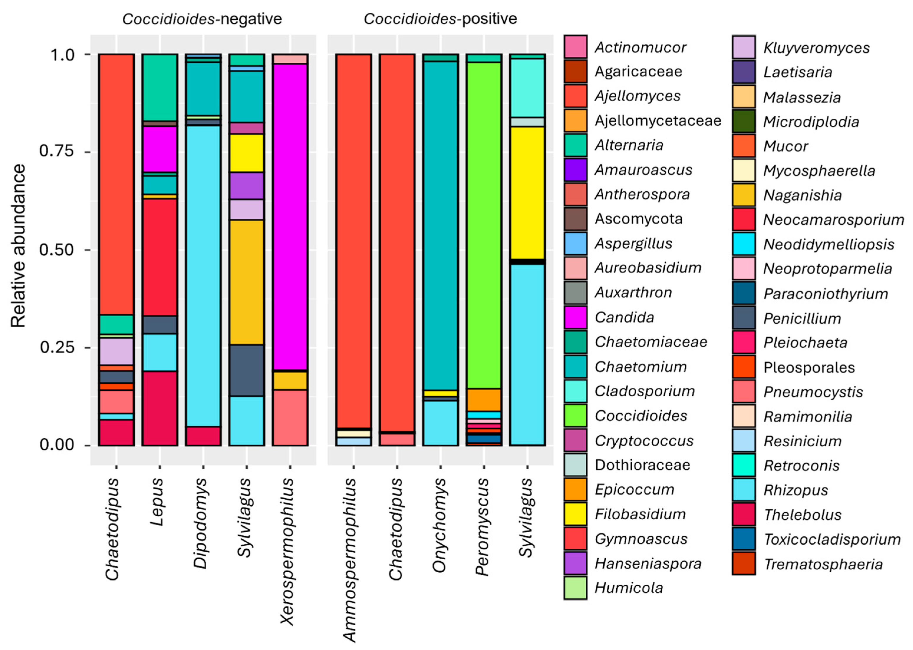
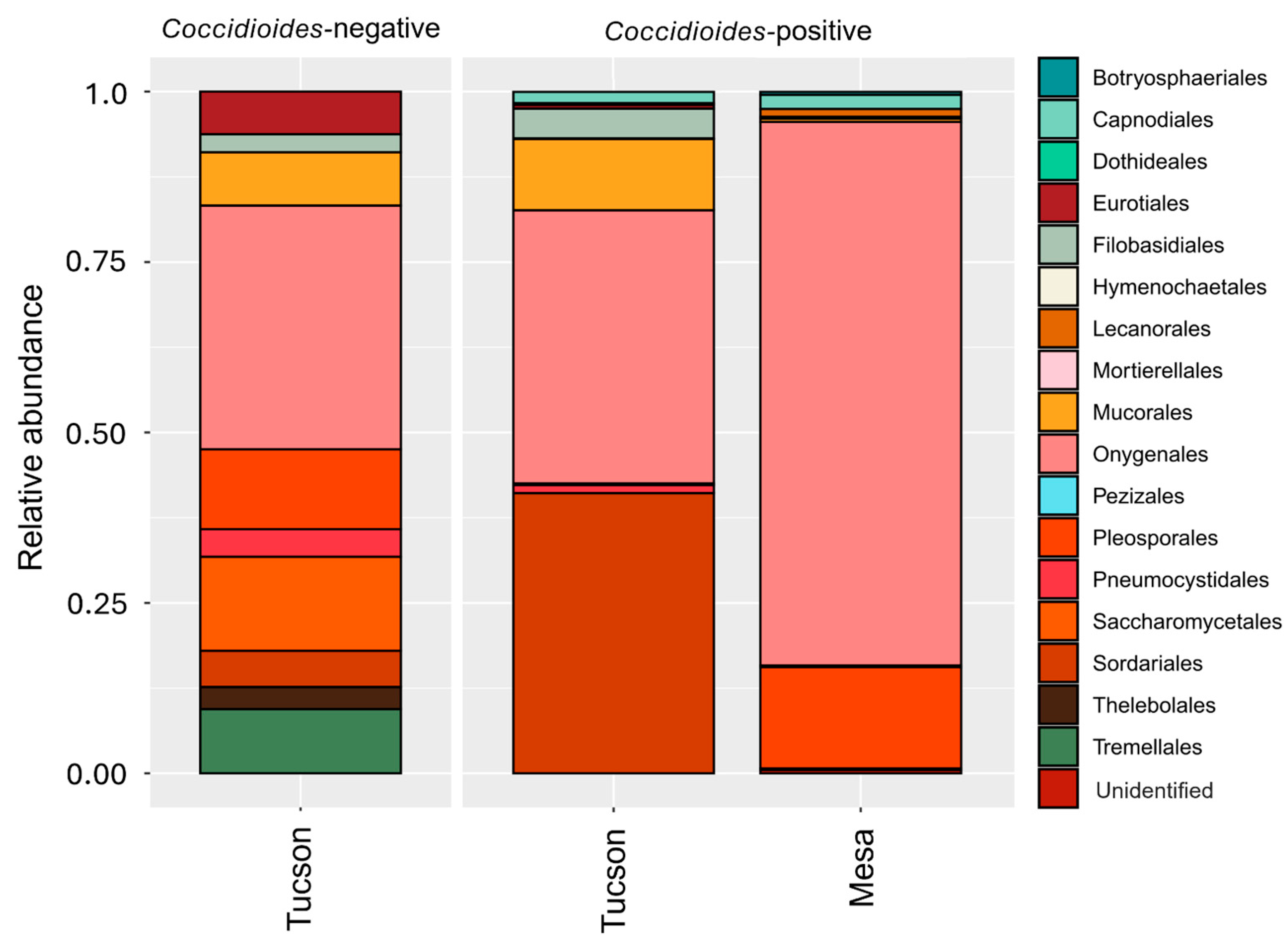
In the Mesa site, we identified 5389 amplicon sequences across the 14 samples from different Peromyscus individuals. We found an average of 384.9 ± 953.5 amplicon reads per sample with 16.8 ± 8.8 genus identified. The ten most prevalent genera accounted for 94.3% of the total reads (Figure 1 and Figure 2), while the ten most prevalent orders, accounted for 98.9% of the total reads (Figure 3). Given the limited sampling depth in the Mesa site samples, we only report fungal taxa and their relative abundances. In the Tucson site, 2,982,366 amplicon sequences were identified across 26 samples of different individuals belonging to distinct species. On average there were 114,706.4 ± 57,945.8 amplicon reads per sample with 12 ± 7.8 genus identified. The ten most prevalent genera accounted for 86.5% of the total reads (Figure 1 and Figure 2), while the ten most prevalent orders identified accounted for 98.4% of the total reads (Figure 3).

3.3. Comparisons of Alpha and Beta Diversity

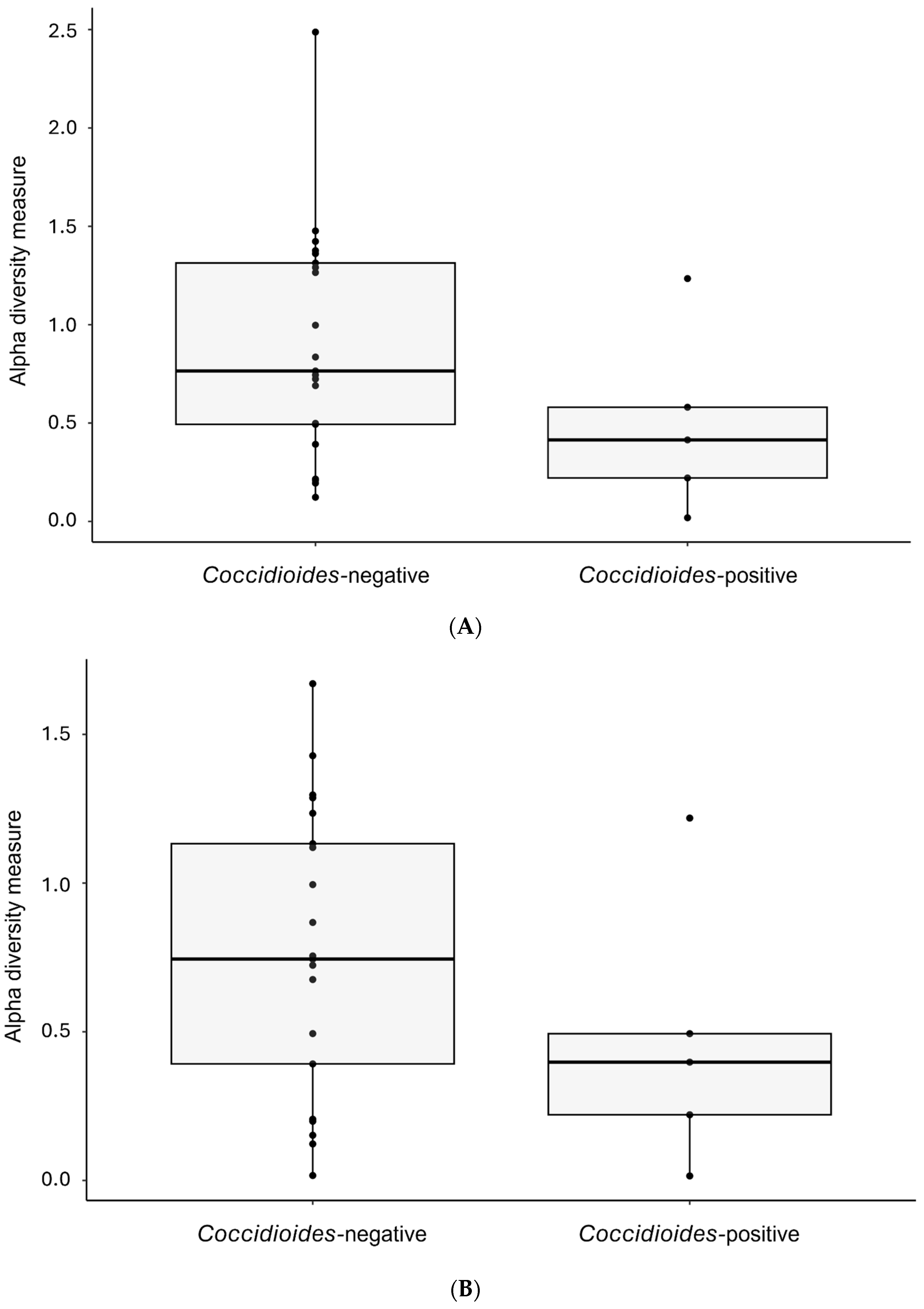
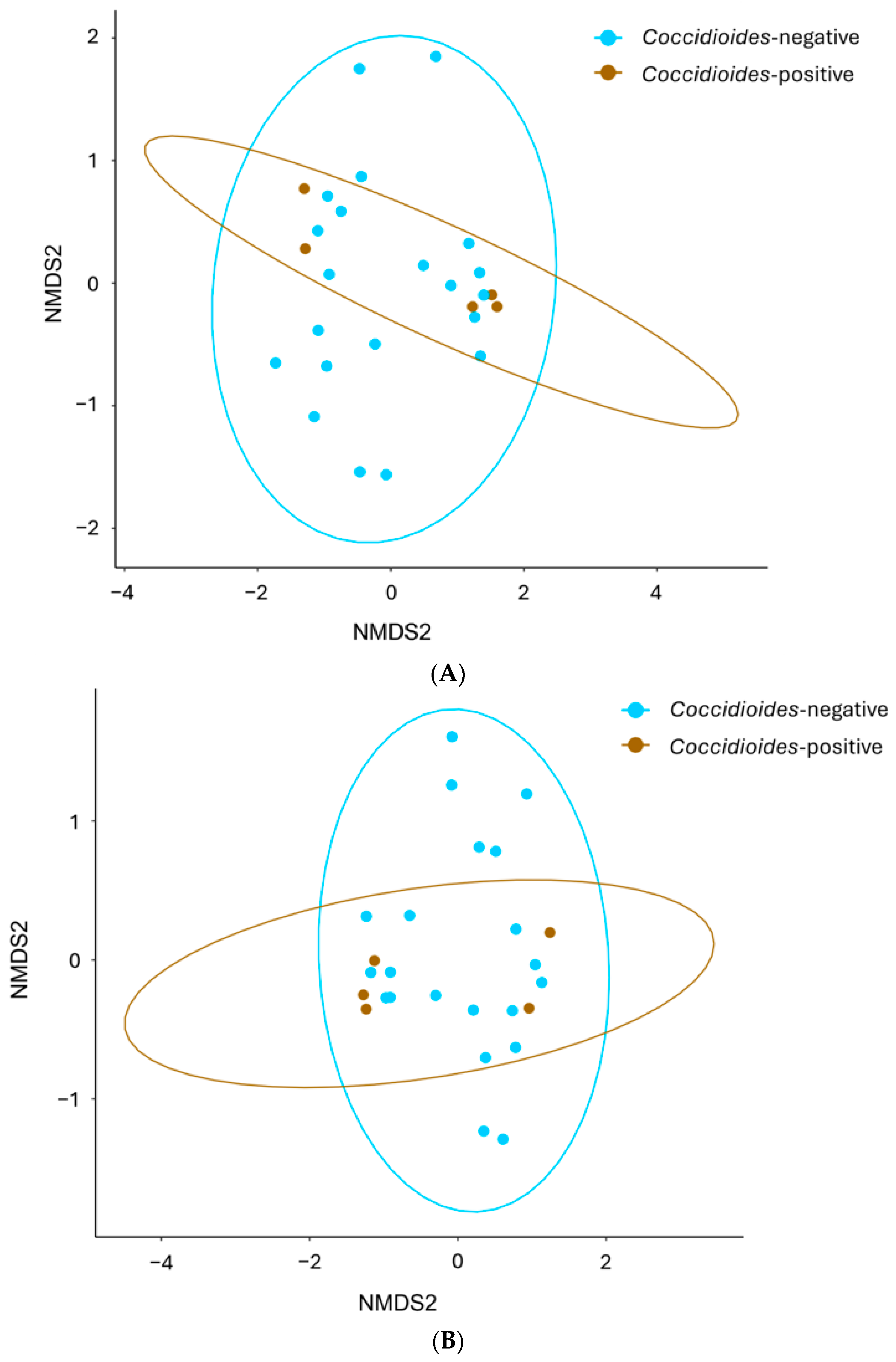
There were no significant differences in alpha diversity assessed by the Shannon diversity index between Coccidioides-positive and -negative samples at the genus (p = 0.138) or at the order level (p = 0.209, Figure 4). No differences in beta diversity were found between Coccidioides-positive and -negative samples when OTUs are grouped at the genus level (p = 0.487) or at the order level (p = 0.333, Figure 5). Host family showed a significant effect on beta diversity both when OTUs are grouped at the genus level (p = 0.001) and at the order level (p = 0.003; Figure 6). Similarly, the host genus has a significant effect on beta diversity when OTUs are grouped at genus (p = 0.001) and order levels (p = 0.002; Figure 6).

3.4. Animal-Associated Fungal Community

Most of the genera found in the samples were previously reported to be associated with vertebrates (Figure 7). The animal-associated dataset represents 89.2% of the original reads in the Tucson samples (2,659,415 reads) and 88.3% in the Mesa samples (4761 reads). For Tucson, it resulted in an average of 102,285.2 ± 61,770.6 reads per sample that were assigned to 7.8 ± 3.5 different fungal genera (Figures S1 and S2). In the Mesa site, the animal-associated dataset has an average of 340.1 ± 923.8 reads per sample that were assigned to 4.6 ± 3.4 different genera (Figures S1 and S2). Beta and alpha diversity comparisons had the same results found in the analyses of the whole dataset. No significant effects of Coccidioides presence (p = 0.184), host genus (p = 0.442) and host family (p = 0.488) in alpha diversity. Host genus (p = 0.001) and host family (p = 0.001) have a significant effect on beta diversity.

4. Discussion

In this study, we describe the prevalence of Coccidioides in mammal lung samples from sites in Tucson and Mesa, Arizona, USA. We also describe the mycobiome in these samples and compare fungal community diversity between Coccidioides-positive and -negative at order and genus levels. We found that small mammals in endemic areas carry a diverse community of fungi in their lungs, but no significant differences were found between Coccidioides-positive and -negative samples, suggesting that Coccidioides does not significantly alter the lung mycobiome in these hosts.
In both sites, most OTU were assigned to the order Onygenales. Tucson site samples exhibited lower Coccidioides prevalence, with only certain host genera (i.e., Chaetodipus, Onychomys, Ammospermophilus and Sylvilagus) testing positive for Coccidioides. In contrast, Coccidioides was highly prevalent in the lungs of mammals from the Mesa site, where all Peromyscus tested positive for Coccidioides. Given that all Mesa site samples were Coccidioides -positive, and considering the limited sequencing depth and overall low read counts, no statistical analyses were performed using this dataset. We suspect these limitations may be due to issues during the DNA extraction or the library preparation.
In terms of mycobiome diversity, the Mesa site revealed a lung mycobiome dominated by Coccidioides (78.7% of the reads), with other common genera including Epicoccum, Toxicocladosporium, and Alternaria. The Tucson site exhibited a more even distribution of fungal genera, with taxa such as Ajellomyces, Chaetomium, and Rhizopus predominating in the lungs. These findings align with what was found in soil samples from these sites. In Tucson, 10.4% of the burrow-associated soils screened in this study were positive (Table S1). In Mesa, 43% of soil samples were positive when collected near rodent burrows [46].
Host taxonomy was associated with differences in beta diversity, suggesting that host physiology and behavior might shape the lung mycobiome of mammals obtained in the same location [66]. However, it is important to note that host genera were identified based on morphological characteristics which can lead to misidentification [3,67]. There were no significant differences in alpha and beta diversity between Coccidioides-positive and -negative samples. This result aligns with previous research that showed no differences in the mycobiome of Coccidioides-positive and -negative lungs from museum specimens [2]. The absence of differences in the lung mycobiome of Coccidioides-positive and -negative samples, suggests that Coccidioides is behaving as a commensal, as pathogens are expected to be associated with shifts in the microbiome [5,6,7]. Therefore, our fungal diversity results are consistent with the endozoan hypothesis which states that the fungus will persist in small-mammal hosts from endemic areas without causing disease [40].
Studying the mycobiome in the lungs is challenging and given the importance of detecting Coccidioides for the objective of this study, we used two methods to ensure detection: CocciDx and an Illumina-based ITS2 amplicon sequencing. Although both methods successfully detected Coccidioides presence, CocciDx detected Coccidioides in 19 samples, while the ITS2 amplicon sequencing detected Coccidioides in six samples. ITS2 amplicon sequencing is a powerful tool to characterize fungal diversity, but it can underestimate or overestimate certain taxa [50]. In contrast, CocciDx is a qPCR-based assay that targets a Coccidioides-specific sequence [48,49]. This result highlights the limitations of using a single detection method when looking for a specific genus or species.
The dynamicity of the lung environment leads to the presence of organisms that are not part of the established mycobiome, but only transiently present in the lung [10,11]. However, in this study, the presence of potentially transient genera did not seem to have an impact on our dataset and analyses. Once we excluded the fungal genera not previously known to be associated with vertebrate animals (e.g., associated with plant, feces, soil, etc.), we still retained most of the OTUs. In the Mesa site samples, animal-associated fungal genera accounted for 89.2% of the reads and 88.3% of the reads in Tucson. Additionally, most of the genera within the ten most prevalent were retained, especially those with higher number of reads. The alpha and beta diversity comparisons of the animal-associated dataset had the same outcome as the whole dataset analysis. Host taxonomy has a significant effect on mycobiome beta diversity, but Coccidioides presence does not. We conclude that fungal genera that were transiently present in the lungs did not affect the outcome of our study. We hypothesize this is because the noise caused by these fungi is similar across all individuals obtained in the same site.
We acknowledge that conclusions from our study are limited by its cross-sectional nature, small sample size and the lack of information on the specimens from which the lung samples were obtained. Studies have shown that different factors can shape the lung microbiome including age and disease progression [10,68,69,70]. Although we have not observed an effect of Coccidioides presence in the lung mycobiome, Coccidioides could be affecting the diversity of viruses and bacteria which our study did not characterize. Moreover, we do not have information on the timing of infection by Coccidioides or by any other pathogens for these individuals. The timing of Coccidioides infection and presence of other pathogens could play a role in the lack of mycobiome differences between Coccidioides-positive and -negative samples. Also, time since death and freeze/thawing cycles could have caused changes in the mycobiome profiles [71,72].
Future studies could benefit from obtaining samples for which life history information is available. This could be achieved by obtaining samples from zoos, animal diversity management programs, and farms. Additionally, a better understanding of the behavior of Coccidioides in the lungs and its effects on the lung mycobiome could come from laboratory studies comparing the mycobiome of mice from the same background under controlled experimental conditions.

Supplementary Materials

The following supporting information can be downloaded at: https://www.mdpi.com/article/10.3390/pathogens14080807/s1, Figure S1: Animal-associated fungal genera in lung mycobiome plotted for each site when samples are positive or negative for Coccidioides; Figure S2: Animal-associated fungal genera in lung mycobiome plotted for each host genus when they are negative or positive for Coccidioides. Supplement Table S1. Coccidioides positivity in soil samples collected near rodent burrows from Tucson, AZ Spreadsheet S1. FunGuild amended database, and ITS2 amplicon sequencing reads assigned to fungal genus.

Author Contributions

Conceptualization, B.B.; Methodology, A.F.-B. and J.S.; Formal analysis, A.F.-B. and D.R.K.; Investigation, A.F.-B., J.S., M.L.B. and D.R.K.; Writing—original draft, A.F.-B., D.R.K. and B.B.; Writing—review and editing, A.F.-B., J.S., M.L.B. and B.B.; Supervision, B.B.; Project administration, B.B.; Funding acquisition, B.B. All authors have read and agreed to the published version of the manuscript.

Funding

Funding for this work was provided by the NAU/ABOR Translational Research Initiative Fund Regent’s award. This work was made possible through specimens obtained via the nonhuman primate tissue and specimen repository of the Washington National Primate Research center which is supported under NIH awards P51OD010425 and U42OD011123.

Institutional Review Board Statement

This study did not require live animal trapping, handling or sacrifice, as we worked with specimens that were donated to this study post-mortem. Therefore, IACUC approval was not required for this study.

Informed Consent Statement

Not applicable.

Data Availability Statement

The original contributions presented in this study are included in the article/Supplementary Materials. Further inquiries can be directed to the corresponding author.

Acknowledgments

In memory of an individual at Arizona Game and Fish who unfortunately past away before completion of this publication, and whose support was essential to this work. We thank the Arizona Game and Fish and the University of Washington for donating the samples used in this study. We also thank the NAU Genetics Core for running the Illumina amplicon sequencing.

Conflicts of Interest

The authors declare no conflict of interest.

References

  1. Melo-Neto, M.F.; Maranhão, F.C.A.; Guimarães, P.S.; Silva, D.M.W. Mycobiota recovered from the trachea and lungs of pigeons (Columba livia) captured in a grain mill. Res. Soc. Dev. 2022, 11, e57211326802. [Google Scholar] [CrossRef]
  2. Salazar-Hamm, P.S.; Gadek, C.R.; Mann, M.A.; Steinberg, M.; Montoya, K.N.; Behnia, M.; Gyllenhaal, E.F.; Brady, S.S.; Takano, O.M.; Williamson, J.L.; et al. Phylogenetic and ecological drivers of the avian lung mycobiome and its potentially pathogenic component. Commun. Biol. 2025, 8, 634. [Google Scholar] [CrossRef]
  3. Salazar-Hamm, P.S.; Montoya, K.N.; Montoya, L.; Cook, K.; Liphardt, S.; Taylor, J.W.; Cook, J.A.; Natvig, D.O. Breathing can be dangerous: Opportunistic fungal pathogens and the diverse community of the small mammal lung mycobiome. Front. Fungal Biol. 2022, 3, 996574. [Google Scholar] [CrossRef] [PubMed]
  4. Babb-Biernacki, S.J.; Esselstyn, J.A.; Doyle, V.P. Predicting Species Boundaries and Assessing Undescribed Diversity in Pneumocystis, an Obligate Lung Symbiont. J. Fungi 2022, 8, 799. [Google Scholar] [CrossRef] [PubMed]
  5. Allender, M.C.; Baker, S.; Britton, M.; Kent, A.D. Snake fungal disease alters skin bacterial and fungal diversity in an endangered rattlesnake. Sci. Rep. 2018, 8, 12147. [Google Scholar] [CrossRef]
  6. Jani, A.J.; Briggs, C.J. The pathogen Batrachochytrium dendrobatidis disturbs the frog skin microbiome during a natural epidemic and experimental infection. Proc. Natl. Acad. Sci. USA 2014, 111, E5049–E5058. [Google Scholar] [CrossRef]
  7. Lloyd, M.M.; Pespeni, M.H. Microbiome shifts with onset and progression of Sea Star Wasting Disease revealed through time course sampling. Sci. Rep. 2018, 8, 16476. [Google Scholar] [CrossRef]
  8. Richardson, H.; Dicker, A.J.; Barclay, H.; Chalmers, J.D. The microbiome in bronchiectasis. Eur. Respir. Rev. 2019, 28, 190048. [Google Scholar] [CrossRef]
  9. Richardson, M.; Bowyer, P.; Sabino, R. The human lung and Aspergillus: You are what you breathe in? Med. Mycol. 2019, 57, S145–S154. [Google Scholar] [CrossRef]
  10. Li, R.; Li, J.; Zhou, X. Lung microbiome: New insights into the pathogenesis of respiratory diseases. Signal Transduct. Target. Ther. 2024, 9, 19. [Google Scholar] [CrossRef]
  11. Natalini, J.G.; Singh, S.; Segal, L.N. The dynamic lung microbiome in health and disease. Nat. Rev. Microbiol. 2023, 21, 222–235. [Google Scholar] [CrossRef]
  12. Hamm, P.S.; Taylor, J.W.; Cook, J.A.; Natvig, D.O. Decades-old studies of fungi associated with mammalian lungs and modern DNA sequencing approaches help define the nature of the lung mycobiome. PLoS Pathog. 2020, 16, e1008684. [Google Scholar] [CrossRef]
  13. Penders, J.; Thijs, C.; Vink, C.; Stelma, F.F.; Snijders, B.; Kummeling, I.; van den Brandt, P.A.; Stobberingh, E.E. Factors Influencing the Composition of the Intestinal Microbiota in Early Infancy. Pediatrics 2006, 118, 511–521. [Google Scholar] [CrossRef]
  14. Mitchell, C.M.; Mazzoni, C.; Hogstrom, L.; Bryant, A.; Bergerat, A.; Cher, A.; Pochan, S.; Herman, P.; Carrigan, M.; Sharp, K.; et al. Delivery Mode Affects Stability of Early Infant Gut Microbiota. Cell Rep. Med. 2020, 1, 100156. [Google Scholar] [CrossRef]
  15. Reyman, M.; van Houten, M.A.; van Baarle, D.; Bosch, A.A.T.M.; Man, W.H.; Chu, M.L.J.N.; Arp, K.; Watson, R.L.; Sanders, E.A.M.; Fuentes, S.; et al. Impact of delivery mode-associated gut microbiota dynamics on health in the first year of life. Nat. Commun. 2019, 10, 4997. [Google Scholar] [CrossRef]
  16. Badal, V.D.; Vaccariello, E.D.; Murray, E.R.; Yu, K.E.; Knight, R.; Jeste, D.V.; Nguyen, T.T. The Gut Microbiome, Aging, and Longevity: A Systematic Review. Nutrients 2020, 12, 3759. [Google Scholar] [CrossRef]
  17. Bartley, J.M.; Zhou, X.; Kuchel, G.A.; Weinstock, G.M.; Haynes, L. Impact of Age, Caloric Restriction, and Influenza Infection on Mouse Gut Microbiome: An Exploratory Study of the Role of Age-Related Microbiome Changes on Influenza Responses. Front. Immunol. 2017, 8, 1164. [Google Scholar] [CrossRef]
  18. Emmons, C.W.; Ashburn, L.L. The Isolation of Haplosporangium parvum n. sp. and Coccidioides immitis from Wild Rodents. Their Relationship to Coccidioidomycosis. Public Health Rep. (1896–1970) 1942, 57, 1715–1727. [Google Scholar] [CrossRef]
  19. Emmons, C.W. Coccidioidomycosis in Wild Rodents. A Method of Determining the Extent of Endemic Areas. Public Health Rep. (1896–1970) 1943, 58, 1–5. [Google Scholar] [CrossRef]
  20. Bakerspigel, A. Haplosporangium in Saskatchewan Rodents. Mycologia 1956, 48, 568–572. [Google Scholar] [CrossRef]
  21. Jellison, W.L. Haplomycosis in Montana Rabbits, Rodents, and Carnivores. Public Health Rep. (1896–1970) 1950, 65, 1057–1063. [Google Scholar] [CrossRef]
  22. Delhaes, L.; Monchy, S.; Fréalle, E.; Hubans, C.; Salleron, J.; Leroy, S.; Prevotat, A.; Wallet, F.; Wallaert, B.; Dei-Cas, E.; et al. The Airway Microbiota in Cystic Fibrosis: A Complex Fungal and Bacterial Community—Implications for Therapeutic Management. PLoS ONE 2012, 7, e36313. [Google Scholar] [CrossRef]
  23. Kollath, D.R.; Dolby, G.A.; Webster, T.H.; Barker, B.M. Characterizing fungal communities on Mojave desert tortoises (Gopherus agassizii) in Arizona to uncover potential pathogens. J. Arid Environ. 2023, 219, 105089. [Google Scholar] [CrossRef]
  24. Zhang, H.; Zhang, K.; Ma, H.; Deng, J.; Fang, C.; Zhao, H.; An, X.; Zhang, J.; Wang, Q.; Jiang, W.; et al. Seasonality Has Greater Influence on Amphibian Cutaneous Mycobiome than Host Species. J. Fungi 2025, 11, 473. [Google Scholar] [CrossRef]
  25. Kearns, P.J.; Fischer, S.; Fernández-Beaskoetxea, S.; Gabor, C.R.; Bosch, J.; Bowen, J.L.; Tlusty, M.F.; Woodhams, D.C. Fight Fungi with Fungi: Antifungal Properties of the Amphibian Mycobiome. Front. Microbiol. 2017, 8, 2494. [Google Scholar] [CrossRef] [PubMed]
  26. Kearns, P.J.; Winter, A.S.; Woodhams, D.C.; Northup, D.E. The Mycobiome of Bats in the American Southwest Is Structured by Geography, Bat Species, and Behavior. Microb. Ecol. 2023, 86, 1565–1574. [Google Scholar] [CrossRef] [PubMed]
  27. Weinstein, S.B.; Stephens, W.Z.; Greenhalgh, R.; Round, J.L.; Dearing, M.D. Wild herbivorous mammals (genus Neotoma) host a diverse but transient assemblage of fungi. Symbiosis 2022, 87, 45–58. [Google Scholar] [CrossRef]
  28. Vargas-Gastélum, L.; Romer Alexander, S.; Ghotbi, M.; Dallas Jason, W.; Alexander, N.R.; Moe Kylie, C.; McPhail Kerry, L.; Neuhaus George, F.; Shadmani, L.; Spatafora Joseph, W.; et al. Herptile gut microbiomes: A natural system to study multi-kingdom interactions between filamentous fungi and bacteria. mSphere 2024, 9, e00475-23. [Google Scholar] [CrossRef]
  29. Hathaway, J.J.M.; Salazar-Hamm, P.S.; Caimi, N.A.; Natvig, D.O.; Buecher, D.C.; Northup, D.E. Comparison of Fungal and Bacterial Microbiomes of Bats and Their Cave Roosting Environments at El Malpais National Monument, New Mexico, USA. Geomicrobiol. J. 2024, 41, 82–97. [Google Scholar] [CrossRef]
  30. Zapanta, K.; Kavanagh, M.; Keller, K.; Nguyen, L.; Rosenkrantz, W.; Krumbeck, J.A. The cutaneous microbiota and Nannizziomycosis in bearded dragons (Pogona vitticeps): Associations between infectious Nannizziopsis species and common bacterial pathogens. Vet. Dermatol. 2025, 36, 506–515. [Google Scholar] [CrossRef]
  31. Insuk, C.; Cheeptham, N.; Lausen, C.; Xu, J. DNA metabarcoding analyses reveal fine-scale microbiome structures on Western Canadian bat wings. Microbiol. Spectr. 2024, 12, e00376-24. [Google Scholar] [CrossRef]
  32. Fisher, M.C.; Koenig, G.L.; White, T.J.; Taylor, J.W. Molecular and phenotypic description of Coccidioides posadasii sp. nov., previously recognized as the non-California population of Coccidioides immitis. Mycologia 2002, 94, 73–84. [Google Scholar] [CrossRef]
  33. de Perio, M.; Niemeier, R.T.; Burr, G. Coccidioides Exposure and Coccidioidomycosis among Prison Employees, California, United States. Emerg. Infect. Dis. J. 2015, 21, 1031. [Google Scholar] [CrossRef] [PubMed]
  34. Wack, E.E.; Ampel, N.M.; Sunenshine, R.H.; Galgiani, J.N. The Return of Delayed-Type Hypersensitivity Skin Testing for Coccidioidomycosis. Clin. Infect. Dis. 2015, 61, 787–791. [Google Scholar] [CrossRef] [PubMed]
  35. Drips, W., Jr.; Smith, C.E. Epidemiology of Coccidioidomycosis: A Contemporary Military Experience. JAMA 1964, 190, 1010–1012. [Google Scholar] [CrossRef] [PubMed]
  36. Smith, C.E.; Saito, M.T.; Simons, S.A. Pattern of 39,500 serologic tests in coccidioidomycosis. J. Am. Med. Assoc. 1956, 160, 546–552. [Google Scholar] [CrossRef]
  37. CDC, C.f.D.C.a.P. Valley Fever (Coccidioidomycosis). Available online: https://www.cdc.gov/valley-fever/php/statistics/index.html (accessed on 3 March 2025).
  38. Grizzle, A.J.; Wilson, L.; Nix, D.E.; Galgiani, J.N. Clinical and Economic Burden of Valley Fever in Arizona: An Incidence-Based Cost-of-Illness Analysis. Open Forum Infect. Dis. 2021, 8, ofaa623. [Google Scholar] [CrossRef] [PubMed]
  39. Barker, B.M. Coccidioidomycosis in Animals. In Emerging and Epizootic Fungal Infections in Animals; Seyedmousavi, S., de Hoog, G.S., Guillot, J., Verweij, P.E., Eds.; Springer International Publishing: Cham, Switzerland, 2018; pp. 81–114. [Google Scholar] [CrossRef]
  40. Taylor, J.W.; Barker, B.M. The endozoan, small-mammal reservoir hypothesis and the life cycle of Coccidioides species. Med. Mycol. 2019, 57, S16–S20. [Google Scholar] [CrossRef]
  41. Andreote, F.D.; Gumiere, T.; Durrer, A. Exploring interactions of plant microbiomes. Sci. Agrícola 2014, 71, 528–539. [Google Scholar] [CrossRef]
  42. Elconin, A.F.; Egeberg, R.O.; Egeberg, M.C. Significance of soil salinity on the ecology of Coccidioides immitis. J. Bacteriol. 1964, 87, 500–503. [Google Scholar] [CrossRef]
  43. Lacy, G.H.; Swatek, F.E. Soil Ecology of Coccidioides immitis at Amerindian Middens in California. Appl. Microbiol. 1974, 27, 379–388. [Google Scholar] [CrossRef]
  44. Chow, N.A.; Kangiser, D.; Gade, L.; McCotter Orion, Z.; Hurst, S.; Salamone, A.; Wohrle, R.; Clifford, W.; Kim, S.; Salah, Z.; et al. Factors Influencing Distribution of Coccidioides immitis in Soil, Washington State, 2016. mSphere 2021, 6, e00598-21. [Google Scholar] [CrossRef]
  45. Kollath, D.R.; Teixeira, M.M.; Funke, A.; Miller, K.J.; Barker, B.M. Investigating the Role of Animal Burrows on the Ecology and Distribution of Coccidioides spp. in Arizona Soils. Mycopathologia 2020, 185, 145–159. [Google Scholar] [CrossRef]
  46. Ramsey, M.; Barker Bridget, M. Investigating Prevalence of Coccidioides Over 3 Years (2022–2025) at a Single Site in Mesa, Arizona, United States; Northern Arizona University: Flagstaff, AZ, USA, 2025; to be submitted. [Google Scholar]
  47. Lauer, A.; Baal, J.D.H.; Baal, J.C.H.; Verma, M.; Chen, J.M. Detection of Coccidioides immitis in Kern County, California, by multiplex PCR. Mycologia 2012, 104, 62–69. [Google Scholar] [CrossRef]
  48. Bowers, J.R.; Parise, K.L.; Kelley, E.J.; Lemmer, D.; Schupp, J.M.; Driebe, E.M.; Engelthaler, D.M.; Keim, P.; Barker, B.M. Direct detection of Coccidioides from Arizona soils using CocciENV, a highly sensitive and specific real-time PCR assay. Med. Mycol. 2019, 57, 246–255. [Google Scholar] [CrossRef] [PubMed]
  49. Litvintseva, A.P.; Marsden-Haug, N.; Hurst, S.; Hill, H.; Gade, L.; Driebe, E.M.; Ralston, C.; Roe, C.; Barker, B.M.; Goldoft, M.; et al. Valley Fever: Finding New Places for an Old Disease: Coccidioides immitis Found in Washington State Soil Associated with Recent Human Infection. Clin. Infect. Dis. 2015, 60, e1–e3. [Google Scholar] [CrossRef] [PubMed]
  50. Taylor, D.L.; Walters William, A.; Lennon Niall, J.; Bochicchio, J.; Krohn, A.; Caporaso, J.G.; Pennanen, T. Accurate Estimation of Fungal Diversity and Abundance through Improved Lineage-Specific Primers Optimized for Illumina Amplicon Sequencing. Appl. Environ. Microbiol. 2016, 82, 7217–7226. [Google Scholar] [CrossRef] [PubMed]
  51. Colman, R.E.; Schupp, J.M.; Hicks, N.D.; Smith, D.E.; Buchhagen, J.L.; Valafar, F.; Crudu, V.; Romancenco, E.; Noroc, E.; Jackson, L.; et al. Detection of Low-Level Mixed-Population Drug Resistance in Mycobacterium tuberculosis Using High Fidelity Amplicon Sequencing. PLoS ONE 2015, 10, e0126626. [Google Scholar] [CrossRef]
  52. Martin, T.; Lu, S.-W.; van Tilbeurgh, H.; Ripoll, D.R.; Dixelius, C.; Turgeon, B.G.; Debuchy, R. Tracing the Origin of the Fungal α1 Domain Places Its Ancestor in the HMG-Box Superfamily: Implication for Fungal Mating-Type Evolution. PLoS ONE 2010, 5, e15199. [Google Scholar] [CrossRef]
  53. Bolyen, E.; Rideout, J.R.; Dillon, M.R.; Bokulich, N.A.; Abnet, C.C.; Al-Ghalith, G.A.; Alexander, H.; Alm, E.J.; Arumugam, M.; Asnicar, F.; et al. Reproducible, interactive, scalable and extensible microbiome data science using QIIME 2. Nat. Biotechnol. 2019, 37, 852–857. [Google Scholar] [CrossRef]
  54. Callahan, B.J.; McMurdie, P.J.; Rosen, M.J.; Han, A.W.; Johnson, A.J.A.; Holmes, S.P. DADA2: High-resolution sample inference from Illumina amplicon data. Nat. Methods 2016, 13, 581–583. [Google Scholar] [CrossRef]
  55. Rognes, T.; Flouri, T.; Nichols, B.; Quince, C.; Mahé, F. VSEARCH: A versatile open source tool for metagenomics. PeerJ 2016, 4, e2584. [Google Scholar] [CrossRef]
  56. Bengtsson-Palme, J.; Ryberg, M.; Hartmann, M.; Branco, S.; Wang, Z.; Godhe, A.; De Wit, P.; Sánchez-García, M.; Ebersberger, I.; de Sousa, F.; et al. Improved software detection and extraction of ITS1 and ITS2 from ribosomal ITS sequences of fungi and other eukaryotes for analysis of environmental sequencing data. Methods Ecol. Evol. 2013, 4, 914–919. [Google Scholar] [CrossRef]
  57. Van Nguyen, H.; Lavenier, D. PLAST: Parallel local alignment search tool for database comparison. BMC Bioinform. 2009, 10, 329. [Google Scholar] [CrossRef]
  58. Hibbett, D.; Abarenkov, K.; Kõljalg, U.; Öpik, M.; Chai, B.; Cole, J.; Wang, Q.; Crous, P.; Robert, V.; Helgason, T.; et al. Sequence-based classification and identification of Fungi. Mycologia 2016, 108, 1049–1068. [Google Scholar] [CrossRef]
  59. Nguyen, N.H.; Song, Z.; Bates, S.T.; Branco, S.; Tedersoo, L.; Menke, J.; Schilling, J.S.; Kennedy, P.G. FUNGuild: An open annotation tool for parsing fungal community datasets by ecological guild. Fungal Ecol. 2016, 20, 241–248. [Google Scholar] [CrossRef]
  60. Lahti, L.; Shetty, S. Microbiome R Package, Bioconductor. 2017. Available online: https://github.com/microbiome/microbiome (accessed on 6 August 2025).
  61. McMurdie, P.J.; Holmes, S. phyloseq: An R Package for Reproducible Interactive Analysis and Graphics of Microbiome Census Data. PLoS ONE 2013, 8, e61217. [Google Scholar] [CrossRef]
  62. Wickham, H. ggplot2: Elegant Graphics for Data Analysis; Springer: New York, NY, USA, 2016. [Google Scholar]
  63. Oksanen, J.; Blanchet, F.G.; Friendly, M.; Kindt, R.; Legendre, P.; Minchin, P.R.; O’Hara, R.B.; Solymos, P.; McGlinn, D.; Szoecs, E.; et al. Vegan: Community Ecology Package, 2.5-6. 2019. Available online: https://cran.r-project.org/web/packages/vegan/index.html (accessed on 6 August 2025).
  64. Wickham, H.; Averick, M.; Bryan, J.; Chang, W.; McGowan, L.D.A.; François, R.; Grolemund, G.; Hayes, A.; Henry, L.; Hester, J.; et al. Welcome to the tidyverse. J. Open Source Softw. 2019, 4, 1686. [Google Scholar] [CrossRef]
  65. Wickham, H.; François, R.; Henry, L.; Müller, K.; Vaughan, D. dplyr: A Grammar of Data Manipulation. 2023. Available online: https://github.com/tidyverse/dplyr (accessed on 6 August 2025).
  66. Araujo, G.; Montoya, J.M.; Thomas, T.; Webster, N.S.; Lurgi, M. A mechanistic framework for complex microbe-host symbioses. Trends Microbiol. 2025, 33, 96–111. [Google Scholar] [CrossRef]
  67. Müller, L.; Gonçalves, G.L.; Cordeiro-Estrela, P.; Marinho, J.R.; Althoff, S.L.; Testoni, A.F.; González, E.M.; Freitas, T.R.O. DNA Barcoding of Sigmodontine Rodents: Identifying Wildlife Reservoirs of Zoonoses. PLoS ONE 2013, 8, e80282. [Google Scholar] [CrossRef]
  68. Madan, J.C.; Koestler, D.C.; Stanton, B.A.; Davidson, L.; Moulton, L.A.; Housman, M.L.; Moore, J.H.; Guill, M.F.; Morrison, H.G.; Sogin, M.L.; et al. Serial Analysis of the Gut and Respiratory Microbiome in Cystic Fibrosis in Infancy: Interaction between Intestinal and Respiratory Tracts and Impact of Nutritional Exposures. mBio 2012, 3, 10–1128. [Google Scholar] [CrossRef] [PubMed]
  69. Coburn, B.; Wang, P.W.; Diaz Caballero, J.; Clark, S.T.; Brahma, V.; Donaldson, S.; Zhang, Y.; Surendra, A.; Gong, Y.; Elizabeth Tullis, D.; et al. Lung microbiota across age and disease stage in cystic fibrosis. Sci. Rep. 2015, 5, 10241. [Google Scholar] [CrossRef]
  70. Chen, R.; Wang, L.; Koch, T.; Curtis, V.; Yin-DeClue, H.; Handley, S.A.; Shan, L.; Holtzman, M.J.; Castro, M.; Wang, L. Sex effects in the association between airway microbiome and asthma. Ann. Allergy Asthma Immunol. 2020, 125, 652–657.e653. [Google Scholar] [CrossRef]
  71. Moitas, B.; Caldas, I.M.; Sampaio-Maia, B. Microbiology and postmortem interval: A systematic review. Forensic Sci. Med. Pathol. 2024, 20, 696–715. [Google Scholar] [CrossRef] [PubMed]
  72. Pechal, J.L.; Schmidt, C.J.; Jordan, H.R.; Benbow, M.E. Frozen: Thawing and Its Effect on the Postmortem Microbiome in Two Pediatric Cases. J. Forensic Sci. 2017, 62, 1399–1405. [Google Scholar] [CrossRef] [PubMed]
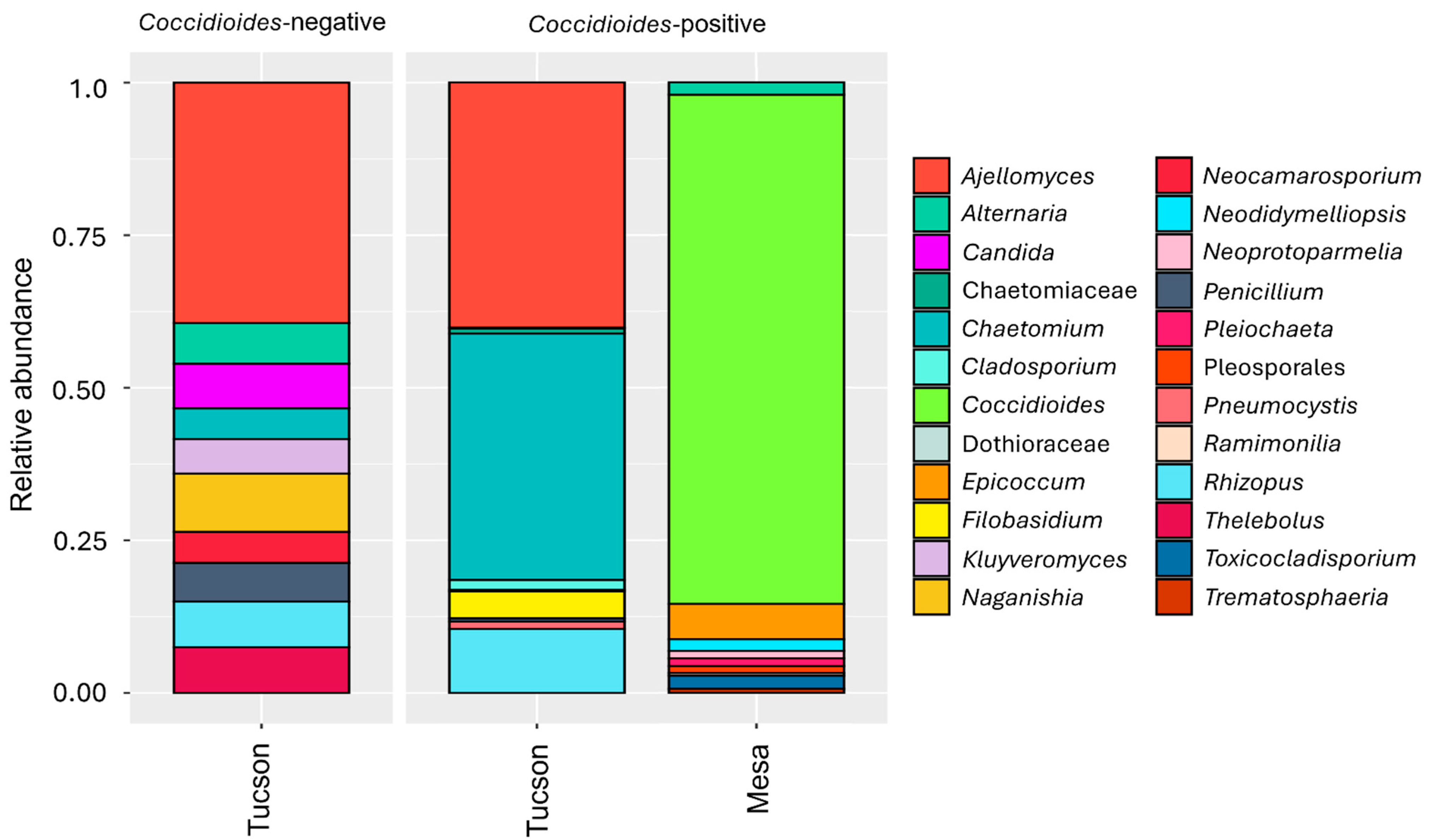
Figure 1. The ten most prevalent genera in the lung mycobiome are plotted for each site when samples are positive or negative for Coccidioides. In the Tucson site (n = 26), the ten most prevalent fungal genus are Ajellomyces (34.8%), Chaetomium (10.9%), Rhizopus (7.1%), Naganishia (6.7%), Thelebolus (5.2%), Candida (5.1%), Alternaria (4.7%), Penicillium (4.5%), Kluyveromyces (4.0%), Neocamarosporium (3.5%). In the Mesa site (n = 14), the most prevalent fungal genus are Coccidioides (78.7%), Epicoccum (5.5%), Toxicocladosporium (2.0%), Alternaria (1.9%), Neodidymelliopsis (1.8%), Pleiochaeta (1.2%), Neoprotoparmelia (1.1%), Pleosporales (1.1%), Trematosphaeria (0.6%) and Ramimonilia (0.4%).
Pathogens 14 00807 g001
Figure 2. The ten most prevalent fungal genera in lung mycobiome plotted for each host genus when they are negative or positive for Coccidioides. In Dipodomys (n = 1) the most abundant genera were Rhizopus (76.9%), Chaetomium (13.7%), Thelebolus (4.8%), Penicillium (1.5%), Chaetomiaceae (1.1%); in Onychomys (n = 1), Chaetomium (84.1%), Rhizopus (11.5%), Chaetomiaceae (1.8%), Filobasidium (1.6%), Penicillium (1.0%); in Ammospermophilus (n = 1), Ajellomyces (95.6%), Resinicium (2.1%), Mycosphaerella (1.9%), Ascomycota (0.4%); in Xerospermophilus (n = 1), Candida (78.3%), Pneumocystis (14.3%), Naganishia (4.6%), Aureobasidium (2.4%), Chaetomium (0.4%); in Chaetodipus (n = 12), Ajellomyces (68.1%), Kluyveromyces (5.8%), Thelebolus (5.4%), Pneumocystis (5.3%), Alternaria (4.1%); in Sylvilagus (n = 6), Naganishia (28.4%), Rhizopus (15.3%), Chaetomium (11.7%), Penicillium (11.7%), Filobasidium (11.7%); in Lepus (n = 4), Neocamarosporium (28.9%), Thelebolus (18.4%), Alternaria (16.5%), Candida (11.4%), Rhizopus (9.3%). In Peromyscus (n = 14; Mesa site), the most abundant genera are Coccidioides (78.7%), Epicoccum (5.5%), Toxicocladosporium (2.0%), Alternaria (1.9%), Neodidymelliopsis (1.8%), Pleiochaeta (1.2%), Neoprotoparmelia (1.1%), Pleosporales (1.1%), Trematosphaeria (0.6%) and Ramimonilia (0.4%).
Pathogens 14 00807 g002
Figure 3. The ten most prevalent orders in the lung mycobiome are plotted for each site when samples are positive or negative for Coccidioides. In the Tucson site (n = 26), the most prevalent fungal orders are Onygenales (36.1%), Sordariales (12.0%), Saccharomycetales (11%), Pleosporales (9.4%), Mucorales (8.2%), Tremellales (7.5%), Eurotiales (5.1%), Pneumocystidales (3.5%), Filobasidiales (2.9%), and Thelebolales (2.6%). In the Mesa site (n = 14), the most prevalent fungal orders are Onygenales (78.8%), Pleosporales (14.7%), Capnodiales (2.1%), Lecanorales (1.1%), Unidentified (0.5%), Mucorales (0.5%), Botryosphaeriales (0.4%), Mortierellales (0.3%), Pezizales (0.2%) and Sordariales (0.2%) and Spizellomycetales (0.2%).
Pathogens 14 00807 g003
Figure 4. Alpha diversity comparisons calculated with the Shannon diversity index (Tucson samples only). (A) Alpha diversity plot at genus level. There are no differences in alpha diversity between Coccidioides-positive and -negative samples (p = 0.138). (B) Alpha diversity plot at order level. There are no differences in alpha diversity between Coccidioides-positive and -negative samples (p = 0.209).
Pathogens 14 00807 g004
Figure 5. Beta diversity comparisons calculated with the Bray–Curtis measure of dissimilarity (Tucson samples only). (A) Beta diversity plot at genus level. There are no differences in beta diversity between Coccidioides-positive and -negative samples (p = 0.487). (B) Beta diversity plot at order level. There are no differences in beta diversity between Coccidioides-positive and -negative samples (p = 0.333).
Pathogens 14 00807 g005
Figure 6. Beta diversity comparisons at genus level calculated with the Bray–Curtis measure of dissimilarity (Tucson samples only). (A) Host genus has a significant effect on lung mycobiome beta diversity (p = 0.001). (B) Host family has a significant effect on lung mycobiome beta diversity (p = 0.001).
Pathogens 14 00807 g006
Figure 7. Assignment of fungal genera into functional categories: animal associated, plant associated, dung associated, other, unknown (no information available), or NA (OTU not identified to genus). Animal associated genera represent 89.2% of the original dataset for the Tucson samples and 88.3% for the Mesa samples.
Pathogens 14 00807 g007
Table 1. Coccidioides positivity in lung samples comparing qPCR (CocciDx) CT values and amplicon sequencing results.
SiteHost SpeciesIDCT Value
(CocciDx)
Amplicon Count 1
TucsonChaetodipusPM_43156 (0.04%)
PM_532111 (0.18%)
SylvilagusCT_638.7
OnychomysGH_M39.6
AmmospermophilusA_GS39.8
MesaPeromyscus17L38.9
24L34.5
29L38.5
31L28.6
33L36.39 (11.4%)
34L31.5431(60.4%)
35L31.53606 (95.9%)
36L33.7
37L34.1
38L36.4193 (89.8%)
39L31.7
40L34.1
41L33.3
43L33.8
1 Total amplicon reads (Relative abundance).
Disclaimer/Publisher’s Note: The statements, opinions and data contained in all publications are solely those of the individual author(s) and contributor(s) and not of MDPI and/or the editor(s). MDPI and/or the editor(s) disclaim responsibility for any injury to people or property resulting from any ideas, methods, instructions or products referred to in the content.