Article

Occurrence of Echinococcus felidis in Apex Predators and Warthogs in Tanzania: First Molecular Evidence of Leopards as a New, Definitive Host and Implications for Ecosystem Health

by
Barakaeli Abdieli Ndossi
1,2,
Eblate Ernest Mjingo
1,
Mary Wokusima Zebedayo
1,
Seongjun Choe
3,
Hansol Park
2,
Lee Dongmin
2,
Keeseon S. Eom
2,3,* and
Mohammed Mebarek Bia
2,3,*
1
Tanzania Wildlife Research Institute, Arusha P.O. Box 661, Tanzania
2
International Parasite Resource Bank, Cheongju 28644, Republic of Korea
3
Department of Parasitology, Parasitology Research Center Chungbuk National University, School of Medicine, Cheongju 28644, Republic of Korea
*
Authors to whom correspondence should be addressed.
Pathogens 2025, 14(5), 443; https://doi.org/10.3390/pathogens14050443
Submission received: 26 March 2025 / Revised: 25 April 2025 / Accepted: 28 April 2025 / Published: 30 April 2025
(This article belongs to the Special Issue Zoonotic Cestodoses: Echinococcosis and Taeniosis)

Abstract

:
(1) Background: Limited information on Echinococcus species among the wildlife in Tanzania has created a significant knowledge gap regarding their distribution, host range, and zoonotic potential. This study aimed to enhance the understanding of Echinococcus felidis transmission dynamics within the great Serengeti ecosystem. (2) Methods: A total of 37 adult Echinococcus specimens were collected from a leopard (Panthera pardus) (n = 1) in Maswa Game Reserve and 7 from a lion (Panthera leo) (n = 1) in Loliondo. Two hydatid cysts were also obtained from warthogs (n = 2) in the Serengeti National Park. (3) Results: Morphological examination revealed infertile cysts in warthogs that were molecularly identified as E. felidis. This marks the first molecular evidence of E. felidis in leopards and warthogs in Tanzania. Pairwise similarity analysis showed 98.7%–99.5% identity between Tanzanian, Ugandan, and South African isolates. Thirteen unique haplotypes were identified, with a haplotype diversity of (Hd = 0.9485) indicating genetic variability. Phylogenetic analysis grouped E. felidis into a single lineage, with the leopard isolate forming a distinct haplotype, suggesting leopards as an emerging host. Lion and warthog isolates shared multiple mutational steps, suggesting possible genetic divergence. (4) Conclusions: This study confirms African lions and leopards as definitive hosts and warthogs as potential intermediate hosts of E. felidis in the Serengeti ecosystem. Our findings highlight disease spillover risks and stress the importance of ecosystem-based conservation in wildlife–livestock overlap areas. Although E. felidis is believed to be confined to wildlife, the proximity of infected animals to pastoralist communities raises concerns for spillover. These findings highlight the importance of ecosystem-based surveillance, especially in wildlife–livestock–human interface areas.

1. Introduction

Echinococcosis is a major zoonotic disease caused by the larval (metacestodes) stage of Echinococcus (Cestoda: Taeniidae), imposing significant global medical and economic burdens [1,2]. The genus Echinococcus comprises several genetically distinct species, such as E. granulosus sensu lato (G1–G10), E. multilocularis, E. vogeli, E. oligarthra, and E. shiquicus [3,4]. Within the E. granulosus complex, molecular studies have revealed cryptic species with specific hosts associations and geographical distributions. These include E. granulosus sensu stricto (s. s.) (G1, G3), E. equinus (G4), E. ortleppi (G5), E. canadensis (G6–8, G10), and E. felidis, each exhibiting unique host specificity and epidemiological significance [5,6,7,8,9]. Adult Echinococcus spp. primarily infect definitive hosts such as domestic dogs (Canis familiaris) and wild canids [10,11,12,13]. Gravid proglottids are shed in feces, and the released eggs contaminate the environment, where they can be ingested by intermediate hosts, resulting in Cyst echinococcosis (CE) predominantly in the liver and lungs [14,15,16,17,18].
Humans serve as accidental intermediate hosts, becoming infected through ingestion of eggs via contaminated food, water, or fomites [17]. The disease can lead to severe clinical outcomes in both humans and animals, including organ failure, secondary infections, and death [19,20,21]. Despite its endemic status in several African countries, molecular clarification on Echinococcus spp., particularly in wildlife, remains limited. Existing studies are primarily from Mauritania [22], Sudan [23,24], Ethiopia [25,26], Somalia [27], Namibia [28], and Kenya [12,13,29,30,31,32], with limited references to Tanzania [33]. Reports of E. felidis in wildlife hosts have emerged from South Africa [34,35], Uganda [31,36], Kenya [37,38], and Namibia [39].
Lions (Panthera leo), spotted hyenas (Crocuta crocuta), and domestic dogs (Canis familiaris) have been identified as definitive hosts of E. felidis, whereas warthogs (Phacochoerus africanus) and hippopotamuses (Hippopotamus amphibius) are among the few documented intermediate hosts [33,35,40]. Molecular diagnostic tools targeting genes such as cytochrome oxidase subunit 1 (Cox 1), NADH dehydrogenase gene (nad1), ribosomal DNA (ITS1 & ITS2), and RAPD-PCR have enabled the precise identification and genotyping of Echinococcus species [27,37,41,42]. However, the epidemiology, geographic distribution, and host range of E. felidis in sub-Saharan Africa, especially Tanzania, remain poorly understood [43].
In Tanzania, Copro-ELISA has detected Echinococcus antigens in cheetahs, lions, and spotted hyenas [44]; however, this method may cross-react with other cestodes and lacks species level specificity [45,46]. Therefore, molecular studies are essential to better understand CE epidemiology and transmission dynamics within wildlife, livestock, and human interfaces [47]. Previous studies have emphasized the value of molecular characterization for clarifying taxonomy, epidemiology, host specificity, and ecology of Echinococcus spp., particularly in regions like sub-Saharan Africa that are underrepresented [48]. Additionally, an integrated One Health approach combining molecular and ecological studies is needed to assess zoonotic risks and inform control strategies [49]. Although Tanzania is recognized for its mega biodiversity in East Africa [50,51], molecular data on Echinococcus spp., including E. felidis in wildlife, remain scarce. This gap hinders a broad understanding of transmission patterns and epidemiological risks to livestock and humans. This study provides the first molecular confirmation of E. felidis in lions and leopards as definitive hosts and warthogs as a suspected intermediate host in the Serengeti ecosystem, providing insights into its transmission dynamics in a landscape shared by wildlife, livestock, and humans [52]. These findings provide baseline data that contribute to a better understanding of the presence of E. felidis in the Serengeti ecosystem, emphasizing the need for broader surveillance and targeted research to enhance ecosystem health and inform One Health-based control strategies.

2. Materials and Methods
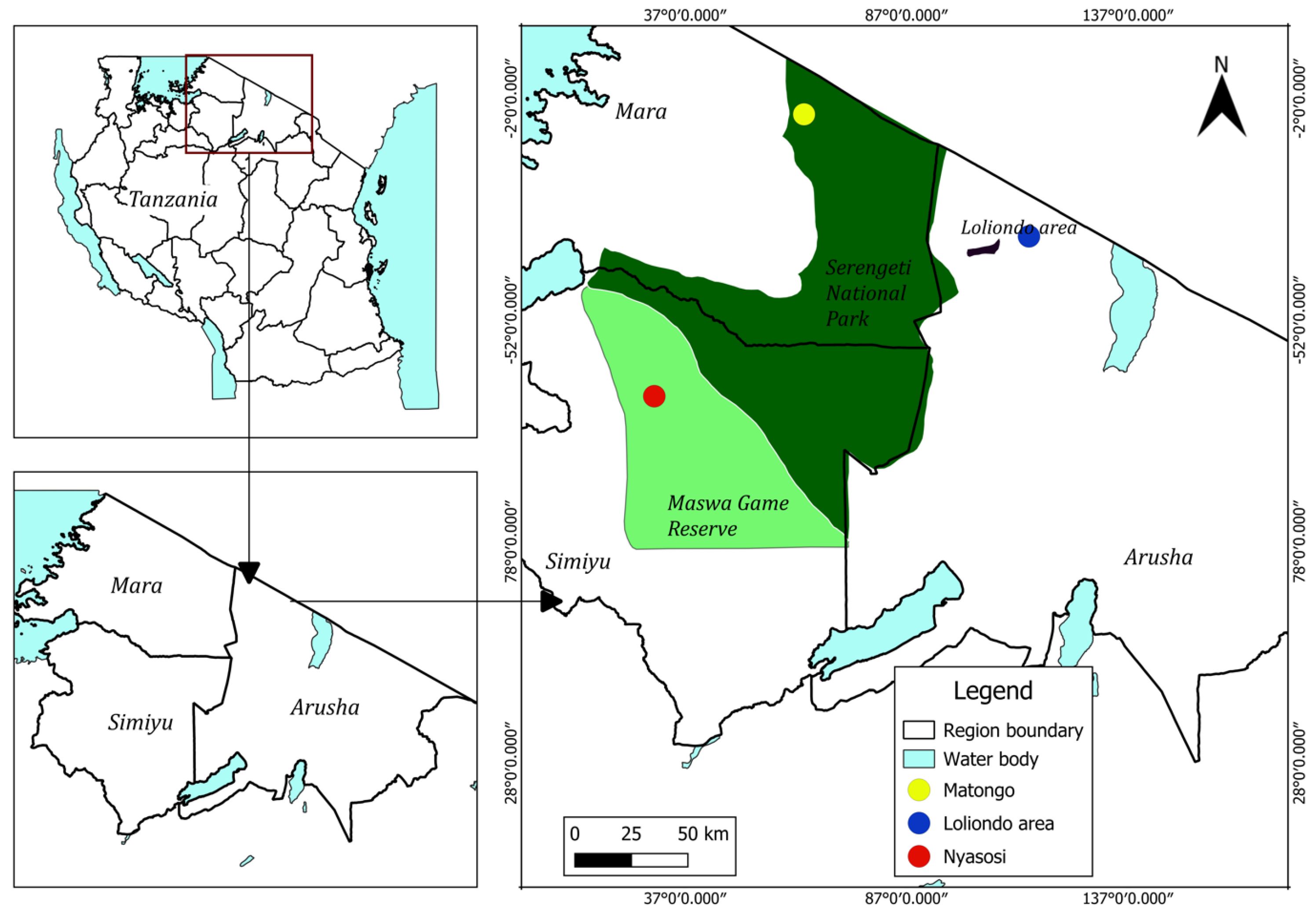
2.1. Study Area

This study was conducted between 2014 and 2024 in Serengeti National Park, Maswa Game Reserve, and the Loliondo area within the greater Serengeti ecosystem in Tanzania (Figure 1). A total of 37 adult Echinococcus spp. specimens were collected from the small intestine of a leopard (Panthera pardus) (n = 1) in the Maswa Game Reserve (S 3°18′43.07400″ S″, 34°30′30.85560″ E″), and 7 specimens were recovered from the small intestine of a lion (Panthera leo) (n = 1) in the Loliondo area (2°22′52.65120″ S″, 35°16′51.16440″ E″). Two hydatid cysts were collected from the lungs of the two warthogs (Phacochoerus africanus) (n = 2) along the Matongo area (1°38′35.81650″ S″, 34°6′13.50080″ E″) in Serengeti National Park. The cysts were spherical, thin-walled, and measured approximately 2–3 cm in diameter. Morphological examination revealed that the cysts were infertile, as no protoscoleces were observed. All animals were found dead during routine wildlife disease surveillance within the Serengeti ecosystem. Due to the conservation status of these species in Tanzania, sampling was opportunistic and restricted to carcasses obtained through authorized surveillance activities.
Each intestine was sectioned and opened lengthwise, and the contents were washed with physiological saline (0.85%) to isolate the parasites under a stereoscopic microscope [53]. Adult worms were preserved in 10% formalin for morphological examination and in 70% ethanol for molecular studies.
The collected cysts were prepared according to Thompson and McManus’ method [54]. Briefly, the hydatid liquid was drained from the collected cysts by using a 0.5 mm sterile syringe and stored in 15 mL sterilized and labelled falcon tubes with the immediate addition of 70% ethanol, then kept at room temperature. In addition, the germinal membranes from the cysts were collected, and Thompson’s procedures were utilized until further use [54].

2.2. Morphological Analysis

For the morphological analysis, a subset of 15 adult worms (10 from the leopard and 5 from the lion) were stained by using Semichon’s acetocarmine. Prior to staining, the worms were cleaned in a physiological saline solution (0.85%) to remove debris and mucus from the tegument. After cleaning, the worms were transferred to 70% ethanol for 30 min and transferred to Semichon’s acetocarmine stain for 10 h. The worms were destained in a solution of acid ethanol (Ethanol 70% with 1 mL 1 N HCl/10 mL), followed by dehydrating in 70% ethanol for 30 min, 80% Ethanol for 30 min, 90% ethanol for 30 min, and 100% × 2 ethanol for 30 min. In the last step, the worms were cleaned in xylene for 1 min and mounted on the slide by Canada balsam [55]. Morphometric parameters such as total body length, the number and size of proglottids, scolex structure, and rostellar hooks were measured and compared. Although 7 adult worms were recovered from the lion, the poor preservation state of these samples post-collection prevented detailed morphological study. Only molecular identification was performed on these specimens.

2.3. DNA Extraction and PCR Amplification

DNA was extracted from 7 individual adult worms (5 from the leopard and 2 from the lion) and from each warthog cyst using the DNeasy Blood and Tissue Kit (QIAGEN®, Hilden, Germany) according to the manufacturer’s protocol with some modifications. For the larval stage, small (<2 mm) pieces of cysts, previously kept at 4 °C in 70% ethanol, were prepared and washed with PBS x1 using a shaker (LABOGENE R100) overnight to thoroughly centrifuge the membrane at (15,345× g). Protoscoleces and adult worms were washed with PBS X1, centrifuged at (15,345× g), and had the supernatants discarded a total of three times. Immediately, the samples were crushed with a sterile pestle, and genomic DNA was extracted and kept at −20 °C until further use.
The polymerase chain reaction (PCR) was carried out targeting the Cytochrome C oxidase 1 (Cox 1) from mitochondrial genomes. The primers JB3 (5′-TTT TTT GGG CAT CCT GAG GTT TAT-3′) and JB4.5 (5′TAA AGA AAG AAC ATA ATG AAA ATG-3′) were used for the amplification of the Cox 1 gene region [56]. The PCR cocktail was prepared by using 8 μL HiPi Plus 5× PCR Master Mix, 12.5 pmol forward primer, 12.5 pmol reverse primer, 26 μL distilled water, and 4–20 ng DNA sample to make a final volume of 40 μL. The reaction was carried out in an automatic thermal cycler whereby the pre-denaturation was set for 3 min at 95 °C. Thirty-five cycles were well-set whereby in each cycle, DNA separation was conducted for 30 s at 95 °C, annealing for 30 s at 47 °C, and the extended time for DNA synthesis was 1 min for 72 °C, with a final extension step at 72 °C for 10 min. The PCR products were run on 1% agarose gel, and the images were obtained on a gel-imaging device (Gel Doc XR + System).

2.4. Molecular Analysis

The sequencing was conducted in a biomolecular company (Cosmogenetech Co., Ltd., Seoul, Republic of Korea). The sequence analysis and alignment of sequences were performed by Geneious software Version 9.0. [57]. The sequenced samples were trimmed and assembled using de novo sequence assemblers to generate the consensus sequences. As a result, Cox1 sequences of 228 bp from the lion, 258 bp from the leopard, and 224 bp from the warthog were obtained. The National Center for Biotechnology Information (NCBI) https://blast.ncbi.nlm.nih.gov/Blast.cgi (accessed on 10 March 2025) was used to determine sequence similarity and identity matches with existing reference sequences.
The obtained sequences were compared with the sequence of E. felidis and with other Echinococcus species from the GenBank (Table 1) to observe the phylogenetic relationships between individuals. In addition, Taenia solium was added in the analysis as an outgroup [58]. Using MEGA v.6. software [59] and the Maximum Likelihood method [60] to root the phylogenetic tree, Bayesian Information Criterion (BIC) value was determined with the HKY + I (Hasegawa–Kishino–Yano + invariant sites) method, with 1000 bootstrap replication estimated to obtain a high level of confidence. The haplotype networks were generated according to mutation steps by using the Network software (PopArt V.1.7) that depends on statistical parsimony after creating the data file for the Cox 1 genetic locus by utilizing DnaSP v.6. software [61].

3. Results

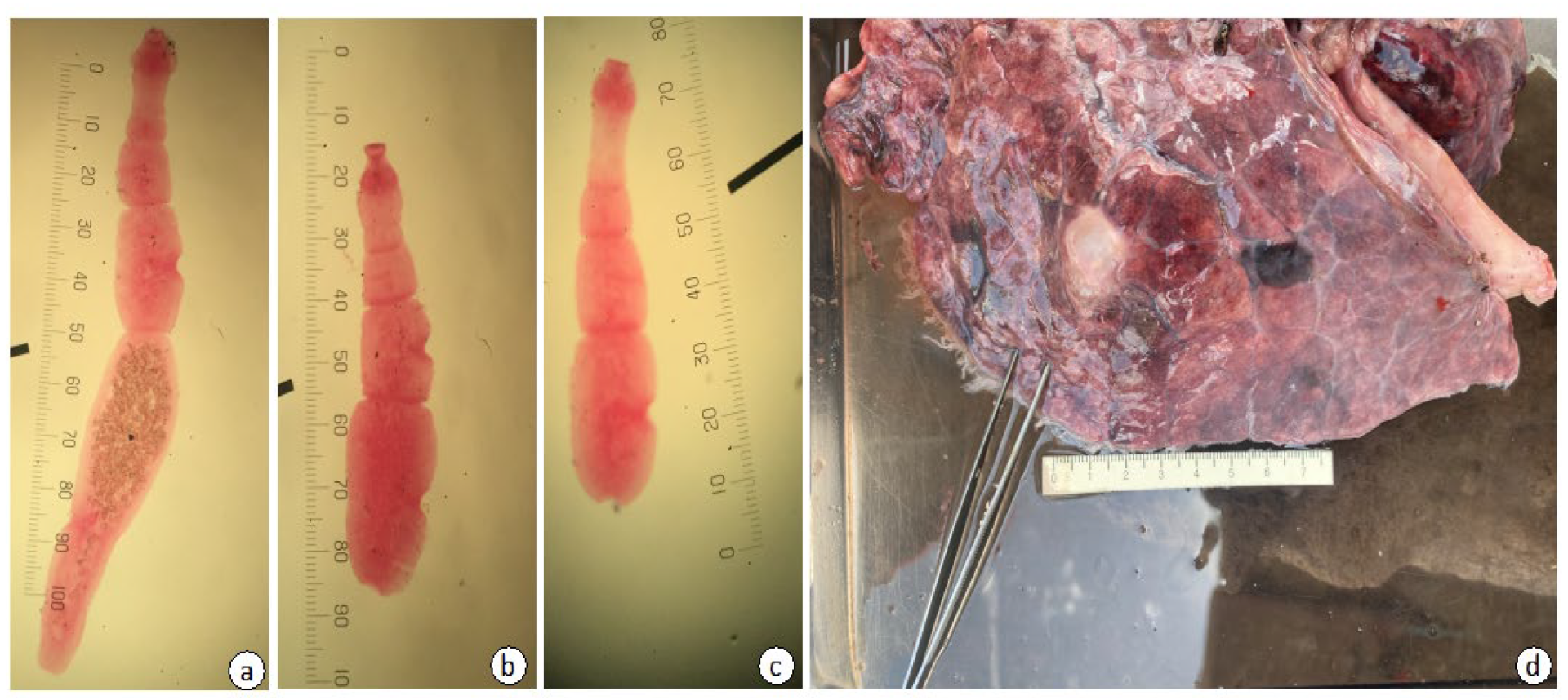
3.1. Morphological Identification of Echinococcus felidis

The morphometric features of E. felidis worms from the leopard were as follows: having four to five proglottids with a total body length ranging from 1.65 to 3.10 mm (average 2.02 mm) (Figure 2). For instance, the gravid proglottids located at the posterior end of the strobila contained immature eggs within the uterus, measuring approximately 29.63 × 39.50 µm (Figure 2). The genital pore was positioned near the middle to posterior region of the proglottids, while the number of testes varied between 12 and 60, and they were distributed both anterior and posterior to the genital pore. The uterine structure was sac-like, with no lateral branches (Figure 2), while the cirrus sac, measuring between 69.13 and 188.89 µm in length, was slightly smaller compared to other species (Table 2).

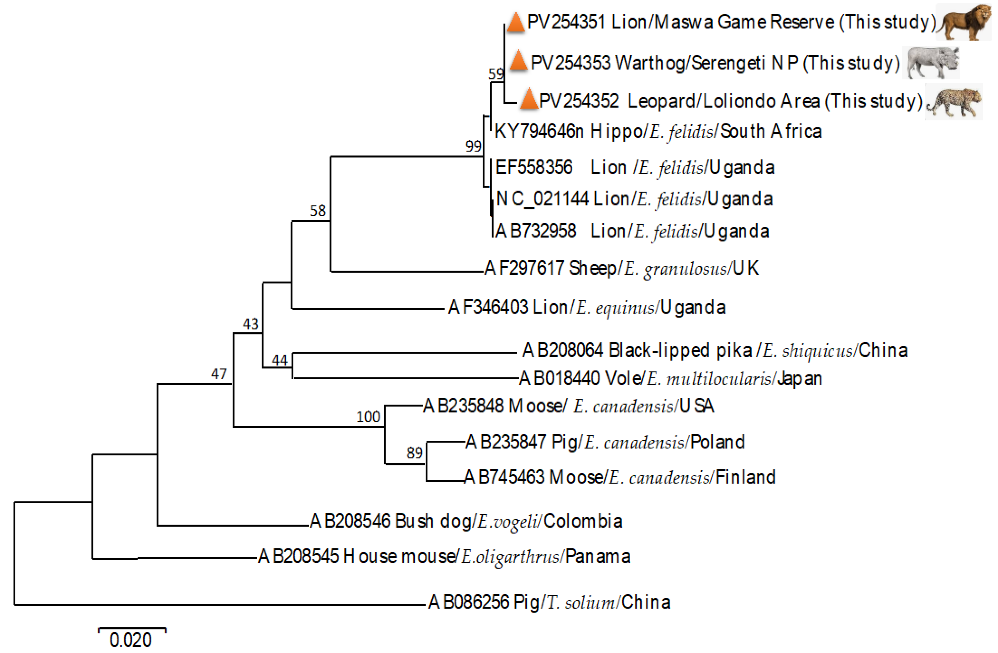
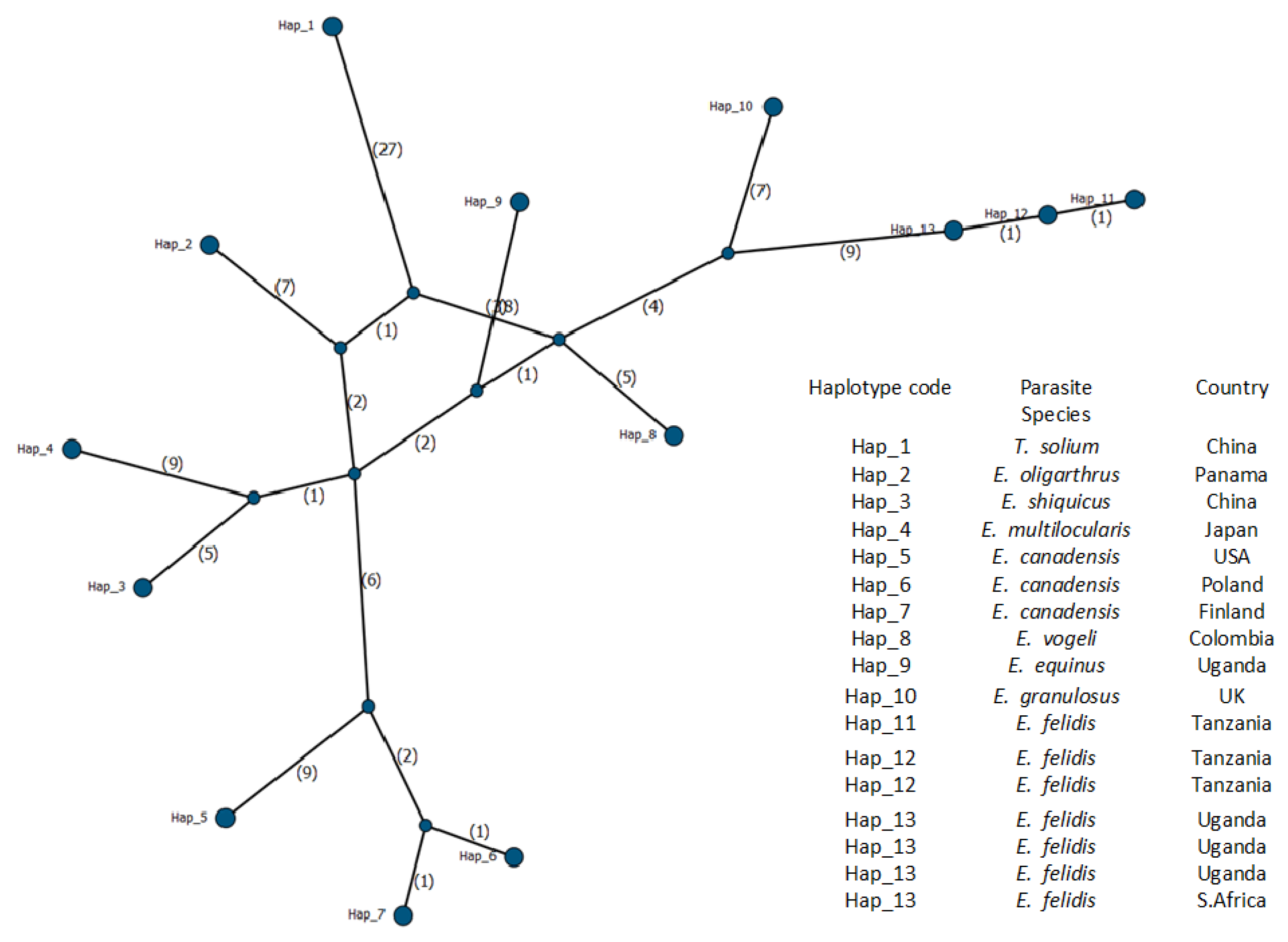
3.2. Molecular Identification of Echinococcus felidis and Phylogenetic Analysis

The obtained sequences were aligned with the published Echinococcus sequences from GenBank (Table 1). Pairwise similarity analysis revealed intraspecies variation (98.7%–99.5%) among E. felidis. The leopard isolate (PV254352) exhibited 98.7%–99.5% similarity with sequences NC_021144, AB732958, EF558356, and KY794646, while the lion isolate (PV254351) showed 99.7%–99.8% similarity with the same sequences (Figure 3). E. felidis from the warthog (PV254351) showed high genetic similarity with both lion- and leopard-derived E. felidis sequences. The close genetic relationship revealed minimal divergence among hosts. For instance, the warthog sequence showed 99.6% similarity with the lion (PV254351) and 99.5% similarity with the leopard (PV254352) sequences. Additionally, E. felidis from the warthog exhibited a high similarity of 99.7% to 99.8% with sequences from other regions, including NC_021144, AB732958, EF558356, and KY794646.
The maximum likelihood analysis revealed the phylogenetic clusters among the sequences with a high level of confidence (>95%). It placed the E. felidis sequences into a single cluster (Figure 3). The obtained genetic distance of 0.12 to 0.18 showed that E. felidis forms a distinct lineage compared with other Echinococcus species (Figure 3).
Genetic diversity and haplotype distribution were generated among the E. felidis and other Echinococcus species from various regions. A total of 17 sequences (Table 1) were used for the analysis. Only 189 informative sites were considered after excluding missing data and sites with gaps to identify genetic variation among the sequences. Among these, 57 variable sites were identified, reflecting genetic polymorphism. Among the haplotypes identified, Hap_13 was shared across different hosts, such as African lion and hippopotamus isolates from Uganda and South Africa, suggesting a widespread and possibly conserved lineage; the overall haplotype diversity (Hd) was 0.9485, indicating high genetic variation within the dataset (Figure 4).
The haplotype network revealed 13 unique haplotypes, 11 of which were represented by single sequences (Figure 4). E. felidis formed a distinct lineage clustered on a single branch that includes Hap_11, Hap_12, and Hap_13. Among these, Hap_13 appears to be the central haplotype, linking E. felidis with closely related species. Hap_12 comprises two sequences identified in the present study, one originating from a lion and the other from a warthog. The cysts recovered from the warthog were morphologically characterized as infertile. Genetic analysis confirmed the presence of E. felidis. Despite this finding, the role of warthogs as an intermediate host for E. felidis remains undefined. Hap_13 includes four previously reported E. felidis from Uganda (NC_021144, AB732958, EF558356) and South Africa (KY794646). The isolate from the leopard in this study (PV254352) formed Hap_11, which appeared genetically distinct and potentially more recently derived. Its position in the network suggests a unique lineage or emerging variant, supporting the possibility of the leopard as a new or incidental host for E. felidis.
Additionally, several haplotypes radiated from the central Hap_8, which may represent the common ancestor for the broader Echinococcus spp. lineage in this study. In contrast, Hap_1 through Hap_7 formed separate branches, reflecting host-specific divergence, geographic isolation, or localized evolutionary adaptations (Figure 4).

4. Discussion

Infection with Echinococcus spp. has been reported in various wildlife species across several African countries [69]. African lions, spotted hyenas, jackals, and wild dogs are among the definitive hosts reported with Echinococcus spp. infections [37]. Common intermediate hosts susceptible to CE include a wide range of wild herbivores, such as hippopotamuses, waterbucks, buffalo, warthogs, blue duikers, wildebeests, impalas, topis, baboons, giraffes, gazelles, and zebras [40]. These species serve as natural reservoirs for larval stages of Echinococcus spp. that are essential for maintaining the sylvatic cycle [40]. Although the hydatid cysts recovered from the warthog were infertile, molecular identification in this study provides preliminary evidence confirming E. felidis, suggesting that warthogs can harbor this species, yet its role as an intermediate host remains inconclusive.
The spatial distribution of Echinococcus spp. infections in wildlife species is influenced by ecological factors, landscape features, and regional climate, all of which affect the survival and viability of Echinococcus spp. eggs in the environment [70,71]. This study provides the first molecular confirmation of E. felidis in Tanzania, identifying an African lion and a leopard as definitive hosts. The detection of E. felidis in the leopard represents a new host record, raising important questions about host adaptability, predator–prey dynamics, and potential cross-species transmission.
Morphometric features observed in this study align with previous descriptions of E. felidis [66,67,68]. However, morphology alone is insufficient for species confirmation due to variations caused by geographical differences, host adaptation, or intraspecific morphological plasticity [72]. The molecular identification of Echinococcus spp. in livestock and humans in Tanzania have been studied [47,73]. However, there is limited information about Echinococcus spp. infections in wildlife, with only Copro-ELISA studies identifying infections in spotted hyenas and cheetahs [44]. This study fills a critical knowledge gap by providing molecular evidence of E. felidis in wild carnivores and warthogs from the Serengeti ecosystem.
The observed genetic variation in E. felidis indicates species-specific divergence and gene conservation within the genus Echinococcus [74,75]. Although we observed divergence among the isolates from the lion, leopard, and two warthogs, the relatively short fragment of the Cox1 gene used in this study may limit the resolution of genetic differentiation [76]. While Cox1 is suitable for species-level identification, more robust phylogenetic analyses would benefit from longer mitochondrial fragments or whole-genome sequencing. Further research is necessary to clarify population structure, genetic variation, and host-specific adaptations [77,78]. Phylogenetic analysis using the maximum likelihood method demonstrated clear clustering of E. felidis sequences with strong bootstrap support (>95%). The phylogenetic tree also confirmed E. felidis as a sister taxon to E. granulosus (AF297617), consistent with previous findings [65].
Our findings are supported by dietary studies showing that apex predators are more exposed to Echinococcus spp. infection due to prey consumption pattens [79,80,81]. Host population density, availability of prey, reproduction, and habitat use predator–prey interactions and influence transmission dynamics [82,83]. The detection of E. felidis in a leopard may suggest either accidental infection or potential adaptability to other carnivores driven by overlapping ecological niches and/or environmental factors.
The proximity of the Maswa and Loliondo areas to pastoralist communities, particularly the Maasai and Sukuma tribes, increases the potential for human and livestock exposure to Echinococcus species, including E. felidis [84,85]. Human activities such as bushmeat hunting, livestock grazing, and encroachment into protected areas could facilitate cross-species transmission [86]. Although E. felidis has traditionally been thought to remain confined to wildlife [43], its potential for zoonotic transmission is a growing concern, especially with the expanding human–wildlife interface. The detection of E. felidis in multiple hosts coupled with observed genetic divergence suggests a dynamic sylvatic cycle with implications for ecosystem health.
Among the limitations of this study is the small sample size, comprising one leopard, one lion, and two warthogs collected opportunistically during surveillance activities. These species are highly protected and can only be sampled under special conditions or permits in Tanzania Protected Areas. Despite this limitation, our findings provide valuable insight into the ecology and transmission of E. felidis. Broader sampling is essential to fully understand the role of different hosts and the extent of transmission.
Our study emphasizes the need for considering interspecies parasite transmission in the management of carnivore and ungulate populations in protected areas. The close genetic relationship among E. felidis isolates from different host species highlights the risk of disease spillover. Conservation strategies must incorporate disease ecology, especially in areas where wildlife, livestock, and human populations intersect.

5. Conclusions

This study provides the first molecular confirmation of Echinococcus felidis in Tanzania, identifying an African lion and a leopard as definitive hosts within the Serengeti ecosystem. Although the cysts recovered from a warthog were infertile, molecular analysis confirmed the presence of E. felidis DNA. However, the absence of viable protoscoleces made its role in transmission inconclusive. While the warthog cannot be classified as an intermediate host, its contribution to the parasite’s life cycle should not be entirely excluded. Further research is needed to clarify its ecological role and to better understand the broader transmission dynamics of E. felidis in East Africa.

Author Contributions

B.A.N., E.E.M., H.P. and L.D. were responsible for the conceptualization. M.M.B. and B.A.N. were responsible for the formal analysis. B.A.N., H.P. and M.W.Z. were responsible for the methodology. B.A.N., H.P., S.C. and L.D. were responsible for the resources. K.S.E., M.M.B. and H.P. were responsible for the validation. B.A.N. was responsible for the writing of the original draft. H.P., E.E.M., M.M.B. and B.A.N. were responsible for writing, review, and editing. All authors have read and agreed to the published version of the manuscript.

Funding

This research was funded by the International Parasite Resource Bank and Inclusive Business Solution (IBS) project, Korea (Grant No. 2020-0042-3), and the National Research Foundation of Korea (Grant No. 2020 H1 D3 A1 A0 20 81496).

Institutional Review Board Statement

The study was conducted in accordance with the Joint Management Research Committee of the Tanzania Wildlife Research Institute. Additionally, the research permits were granted by the Commission for Science and Technology Tanzania (CST00000898-2024-2024-00979, 04 October 2024).

Informed Consent Statement

Not applicable.

Data Availability Statement

All Raw-DNA sequences were deposited in The National Center for Biotechnology Information (NCBI) PV254351-PV254353.

Acknowledgments

We thank the Korea International Cooperation Agency (KOICA) and Cocoon Inc. through the Inclusive Business Solution (IBS) project for supporting field activities and the collection of samples. Thanks are also due to the International Parasite Resource Bank (iPRB) for supporting this study.

Conflicts of Interest

The authors declare no conflicts of interest.

References

  1. Yıldız, F. Echinococcus infection: The effects of echinococcosis on public health and economy. Int. J. Vet. Anim. Res. 2019, 2, 51–59. [Google Scholar]
  2. Kakundi, E.M. Molecular Epidemiology of Echinococcus and Taenia Species in Dogs from Cystic Echinococcosis Endemic Areas in Kenya. Ph.D. Thesis, University of Nairobi, Nairobi, Kenya, 2020. [Google Scholar]
  3. Lymbery, A. Phylogenetic pattern, evolutionary processes and species delimitation in the genus Echinococcus. Adv. Parasitol. 2017, 95, 111–145. [Google Scholar] [PubMed]
  4. Thompson, R.A.; McManus, D.P. Towards a taxonomic revision of the genus Echinococcus. Trends Parasitol. 2002, 18, 452–457. [Google Scholar] [CrossRef]
  5. Casulli, A.; Massolo, A.; Saarma, U.; Umhang, G.; Santolamazza, F.; Santoro, A. Species and genotypes belonging to Echinococcus granulosus sensu lato complex causing human cystic echinococcosis in Europe (2000–2021): A systematic review. Parasites Vectors 2022, 15, 109. [Google Scholar] [CrossRef] [PubMed]
  6. Nakao, M.; Lavikainen, A.; Yanagida, T.; Ito, A. Phylogenetic systematics of the genus Echinococcus (Cestoda: Taeniidae). Int. J. Parasitol. 2013, 43, 1017–1029. [Google Scholar] [CrossRef]
  7. Nakao, M.; Yanagida, T.; Konyaev, S.; Lavikainen, A.; Odnokurtsev, V.; Zaikov, V.; Ito, A. Mitochondrial phylogeny of the genus Echinococcus (Cestoda: Taeniidae) with emphasis on relationships among Echinococcus canadensis genotypes. Parasitology 2013, 140, 1625–1636. [Google Scholar] [CrossRef]
  8. Vuitton, D.; McManus, D.; Rogan, M.; Romig, T.; Gottstein, B.; Naidich, A.; Tuxun, T.; Wen, H.; Menezes da Silva, A. International consensus on terminology to be used in the field of Echinococcoses. Parasite 2020, 27, 41. [Google Scholar] [CrossRef]
  9. Wassermann, M.; Woldeyes, D.; Gerbi, B.; Ebi, D.; Zeyhle, E.; Mackenstedt, U.; Petros, B.; Tilahun, G.; Kern, P.; Romig, T. A novel zoonotic genotype related to Echinococcus granulosus sensu stricto from southern Ethiopia. Int. J. Parasitol. 2016, 46, 663–668. [Google Scholar] [CrossRef]
  10. Laurimaa, L. Echinococcus Multilocularis and Other Zoonotic Parasites in Estonian Canids. Ph.D. Thesis, University of Tartu, Tartu, Estonia, 2016. [Google Scholar]
  11. Moks, E.; Jõgisalu, I.; Valdmann, H.; Saarma, U. First report of Echinococcus granulosus G8 in Eurasia and a reappraisal of the phylogenetic relationships of ‘genotypes’ G5-G10. Parasitology 2008, 135, 647–654. [Google Scholar] [CrossRef]
  12. Nelson, G.S.; Rausch, R.L. Echinococcus infections in man and animals in Kenya. Ann. Trop. Med. Parasitol. 1963, 57, 136–149. [Google Scholar] [CrossRef]
  13. Macpherson, C.N.L.; Karstad, L.; Stevenson, P.; Arundel, J.H. Hydatid disease in the Turkana District of Kenya: III. The significance of wild animals in the transmission of Echinococcus granulosus, with particular reference to Turkana and Masailand in Kenya. Ann. Trop. Med. Parasitol. 1983, 77, 61–73. [Google Scholar] [CrossRef] [PubMed]
  14. Pal, M.; Alemu, H.H.; Marami, L.M.; Garedo, D.R.; Bodena, E.B. Cystic echinococcosis: A comprehensive review on life cycle, epidemiology, pathogenesis, clinical Spectrum, diagnosis, public health and economic implications, treatment, and control. Int. J. Clin. Exp. Med. Res. 2022, 6, 131–141. [Google Scholar] [CrossRef]
  15. Jenkins, D.; Macpherson, C. Transmission ecology of Echinococcus in wild-life in Australia and Africa. Parasitology 2003, 127, S63–S72. [Google Scholar] [CrossRef]
  16. Otero-Abad, B.; Torgerson, P.R. A systematic review of the epidemiology of echinococcosis in domestic and wild animals. PLoS Neglected Trop. Dis. 2013, 7, e2249. [Google Scholar] [CrossRef] [PubMed]
  17. Eckert, J.; Deplazes, P. Biological, epidemiological, and clinical aspects of echinococcosis, a zoonosis of increasing concern. Clin. Microbiol. Rev. 2004, 17, 107–135. [Google Scholar] [CrossRef]
  18. Thompson, R. Biology and systematics of Echinococcus. Adv. Parasitol. 1995, 95, 65–109. [Google Scholar]
  19. Deplazes, P.; Eckert, J. Veterinary aspects of alveolar echinococcosis—A zoonosis of public health significance. Vet. Parasitol. 2001, 98, 65–87. [Google Scholar] [CrossRef]
  20. Agudelo Higuita, N.I.; Brunetti, E.; McCloskey, C. Cystic echinococcosis. J. Clin. Microbiol. 2016, 54, 518–523. [Google Scholar] [CrossRef] [PubMed]
  21. Jenkins, D.; Romig, T.; Thompson, R. Emergence/re-emergence of Echinococcus spp.—A global update. Int. J. Parasitol. 2005, 35, 1205–1219. [Google Scholar] [CrossRef]
  22. Maillard, S.; Gottstein, B.; Haag, K.L.; Ma, S.; Colovic, I.; Benchikh-Elfegoun, M.C.; Knapp, J.; Piarroux, R. The EmsB tandemly repeated multilocus microsatellite: A new tool to investigate genetic diversity of Echinococcus granulosus sensu lato. J. Clin. Microbiol. 2009, 47, 3608–3616. [Google Scholar] [CrossRef]
  23. Omer, R.; Dinkel, A.; Romig, T.; Mackenstedt, U.; Aradaib, I. Strain characterization of cystic echinococcosis in livestock in Sudan. Int. J. Med. Microbiol. 2004, 293, 59. [Google Scholar]
  24. Omer, R.A.; Dinkel, A.; Romig, T.; Mackenstedt, U.; Elnahas, A.; Aradaib, I.; Ahmed, M.; Elmalik, K.; Adam, A. A molecular survey of cystic echinococcosis in Sudan. Vet. Parasitol. 2010, 169, 340–346. [Google Scholar] [CrossRef]
  25. Maillard, S.; Benchikh-Elfegoun, M.C.; Knapp, J.; Bart, J.M.; Koskei, P.; Gottstein, B.; Piarroux, R. Taxonomic position and geographical distribution of the common sheep G1 and camel G6 strains of Echinococcus granulosus in three African countries. Parasitol. Res. 2007, 100, 495–503. [Google Scholar] [CrossRef]
  26. Tigre, W.; Deresa, B.; Haile, A.; Gabriël, S.; Victor, B.; Van Pelt, J.; Devleesschauwer, B.; Vercruysse, J.; Dorny, P. Molecular characterization of Echinococcus granulosus sl cysts from cattle, camels, goats and pigs in Ethiopia. Vet. Parasitol. 2016, 215, 17–21. [Google Scholar] [CrossRef] [PubMed]
  27. Bowles, J.; Blair, D.; McManus, D.P. Genetic variants within the genus Echinococcus identified by mitochondrial DNA sequencing. Mol. Biochem. Parasitol. 1992, 54, 165–173. [Google Scholar] [CrossRef] [PubMed]
  28. Obwaller, A.; Schneider, R.; Walochnik, J.; Gollackner, B.; Deutz, A.; Janitschke, K.; Aspöck, H.; Auer, H. Echinococcus granulosus strain differentiation based on sequence heterogeneity in mitochondrial genes of cytochrome c oxidase-1 and NADH dehydrogenase-1. Parasitology 2004, 128, 569–575. [Google Scholar] [CrossRef]
  29. Wachira, T. Host influence on the rate of maturation of Echinococcus granulosus in dogs in Kenya. Ann. Trop. Med. Parasitol. 1993, 87, 607–609. [Google Scholar] [CrossRef] [PubMed]
  30. Dinkel, A.; Njoroge, E.M.; Zimmermann, A.; Wälz, M.; Zeyhle, E.; Elmahdi, I.E.; Romig, T. A PCR system for detection of species and genotypes of the Echinococcus granulosus-complex, with reference to the epidemiological situation in eastern Africa. Int. J. Parasitol. 2004, 34, 645–653. [Google Scholar] [CrossRef]
  31. Hüttner, M.; Siefert, L.; Mackenstedt, U.; Romig, T. A survey of Echinococcus species in wild carnivores and livestock in East Africa. Int. J. Parasitol. 2009, 39, 1269–1276. [Google Scholar] [CrossRef]
  32. Casulli, A.; Zeyhle, E.; Brunetti, E.; Pozio, E.; Meroni, V.; Genco, F.; Filice, C. Molecular evidence of the camel strain (G6 genotype) of Echinococcus granulosus in humans from Turkana, Kenya. Trans. R. Soc. Trop. Med. Hyg. 2010, 104, 29–32. [Google Scholar] [CrossRef]
  33. Rodgers, W. Weights, measurement and parasitic infestation of six lions from southern Tanzania. East Afr. Wildl. J. 1974, 12, 157–158. [Google Scholar] [CrossRef]
  34. Halajian, A.; Luus-Powell, W.J.; Roux, F.; Nakao, M.; Sasaki, M.; Lavikainen, A. Echinococcus felidis in hippopotamus, South Africa. Vet. Parasitol. 2017, 243, 24–28. [Google Scholar] [CrossRef] [PubMed]
  35. Young, E. Echinococcosis (hydatodosis) in wild animals of the Kruger National Park. J. S. Afr. Vet. Assoc. 1975, 46, 285–286. [Google Scholar]
  36. Hüttner, M.; Nakao, M.; Wassermann, T.; Siefert, L.; Boomker, J.D.; Dinkel, A.; Sako, Y.; Mackenstedt, U.; Romig, T.; Ito, A. Genetic characterization and phylogenetic position of Echinococcus felidis (Cestoda: Taeniidae) from the African lion. Int. J. Parasitol. 2008, 38, 861–868. [Google Scholar] [CrossRef] [PubMed]
  37. Kagendo, D.; Magambo, J.; Agola, E.L.; Njenga, S.M.; Zeyhle, E.; Mulinge, E.; Gitonga, P.; Mbae, C.; Muchiri, E.; Wassermann, M. A survey for Echinococcus spp. of carnivores in six wildlife conservation areas in Kenya. Parasitol. Int. 2014, 63, 604–611. [Google Scholar] [CrossRef] [PubMed]
  38. Mulinge, E.; Magambo, J.; Odongo, D.; Njenga, S.; Zeyhle, E.; Mbae, C.; Kagendo, D.; Addy, F.; Ebi, D.; Wassermann, M. Molecular characterization of Echinococcus species in dogs from four regions of Kenya. Vet. Parasitol. 2018, 255, 49–57. [Google Scholar] [CrossRef]
  39. Aschenborn, O.; Aschenborn, J.; Beytell, P.; Wachter, B.; Melzheimer, J.; Dumendiak, S.; Rüffler, B.; Mackenstedt, U.; Kern, P.; Romig, T. High species diversity of Echinococcus spp. in wild mammals of Namibia. Int. J. Parasitol. Parasites Wildl. 2023, 21, 134–142. [Google Scholar] [CrossRef]
  40. Hüttner, M.; Romig, T. Echinococcus species in African wildlife. Parasitology 2009, 136, 1089–1095. [Google Scholar] [CrossRef]
  41. Reddy, Y.; Rao, J.R.; Butchaiah, G.; Sharma, B. Random amplified polymorphic DNA for the specific detection of bubaline Echinococcus granulosus by hybridization assay. Vet. Parasitol. 1998, 79, 315–323. [Google Scholar] [CrossRef]
  42. Nejad, M.R.; Taghipour, N.; Nochi, Z.; Mojarad, E.N.; Mohebbi, S.; Harandi, M.F.; Zali, M. Molecular identification of animal isolates of Echinococcus granulosus from Iran using four mitochondrial genes. J. Helminthol. 2012, 86, 485–492. [Google Scholar] [CrossRef]
  43. Romig, T.; Omer, R.A.; Zeyhle, E.; Huettner, M.; Dinkel, A.; Siefert, L.; Elmahdi, I.E.; Magambo, J.; Ocaido, M.; Menezes, C.N. Echinococcosis in sub-Saharan Africa: Emerging complexity. Vet. Parasitol. 2011, 181, 43–47. [Google Scholar] [PubMed]
  44. Ernest, E.; Nonga, H.; Cleaveland, S. Prevalence of echinococcosis in dogs and wild carnivores in selected Serengeti ecosystem areas of Tanzania. Tanzan. Vet. J. 2013, 28, 1–7. [Google Scholar]
  45. Brunetti, E.; Kern, P.; Vuitton, D.A. Expert consensus for the diagnosis and treatment of cystic and alveolar echinococcosis in humans. Acta Trop. 2010, 114, 1–16. [Google Scholar] [CrossRef] [PubMed]
  46. Siracusano, A.; Bruschi, F. Cystic echinococcosis: Progress and limits in epidemiology and immunodiagnosis. Parassitologia 2006, 48, 65–66. [Google Scholar]
  47. Tamarozzi, F.; Kibona, T.; de Glanville, W.A.; Mappi, T.; Adonikamu, E.; Salewi, A.; Misso, K.; Maro, V.; Casulli, A.; Santoro, A. Cystic echinococcosis in northern Tanzania: A pilot study in Maasai livestock-keeping communities. Parasites Vectors 2022, 15, 396. [Google Scholar] [CrossRef]
  48. Thompson, R.C.A.; Deplazes, P.; Lymbery, A.J. Echinococcus and Echinococcosis, Part A. In Advances in Parasitology; Academic Press: Cambridge, MA, USA, 2017; Volume 95, ISBN 9780128114714. [Google Scholar]
  49. Thompson, R.C.A.; Deplazes, P.; Lymbery, A.J. Echinococcus and Echinococcosis, Part B. In Advances in Parasitology; Academic Press: Cambridge, MA, USA, 2017; Volume 96, ISBN 9780128114721. [Google Scholar]
  50. Heritage, N. United Nations Educational, Scientific and Cultural Organisation. UNESCO. 2014. Available online: https://ich.unesco.org/en/performing-arts-00054 (accessed on 2 January 2025).
  51. Thaxton, M. Integrating population, health, and environment in Tanzania. Life 2004, 7, 285.7. [Google Scholar]
  52. Sinclair, A.R.; Mduma, S.A.; Hopcraft, J.G.C.; Fryxell, J.M.; Hilborn, R.; Thirgood, S. Long-term ecosystem dynamics in the Serengeti: Lessons for conservation. Conserv. Biol. 2007, 21, 580–590. [Google Scholar]
  53. Avcioglu, H.; Guven, E.; Balkaya, I.; Kirman, R.; Akyuz, M.; Bia, M.M.; Gulbeyen, H.; Yaya, S. Echinococcus multilocularis in Red Foxes in Turkey: Increasing risk in urban. Acta Trop. 2021, 216, 105826. [Google Scholar] [CrossRef] [PubMed]
  54. Eckert, J.; Gemmell, M.; Meslin, F.-X.; Pawlowski, Z. WHO/OIE Manual on Echinococcosis in Humans and Animals: A Public Health Problem of Global Concern; World Organisation for Animal Health: Paris, France, 2001. [Google Scholar]
  55. Thaenkham, U.; Chaisiri, K.; Hui En Chan, A. Parasitic helminth sample preparation for taxonomic Study. In Molecular Systematics of Parasitic Helminths; Springer: Berlin/Heidelberg, Germany, 2022; pp. 225–242. [Google Scholar]
  56. Morgan, J.; Blair, D. Relative merits of nuclear ribosomal internal transcribed spacers and mitochondrial CO1 and ND1 genes for distinguishing among Echinostoma species (Trematoda). Parasitology 1998, 116, 289–297. [Google Scholar] [CrossRef]
  57. Olsen, C.; Qaadri, K.; Moir, R.; Kearse, M.; Buxton, S.; Cheung, M. Geneious R7: A bioinformatics platform for biologists. In International Plant and Animal Genome Conference XXII; Biomatters, Inc.: Newark, NJ, USA, 2014. [Google Scholar]
  58. Jeon, H.-K.; Kim, K.-H.; Eom, K.S. Complete sequence of the mitochondrial genome of Taenia saginata: Comparison with T. solium and T. asiatica. Parasitol. Int. 2007, 56, 243–246. [Google Scholar]
  59. Tamura, K.; Stecher, G.; Peterson, D.; Filipski, A.; Kumar, S. MEGA6: Molecular evolutionary genetics analysis version 6.0. Mol. Biol. Evol. 2013, 30, 2725–2729. [Google Scholar] [CrossRef] [PubMed]
  60. Felsenstein, J. Evolutionary trees from DNA sequences: A maximum likelihood approach. J. Mol. Evol. 1981, 17, 368–376. [Google Scholar] [CrossRef] [PubMed]
  61. Rozas, J.; Ferrer-Mata, A.; Sánchez-DelBarrio, J.C.; Guirao-Rico, S.; Librado, P.; Ramos-Onsins, S.E.; Sánchez-Gracia, A. DnaSP 6: DNA sequence polymorphism analysis of large data sets. Mol. Biol. Evol. 2017, 34, 3299–3302. [Google Scholar] [CrossRef] [PubMed]
  62. Nakao, M.; Sako, Y.; Ito, A. The mitochondrial genome of the tapeworm Taenia solium: A finding of the abbreviated stop codon U. J. Parasitol. 2003, 89, 633–635. [Google Scholar] [CrossRef]
  63. Nakao, M.; McManus, D.; Schantz, P.; Craig, P.; Ito, A. A molecular phylogeny of the genus Echinococcus inferred from complete mitochondrial genomes. Parasitology 2006, 134, 713–722. [Google Scholar] [CrossRef]
  64. Nakao, M.; Yokoyama, N.; Sako, Y.; Fukunaga, M.; Ito, A. The complete mitochondrial DNA sequence of the cestode Echinococcus multilocularis (Cyclophyllidea: Taeniidae). Mitochondrion 2002, 1, 497–509. [Google Scholar] [CrossRef]
  65. Le, T.; Pearson, M.; Blair, D.; Dai, N.; Zhang, L.; McManus, D. Complete mitochondrial genomes confirm the distinctiveness of the horse-dog and sheep-dog strains of Echinococcus granulosus. Parasitology 2002, 124, 97–112. [Google Scholar] [CrossRef]
  66. Verster, A.J.M. Review of Echinococcus species in South Africa. J. Vet. Res. 1965, 32, 7–118. [Google Scholar]
  67. Ortlepp, R. South African helminths Part I. J. Veterinalry Sci. Anim. Ind. 1937, 9, 311–336. [Google Scholar]
  68. Rausch, R.L. The Taxonomic Value and Variability of Certain Structures in the Cestode genus Echinococcus (Rudolphi, 1801) and a Review of Recognized Species; Thapar Commemoration; University of Nebraska: Lincoln, UK, 1953. [Google Scholar]
  69. Mathis, A.; Deplazes, P.; Eckert, J. An improved test system for PCR-based specific detection of Echinococcus multilocularis eggs. J. Helminthol. 1996, 70, 219–222. [Google Scholar] [CrossRef]
  70. Atkinson, J.A.M.; Gray, D.J.; Clements, A.C.; Barnes, T.S.; McManus, D.P.; Yang, Y.R. Environmental changes impacting Echinococcus transmission: Research to support predictive surveillance and control. Glob. Change Biol. 2013, 19, 677–688. [Google Scholar] [CrossRef] [PubMed]
  71. Di, X.; Li, S.; Ma, B.; Di, X.; Li, Y.; An, B.; Jiang, W. How climate, landscape, and economic changes increase the exposure of Echinococcus Spp. BMC Public Health 2022, 22, 2315. [Google Scholar] [CrossRef]
  72. Said, I.M.; Abdel-Hafez, S.K.; Al-Yaman, F.M. Morphological variation of Echinococcus granulosus protoscoleces from hydatid cysts of human and various domestic animals in Jordan. Int. J. Parasitol. 1988, 18, 1111–1114. [Google Scholar] [CrossRef] [PubMed]
  73. Bia, M.M.; Choe, S.; Ndosi, B.A.; Park, H.; Kang, Y.; Eamudomkarn, C.; Nath, T.C.; Kim, S.; Jeon, H.-K.; Lee, D. Genotypes of Echinococcus species from cattle in Tanzania. Korean J. Parasitol. 2021, 59, 457. [Google Scholar] [CrossRef]
  74. Maldonado, L.L.; Assis, J.; Araújo, F.M.G.; Salim, A.C.; Macchiaroli, N.; Cucher, M.; Federico Camicia, F.; Fox, A.; Rosenzvit, M.; Oliveira, G.; et al. The Echinococcus canadensis (G7) genome: A key knowledge of parasitic platyhelminth human diseases. BMC Genom. 2017, 18, 204. [Google Scholar] [CrossRef] [PubMed]
  75. Van Herwerden, L.; Gasser, R.B.; Blair, D. ITS-1 ribosomal DNA sequence variants are maintained in different species and strains of Echinococcus. Int. J. Parasitol. 2000, 30, 157–169. [Google Scholar] [CrossRef]
  76. Ebert, D.; Fields, P.D. Host–parasite co-evolution and its genomic signature. Nat. Rev. Genet. 2020, 21, 754–768. [Google Scholar] [CrossRef]
  77. Nosil, P.; Funk, D.J.; Ortiz-Barrientos, D. Divergent selection and heterogeneous genomic divergence. Mol. Ecol. 2009, 18, 375–402. [Google Scholar] [CrossRef]
  78. Doña, J.; Proctor, H.; Mironov, S.; Serrano, D.; Jovani, R. Host specificity, infrequent major host switching and the diversification of highly host-specific symbionts: The case of vane-dwelling feather mites. Glob. Ecol. Biogeogr. 2018, 27, 188–198. [Google Scholar] [CrossRef]
  79. Raoul, F.; Deplazes, P.; Rieffel, D.; Lambert, J.-C.; Giraudoux, P. Predator dietary response to prey density variation and consequences for cestode transmission. Oecologia 2010, 164, 129–139. [Google Scholar] [CrossRef]
  80. Budke, C.M.; Campos-Ponce, M.; Qian, W.; Torgerson, P.R. A canine purgation study and risk factor analysis for echinococcosis in a high endemic region of the Tibetan plateau. Vet. Parasitol. 2005, 127, 49–55. [Google Scholar] [CrossRef] [PubMed]
  81. Ziadinov, I.; Mathis, A.; Trachsel, D.; Rysmukhambetova, A.; Abdyjaparov, T.; Kuttubaev, O.; Deplazes, P.; Torgerson, P.R. Canine echinococcosis in Kyrgyzstan: Using prevalence data adjusted for measurement error to develop transmission dynamics models. Int. J. Parasitol. 2008, 38, 1179–1190. [Google Scholar] [CrossRef] [PubMed]
  82. Forbes, R.E. Lion and Leopard Diet and Dispersal in Human-Dominated Landscapes. Master’s Thesis, Nelson Mandela University, Gqeberha, South Africa, 2024. [Google Scholar]
  83. Krebs, C.J.; Myers, J.H. Population cycles in small mammals. Adv. Ecol. Res. 1974, 8, 267–399. [Google Scholar]
  84. Allan, F.K. East Coast Fever and Vaccination at the Livestock/Wildlife Interface. Ph.D. Thesis, University of Edinburgh, Old College, South Bridge, MA, USA, 2020. [Google Scholar]
  85. Hampson, K.; McCabe, J.T.; Estes, A.B.; Ogutu, J.O.; Rentsch, D.; Craft, M.; Hemed, C.B.; Ernest, E.; Hoare, R.; Kissui, B. Living in the Greater Serengeti Ecosystem: Human-Wildlife Conflict and Coexistence. Serengeti IV: Sustaining Biodiversity in a Coupled Humannatural System; The University of Chicago Press: Chicago, IL, USA, 2015; pp. 607–645. [Google Scholar]
  86. Kurpiers, L.A.; Schulte-Herbrüggen, B.; Ejotre, I.; Reeder, D.M. Bushmeat and emerging infectious diseases: Lessons from Africa. In Problematic Wildlife: A Cross-Disciplinary Approach; Springer: Berlin/Heidelberg, Germany, 2016; Volume 24, pp. 507–551. [Google Scholar]
Figure 1. Study area within the Serengeti–Mara ecosystem.
Pathogens 14 00443 g001
Figure 2. Morphological examination of adult E. felidis from the leopard (ac) showing strobila composed of 4–5 segments, with a total length ranging from 1.65 to 3.10 mm (av.2.02 mm). A hydatid cyst (d) in the lung of a warthog measured approximately 2 × 3 cm in diameter.
Pathogens 14 00443 g002
Figure 3. Phylogenetic relationship tree of the Mitochondrial Cox 1 gene of adult worms and hydatid cysts from the African lion, leopard, and warthog rooted with different sequences of Echinococcus spp. The tree shows the samples from Tanzania grouped with Echinococcus felidis sequences already published.
Pathogens 14 00443 g003
Figure 4. Median-joining haplotype network of 13 mitochondrial Cox1 haplotypes of Echinococcus spp. Haplotypes are represented by circles scaled to sequence frequency. E. felidis forms a distinct cluster (Hap_11–Hap_13), with Hap_13 linking previously reported isolates from Uganda and South Africa. Hap_12 includes lion and warthog samples from this study, while Hap_11, from a leopard, appears genetically distinct. The central position of Hap_8 suggests an ancestor haplotype, with peripheral haplotypes (Hap_1–Hap_7) reflecting host and geographic divergence.
Pathogens 14 00443 g004
Table 1. Reference sequences of Echinococcus species analyzed in this study.
Host NameScientific NameCountryParasite
Species
Haplotype CodeAccession NumberReferences
PigSus scrofaChinaTaenia soliumHap_1AB086256[62]
House mouseMus musculusPanamaE. oligarthrusHap_2AB208545[63]
Black-lipped pikaOchotona curzoniaeChinaE. shiquicusHap_3AB208064[63]
VoleCraseomys rufocanusJapanE. multilocularisHap_4AB018440[64]
MooseAlces alcesUSAE. canadensisHap_5AB235848[63]
PigSus scrofaPolandE. canadensisHap_6AB235847[63]
MooseAlces alcesFinlandE. canadensisHap_7AB745463[7]
Bush dogSpeothos venaticusColombiaE. vogeliHap_8AB208546[63]
African lionPanthera leoUgandaE. equinusHap_9AF346403[65]
SheepOvis ariesUKE. granulosusHap_10AF297617[65]
African lionPanthera leoTanzaniaE. felidisHap_11PV254351This study
LeopardPanthera pardusTanzaniaE. felidisHap_12PV254352This study
WarthogPhacochoerus africanusTanzaniaE. felidisHap_12PV254353This study
African lionPanthera leoUgandaE. felidisHap_13NC_021144[7]
African lionPanthera leoUgandaE. felidisHap_13AB732958[7]
African lionPanthera leoUgandaE. felidisHap_13EF558356[36]
HippopotamusHippopotamus amphibiusS. AfricaE. felidisHap_13KY794646[34]
Table 2. Morphometric features of E. felidis isolates from the leopard in relation to others.
FeatureE. felidis from Leopard
(This Study)
E. felidis Verster, 1965 [66]E. felidis Ortlepp, 1935 [67]E. felidis
Raush, 1953 [68]
Body length (mm)1.65–3.10 (av.2.02)2.12–5.220
(av.3.239) ± 0.3
3.42–5.22 (av.4.21 ± 0.6)6 mm
Number of segments4.0–5.03 (18.5%), 4 (47.0%),
5 (34.5%)
4–5 usually 43.0–4.0
Number of hooklets28–32---
Number of testes12.0–60.028–45 (av.35.9 ± 3.5)30–46 (av.6 ± 4.2)32–46
Cirrus sac69.13–188.89 (av.128.39 µm long, 49.38–164.19 µm (av.88.88 µm))Mt: 82.8–202.4 (129.4 ± 2.4) µm long, 46.0–105.8 (64.9 ± 99) µm with gravid segment 147.2–184 (159.9 ± 13.7 µm, 56.0–79.2 (717 ± 9.3))Mt: 115.0–174.8 (148.2 ± 15.5 µm long, 55.2–92.0 (77.3 + 133)) width. Gravid segment 142.6–207.0 (176.9 ± 21.6) µm, 64.4–96.6 (83.2 ± 10.2) µm-
Egg size29.63 × 30.50 µm--37–40.5 (32–35.5 µm)
Disclaimer/Publisher’s Note: The statements, opinions and data contained in all publications are solely those of the individual author(s) and contributor(s) and not of MDPI and/or the editor(s). MDPI and/or the editor(s) disclaim responsibility for any injury to people or property resulting from any ideas, methods, instructions or products referred to in the content.

