Article

Comparative Proteomic Insights into the Immune Response of Conogethes punctiferalis Challenged with Beauveria bassiana

1
College of Plant Protection, Shandong Agricultural University, Tai’an 271018, China
2
College of Resources and Environment, Shandong Agricultural University, Tai’an 271018, China
3
College of Life Science, Hebei University, Baoding 071002, China
4
Institute of Plant Protection, Shandong Academy of Agricultural Sciences, Jinan 250100, China
5
Institute of Agricultural Resources and Regional Planning, Chinese Academy of Agricultural Sciences, Beijing 100081, China
*
Authors to whom correspondence should be addressed.
Insects 2025, 16(7), 667; https://doi.org/10.3390/insects16070667
Submission received: 17 May 2025 / Revised: 17 June 2025 / Accepted: 24 June 2025 / Published: 26 June 2025
(This article belongs to the Section Insect Physiology, Reproduction and Development)

Simple Summary

The yellow peach moth (YPM), Conogethes punctiferalis, is a polyphagous insect pest that has been reported to cause severe damage to many crop species in China. Beauveria bassiana, as a common and effective entomopathogenic fungus, is extensively used for the biocontrol of various insect pests. In this study, we carried out the proteomic analysis of YPM larvae infected with B. bassiana using the isobaric tags for relative and absolute quantification (iTRAQ) technique. The immune-related proteins were screened based on the proteome data. The differentially expressed proteins (DEPs) were screened and identified, and then these DEPs were subjected to functional enrichment analysis. The accuracy and reliability of the proteome sequencing data were validated by qRT-PCR. Our results offer a new insight into the immune responses of YPM larvae infected with B. bassiana at the protein level and contribute to understanding the immune mechanisms of YPM larvae in response to B. bassiana infection.

Abstract

The yellow peach moth (YPM), Conogethes punctiferalis, is an important agricultural insect pest causing severe damage to corn in eastern China. Beauveria bassiana is an effective, eco-friendly, and promising alternative agent for controlling this insect pest. However, insect immunity can limit the ability of fungal infections. In order to understand the immune response mechanism of YPM, a comparative proteomic analysis of non-infected and B. bassiana-infected larvae was conducted using the isobaric tags for relative and absolute quantification (iTRAQ) technique. On the basis of proteomic analysis, 4195 quantifiable proteins were identified from a total of 29,155 peptides. The functions of the identified proteins were annotated in four databases (GO, COG, KEGG, and IPR). A total of 132 immune-related proteins were screened, including 46 pathogen recognition proteins, 27 extracellular signal modulation proteins, and 59 immune effectors. Furthermore, 70 differentially expressed proteins (DEPs) were identified, including 57 up-regulated proteins and 13 down-regulated proteins. Among these, four DEPs were related to immunity, namely one defense protein and three peptidoglycan recognition proteins. Six randomly selected immune-related proteins associated with target genes were validated for qRT-PCR, and the results indicated that the accuracy and reliability of the proteome sequencing data were high. Taken together, the results enrich the fundamental knowledge of YPM immune responses to B. bassiana infection and provide a new insight into insect−pathogen interactions.

1. Introduction

The yellow peach moth (YPM), Conogethes punctiferalis (Lepidoptera: Crambidae), is an important polyphagous insect pest extensively distributed across subtropical and tropical Asia and Australia and can attack over 100 species of field crops, fruits, and vegetables [1,2]. In China, Ostrinia furnacalis is considered a highly destructive insect pest of corn, and yet YPM has replaced O. furnacalis as the dominant insect pest on corn in the Huang-Huai-Hai region in recent years [3,4]. Currently, the utilization of chemical insecticides is the most common method for controlling YPM populations [5]. Regrettably, the overuse of chemical insecticides can lead to undesirable consequences, including environmental pollution and insect resistance to chemical insecticides [6,7].
Entomopathogenic fungi have been developed as an eco-friendly alternative to chemical insecticides because of their specific mode of action and ability to infect a vast array of insect pests [8,9]. Among them, Beauveria and Metarhizium are widely applied for the biocontrol of diverse insect pests [10,11]. For example, the utilization of Beauveria bassiana in the management of Dendrolimus punctatus has become a successful biological control scheme in China [12]. A highly virulent strain of Metarhizium pingshaense had a mortality in excess of 86% against YPM larvae under laboratory conditions [13]. The potential suppression of YPM larvae by B. bassiana would make it a promising alternative agent for controlling the insect pest in a previous study [14]. In fact, the well-developed innate immunity of insects to entomopathogenic fungi greatly limits the development and application of fungal biopesticides [15]. Despite lacking adaptive immunity in insects, their powerful innate immune systems, consisting of cellular and humoral immunity, are still capable of eliminating microbial pathogenic infections [16,17]. Therefore, it is crucial to explore the immune mechanisms of insects for enhancing the efficiency of fungal infections.
Due to the rapid development of molecular biology technology, numerous studies on insect immunity have been reported based on genome, transcriptome, and proteome analyses [18,19,20]. The genome of YPM has recently been published, constituting invaluable information for exploring the evolutionary mechanism of the YPM immune system [21]. However, proteins participating in the YPM immune response have not yet been comprehensively characterized. Proteomic analysis is able to provide highly valuable insights into the changes to the proteins in response to a specific stimulus or stress [22]. The isobaric tags for relative and absolute quantification (iTRAQ) technique has recently been increasingly applied due to its high sensitivity and accurate quantification [23,24,25]. To obtain more information on the immune responses of YPM to B. bassiana infection, the present study firstly reports a comparative iTRAQ-based quantitative proteomic analysis of YPM larvae infected with B. bassiana, thereby providing new insights into the immune molecular mechanism of insect−pathogen interactions.

2. Materials and Methods

2.1. Insect Rearing

YPM larvae were fed on fresh corn kernels in an artificial climate chamber (RXZ-380C, Ningbo, Zhejing, China) under the conditions of 25 ± 1 °C, 70 ± 5% relative humidity, and a 14 h light (with 5000 lx)/10 h dark cycle. The trials were conducted using three-day-old 5th-instar larvae.

2.2. Fungal Infection

B. bassiana strain (ACCC30107) was cultured on potato dextrose agar (PDA) plates at 26 °C and 80% relative humidity. Mature conidia were collected and the suspensions were prepared using sterile phosphate-buffered saline (PBS, pH = 7.4). The concentration was adjusted to 1 × 10⁵ conidia/μL by the hemocytometer. Three-day-old 5th-instar larvae were inoculated with 2 μL conidial suspensions (1 × 10⁵ conidia/μL) or sterile PBS (as control) using a microinjector (Hamilton, Bonaduz, Switzerland). Each treatment or control group included 30 larvae. Each bioassay was performed in triplicate. According to a previous study [14], the whole body of YPM larvae was frozen in liquid nitrogen at 36 h post-infection for further use.

2.3. Total Protein Extraction

The whole YPM larvae were ground into powder in liquid nitrogen and immediately transferred to the pre-cooled centrifuge tubes. The PASP protein lysis buffer (100 mmol/L ammonium bicarbonate, 8 mol/L urea, pH = 8) was added and then vortexed and mixed thoroughly. The samples were subjected to 5 min of ultrasonication in an ice-water bath to ensure complete lysis. The lysate was centrifuged (12,000× g, 4 °C) for 15 min, and the supernatant was collected. The dithiothreitol (DTT, 10 mmol/L) was added to the mix for 1 h at 56 °C. The iodoacetamide (IAM) was then added and placed for 1 h at room temperature in a condition of complete darkness. The above mixture was precipitated with four times the volume of pre-cooled acetone at –20 °C for at least 2 h, and the precipitation was collected using a centrifuge (12,000× g, 4 °C) for 15 min. Subsequently, the precipitation was resuspended and washed with 1 mL of –20 °C pre-cooled acetone and collected again by centrifuging at 12,000× g for 15 min at 4 °C. The samples were air-dried, and the protein dissolving solution (8 mol/L urea, 100 mmol/L triethylammonium bicarbonate (TEAB), pH = 8.5) was added to dissolve protein precipitation.

2.4. Protein Quality Test

According to the method of the Bradford protein quantitative kit, Bovine serum albumin (BSA) standard protein solution was prepared. The concentration gradient ranged from 0 to 0.5 g/L. BSA standard protein solutions with different concentration gradients were prepared and added into a 96-well plate. The sample solutions were then diluted to different concentrations and also added into the above plate. The volume of each well was 20 µL, and each gradient was repeated three times. A volume of 180 μL Coomassie Brilliant Blue G-250 dye solution was quickly added into the plate and left for 5 min at room temperature. The absorbance at 595 nm was then detected. The standard curve was drawn according to the absorbance, and the concentration of the protein samples was then calculated.

2.5. iTRAQ Labeling

The protein samples were made up to 100 μL using the dissolution buffer (DB buffer) (8 mol/L urea, 100 mmol/L TEAB, pH = 8.5). Trypsin (1 μg/μL) and TEAB (100 mmol/L) buffer were added, mixed well, and the mixture was digested at 37 °C for 4 h. Formic acid was added, used to adjust the pH to <3, and mixed well. The samples were then centrifuged (12,000× g) for 5 min. The sample was gradually loaded onto the C18 desalting column and washed three times consecutively with the washing buffer (0.1% formic acid, 3% acetonitrile). A volume of 300 μL eluent solution (0.1% formic acid, 70% acetonitrile) was added, and then the eluents were collected and lyophilized. A volume of 20 μL of 1 mol/L TEAB buffer was added to reconstitute. Each sample was added 5.5 μL of iTRAQ labeling reagent and mixed with shaking for 2 h. After that, a volume of 100 μL of 50 mmol/L Tris-HCl (pH = 8) was added and used for terminating the reaction. The labeling samples were mixed at an equal volume, desalted, and then lyophilized.

2.6. Separation of Fractions

The mobile phases A (2% acetonitrile, 98% water, pH = 10) and B (98% acetonitrile) were prepared to achieve a gradient elution effect. Solution A with dissolved lyophilized powder was centrifuged (14,000× g, 4 °C) for 20 min. The samples were fractionated using a Waters BEH C18 (Waters Corporation, Milford, MA, USA) chromatographic column (4.6 × 250 mm, 5 μm) on the L-3000 HPLC system. A tube was collected every minute, combined into ten fractions, lyophilized, and dissolved by the addition of 0.1% (v/v) formic acid.

2.7. Liquid Chromatography and Mass Spectrometry (LC-MS/MS) Analysis

The shotgun proteomic analyses were carried out using an EASY-nLCTM 1200 UHPLC system (Thermo Fisher, Waltham, MA, USA) and a Q ExactiveTM HF-X mass spectrometer (Thermo Fisher, Waltham, MA, USA). Samples were added into a C18 Nano-Trap column (4.5 cm × 75 μm, 3 μm). The separation of peptides was achieved through the utilization of an analytical column (15 cm × 150 μm, 1.9 μm) employing a linear gradient elution technique (Table S1). The separated peptides were checked by means of a Q ExactiveTM HF-X mass spectrometer. The full scanning range was from 407 to 1500 m/z. The automatic gain control (AGC) target value was set at 3 × 10⁶, and the maximum ion injection time was 20 ms. The top 40 most abundant precursors were selected for fragmentation, followed by detection using MS/MS. The raw data used for MS detection was designated “.raw”.

2.8. Identification and Quantitation of Proteins

The resulting spectra of each run were searched individually in the protein database (761199-X101SC21043487-Z01-Conogethes_punctiferalis-customer.pep.fasta (38033 sequences)) using the Proteome Discoverer 2.2 software. The searched parameters were set as follows: a mass tolerance of 10 mg/L for precursor ions and a mass tolerance of 0.02 Da for the product ions. The Proteome Discoverer 2.2 software was used to filter the retrieval results. The peptide spectrum matches (PSMs) with over 99% credibility were identified, and credible proteins contained a minimum of one specific peptide. Only credible PSMs and proteins were retained and conducted with FDR less than 1%. The results of the protein quantitation were analyzed using a t-test. The differentially expressed proteins (DEPs) (the up-regulated expression protein: fold change (FC) ≥ 1.5, p value ≤ 0.05; the down-regulated expression protein: FC ≤ 0.67, p value ≤ 0.05) were screened and identified.

2.9. Functional Annotation and Enrichment Analysis

Gene Ontology (GO) and InterPro (IPR) functional annotation were performed using the IterProScan 5.0 software in the non-redundant protein database (Pfam, PRINTS, ProDom, SMART, ProSite, and PANTHER). Clusters of Orthologous Groups (COG) and Kyoto Encyclopedia of Genes and Genomes (KEGG) annotations were conducted by subjecting the identified proteins to BLAST 2.2.26 software comparison (blastp, E value ≤ 10−4), then filtering the BLAST results for each sequence and selecting the comparison with the highest score for annotation. The protein family and pathway analyses were conducted through COG and KEGG [26]. The cluster heatmap, GO, KEGG pathway, and IPR enrichment analyses of DEPs were performed [27]. The cloud tools at NovoMagic (https://magic-plus.novogene.com/#/tool/list, accessed on 7 September 2024) were used for data visualization.

2.10. qRT-PCR Validation

The total RNA of YPM larvae was extracted and reverse transcribed into cDNA according to the kit method (Tiangen, Beijing, China). Specific primers were designed by Primer Premier 6 software (Table S2), and ribosomal protein 49 (RP49) was selected as the internal reference gene. The reaction system with a volume of 20 μL, consisting of 10 μL SuperReal PreMix Plus (2×), 1 μL upstream and downstream primers, 1 μL cDNA template, and 7 μL RNase-free ddH2O, was used for qRT-PCR by Bio-Rad CFX96 Touch Real Time PCR Detection System (Bio-Rad, Hercules, CA, USA). The reaction conditions were as follows: pre-denaturation at 95 °C for 15 min, followed by 40 cycles of denaturation at 95 °C for 10 s and annealing/extension at 60 °C for 30 s. All samples were analyzed in triplicate and repeated thrice as independent biological replicates. The qRT-PCR data were calculated using the 2−ΔΔCT method [28].

3. Results

3.1. Identification and Quality Control of Proteome

In the present study, a total of 62,669 (11.08%) matched spectra in the 565,469 total spectra and 29,155 peptides, 4197 identified proteins, and 4195 all quantifiable proteins were detected and identified (Table 1).
Quality control of proteome data was performed to ensure accuracy and reliability. The results showed that more than 90% of the proteins had a coverage between 0 and 0.5 (Figure 1A). In total, 766 proteins with a mass ranging from 20 to 30 kDa were identified, followed by 701, 633, and 523 proteins with a mass of 10–20, 30–40, and 40–50 kDa, respectively. The remaining 346 proteins had a mass greater than 100 kDa (Figure 1B).

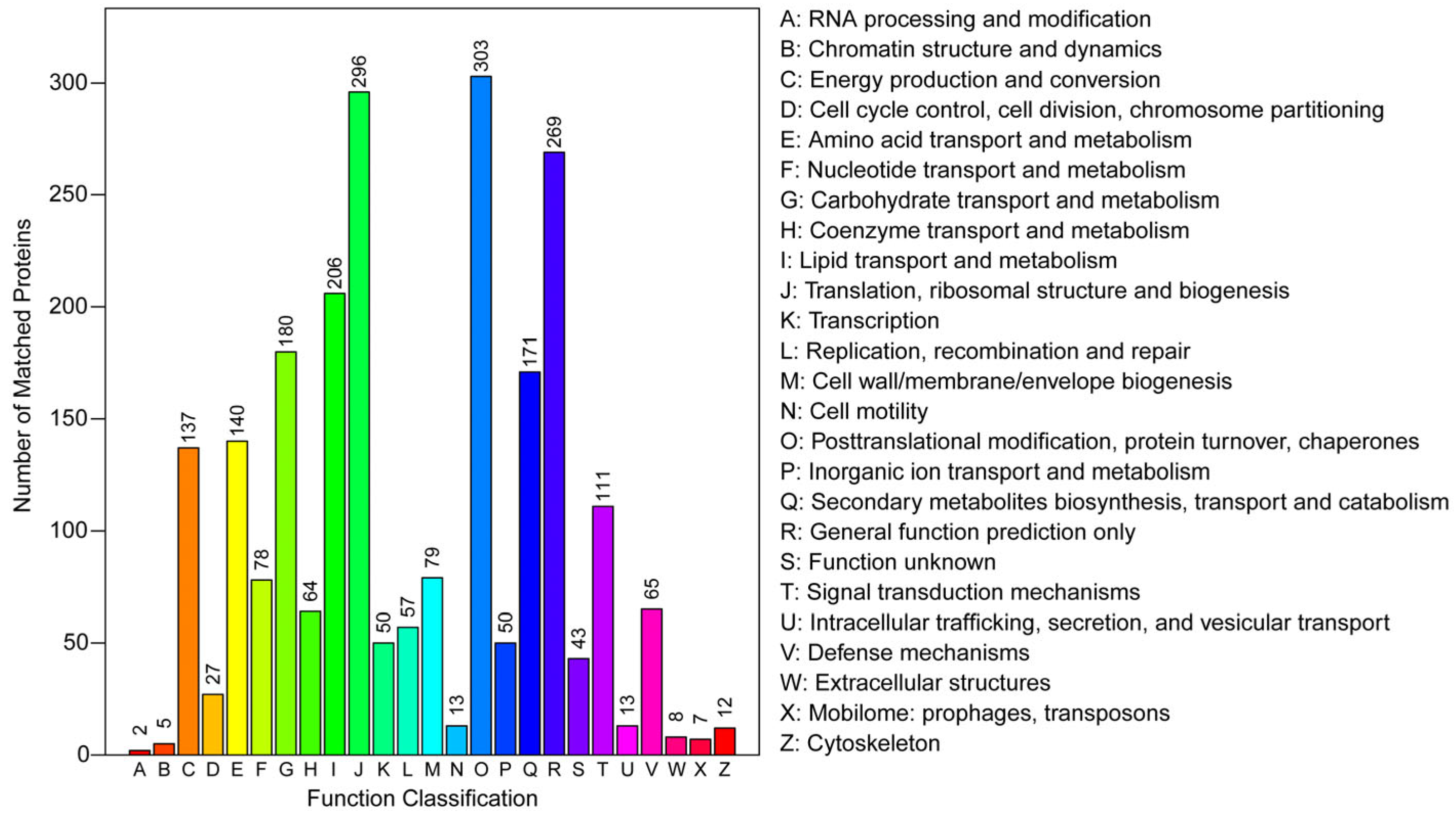
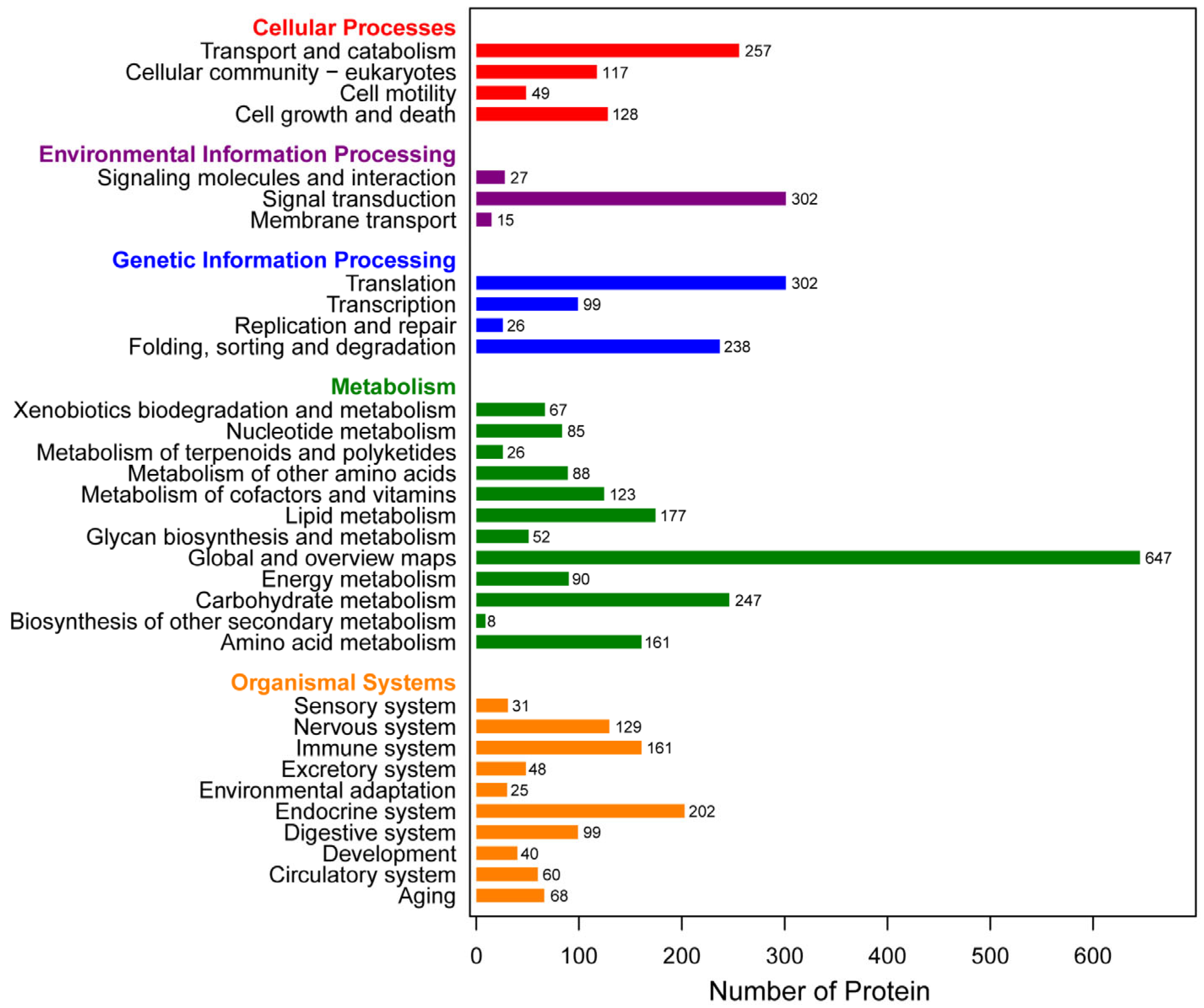
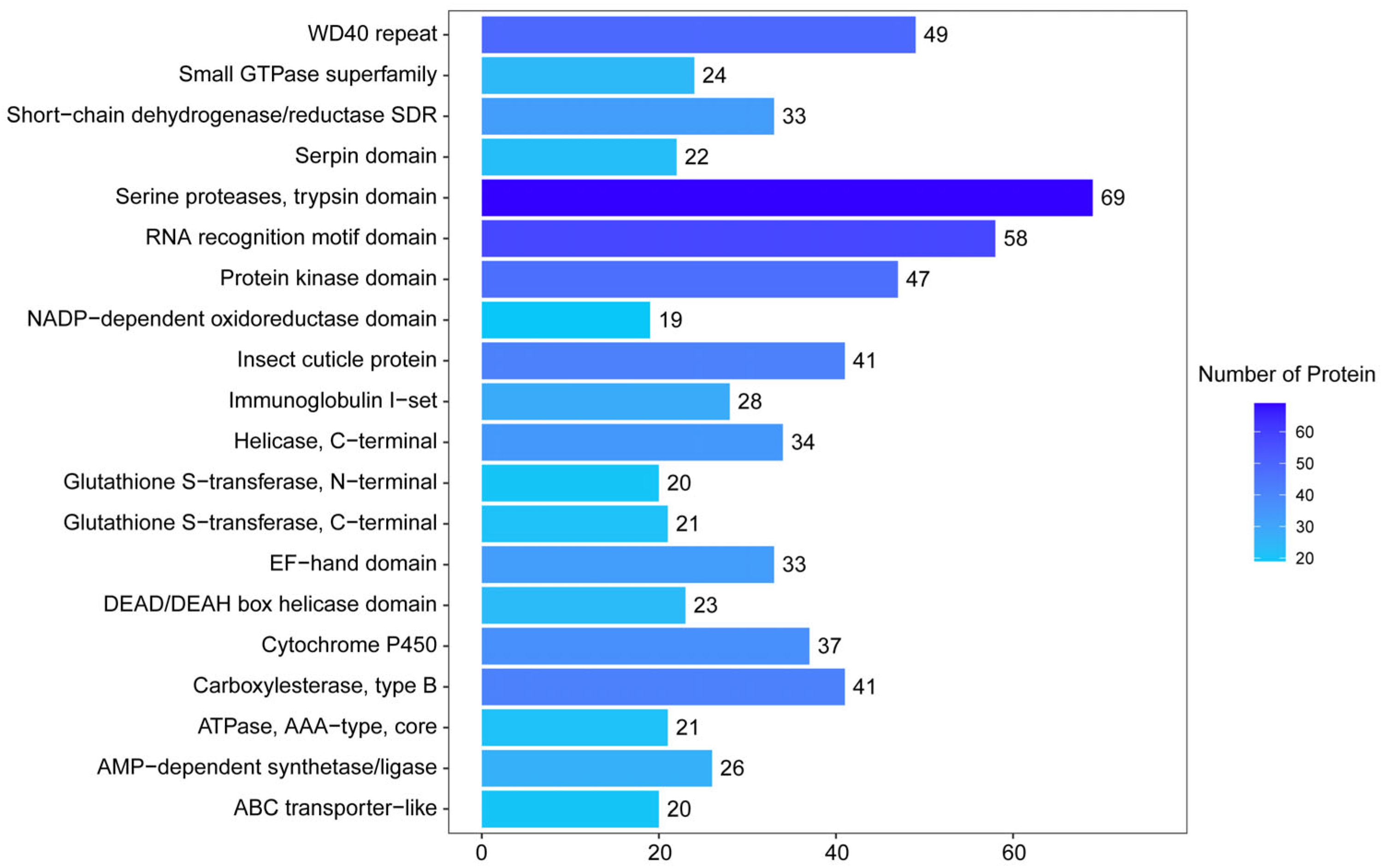
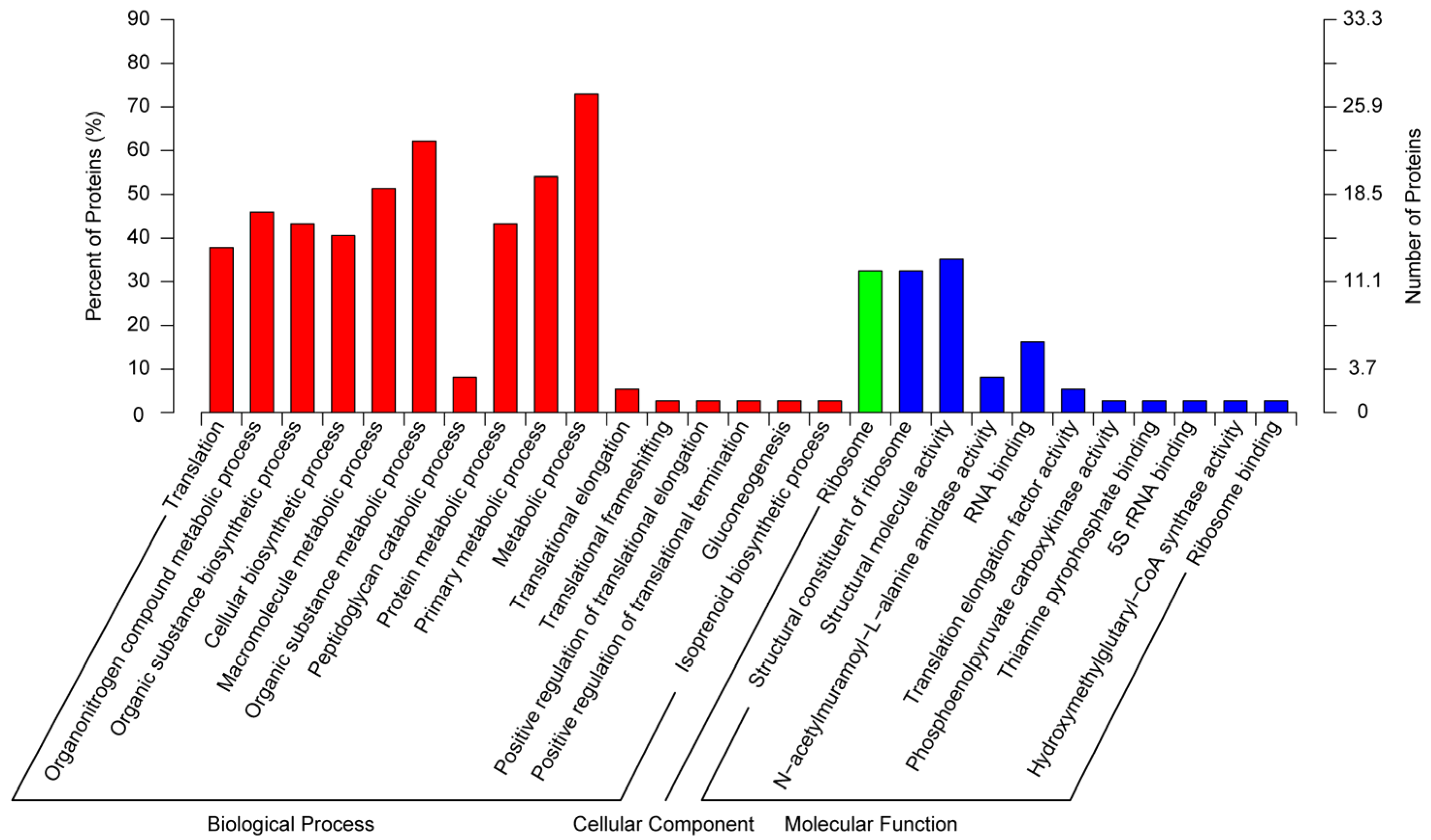
3.2. Functional Annotation and Classification of Proteome

According to the GO annotations, a total of 2312 proteins were divided into three categories (Figure 2). In biological processes, the top three most frequent categories were oxidation–reduction process (206 proteins), metabolic process (139 proteins), and proteolysis (130 proteins). In cellular components, the top three most frequent categories were integral components of the membrane (104 proteins), intracellular (97 proteins), and ribosome (72 proteins). In molecular function, the top three most frequent categories were protein binding (337 proteins), ATP binding (230 proteins), and nucleic acid binding (104 proteins).
A total of 2122 proteins were classified into 25 categories based on the COG function classification (Figure 3). Among these, the largest group was posttranslational modification, protein turnover, and chaperones (303 proteins), followed by translation, ribosomal structure, and biogenesis (296 proteins), and general function prediction only (269 proteins).
In total, 4107 proteins were mapped against the KEGG pathway and were categorized into five groups (Figure 4). The most enriched pathways were transport and catabolism (257 proteins), signal transduction (302 proteins), translation, global, and overview maps (647 proteins), and the endocrine system (202 proteins) in each category.
The IPR analysis showed that a total of 3505 proteins were annotated, and the top three most frequent categories were serine protease, trypsin domain (69 proteins), RNA recognition motif domain (58 proteins), and WD40 repeat (49 proteins) (Figure 5).

3.3. Identification of the Immune-Related Proteins

A total of 132 immune-related proteins were screened and identified based on the YPM larval proteome data, including 46 pathogen recognition proteins, 27 extracellular signal modulation proteins, and 59 immune effectors (Table S3).

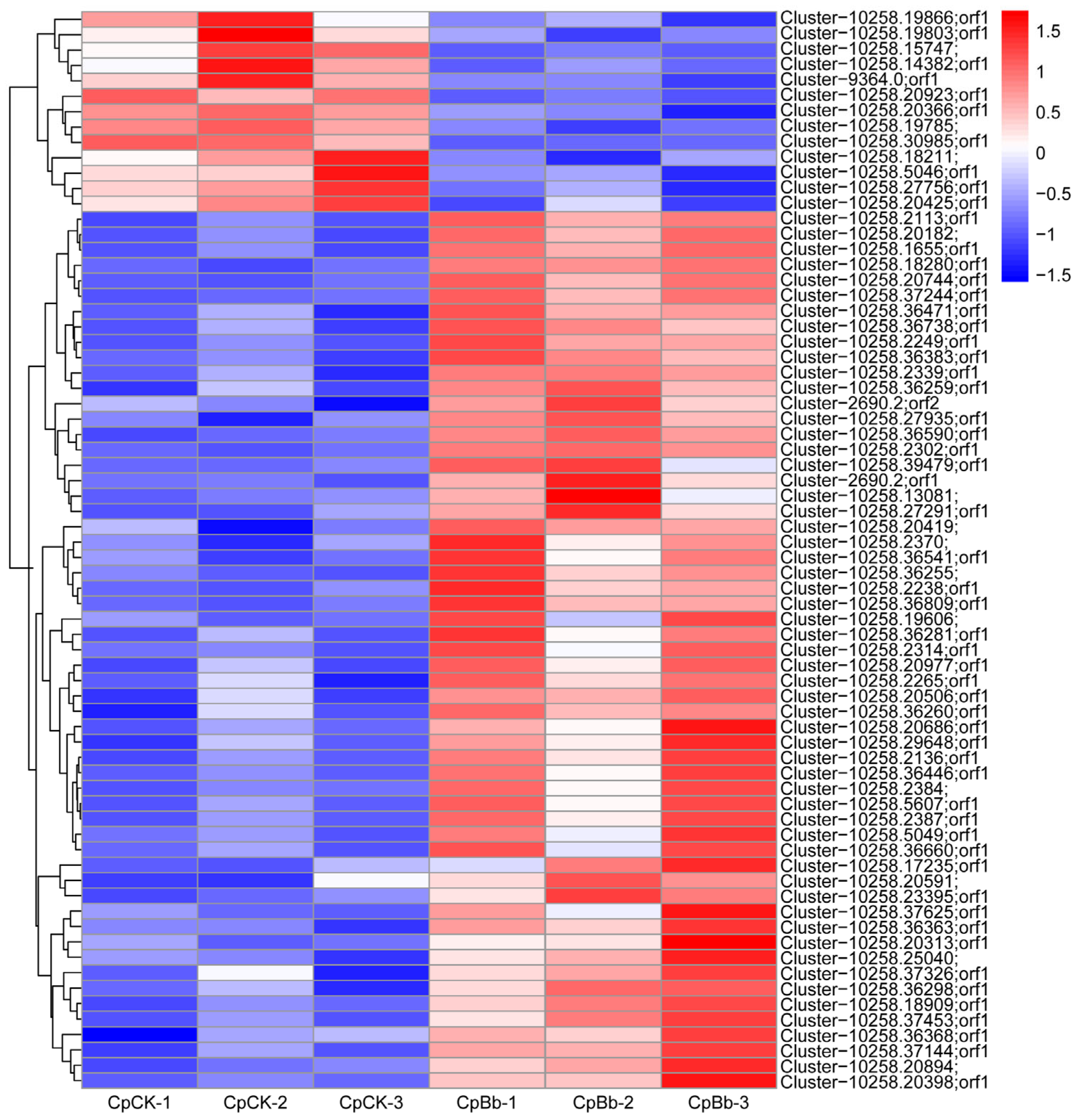
3.4. Statistics of DEPs

The results of the differential protein screening showed that a total of 70 DEPs were identified, including 57 up-regulated proteins and 13 down-regulated proteins (Table 2). The cluster heatmap of DEPs is shown in Figure 6. After eliminating the undescribed proteins, 57 DEPs were identified, of which 47 were up-regulated and 10 were down-regulated (Table 3). Among these DEPs, four up-regulated DEPs were related to immunity, namely defense protein 3-like, peptidoglycan recognition protein B (PGRP-B), peptidoglycan recognition protein-like (PGRP), and peptidoglycan recognition protein LB-like (PGRP-LB).

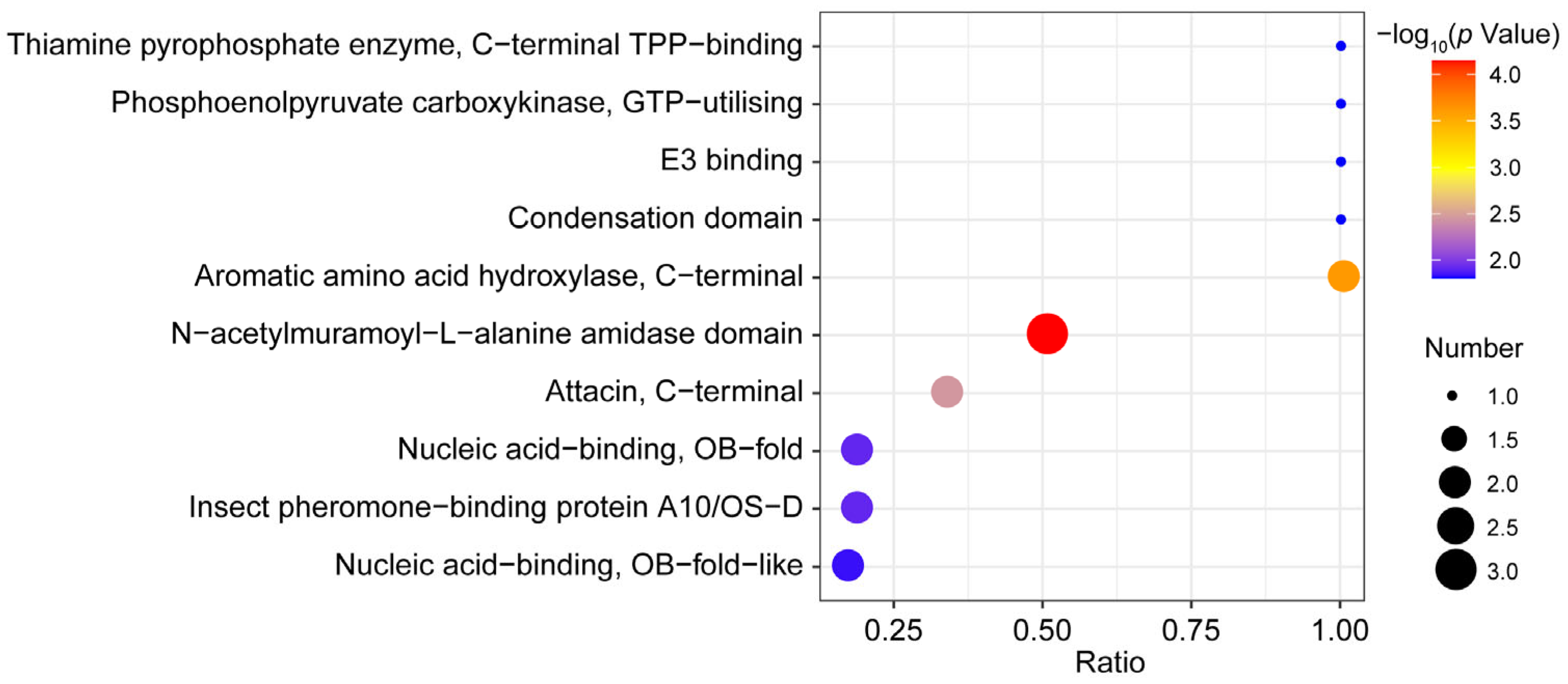
3.5. Functional Enrichment Analysis of DEPs

In total, 37 DEPs were enriched in GO enrichment analysis (Figure 7). In the biological process group, the DEPs were present in the metabolic process (27 DEPs), the organic substance metabolic process (23 DEPs), and the primary metabolic process (20 DEPs). In the cellular component group, the DEPs were mainly present in the ribosome (12 DEPs). In the molecular function group, more DEPs existed in the structural molecule activity (13 DEPs), structural constituent of ribosome (12 DEPs), and organic cyclic compound binding (11 DEPs).
Based on the KEGG pathway analysis, 29 DEPs were enriched, leading to the generation of 47 maps. The bubble chart shows the 20 most enriched KEGG pathways (Figure 8). The top three enriched pathways were ribosome (13 DEPs), AMPK signaling pathway (3 DEPs), and folate biosynthesis (3 DEPs), respectively.
A total of 55 DEPs were enriched according to IPR enrichment analysis. The bubble chart shows the 10 most enriched IPR (Figure 9). The top category was N-acetylmuramoyl-L-alanine amidase domain (3 DEPs).

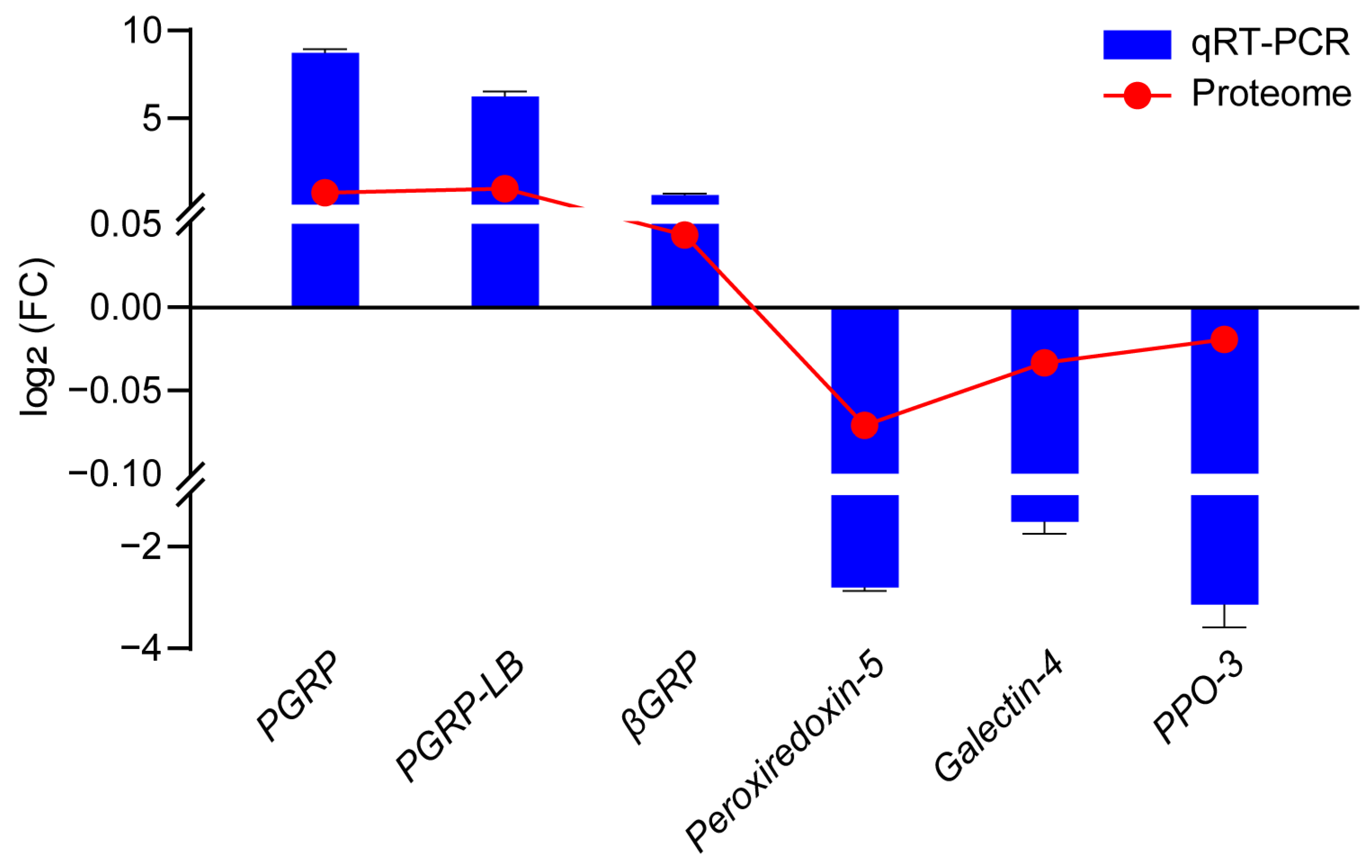
3.6. Validation of Several DEPs by qRT-PCR

To further verify the consistency of gene expression changes at both the mRNA and protein levels, six immune-related genes encoding proteins were selected for qRT-PCR analysis (Table S2 and Figure 10). The results revealed that the expression patterns of six genes at the mRNA level were consistent with those at the protein level, indicating that the accuracy of the proteome data was high.

4. Discussion

Transcriptomics or genomics are used to evaluate the messenger alone; by contrast, proteomics is a more direct means of describing molecular reactions, which can offer extremely useful information on the changes in proteins under various conditions and factors [22,25]. With the rapid development of omics techniques, iTRAQ has become a powerful and effective technique in proteomics due to its high sensitivity and accurate quantification [23,29]. Proteins are the pivotal final products of cells, which are capable of performing physiological functions [25]. The detection of changes at the protein level is capable of offering more direct evidence to study the immune mechanisms of insects. Here, a comparative iTRAQ-based quantitative proteomic analysis was carried out to obtain systematic information of YPM against B. bassiana infection.
On the basis of proteomic approaches, immune-related proteins can be detected in certain tissues or organs in insects. For example, a total of 81 immune-related proteins were screened in the Bombyx mori proteome, including 32 recognition proteins, 28 signaling proteins, and 21 effector proteins [30]. Meanwhile, in the Plutella xylostella proteome, only 58 immune-related proteins were identified [31]. In this study, 132 immune-related proteins were identified, including 46 pathogen recognition proteins, 27 extracellular signal modulation proteins, and 59 immune effectors. In comparison, a greater number of the immune-related proteins were identified from the YPM proteome. However, further functional analyses of the immune-related proteins are required in order to reveal the molecular mechanism of immune recognition that underlies the YPM response to B. bassiana infection.
Peptidoglycan recognition proteins (PGRPs) are an important class of pattern recognition receptors, and play a critical role in the innate immune response of insects [32,33]. In addition, some PGRPs are able to recognize structural diversity of peptidoglycans (PGNs) and activate various innate immune pathways, including the Toll pathway, the Imd pathway, and the prophenoloxidase (PPO) activation pathway [34,35,36]. They can initiate downstream immune responses through the recognition and binding of pathogen-associated molecular patterns (PAMPs), thereby inducing the expression of genes that encode antimicrobial peptides (AMPs) [34,37]. For example, the PGRP1 of O. furnacalis can recognize the invading microbes and the transcript levels of PGRP1 increased in response to bacterial and fungal challenges [38]. In Antheraea pernyi, the interference of ApPGRP-B resulted in a significant increase in the AMP genes in the immune response [39]. In this study, the expressions of PGRP-B (ADU33185.1), PGRP (XP_028160373.1), and PGRP-LB (XP_013143081.1) were up-regulated, indicating that the immune recognition of YPM was activated upon B. bassiana challenge. Remarkably, PGRP-B, PGRP, and PGRP-LB possess an N-acetylmuramoyl-L-alanine amidase domain according to the IPR enrichment analysis. The catalytic PGRPs are capable of cleaving the amide bond of bacterial PGNs, thereby exerting direct bactericidal activity [39,40]. However, further study is required to determine whether the PGRPs in this study indeed contain catalytic enzyme activity. Serine proteases (SPs) are important proteolytic enzymes and widely found in insects [41]. Extracellular SPs have been demonstrated to form cascades, and the reaction is rapidly stimulated; meanwhile, the signals of pathogen invasion are amplified [42,43]. Some SPs with one or more disulfide-bridged structures are designated as clips, which indicate the presence of clip domains [42,44]. Clip-domain serine proteases (Clip-SPs) constitute the main members of the extracellular SP cascade pathway in insects and participate in various physiological processes (such as embryonic development and immune responses) [43,45,46]. Studies have demonstrated that Clip-SPs involving the immune cascade pathways can lead to the activation of PPO and the Toll-ligand Spätzle [42,47]. In the present study, a Clip-SP (XP_013184392.1) with up-regulated expression was identified on the basis of the proteome data. Therefore, we speculated that the Clip-SP of YPM may be involved in the activation of the PPO cascade and the Spätzle–Toll pathway. However, due to the limitations of this study, the function of this Clip-SP needs to be investigated further.
AMPs produced and secreted from the fat body are crucial components of humoral immunity [48]. They form a first line of host defense against a variety of potential invaders, including but not limited to bacteria, fungi, viruses, and parasites [49]. In insects, AMPs are generally classified into four groups on the basis of the secondary structures [48,50]. Among them, attacins are large glycine-rich peptides containing a signal peptide, a pro-peptide domain, an N-terminal domain, a conserved motif, and two glycine-rich domains at the C-terminus [48,51]. Previous studies have demonstrated that attacins were active against bacteria and fungi in lepidopteran insects [52,53]. As an example, in Drosophila melanogaster, attacin C was activated against Gram-negative bacteria and played a key role in the antimicrobial defense [54]. Similarly, an attacin from Hermetia illucens possessed effective antimicrobial activity against the Gram-negative bacteria [55]. In this study, defense protein 3-like (XP_023937619.1) was identified based on the proteome data, which belonged to an attacin C. Given these findings, we speculated that the up-regulation of this protein in YPM after B. bassiana infection may imply its potential role in antifungal immunity.
Through GO categories and KEGG pathway analyses, more DEPs were found to be mainly related to metabolic processes and ribosomes. Although there are clear distinctions between the concepts of immunity and metabolism in biological systems, they are interlinked in animal physiology [56]. In invertebrates, the interactions between the immune system and metabolism are an evolutionarily conserved phenomenon [57]. Initiating the immune system is an energy-consuming process. During a pathogenic infection, the immune response is activated, which is related to a systemic metabolic switch that redirects nutrient flow towards the immune system for the elimination of pathogens [56,58]. Meanwhile, a lot of proteins involved in the ribosome were differentially expressed throughout the B. bassiana infection, including ribosomal protein L17, L6, S8, and S5, as well as the 60S ribosomal protein and 40S ribosomal protein. It was reported that certain ribosome proteins were associated with cell structure, protein translation, and protein biosynthesis [59,60]. The present results suggested that changes in DEPs may potentially affect protein translation and protein biosynthesis during the B. bassiana infection.

5. Conclusions

In summary, the protein changes in YPM larvae in response to B. bassiana infection were investigated by a comparative iTRAQ-based quantitative proteomic analysis. The immune-related proteins were identified based on the proteome data. The DEPs were screened and identified and then subjected to functional enrichment analysis. These findings will be conducive to further understanding the immune mechanism of YPM and providing relevant information for insect pest management.

Supplementary Materials

The following supporting information can be downloaded at: https://www.mdpi.com/article/10.3390/insects16070667/s1, Table S1: Liquid chromatography elution gradient; Table S2: Primers used for qRT-PCR in the present study; Table S3: Immune-related proteins identified in YPM proteome.

Author Contributions

Conceptualization, S.L., X.Y., F.Z. and F.L.; methodology, S.L., X.Y., F.Z., and F.L.; software, S.L. and Z.K.; validation, S.L. and Z.K.; formal analysis, S.L.; investigation, S.L. and Z.K.; resources, all authors; data curation, S.L. and Z.K.; writing—original draft preparation, S.L.; writing—review and editing, all authors; visualization, S.L. and Z.K.; supervision, X.L., H.W., X.Y., F.Z. and F.L.; project administration, X.L., X.Y., F.Z. and F.L.; funding acquisition, X.L., F.Z. and F.L. All authors have read and agreed to the published version of the manuscript.

Funding

This research was funded by the National Science Foundation of Shandong Province (grant numbers ZR2021QC031), the National Key Research and Development Plan in China (grant numbers 2016YFD0300701), and the Shandong Modern Agricultural Technology and Industry System (grant numbers SDAIT-02-10).

Data Availability Statement

The proteome data are available via ProteomeXchange with the identifier PXD063941 (https://proteomecentral.proteomexchange.org/cgi/GetDataset?ID=PXD063941, accessed on 14 May 2025).

Conflicts of Interest

The authors declare no conflicts of interest.

References

  1. Du, Y.L.; Li, J.; Wang, Z.Y. Research progress of Conogethes punctiferalis (Lepidoptera: Crambidae) in China. In The Black Spotted, Yellow Borer, Conogethes punctiferalis Guenée and Allied Species, 1st ed.; Chakravarthy, A.K., Ed.; Springer: Singapore, 2018; pp. 45–66. [Google Scholar]
  2. Rojas-Sandoval, J. Conogethes punctiferalis (yellow peach moth). In CABI Compendium; CABI Digital Library: Wallingford, UK, 2023; pp. 1–11. [Google Scholar]
  3. Wang, Z.Y.; He, K.L.; Shi, J.; Ma, S.Y. Analysis of the heavy occurrence trend of the yellow peach moth in corn and its management strategies. Plant Prot. 2006, 32, 67–69. [Google Scholar]
  4. Guo, J.F.; Qi, J.F.; He, K.L.; Wu, J.Q.; Bai, S.X.; Zhang, T.T.; Zhao, J.R.; Wang, Z.Y. The Asian corn borer Ostrinia furnacalis feeding increases the direct and indirect defence of mid-whorl stage commercial maize in the field. Plant Biotechnol. J. 2019, 17, 88–102. [Google Scholar] [CrossRef]
  5. Kim, K.; Baek, S.; Kim, M.J.; Jung, J.K.; Jung, C.; Lee, J.H. Efficiency of chemical and organic pesticides for Conogethes punctiferalis (Lepidoptera: Crambidae) in commercial chestnut and walnut fields. J. Asia-Pac. Entomol. 2022, 25, 101897. [Google Scholar] [CrossRef]
  6. Jaworski, C.C.; Thomine, E.; Rusch, A.; Lavoir, A.V.; Wang, S.; Desneux, N. Crop diversification to promote arthropod pest management: A review. Agric. Commun. 2023, 1, 100004. [Google Scholar] [CrossRef]
  7. Punniyakotti, P.; Vinayagam, S.; Rajamohan, R.; Priya, S.D.; Moovendhan, M.; Sundaram, T. Environmental fate and ecotoxicological behaviour of pesticides and insecticides in non-target environments: Nanotechnology-based mitigation strategies. J. Environ. Chem. Eng. 2024, 12, 113349. [Google Scholar] [CrossRef]
  8. Wagner, B.L.; Lewis, L.C. Colonization of corn, Zea mays, by the entomopathogenic fungus Beauveria bassiana. Appl. Environ. Microb. 2000, 66, 3468–3473. [Google Scholar] [CrossRef]
  9. Wang, C.S.; Wang, S.B. Insect pathogenic fungi: Genomics, molecular interactions, and genetic improvements. Annu. Rev. Entomol. 2017, 62, 73–90. [Google Scholar] [CrossRef]
  10. Qu, S.; Wang, S.B. Interaction of entomopathogenic fungi with the host immune system. Dev. Comp. Immunol. 2018, 83, 96–103. [Google Scholar] [CrossRef]
  11. Mei, L.J.; Chen, M.J.; Shang, Y.F.; Tang, G.R.; Tao, Y.; Zeng, L.; Huang, B.; Li, Z.Z.; Zhan, S.; Wang, C.S. Population genomics and evolution of a fungal pathogen after releasing exotic strains to control insect pests for 20 years. ISME J. 2020, 14, 1422–1434. [Google Scholar] [CrossRef]
  12. Li, Z.Z. History, progress and current status of the application of fungi against pest insects in China. Chin. J. Biol. Control 2015, 31, 699–711. [Google Scholar]
  13. Senthil Kumar, C.M.; Jacob, T.K.; Devasahayam, S.; Geethu, C.; Hariharan, V. Characterization and biocontrol potential of a naturally occurring isolate of Metarhizium pingshaense infecting Conogethes punctiferalis. Microbiol. Res. 2021, 243, 126645. [Google Scholar] [CrossRef]
  14. Li, S.H.; Liu, F.H.; Kang, Z.W.; Li, X.D.; Lu, Y.; Li, Q.Y.; Pang, Y.S.; Zheng, F.Q.; Yin, X.C. Cellular immune responses of the yellow peach moth, Conogethes punctiferalis (Lepidoptera: Crambidae), to the entomopathogenic fungus, Beauveria bassiana (Hypocreales: Cordycipitaceae). J. Invertebr. Pathol. 2022, 194, 107826. [Google Scholar] [CrossRef]
  15. Tan, S.Q.; Wei, H.S.; Camara, I.; Jia, H.R.; Cao, K.L.; Shi, W.P. Symbiotic bacteria system of Locusta migratoria showed antifungal capabilities against Beauveria bassiana. Int. J. Mol. Sci. 2023, 24, 3138. [Google Scholar] [CrossRef]
  16. Hoffmann, J.A. The immune response of Drosophila. Nature 2003, 426, 33. [Google Scholar] [CrossRef]
  17. Castillo, J.C.; Reynolds, S.E.; Eleftherianos, I. Insect immune responses to nematode parasites. Trends Parasitol. 2011, 27, 537–547. [Google Scholar] [CrossRef]
  18. He, Y.; Cao, X.L.; Li, K.; Hu, Y.X.; Chen, Y.R.; Blissard, G.; Kanost, M.R.; Jiang, H.B. A genome-wide analysis of antimicrobial effector genes and their transcription patterns in Manduca sexta. Insect Biochem. Mol. Biol. 2015, 62, 23–37. [Google Scholar] [CrossRef]
  19. Xiong, G.H.; Xing, L.S.; Lin, Z.; Saha, T.T.; Wang, C.S.; Jiang, H.B.; Zou, Z. High throughput profiling of the cotton bollworm Helicoverpa armigera immunotranscriptome during the fungal and bacterial infections. BMC Genom. 2015, 16, 321. [Google Scholar] [CrossRef]
  20. Zhang, Z.T.; Zhu, S.J.; Mandal, S.D.; Gao, Y.F.; Yu, J.; Zeng, L.; Huang, J.L.; Zafar, J.; Jin, F.L.; Xu, X.X. Combined transcriptomic and proteomic analysis of developmental features in the immune system of Plutella xylostella during larva-to-adult metamorphosis. Genomics 2022, 114, 110381. [Google Scholar] [CrossRef]
  21. Gao, B.J.; Peng, Y.; Jin, M.H.; Zhang, L.; Han, X.; Wu, C.; Yuan, H.; Awawing, A.; Zheng, F.Q.; Li, X.D.; et al. Chromosome genome assembly and whole genome sequencing of 110 individuals of Conogethes punctiferalis (Guenée). Sci. Data 2023, 10, 805. [Google Scholar] [CrossRef]
  22. Lü, D.D.; Xu, P.; Hou, C.X.; Li, R.L.; Hu, C.W.; Guo, X.J. iTRAQ-based quantitative proteomic analysis of silkworm infected with Beauveria bassiana. Mol. Immunol. 2021, 135, 204–216. [Google Scholar] [CrossRef]
  23. Wen, B.; Zhou, R.; Feng, Q.; Wang, Q.H.; Wang, J.; Liu, S.Q. IQuant: An automated pipeline for quantitative proteomics based upon isobaric tags. Proteomics 2014, 14, 2280–2285. [Google Scholar] [CrossRef] [PubMed]
  24. Li, Y.D.; Li, X.; Xu, W.B.; Han, Z.B.; Zhao, Y.Y.; Dong, J.; Wei, H.; Chen, Q.J. Comparative iTRAQ-based quantitative proteomic analysis of the Chinese grass shrimp (Palaemonetes sinensis) infected with the isopod parasite Tachaea chinensis. Parasites Vectors 2019, 12, 415. [Google Scholar] [CrossRef] [PubMed]
  25. Yu, J.Q.; Cen, X.F.; Chen, G.F.; Tang, M.L.; Mo, L.; Li, J.J. iTRAQ-based quantitative proteomic analysis in liver of Pomacea canaliculata induced by oleanolic acid stress. Pest Manag. Sci. 2022, 78, 3467–3478. [Google Scholar] [CrossRef] [PubMed]
  26. Jones, P.; Binns, D.; Chang, H.Y.; Fraser, M.; Li, W.Z.; McAnulla, C.; McWilliam, H.; Maslen, J.; Mitchell, A.; Nuka, G.; et al. InterProScan 5: Genome-scale protein function classification. Bioinformatics 2014, 30, 1236–1240. [Google Scholar] [CrossRef]
  27. Huang, D.W.; Sherman, B.T.; Lempicki, R.A. Bioinformatics enrichment tools: Paths toward the comprehensive functional analysis of large gene lists. Nucleic Acids Res. 2009, 37, 1–13. [Google Scholar] [CrossRef]
  28. Livak, K.J.; Schmittgen, T.D. Analysis of relative gene expression data using real-time quantitative PCR and the 2-ΔΔCT method. Methods 2001, 25, 402–408. [Google Scholar] [CrossRef]
  29. Kang, S.; Zhu, X.; Wu, Q.J.; Wang, S.L.; Crickmore, N.; Zhang, Y.J.; Guo, Z.J. Critical analysis of multi-omic data from a strain of Plutella xylostella resistant to Bacillus thuringiensis Cry1Ac toxin. J. Agric. Food Chem. 2022, 70, 11419–11428. [Google Scholar] [CrossRef]
  30. Chen, S.Y.; Dong, Z.M.; Ren, X.; Zhao, D.C.; Zhang, Y.; Tang, M.Y.; Han, J.X.; Ye, L.; Zhao, P. Proteomic identification of immune-related silkworm proteins involved in the response to bacterial infection. J. Insect Sci. 2019, 19, 13. [Google Scholar] [CrossRef]
  31. Lin, J.H.; Yu, X.Q.; Wang, Q.; Tao, X.P.; Li, J.Y.; Zhang, S.S.; Xia, X.F.; You, M.S. Immune responses to Bacillus thuringiensis in the midgut of the diamondback moth, Plutella xylostella. Dev. Comp. Immunol. 2020, 107, 103661. [Google Scholar] [CrossRef]
  32. Dziarski, R.; Gupta, D. The peptidoglycan recognition proteins (PGRPs). Genome Biol. 2006, 7, 232. [Google Scholar] [CrossRef]
  33. Qiao, P.T.; Mei, X.H.; Li, R.X.; Xu, Y.Y.; Qiu, Z.Y.; Xia, D.G.; Zhao, Q.L.; Shen, D.X. Transcriptome analysis of immune-related genes of Asian corn borer (Ostrinia furnacalis [Guenée]) after oral bacterial infection. Arch. Insect Biochem. Physiol. 2023, 114, e22044. [Google Scholar] [CrossRef] [PubMed]
  34. Wang, Q.; Ren, M.J.; Liu, X.Y.; Xia, H.C.; Chen, K.P. Peptidoglycan recognition proteins in insect immunity. Mol. Immunol. 2019, 106, 69–76. [Google Scholar] [CrossRef] [PubMed]
  35. Lu, Y.Z.; Su, F.H.; Li, Q.L.; Zhang, J.; Li, Y.J.; Tang, T.; Hu, Q.H.; Yu, X.Q. Pattern recognition receptors in Drosophila immune responses. Dev. Comp. Immunol. 2020, 102, 103468. [Google Scholar] [CrossRef]
  36. Zhao, L.; Niu, J.L.; Feng, D.S.; Wang, X.L.; Zhang, R. Immune functions of pattern recognition receptors in Lepidoptera. Front. Immunol. 2023, 14, 1203061. [Google Scholar] [CrossRef]
  37. Garver, L.S.; Wu, J.L.; Wu, L.P. The peptidoglycan recognition protein PGRP-SC1a is essential for Toll signaling and phagocytosis of Staphylococcus aureus in Drosophila. Proc. Nat. Acad. Sci. USA 2006, 103, 660–665. [Google Scholar] [CrossRef]
  38. Shen, D.X.; Ji, J.Y.; Zhang, S.S.; Liu, J.H.; An, C.J. A short-type peptidoglycan recognition protein 1 (PGRP1) is involved in the immune response in Asian corn borer, Ostrinia furnacalis (Guenée). Int. J. Mol. Sci. 2021, 22, 8198. [Google Scholar] [CrossRef]
  39. Liu, W.; Wang, Y.; Zhou, J.L.; Zhang, Y.; Ma, Y.Y.; Wang, D.Y.; Jiang, Y.R.; Shi, S.L.; Qin, L. Peptidoglycan recognition proteins regulate immune response of Antheraea pernyi in different ways. J. Invertebr. Pathol. 2019, 166, 107204. [Google Scholar] [CrossRef]
  40. Kim, M.S.; Byun, M.; Oh, B.H. Crystal structure of peptidoglycan recognition protein LB from Drosophila melanogaster. Nat. Immunol. 2003, 4, 787–793. [Google Scholar] [CrossRef]
  41. Di Cera, E. Serine proteases. IUBMB Life 2009, 61, 510–515. [Google Scholar] [CrossRef]
  42. Kanost, M.R.; Jiang, H.B. Clip-domain serine proteases as immune factors in insect hemolymph. Curr. Opin. Insect Sci. 2015, 11, 47–55. [Google Scholar] [CrossRef]
  43. Veillard, F.; Troxler, L.; Reichhart, J.M. Drosophila melanogaster clip-domain serine proteases: Structure, function and regulation. Biochimie 2016, 122, 255–269. [Google Scholar] [CrossRef] [PubMed]
  44. Cao, X.L.; He, Y.; Hu, Y.X.; Zhang, X.F.; Wang, Y.; Zou, Z.; Chen, Y.C.; Blissard, G.W.; Kanost, M.R.; Jiang, H.B. Sequence conservation, phylogenetic relationships, and expression profiles of nondigestive serine proteases and serine protease homologs in Manduca sexta. Insect Biochem. Mol. Biol. 2015, 62, 51–63. [Google Scholar] [CrossRef] [PubMed]
  45. Jiang, H.B.; Kanost, M.R. The clip-domain family of serine proteinases in arthropods. Insect Biochem. Mol. Biol. 2000, 30, 95–105. [Google Scholar] [CrossRef] [PubMed]
  46. Wu, C.Y.; Xiao, K.R.; Wang, L.Z.; Wang, J.; Song, Q.S.; Stanley, D.; Wei, S.J.; Zhu, J.Y. Identification and expression profiling of serine protease-related genes in Tenebrio molitor. Arch. Insect Biochem. Physiol. 2022, 111, e21963. [Google Scholar] [CrossRef]
  47. Liu, H.W.; Heng, J.Y.; Wang, L.L.; Tang, X.; Guo, P.C.; Li, Y.S.; Xia, Q.Y.; Zhao, P. Identification, characterization, and expression analysis of clip-domain serine protease genes in the silkworm, Bombyx mori. Dev. Comp. Immunol. 2020, 105, 103584. [Google Scholar] [CrossRef]
  48. Eleftherianos, I.; Zhang, W.; Heryanto, C.; Mohamed, A.; Contreras, G.; Tettamanti, G.; Wink, M.; Bassal, T. Diversity of insect antimicrobial peptides and proteins—A functional perspective: A review. Int. J. Biol. Macromol. 2021, 191, 277–287. [Google Scholar] [CrossRef]
  49. Bulet, P.; Stöcklin, R.; Menin, L. Anti-microbial peptides: From invertebrates to vertebrates. Immunol. Rev. 2004, 198, 169–184. [Google Scholar] [CrossRef]
  50. Zhou, L.Z.; Meng, G.L.; Zhu, L.; Ma, L.; Chen, K.K. Insect antimicrobial peptides as guardians of immunity and beyond: A review. Int. J. Mol. Sci. 2024, 25, 3835. [Google Scholar] [CrossRef]
  51. Hedengren, M.; Borge, K.; Hultmark, D. Expression and evolution of the Drosophila attacin/diptericin gene family. Biochem. Biophys. Res. Commun. 2000, 279, 574–581. [Google Scholar] [CrossRef]
  52. Bang, K.; Park, S.; Yoo, J.Y.; Cho, S. Characterization and expression of attacin, an antibacterial protein-encoding gene, from the beet armyworm, Spodoptera exigua (Hübner) (Insecta: Lepidoptera: Noctuidae). Mol. Biol. Rep. 2012, 39, 5151–5159. [Google Scholar] [CrossRef]
  53. Nesa, J.; Sadat, A.; Buccini, D.F.; Kati, A.; Mandal, A.K.; Franco, O.L. Antimicrobial peptides from Bombyx mori: A splendid immune defense response in silkworms. RSC Adv. 2020, 10, 512–523. [Google Scholar] [CrossRef] [PubMed]
  54. Rabel, D.; Charlet, M.; Ehret-Sabatier, L.; Cavicchioli, L.; Cudic, M.; Otvos, L.; Bulet, P. Primary structure and in vitro antibacterial properties of the Drosophila melanogaster attacin C pro-domain. J. Biol. Chem. 2004, 279, 14853–14859. [Google Scholar] [CrossRef] [PubMed]
  55. Fahmy, L.; Ali, Y.M.; Seilly, D.; McCoy, R.; Owens, R.M.; Pipan, M.; Christie, G.; Grant, A.J. An attacin antimicrobial peptide, Hill_BB_C10074, from Hermetia illucens with anti-Pseudomonas aeruginosa activity. BMC Microbiol. 2023, 23, 378. [Google Scholar] [CrossRef] [PubMed]
  56. Hotamisligil, G.S. Inflammation, metaflammation and immunometabolic disorders. Nature 2017, 542, 177–185. [Google Scholar] [CrossRef]
  57. Li, S.R.; Wang, J.; Tian, X.; Toufeeq, S.; Huang, W.R. Immunometabolic regulation during the presence of microorganisms and parasitoids in insects. Front. Immunol. 2023, 14, 905467. [Google Scholar] [CrossRef]
  58. Dolezal, T.; Krejcova, G.; Bajgar, A.; Nedbalova, P.; Strasser, P. Molecular regulations of metabolism during immune response in insects. Insect Biochem. Mol. Biol. 2019, 109, 31–42. [Google Scholar] [CrossRef]
  59. Bursać, S.; Brdovčak, M.C.; Pfannkuchen, M.; Orsolić, I.; Golomb, L.; Zhu, Y.; Katz, C.; Daftuar, L.; Grabušić, K.; Vukelić, I.; et al. Mutual protection of ribosomal proteins L5 and L11 from degradation is essential for p53 activation upon ribosomal biogenesis stress. Proc. Nat. Acad. Sci. USA 2012, 109, 20467–20472. [Google Scholar] [CrossRef]
  60. Yu, Q.; Xiong, Y.H.; Gao, H.; Liu, J.L.; Chen, Z.Q.; Wang, Q.; Wen, D.L. Comparative proteomics analysis of Spodoptera frugiperda cells during Autographa californica multiple nucleopolyhedrovirus infection. Virol. J. 2015, 12, 115. [Google Scholar] [CrossRef]
Figure 1. Distribution of protein coverage (A) and protein mass (B).
Insects 16 00667 g001
Figure 2. GO function annotation.
Insects 16 00667 g002
Figure 3. COG function annotation.
Insects 16 00667 g003
Figure 4. KEGG function annotation.
Insects 16 00667 g004
Figure 5. IPR function annotation.
Insects 16 00667 g005
Figure 6. The cluster heatmap of DEPs.
Insects 16 00667 g006
Figure 7. GO functional enrichment analysis of DEPs.
Insects 16 00667 g007
Figure 8. KEGG pathway analysis of DEPs.
Insects 16 00667 g008
Figure 9. IPR enrichment analysis of DEPs.
Insects 16 00667 g009
Figure 10. Verification of the proteome data.
Insects 16 00667 g010
Table 1. Statistics of protein identification.
Total SpectraMatched SpectraPeptidesIdentified ProteinsAll Quantifiable Proteins
565,46962,66929,15541974195
Table 2. Statistics of protein difference analysis from the YPM larvae proteome.
Number of Total ProteinsRegulated TypeFC > 1.2FC > 1.3FC > 1.5FC > 2.0
565,469up-regulated126925711
down-regulated7247130
Table 3. Differentially expressed proteins of YPM larvae after treatment with B. bassiana.
ProteinDescriptionAccession No.FCp ValueType
Cluster-10258.20366; orf1arylphorin subunit alpha-likeXP_028169947.10.620.0028down
Cluster-10258.18909; orf1protein hennaXP_026753049.11.570.0043up
Cluster-10258.20977; orf1peptidoglycan recognition protein BADU33185.11.970.0171up
Cluster-10258.20744; orf1GTP cyclohydrolase 1 isoform X1XP_028166842.11.636.01 × 10−4up
Cluster-10258.5049; orf1elongation factor 1-alphaXP_022204799.11.850.0211up
Cluster-10258.20686; orf1pancreatic triacylglycerol lipase-likeXP_023935092.11.630.0258up
Cluster-10258.39479; orf1ribosomal L6 and ribosomal S8 and ribosomal S5 C and ribosomal S5 domain containing proteinCDW61069.12.150.0234up
Cluster-10258.20398; orf1tyrosine 3-monooxygenase isoform X1XP_028171956.11.720.0136up
Cluster-10258.20923; orf1hypothetical protein evm_001044RVE54217.10.616.82 × 10−4down
Cluster-10258.14382; orf1chemosensory protein 2AHX37219.10.630.0257down
Cluster-10258.2265; orf1heat shock protein 70-4AQP31364.11.560.0203up
Cluster-10258.5046; orf1hypothetical protein B5V51_858PCG72390.10.650.0305down
Cluster-10258.19866; orf1phosphoenolpyruvate carboxykinase [GTP]-likeXP_028168927.10.650.0316down
Cluster-10258.5607; orf1serine protease easter-like isoform X2XP_013184392.11.530.0136up
Cluster-10258.17235; orf1tryptase-likeXP_013193793.11.850.0448up
Cluster-10258.20506; orf1peptidoglycan recognition protein-likeXP_028160373.11.650.0114up
Cluster-10258.37244; orf160S ribosomal protein L13XP_008552884.12.025.25 × 10−4up
Cluster-10258.20313; orf1uncharacterized protein LOC114360519XP_028171047.11.570.0475up
Cluster-10258.36590; orf140S ribosomal protein S4-like, partialXP_021339550.11.622.55 × 10−4up
Cluster-10258.36281; orf1hypothetical protein DDB_G0285741XP_638067.11.640.0211up
Cluster-10258.19803; orf1chemosensory proteinAPG32552.10.570.0455down
Cluster-10258.36541; orf1S10AAX48886.11.860.0158up
Cluster-10258.18280; orf1uncharacterized protein LOC114364556XP_028176547.11.747.80 × 10−5up
Cluster-9364.0; orf1endocuticle structural glycoprotein ABD-4-likeXP_028173253.10.580.0124down
Cluster-10258.27935; orf1peptidoglycan-recognition protein LB-likeXP_013143081.11.940.0049up
Cluster-10258.36298; orf1uncharacterized protein LOC111678542XP_023295698.11.580.0112up
Cluster-10258.2249; orf160S ribosomal protein L2-A-likeXP_021339551.11.800.0019up
Cluster-10258.30985; orf1venom serine carboxypeptidase-likeXP_028155892.10.606.37 × 10−4down
Cluster-10258.23395; orf1defense protein 3-likeXP_023937619.12.450.0072up
Cluster-10258.2238; orf1elongation factor, putativeXP_002783366.11.690.0073up
Cluster-10258.1655; orf1ribonucleoprotein, putativeELP90168.12.299.19 × 10−4up
Cluster-2690.2; orf2uncharacterized protein LOC111689114XP_023307388.11.650.0198up
Cluster-10258.36368; orf160S ribosomal protein L7-likeXP_028405051.11.520.0338up
Cluster-10258.2136; orf1elongation factor 1-beta-likeXP_022205053.11.670.0090up
Cluster-10258.36383; orf1predicted proteinXP_001625520.11.520.0025up
Cluster-10258.37625; orf1pyruvate dehydrogenase complex dihydrolipoamide acetyltransferaseXP_013761136.12.840.0303up
Cluster-10258.2339; orf1hypothetical proteinAIU94794.11.970.0024up
Cluster-10258.36446; orf160S ribosomal protein L10-likeXP_015760823.12.300.0114up
Cluster-2690.2; orf1uncharacterized protein LOC111689114XP_023307388.13.720.0123up
Cluster-10258.36809; orf140S ribosomal protein S14KXJ11429.12.140.0036up
Cluster-10258.36363; orf160S ribosomal protein L5-like, partialXP_021339565.11.870.0078up
Cluster-10258.2314; orf1stress-70 protein, mitochondrialRDD38839.11.740.0143up
Cluster-10258.27756; orf1uncharacterized protein LOC114364166XP_028175991.10.600.0126down
Cluster-10258.2302; orf1guanine nucleotide-binding proteinXP_004343796.11.983.34 × 10−5up
Cluster-10258.36260; orf1fatty acid-binding protein-likeXP_022204143.11.540.0127up
Cluster-10258.20425; orf1uncharacterized protein LOC114366599XP_028179325.10.590.0242down
Cluster-10258.36738; orf1K+ channel proteinKJE97207.11.720.0054up
Cluster-10258.36471; orf140S ribosomal protein S24XP_013405352.11.530.0059up
Cluster-10258.2113; orf160S ribosomal protein L25-B-likeXP_022204254.12.460.0014up
Cluster-10258.36660; orf1hypothetical protein pdam_00013747RMX60612.11.540.0269up
Cluster-10258.27291; orf1putative ferric-chelate reductase 1 homologXP_028167929.11.520.0127up
Cluster-10258.36259; orf1transaldolaseOQV22424.11.780.0086up
Cluster-10258.37144; orf1eukaryotic translation initiation factor 5A-1XP_015794685.11.570.0052up
Cluster-10258.2387; orf1ribosomal protein L17ABO26685.11.560.0107up
Cluster-10258.37453; orf1trichothecene biosynthesis protein 14 OSG0KYA71.930.0096up
Cluster-10258.37326; orf1hydroxymethylglutaryl-CoA synthase 1-likeXP_027216736.11.550.0400up
Cluster-10258.29648; orf1hypothetical protein evm_006310RVE49064.11.590.0257up
Disclaimer/Publisher’s Note: The statements, opinions and data contained in all publications are solely those of the individual author(s) and contributor(s) and not of MDPI and/or the editor(s). MDPI and/or the editor(s) disclaim responsibility for any injury to people or property resulting from any ideas, methods, instructions or products referred to in the content.

