
3D-Printed Devices in Interventional Radiotherapy (Brachytherapy) Applications: A Literature Review
 , , ,
, , ,  ,
,  and
and 
Abstract
1. Introduction
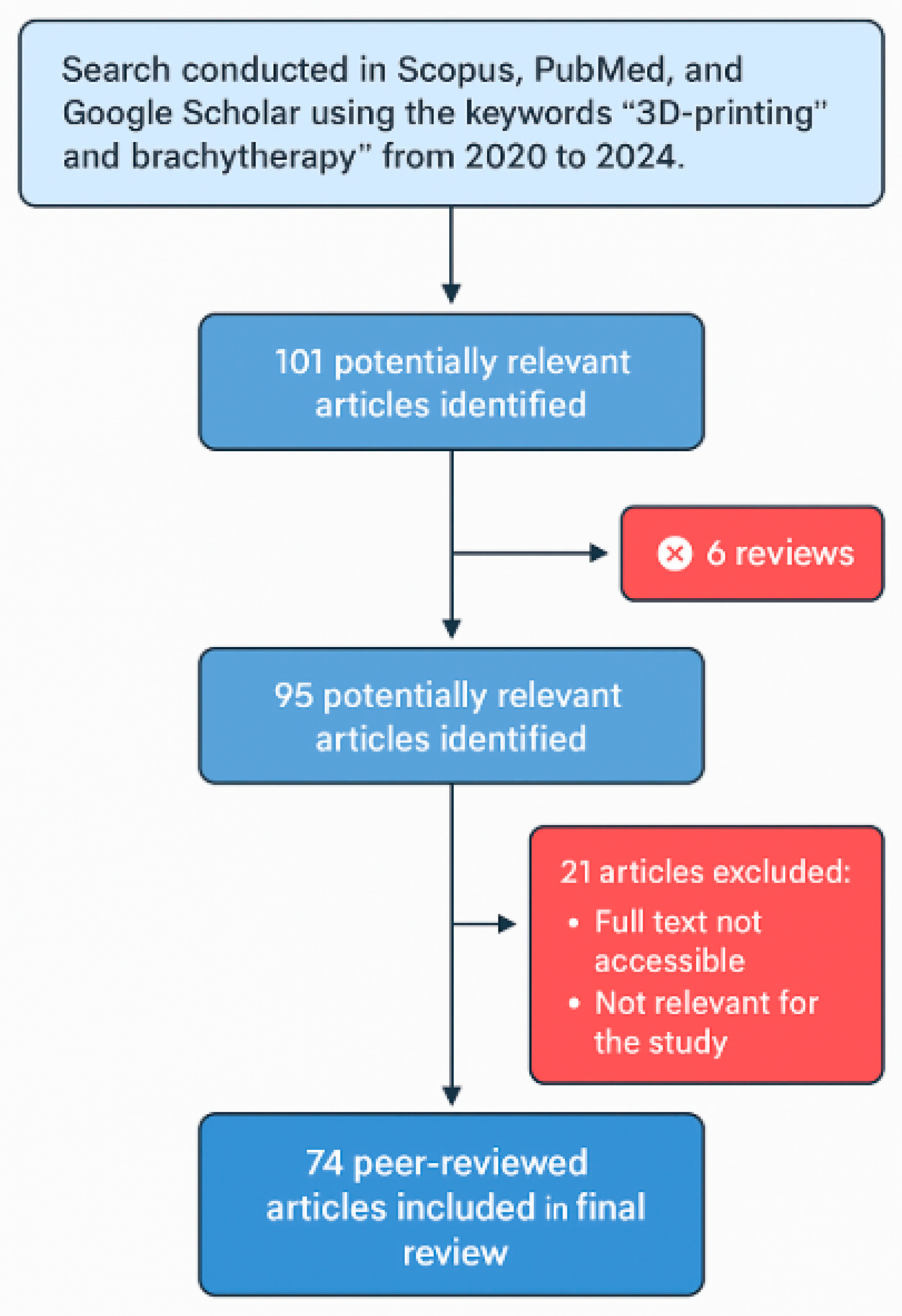
2. Methods
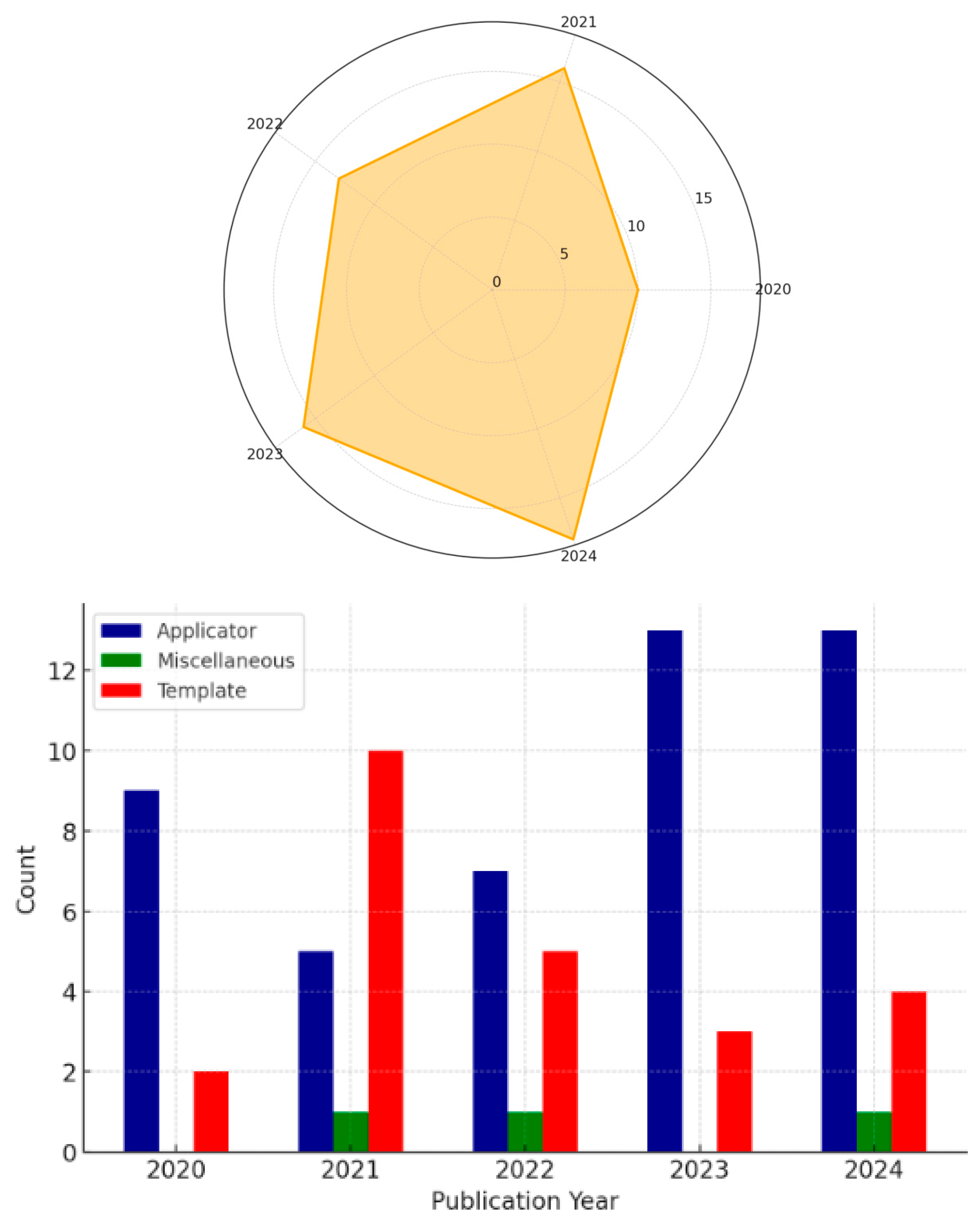
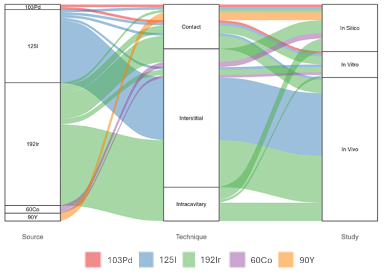
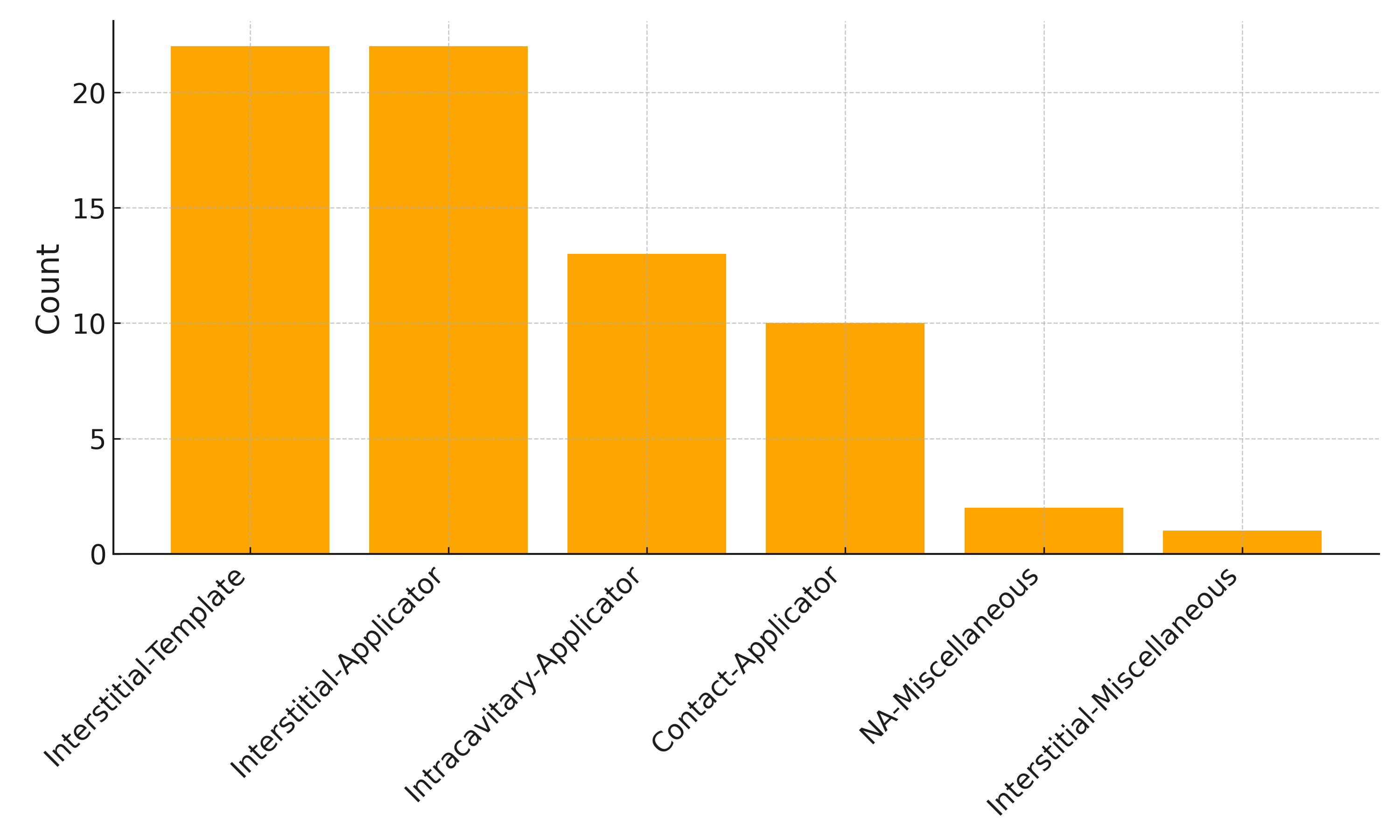
3. Results
4. Discussion
5. Conclusions
Author Contributions
Funding
Conflicts of Interest
Abbreviations
| 3D | Three-dimensional |
| BT | Brachytherapy |
| CT | Computed Tomography |
| CTV-HR | High-risk Clinical Target Volume |
| Gyn | Gynecology |
| HDR | High Dose Rate |
| IRT | Interventional Radiotherapy |
| MRI | Magnetic Resonance Imaging |
| NA | Not Available |
| OAR | Organ at Risk |
| QA | Quality Assurance |
References
- Pötter, R.; Tanderup, K.; Kirisits, C.; De Leeuw, A.; Kirchheiner, K.; Nout, R.; Tan, L.T.; Haie-Meder, C.; Mahantshetty, U.; Segedin, B.; et al. The EMBRACE II study: The outcome and prospect of two decades of evolution within the GEC-ESTRO GYN working group and the EMBRACE studies. Clin. Transl. Radiat. Oncol. 2018, 9, 48–60. [Google Scholar] [CrossRef] [PubMed]
- Bellis, R.; Rembielak, A.; Barnes, E.A.; Paudel, M.; Ravi, A. Additive manufacturing (3D printing) in superficial brachytherapy. J. Contemp. Brachytherapy 2021, 13, 468–482. [Google Scholar] [CrossRef]
- Tanderup, K.; Viswanathan, A.N.; Kirisits, C.; Frank, S.J. Magnetic Resonance Image Guided Brachytherapy. Semin. Radiat. Oncol. 2014, 24, 181–191. [Google Scholar] [CrossRef] [PubMed]
- Poltorak, M.; Banatkiewicz, P.; Poltorak, L.; Sobolewski, P.; Zimon, D.; Szwast, M.; Walecka, I. Brachytherapy and 3D printing for skin cancer: A review paper. J. Contemp. Brachytherapy 2024, 16, 156–169. [Google Scholar] [CrossRef] [PubMed]
- Chen, E.; Zhang, Y.; Zhang, H.; Jia, C.; Liang, Y.; Wang, J. Dosimetry study of three-dimensional print template for 125I implantation therapy. Radiat. Oncol. 2021, 16, 115. [Google Scholar] [CrossRef]
- Segedin, B.; Kobav, M.; Zobec Logar, H.B. The Use of 3D Printing Technology in Gynaecological Brachytherapy—A Narrative Review. Cancers 2023, 15, 4165. [Google Scholar] [CrossRef]
- Biltekin, F.; Akyol, H.; Gültekin, M.; Yildiz, F. 3D printer-based novel intensity-modulated vaginal brachytherapy applicator: Feasibility study. J. Contemp. Brachytherapy 2020, 12, 17–26. [Google Scholar] [CrossRef]
- Subashi, E.; Jacobs, C.; Hood, R.; Kirsch, D.G.; Craciunescu, O. A design process for a 3D printed patient-specific applicator for HDR brachytherapy of the orbit. 3D Print. Med. 2020, 6, 15. [Google Scholar] [CrossRef]
- Pashazadeh, A.; Robatjazi, M.; Castro, N.J.; Friebe, M. A multiwell applicator for conformal brachytherapy of superficial skin tumors: A simulation study. Ski. Res. Technol. 2020, 26, 537–541. [Google Scholar] [CrossRef]
- He, X.; Liu, M.; Zhang, M.; Sequeiros, R.B.; Xu, Y.; Wang, L.; Liu, C.; Wang, Q.; Zhang, K.; Li, C. A novel three-dimensional template combined with MR-guided 125I brachytherapy for recurrent glioblastoma. Radiat. Oncol. 2020, 15, 146. [Google Scholar] [CrossRef]
- Kunogi, H.; Yamaguchi, N.; Sasai, K. Evaluation of a new bi-valve vaginal speculum applicator design for gynecologic interstitial brachytherapy. J. Contemp. Brachytherapy 2020, 12, 27–34. [Google Scholar] [CrossRef] [PubMed]
- Zhang, D.; Yang, Z.; Jiang, S.; Zhou, L.; Zhou, Z.; Wang, W. Individualized and inverse optimized needle configuration for combined intracavitary-interstitial brachytherapy in locally advanced cervical cancer. J. Cancer Res. Ther. 2019, 15, 1589. [Google Scholar] [CrossRef] [PubMed]
- Is 3D printing-gided three-dimensional brachytherapy suitable for cervical cancer: From one single research institute? Eur. J. Gynaecol. Oncol. 2020, 41, 591. [CrossRef]
- Jiang, W.; Jiang, P.; Wei, S.; Jiang, Y.; Ji, Z.; Sun, H.; Fan, J.; Li, W.; Shao, Y.; Wang, J. The accuracy and safety of CT-guided iodine-125 seed implantation assisted by 3D non-coplanar template for retroperitoneal recurrent carcinoma. World J. Surg. Oncol. 2020, 18, 307. [Google Scholar] [CrossRef] [PubMed]
- Wang, L.; Wang, H.; Jiang, Y.; Ji, Z.; Guo, F.; Jiang, P.; Li, X.; Chen, Y.; Sun, H.; Fan, J.; et al. The efficacy and dosimetry analysis of CT-guided 125I seed implantation assisted with 3D-printing non-co-planar template in locally recurrent rectal cancer. Radiat. Oncol. 2020, 15, 179. [Google Scholar] [CrossRef] [PubMed]
- Jiang, P.; Qu, A.; Wei, S.; Sun, H.; Zhang, X.; Li, X.; Wang, J. The Preliminary Results of 3-Dimensional Printed Individual Template Assisted 192Ir High-Dose Rate Interstitial Brachytherapy for Central Recurrent Gynecologic Cancer. Technol. Cancer Res. Treat. 2020, 19, 1533033820971607. [Google Scholar] [CrossRef]
- Pashazadeh, A.; Friebe, M. Transverse dose profile simulation of extruded lines for a 3D printed models for superficial skin cancer therapy. Curr. Dir. Biomed. Eng. 2020, 6, 559–562. [Google Scholar] [CrossRef]
- Simpson-Page, E.; Hamlett, L.; Lew, D.; Stephens, H.; Wilks, R.; Kairn, T.; Crowe, S.B. 3D printed brachytherapy jig for Reference Air Kerma Rate calibration. Phys. Eng. Sci. Med. 2021, 44, 1141–1150. [Google Scholar] [CrossRef]
- Bielęda, G.; Marach, A.; Boehlke, M.; Zwierzchowski, G.; Malicki, J. 3D-printed surface applicators for brachytherapy: A phantom study. J. Contemp. Brachytherapy 2021, 13, 549–562. [Google Scholar] [CrossRef]
- Ji, Z.; Jiang, Y.; Sun, H.; Chen, Y.; Guo, F.; Fan, J.; Wang, J. 3D-printed template and optical needle navigation in CT-guided iodine-125 permanent seed implantation. J. Contemp. Brachytherapy 2021, 13, 410–418. [Google Scholar] [CrossRef]
- Qu, A.; Jiang, P.; Wei, S.; Jiang, Y.; Ji, Z.; Sun, H.; Li, W.; Shao, Y.; Fan, J.; Wang, J. Accuracy and dosimetric parameters comparison of 3D-printed non-coplanar template-assisted computed tomography-guided iodine-125 seed ablative brachytherapy in pelvic lateral recurrence of gynecological carcinomas. J. Contemp. Brachytherapy 2021, 13, 39–45. [Google Scholar] [CrossRef] [PubMed]
- Ji, Z.; Sun, H.; Jiang, Y.; Chen, Y.; Guo, F.; Fan, J.; Wang, J. Analysis on the accuracy of CT-guided radioactive I-125 seed implantation with 3D printing template assistance in the treatment of thoracic malignant tumors. J. Radiat. Res. 2021, 62, 910–917. [Google Scholar] [CrossRef] [PubMed]
- Yan, J.; Qin, X.; Zhang, F.; Hou, X.; Yu, L.; Qiu, J. Comparing multichannel cylinder and 3D-printed applicators for vaginal cuff brachytherapy with preliminary exploration of post-hysterectomy vaginal morphology. J. Contemp. Brachytherapy 2021, 13, 641–648. [Google Scholar] [CrossRef]
- Kang, W.; Zhang, H.; Liang, Y.; Chen, E.; Zhao, J.; Gao, Z.; Wang, J. Comparison of three-dimensional-printed template-guided and traditional implantation of125I seeds for gynecological tumors: A dosimetric and efficacy study. J. Cancer Res. Ther. 2021, 17, 688–694. [Google Scholar] [CrossRef] [PubMed]
- Semeniuk, O.; Cherpak, A.; Robar, J. Design and evaluation of 3D printable patient-specific applicators for gynecologic HDR brachytherapy. Med. Phys. 2021, 48, 4053–4063. [Google Scholar] [CrossRef]
- Wang, L.; Wang, H.; Jiang, Y.; Ji, Z.; Guo, F.; Jiang, P.; Qiu, B.; Sun, H.; Fan, J.; Li, W.; et al. Dosimetric comparison of computed tomography-guided iodine-125 seed implantation assisted with and without three-dimensional printing non-coplanar template in locally recurrent rectal cancer: A propensity score matching study. J. Contemp. Brachytherapy 2021, 13, 18–23. [Google Scholar] [CrossRef]
- Han, X.; Fang, S.; Sheng, R.; Wang, Y.; Zhou, J.; Wang, J. Dosimetry verification of three-dimensional printed polylactic acid template-guided precision125 I seed implantation for lung cancer using a desktop three-dimensional printer. J. Appl. Clin. Med. Phys. 2021, 22, 202–209. [Google Scholar] [CrossRef]
- Deufel, C.L.; Dalvin, L.A.; Qian, J.; Vaishnav, B.; Cutsinger, J.M.; Wittich, M.N.; Petersen, I.A. How to design, fabricate, and validate a customized COMS-style eye plaque: Illustrated with a narrow-slotted plaque example. Brachytherapy 2021, 20, 1235–1244. [Google Scholar] [CrossRef]
- Mohammadi, R.; Siavashpour, Z.; Rashid Hosseini Aghdam, S.; Fazli, S.; Major, T.; Asghar Rohani, A. Manufacturing and evaluation of multi-channel cylinder applicator with 3D printing technology. J. Contemp. Brachytherapy 2021, 13, 80–90. [Google Scholar] [CrossRef]
- Liu, Y.; Jiang, P.; Zhang, H.; Wang, J. Safety and efficacy of 3D-printed templates assisted CT-guided radioactive iodine-125 seed implantation for the treatment of recurrent cervical carcinoma after external beam radiotherapy. J. Gynecol. Oncol. 2021, 32, e15. [Google Scholar] [CrossRef]
- Gong, W.; Chen, Y.; Ji, Z.; Jiang, Y.; Qiu, B.; Sun, H.; Wang, J. The accuracy and dosimetric analysis of 3D-printing non-coplanar template-assisted iodine-125 seed implantation for recurrent chest wall cancer. J. Contemp. Brachytherapy 2021, 13, 273–279. [Google Scholar] [CrossRef] [PubMed]
- Qiu, B.; Jiang, Y.; Ji, Z.; Sun, H.; Fan, J.; Li, W.; Shao, Y.; Jiang, P.; Wang, J. The Accuracy of Individualized 3D-Printing Template-Assisted I125 Radioactive Seed Implantation for Recurrent/Metastatic Head and Neck Cancer. Front. Oncol. 2021, 11, 664996. [Google Scholar] [CrossRef]
- Wang, H.; Peng, R.; Li, X.; Wang, Y.; Jiang, Y.; Ji, Z.; Guo, F.; Tian, S.; Sun, H.; Fan, J.; et al. The dosimetry evaluation of 3D printing non-coplanar template-assisted CT-guided 125I seed stereotactic ablation brachytherapy for pelvic recurrent rectal cancer after external beam radiotherapy. J. Radiat. Res. 2021, 62, 473–482. [Google Scholar] [CrossRef]
- Bielęda, G.; Chicheł, A.; Boehlke, M.; Zwierzchowski, G.; Chyrek, A.; Burchardt, W.; Stefaniak, P.; Wiśniewska, N.; Czereba, K.; Malicki, J. 3D printing of individual skin brachytherapy applicator: Design, manufacturing, and early clinical results. J. Contemp. Brachytherapy 2022, 14, 205–214. [Google Scholar] [CrossRef] [PubMed]
- Liu, Y.; Shen, Z.; Qu, A.; Jiang, P.; Jiang, Y.; Wang, J. A comparative study of dosimetric parameters of 3D-printed non-coplanar template-assisted CT-guided iodine-125 seed implantation brachytherapy in patients with inguinal lymph node metastatic carcinomas. J. Contemp. Brachytherapy 2022, 14, 452–461. [Google Scholar] [CrossRef]
- Sohn, J.J.; Polizzi, M.; Richeson, D.; Gholami, S.; Das, I.J.; Song, W.Y. A Novel Workflow with a Customizable 3D Printed Vaginal Template and a Direction Modulated Brachytherapy (DMBT) Tandem Applicator for Adaptive Interstitial Brachytherapy of the Cervix. J. Clin. Med. 2022, 11, 6989. [Google Scholar] [CrossRef]
- Di, X.; Zhang, H.; Liu, X.; Zhao, J.; Gao, Z.; Yu, H.; Su, X.; Liang, Y.; Wang, J. A new technique for trans-perirectal iodine-125 seed implantation in prostatic cancer guided by CT and 3D printed template: Two case reports. Front. Oncol. 2022, 12, 1031970. [Google Scholar] [CrossRef] [PubMed]
- Marar, M.; Simiele, E.; Niedermayr, T.; Kidd, E.A. Applying 3D-Printed Templates in High-Dose-Rate Brachytherapy for Cervix Cancer: Simplified Needle Insertion for Optimized Dosimetry. Int. J. Radiat. Oncol. 2022, 114, 111–119. [Google Scholar] [CrossRef]
- Hagan, C.T.; Bloomquist, C.; Kim, I.; Knape, N.M.; Byrne, J.D.; Tu, L.; Wagner, K.; Mecham, S.; DeSimone, J.; Wang, A.Z. Continuous liquid interface production of 3D printed drug-loaded spacers to improve prostate cancer brachytherapy treatment. Acta Biomater. 2022, 148, 163–170. [Google Scholar] [CrossRef]
- Chatzikonstantinou, G.; Diefenhardt, M.; Fleischmann, M.; Meissner, M.; Scherf, C.; Trommel, M.; Ramm, U.; Rödel, C.; Tselis, N.; Licher, J. Customized 3D-printed molds for high dose-rate brachytherapy in facial skin cancer: First clinical experience. JDDG J. Dtsch. Dermatol. Ges. 2023, 21, 35–41. [Google Scholar] [CrossRef]
- Wang, B.; Qiu, B.; Wu, L.; Liu, Y.; Zhang, J.; Wang, R.; Zhang, K.; Wang, J. Efficacy and safety of 3D printing coplanar template-assisted iodine-125 seed implantation as palliative treatment for inoperable pancreatic cancer. J. Contemp. Brachytherapy 2022, 14, 140–147. [Google Scholar] [CrossRef]
- Qin, X.; Zhang, F.; Hou, X.; Yu, L.; Yu, L.; Yan, J.; Qiu, J. Efficacy and safety of a 3D-printed applicator for vaginal brachytherapy in patients with central pelvic-recurrent cervical cancer after primary hysterectomy. Brachytherapy 2022, 21, 193–201. [Google Scholar] [CrossRef] [PubMed]
- Sohn, J.J.; Polizzi, M.; Kang, S.-W.; Ko, W.-H.; Cho, Y.-H.; Eom, K.-Y.; Chung, J.-B. Intensity Modulated High Dose Rate (HDR) Brachytherapy Using Patient Specific 3D Metal Printed Applicators: Proof of Concept. Front. Oncol. 2022, 12, 829529. [Google Scholar] [CrossRef]
- Li, P.; Fan, J.; Zhang, K.; Wang, J.; Hu, M.; Yang, S.; Xing, C.; Yuan, Q. Interstitial125 I Brachytherapy as a Salvage Treatment for Refractory Cervical Lymph Node Metastasis of Thoracic Esophageal Squamous Cell Carcinoma After External Irradiation With a CT-Guided Coplanar Template-Assisted Technique: A Retrospective Study. Technol. Cancer Res. Treat. 2022, 21, 15330338221103102. [Google Scholar] [CrossRef] [PubMed]
- Zwierzchowski, G.; Bielęda, G.; Szymbor, A.; Boehlke, M. Personalized Superficial HDR Brachytherapy—Dosimetric Verification of Dose Distribution with Lead Shielding of Critical Organs in the Head and Neck Region. J. Pers. Med. 2022, 12, 1432. [Google Scholar] [CrossRef]
- Morcos, M.; Vogel, J.; Garcia, J.R.; Gomez-Lobo, V.; Bartolac, S. Treatment of pediatric vaginal rhabdomyosarcoma with the use of a real-time tracked custom applicator. Brachytherapy 2022, 21, 291–299. [Google Scholar] [CrossRef] [PubMed]
- Mao, Z.; Zhao, H. 3D-printed vaginal cylindrical template with curved needle channels in MRI-guided vaginal cuff brachytherapy. J. Contemp. Brachytherapy 2023, 15, 422–431. [Google Scholar] [CrossRef]
- Wilks, R.; Crowe, S.B.; Chan, P.; Cheuk, R.; Kairn, T. A 3D printed patient-specific vaginal mould for brachytherapy. J. Phys. Conf. Ser. 2023, 2630, 012026. [Google Scholar] [CrossRef]
- Rane, S.; Hanania, A.; Arango, E.; Kumar, K.; Payne, L.; Dittmar, S.; Gomber, G.; Ugarte, V.; Ludwig, M. A 3D-Printable, Low-Cost Obturator for Less Invasive Gynecologic Brachytherapy. Cureus 2023, 15, e41162. [Google Scholar] [CrossRef]
- Quiñones Rodríguez, L.Á.; Pavón, J.P.; Ramírez, I.C.; Bayard, L.G.; Oquendo, M.A.I. Clinical implementation of 3D printed plesiotherapy moulds using free open source software. Ann. 3D Print. Med. 2023, 12, 100124. [Google Scholar] [CrossRef]
- Cobussen, A.; Petric, P.; Wulff, C.N.; Buus, S.; Spejlborg, H.; Nielsen, S.K.; Traberg, A.; Meisner, B.; Hokland, S.; Lindegaard, J.C. Clinical outcomes using a 3D printed tandem-needle-template and the EMBRACE-II planning aims for image guided adaptive brachytherapy in locally advanced cervical cancer. Acta Oncol. 2023, 62, 1470–1478. [Google Scholar] [CrossRef] [PubMed]
- Marar, M.; Niedermayr, T.; Kidd, E.A. Developing Next-Generation 3-Dimensional Printing for Cervical Cancer Hybrid Brachytherapy: A Guided Interstitial Technique Enabling Improved Flexibility, Dosimetry, and Efficiency. Int. J. Radiat. Oncol. 2023, 117, 312–320. [Google Scholar] [CrossRef] [PubMed]
- Wang, C.; Cheng, Y.; Song, Y.; Lei, J.; Li, Y.; Li, X.; Shi, H. Dosimetric parameters and safety analysis of 3D-printing non-coplanar template-assisted interstitial brachytherapy for non-centrally recurrent cervical cancer. Front. Oncol. 2023, 13, 1174470. [Google Scholar] [CrossRef] [PubMed]
- Liu, B.; Wu, Y.; Sun, L.; Guo, C.; Wang, Q.; Mu, Z. Dosimetry and plan parameters study of three-dimensional-printed template-based intra-cavitary/interstitial interpolation technology using computed tomography-guided high-dose-rate brachytherapy in locally advanced cervical cancer. J. Contemp. Brachytherapy 2023, 15, 325–333. [Google Scholar] [CrossRef]
- Lee, V.W.Y.; Yip, W.W.L.; Tang, S.Y.K.; Leung, M.P.H.; Kwan, K.K.K.; Liu, A.C.H.; Chan, V.N.Y.; Wu, J.W.S.; Cheng, J.N.S.; Chiang, C.-L.; et al. Efficacy and feasibility of 3D printed redesigned VeneziaTM applicator for treating advanced cervix and recurrent endometrial cancer. Phys. Med. 2023, 114, 103150. [Google Scholar] [CrossRef]
- Kiseleva, M.; Lescot, T.; Selivanova, S.V.; Fortin, M. Gold-Enhanced Brachytherapy by a Nanoparticle-Releasing Hydrogel and 3D-Printed Subcutaneous Radioactive Implant Approach. Adv. Healthc. Mater. 2023, 12, 2300305. [Google Scholar] [CrossRef]
- Zeng, Z.; Lu, Y.; Zhang, F.; Zhang, J.; Zhang, W.; Luo, C.; Guo, Y.; Yan, J.; Yu, L. Personalized Brachytherapy for a Herlyn-Werner-Wunderlich Syndrome Patient with Endometrial Cancer: A Case Report. Cancer Manag. Res. 2023, 15, 691–697. [Google Scholar] [CrossRef]
- Chen, Y.; Ji, Z.; He, C.; Dai, J.; Zhang, K.; Li, C.; Song, Y.; Yan, L.; Ma, Y.; Jiang, Y.; et al. Stereotactic ablative brachytherapy with or without assistance of 3D-printing templates for inoperable locally recurrent or oligometastatic soft-tissue sarcoma: A multicenter real-world study. Am. J. Cancer Res. 2023, 13, 6226. [Google Scholar]
- Bienvenido, R.; Quiñones, L.Á.; Pérez, J.; Castro, I.; Gutiérrez, L.; López, J.D.D.; Botana, J.; Iborra, M.A. Study of dose dependence on density in planar 3D-printed applicators for HDR Ir192 surface brachytherapy. Brachytherapy 2023, 22, 250–259. [Google Scholar] [CrossRef]
- Kamio, Y.; Roy, M.-È.; Morgan, L.-A.; Barkati, M.; Beauchemin, M.-C.; DeBlois, F.; Basaric, B.; Carrier, J.-F.; Bedwani, S. The Montreal split ring applicator: Towards highly adaptive gynecological brachytherapy using 3D-printed biocompatible patient-specific interstitial caps. J. Contemp. Brachytherapy 2023, 15, 453–464. [Google Scholar] [CrossRef]
- He, X.; Xu, Y.; Liu, M.; Fang, J.; Zhang, K.; Guo, X.; Yan, X.; Li, C. Three-dimensional template combined with MR-guided iodine-125 brachytherapy for recurrent brain metastases. J. Contemp. Brachytherapy 2023, 15, 174–183. [Google Scholar] [CrossRef] [PubMed]
- Lescot, T.; Lebel-Cormier, M.; Seniwal, B.; Gros-Louis, P.; Bellerive, C.; Landreville, S.; Beaulieu, L.; Fortin, M. Tumor Shape-Specific Brachytherapy Implants by 3D-Printing, Precision Radioactivity Painting, and Biomedical Imaging. Adv. Healthc. Mater. 2023, 12, 2300528. [Google Scholar] [CrossRef] [PubMed]
- Straathof, R.; Van Vliet-Pérez, S.M.; Kolkman-Deurloo, I.-K.K.; Wauben, L.S.G.L.; Nout, R.A.; Heijmen, B.J.M.; Rossi, L.; Dankelman, J.; Van De Berg, N.J. Automated planning of curved needle channels in 3D printed patient-tailored applicators for cervical cancer brachytherapy. Phys. Med. Biol. 2024, 69, 235007. [Google Scholar] [CrossRef]
- Han, P.; Li, F.; Zhang, Y.; Gao, L.; Zhang, G.; Guo, Q.; Zhu, Y.; Su, Q. B-ultrasound or CT-guided 3D-printing individualized non-coplanar template brachytherapy for the treatment of locally uncontrolled recurrent head and neck squamous cell carcinoma. Adv. Dermatol. Allergol. 2024, 41, 41–48. [Google Scholar] [CrossRef] [PubMed]
- Di, X.; Gao, Z.; Yu, H.; Liu, X.; Zhao, J.; Wang, J.; Zhang, H. 125I seed brachytherapy for non-central pelvic recurrence of cervical cancer after external beam radiotherapy. Radiat. Oncol. 2024, 19, 70. [Google Scholar] [CrossRef]
- Lu, Z.; Zhu, G.; Qiu, Z.; Guo, H.; Li, J.; Zheng, L.; Chen, C.; Che, J.; Xiang, Y.; Wang, Y. 3D-printed brachytherapy in patients with cervical cancer: Improving efficacy and safety outcomes. Radiat. Oncol. 2024, 19, 152. [Google Scholar] [CrossRef]
- Feng, C.; Wen, X.; Li, S.; Hua, L.; Chen, S. Comparison of 3D-printed multichannel non–co-planar vaginal applicators and single-channel vaginal applicators for brachytherapy with positive or close surgical margins in cervical cancer. Brachytherapy 2024, 23, 641–647. [Google Scholar] [CrossRef]
- AK, S.N.; Saxena, K.; Puzhakkal, N.; Makuny, D.; Mathew, J.; Lawrence, K.D. Design and development of additivemanufactured multi-channel brachytherapy applicators for cancer treatment. J. Micromanufacturing 2024, 6, 25165984231215888. [Google Scholar] [CrossRef]
- Ewongwo, A.; Niedermayr, T.; Kidd, E.A. Design approach and benefits of the 3D-printed vaginal individualized applicator (VIA). Brachytherapy 2024, 23, 282–289. [Google Scholar] [CrossRef]
- Wang, K.; Qu, A.; Deng, X.; Jiang, W.; Sun, H.; Wang, J.; Jiang, P. Efficacy and safety of 3-dimensional printing noncoplanar template (3D-PNCT)-assisted high-dose-rate interstitial brachytherapy (HDR-ISBT) for reirradiation of recurrent cervical cancer: A prospective cohort. J. Gynecol. Oncol. 2025, 36, e20. [Google Scholar] [CrossRef]
- Shen, J.; Chen, M.; Qiu, H.; Yang, C.; Liu, H.; Chen, J.; Wang, D.; Zhao, H.; Sun, S.; Mei, Z.; et al. Evaluation and improvement of the safety of 3D-printed template assisted intracavitary/interstitial brachytherapy for cervical cancer using repeat FMEA. Brachytherapy 2024, 23, 580–589. [Google Scholar] [CrossRef]
- Mobit, P.; Yang, C.C.; Nittala, M.R.; He, R.; Ahmed, H.Z.; Shultz, G.; Lin, A.; Vijayakumar, S. Eye Plaque Brachytherapy for Choroidal Malignant Melanoma: A Case Report on the Use of Innovative Technology to Expand Access, Improve Practice, and Enhance Outcomes. Cureus 2024, 16, e54572. [Google Scholar] [CrossRef]
- Poltorak, M.; Banatkiewicz, P.; Poltorak, L.; Sobolewski, P.; Zimon, D.; Szwast, M.; Walecka, I. Individualized 3D printing for skin cancer brachytherapy: Development, implementation, clinical applications, and treatment assessment. J. Contemp. Brachytherapy 2024, 16, 173–183. [Google Scholar] [CrossRef]
- De Ridder, M.; Smolic, M.; Kastelijns, M.; Kloosterman, S.; Van Der Vegt, S.; Rijken, J.A.; Jürgenliemk-Schulz, I.M.; Dehnad, H.; Kroon, P.S.; Moerland, M.A. Individualized 3D-printed applicators for magnetic resonance imaging-guided brachytherapy in nasal vestibule cancer. Phys. Imaging Radiat. Oncol. 2024, 31, 100629. [Google Scholar] [CrossRef] [PubMed]
- Jin, N.; Meng, F.; Zhu, L.; Xing, L.; Lin, Q.; Zhang, H. Multimodal image-guided surgical robot versus 3D-printed template for brachytherapy of malignant tumours in the skull base and deep facial region: A clinical comparative study. Int. J. Oral Maxillofac. Surg. 2025, 54, 217–224. [Google Scholar] [CrossRef] [PubMed]
- Wang, W.; Emrich, J.; Mourtada, F. Novel 3D printed universal conical holder for eye plaque quality assurance. J. Appl. Clin. Med. Phys. 2024, 25, e14395. [Google Scholar] [CrossRef] [PubMed]
- Friebe, M.; Boese, A.; Nathan, J.C.; Hutmacher, D.W.; Pashazadeh, A. Personalized 3D Printed Patches for Fast and Safe Radiation Therapy of Non Melanoma Skin Cancer. Phys. Sci. 2024, 9, 0097. [Google Scholar] [CrossRef]
- Poltorak, M.; Banatkiewicz, P.; Poltorak, L.; Sobolewski, P.; Zimon, D.; Szwast, M.; Walecka, I. Reproducibility and air gap pockets of 3D-printed brachytherapy applicator placement in high-dose-rate skin cancer. Phys. Med. 2024, 123, 103401. [Google Scholar] [CrossRef]
- Sekii, S.; Morita, K.; Yada, R.; Tsujino, K. Semi-automatic design concept of 3D-printed individualized template for interstitial brachytherapy of vaginal tumors. Ann. 3D Print. Med. 2024, 14, 100147. [Google Scholar] [CrossRef]
- Ji, Z.; Jiang, Y.L.; Sun, H.T.; Qiu, B.; Li, M.; Fan, J.H.; Wang, J.J. Three-Dimensional-Printed Template-Guided Radioactive Seed Brachytherapy via a Submental Approach for Recurrent Base of Tongue and Floor of Mouth Cancer. World J. Oncol. 2024, 15, 414–422. [Google Scholar] [CrossRef]

| Publication Year | Source | Technique | Endpoint | Study Type | Disease | Ref |
|---|---|---|---|---|---|---|
| 2020 | 192Ir | Intracavitary | Applicator | In Vitro | Gyn | [7] |
| 2020 | 192Ir | Interstitial | Applicator | In Vivo | Ocular | [8] |
| 2020 | 90Y | Contact | Applicator | In Silico | Skin | [9] |
| 2020 | 125I | Interstitial | Template | In Vivo | Brain | [10] |
| 2020 | 192Ir | Interstitial | Applicator | In Vivo | Gyn | [11] |
| 2020 | 192Ir | Intracavitary/Interstitial | Applicator | In Silico | Gyn | [12] |
| 2020 | 192Ir | Intracavitary | Applicator | In Vivo | Gyn | [13] |
| 2020 | 125I | Interstitial | Template | In Vivo | Perineal | [14] |
| 2020 | 125I | Interstitial | Template | In Vivo | Perineal | [15] |
| 2020 | 192Ir | Interstitial | Applicator | In Vivo | Gyn | [16] |
| 2020 | 90Y | Contact | Applicator | In Silico | Skin | [17] |
| 2021 | 192Ir | NA | Miscellaneous | In Vitro | NA | [18] |
| 2021 | 192Ir | Contact | Applicator | In Vitro | NA | [19] |
| 2021 | 125I | Interstitial | Template | In Vivo | Ocular | [20] |
| 2021 | 125I | Interstitial | Template | In Vivo | Gyn | [21] |
| 2021 | 125I | Interstitial | Template | In Vivo | Thoracic | [22] |
| 2021 | 192Ir | Intracavitary | Applicator | In Vivo | Gyn | [23] |
| 2021 | 125I | Interstitial | Template | In Vivo | Gyn | [24] |
| 2021 | 192Ir | Intracavitary | Applicator | In Silico | Gyn | [25] |
| 2021 | 125I | Interstitial | Template | In Vivo | Rectal | [26] |
| 2021 | 125I | Interstitial | Template | In Vivo | Thoracic | [27] |
| 2021 | 125I | Contact | Applicator | In Silico/In Vitro | Ocular | [28] |
| 2021 | 60Co | Interstitial | Applicator | In Silico/In Vitro | Gyn | [29] |
| 2021 | 125I | Interstitial | Template | In Vivo | Gyn | [30] |
| 2021 | 125I | Interstitial | Template | In Vivo | Thoracic | [31] |
| 2021 | 125I | Interstitial | Template | In Vivo | Head and Neck | [32] |
| 2021 | 125I | Interstitial | Template | In Vivo | Rectal | [33] |
| 2022 | 192Ir | Contact | Applicator | In Vivo | Skin | [34] |
| 2022 | 125I | Interstitial | Template | In Vivo | Pelvic | [35] |
| 2022 | 192Ir | Interstitial | Applicator | In Silico | Gyn | [36] |
| 2022 | 125I | Interstitial | Template | In Vivo | Prostate | [37] |
| 2022 | 192Ir | Interstitial | Applicator | In Vivo | Gyn | [38] |
| 2022 | 125I | Interstitial | Miscellaneous | In Vitro/In Vivo | Prostate | [39] |
| 2022 | 192Ir | Contact | Applicator | In Vivo | Skin | [40] |
| 2022 | 125I | Interstitial | Template | In Vivo | Pancreatic | [41] |
| 2022 | 192Ir | Intracavitary/Interstitial | Applicator | In Vivo | Gyn | [42] |
| 2022 | 192Ir | Intracavitary | Applicator | In Silico | NA | [43] |
| 2022 | 125I | Interstitial | Template | In Vivo | Esophageal | [44] |
| 2022 | 192Ir | Contact | Applicator | In Vitro | Head and Neck | [45] |
| 2022 | 192Ir | Intracavitary | Applicator | In Vivo | Gyn | [46] |
| 2023 | 192Ir | Interstitial | Applicator | In Vivo | Gyn | [47] |
| 2023 | 192Ir | Intracavitary | Applicator | In Silico | Gyn | [48] |
| 2023 | 192Ir | Interstitial | Applicator | In Vitro | Gyn | [49] |
| 2023 | 192Ir | Contact | Applicator | In Vivo | Skin | [50] |
| 2023 | 192Ir | Interstitial | Applicator | In Vivo | Gyn | [51] |
| 2023 | 192Ir | Interstitial | Applicator | In Vitro/In Vivo | Gyn | [52] |
| 2023 | 192Ir | Interstitial | Template | In Vivo | Gyn | [53] |
| 2023 | 192Ir | Intracavitary/Interstitial | Applicator | In Vivo | Gyn | [54] |
| 2023 | 192Ir | Interstitial | Applicator | In Vivo | Gyn | [55] |
| 2023 | 125I | Interstitial | Applicator | In Vivo | Skin | [56] |
| 2023 | 192Ir | Intracavitary | Applicator | In Vivo | Gyn | [57] |
| 2023 | 125I | Interstitial | Template | In Vivo | Sarcoma | [58] |
| 2023 | 192Ir | Contact | Applicator | In Vitro | Skin | [59] |
| 2023 | 192Ir/60Co | Interstitial | Applicator | In Silico | Gyn | [60] |
| 2023 | 125I | Interstitial | Template | In Vivo | Brain | [61] |
| 2023 | 103Pd | Contact | Applicator | In Silico/In Vitro | Ocular | [62] |
| 2024 | 192Ir | Interstitial | Applicator | In Silico | Gyn | [63] |
| 2024 | 192Ir | Interstitial | Template | In Vivo | Head and Neck | [64] |
| 2024 | 125I | Interstitial | Template | In Vivo | Gyn | [65] |
| 2024 | 192Ir | Interstitial | Applicator | In Vivo | Gyn | [66] |
| 2024 | 192Ir | Interstitial | Applicator | In Vivo | Gyn | [67] |
| 2024 | 192Ir | Intracavitary/Contact | Applicator | In Silico | Gyn | [68] |
| 2024 | 192Ir | Interstitial | Applicator | In Vivo | Gyn | [69] |
| 2024 | 192Ir | Interstitial | Applicator | In Vivo | Gyn | [70] |
| 2024 | 192Ir | Intracavitary/Interstitial | Applicator | In Vivo | Gyn | [71] |
| 2024 | 125I | Contact | Applicator | In Vivo | Skin | [72] |
| 2024 | 192Ir | Contact | Applicator | In Vivo | Skin | [73] |
| 2024 | 192Ir | Interstitial | Applicator | In Vivo | Head and Neck | [74] |
| 2024 | 125I | Interstitial | Template | In Vivo | Head and Neck | [75] |
| 2024 | NA | NA | Miscellaneous | NA | Ocular | [76] |
| 2024 | 90Y | Contact | Applicator | In Silico | Skin | [77] |
| 2024 | 192Ir | Contact | Applicator | In Vivo | Skin | [78] |
| 2024 | 192Ir | Interstitial | Applicator | In Silico | Gyn | [79] |
| 2024 | 125I | Interstitial | Template | In Vivo | Head and Neck | [80] |
Disclaimer/Publisher’s Note: The statements, opinions and data contained in all publications are solely those of the individual author(s) and contributor(s) and not of MDPI and/or the editor(s). MDPI and/or the editor(s) disclaim responsibility for any injury to people or property resulting from any ideas, methods, instructions or products referred to in the content. |
© 2025 by the authors. Licensee MDPI, Basel, Switzerland. This article is an open access article distributed under the terms and conditions of the Creative Commons Attribution (CC BY) license (https://creativecommons.org/licenses/by/4.0/).
Share and Cite
Rosa, E.; Raponi, S.; Fionda, B.; Vaccaro, M.; Lancellotta, V.; Napolitano, A.; Ciasca, G.; Bannoni, L.; Cornacchione, P.; Tagliaferri, L.; et al. 3D-Printed Devices in Interventional Radiotherapy (Brachytherapy) Applications: A Literature Review. J. Pers. Med. 2025, 15, 262. https://doi.org/10.3390/jpm15060262
Rosa E, Raponi S, Fionda B, Vaccaro M, Lancellotta V, Napolitano A, Ciasca G, Bannoni L, Cornacchione P, Tagliaferri L, et al. 3D-Printed Devices in Interventional Radiotherapy (Brachytherapy) Applications: A Literature Review. Journal of Personalized Medicine. 2025; 15(6):262. https://doi.org/10.3390/jpm15060262
Chicago/Turabian StyleRosa, Enrico, Sofia Raponi, Bruno Fionda, Maria Vaccaro, Valentina Lancellotta, Antonio Napolitano, Gabriele Ciasca, Leonardo Bannoni, Patrizia Cornacchione, Luca Tagliaferri, and et al. 2025. "3D-Printed Devices in Interventional Radiotherapy (Brachytherapy) Applications: A Literature Review" Journal of Personalized Medicine 15, no. 6: 262. https://doi.org/10.3390/jpm15060262
APA StyleRosa, E., Raponi, S., Fionda, B., Vaccaro, M., Lancellotta, V., Napolitano, A., Ciasca, G., Bannoni, L., Cornacchione, P., Tagliaferri, L., De Spirito, M., & Placidi, E. (2025). 3D-Printed Devices in Interventional Radiotherapy (Brachytherapy) Applications: A Literature Review. Journal of Personalized Medicine, 15(6), 262. https://doi.org/10.3390/jpm15060262