
Radiological Reporting of Brain Atrophy in MRI: Real-Life Comparison Between Narrative Reports, Semiquantitative Scales and Automated Software-Based Volumetry

 ,
,  , and
, and
Abstract
1. Introduction
2. Materials and Methods
2.1. Study Population
2.2. MRI Acquisition Protocol
2.3. Automated Brain Volumetry
2.4. Visual Rating Scales (VRSs)
2.5. Qualitative Evaluation of Original Radiology Reports
- NA: No mention of atrophy or related findings.
- 0: Atrophy explicitly reported as absent or normal.
- 1: Atrophy mentioned in vague or mild terms, without specific grading.
- 2: Atrophy described as moderate.
- 3: Atrophy described as severe.
2.6. Statistical Analysis
3. Results
3.1. Report - VRS Comparison
3.2. VRS - Software Comparison
3.3. Report—Software Comparison
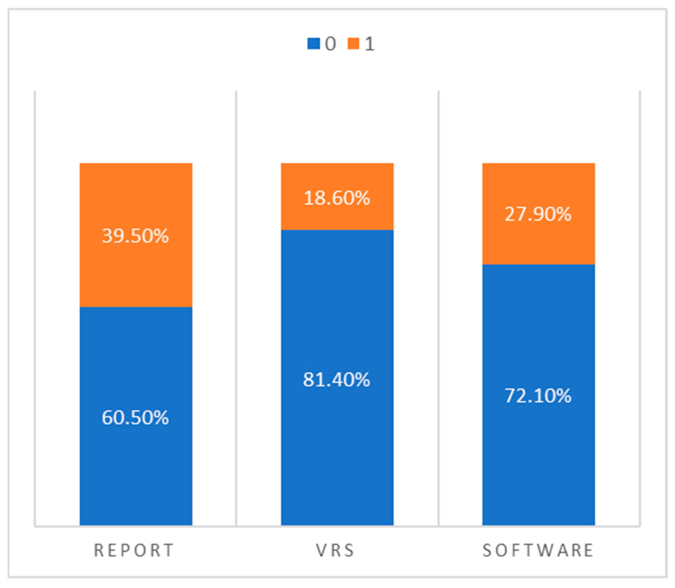
- Narrative radiology reports classified 17 cases as pathological and 26 as normal.
- Visual rating scales reported only 8 cases as pathological and 35 as normal.
- Software-based volumetry identified 12 cases as pathological and 31 as normal.
4. Discussion
Author Contributions
Funding
Institutional Review Board Statement
Informed Consent Statement
Data Availability Statement
Conflicts of Interest
References
- Agren, R.; Awad, A.; Blomstedt, P.; Fytagoridis, A. Voxel-Based Morphometry of Cerebellar Lobules in Essential Tremor. Front. Aging Neurosci. 2021, 13, 667854. [Google Scholar] [CrossRef] [PubMed]
- Amiri, H.; de Sitter, A.; Bendfeldt, K.; Battaglini, M.; Gandini Wheeler-Kingshott, C.A.M.; Calabrese, M.; Geurts, J.J.G.; Rocca, M.A.; Sastre-Garriga, J.; Enzinger, C.; et al. Urgent challenges in quantification and interpretation of brain grey matter atrophy in individual MS patients using MRI. Neuroimage Clin. 2018, 19, 466–475. [Google Scholar] [CrossRef] [PubMed]
- Cameron, E.; Dyke, J.P.; Hernandez, N.; Louis, E.D.; Dydak, U. Cerebral gray matter volume losses in essential tremor: A case-control study using high resolution tissue probability maps. Park. Relat. Disord. 2018, 51, 85–90. [Google Scholar] [CrossRef]
- Torisson, G.; van Westen, D.; Stavenow, L.; Minthon, L.; Londos, E. Medial temporal lobe atrophy is underreported and may have important clinical correlates in medical inpatients. BMC Geriatr. 2015, 15, 65. [Google Scholar] [CrossRef]
- Zivadinov, R.; Bergsland, N.; Korn, J.R.; Dwyer, M.G.; Khan, N.; Medin, J.; Price, J.C.; Weinstock-Guttman, B.; Silva, D.; Group, M.-M.S. Feasibility of Brain Atrophy Measurement in Clinical Routine without Prior Standardization of the MRI Protocol: Results from MS-MRIUS, a Longitudinal Observational, Multicenter Real-World Outcome Study in Patients with Relapsing-Remitting MS. AJNR Am. J. Neuroradiol. 2018, 39, 289–295. [Google Scholar] [CrossRef]
- Grisoli, M.; Nigri, A.; Medina Carrion, J.P.; Palermo, S.; Demichelis, G.; Giacosa, C.; Mongelli, A.; Fichera, M.; Nanetti, L.; Mariotti, C. Tracking longitudinal thalamic volume changes during early stages of SCA1 and SCA2. Radiol. Med. 2024, 129, 1215–1223. [Google Scholar] [CrossRef]
- Giorgio, A.; De Stefano, N. Advanced Structural and Functional Brain MRI in Multiple Sclerosis. Semin. Neurol. 2016, 36, 163–176. [Google Scholar] [CrossRef]
- Kaushik, S.; Vani, K.; Chumber, S.; Anand, K.S.; Dhamija, R.K. Evaluation of MR Visual Rating Scales in Major Forms of Dementia. J Neurosci Rural Pr. 2021, 12, 16–23. [Google Scholar] [CrossRef]
- Pizzini, F.B.; Conti, E.; Bianchetti, A.; Splendiani, A.; Fusco, D.; Caranci, F.; Bozzao, A.; Landi, F.; Gandolfo, N.; Farina, L.; et al. Radiological assessment of dementia: The Italian inter-society consensus for a practical and clinically oriented guide to image acquisition, evaluation, and reporting. Radiol. Medica 2022, 127, 998–1022. [Google Scholar] [CrossRef]
- Rocca, M.A.; Battaglini, M.; Benedict, R.H.; De Stefano, N.; Geurts, J.J.; Henry, R.G.; Horsfield, M.A.; Jenkinson, M.; Pagani, E.; Filippi, M. Brain MRI atrophy quantification in MS: From methods to clinical application. Neurology 2017, 88, 403–413. [Google Scholar] [CrossRef]
- Sinnecker, T.; Schadelin, S.; Benkert, P.; Ruberte, E.; Amann, M.; Lieb, J.M.; Naegelin, Y.; Muller, J.; Kuhle, J.; Derfuss, T.; et al. Brain atrophy measurement over a MRI scanner change in multiple sclerosis. Neuroimage Clin. 2022, 36, 103148. [Google Scholar] [CrossRef] [PubMed]
- Pizzini, F.B.; Boscolo Galazzo, I.; Natale, V.; Ribaldi, F.; Scheffler, M.; Caranci, F.; Lovblad, K.O.; Menegaz, G.; Frisoni, G.B.; Gunther, M. Insights into single-timepoint ASL hemodynamics: What visual assessment and spatial coefficient of variation can tell. Radiol. Med. 2024, 129, 467–477. [Google Scholar] [CrossRef] [PubMed]
- Hakansson, C.; Torisson, G.; Londos, E.; Hansson, O.; Bjorkman-Burtscher, I.M.; van Westen, D. Reporting frequency of radiology findings increases after introducing visual rating scales in the primary care diagnostic work up of subjective and mild cognitive impairment. Eur. Radiol. 2021, 31, 666–673. [Google Scholar] [CrossRef] [PubMed]
- Harper, L.; Barkhof, F.; Fox, N.C.; Schott, J.M. Using visual rating to diagnose dementia: A critical evaluation of MRI atrophy scales. J. Neurol. Neurosurg. Psychiatry 2015, 86, 1225–1233. [Google Scholar] [CrossRef]
- Kim, G.H.; Kim, J.E.; Choi, K.G.; Lim, S.M.; Lee, J.M.; Na, D.L.; Jeong, J.H. T1-weighted axial visual rating scale for an assessment of medial temporal atrophy in Alzheimer’s disease. J. Alzheimers Dis. 2014, 41, 169–178. [Google Scholar] [CrossRef]
- Loreto, F.; Gontsarova, A.; Scott, G.; Patel, N.; Win, Z.; Carswell, C.; Perry, R.; Malhotra, P. Visual atrophy rating scales and amyloid PET status in an Alzheimer’s disease clinical cohort. Ann. Clin. Transl. Neurol. 2023, 10, 619–631. [Google Scholar] [CrossRef]
- Mossa-Basha, M.; Andre, J.B.; Yuh, E.; Hunt, D.; LaPiana, N.; Howlett, B.; Krakauer, C.; Crane, P.; Nelson, J.; DeZelar, M.; et al. Comparison of brain imaging and physical health between research and clinical neuroimaging cohorts of ageing. Br. J. Radiol. 2024, 97, 614–621. [Google Scholar] [CrossRef]
- Zhang, Y.; Tartaglia, M.C.; Schuff, N.; Chiang, G.C.; Ching, C.; Rosen, H.J.; Gorno-Tempini, M.L.; Miller, B.L.; Weiner, M.W. MRI signatures of brain macrostructural atrophy and microstructural degradation in frontotemporal lobar degeneration subtypes. J. Alzheimers Dis. 2013, 33, 431–444. [Google Scholar] [CrossRef]
- Brusini, I.; MacNicol, E.; Kim, E.; Smedby, O.; Wang, C.; Westman, E.; Veronese, M.; Turkheimer, F.; Cash, D. MRI-derived brain age as a biomarker of ageing in rats: Validation using a healthy lifestyle intervention. Neurobiol. Aging 2022, 109, 204–215. [Google Scholar] [CrossRef]
- Hofmann, S.M.; Beyer, F.; Lapuschkin, S.; Goltermann, O.; Loeffler, M.; Muller, K.R.; Villringer, A.; Samek, W.; Witte, A.V. Towards the interpretability of deep learning models for multi-modal neuroimaging: Finding structural changes of the ageing brain. Neuroimage 2022, 261, 119504. [Google Scholar] [CrossRef]
- Persson, K.; Barca, M.L.; Edwin, T.H.; Cavallin-Eklund, L.; Tangen, G.G.; Rhodius-Meester, H.F.M.; Selbaek, G.; Knapskog, A.B.; Engedal, K. Regional MRI volumetry using NeuroQuant versus visual rating scales in patients with cognitive impairment and dementia. Brain Behav. 2024, 14, e3397. [Google Scholar] [CrossRef]
- Zilioli, A.; Rosenberg, A.; Mohanty, R.; Matton, A.; Granberg, T.; Hagman, G.; Lotjonen, J.; Kivipelto, M.; Westman, E. Brain MRI volumetry and atrophy rating scales as predictors of amyloid status and eligibility for anti-amyloid treatment in a real-world memory clinic setting. J. Neurol. 2024, 272, 84. [Google Scholar] [CrossRef] [PubMed]
- Vosshenrich, J.; Brantner, P.; Cyriac, J.; Jadczak, A.; Lieb, J.M.; Blackham, K.A.; Heye, T. Quantifying the Effects of Structured Reporting on Report Turnaround Times and Proofreading Workload in Neuroradiology. Acad. Radiol. 2023, 30, 727–736. [Google Scholar] [CrossRef]
- Goodkin, O.; Pemberton, H.; Vos, S.B.; Prados, F.; Sudre, C.H.; Moggridge, J.; Cardoso, M.J.; Ourselin, S.; Bisdas, S.; White, M.; et al. The quantitative neuroradiology initiative framework: Application to dementia. Br. J. Radiol. 2019, 92, 20190365. [Google Scholar] [CrossRef]
- Mallio, C.A.; Sertorio, A.C.; Bernetti, C.; Beomonte Zobel, B. Large language models for structured reporting in radiology: Performance of GPT-4, ChatGPT-3.5, Perplexity and Bing. Radiol. Med. 2023, 128, 808–812. [Google Scholar] [CrossRef]
- Mallio, C.A.; Bernetti, C.; Sertorio, A.C.; Beomonte Zobel, B. Large language models and structured reporting: Never stop chasing critical thinking. Radiol. Med. 2023, 128, 1445–1446. [Google Scholar] [CrossRef]
- Mamlouk, M.D.; Chang, P.C.; Saket, R.R. Contextual Radiology Reporting: A New Approach to Neuroradiology Structured Templates. AJNR Am. J. Neuroradiol. 2018, 39, 1406–1414. [Google Scholar] [CrossRef]
- Granata, V.; Fusco, R.; Coluccino, S.; Russo, C.; Grassi, F.; Tortora, F.; Conforti, R.; Caranci, F. Preliminary data on artificial intelligence tool in magnetic resonance imaging assessment of degenerative pathologies of lumbar spine. Radiol. Med. 2024, 129, 623–630. [Google Scholar] [CrossRef]
- Lee, J.; Lee, J.Y.; Oh, S.W.; Chung, M.S.; Park, J.E.; Moon, Y.; Jeon, H.J.; Moon, W.J. Evaluation of Reproducibility of Brain Volumetry between Commercial Software, Inbrain and Established Research Purpose Method, FreeSurfer. J. Clin. Neurol. 2021, 17, 307–316. [Google Scholar] [CrossRef]
- Kim, S.H.; Schramm, S.; Riedel, E.O.; Schmitzer, L.; Rosenkranz, E.; Kertels, O.; Bodden, J.; Paprottka, K.; Sepp, D.; Renz, M.; et al. Automation bias in AI-assisted detection of cerebral aneurysms on time-of-flight MR angiography. Radiol. Med. 2025, 130, 555–566. [Google Scholar] [CrossRef]
- Cover, K.S.; van Schijndel, R.A.; van Dijk, B.W.; Redolfi, A.; Knol, D.L.; Frisoni, G.B.; Barkhof, F.; Vrenken, H.; neuGrid; Alzheimer’s Disease Neuroimaging, Initiative. Assessing the reproducibility of the SienaX and Siena brain atrophy measures using the ADNI back-to-back MP-RAGE MRI scans. Psychiatry Res. 2011, 193, 182–190. [Google Scholar] [CrossRef] [PubMed]
- Koussis, P.; Toulas, P.; Glotsos, D.; Lamprou, E.; Kehagias, D.; Lavdas, E. Reliability of automated brain volumetric analysis: A test by comparing NeuroQuant and volBrain software. Brain Behav. 2023, 13, e3320. [Google Scholar] [CrossRef]
- Lee, J.Y.; Park, J.E.; Chung, M.S.; Oh, S.W.; Moon, W.J. [Expert Opinions and Recommendations for the Clinical Use of Quantitative Analysis Software for MRI-Based Brain Volumetry]. Taehan Yongsang Uihakhoe Chi 2021, 82, 1124–1139. [Google Scholar] [CrossRef]
- Tanabe, J.; Lim, M.F.; Dash, S.; Pattee, J.; Steach, B.; Pressman, P.; Bettcher, B.M.; Honce, J.M.; Potigailo, V.A.; Colantoni, W.; et al. Automated Volumetric Software in Dementia: Help or Hindrance to the Neuroradiologist? AJNR Am. J. Neuroradiol. 2024, 45, 1737–1744. [Google Scholar] [CrossRef]
- Bruno, F.; Tommasino, E.; Catalucci, A.; Pastorelli, C.; Borea, F.; Caldarelli, G.; Bellini, M.; Badini, P.; Mancini, S.; Santobuono, C.; et al. Evaluation of Cerebral Volume Changes in Patients with Tremor Treated by MRgFUS Thalamotomy. Life 2022, 13, 16. [Google Scholar] [CrossRef]
- Scheltens, P.; Leys, D.; Barkhof, F.; Huglo, D.; Weinstein, H.C.; Vermersch, P.; Kuiper, M.; Steinling, M.; Wolters, E.C.; Valk, J. Atrophy of medial temporal lobes on MRI in "probable" Alzheimer’s disease and normal ageing: Diagnostic value and neuropsychological correlates. J. Neurol. Neurosurg. Psychiatry 1992, 55, 967–972. [Google Scholar] [CrossRef]
- Pasquier, F.; Leys, D.; Weerts, J.G.; Mounier-Vehier, F.; Barkhof, F.; Scheltens, P. Inter- and intraobserver reproducibility of cerebral atrophy assessment on MRI scans with hemispheric infarcts. Eur. Neurol. 1996, 36, 268–272. [Google Scholar] [CrossRef]
- Koedam, E.L.; Lehmann, M.; van der Flier, W.M.; Scheltens, P.; Pijnenburg, Y.A.; Fox, N.; Barkhof, F.; Wattjes, M.P. Visual assessment of posterior atrophy development of a MRI rating scale. Eur. Radiol. 2011, 21, 2618–2625. [Google Scholar] [CrossRef]
- Cole, J.H.; Franke, K. Predicting Age Using Neuroimaging: Innovative Brain Ageing Biomarkers. Trends Neurosci. 2017, 40, 681–690. [Google Scholar] [CrossRef]
- Parillo, M.; Vaccarino, F.; Beomonte Zobel, B.; Mallio, C.A. ChatGPT and radiology report: Potential applications and limitations. Radiol. Med. 2024, 129, 1849–1863. [Google Scholar] [CrossRef]
- Pemberton, H.G.; Zaki, L.A.M.; Goodkin, O.; Das, R.K.; Steketee, R.M.E.; Barkhof, F.; Vernooij, M.W. Technical and clinical validation of commercial automated volumetric MRI tools for dementia diagnosis-a systematic review. Neuroradiology 2021, 63, 1773–1789. [Google Scholar] [CrossRef] [PubMed]


| Scale | Region Assessed | Imaging Plane/Sequence | Scoring Range | Scoring Criteria | Pathological Cut-Off |
|---|---|---|---|---|---|
| MTA (Scheltens) | Medial temporal lobe (hippocampus, choroid fissure, temporal horn) | Coronal T1-weighted | 0–4 | 0 = normal; 1 = mild choroid fissure widening; 2 = +mild temporal horn enlargement; 3 = +moderate hippocampal atrophy; 4 = severe atrophy with structural loss | ≥2 (<75 yrs); ≥3 (≥75 yrs) |
| GCA (Pasquier) | Global Cortical Atrophy (frontal, parietal, temporal, occipital lobes) | Axial T1-weighted | 0–3 (per hemisphere) | 0 = normal; 1 = mild sulcal widening; 2 = moderate; 3 = severe “knife blade” atrophy | ≥2 (any age) |
| Koedam | Posterior parietal regions (precuneus, parieto-occipital sulcus, posterior cingulate) | Axial, sagittal, coronal T1-weighted | 0–3 | 0 = no sulcal widening; 1 = mild; 2 = moderate; 3 = severe widening and atrophy | ≥2 (any age) |
| Visual Rating Scale | ICC (Single Measures) | ICC (Average Measures) |
|---|---|---|
| Pasquier (GCA) | 0.54 (95% CI: 0.31–0.72) | 0.70 (95% CI: 0.47–0.83) |
| Koedam | 0.55 (95% CI: 0.07–0.78) | 0.71 (95% CI: 0.14–0.87) |
| MTA (Scheltens) | 0.47 (95% CI: 0.23–0.72) | 0.68 (95% CI: 0.18–0.78) |
| VRS—Software | Cohen Kappa Test | Concordance | McNemar Test—p Value |
|---|---|---|---|
| MTA | 0.14 | 71.73% | 0.005 |
| Koedam | 0.33 | 82.61% | 0.28 |
| Pasquier | 0.29 | 80.43% | 0.18 |
| Report—Software | Cohen Kappa | Concordance | McNemar Test—p Value |
|---|---|---|---|
| Temporal | 0.29 | 84.78% | 0.13 |
| Global | 0.03 | 69.56% | 0.50 |
| Posterior | 0.20 | 80.43% | 0.78 |
Disclaimer/Publisher’s Note: The statements, opinions and data contained in all publications are solely those of the individual author(s) and contributor(s) and not of MDPI and/or the editor(s). MDPI and/or the editor(s) disclaim responsibility for any injury to people or property resulting from any ideas, methods, instructions or products referred to in the content. |
© 2025 by the authors. Licensee MDPI, Basel, Switzerland. This article is an open access article distributed under the terms and conditions of the Creative Commons Attribution (CC BY) license (https://creativecommons.org/licenses/by/4.0/).
Share and Cite
Bruno, F.; Fagotti, C.; Saltarelli, G.; Di Cerbo, G.; Sabatelli, A.; De Felici, C.; Innocenzi, A.; Di Cesare, E.; Splendiani, A. Radiological Reporting of Brain Atrophy in MRI: Real-Life Comparison Between Narrative Reports, Semiquantitative Scales and Automated Software-Based Volumetry. Diagnostics 2025, 15, 1246. https://doi.org/10.3390/diagnostics15101246
Bruno F, Fagotti C, Saltarelli G, Di Cerbo G, Sabatelli A, De Felici C, Innocenzi A, Di Cesare E, Splendiani A. Radiological Reporting of Brain Atrophy in MRI: Real-Life Comparison Between Narrative Reports, Semiquantitative Scales and Automated Software-Based Volumetry. Diagnostics. 2025; 15(10):1246. https://doi.org/10.3390/diagnostics15101246
Chicago/Turabian StyleBruno, Federico, Cristina Fagotti, Gaspare Saltarelli, Giovanni Di Cerbo, Alessandra Sabatelli, Claudia De Felici, Antonio Innocenzi, Ernesto Di Cesare, and Alessandra Splendiani. 2025. "Radiological Reporting of Brain Atrophy in MRI: Real-Life Comparison Between Narrative Reports, Semiquantitative Scales and Automated Software-Based Volumetry" Diagnostics 15, no. 10: 1246. https://doi.org/10.3390/diagnostics15101246
APA StyleBruno, F., Fagotti, C., Saltarelli, G., Di Cerbo, G., Sabatelli, A., De Felici, C., Innocenzi, A., Di Cesare, E., & Splendiani, A. (2025). Radiological Reporting of Brain Atrophy in MRI: Real-Life Comparison Between Narrative Reports, Semiquantitative Scales and Automated Software-Based Volumetry. Diagnostics, 15(10), 1246. https://doi.org/10.3390/diagnostics15101246