
Laboratory Assessment of Unfractionated Heparin (UFH) with Activated Clotting Time (ACT) and Anti-Xa Activity during Peripheral Arterial Angiographic Procedure
 ,
,
Abstract
1. Introduction
2. Materials and Methods
2.1. Patients
2.2. Methods
2.3. Statistical Methods
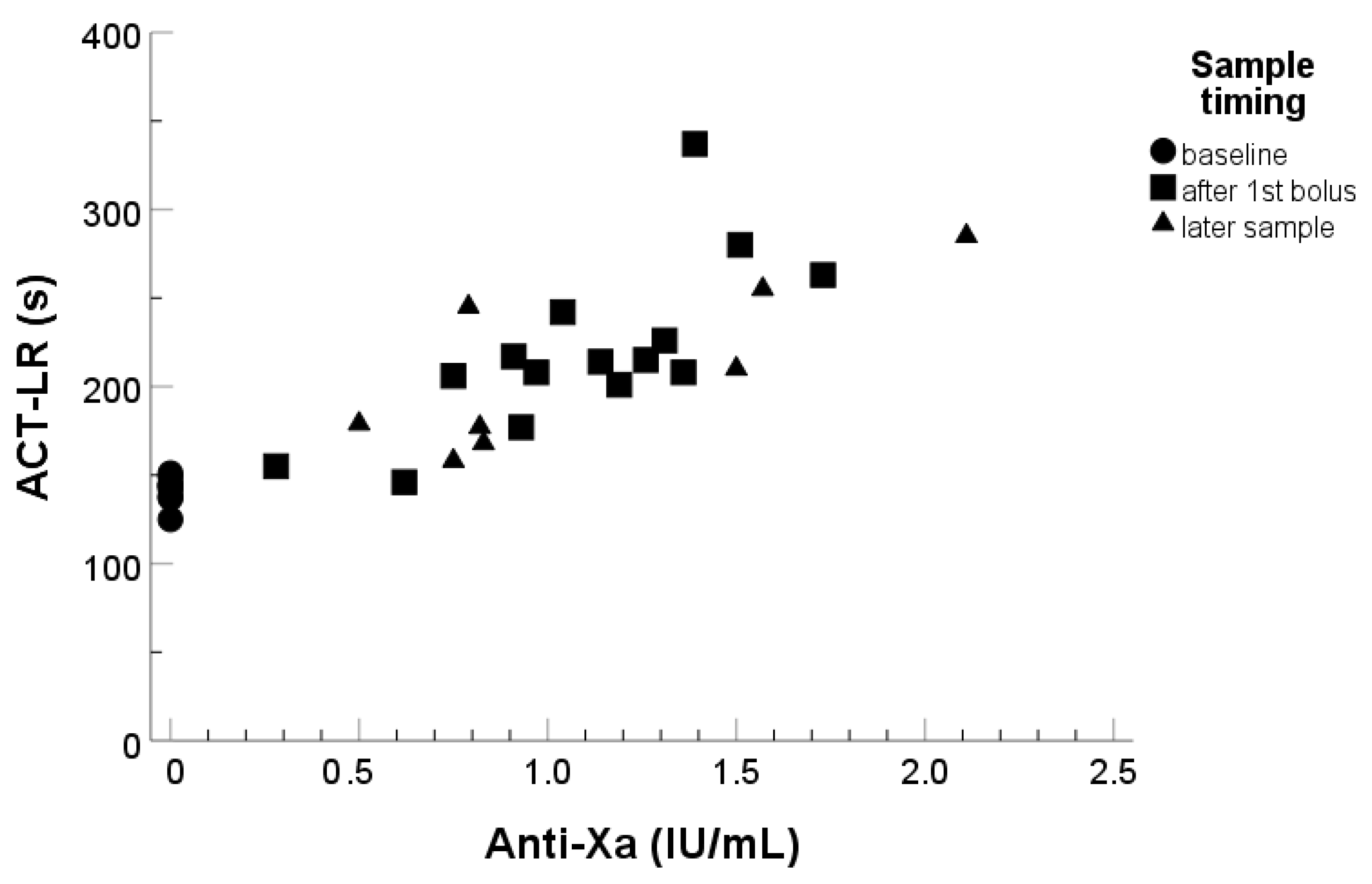
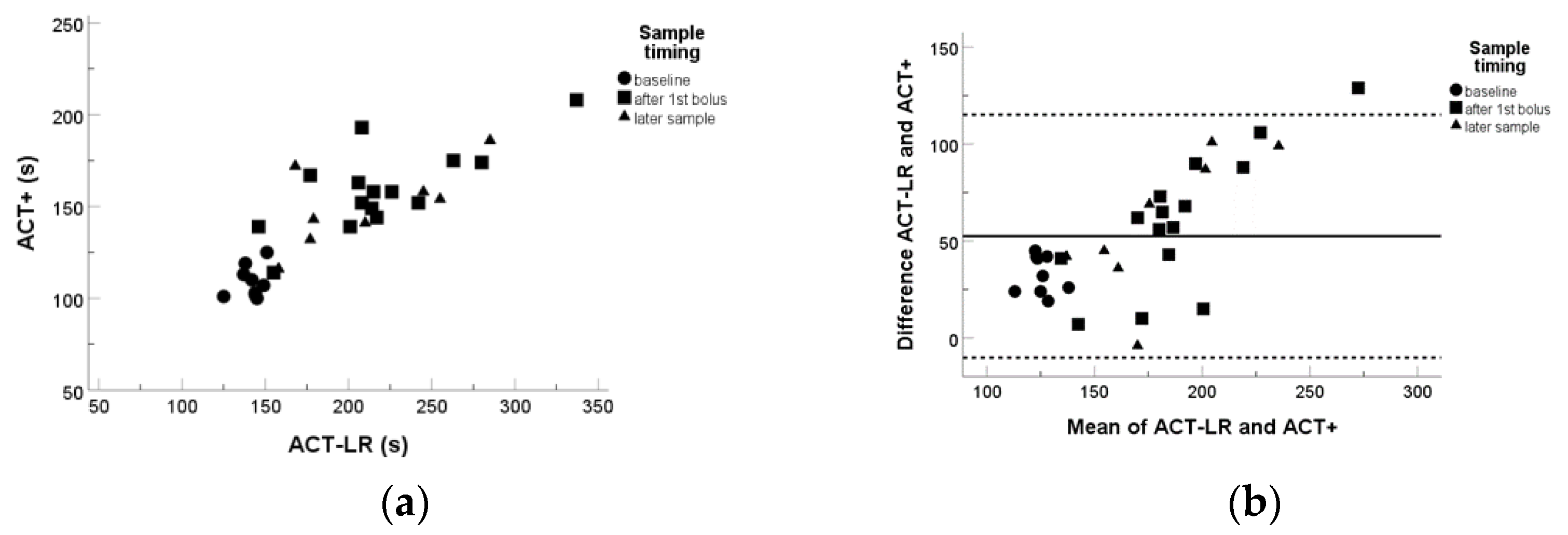
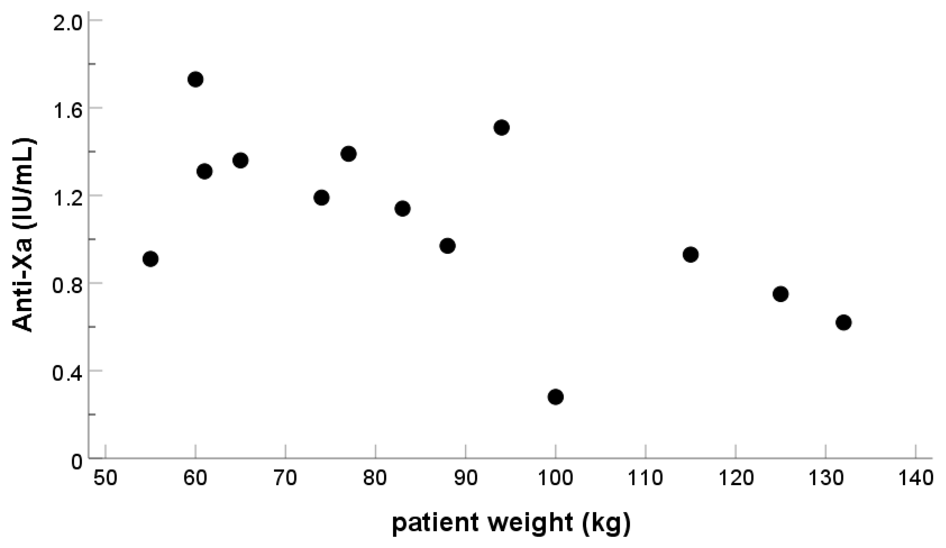
3. Results
4. Discussion
5. Conclusions
Author Contributions
Funding
Institutional Review Board Statement
Informed Consent Statement
Data Availability Statement
Acknowledgments
Conflicts of Interest
References
- Norgren, L.; Hiatt, W.R.; Dormandy, J.A.; Nehler, M.R.; Harris, K.A.; Fowkes, F.G.; TASCIIWorking Group Bell, K.; Caporusso, J.; Durand-Zaleski, I.; Komori, K.; et al. Inter-Society Consensus for the Management of Peripheral Arterial Disease (TASC II). Eur. J. Vasc. Endovasc. Surg. 2007, 33 (Suppl. 1), S1–S75. [Google Scholar] [CrossRef] [PubMed]
- Durran, A.C.; Watts, C. Current Trends in Heparin Use During Arterial Vascular Interventional Radiology. Cardiovasc. Interv. Radiol. 2012, 35, 1308–1314. [Google Scholar] [CrossRef] [PubMed]
- Hattersley, P.G. Activated coagulation time of whole blood. JAMA 1966, 196, 436–440. [Google Scholar] [CrossRef] [PubMed]
- Young, J.A.; Kisker, C.T.; Doty, D.B. Adequate Anticoagulation During Cardiopulmonary Bypass Determined by Activated Clotting Time and the Appearance of Fibrin Monomer. Ann. Thorac. Surg. 1978, 26, 231–240. [Google Scholar] [CrossRef]
- Shore-Lesserson, L.; Baker, R.A.; Ferraris, V.A.; Greilich, P.E.; Fitzgerald, D.; Roman, P.; Hammon, J.W. The Society of Thoracic Surgeons, The Society of Cardiovascular Anesthesiologists, and The American Society of ExtraCorporeal Technology: Clinical Practice Guidelines-Anticoagulation During Cardiopulmonary Bypass. Anesth. Analg. 2018, 126, 413–424. [Google Scholar] [CrossRef]
- Kasapis, C.; Gurm, H.S.; Chetcuti, S.J.; Munir, K.; Luciano, A.; Smith, D.; Aronow, H.D.; Kassab, E.H.; Knox, M.F.; Moscucci, M.; et al. Defining the optimal degree of heparin anticoagulation for peripheral vascular interventions: Insight from a large, regional, multicenter registry. Circ. Cardiovasc. Interv. 2010, 3, 593–601. [Google Scholar] [CrossRef]
- Nissborg, E.; Wahlgren, C.M. Anticoagulant Effect of Standard Dose Heparin During Peripheral Endovascular Intervention. Ann. Vasc. Sur.g 2019, 60, 286–292. [Google Scholar] [CrossRef]
- Rossi, M.; Iezzi, R. Cardiovascular and Interventional Radiological Society of Europe Guidelines on Endovascular Treatment in Aortoiliac Arterial Disease. Cardiovasc. Interv. Radiol. 2013, 37, 13–25. [Google Scholar] [CrossRef]
- Tremey, B.; Szekely, B.; Schlumberger, S.; François, D.; Liu, N.; Sievert, K.; Fischler, M. Anticoagulation monitoring during vascular surgery: Accuracy of the Hemochron ® low range activated clotting time (ACT-LR). Br. J. Anaesth. 2006, 97, 453–459. [Google Scholar] [CrossRef]
- Miles, L.F.; Coulson, T.G.; Galhardo, C.; Falter, F. Pump Priming Practices and Anticoagulation in Cardiac Surgery: Results From the Global Cardiopulmonary Bypass Survey. Anesth. Analg. 2017, 125, 1871–1877. [Google Scholar] [CrossRef]
- Korpi-Steiner, N.L.; Walz, J.M.; Schanzer, A.; Rao, L.V. Comparison of Point-of-Care Activated Clotting Time Methods in Different Clinical Settings in a Large Academic Medical Center. Jrnl. App. Lab. Med. 2017, 2, 356–366. [Google Scholar] [CrossRef] [PubMed]
- Wiersema, A.M.; Vos, J.A.; Bruijninckx, C.M.A.; van Delden, O.M.; Reijnen, M.M.P.J.; Vahl, A.; Zeebregts, C.J.; Moll, F.L. Periprocedural prophylactic antithrombotic strategies in interventional radiology: Current practice in the Netherlands and comparison with the United kingdom. Cardiovasc. Interv. Radiol 2013, 36, 1477–1492. [Google Scholar] [CrossRef] [PubMed]
- Goldhammer, J.E.; Zimmerman, D. Pro: Activated Clotting Time Should Be Monitored During Heparinization For Vascular Surgery. J. Cardiothorac. Vasc. Anesth. 2018, 32, 1494–1496. [Google Scholar] [CrossRef]
- Wolo, E.; Herman, C. Con: Activated Clotting Time Should Not Be Monitored During Heparinization for Vascular Surgery. J. Cardiothorac. Vasc. Anesth. 2018, 32, 1497–1499. [Google Scholar] [CrossRef]
- Doherty, T.M.; Shavelle, R.M.; French, W.J. Reproducibility and variability of activated clotting time measurements in the cardiac catheterization laboratory. Catheter. Cardiovasc. Interv. 2005, 65, 330–337. [Google Scholar] [CrossRef]
- Garcia, D.A.; Baglin, T.P.; Weitz, J.I.F.C.C.P.; Samama, M.M. Parenteral Anticoagulants: Antithrombotic Therapy and Prevention of Thrombosis, 9th ed: American College of Chest Physicians Evidence-Based Clinical Practice Guidelines. Chest 2012, 141, e24S–e43S. [Google Scholar] [CrossRef] [PubMed]
- Bounameaux, H.; Marbet, G.A.; Lammle, B.; Eichlisberger, R.; Duckert, F. Monitoring of heparin treatment. Comparison of thrombin time, activated partial thromboplastin time, and plasma heparin concentration, and analysis of the behavior of antithrombin III. Am. J. Clin. Pathol. 1980, 74, 68–73. [Google Scholar] [CrossRef] [PubMed]
- Burki, S.; Brand, B.; Escher, R.; Wuillemin, W.A.; Nagler, M. Accuracy, reproducibility and costs of different laboratory assays for the monitoring of unfractionated heparin in clinical practice: A prospective evaluation study and survey among Swiss institutions. BMJ Open 2018, 8, e022943. [Google Scholar] [CrossRef]
- Dieplinger, B.; Egger, M.; Luft, C.; Hinterreiter, F.; Pernerstorfer, T.; Haltmayer, M.; Mueller, T. Comparison between activated clotting time and anti-activated factor X activity for the monitoring of unfractionated heparin therapy in patients with aortic aneurysm undergoing an endovascular procedure. J. Vasc. Surg. 2018, 68, 400–407. [Google Scholar] [CrossRef]
- Delmas, C.; Jacquemin, A.; Vardon-Bounes, F.; Georges, B.; Guerrero, F.; Hernandez, N.; Marcheix, B.; Seguin, T.; Minville, V.; Conil, J.M.; et al. Anticoagulation Monitoring Under ECMO Support: A Comparative Study Between the Activated Coagulation Time and the Anti-Xa Activity Assay. J. Intensive. Care. Med. 2018, 35, 885066618776937. [Google Scholar] [CrossRef]
- Rosenberg, A.F.; Zumberg, M.; Taylor, L.; LeClaire, A.; Harris, N. The use of anti-Xa assay to monitor intravenous unfractionated heparin therapy. J. Pharm. Pract. 2010, 23, 210–216. [Google Scholar] [CrossRef] [PubMed]
- Fowler, A.J.; Ahmad, T.; Phull, M.K.; Allard, S.; Gillies, M.A.; Pearse, R.M. Meta-analysis of the association between preoperative anaemia and mortality after surgery. Br. J. Surg. 2015, 102, 1314–1324. [Google Scholar] [CrossRef] [PubMed]
- Bodewes, T.C.F.; Pothof, A.B.; Darling, J.D.; Deery, S.E.; Jones, D.W.; Soden, P.A.; Moll, F.L.; Schermerhorn, M.L. Preoperative anemia associated with adverse outcomes after infrainguinal bypass surgery in patients with chronic limb-threatening ischemia. J. Vasc. Surg. 2017, 66, 1775–1785.e2. [Google Scholar] [CrossRef]
- Girardi, L.; Sudi, K.; Muntean, W. Effect of heparin, platelets, activated platelets, platelet fragments, and hematocrit on activated clotting time. Artif. Organs 2000, 24, 507–513. [Google Scholar] [CrossRef] [PubMed]
- Konrath, S.; Mailer, R.K.; Renné, T. Mechanism, Functions, and Diagnostic Relevance of FXII Activation by Foreign Surfaces. Hamostaseologie 2021, 41, 489–501. [Google Scholar] [CrossRef] [PubMed]
- Ambulgekar, N.V.; Grey, S.F.; Rosman, H.S.; Othman, H.; Davis, T.P.; Nypaver, T.J.; Schreiber, T.; Yamasaki, H.; Lalonde, T.A.; Henke, P.K.; et al. Blue Cross Blue Shield of Michigan Cardiovascular Consortium (BMC2) Investigators. Association of Anemia With Outcomes in Patients Undergoing Percutaneous Peripheral Vascular Intervention: Insights From the Blue Cross Blue Shield of Michigan Cardiovascular Consortium (BMC2 VIC). J. Invasive Cardiol. 2018, 30, 35–42. [Google Scholar]
- Liveris, A.; Bello, R.A.; Friedmann, P.; Duffy, M.A.; Manwani, D.; Killinger, J.S.; Rodriquez, D.; Weinstein, S. Anti-factor Xa assay is a superior correlate of heparin dose than activated partial thromboplastin time or activated clotting time in pediatric extracorporeal membrane oxygenation. Pediatr. Crit. Care Med. 2014, 15, e72–e79. [Google Scholar] [CrossRef]


| Patient characteristics (n = 15) | |||
| Age, years median (range) | 62 (32–93) | ||
| Gender (men/women) | 13/2 | ||
| Weight, kg, median (range) | 83 (55–132) | ||
| Receiving aspirin/clopidogrel | 9/4 | ||
| N | % | ||
| Indication for the endovascular procedure | |||
| Lower limb artery stenosis (left/right side) | 11 (8/3) | 73 | |
| Vascular malformation, AV upper limb or pulmonary | 2 | 13 | |
| Iliacal thrombectomy, deep vein thrombosis (right side) | 1 | 7 | |
| Dialysis fistula malfunction | 1 | 7 | |
| UFH doses received | |||
| Only single dose 5000 IU | 10 | 67 | |
| Single dose 5000 IU and subsequent dose 2500–3000 IU | 2 | 13 | |
| Single dose 5000 IU, subsequent, third dose 2500–4000 IU | 2 | 13 | |
| Single dose 2500 IU (dialysis patient) | 1 | 7 | |
| LMWH medication before procedure | |||
| Yes | 2 | 13 | |
| No | 13 | 87 | |
Disclaimer/Publisher’s Note: The statements, opinions and data contained in all publications are solely those of the individual author(s) and contributor(s) and not of MDPI and/or the editor(s). MDPI and/or the editor(s) disclaim responsibility for any injury to people or property resulting from any ideas, methods, instructions or products referred to in the content. |
© 2023 by the authors. Licensee MDPI, Basel, Switzerland. This article is an open access article distributed under the terms and conditions of the Creative Commons Attribution (CC BY) license (https://creativecommons.org/licenses/by/4.0/).
Share and Cite
Helin, T.; Tirri, T.; Korkala, H.; Lappalainen, K.; Joutsi-Korhonen, L. Laboratory Assessment of Unfractionated Heparin (UFH) with Activated Clotting Time (ACT) and Anti-Xa Activity during Peripheral Arterial Angiographic Procedure. Diagnostics 2023, 13, 1489. https://doi.org/10.3390/diagnostics13081489
Helin T, Tirri T, Korkala H, Lappalainen K, Joutsi-Korhonen L. Laboratory Assessment of Unfractionated Heparin (UFH) with Activated Clotting Time (ACT) and Anti-Xa Activity during Peripheral Arterial Angiographic Procedure. Diagnostics. 2023; 13(8):1489. https://doi.org/10.3390/diagnostics13081489
Chicago/Turabian StyleHelin, Tuukka, Tomi Tirri, Heidi Korkala, Kimmo Lappalainen, and Lotta Joutsi-Korhonen. 2023. "Laboratory Assessment of Unfractionated Heparin (UFH) with Activated Clotting Time (ACT) and Anti-Xa Activity during Peripheral Arterial Angiographic Procedure" Diagnostics 13, no. 8: 1489. https://doi.org/10.3390/diagnostics13081489
APA StyleHelin, T., Tirri, T., Korkala, H., Lappalainen, K., & Joutsi-Korhonen, L. (2023). Laboratory Assessment of Unfractionated Heparin (UFH) with Activated Clotting Time (ACT) and Anti-Xa Activity during Peripheral Arterial Angiographic Procedure. Diagnostics, 13(8), 1489. https://doi.org/10.3390/diagnostics13081489