Article

Biosolutions from Native Trichoderma Strains Against Grapevine Trunk Diseases

by
Laura Zanfaño
1,
Guzmán Carro-Huerga
1,
Álvaro Rodríguez-González
1,
Daniela Ramírez-Lozano
1,
Sara Mayo-Prieto
1,
Santiago Gutiérrez
1,2 and
Pedro A. Casquero
1,*
1
Grupo Universitario de Investigación en Ingeniería y Agricultura Sostenible (GUIIAS), Instituto de Medio Ambiente, Recursos Naturales y Biodiversidad, Universidad de León, Avenida Portugal 41, 24071 León, Spain
2
Área de Microbiología, Grupo Universitario de Investigación en Ingeniería y Agricultura Sostenible (GUIIAS), Instituto de Medio Ambiente, Recursos Naturales y Biodiversidad, Universidad de León, Avenida Portugal 41, 24071 León, Spain
*
Author to whom correspondence should be addressed.
Agronomy 2025, 15(8), 1901; https://doi.org/10.3390/agronomy15081901
Submission received: 25 June 2025 / Revised: 30 July 2025 / Accepted: 5 August 2025 / Published: 7 August 2025
(This article belongs to the Special Issue Molecular Advances in Crop Protection and Agrobiotechnology)

Abstract

Fungi of the genus Trichoderma show strong potential as biological control agents (BCAs) against grapevine trunk diseases (GTDs) through mechanisms like antibiotic metabolite production and lytic enzymes. This study evaluated the biocontrol activity of four native Trichoderma strains—T. gamsii T065 and T071, T. carraovejensis T154, and T. harzianum T214—against Phaeoacremonium minimum, Phaeomoniella chlamydospora, and Diplodia seriata. Culture filtrates obtained at 8, 16, and 24 days post-incubation were tested using antibiogram and mycelial inhibition assays. Strains T071, T154, and T214 effectively inhibited D. seriata, while T154 and T214 also suppressed P. chlamydospora. Nevertheless, the limited effectiveness of all filtrates against P. minimum suggests that antibiosis is not the predominant mechanism involved in its control. These findings highlight the potential of specific Trichoderma strains and incubation times to directly control GTD pathogens and support the development of scalable biocontrol solutions.

1. Introduction

To ensure optimum crop productivity, farmers currently rely heavily on chemical insecticides and inorganic fertilizers. Over-reliance on these chemicals has several side effects on users, non-target organisms and the environment. The future scenario of the agricultural sector, increasingly pressured by regulations, public concern and environmental issues, continues to motivate the development of alternative methods to chemicals for applications such as fertilizers and pesticides [1,2]. Bioactive fungal compounds offer a promising alternative to chemical pesticides for crop protection and pest management. Multiple mechanisms of action, selective pest control, environmental friendliness and sustainability are some of the advantages of using bioactive fungal compounds [3]. Among the microorganisms capable of the production of bioactive compounds are fungi of the genus Trichoderma.
Trichoderma species are fungi that are highly interactive in root, soil and foliar environments. These filamentous fungi are known for their diverse biological functions, including their ability to promote plant growth [4], protect against pathogens [5,6] and produce secondary metabolites [7]. For example, different strains produce more than 100 different metabolites with known antibiotic activities [8]. Trichoderma show a high level of genetic diversity and can be used to produce a wide range of products of commercial and environmental interest. They are prolific producers of extracellular proteins and are best known for their ability to produce enzymes that degrade cellulose and chitin although they also produce other useful enzymes [9].
These fungi are particularly important in agriculture, where they act as biocontrol agents and contribute to integrated pest and disease management strategies. Trichoderma species, such as Trichoderma harzianum and Trichoderma viride, have been extensively studied for their mechanisms of action, which include the production of lytic enzymes, competition for nutrients and the synthesis of secondary metabolites with antimicrobial properties [10,11]. Some of these secondary metabolites are peptaibols, NRPs, volatile and non-volatile terpenes, pyrones, siderophores and nitrogen-containing compounds. As many as 373 different molecules have been identified, but in many cases the specific activity of these molecules is unknown [12]. These compounds have attracted considerable attention for their potential use in sustainable crop protection. The growing interest in Trichoderma secondary metabolites has stimulated research on their chemical diversity, biological activities and applications in crop and forest management [13]. Some studies have shown that secondary metabolites of Trichoderma spp. may also play a role in both growth regulation and activation of plant defense responses [14]. Trichoderma also secretes Cell Wall Degrading Enzymes (CWDEs), with the ability to break down the cell walls of pathogens [15]. The most important CWDEs are chitinases, glucanases and proteases. Among these, chitinases stand out as the best studied in the case of Trichoderma, as they are involved in the maintenance of the fungal wall itself and in the degradation of the cell wall of other fungal species [16].
Trichoderma species and the compounds they are capable of producing, such as secondary metabolites and lytic enzymes, have attracted attention in viticulture for their potential in the control of grapevine trunk diseases (GTDs), which are among the most destructive and economically significant diseases affecting vineyards worldwide. These chronic fungal infections impact the longevity, productivity, and quality of grapevines, posing a major challenge to viticulture. GTDs affect nearly 20% of global vineyards, with annual economic losses estimated at 1.5 billion USD globally [17], while yield losses range from 30% to 60% in severely affected regions like California, Australia, and New Zealand [18,19]. Some of the most important GTDs in vineyards are esca and Petri disease, caused by the pathogens Phaeoacremonium minimum, Phaeomoniella chlamydospora and Fomitiporia mediterranea. Symptoms of these diseases include foliar discoloration, necrosis, and dieback of wood tissue. Another GTD caused by Diplodia seriata, Lasiodiplodia theobromae and Neofusicoccum parvum is Botryosphaeria dieback, which produces necrotic lesions that spread along the trunk of the vine [20,21]. The control of these diseases by secondary metabolites and lytic enzymes produced by Trichoderma is becoming a point of great interest in biological control.
The most effective secondary metabolites produced by Trichoderma species against grapevine pathogens include a variety of bioactive compounds with antifungal and plant defense-inducing properties [22]. Secondary metabolites such as harzianic acid (HA) and 6-pentyl-α-pyrone (6PP) produced by Trichoderma strains have been shown to inhibit pathogens such as Eutypa lata (Eutypa dieback) and other fungi that cause GTDs [23]. These metabolites suppress disease development and induce systemic resistance in grapevines, reducing the need for chemical fungicides. There has also been evidence that some secondary metabolites of Trichoderma, such as 6PP are able to improve crop yield and increase the total amount of polyphenols and antioxidant activity in the grapes [24]. With regard to Trichoderma CWDEs, they act through several mechanisms to control grapevine diseases. One of the primary mechanisms involves direct mycoparasitism, where Trichoderma species can invade or wound the mycelium of pathogens, causing cell deformation, protoplasm shrinkage, and ultimately cell wall breakage through the action of their CWDEs [25]. Beyond direct antagonism, enzymes produced by Trichoderma can induce systemic resistance in grapevines, enhancing their natural defenses against a wide range of pathogens [26,27]. A laboratory model developed to assess root colonization by T. atroviride and its effect on defense activation showed that this colonization led to the up-regulation of targeted defense genes [28].
The proposed hypothesis is that culture filtrates from native Trichoderma strains produce bioactive compounds with differential inhibitory effects on key GTD pathogens, depending on the incubation period and the specific metabolism of each strain. These compounds collectively contribute to Trichoderma’s effectiveness as a biocontrol agent in vineyards, supporting sustainable viticulture practices.

2. Materials and Methods

2.1. Fungal Strains

The four Trichoderma isolates used in this study were Trichoderma gamsii T065, Trichoderma gamsii T071, Trichoderma carraovejensis T154, and Trichoderma harzianum T214 (Table 1). These isolates belong to the collection of the “Laboratorio de Diagnóstico de Plagas y Enfermedades Vegetales” (Plant and Pest Diagnostic Laboratory) (LDPEV), Universidad de León, Spain. The chosen isolates were obtained from vine plots in different regions of Spain as well as from different parts of the grapevine plant. T065 [29] and T071 [30] were isolated from Albarín and Tempranillo vineyard soil, respectively. T154 is a new species (Trichoderma carraovejensis [6]) that was isolated from the trunk wood of Tempranillo grapevines, and T214 was isolated from Verdejo grapevine bark [29].
The pathogens used in the antifungal assays are available at the LDPEV collection with the following codes: Phaeoacremonium minimum Y038-05-3, Phaeomoniella chlamydospora Y116-18-03c, and Diplodia seriata ULEP32 (Table 2). These pathogens belong to the main GTDs. P. minimum and P. chlamydospora are two of the main pathogens that cause Petri and esca disease and Diplodia seriata is the most representative and aggressive pathogen of Botryosphaeria dieback disease [31].

2.2. Culture Filtrates from Trichoderma

2.2.1. Fungal Culture Preparation

Four Trichoderma isolates (T065, T071, T154 and T214) were used to extract culture filtrate with potential against GTDs. Initially, Trichoderma culture filtrates were produced by cultivation in potato dextrose broth (PDB) liquid medium (Conda Laboratory, Madrid, Spain). This process was carried out in 250 mL Erlenmeyer flasks with 50 mL of PDB. Three 6-mm diameter plugs from each Trichoderma strain obtained from PDA (Sigma-Aldrich Chemie GmbH, Steinheim, Germany) cultures of 7 days growth were inoculated onto this medium. The flasks were incubated in static conditions at 25 °C in the dark for different periods of time.

2.2.2. Filtrate Extraction

At the end of the incubation period, the compounds produced by Trichoderma were extracted by filtration. For this purpose, the liquid solution in the flasks was centrifuged for 5 min at the maximum speed allowed by the centrifuge. This liquid solution was then filtered through a 0.45 μm filter (cellulose acetate syringe filters. Filter-Lab, Barcelona, Spain), thus retaining the Trichoderma spores in the filter and preserving only the compounds produced by the fungus during the incubation period. Finally, the pH of the culture medium just before the extraction of the culture filtrates was also evaluated, a factor related to the type of compound produced that also influences the development of the microorganisms. The pH measurements were carried out with a VioLab PC 50 sensor (labprocess 25, Barcelona, Spain).

2.3. Experimental Design

From each Trichoderma isolate (T065, T071, T154 and T214) used in this study, culture filtrates were extracted at three different times, 8 days after PDB inoculation, 16 days and 24 days. This allowed for the study of the compounds produced during different time periods. A total of 6 flasks were inoculated from each isolate, with two replicates for each incubation period (8, 16 and 24 days). A total of 24 Erlenmeyer flasks were used in the experiment. These trials were developed from September to November 2024.

2.4. Antifungal Assays

Two in vitro assays were performed to evaluate the efficacy of the culture filtrates produced by Trichoderma against the pathogens P. minimum, P. chlamydospora and D. serita.

2.4.1. Antibiograms

Experiments were conducted according to the methodology of Malmierca et al. (2012) [34], where this bioassay was performed using Trichoderma culture filtrates against the three grapevine pathogens. Bioassay plates were prepared by inoculating 1 × 10⁶ spores/mL of P. minimum and P. chlamydospora and 1 × 10³ spores/mL of D. seriata on PDA medium (PDB with 1% agar) at 45–55 °C. A total of 15 mL of this solution was added to each plate. Five 6 mm holes were made per plate using a punch and 20 μL of the culture filtrate was added to each hole. Plates were incubated at 25 °C for 24 to 48 h to visualize the growth inhibition zone. For evaluation of the inhibition zone, two perpendicular measurements of the diameter of the inhibition ring around each hole were taken. The experimental design consisted of a total of 27 plates, 9 plates of each pathogen and 3 replicates of each culture filtrate extraction time (8, 16 and 24 days). In each of these plates, four of the five holes were destined for the culture filtrates of the four Trichoderma isolates and one for the control, which was carried out with autoclaved distilled water.

2.4.2. Inhibition of Mycelial Growth

This assay was performed in 55 mm diameter Petri dishes containing PDA medium. A total of 200 μL of culture filtrate was added to each plate and spread with a seeding loop. Then, 6 mm diameter plugs of each pathogen, obtained from the actively growing edges of the PDA cultures, were inoculated into the Petri dishes. Plates were incubated at 25 °C for 3 days for D. seriata and 15 days for P. minimum and P. chlamydospora. The experimental design consisted of a total of 117 plates. Each pathogen was tested on the culture filtrates produced after 8, 16, and 24 days by the four Trichoderma isolates. A control for each pathogen was also performed by applying autoclaved distilled water to the plate. Three replicates of each treatment were performed. The results were evaluated with two perpendicular measurements of pathogen growth and by calculating the percentage inhibition of the pathogen relative to the control. The inhibition percentage caused by Trichoderma was calculated with the following equation:
G r o w t h   i n h i b i t i o n   % = D 2 D 1 D 2 · 100
where D1 is the diameter of the pathogen mycelium grown in the presence of the culture filtrates and D2 is the diameter of the pathogen mycelium grown alone in the control plate.

2.5. Statistical Analysis

All of the tests carried out were analyzed using IBM SPSS® statics 21 (IBM Corp. Armonk, NY, USA). This software was used for the statistical analyses as follows: first, to check if they had a normal distribution using the Shapiro-Wilk test (n-values < 50). Then the homogeneity of variances was evaluated using Levene’s test and one-way ANOVAs were carried out to determine whether there were significant differences. A post hoc test (LSD, p ≤ 0.05) was performed to establish differences between groups.

3. Results

3.1. pH Evaluation

The results of the pH value of the culture medium containing the compounds produced by Trichoderma are shown in Figure 1. The pH on Day 0 showed a value of 5.25, which corresponded to the pH measured on the PDB medium before inoculating the Trichoderma isolates. The pH values on Day 8 decreased with respect to Day 0 for all Trichoderma isolates, to values between 2.72 as in the case of T071 and 4.25 in the case of T154. It was observed from Day 8 that the pH values increase significantly for all Trichoderma until Day 24, when all Trichoderma pH values reach between 7.97 and 8.38.

3.2. Antibiograms

The antibiogram test was performed with the three pathogens described above but was only effective with D. seriata. In the case of P. minimum and P. chlamydospora, there was no inhibition of their development by any of the culture filtrates used during the assays.
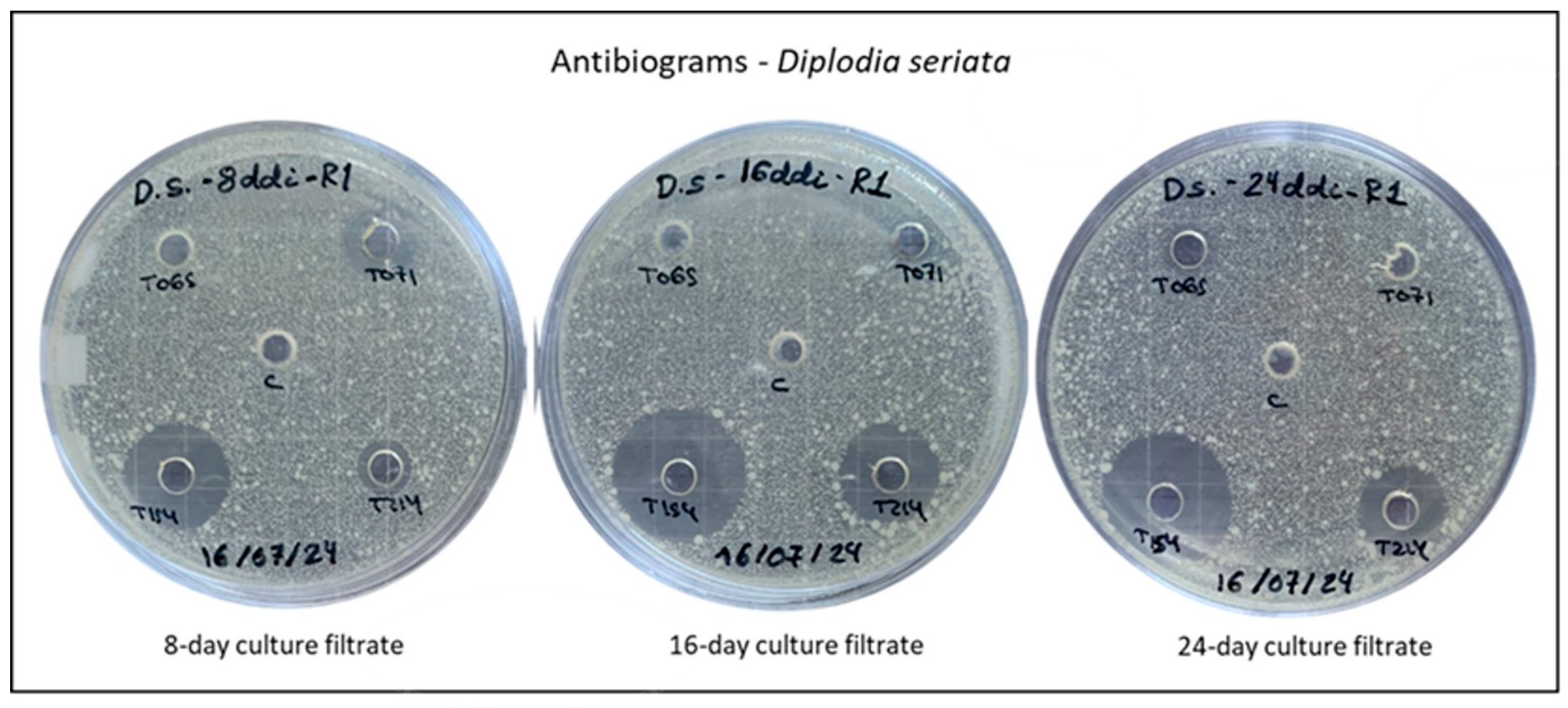
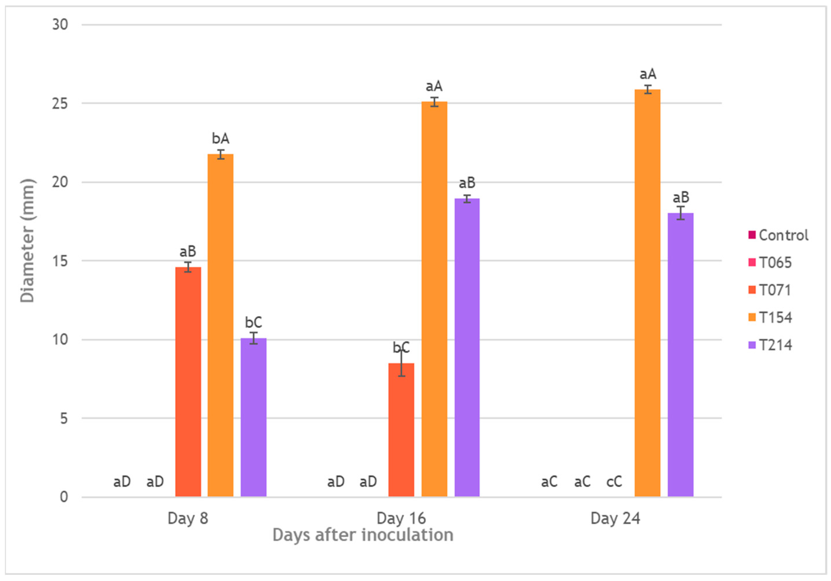
The results of the antibiograms for D. seriata are shown in Figure 2 and Figure 3. The compounds produced by isolate T065 at the different time periods showed no inhibitory activity against D. seriata. In the case of T071, only the culture filtrates extracted on Days 8 and 16 produced an inhibition ring of 14.6 ± 1 mm and 8.52 ± 1 mm respectively, with significant differences between them (F = 212.712; df (2,15); p ≤ 0.001). T154 produced an inhibition ring between 21.77 ± 1 and 25.89 ± 1 mm with no significant differences between 16 and 24 days of culture filtrate extraction. However, there were significantly higher differences between these two and the culture filtrate at 8 days (F = 66.277; df (2,15); p ≤ 0.001). Finally, for the T214 culture filtrate, significant differences were observed between the extraction times of 16 and 24 days compared with 8 days (F = 206.369; df (2,15); p ≤ 0.001), with the 16-day compounds reaching an inhibition diameter of 18.94 ± 1 mm.
Analyzing the results by days of culture filtrate extraction, on Day 8 of extraction, the compounds produced by T154 produce the greatest inhibition of D. seriata (21.77 ± 1 mm), followed by those produced by T071 (14.60 ± 1 mm), and finally by T214 (10.11 ± 1 mm), with significant differences between them (F = 334.237; df (2,15); p ≤ 0.001). The compounds extracted after 16 days also showed significant differences between T071 (8.52 ± 1 mm), T154 (25.09 ± 1 mm) and T214 (18.94 mm) (F = 270.046; df (2,15); p ≤ 0.001). At 24 days, significant differences in the inhibition of D. seriata were also observed between T154 (25.89 ± 1 mm) and T214 (18.02 ± 1 mm) (F = 2319.447; df (2,15); p ≤ 0.001).

3.3. Inhibition of Mycelial Growth

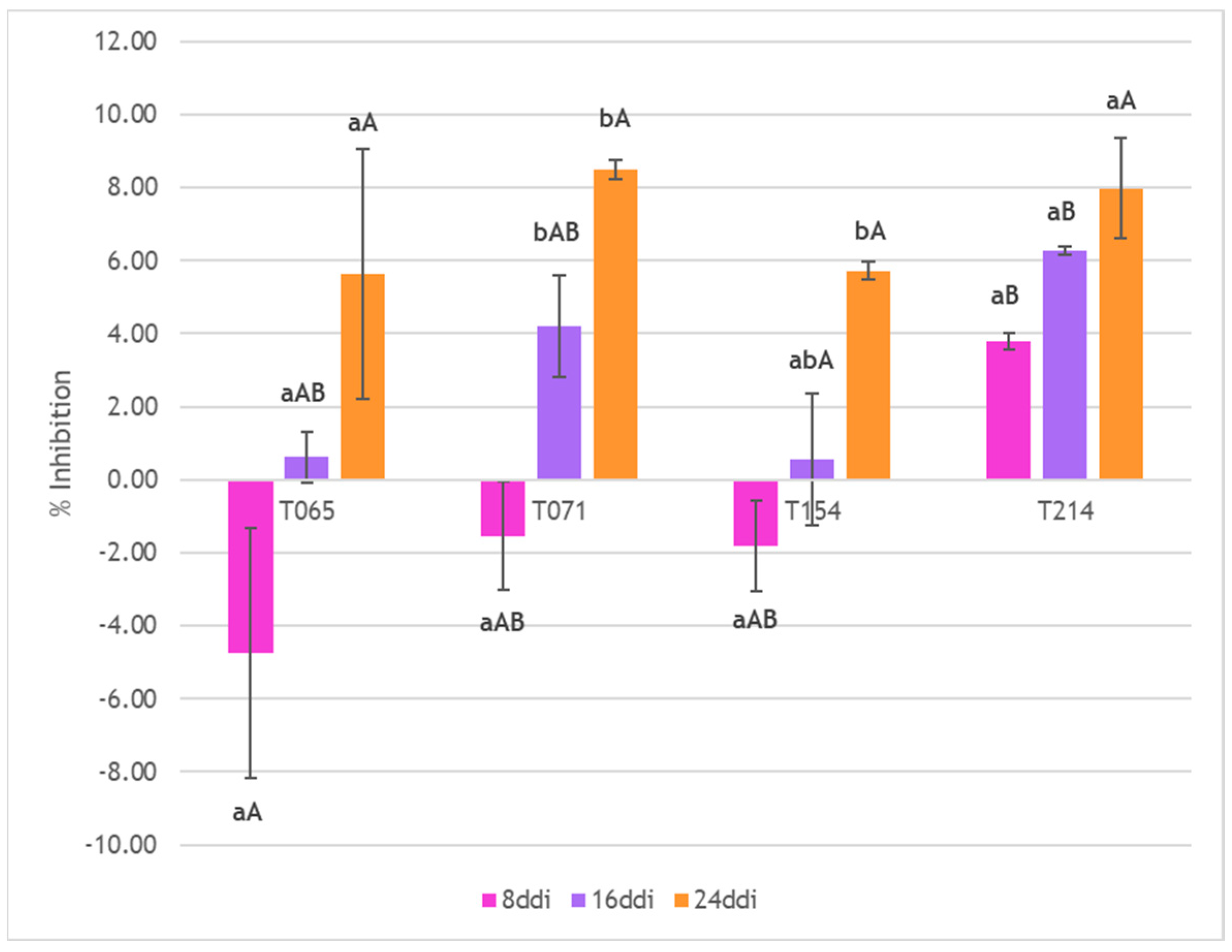
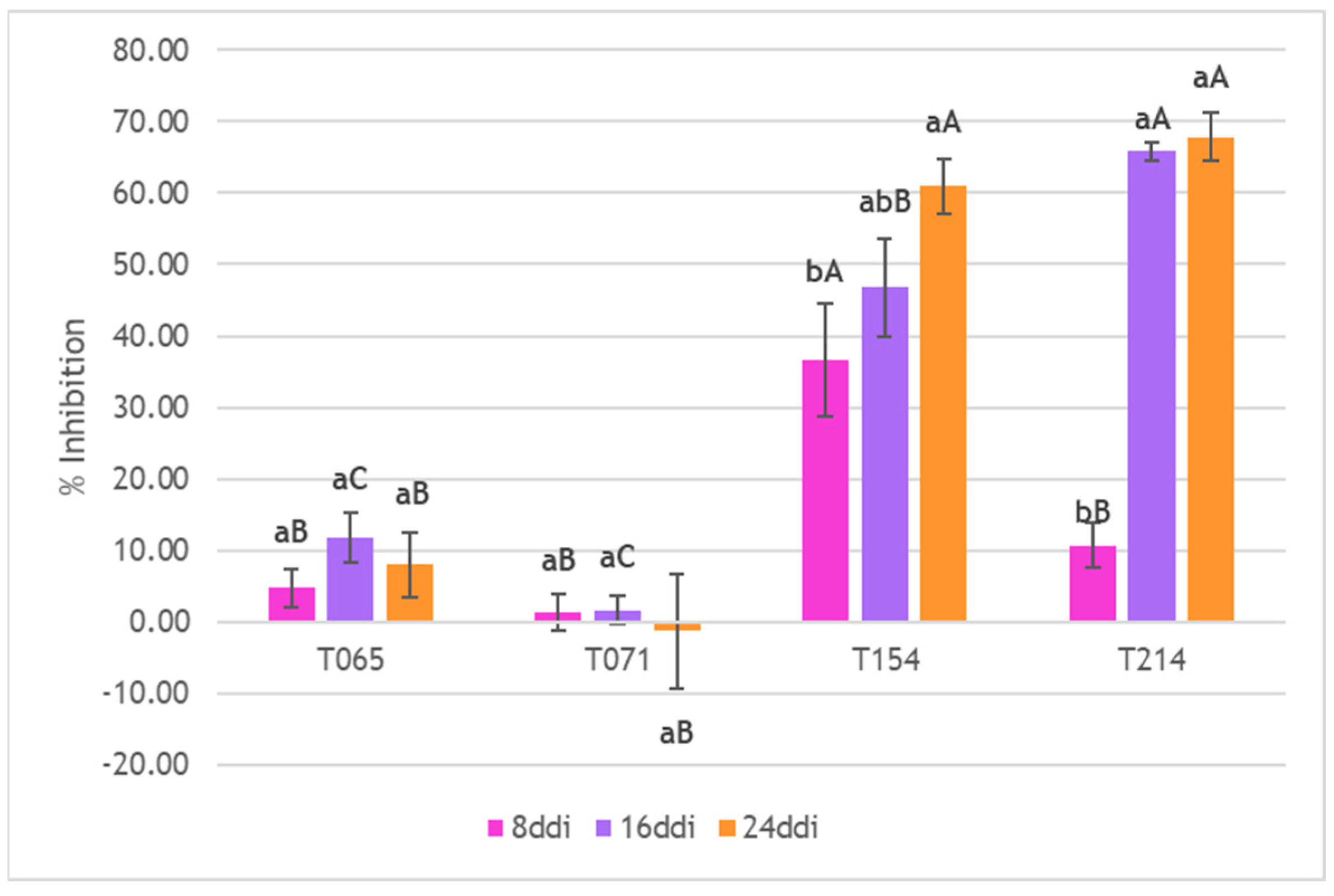
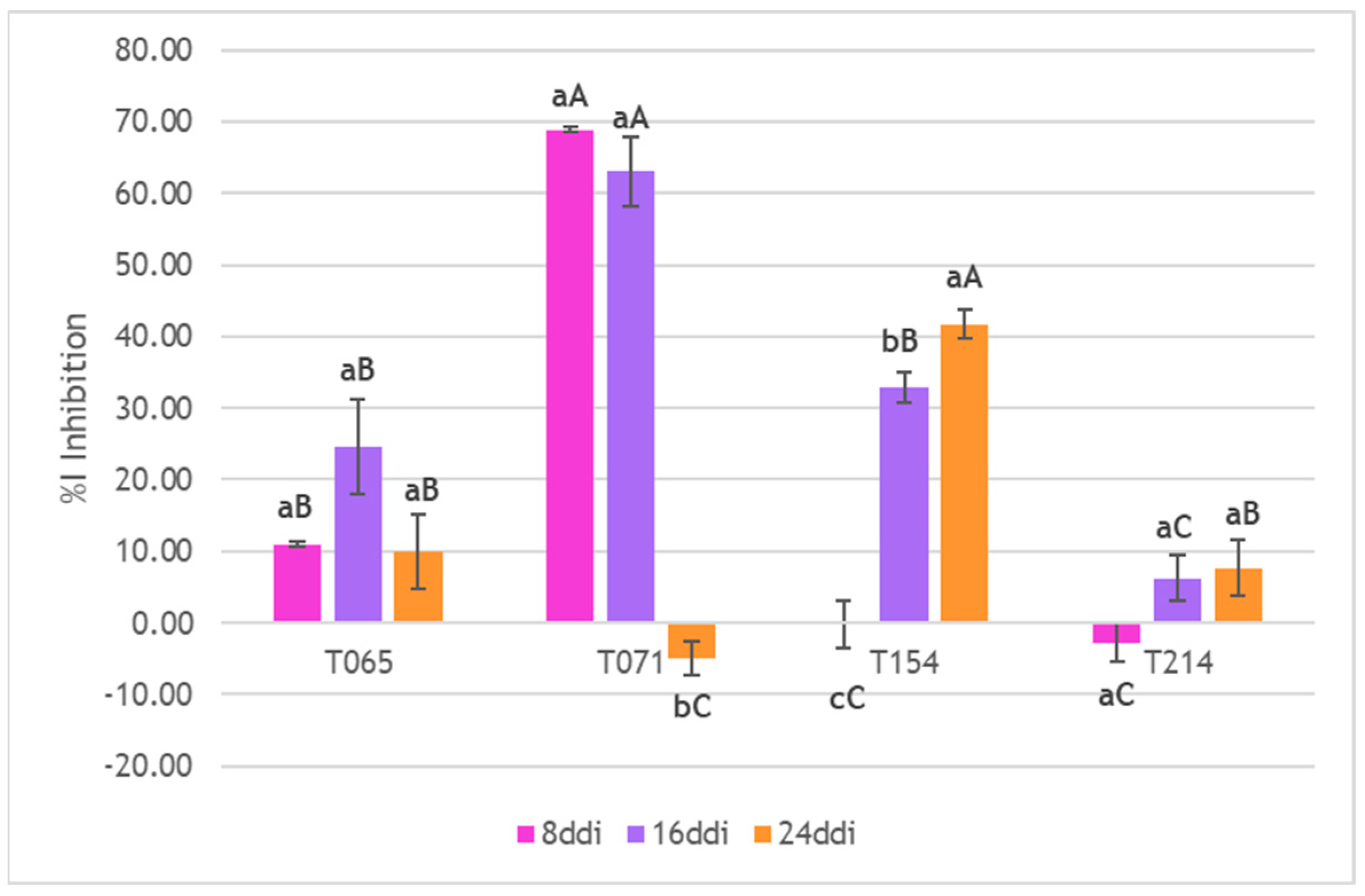
Starting with P. minimum, the results generally showed very low mycelial inhibition values of this pathogen, less than 8%. There were no significant differences between the 8-, 16- and 24-day culture filtrates of the T065 and T214 isolates. However, culture filtrates from Days 16 (F = 12.671; df (2,6); p < 0.025) and 24 (F = 12.671; df (2,6); p < 0.004) of isolate T071 showed significantly higher inhibition data than those extracted on Day 8. In the case of T154, significant differences were observed between the culture filtrates on Days 8 and 24 (F = 5.946; df (2,6); p < 0.019). No significant differences were observed between the culture filtrates extracted from the different Trichoderma isolates on Day 24. Significant differences were observed between the inhibition values of the culture filtrates extracted on Day 8 from isolates T065 (−4.75%) and T214 (3.79%) (F = 3.321; df (3,8); p < 0.022); and between the culture filtrates extracted on Day 16 from isolates T154 (0.56%) and T214 (7.70%) (F = 3.387; df (3,8); p < 0.035) (Figure 4). Higher inhibition values were observed for P. chlamydospora than for P. minimum. For isolate T154, significant differences were observed between the culture filtrate extracted on Day 8 (36.68%) and those extracted on Day 24 (60.98%) (F = 3.603; df (2,6); p < 0.037). T214 showed the highest inhibition values, with significant differences between the culture filtrates extracted on Days 16 (65.80%) and 24 (67.87%) and those extracted on Day 8 (10.71%) (F = 137.057; df (2,6); p ≤ 0.001). Analyzing the differences between Trichoderma isolates for Day 8 of culture filtrate extraction, isolate T154 showed an inhibition value (36.68%) significantly higher than the rest of the isolates (F = 12.084; df (3,8); p < 0.004). For the culture filtrates extracted on Day 16, there were significant differences between T154 (46.81%) and T214 (65.80%) (F = 55.678; df (3,8); p < 0.010) and also between these two and T065 (11.69%) and T071 (1.58%) (F = 55.678; df (3,8); p ≤ 0.001). Of those extracted on Day 24, T154 (60.98%) and T214 (67.87%) showed significant differences with respect to T065 (8.04%) and T071 (14.69%) (F = 56.999; df (3,8); p ≤ 0.001) (Figure 5). Finally, in the case of D. seriata, the highest inhibition values were those produced by the culture filtrates extracted on Days 8 (68.90%) and 16 (63.06%) from isolate T071. Both culture filtrates from T071 showed significantly higher inhibition values than the culture filtrate extracted on Day 24 from T071 (−4.88%) (F = 172.098; df (2,6); p ≤ 0.001) and were also significantly higher than the culture filtrates extracted on Day 8 (F = 211.452; df (3,8); p ≤ 0.001) and on Day 16 (F = 27.584; df (3,8); p ≤ 0.001) from the other isolates (Figure 6).
From these results it can be concluded that the culture filtrates with the highest inhibitory activity against P. minimum were those extracted on Days 16 (7.70%) and 24 (7.98%) from isolate T214. For P. chlamydospora, the highest inhibition was produced by the metabolites from Day 24 of T154 (60.98%) and T214 (67.87%), as well as those from Day 16 of T214 (65.80%), and for D. seriata, the Day 8 (68.90%) and Day 16 (63.06%) metabolites of isolate T071.

4. Discussion

To our knowledge, this is the first time that culture filtrates of Trichoderma strains have been tested against Phaeacremonium minimum and Diplodia seriata as an alternative biosolution to biocontrol fungi involved in GTDs. It is particularly noteworthy that in this experiment it was shown to have a direct effect, inhibiting the growth of mycelium or the development of spores for Diplodia seriata and Phaeomoniella chlamydospora apart from Phaeoacremonium minimum.
In previous experiments, it was tested against Eutypa lata [27], describing an indirect effect in terms of up- or down-regulation of grapevine genes to control GTDs. Additionally, the use of a secondary metabolite (6-pentyl-α-pyrone (6PP)) produced by three Trichoderma strains (Trichoderma harzianum T77, Trichoderma atroviride UST1, UST2) showed an in vitro effect of the inhibition of spores and mycelium of fungi producing GTDs [23]. This first in vitro assay shows that, depending on the type of strain and incubation time, it is possible to directly biocontrol fungi involved in GTDs. This experiment opens a new line of research due to itsgreat potential for GTDs control. In our previous research [29,35] isolate T065 demonstrated a significant activity against P. minimum in a membrane assay. This assay was redirected to evaluate the effectiveness of diffusible compounds through a membrane (mainly secondary metabolites). Therefore, the results in this manuscript are in accordance with previous research but it is necessary to identify the compound or compounds that had this activity and are presumed to be secondary metabolites. The other isolates, T071, T154 and T214, demonstrated significant activity against P. minimum in the dual culture assay where different mechanisms act against P. minimum and successfully reduced its growth. In these previous assays lytic enzymes could play an important role, as well as mycoparasitism, where different compounds are involved.
The mechanisms of action of Trichoderma as biocontrol agents include a variety of processes such as mycoparasitism, competition for nutrients, and the activation of plant defense mechanisms, all supported by the production of extracellular enzymes and secondary metabolites (antibiosis). The role of Trichoderma secondary metabolites as antibiotics and CWDEs is under discussion due to the wide variety of these compounds found in different Trichoderma species and their varying activity depending on environmental conditions [7].
This study focused on the antifungal activity of culture filtrates produced by native Trichoderma strains T065, T071, T154, and T214, all isolated from different vineyards in Spain. This antifungal activity was studied against P. minimum, P. chlamydospora and D. seriata, three of the main pathogens causing GTDs.
In terms of different effectiveness, strains T154 and T214 showed a significantly higher reduction in the growth of GTDs pathogens in comparison to the other strains. This could be due to both of them belonging to the Harzianum clade [29]. Genomic and phylogenetic studies confirm the Harzianum clade’s broad genetic endowment for biocontrol, supporting its reputation as the most effective group among Trichoderma species [36,37,38].
Firstly, the production of secondary metabolites is closely related to the pH of the medium in which they are produced. Trichoderma species can grow in soils with a pH ranging from 5.5 to 8.5, although the optimum values are between 5.5 and 6.5 (i.e., in a slightly acidic environment) [35]. This optimal pH range influences the production of secondary metabolites by the fungus. Furthermore, the production of secondary metabolites by Trichoderma can vary depending on the species and especially strains. This suggests that different Trichoderma strains may have different responses to pH changes in terms of secondary metabolite production [39,40]. In the present study, the initial pH of the production medium (PDB) was slightly acidic at 5.25, but the pH of the medium increased to values close to 8 as the days progressed. These variations in the pH of the medium may be the cause or the consequence of the production of different types of secondary metabolites, as these can be produced at different pH levels, but some are only synthesized at certain pH ranges. Some studies, such as that of Chalimah et al. (2020) [41], have shown that the most effective metabolites against some pathogenic fungi are produced at acidic pH. In our study, this effect was only observed in the case of strain T071, whose most effective metabolites against D. seriata were found on Days 8 and 16, when the pH of the medium was around 3 and 5 respectively. Other studies have shown that fungi respond to environmental pH by activating a specific transcription factor, PacC, which is involved in the biosynthesis of secondary metabolites [42]. Thus, it can be said that the pH of the medium plays a crucial role in determining which specific secondary metabolites are produced by the Trichoderma species, affecting their ecological functions and biocontrol potential.
The most significant method of spread for most grapevine trunk pathogens is through airborne spores released from infected plant material. Fungal pathogens overwinter in dead, infected wood where they produce fruiting bodies (pycnidia and perithecia) that contain spores. These structures release spores primarily during and after rainfall events, creating the ideal conditions for widespread dispersal [18,20]. Spores of some pathogens, such as those causing Botryosphaeria dieback, can be transported up to 20 m by wind [19]. The biocontrol capacity of the Trichoderma culture filtrate on these structures is therefore crucial. For this reason, the ability of culture filtrates to control spores of grapevine pathogens were investigated by performing antibiogram tests. Some studies have investigated the antimicrobial activity of Trichoderma culture filtrates using antibiogram tests. There are various methods of applying antibiogram techniques, such as the agar disk-diffusion method, in which the spores of the pathogen to be controlled is mixed with the culture medium and the antimicrobial compounds are impregnated onto paper discs, which are then placed on the culture medium in Petri dishes. Another method used is the Agar well diffusion method, in which a volume of microbial inoculum is inoculated on the surface of the culture medium of the plate, then holes are made in the culture medium and the antimicrobial compounds to be tested are poured into them [43]. A new method, a mixture of the two methods described above, was used in this work, as described in the Materials and Methods section. After a long process of optimizing the methodology used in this study, it was possible to demonstrate the efficacy of some of the culture filtrates produced by different Trichoderma strains in controlling D. seriata under in vitro conditions. The compounds produced by strain T071 after 8 days and those produced by strains T154 and T214 after 8, 16 and 24 days were the most effective in inhibiting D. seriata. Studies evaluating the efficacy of Trichoderma secondary metabolites against GTDs using the antibiogram method are practically non-existent. For this reason, the results of this study were compared with other studies using a similar methodology to evaluate the control of other types of disease. For example, the efficacy of the trichothecene harzianum A (HA) produced by Trichoderma against pathogens such as B. cinerea has been demonstrated [34], with inhibition diameters of less than 4.47 mm. Another study by Sedjati et al. (2022) [44] using secondary metabolites produced by Trichoderma longibrachiatum against some bacteria also produced inhibition results of around 10.00 mm in diameter. The inhibition values of these studies were significantly lower than those achieved by the culture filtrates of strains T071, T154 and T214 against D. seriata.
The mycelial inhibition assays against the GTDs pathogens yielded highly variable data. Firstly, P. minimum showed the lowest inhibition values against the culture filtrates of Trichoderma. Several studies have demonstrated the efficacy of Trichoderma against P. minimum [35], so it could be concluded that among the mechanisms of action of Trichoderma for the control of this pathogen, control by antibiosis or lytic enzymes does not stand out, with spore adhesion or niche exclusion being more relevant [45]. In the case of P. chlamydospora, the inhibition values of the culture filtrates produced by strains T154 and T214 were notably higher than those of P. minimum. Several studies have demonstrated the efficacy of Trichoderma against P. chlamydospora [46,47], but this study highlights the importance of the antibiosis mechanisms of some Trichoderma strains in combating P. chlamydospora. In the case of D. seriata, the culture filtrates of strain T071 could be seen to achieve high inhibition rates of the mycelium of this pathogen. Meanwhile, the strains that stood out in terms of inhibition in the antibiogram assays were T154 and T214. This may be because the effect of the culture filtrate of some Trichoderma strains was different when acting on the spores of the pathogen (antibiograms) than when acting on the mycelium of the pathogen. Antibiograms results could be due to the effect of antibiotic compounds and secondary metabolites that successfully biocontrol P minimum, as shown in the previous researchs of membrane assays [35]. These successful results reinforce the idea that these Trichoderma could produce secondary metabolites that could be involved in biological control and that further chemical analysis is necessary, such as the ones performed by Vinale et al. (2016) [48].
The successful control of the mycelium of these pathogens by some of the culture filtrates obtained in this study may be due to the action of lytic enzymes. As already shown in some studies, the production of enzymes by different Trichoderma isolates showed a high activity of endochitinases and 1,3-β-glucanases with the ability to break the cell wall of pathogenic fungi [49]. In this case, culture filtrates are distributed through all surface, so chemical compounds are on the surface and no filtering or diffusion is achieved in comparison to antibiograms. Thus, CWDEs and secondary metabolites could both be responsible for biocontrol activities.
Strain T065, described as T. gamsii, was selected for this study because other T. gamsii strains have been described as effective BCAs against GTDs such as esca [50]. However, it was observed that the biocompounds produced by T065 did not have the ability to control the pathogens that caused the GTDs, as studied after 8, 16, 24 days of evaluating the culture filtrates. This does not mean that strain T065 is not capable of controlling these diseases by other control mechanisms, or by evaluating different times of production after cultivation such as two or three days after being cultivated or evaluating a co-culture with pathogens or other Trichoderma strains according to [51]. Also, strain T154 and T214 isolated from woody tissues as endophytes, and especially T154, have demonstrated their effects as endophytes in biocontrol [45]. Other endophytes have demonstrated a better effect of biocontrol as described in [52,53]. Therefore, this could help in having a better biocontrol performance in comparison to the other strains assayed.
This study represents a starting point for further investigation into the effects of bioactive compounds produced by Trichoderma in the control of GTDs. Future efforts are needed to optimize the harvest time of culture filtrates, assess different incubation periods to reduce the collection time, evaluate their toxicological profile in plants, and distinguish between the mechanisms responsible for biocontrol activity (enzyme production or secondary metabolites). It is of great importance to conduct subsequent trials on plants under controlled greenhouse conditions and later in the field to reliably validate this biocontrol method. Future experiments will try to characterize secondary metabolites and lytic enzymes, especially according to 6PP as a key metabolite involved in plant-Trichoderma-pathogen interaction. Thus, this experiment marks the beginning of future work to open new assays to characterize culture filtrates as an ecological and environmentally friendly solution to limit the spread of GTDs.

5. Conclusions

Native Trichoderma strains (T071, T154, and T214) produce effective biocompounds for GTDs biocontrol. Biocompounds produced by strains T071, T154 and T214 are highly effective in inhibiting the development of spores and mycelium of D. seriata. Bioactive fungal compounds of T154 and T214 have the capacity to control the mycelial development of P. chlamydospora. The low efficacy of the biocompounds of native Trichoderma strains against P. minimum corroborates that the mechanism of action of Trichoderma through antibiosis is not the most relevant to control this pathogen [45]. This work shows that, depending on the type of strain and incubation time, it is possible to directly biocontrol fungi involved in GTDs. This in vitro study offers preliminary evidence supporting the potential of culture filtrates from native Trichoderma strains as a solution for the biocontrol of GTDs.

Author Contributions

Conceptualization, L.Z., G.C.-H. and P.A.C.; Methodology, L.Z., G.C.-H., D.R.-L. and S.M.-P.; Software, L.Z., D.R.-L., and Á.R.-G.; Validation, L.Z., P.A.C. and S.G.; Formal Analysis, L.Z. and G.C.-H.; Investigation, L.Z.; Resources, L.Z. and G.C.-H.; Data Curation, L.Z.; Writing—Original Draft Preparation, L.Z.; Writing—Review and Editing, L.Z., G.C.-H., P.A.C. and S.G.; Visualization, G.C.-H. and Á.R.-G.; Supervision, P.A.C. and S.G.; Project Administration, P.A.C.; Funding Acquisition, P.A.C. All authors have read and agreed to the published version of the manuscript.

Funding

The authors declare that this study received funding from project “LOWPHWINE IDI–20210391” “Estudio de nuevos factores relacionados con el suelo, la planta y la microbiota enológica que influyen en el equilibrio de la acidez de los vinos y en su garantía de calidad y estabilidad en climas cálidos” which was granted by the Centro para el Desarrollo tecnológico Industrial (CDTI), the Ministerio de Ciencia, Innovación y Universidades (Spain), who awarded a grant to Laura Zanfaño González (FPU 20/03040) and by the Research Program of the Universidad de León (2022) for the grant awarded to Daniela Ramírez Lozano. The funder had the following involvement with the study: in this case only economic support was involved by the funder.

Data Availability Statement

The original contributions presented in this study are included in the article. Further inquiries can be directed to the corresponding author(s).

Acknowledgments

We thank Enrique Barajas Tola and Juan Antonio Rubio Cano from the Instituto Tecnológico Agrario de Castilla y León (ITACyL) for kindly providing Phaeoacremonium minimum strain Y038-05-03a and P. chlamydospora Y116-18-03c as well as research staff of the GUIIAS group for their technical support.

Conflicts of Interest

The authors declare no conflicts of interest.

References

  1. Skinner, M.; Parker, B.L.; Kim, J.S. Role of Entomopathogenic Fungi in Integrated Pest Management. In Integrated Pest Management: Current Concepts and Ecological Perspective; Elsevier Inc.: Amsterdam, The Netherlands, 2014; pp. 169–191. ISBN 9780124017092. [Google Scholar]
  2. Bamisile, B.S.; Akutse, K.S.; Siddiqui, J.A.; Xu, Y. Model Application of Entomopathogenic Fungi as Alternatives to Chemical Pesticides: Prospects, Challenges, and Insights for next-Generation Sustainable Agriculture. Front. Plant Sci. 2021, 12, 741804. [Google Scholar] [CrossRef]
  3. Fenibo, E.O.; Ijoma, G.N.; Matambo, T. Biopesticides in Sustainable Agriculture: A Critical Sustainable Development Driver Governed by Green Chemistry Principles. Front. Sustain. Food Syst. 2021, 5, 619058. [Google Scholar] [CrossRef]
  4. Nieto-Jacobo, M.F.; Steyaert, J.M.; Salazar-Badillo, F.B.; Vi Nguyen, D.; Rostás, M.; Braithwaite, M.; De Souza, J.T.; Jimenez-Bremont, J.F.; Ohkura, M.; Stewart, A.; et al. Environmental Growth Conditions of Trichoderma spp. Affects Indole Acetic Acid Derivatives, Volatile Organic Compounds, and Plant Growth Promotion. Front. Plant Sci. 2017, 8, 102. [Google Scholar] [CrossRef] [PubMed]
  5. Neme, A.; Leta, A.; Yones, A.M.; Tahir, M. Seedborne Mycoflora of Faba Bean (Vicia fabae L.) and Evaluation of Plant Extract and Trichoderma Species against Mycelium Growth of Selected Fungi. Heliyon 2023, 9, e17291. [Google Scholar] [CrossRef] [PubMed]
  6. Zanfaño, L.; Carro-Huerga, G.; Rodríguez-González, Á.; Mayo-Prieto, S.; Cardoza, R.E.; Gutiérrez, S.; Casquero, P.A. Trichoderma carraovejensis: A New Species from Vineyard Ecosystem with Biocontrol Abilities against Grapevine Trunk Disease Pathogens and Ecological Adaptation. Front. Plant Sci. 2024, 15, 1388841. [Google Scholar] [CrossRef]
  7. Vinale, F.; Marra, R.; Scala, F.; Ghisalberti, E.L.; Lorito, M.; Sivasithamparam, K. Major Secondary Metabolites Produced by Two Commercial Trichoderma Strains Active against Different Phytopathogens. Lett. Appl. Microbiol. 2006, 43, 143–148. [Google Scholar] [CrossRef]
  8. Harman, G.E.; Howell, C.R.; Viterbo, A.; Chet, I.; Lorito, M. Trichoderma Species—Opportunistic, Avirulent Plant Symbionts. Nat. Rev. Microbiol. 2004, 2, 43–56. [Google Scholar] [CrossRef]
  9. Ghasemi, S.; Safaie, N.; Shahbazi, S.; Shams-Bakhsh, M.; Askari, H. The Role of Cell Wall Degrading Enzymes in Antagonistic Traits of Trichoderma virens against Rhizoctonia solani. Iran. J. Biotechnol. 2020, 18, 18–28. [Google Scholar] [CrossRef]
  10. Rajesh, R.W.; Rahul, M.S.; Ambalal, N.S. Trichoderma: A Significant Fungus for Agriculture and Environment. Afr. J. Agric. Res. 2016, 11, 1952–1965. [Google Scholar] [CrossRef]
  11. Rauf, A.; Subhani, M.N.; Siddique, M.; Shahid, H.; Chattha, M.B.; Alrefaei, A.F.; Hasan Naqvi, S.A.; Ali, H.; Lucas, R.S. Cultivating a Greener Future: Exploiting Trichoderma Derived Secondary Metabolites for Fusarium Wilt Management in Peas. Heliyon 2024, 10, e29031. [Google Scholar] [CrossRef]
  12. Contreras-Cornejo, H.A.; Macías-Rodríguez, L.; del-Val, E.; Larsen, J. Ecological Functions of Trichoderma spp. and Their Secondary Metabolites in the Rhizosphere: Interactions with Plants. FEMS Microbiol. Ecol. 2016, 92, fiw036. [Google Scholar] [CrossRef] [PubMed]
  13. Mukherjee, P.K.; Horwitz, B.A.; Kenerley, C.M. Secondary Metabolism in Trichoderma—A Genomic Perspective. Microbiology 2012, 158, 35–45. [Google Scholar] [CrossRef]
  14. Vinale, F.; Sivasithamparam, K.; Ghisalberti, E.L.; Marra, R.; Barbetti, M.J.; Li, H.; Woo, S.L.; Lorito, M. A Novel Role for Trichoderma Secondary Metabolites in the Interactions with Plants. Physiol. Mol. Plant Pathol. 2008, 72, 80–86. [Google Scholar] [CrossRef]
  15. Woo, S.L.; Scala, F.; Ruocco, M.; Lorito, M. The Molecular Biology of the Interactions between Trichoderma spp., Phytopathogenic Fungi, and Plants. Phytopathology 2006, 96, 181–185. [Google Scholar] [CrossRef]
  16. Gruber, S.G.; Seidl-Seiboth, V. Self versus Non-Self: Fungal Cell Wall Degradation in Trichoderma. Microbiology 2012, 158, 26–34. [Google Scholar] [CrossRef] [PubMed]
  17. Mercer, C. Scientists Find New Clues to ‘Billion-Dollar’ Vine Diseases. Available online: https://www.decanter.com/wine-news/scientists-find-new-clues-to-billion-dollar-vine-diseases-484958/ (accessed on 12 February 2025).
  18. Sosnowski, M. Best Practice Management Guide. In Grapevine Trunk Disease; Wine Australia: Adelaide, SAU, Australia, 2019. [Google Scholar]
  19. Jaspers, M.; Sosnowski, M. Grapevine Trunk Diseases. In Eutypa & Botryosphaeria Dieback in Vineyards; Bragato Research Institute: Bienheim, New Zeland, 2021. [Google Scholar]
  20. Kenfaoui, J.; Radouane, N.; Mennani, M.; Tahiri, A.; El Ghadraoui, L.; Belabess, Z.; Fontaine, F.; El Hamss, H.; Amiri, S.; Lahlali, R.; et al. A Panoramic View on Grapevine Trunk Diseases Threats: Case of Eutypa Dieback, Botryosphaeria Dieback, and Esca Disease. J. Fungi 2022, 8, 595. [Google Scholar] [CrossRef]
  21. Travadon, R.; Lawrence, D.P.; Moyer, M.M.; Fujiyoshi, P.T.; Baumgartner, K. Fungal Species Associated with Grapevine Trunk Diseases in Washington Wine Grapes and California Table Grapes, with Novelties in the Genera Cadophora, Cytospora, and Sporocadus. Front. Fungal Biol. 2022, 3, 1018140. [Google Scholar] [CrossRef] [PubMed]
  22. Khan, R.A.A.; Najeeb, S.; Hussain, S.; Xie, B.; Li, Y. Bioactive Secondary Metabolites from Trichoderma spp. against Phytopathogenic Fungi. Microorganisms 2020, 8, 817. [Google Scholar] [CrossRef]
  23. Mutawila, C.; Vinale, F.; Halleen, F.; Lorito, M.; Mostert, L. Isolation, Production and in Vitro Effects of the Major Secondary Metabolite Produced by Trichoderma Species Used for the Control of Grapevine Trunk Diseases. Plant Pathol. 2016, 65, 104–113. [Google Scholar] [CrossRef]
  24. Pascale, A.; Vinale, F.; Manganiello, G.; Nigro, M.; Lanzuise, S.; Ruocco, M.; Marra, R.; Lombardi, N.; Woo, S.L.; Lorito, M. Trichoderma and Its Secondary Metabolites Improve Yield and Quality of Grapes. Crop Prot. 2017, 92, 176–181. [Google Scholar] [CrossRef]
  25. Mesguida, O.; Haidar, R.; Yacoub, A.; Dreux-Zigha, A.; Berthon, J.Y.; Guyoneaud, R.; Attard, E.; Rey, P. Microbial Biological Control of Fungi Associated with Grapevine Trunk Diseases: A Review of Strain Diversity, Modes of Action, and Advantages and Limits of Current Strategies. J. Fungi 2023, 9, 638. [Google Scholar] [CrossRef] [PubMed]
  26. Jangid, P.K.; Mathur, P.N.; Mathur, K. Role of Chitinase and β-1,3 Glucanase Elicitation in the Trichoderma harzianum Induced Systemic Resistance in Capsicum. Indian Phytopath 2004, 57, 217–221. [Google Scholar]
  27. Mutawila, C.; Stander, C.; Halleen, F.; Vivier, M.A.; Mostert, L. Response of Vitis vinifera Cell Cultures to Eutypa lata and Trichoderma atroviride Culture Filtrates: Expression of Defence-Related Genes and Phenotypes. Protoplasma 2017, 254, 863–879. [Google Scholar] [CrossRef]
  28. Stempien, E.; Pierron, R.J.G.; Adendorff, I.; Van Jaarsveld, W.J.; Halleen, F.; Mostert, L. Host Defence Activation and Root Colonization of Grapevine Rootstocks by the Biological Control Fungus Trichoderma atroviride. Phytopathol. Mediterr. 2020, 59, 615–626. [Google Scholar] [CrossRef]
  29. Carro-Huerga, G.; Mayo-Prieto, S.; Rodríguez-González, Á.; Álvarez-García, S.; Gutiérrez, S.; Casquero, P.A. The Influence of Temperature on the Growth, Sporulation, Colonization, and Survival of Trichoderma spp. in Grapevine Pruning Wounds. Agronomy 2021, 11, 1771. [Google Scholar] [CrossRef]
  30. Rodríguez-González, A.; Carro-Huerga, G.; Mayo-Prieto, S.; Lorenzana, A.; Gutiérrez, S.; Peláez, H.J.; Casquero, P.A. Investigations of Trichoderma spp. and Beauveria bassiana as Biological Control Agent for Xylotrechus arvicola, a Major Insect Pest in Spanish Vineyards. J. Econ. Entomol. 2018, 111, 2585–2591. [Google Scholar] [CrossRef] [PubMed]
  31. Gramaje, D.; Úrbez-Torres, J.R.; Sosnowski, M.R. Managing Grapevine Trunk Diseases with Respect to Etiology and Epidemiology: Current Strategies and Future Prospects. Plant Dis. 2018, 102, 12–39. [Google Scholar] [CrossRef]
  32. Martín, L.; Martín, M.T. Characterization of Fungicide Resistant Isolates of Phaeoacremonium aleophilum Infecting Grapevines in Spain. Crop Prot. 2013, 52, 141–150. [Google Scholar] [CrossRef]
  33. Martín, M.T.; Cobos, R.; Martín, L.; López-Enríquez, L. Real-Time PCR Detection of Phaeomoniella chlamydospora and Phaeoacremonium aleophilum. Appl. Environ. Microbiol. 2012, 78, 3985–3991. [Google Scholar] [CrossRef]
  34. Malmierca, M.G.; Cardoza, R.E.; Alexander, N.J.; McCormick, S.P.; Hermosa, R.; Monte, E.; Gutiérrez, S. Involvement of Trichoderma Trichothecenes in the Biocontrol Activity and Induction of Plant Defense-Related Genes. Appl. Env. Microbiol. 2012, 78, 4856–4868. [Google Scholar] [CrossRef]
  35. Carro-Huerga, G.; Mayo-Prieto, S.; Rodríguez-González, Á.; Cardoza, R.E.; Gutiérrez, S.; Casquero, P.A. Vineyard Management and Physicochemical Parameters of Soil Affect Native Trichoderma Populations, Sources of Biocontrol Agents against Phaeoacremonium minimum. Plants 2023, 12, 887. [Google Scholar] [CrossRef] [PubMed]
  36. Chaverri, P.; Branco-Rocha, F.; Jaklitsch, W.; Gazis, R.; Degenkolb, T.; Samuels, G.J. Systematics of the Trichoderma harzianum Species Complex and the Re-Identification of Commercial Biocontrol Strains. Mycologia 2015, 107, 558–590. [Google Scholar] [CrossRef] [PubMed]
  37. Fanelli, F.; Liuzzi, V.C.; Logrieco, A.F.; Altomare, C. Genomic Characterization of Trichoderma atrobrunneum (T. harzianum Species Complex) ITEM 908: Insight into the Genetic Endowment of a Multi-Target Biocontrol Strain. BMC Genom. 2018, 19, 662. [Google Scholar] [CrossRef] [PubMed]
  38. Ismaiel, A.; Jambhulkar, P.P.; Sinha, P.; Lakshman, D.K. Trichoderma: Harzianum Clade in Soils from Central and South America. J. Fungi 2024, 10, 813. [Google Scholar] [CrossRef]
  39. Benítez, T.; Rincón, A.M.; Limón, M.C.; Codón, A.C. Biocontrol Mechanism of Trichoderma Strains. Int. Microbiol. 2004, 7, 249–260. [Google Scholar]
  40. Maurya, M.K.; Srivastava, M. Harshita Effect of Various PH Levels on the Growth and Sporulation of Trichoderma viride Isolates and Assessing Their Antagonistic Activity against Soil-Borne Pathogens. J. Pure Appl. Microbiol. 2024, 18, 2516–2527. [Google Scholar] [CrossRef]
  41. Chalimah, N.; Soesanto, L.; Suharti, W.S. The Effect of Various PH Medium on the Secondary Metabollites Production from Trichoderma harzianum T10 to Control Damping off on Cucumber Seedlings. J. Trop. Hortic. 2020, 3, 65. [Google Scholar] [CrossRef]
  42. Trushina, N.; Levin, M.; Mukherjee, P.K.; Horwitz, B.A. PacC and PH-Dependent Transcriptome of the Mycotrophic Fungus Trichoderma virens. BMC Genom. 2013, 14, 138. [Google Scholar] [CrossRef]
  43. Balouiri, M.; Sadiki, M.; Ibnsouda, S.K. Methods for in Vitro Evaluating Antimicrobial Activity: A Review. J. Pharm. Anal. 2016, 6, 71–79. [Google Scholar] [CrossRef] [PubMed]
  44. Sedjati, S.; Ambariyanto, A.; Trianto, A.; Ridlo, A.; Supriyantini, E.; Sabdono, A.; Radjasa, O.K.; Firmansyah, T. Antibacterial Activity of the Fungal Metabolite Trichoderma longibrachiatum against Multidrug-Resistant Klebsiella pneumoniae and Methicillin-Resistant Staphylococcus aureus. Jordan. J. Biol. Sci. 2022, 15, 107–113. [Google Scholar] [CrossRef]
  45. Carro-Huerga, G.; Compant, S.; Gorfer, M.; Cardoza, R.E.; Schmoll, M.; Gutiérrez, S.; Casquero, P.A. Colonization of Vitis vinifera L. by the Endophyte Trichoderma sp. Strain T154: Biocontrol Activity against Phaeoacremonium minimum. Front. Plant Sci. 2020, 11, 1170. [Google Scholar] [CrossRef]
  46. Pertot, I.; Prodorutti, D.; Colombini, A.; Pasini, L. Trichoderma atroviride SC1 Prevents Phaeomoniella chlamydospora and Phaeoacremonium aleophilum Infection of Grapevine Plants during the Grafting Process in Nurseries. BioControl 2016, 61, 257–267. [Google Scholar] [CrossRef]
  47. Di Marco, S.; Metruccio, E.G.; Moretti, S.; Nocentini, M.; Carella, G.; Pacetti, A.; Battiston, E.; Osti, F.; Mugnai, L. Activity of Trichoderma asperellum Strain ICC 012 and Trichoderma gamsii Strain ICC 080 toward Diseases of Esca Complex and Associated Pathogens. Front. Microbiol. 2022, 12, 813410. [Google Scholar] [CrossRef] [PubMed]
  48. Vinale, F.; Strakowska, J.; Mazzei, P.; Piccolo, A.; Marra, R.; Lombardi, N.; Manganiello, G.; Pascale, A.; Woo, S.L.; Lorito, M. Cremenolide, a New Antifungal, 10-Member Lactone from Trichoderma cremeum with Plant Growth Promotion Activity. Nat. Prod. Res. 2016, 30, 2575–2581. [Google Scholar] [CrossRef] [PubMed]
  49. Thrane, C.; Funck Jensen, D.; Tronsmo, A. Substrate Colonization, Strain Competition, Enzyme Production in Vitro, and Biocontrol of Pythium ultimum by Trichoderma spp. Isolates P1 and T3. Eur. J. Plant Pathol. 2000, 106, 215–225. [Google Scholar] [CrossRef]
  50. Bigot, G.; Sivilotti, P.; Stecchina, M.; Lujan, C.; Freccero, A.; Mosetti, D. Long-Term Effects of Trichoderma asperellum and Trichoderma gamsii on the Prevention of Esca in Different Vineyards of Northeastern Italy. Crop Prot. 2020, 137, 10526. [Google Scholar] [CrossRef]
  51. Vinale, F.; Nicoletti, R.; Borrelli, F.; Mangoni, A.; Parisi, O.A.; Marra, R.; Lombardi, N.; Lacatena, F.; Grauso, L.; Finizio, S.; et al. Co-Culture of Plant Beneficial Microbes as Source of Bioactive Metabolites. Sci. Rep. 2017, 7, 14330. [Google Scholar] [CrossRef]
  52. del Carmen H. Rodríguez, M.; Evans, H.C.; de Abreu, L.M.; de Macedo, D.M.; Ndacnou, M.K.; Bekele, K.B.; Barreto, R.W. New Species and Records of Trichoderma Isolated as Mycoparasites and Endophytes from Cultivated and Wild Coffee in Africa. Sci. Rep. 2021, 11, 5671. [Google Scholar] [CrossRef]
  53. Nascimento Brito, V.; Lana Alves, J.; Sírio Araújo, K.; de Souza Leite, T.; Borges de Queiroz, C.; Liparini Pereira, O.; de Queiroz, M.V. Endophytic Trichoderma Species from Rubber Trees Native to the Brazilian Amazon, Including Four New Species. Front. Microbiol. 2023, 14, 1095199. [Google Scholar] [CrossRef] [PubMed]
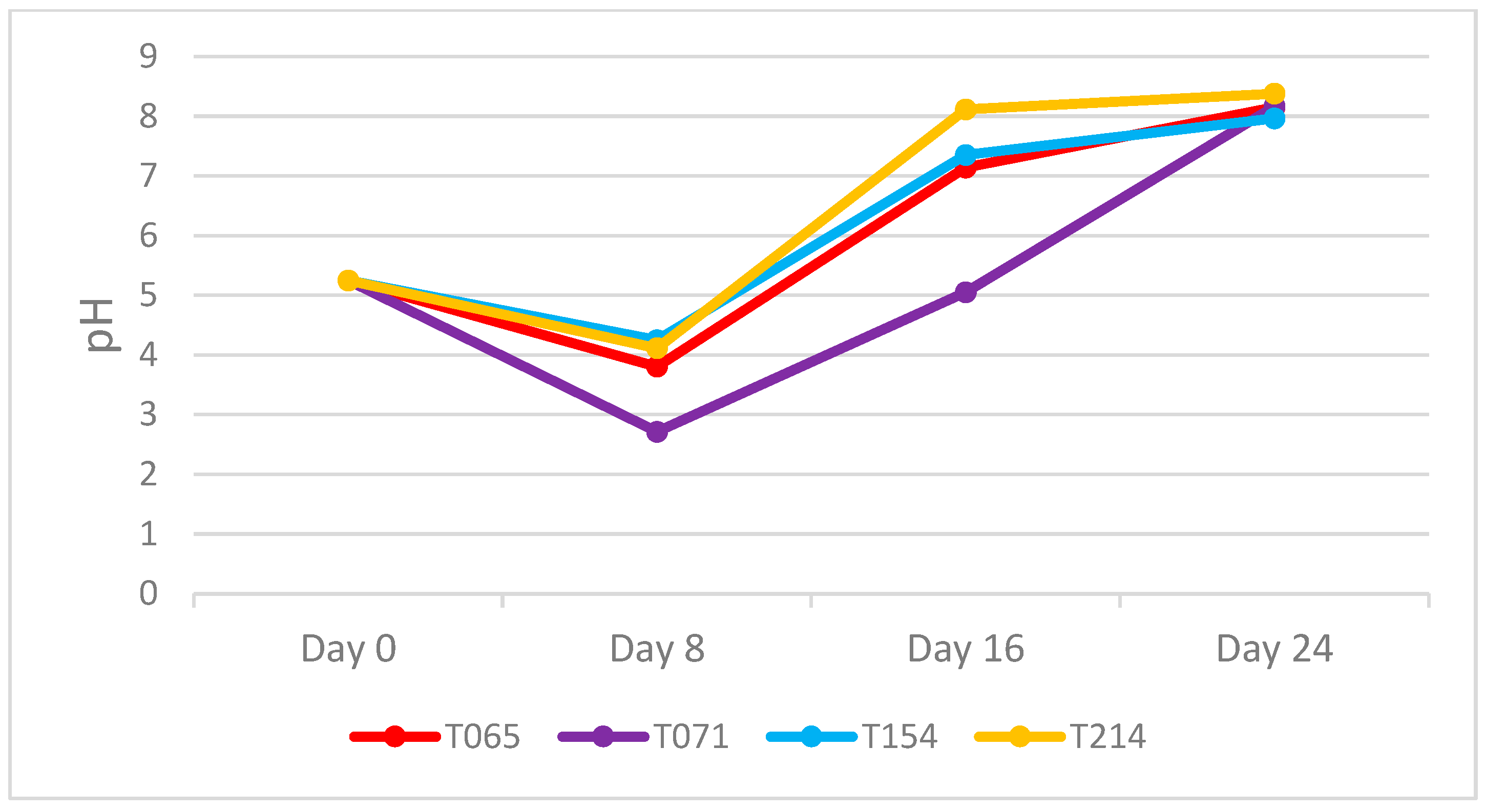
Figure 1. pH evolution in the culture filtrates of Trichoderma on Days 8, 16 and 24 of the trial (2 replicates).
Agronomy 15 01901 g001
Figure 2. Antibiograms of Diplodia seriata with 8-, 16-, and 24-day culture filtrates of all Trichoderma isolates.
Agronomy 15 01901 g002
Figure 3. Antibiograms of D. seriata. Diameter in mm of inhibition by the Trichoderma culture filtrates on the pathogen D. seriata (3 replicates). Different lowercase letters indicate significant differences between the culture filtrates extracted at 8, 16 or 24 days for the same Trichoderma. Different capital letters indicate significant differences between Trichoderma for the same extraction times (8, 16, and 24 days). LSD test (p ≤ 0.05).
Agronomy 15 01901 g003
Figure 4. Percentage inhibition of Trichoderma culture filtrates on P. minimum (3 replicates). Different lower case letters indicate significant differences between the culture filtrates extracted at 8, 16 or 24 days for the same Trichoderma. Different capital letters indicate significant differences between Trichoderma for the same extraction times (8, 16, and 24 days). LSD test (p ≤ 0.05).
Agronomy 15 01901 g004
Figure 5. Percentage inhibition of Trichoderma culture filtrates on P. chlamydospore (3 replicates). Different lowercase letters indicate significant differences between the culture filtrates extracted at 8, 16, or 24 days for the same Trichoderma. Different capital letters indicate significant differences between Trichoderma for the same extraction times (8, 16, and 24 days). LSD test (p ≤ 0.05).
Agronomy 15 01901 g005
Figure 6. Percentage inhibition of Trichoderma culture filtrates on D. seriata (3 replicates). Different lower case letters indicate significant differences between the culture filtrates extracted at 8, 16, or 24 days for the same Trichoderma. Different capital letters indicate significant differences between Trichoderma for the same extraction times (8, 16, and 24 days). LSD test (p ≤ 0.05).
Agronomy 15 01901 g006
Table 1. Trichoderma strains used in this study.
IsolateTrichoderma spp.OriginReferences
T065Trichoderma gamsiiSoil of vineyard, PDO León, Spain[30]
T071Trichoderma gamsiiSoil of vineyard, PDO León, Spain[31]
T154Trichoderma carraovejensisWood of Vitis vinifera cv. Tempranillo, Spain[6]
T214Trichoderma harzianumWood of Vitis vinifera cv. Verdejo, Spain[30]
Table 2. Pathogens used in this study.
IsolatePathogenOriginReference
Y038-05-3Phaeoacremonium minimumValles de Benavente Region, Spain[32]
Y116-18-03cPhaeomoniella chlamydosporaArribes del Duero, Spain[33]
ULEP32Diplodia seriataWood of Vitis vinifera cv., Verdejo, Spain[6]
Disclaimer/Publisher’s Note: The statements, opinions and data contained in all publications are solely those of the individual author(s) and contributor(s) and not of MDPI and/or the editor(s). MDPI and/or the editor(s) disclaim responsibility for any injury to people or property resulting from any ideas, methods, instructions or products referred to in the content.