Next Article in Journal
Mycosynthesis, Characterization of Zinc Oxide Nanoparticles, and Its Assessment in Various Biological Activities
Next Article in Special Issue
The Effect of Titanium Oxyfluoride Morphology on Photocatalytic Activity of Fluorine-Doped Titanium(IV) Oxide
Previous Article in Journal
Pressure-Induced Structural Phase Transitions in the Chromium Spinel LiInCr4O8 with Breathing Pyrochlore Lattice
 
 
Correction published on 28 June 2023, see Crystals 2023, 13(7), 1024.
Article

Photocatalytic Degradation of Textile Orange 16 Reactive Dye by ZnO Nanoparticles Synthesized via Green Route Using Punica Granatum Leaf Extract

1
Chemistry Department, Faculty of Science, University of Ha’il, P.O. Box 2440, Ha’il 81451, Saudi Arabia
2
Bharat Ratna Prof. CNR Rao Research Centre, Basaveshwar Science College, Bagalkot 587101, Karnataka, India
3
Department of Engineering Physics, K.L.E Institute of Technology, Hubballi 580027, Karnataka, India
4
School of Mechanical Engineering, KLE Technological University, Vidya Nagar, Hubballi 580031, Karnataka, India
5
Center of Excellence for Advanced Materials Research, King Abdulaziz University, P.O. Box 80203, Jeddah 21589, Saudi Arabia
6
Department of Chemistry, Bharath Institute of Higher Education and Research (BIHER), Bharath University, Chennai 600073, Tamil Nadu, India
7
Department of Agricultural Technology, Politeknik Pertanian Negeri Payakumbuh, Payakumbuh 26271, West Sumatra, Indonesia
*
Authors to whom correspondence should be addressed.
Crystals 2023, 13(2), 172; https://doi.org/10.3390/cryst13020172
Submission received: 10 December 2022 / Revised: 27 December 2022 / Accepted: 13 January 2023 / Published: 18 January 2023 / Corrected: 28 June 2023
(This article belongs to the Special Issue Advanced Nanomaterials for Photocatalytic Technologies)

Abstract

:
Since it does not use any dangerous chemicals and is a simple, low-cost process, the green synthesis approach for nanoparticle creation has several benefits compared to the physical and chemical synthesis routes. The current study describes an environmentally friendly synthesis of zinc oxide (ZnO) nanoparticles (NPs) using an extract of Punica granatum plant leaves. Fourier-transform infrared spectroscopy (FTIR), ultraviolet-visible spectrophotometer (UV-Vis), field-emission scanning electron microscopy (FESEM), energy-dispersive X-ray spectroscopy, and X-ray diffraction techniques were used to characterize the morphology, composition, and structural properties of the synthesized zinc oxide nanoparticles. The XRD pattern reveals that the ZnO nanoparticles are crystalline and have a diameter of 20 nm. According to the FESEM studies, the ZnO-NPs have sizes ranging from 50 to 100 nm on average and are almost spherical. When exposed to direct sunlight, the produced ZnO-NPs demonstrate impressive photocatalytic oxidation of textile Orange 16, a reactive dye. As a result, our research advances the development of a green photocatalyst for the removal of harmful dyes from water.

1. Introduction

New nanoscale materials are being produced as a result of the advancements in nanotechnology. These materials have a variety of uses, including in consumer goods, nanomedicine, and nanoelectronics [1,2]. Because of their superior chemical and physical characteristics when compared to their bulk counterparts, research on such materials has increased significantly in recent years. Metal oxide nanostructures have been created, and they have a variety of uses in various industries. A semiconductor with a greater band gap (3.4 eV) is zinc oxide (ZnO). Applications for it include dye degradation, gas sensors, solar cells, and many others [3]. Numerous chemical and physical techniques, such as the sol–gel technique [4], the precipitation method [5], the arc discharge technique [6], the hydrothermal method [7], and the laser ablation method [8], have been adopted for the synthesis of ZnO nanoparticles. Due to its numerous advantages over the physical and chemical approaches—including the fact that it does not use dangerous chemicals and is both environmentally friendly and economically advantageous—synthesis via the green method has been used to prepare ZnO nanoparticles [9,10]. This approach makes use of biological components that are readily available from plants.
Because they are readily available, leaves from the Punica grantum plant have been employed. The family of Punicaceae, which consists of two different species, is dominated by the pomegranate species (Punica granatum). Punica grantum plant leaves contain alkaloids, tannins, triterpenic acids, and flavonoids as phytochemicals [11]. Utilizing chemical reduction, these phytoconstituents produce metal oxide nanoparticles by acting as the stabilizing and reducing agents. As far as we are aware, there has not been any research on the usage of P. granatum leaves in the preparation of ZnO-NPs.
Organic substances called dyes are used in the paper, food, leather, and textile industries. Strongly colored, hazardous dyes are present in the effluents from these enterprises. The discharged effluents will enter running water, polluting the surface and groundwater and perhaps endangering the health of aquatic life as well as that of people, fauna, and the environment. Before release, the effluents would be treated to break down the dye into non-toxic species. Different purification methods, including chemical, biological, and physical techniques, have been developed, depending on the type of pollution. ZnO-NPs have been used to degrade dyes with success [12,13,14]. Through catalytic photooxidation with ZnO-NPs, reactive oxidative hydroxide radicals are created, which destroy the dyes. According to its chemical makeup, the textile Orange 16 reactive dye is an organic substance classified as a disulfonated triphenylmethane dye. Although originally designed for the textile industry, it is now frequently used for protein staining in biochemistry. The catalyst should be stable and reusable in addition to having a high degradation efficiency, as these qualities are crucial for industrial applications. Numerous investigations [15,16] have shown that ZnO-NPs are stable and reusable.
The P. granatum leaf extract was used in the current work’s green synthesis to create ZnO-NPs. Under exposure to direct sunlight, the green ZnO-NPs that were generated were used to photocatalytically degrade the textile Orange 16 reactive dye.

2. Materials and Methodology

2.1. Materials

We gathered Punica granatum leaves from a field in Kaladagi near Bagalkot (India). Zinc nitrate hexahydrate of high purity was procured from Sigma-Aldrich, Mumbai, India. All water used in the experiment was deionized distilled water. We bought textile Orange 16 reactive dye from a local market in Bagalkot, Karnataka, India, through a textile business. None of the chemicals were further purified before use. Glassware washed in a prepared Piranha solution (3:1 volume ratio of H2SO4/H2O2) was rinsed with deionized (DI) water with a resistivity of 16.4 MΩ·cm (millipore water), dried in an oven, and returned for later use.

2.2. Methodology

2.2.1. Extraction of Punica granatum Leaf

Young P. granatum leaves were collected from a Punica granatum plantation at a Kaladagi village near Bagalkot (India). The leaves’ middle rib was cut off. The leaves that remained were cleaned with distilled water to get rid of dust, and they were then dried in the shade to get rid of all the moisture. An aqueous extract of Punica granatum leaf was prepared. During the process, the dried leaves were weighed accurately and were finely grounded in a mixture. Then, deionized water was added to the dried fine powder. For about three hours, the mixture was refluxed at 60 °C, using the Soxhlet extractor apparatus. Once the green color turned brownish, the process was stopped. At room temperature, the solution was allowed to cool for the period of ten to fifteen minutes. Furthermore, Whatman No. 41 filter paper was used to filter the solution, and the filtrate was then collected in a dry flask while the residue was thrown away. For future use, the leaf extract was kept at ambient temperature.

2.2.2. Green Synthesis of Zinc Oxide Nanoparticles (ZnO-NPs)

In deionized water, a solution of Zn(NO3)2·6H2O (0.1 M) was made. The mixture was made homogeneously by stirring it without being heated. After that, a magnetic stirrer was used to gradually add drops of the aqueous leaf extract to the zinc nitrate solution. After the leaf extract was completely added, the resulting mixture was stirred with heating at 60 °C for 3–4 h on a hot plate until it took the form of slurry. The obtained slurry substance was filtered and placed in a crucible. The sample was calcined at 500 °C in a muffle furnace at a heating rate of 5 °C for 5 h. After calcination, white powder was obtained and stored properly.

2.2.3. Characterization of ZnO-NPs

A Siemens D 5000 (Malvern, UK) powder X-ray diffractometer was used to perform X-ray diffraction on the powdered zinc oxide nanoparticles. The UV-visible spectrum of the zinc oxide nanoparticles was recorded by employing a UV spectrophotometer (Systonic, Bangalore, India). The samples were evaluated between 200 and 800 nm. A Shimadzu (Model: IR Affinity-1, Tokyo, Japan) FTIR spectrometer was used to perform Fourier-transform spectroscopic measurements. A Carl Zeiss (Model: Sigma 300, Bangalore, India) field-emission scanning electron microscope (FESEM) was used to examine the surface morphology and the dimension of the particles of the prepared ZnO nanoparticles. The Oxford instrument (Buckinghamshire, UK), an energy-dispersive X-ray spectroscopy (EDX), was used to obtain the spectrum to check the purity of the synthesized ZnO nanoparticles and the stoichiometry of the samples. The particle size of the synthesized ZnO-NPs was determined using the Zetasizer (Model 3000HS, Malvern, UK). On a cuvette, the zeta average size of ZnO-NPs suspended in DI water was determined. The measurement was repeated three times, and the average value was used for data analysis.

2.2.4. Photocatalytic Activity of ZnO-NPs

A photodegradation study of textile Orange 16 reactive dye in water by exposure to sunlight was used to determine the ZnO nanoparticles as the photocatalysts. For this, an artificial, laboratory-simulated dye was prepared by dissolving the dye in water until the solution’s absorbance value was more than one, i.e., 0.4 g in 100 mL of water. To the prepared colored mixture, a known amount of ZnO (i.e., 0.1 g), which would act as a photocatalyst, was added. Later, the aqueous solution was sonicated for 30 min. The solution was then kept in sunlight while being constantly stirred. After 30 min, 5 mL of the solution was withdrawn and centrifuged to settle down the ZnO photocatalyst. The supernatant solution was used to measure the absorbance with a UV-Vis spectrophotometer. From the experiment, it was observed that the value of absorbance reduced after each interval, showing the degradation of the dye by the ZnO-NPs. The detailed stepwise green synthesis of ZnO nanoparticles and their photocatalytic action are shown in Figure 1.

3. Results and Discussion

3.1. X-ray Diffraction Analysis

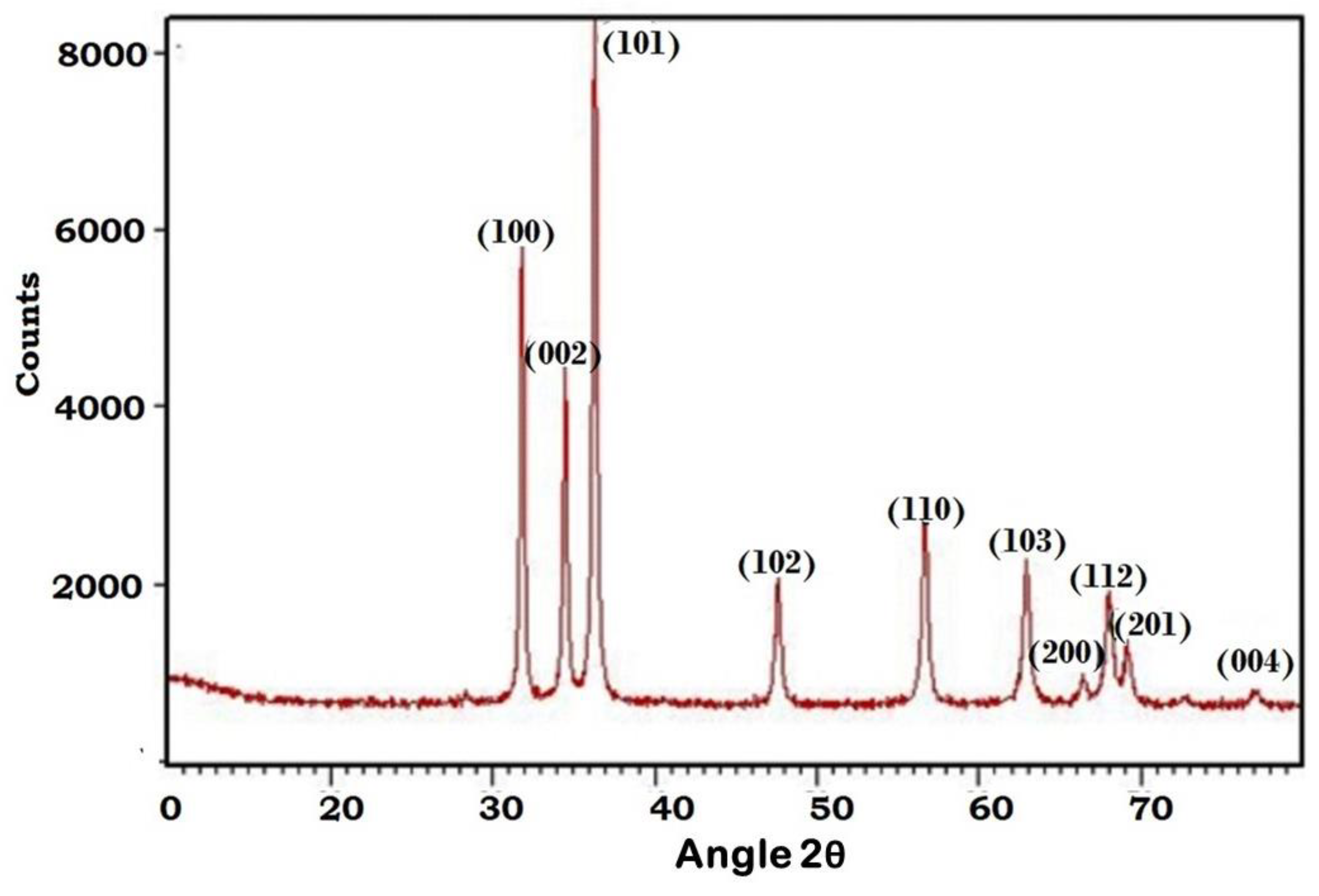
Figure 2 depicts the X-Ray diffraction graph of the green synthesized ZnO nanoparticles. The sample was examined at various angles, ranging from 0° to 70°. ZnO nanoparticles have significant peaks at 2θ = 31.81°, 34.49°, 36.28°, 47.62°, 56.63°, 62.91°, and 68°, which can be designated as (100), (002), (101), (102), (110), (103), and (200), respectively. The orientation and crystallinity of the ZnO-NPs were revealed by using the X-ray pattern. The JCPDS data sheet/ICDD no. 36-1451 was used to compute the XRD pattern. The obtained X-ray pattern demonstrate that ZnO-NPs were synthesized using the green synthesis route, with the establishment of crystalline and wurtzite hexagonal structures. The particle dimension of ZnO-NPs was computed using the Debye–Scherrer equation from the highest peak (101) in the XRD graph:
d   =   K λ β c o s θ
where d is the size of the crystallite; λ is the wavelength for diffraction; β is the corrected value of FWHM; θ is the angle of diffraction; and K is the universal and its value is near unity, i.e., 0.94.
The purity of the ZnO-NPs is confirmed by the absence of diffraction peaks from other phases.
Bragg’s Equations (3)–(5) were used to calculate the inter-planar spacing (d), the lattice parameters (a = b and c) for the hexagonal wurtzite structure, and the volume of the hexagonal system unit cell (V) [17,18,19,20]. Table 1 summarizes all of these parameters.
n λ   =   2 d   s i n   θ
1 d h k l 2   =   4 3 h 2   +   h k   +   k 2 a 2   +   l 2 c 2
V   =   3 2 a 2 c
where dhkl = inter-planar spacing; V = volume of the unit cell; and a, c = lattice parameters for the ZnO-NP.

3.2. Particle Size Analysis

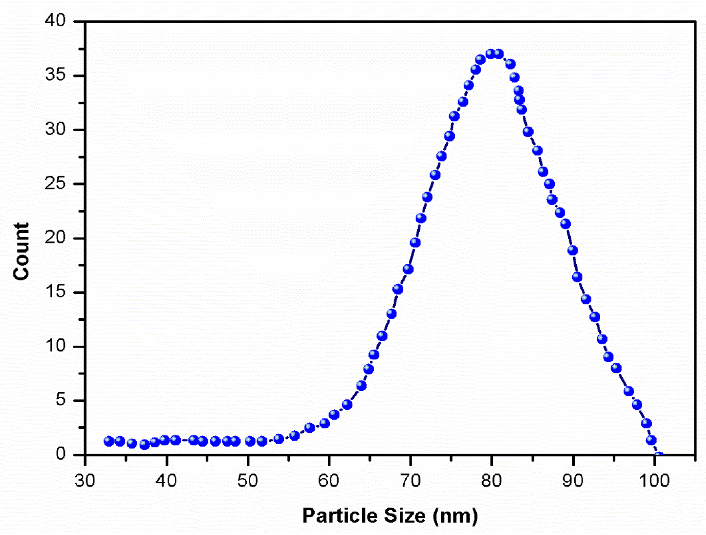
The particle size of the ZnO-NPs is depicted in Figure 3. The obtained results reveal that the size of ZnO particles ranges between 60 and 100 nm. The average particle size is measured at 80 nm.

3.3. UV-Visible Spectroscopic Analysis

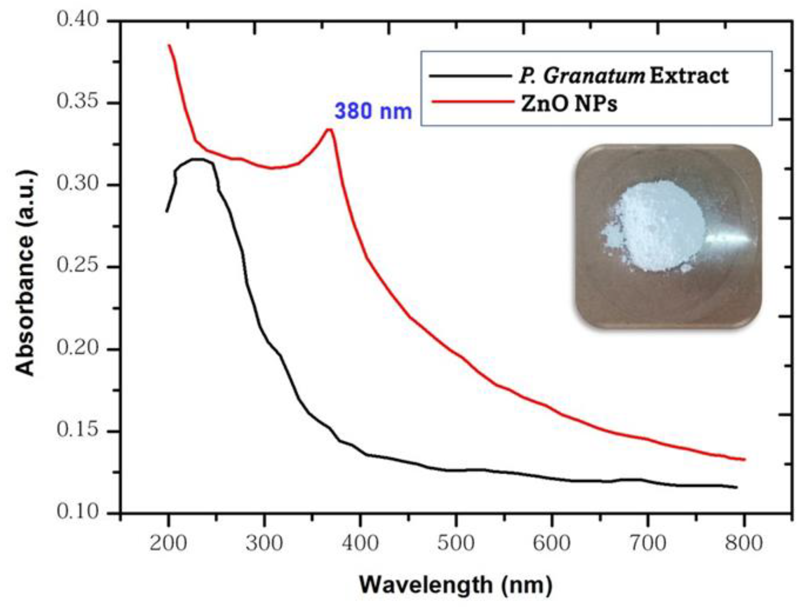
Surface plasmon resonance distinguishes the absorbance pattern of nanoparticles from that of their bulk counterparts. The UV-visible measurement was used to confirm the formation of nanoparticles. Figure 4 depicts the UV-visible absorption spectrum of the ZnO-NPs. Using a systonic UV spectrophotometer, the substance was analyzed between 200 and 800 nm. The color of a solution of zinc nitrate hexahydrate (Zn(NO3)2H2O) changed from white to brown when the P. granatum leaf extract was added. This resulted from the solution’s synthesis of ZnO-NPs. The nanoparticles of zinc oxide are responsible for stimulating the surface plasmon vibrations, which in turn generate color changes. The absorbance peak was found to be centered at 382 nm, indicating that zinc nitrate hexahydrate had been converted to ZnO-NPs [21,22,23].
The optical energy band gap (Eg) was calculated using Formula (5), and the results are shown in Table 2.
E g   =   hc λ
From this formula, ‘h’ represents the Planck’s constant (6.626 × 10⁻³⁴ J s), ‘c’ corresponds to the velocity of light (the value is 3 × 10⁸ m s−1), and λ corresponds to the wavelength of the peak with the maximum intensity [24].
The band gap values were calculated, and the results are nearly equivalent to 3.4 eV, which is consistent with previously reported values in the literature. Suresh et al. (3.33 eV) [25] and Hancock et al. (3.39 eV) [26] reported comparable band gap results. The variation in the shape and size of the ZnO-NPs may explain the difference in Eg with Zn (NO3)2·6H2O concentration. In addition, a decrease in Eg values is attributed to an increase in ZnO particle size [27].

3.4. FESEM and EDX Analysis

ZnO may be produced into several nanostructures, including nanospheres, nanorods, and others. By looking at and analyzing each minute topographical feature using a field-emission scanning electron microscope (FESEM), the particle size and shape may be determined. The particle size and shape of the synthesized ZnO nanoparticles were analyzed with the aid of a Carl Zeiss FESEM. Figure 5 displays a FESEM picture of the ZnO nanoparticles that were synthesized via the green route. This image demonstrates that the particle is composed of nanostructures, which are even smaller structures. The XRD investigation indicated that the nanoparticles are fully spherical, with an average diameter of 80 nm.
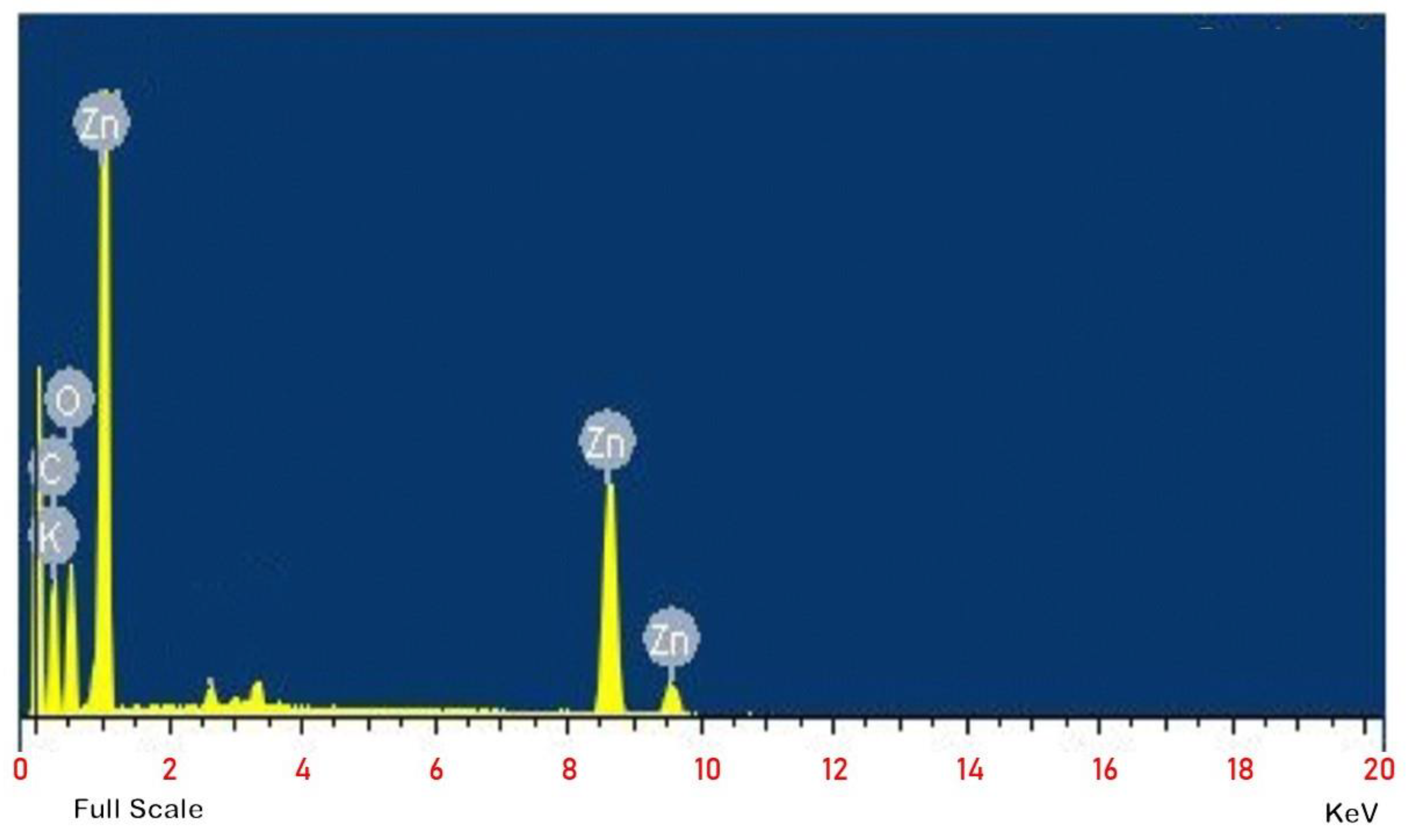
EDX was utilized to undertake an elemental investigation or chemical composition on the ZnO nanoparticles made in an environmentally responsible manner. The stoichiometry and chemical purity of the samples were analyzed using an EDX-equipped equipment from Oxford. The EDX spectrum of the ZnO nanoparticles is illustrated here in Figure 6. The EDX spectrum indicates indisputably that ZnO and oxygen (O) ions are present in the ZnO nanoparticles formed by the P. Granatum reaction. According to the findings of the elemental analysis, the ZnO powder is made up of 76% zinc and 15% oxygen, which shows that it is quite pure and only comprises a minor number of contaminants. The stoichiometry reveals that the mass percentages of zinc and oxygen should be 80.3% and 19.7%, respectively, if we are to trust the formulae.

3.5. FT-IR Analysis

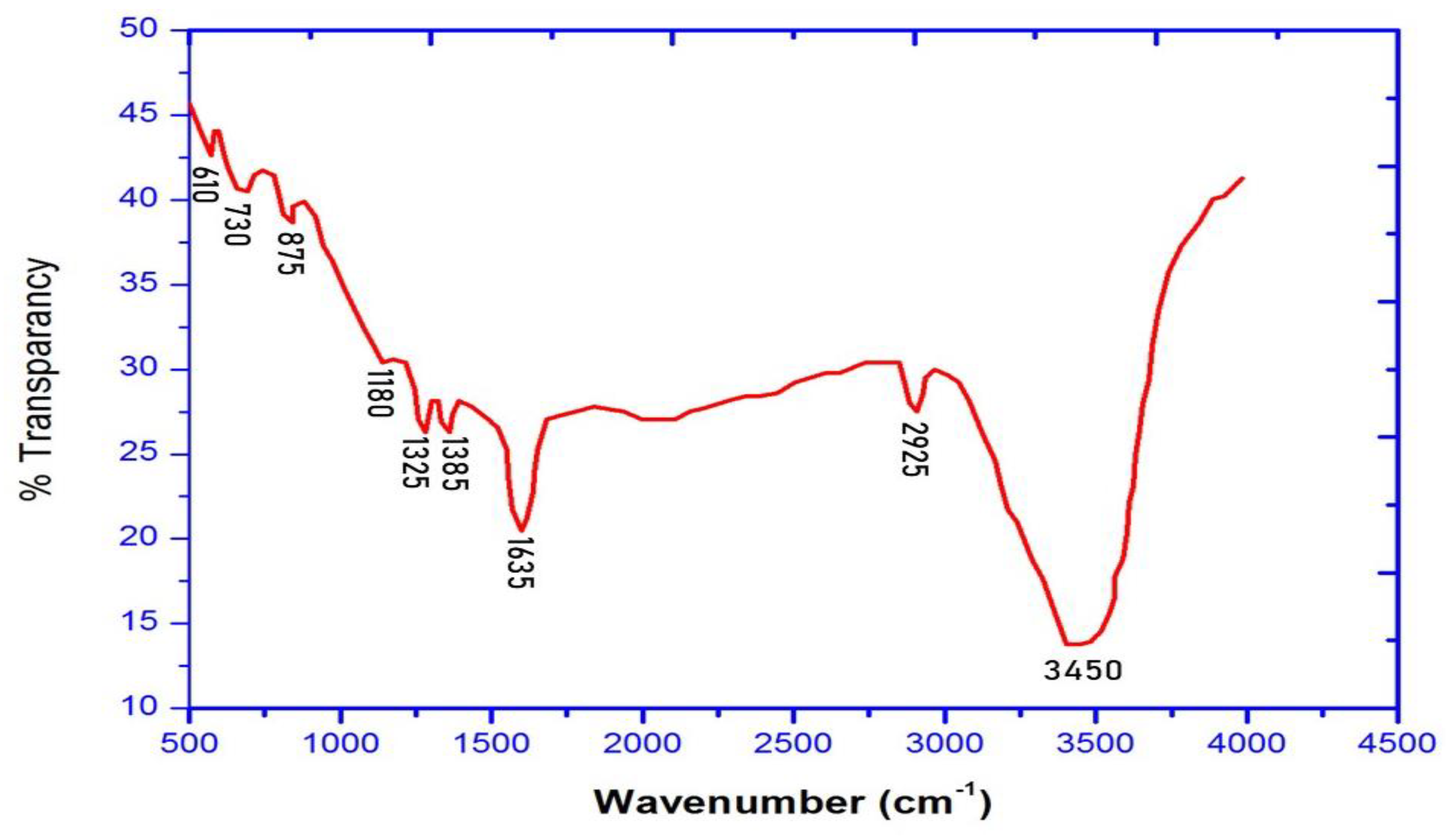
FT-IR spectroscopy was used to establish the presence of the Zn–O bond and its formation mechanism, as well as to detect the photo components that coat the surface of the ZnO-NPs. Fourier-transform infrared spectroscopy was performed using a Bruker Alpha FTIR as the instrument. Figure 7 depicts the FT-IR spectra of the ZnO-NPs that were synthetically generated via the green route. Both the 3610 cm−1 and 3822 cm−1 spectral peaks are the consequence of O–H stretching. The C–H stretch is responsible for the peak that appears at roughly 2354 cm−1. The peak is induced by C=O stretching and is located at about 1512 cm−1. There is a link between the peaks at 1635 cm−1 and ZnO vibrations caused by bending deformation. At 610 cm−1, strong vibrational bands are produced as a result of the stretching modes utilized to form the ZnO nanoparticles. The phytoconstituents of P. granatum prevent the aggregation of ZnO-NPs during their production [22,23,28,29]. This is achieved by stabilizing the nanoparticles’ surface.

3.6. Photocatalytic Activity

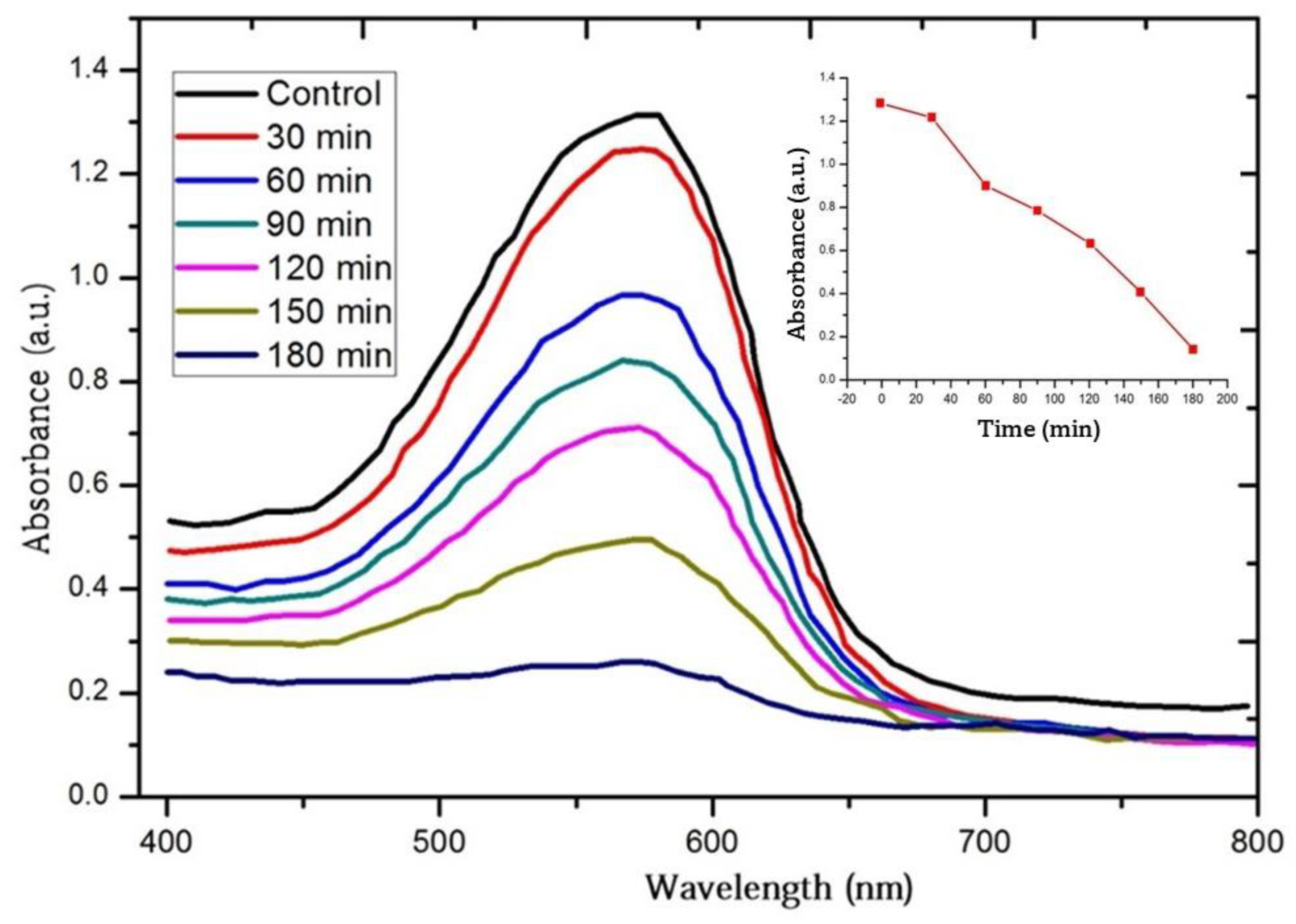
To evaluate the photodegradation of textile Orange 16 reactive dye in the presence of ZnO nanoparticles, the decrease in the absorbance of the dye was determined. The decrease in the absorbance of the dye solution as a function of exposure length is consistent with a decline in the concentration of textile Orange 16 reactive dye. Over time, the concentration of the blue pigment in the dye solution progressively lost its vibrancy, until it ultimately became light blue. Figure 8 depicts the deterioration of the textile Orange 16 reactive dye stain as a function of time in the samples exposed to sunlight. It can be seen that 600 nm is the wavelength at which textile Orange 16 reactive dye has the highest absorption peak. In addition, it reveals that ZnO-NPs are capable of significantly reducing the pollutant within three hours.

3.7. Recyclability and Photostability

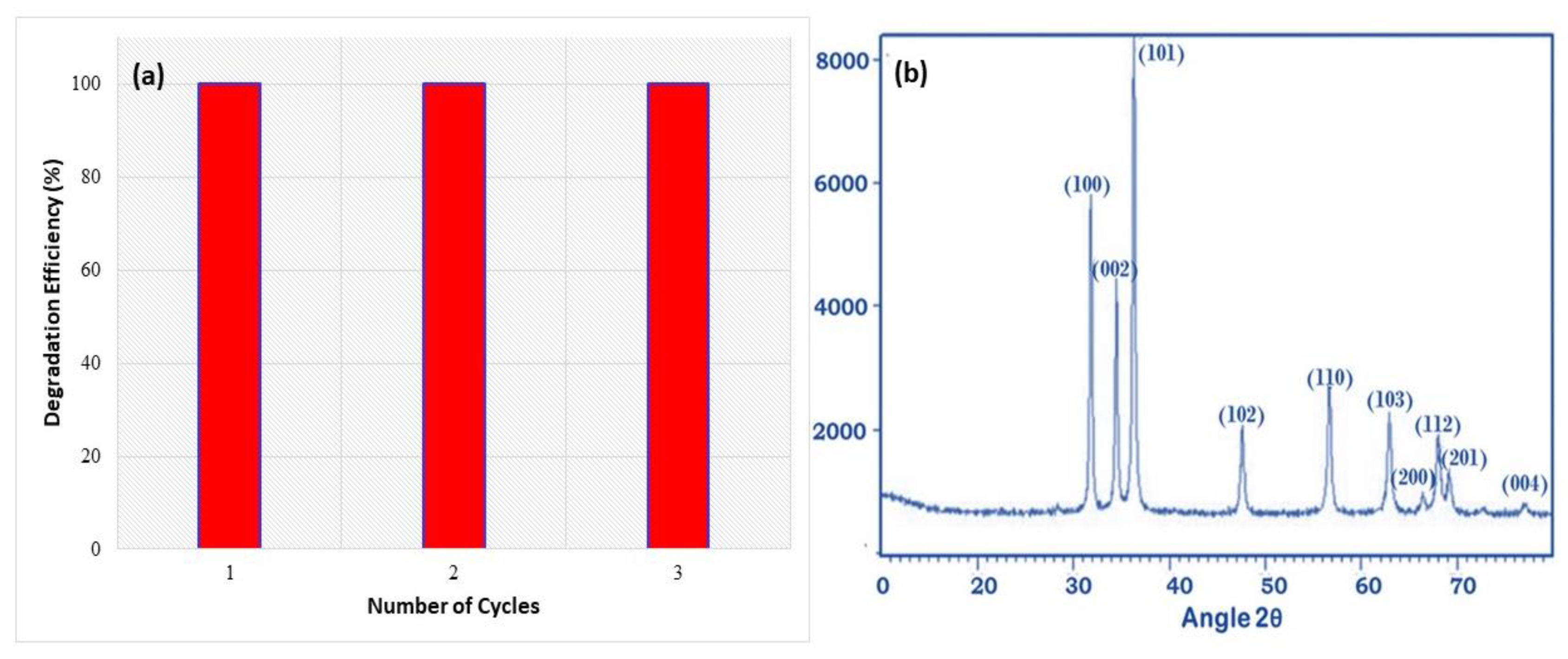
Three cycles of photocatalysis were conducted to assess whether or not the photocatalyst could be recycled. The images are depicted in Figure 9. Figure 9a shows a 100% efficiency even after three cycles of photocatalysis. In addition, as seen in Figure 9b, the XRD measurements performed after the photocatalytic phenomenon show that the crystalline structure of ZnO has not altered when compared to before the photocatalytic process was undertaken.
Figure 10 shows the photodegradation of the Orange 16 dye by using green synthesized ZnO nanoparticles. As observed in Figure 10, the effectiveness of the ZnO-NPs as photocatalysts for the breakdown of organic materials is shown by the decline in the color intensity of the dye with respect to the time of exposure to sunlight.

3.8. Photolysis

There are three types of photocatalytic dye degradation mechanisms: (1) dye sensitization via charge injection, (2) indirect dye degradation via oxidation/reduction, and (3) direct photolysis of the dye. Photocatalytic substances are generally classified into three generations. One-component (e.g., ZnO and TiO2) and multi-component semiconductor metal oxide (e.g., ZnO-TiO2 and Bi2O3-ZnO) photocatalysts are classified as first-generation and second-generation, respectively. Third-generation photocatalysts are photocatalysts that are dispersed on an inert solid substrate (e.g., Ag-Al2O3 and ZnO-C).
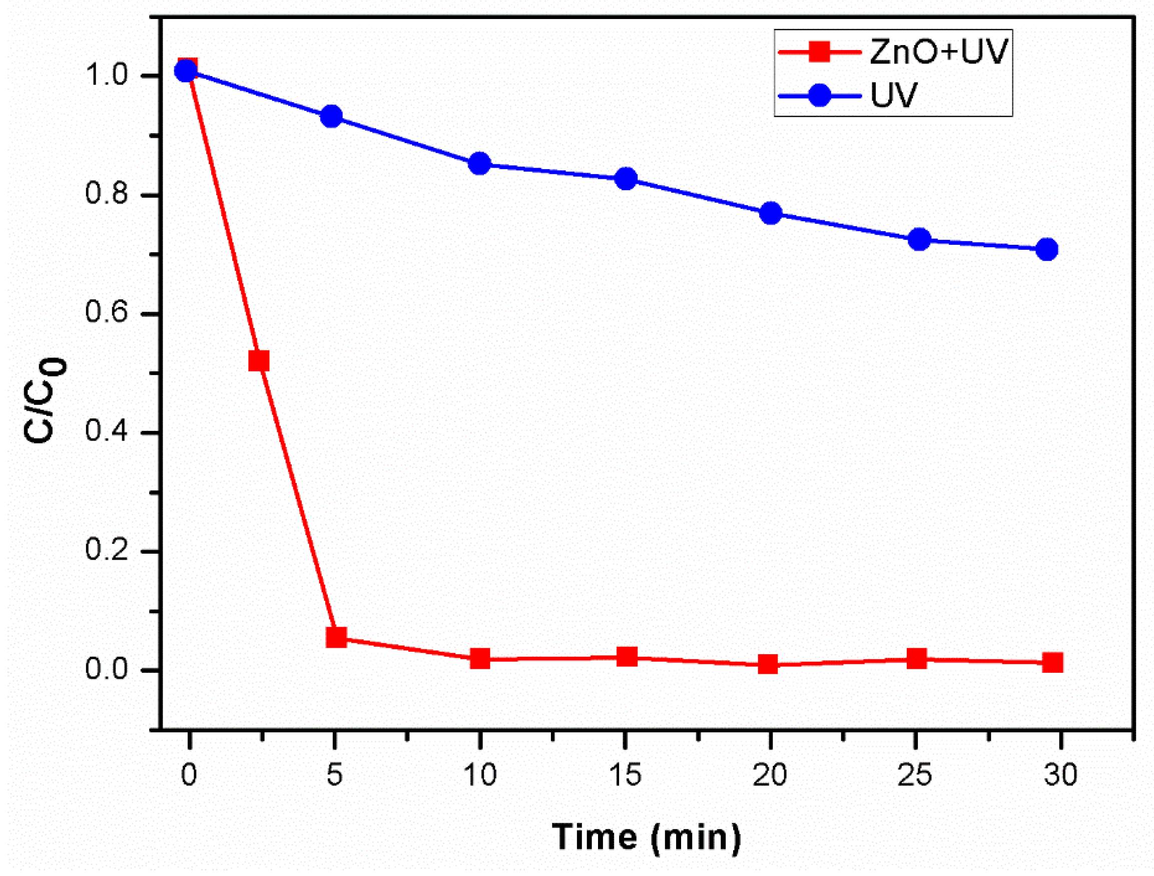
Because some dyes are degraded by direct UV irradiation, it is necessary to investigate the extent to which textile Orange 16 reactive dye is photolyzed in the absence of a photocatalyst. In the absence of the photocatalyst, direct UV radiation exposure photolyzed textile Orange 16 reactive dye by up to 25% in 30 min. When ZnO-NPs were used as a photocatalyst and exposed to UV radiation, it was discovered that the photodegradation of the dye increased by decolorizing the textile Orange 16 reactive dye with an efficiency of 96% in 30 min. The time-dependent photocatalytic degradation and the photolysis of textile Orange 16 reactive dye concentration are shown in Figure 11.

3.9. Comparison

The dye photodegradation in the presence of the synthesized ZnO-NPs was compared to a commercial ZnO and previously reported work. The results are tabulated in Table 3.
Among the tested ZnO NPs, commercial ZnO performed the best when compared to other ZnO NPs.

4. Conclusions

In the current investigation, an extract of the leaves of P. granatum was dissolved in water and utilized in a green synthesis process, which resulted in the successful generation of ZnO-NPs. The preparation of spherical, polydisperse ZnO-NPs with sizes ranging from 50 to 100 nm and an average size of 80 nm was performed. The majority of the nanoparticles are spherical and have an 80 nm diameter. According to the results of the EDX analysis, the ZnO powder has a very high degree of purity and includes nearly no impurities. The powder consists of 76% zinc and 15% oxygen. According to the results of the photocatalytic experiment, the bio-produced ZnO-NPs are able to photodegrade the textile Orange 16 reactive dye with an overall efficiency of 93%. The photolysis of the textile Orange 16 reactive dye concentration without and with ZnO-NPs shows that the ZnO-NPs play the role of photocatalysts effectively. According to the results of this work, the manufacture of ZnO-NPs using the P. granatum leaf extract is safe, inexpensive, straightforward, and environmentally friendly, and these nanoparticles have shown efficacy as green photocatalysts in the actual treatment of wastewater.

Author Contributions

Conceptualization, S.A.A.-Z. and M.B.P.; methodology, S.N.M. and A.Y.P.; software, A.A.O.; validation, N.M., D.M. and A.K.; formal analysis, A.M.; investigation, E.S.; project administration, S.A.A.-Z. All authors have read and agreed to the published version of the manuscript.

Funding

This research has been funded by Scientific Research Deanship at the University of Ha’il-Saudi Arabia through project number RG-21 032.

Data Availability Statement

Data can be provided on the request from the corresponding author.

Acknowledgments

This research has been funded by Scientific Research Deanship at the University of Ha’il-Saudi Arabia through project number RG-21 032.

Conflicts of Interest

The authors declare no conflict of interest.

References

  1. Saini, R.; Saini, S.; Sharma, S. Nanotechnology: The future medicine. J. Cutan. Aesthetic Surg. 2010, 3, 32. [Google Scholar] [CrossRef]
  2. Singh, A. Synthesis, characterization, electrical and sensing properties of ZnO nanoparticles. Adv. Powder Technol. 2010, 21, 609–613. [Google Scholar] [CrossRef]
  3. Nohavica, D.; Gladkov, P. ZnO nanoparticles and their applications—New achievments. NANOCON 2010, 1, 121–124. [Google Scholar] [CrossRef]
  4. Azlina, H.N.; Hasnidawani, J.N.; Norita, H.; Surip, S.N. Synthesis of SiO2 nanostructures using sol-gel method. Acta Phys. Pol. A 2016, 129, 842–844. [Google Scholar] [CrossRef]
  5. Raoufi, D. Synthesis and microstructural properties of ZnO nanoparticles prepared by precipitation method. Renew. Energy 2013, 50, 932–937. [Google Scholar] [CrossRef]
  6. Ashkarran, A.A.; Irajizad, A.; Mahdavi, S.M.; Ahadian, M.M. ZnO nanoparticles prepared by electrical arc discharge method in water. Mater. Chem. Phys. 2009, 118, 6–8. [Google Scholar] [CrossRef]
  7. Aneesh, P.M.; Vanaja, K.A.; Jayaraj, M.K. Synthesis of ZnO nanoparticles by hydrothermal method. In Nanophotonic Materials IV; SPIE: Bellingham, WA, USA, 2007. [Google Scholar]
  8. Singh, S.C.; Gopal, R. Synthesis of colloidal zinc oxide nanoparticles by pulsed laser ablation in aqueous media. Phys. E Low-Dimens. Syst. Nanostruct. 2008, 40, 724–730. [Google Scholar] [CrossRef]
  9. Singh, J.; Kumar, S.; Alok, A.; Upadhyay, S.K.; Rawat, M.; Tsang, D.C.; Bolan, N.; Kim, K.H. The potential of green synthesized zinc oxide nanoparticles as nutrient source for plant growth. J. Clean. Prod. 2019, 214, 1061–1070. [Google Scholar] [CrossRef]
  10. Singh, J.; Dutta, T.; Kim, K.H.; Rawat, M.; Samddar, P.; Kumar, P. ‘Green’ synthesis of metals and their oxide nanoparticles: Applications for environmental remediation. J. Nanobiotechnol. 2018, 16, 84. [Google Scholar] [CrossRef]
  11. Wang, R.; Ding, Y.; Liu, R.; Xiang, L.; Du, L. Pomegranate: Constituents, bioactivities and pharmacokinetics. Fruit Veg. Cereal Sci. Biotechnol. 2010, 4, 77–87. [Google Scholar]
  12. Hassan, S.S.M.; Azab, W.I.M.E.; Ali, H.R.; Mansour, M.S.M. Green synthesis and characterization of ZnO nanoparticles for photocatalytic degradation of anthracene. Adv. Nat. Sci. Nanosci. Nanotechnol. 2015, 6, 045012. [Google Scholar] [CrossRef]
  13. Bhuyan, T.; Mishra, K.; Khanuja, M.; Prasad, R.; Varma, A. Biosynthesis of zinc oxide nanoparticles from Azadirachta indica for antibacterial and photocatalytic applications. Mater. Sci. Semicond. Process. 2015, 32, 55–61. [Google Scholar] [CrossRef]
  14. Davar, F.; Majedi, A.; Mirzaei, A. Green synthesis of ZnO nanoparticles and its application in the degradation of some dyes. J. Am. Ceram. Soc. 2015, 98, 1739–1746. [Google Scholar] [CrossRef]
  15. Kitture, R.; Koppikar, S.J.; Kaul-Ghanekar, R.; Kale, S.N. Catalyst efficiency, photostability and reusability study of ZnO nanoparticles in visible light for dye degradation. J. Phys. Chem. Solids 2011, 72, 60–66. [Google Scholar] [CrossRef]
  16. Zainuri, N.Z.; Hairom, N.H.; Sidik, D.A.; Misdan, N.; Yusof, N.; Mohammad, A.W. Reusability performance of zinc oxide nanoparticles for photocatalytic degradation of POME. E3S Web Conf. 2018, 34, 02013. [Google Scholar] [CrossRef]
  17. Sharma, S.; Kumar, K.; Thakur, N.; Chauhan, S.; Chauhan, M.S. The effect of shape and size of ZnO nanoparticles on their antimicrobial and photocatalytic activities: A green approach. Bull. Mater. Sci. 2020, 43, 20. [Google Scholar] [CrossRef]
  18. Le Pevelen, D.D. (Ed.) Small Molecule X-ray Crystallography, Theory and Workflow. In Encyclopaedia of Spectroscopy and Spectrometry, 2nd ed.; Academic Press: Cambridge, MA, USA, 2010; pp. 2559–2576. [Google Scholar] [CrossRef]
  19. Zak, K.A.; Abd Majid, W.H.; Abrishami, M.E.; Yousefi, R. X-ray analysis of ZnO nanoparticles by Williamson–Hall and size–strain plot methods. Solid State Sci. 2011, 13, 251. [Google Scholar]
  20. Lingaraju, K.; Raja Naika, H.; Manjunath, K.; Basavaraj, R.B.; Nagabhushana, H.; Nagaraju, G.; Suresh, D. Biogenic synthesis of zinc oxide nanoparticles using Ruta graveolens (L.) and their antibacterial and antioxidant activities. Appl. Nanosci. 2016, 6, 703. [Google Scholar] [CrossRef]
  21. Fuku, X.; Diallo, A.; Maaza, M. Nanoscaled electrocatalytic optically modulated ZnO nanoparticles through green process of Punica granatum L. and their antibacterial activities. Int. J. Electrochem. 2016, 2016, 4682967. [Google Scholar] [CrossRef]
  22. Fuku, X.; Kaviyarasu, K.; Matinise, N.; Maaza, M. Punicalagin green functionalized Cu/Cu2O/ZnO/CuO nanocomposite for potential electrochemical transducer and catalyst. Nanoscale Res. Lett. 2016, 11, 386. [Google Scholar] [CrossRef]
  23. Matinise, N.; Fuku, X.G.; Kaviyarasu, K.; Mayedwa, N.; Maaza, M.J. ZnO nanoparticles via Moringa oleifera green synthesis: Physical properties & mechanism of formation. Appl. Surf. Sci. 2017, 406, 339–347. [Google Scholar] [CrossRef]
  24. Choudhary, M.K.; Kataria, J.; Sharma, S. Novel Green Biomimetic Approach for Preparation of Highly Stable Au-ZnO Heterojunctions with Enhanced Photocatalytic Activity. Appl. Nano Mater. 2018, 1, 1870. [Google Scholar] [CrossRef]
  25. Suresh, D.; Nethravathi, P.C.; Lingaraju, K.; Rajanaika, H.; Sharma, S.C.; Nagabhushana, H. EGCG assisted green synthesis of ZnO nanopowders: Photodegradative, antimicrobial and antioxidant activities. Spectrochim. Acta Part A Mol. Biomol. Spectrosc. 2015, 136 Pt C, 1467–1474. [Google Scholar] [CrossRef]
  26. Hancock, J.M.; Rankin, W.M.; Hammad, T.M.; Salem, J.S.; Chesnel KHarrison, R.G. Optical and Magnetic Properties of ZnO Nanoparticles Doped with Co, Ni and Mn and Synthesized at Low Temperature. J. Nanosci. Nanotechnol. 2015, 15, 3809. [Google Scholar] [CrossRef] [PubMed]
  27. Akbarian, M.; Mahjoub, S.; Elahi, S.M.; Zabihi, E.; Ebrahim Zabihi, E.; Tashakkorian, H. Green synthesis, formulation and biological evaluation of a novel ZnO nanocarrier loaded with paclitaxel as drug delivery system on MCF-7 cell line. Colloids Surf. B Biointerfaces 2020, 186, 110686. [Google Scholar] [CrossRef] [PubMed]
  28. Singh, K.; Singh, J.; Rawat, M. Green synthesis of zinc oxide nanoparticles using Punica Granatum leaf extract and its application towards photocatalytic degradation of Coomassie brilliant blue R-250 dye. SN Appl. Sci. 2019, 1, 624. [Google Scholar] [CrossRef]
  29. Otaibi, A.A.; Patil, M.B.; Rajamani, S.B.; Mathad, S.N.; Patil, A.Y.; Amshumali, M.K.; Shaik, J.P.; Asiri, A.M.; Khan, A. Development and Testing of Zinc Oxide Embedded Sulfonated Poly (Vinyl Alcohol) Nanocomposite Membranes for Fuel Cells. Crystals 2022, 12, 1739. [Google Scholar] [CrossRef]
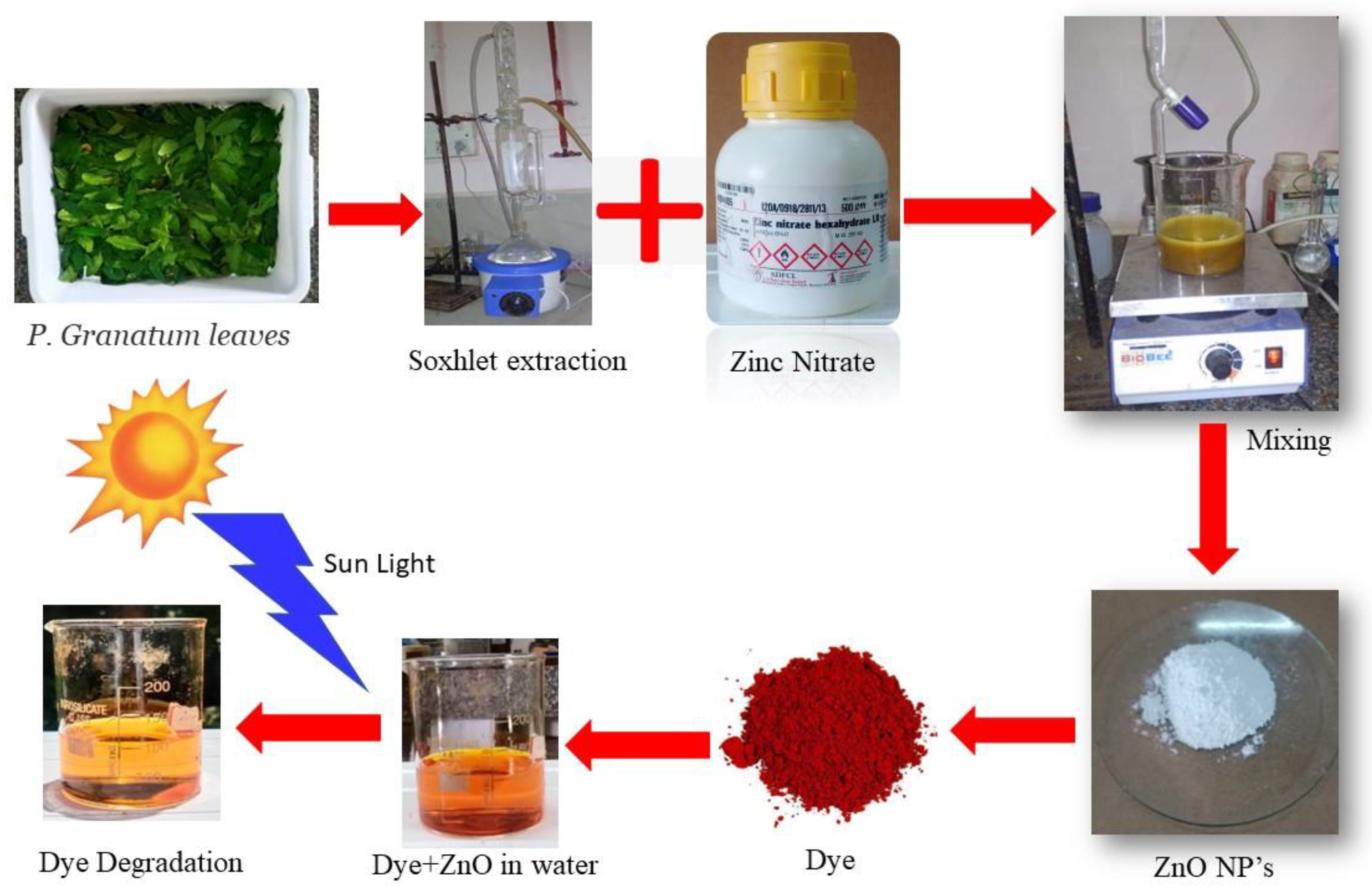
Figure 1. Step-by-step process of ZnO-NP synthesis via green route and the degradation of the dye.
Crystals 13 00172 g001
Figure 2. X-ray diffraction curves of green synthesized ZnO nanoparticles.
Crystals 13 00172 g002
Figure 3. Histogram of ZnO particle size distribution.
Crystals 13 00172 g003
Figure 4. ZnO-NPs and P. granatum leave extract spectra measured using an UV-visible spectrophotometer. (Inside: photograph of ZnO-NPs synthesized via green method).
Crystals 13 00172 g004
Figure 5. FESEM picture of zinc oxide NPs synthesized via the green route.
Crystals 13 00172 g005
Figure 6. EDAX pattern for the green synthesized ZnO-NPs (Zn—zinc, O—oxygen, and C—carbon).
Crystals 13 00172 g006
Figure 7. FTIR spectrum of ZnO-NPs.
Crystals 13 00172 g007
Figure 8. UV-vis absorption spectra showing the degradation of textile Orange 16 reactive dye by photocatalytic action using zinc oxide NPs synthesized via the green route at 30 min time intermissions. Inset: decrease in intensity of textile Orange 16 reactive dye in the presence of ZnO-NPs with time.
Crystals 13 00172 g008
Figure 9. (a) The degradation profile of ZnO-NPs for three cycles and (b) XRD of ZnO after three cycles.
Crystals 13 00172 g009
Figure 10. The color intensity before and after photodegradation using green synthesized ZnO nanoparticles.
Crystals 13 00172 g010
Figure 11. Time-dependent photocatalytic degradation and photolysis of textile Orange 16 reactive dye concentration (C/C0).
Crystals 13 00172 g011
Table 1. Crystallite size (D), inter-planar spacing (d), lattice parameters (a = b and c), and volume (V) were calculated from the XRD measurements at 50 mmol/kg concentrations of Zn(NO3)2·6H2O.
am (mmol/kg)D (nm)d (nm)a = b (nm)c (nm)V (nm3)
5031.1580.2560.3250.5274.721
am represents the molality of Zn (NO3)2·6H2O in water.
Table 2. λmax and energy band gap (Eg) results of ZnO-NPs at three different concentrations of zinc nitrate hexahydrate.
am (mmpl/kg)λmax (nm)Eg (eV)
53813.436 ± 0.3
103863.369 ± 0.4
503903.361 ± 0.3
am represents the molality of Zn(NO3)2·6H2O in water.
Table 3. Comparison of ZnO-NPs as a photocatalyst for dye degradation.
Sl. NoPhotocatalystTime (min)% Photocatalytic
Degradation of Dye
Reference
1ZnO18094Present work
2ZnO18095Commercial ZnO
3ZnO16086.9[17]
Disclaimer/Publisher’s Note: The statements, opinions and data contained in all publications are solely those of the individual author(s) and contributor(s) and not of MDPI and/or the editor(s). MDPI and/or the editor(s) disclaim responsibility for any injury to people or property resulting from any ideas, methods, instructions or products referred to in the content.