

Mycosynthesis, Characterization of Zinc Oxide Nanoparticles, and Its Assessment in Various Biological Activities


(This article belongs to the Section Inorganic Crystalline Materials)
Abstract
1. Introduction
2. Materials and Methods
2.1. Collection of Mushroom Material
2.2. Morphological Identification
2.3. Mycosynthesis of ZnO NPs from Daedalea sp.
2.4. Characterization of Zinc Oxide Nanoparticles from Daedalea sp.
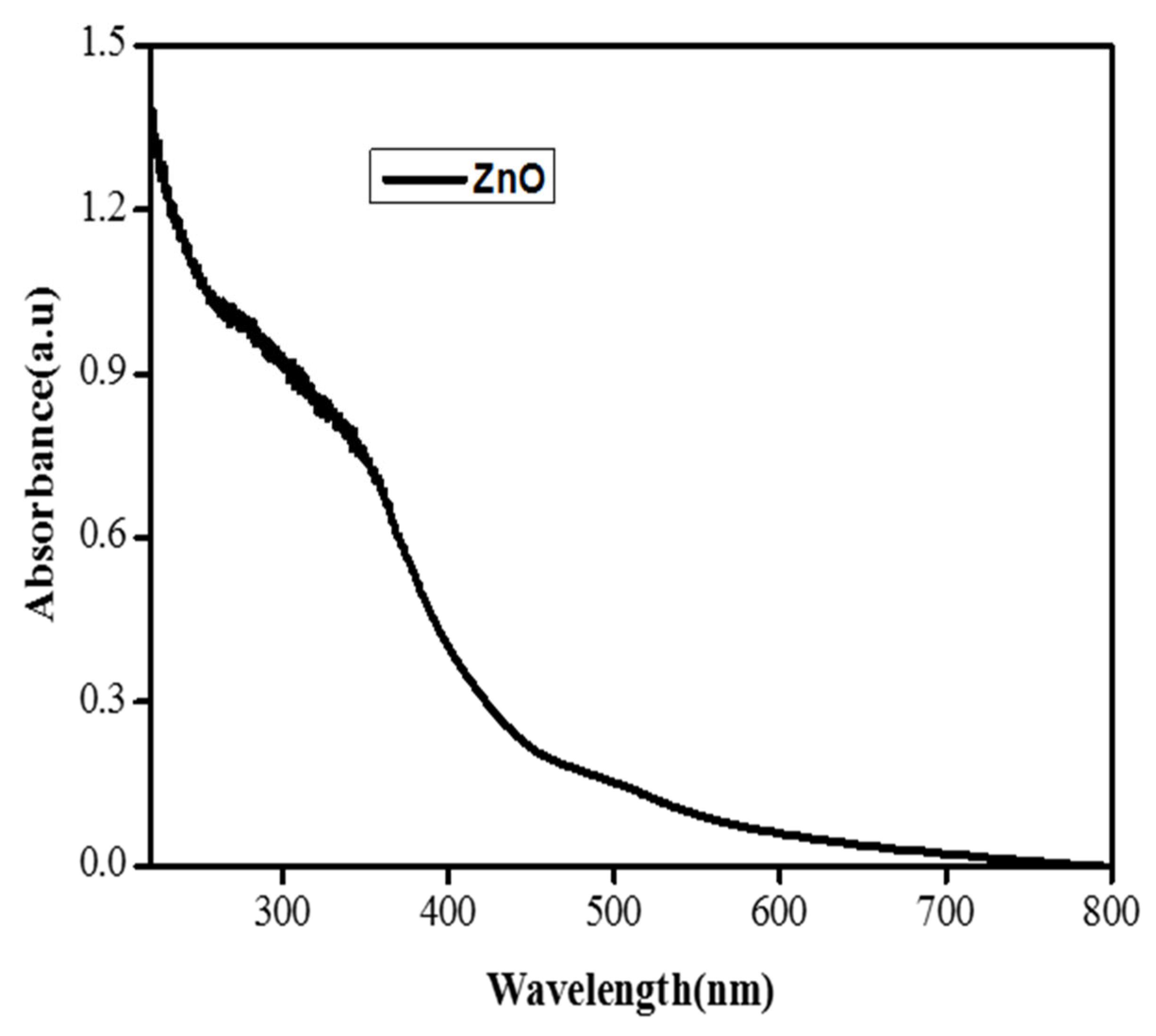
2.4.1. UV-Vis Spectrophotometry Analysis
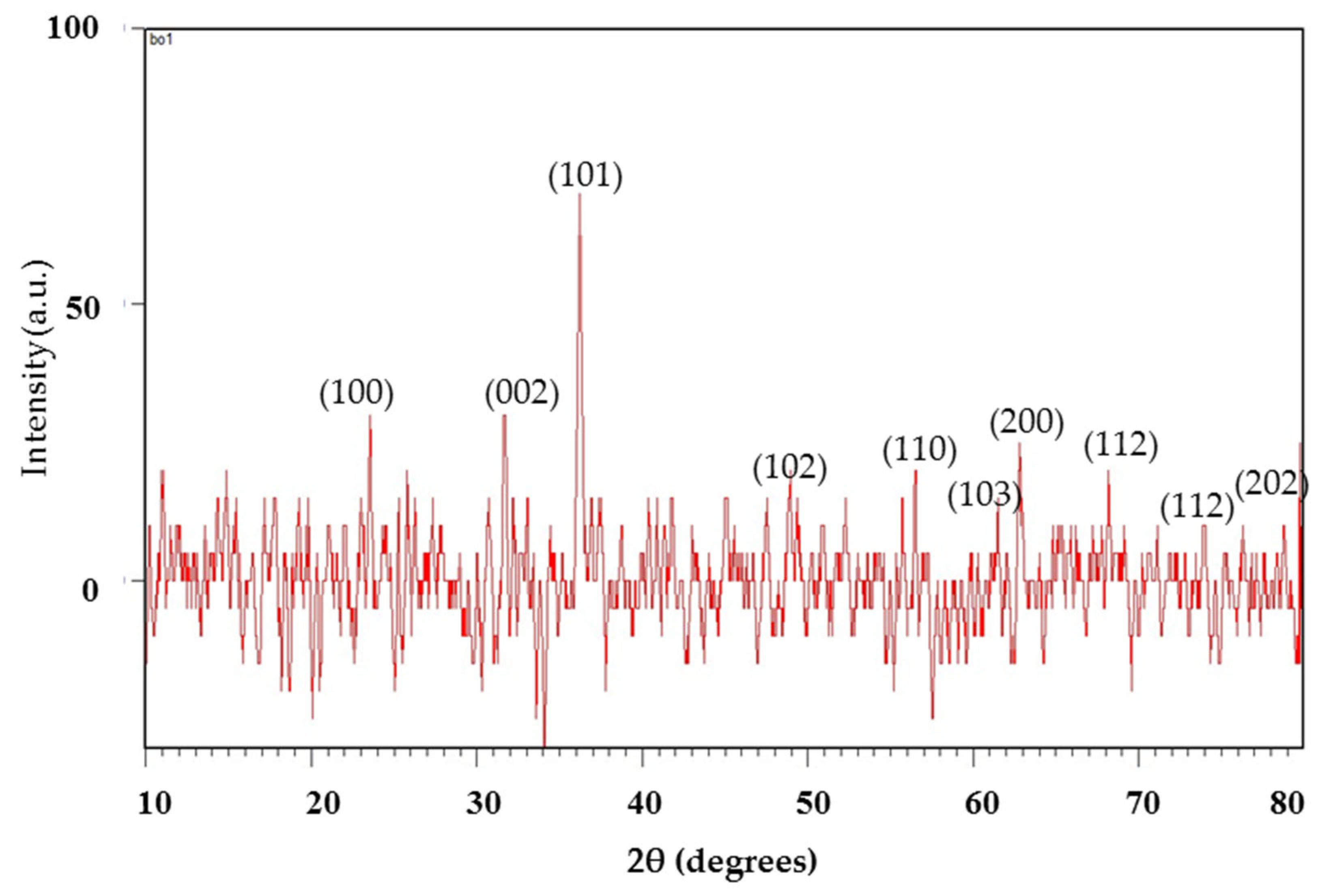
2.4.2. XRD Analysis
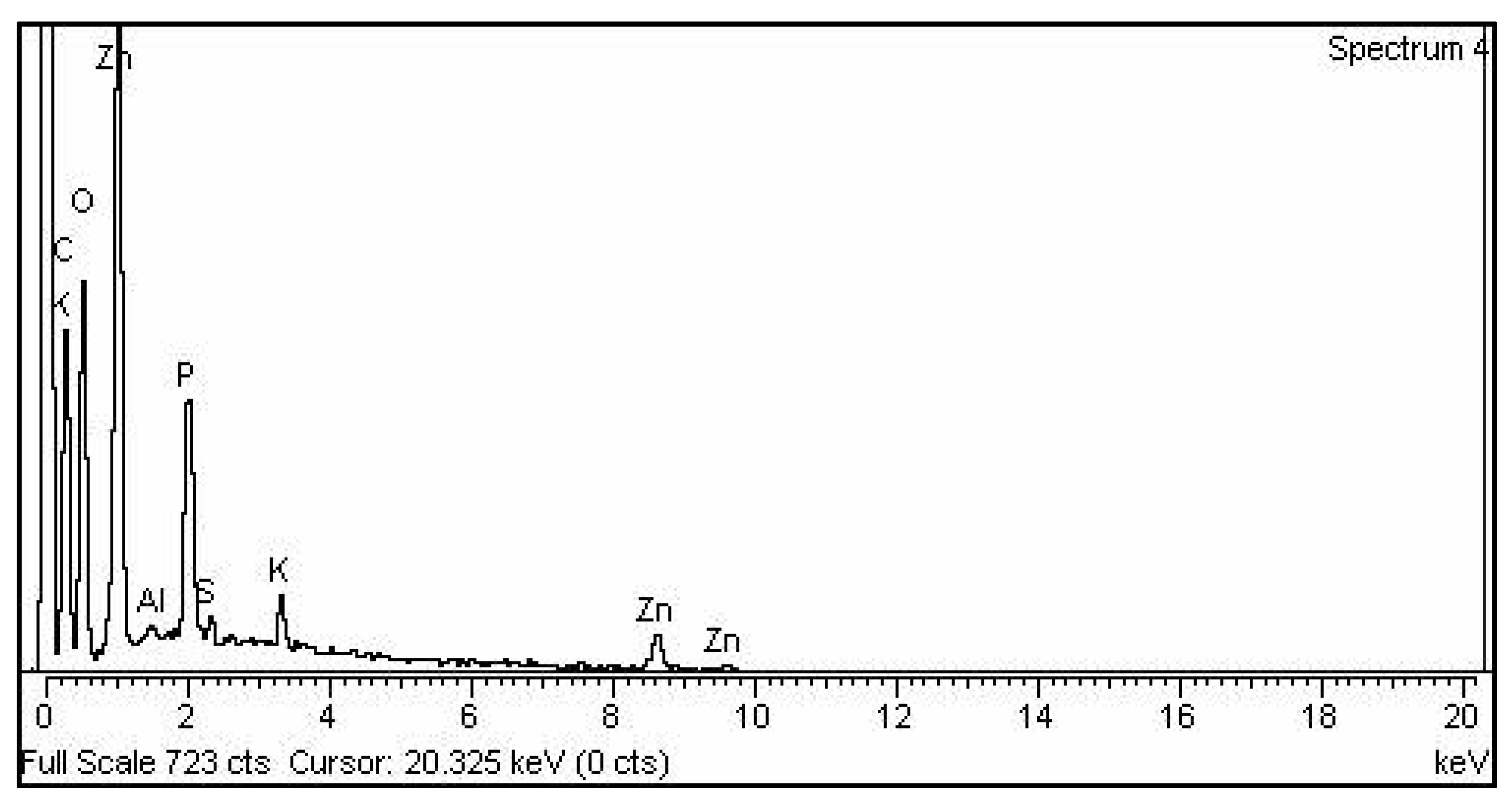
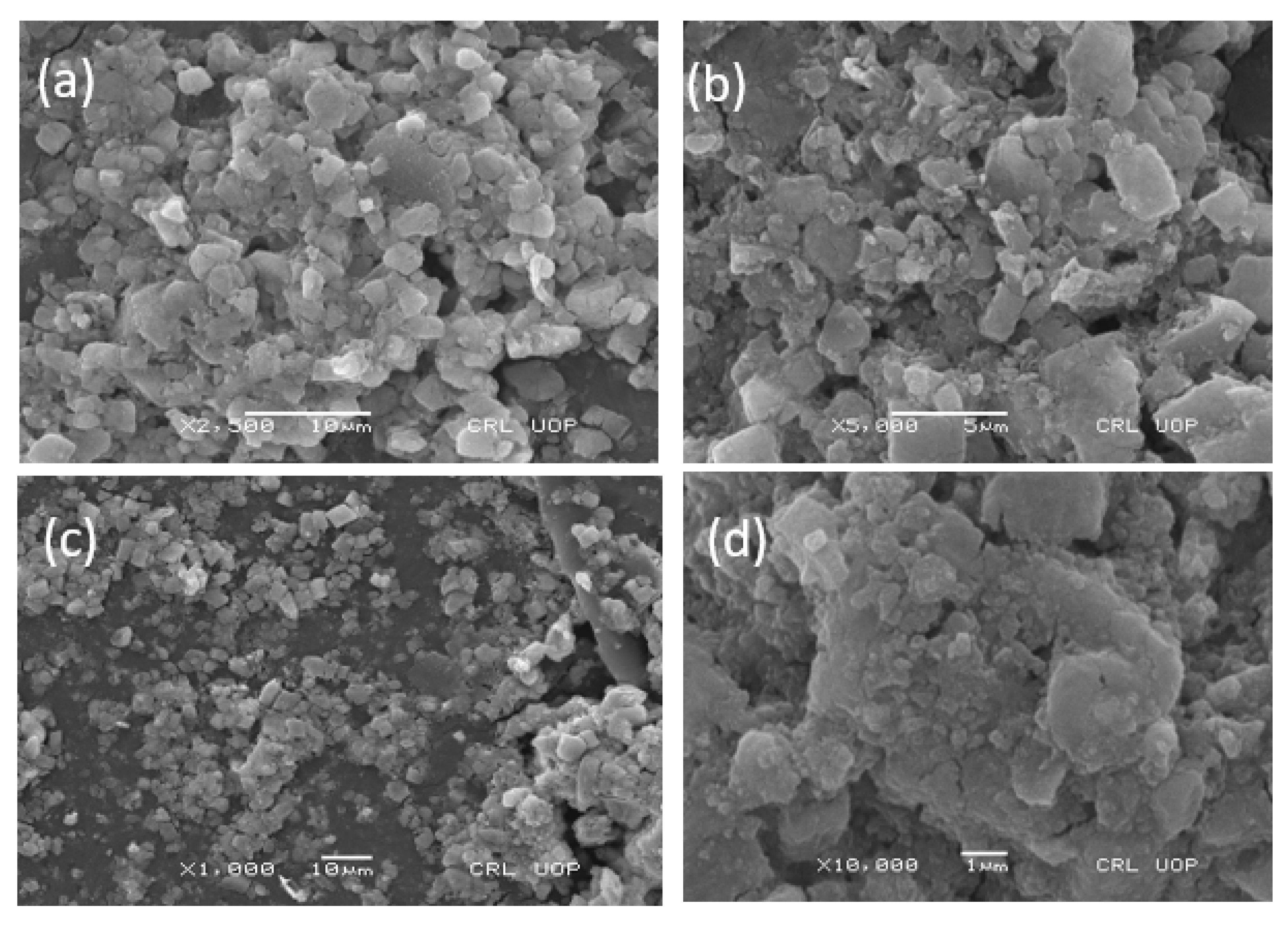
2.4.3. SEM and EDX
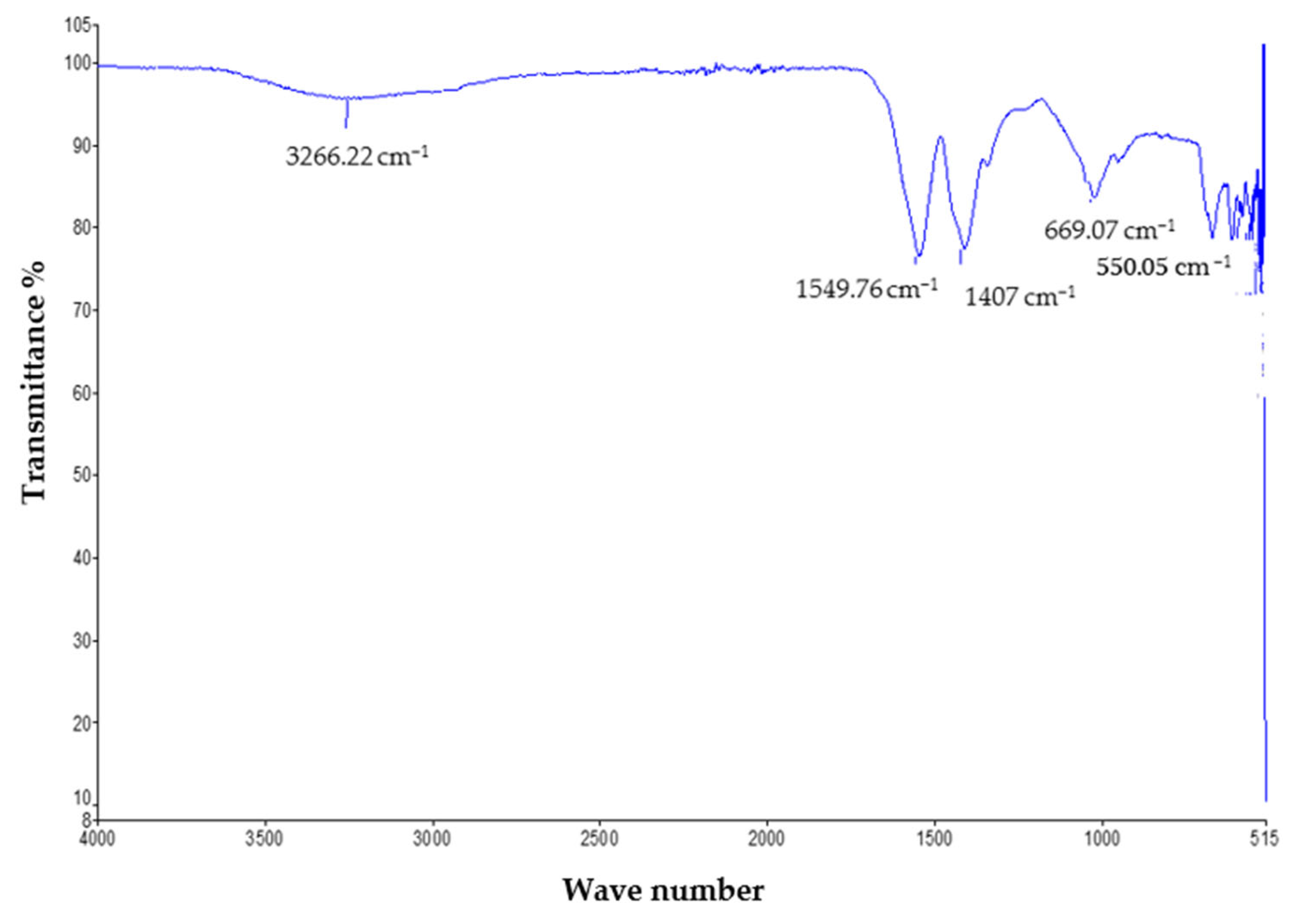
2.4.4. FTIR Analysis
2.5. Biological Application
2.5.1. Antifungal Activity of Zinc Oxide NPs
2.5.2. Antibacterial Activity
2.5.3. Biocompatibility
2.5.4. Anti-Leishmanial Activity
3. Results and Discussion
3.1. XRD Study
3.2. UV Analysis
3.3. FTIR Study
3.4. EDX Analysis
3.5. SEM Study
3.6. Biological Application
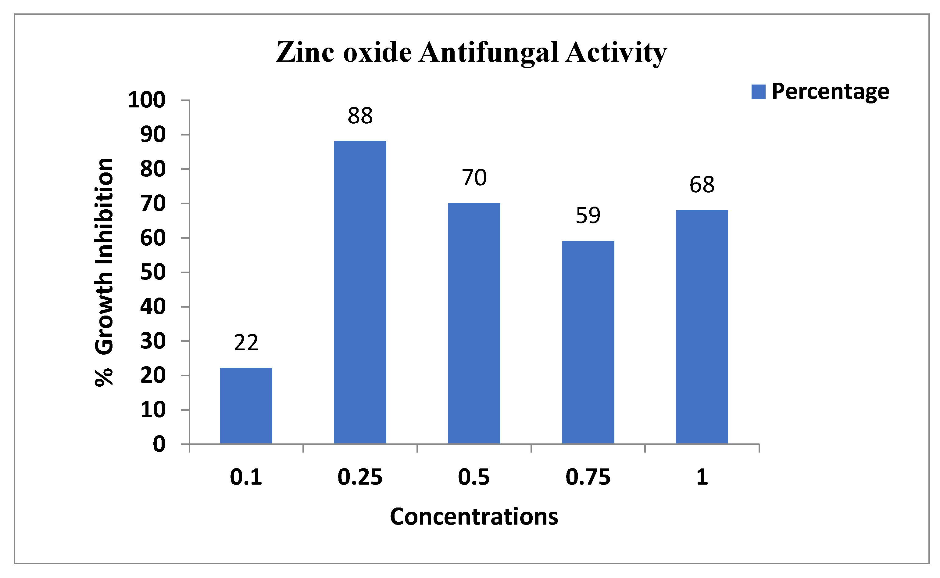
3.6.1. Antifungal Activity
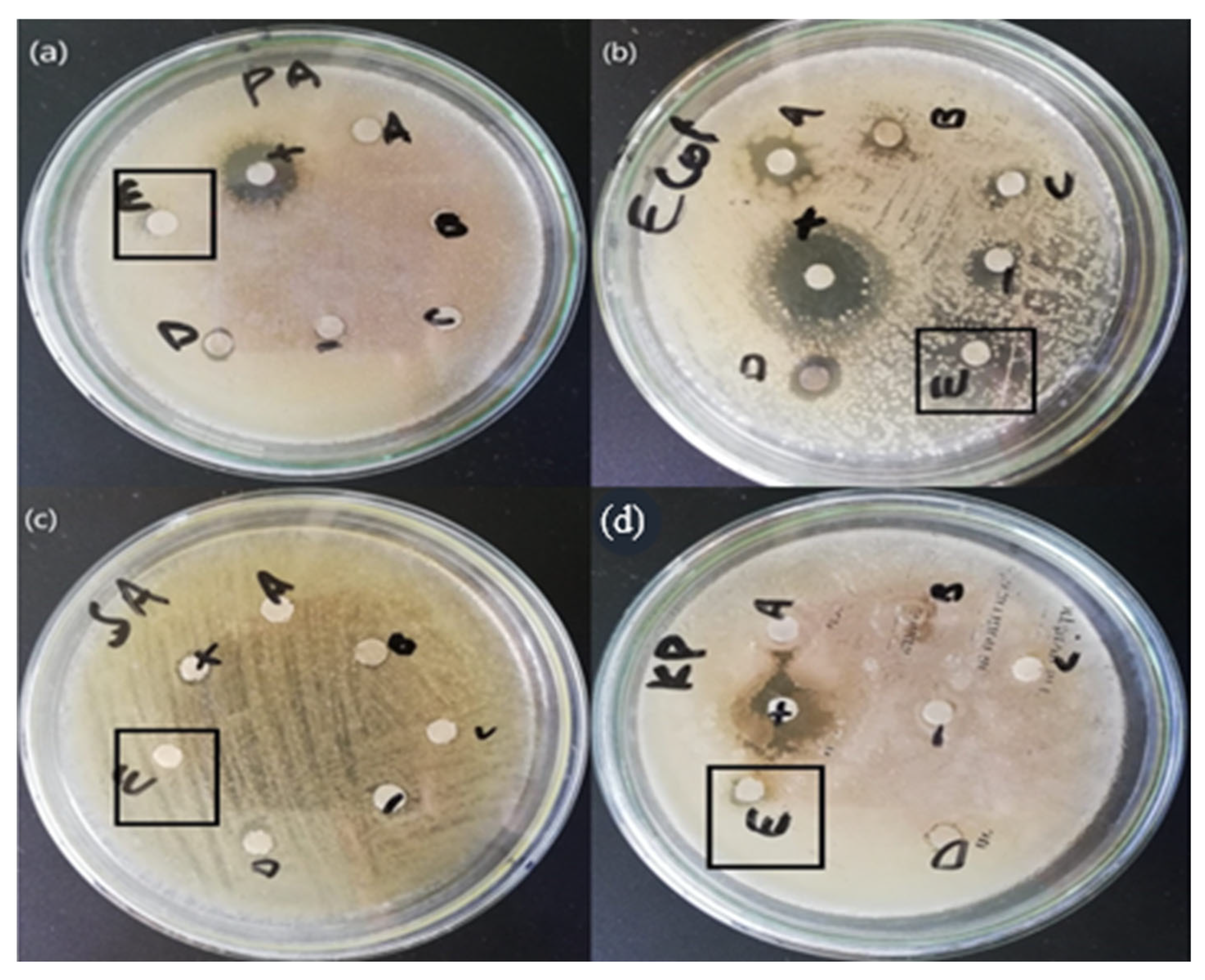
3.6.2. Antibacterial Activity
3.6.3. Biocompatibility
3.6.4. Antileishmanial Activity
4. Conclusions
Author Contributions
Funding
Conflicts of Interest
References
- Albrecht, M.A.; Evans, C.W.; Raston, C.L. Green chemistry and the health implications of nanoparticles. Green Chem. 2006, 8, 417–432. [Google Scholar] [CrossRef]
- Aula, M.; Leppänen, A.; Roininen, J.; Heikkinen, E.-P.; Vallo, K.; Fabritius, T.; Huttula, M. Characterization of process conditions in industrial stainless steelmaking electric arc furnace using optical emission spectrum measurements. Metall. Mater. Trans. 2014, 45, 839–849. [Google Scholar] [CrossRef]
- Balashanmugam, P.; Santhosh, S.; Giyaullah, H.; Balakumaran, M.; Kalaichelvan, P. Mycosynthesis, characterization and antibacterial activity of silver nanoparticles from Microporus xanthopus: A macro mushroom. Int. J. Innov. Res. Sci. Eng. Technol. 2013, 2, 6262–6270. [Google Scholar]
- Brindhadevi, K.; Samuel, M.S.; Verma, T.N.; Vasantharaj, S.; Sathiyavimal, S.; Saravanan, M.; Duc, P.A. Zinc oxide nanoparticles (ZnONPs)-induced antioxidants and photocatalytic degradation activity from hybrid grape pulp extract (HGPE). Agric. Biotechnol. 2020, 28, 101730. [Google Scholar] [CrossRef]
- Chin, S.F.; Azman, A.; Pang, S.C. Size controlled synthesis of starch nanoparticles by a microemulsion method. J. Nanomater. 2014, 22, 3388. [Google Scholar] [CrossRef]
- Dheyab, M.A.; Owaid, M.N.; Rabeea, M.A.; Aziz, A.A.; Jameel, M.S. Mycosynthesis of gold nanoparticles by the Portabello mushroom extract, Agaricaceae, and their efficacy for decolorization of Azo dye. Environ. Nanotechnol. Monit. Manag. 2020, 14, 100312. [Google Scholar] [CrossRef]
- Faisal, S.; Khan, M.A.; Jan, H.; Shah, S.A.; Shah, S.; Rizwan, M.; Akbar, M.T. Edible mushroom (Flammulina velutipes) as biosource for silver nanoparticles: From synthesis to diverse biomedical and environmental applications. Nanotechnology 2020, 32, 065101. [Google Scholar] [CrossRef] [PubMed]
- He, L.; Liu, Y.; Mustapha, A.; Lin, M. Antifungal activity of zinc oxide nanoparticles against Botrytis cinerea and Penicillium expansum. Microbiol. Res. 2011, 166, 207–215. [Google Scholar] [CrossRef]
- Mohamed, Y.; Azzam, A.; Amin, B.; Safwat, N. Mycosynthesis of iron nanoparticles by Alternaria alternata and its antibacterial activity. Afr. J. Biotechnol. 2015, 14, 1234–1241. [Google Scholar] [CrossRef]
- Naeem, G.A.; Jaloot, A.S.; Owaid, M.N.; Muslim, R.F. Green synthesis of gold nanoparticles from coprinus comatus, agaricaceae, and the effect of ultraviolet irradiation on their characteristics. WJST 2021, 18, 9396. [Google Scholar] [CrossRef]
- Hengstmengel, J. Notes on Hymenoscyphus 3: On the nomenclature of Hymenoscyphus subcarneus (Ascomycota, Helotiales). Mycotaxon 2009, 107, 267–276. [Google Scholar] [CrossRef]
- Rajabi, H.R.; Naghiha, R.; Kheirizadeh, M.; Sadatfaraji, H.; Mirzaei, A.; Alvand, Z.M. Microwave assisted extraction as an efficient approach for biosynthesis of zinc oxide nanoparticles: Synthesis, characterization, and biological properties. Mater. Sci. Eng. C 2017, 78, 1109–1118. [Google Scholar] [CrossRef] [PubMed]
- Geiser, D.M.; Aoki, T.; Bacon, C.W.; Baker, S.E.; Bhattacharyya, M.K.; Brandt, M.E.; Zhang, N. One fungus, one name: Defining the genus Fusarium in a scientifically robust way that preserves longstanding use. Phytopathology 2013, 103, 400–408. [Google Scholar] [CrossRef] [PubMed]
- Santa Izabel, T.S.; Cruz, A.C.R.; Gusmão, L.F.P. Conidial fungi from the semi-arid Caatinga biome of Brazil. Ellisembiopsis gen. nov., new variety of Sporidesmiella and some notes on Sporidesmium complex. Mycosphere 2013, 4, 156–163. [Google Scholar] [CrossRef]
- Ahmed, K.; Valeem, E.E.; Khan, M.A. Biosynthesis of alpha amylase from Aspergillus Fumigatus (Fresenius 1863) in submerged fermentation. PJBT 2015, 12, 87–92. [Google Scholar]
- Zhao, K.; Wu, G.; Feng, B.; Yang, Z.L. Molecular phylogeny of Caloboletus (Boletaceae) and a new species in East Asia. Mycol. Prog. 2014, 13, 1127–1136. [Google Scholar] [CrossRef]
- Burdsall, H.H., Jr.; Larsen, M.J. Lazulinospora, a new genus of Corticiaceae, and a note on Tomentella atrocyanea. Mycologia 1974, 66, 96–100. [Google Scholar] [CrossRef]
- Vinokurova, N.G.; Baskunov, B.P.; Zelenkova, N.F.; Arinbasarov, M.U. The alkaloids of Penicillium aurantiogriseum Dierckx (1901) var. aurantiogriseum VKM F-1298. Microbiology 2004, 73, 414–419. [Google Scholar] [CrossRef]
- Thompson, F.; Gomez-Gil, B. International Committee on Systematics of Prokaryotes Subcommittee on the taxonomy of Aeromonadaceae, Vibrionaceae and related organisms Minutes of the meeting, 13 November 2017, Chicago, USA. Int. J. Syst. Evol. Microbiol. 2018, 68, 2111–2112. [Google Scholar] [CrossRef]
- Sharmila, G.; Muthukumaran, C.; Sandiya, K.; Santhiya, S.; Pradeep, R.S.; Kumar, N.M.; Thirumarimurugan, M. Biosynthesis, characterization, and antibacterial activity of zinc oxide nanoparticles derived from Bauhinia tomentosa leaf extract. J. Nanostruct. Chem. 2018, 8, 293–299. [Google Scholar] [CrossRef]
- Sumanth, B.; Lakshmeesha, T.R.; Ansari, M.A.; Alzohairy, M.A.; Udayashankar, A.C.; Shobha, B.; Almatroudi, A. Mycogenic synthesis of extracellular zinc oxide nanoparticles from Xylaria acuta and its nanoantibiotic potential. Int. J. Nanomed. 2020, 15, 8519. [Google Scholar] [CrossRef]
- Vithiya, K.; Sen, S. Biosynthesis of nanoparticles. Int. J. Pharm. Sci. 2011, 2, 2781. [Google Scholar]
- Rani, V.; Verma, Y.; Rana, K.; Rana, S.V.S. Zinc oxide nanoparticles inhibit dimethylnitrosamine induced liver injury in rat. Chem. Biol. Interact. 2018, 295, 84–92. [Google Scholar] [CrossRef]
- Huang, X.; Zheng, X.; Xu, Z.; Yi, C. ZnO-based nanocarriers for drug delivery application: From passive to smart strategies. Int. J. Pharm. 2017, 534, 190–194. [Google Scholar] [CrossRef]
- Jeng, H.A.; Swanson, J. Toxicity of metal oxide nanoparticles in mammalian cells. J. Environ. Sci. Health A 2006, 41, 2699–2711. [Google Scholar] [CrossRef]
- Wojnarowicz, J.; Chudoba, T.; Lojkowski, W. A review of microwave synthesis of zinc oxide nanomaterials: Reactants, process parameters and morphologies. Nanomaterials 2020, 10, 1086. [Google Scholar] [CrossRef]
- Zubair, M.S.; Munis, M.F.H.; Alsudays, I.M.; Alamer, K.H.; Haroon, U.; Kamal, A.; Ali, M.; Ahmed, J.; Ahmad, Z.; Attia, H. First Report of Fruit Rot of Cherry and Its Control Using Fe2O3 Nanoparticles Synthesized in Calotropis procera. Molecules 2022, 27, 4461. [Google Scholar] [CrossRef]
- Bavarsad, M.; Farrokhi-Nejad, R. Identification of Trichoderma Species Using Partial Sequencing of nrRNA and tef1α Genes with Report of Trichoderma capillare in Iran Mycoflore. Majallah-I Ḥifāẓat-I Giyāhān 2018, 113, 5874. [Google Scholar]
- Bocci, F.; Gearhart-Serna, L.; Boareto, M.; Ribeiro, M.; Ben-Jacob, E.; Devi, G.R.; Jolly, M.K. Toward understanding cancer stem cell heterogeneity in the tumor microenvironment. Proc. Natl. Acad. Sci. USA 2019, 116, 148–157. [Google Scholar] [CrossRef]
- Han, M.L.; Vlasak, J.; Cui, B.K. Daedalea americana sp. nov.(Polyporales, Basidiomycota) evidenced by morphological characters and phylogenetic analysis. Phytotaxa 2015, 204, 277–286. [Google Scholar] [CrossRef]
- Rajabi, M.; Mousa, S.A. The role of angiogenesis in cancer treat. Biomedicines 2017, 5, 34. [Google Scholar] [CrossRef]
- Rabeea, M.A.; Owaid, M.N.; Aziz, A.A.; Jameel, M.S.; Dheyab, M.A. Mycosynthesis of gold nanoparticles using the extract of Flammulina velutipes, Physalacriaceae, and their efficacy for decolorization of methylene blue. J. Environ. Chem. Eng. 2020, 8, 103841. [Google Scholar] [CrossRef]


| Element | Weight% | Atomic% |
|---|---|---|
| C k | 24.30 | 52.68 |
| O K | 13.28 | 21.62 |
| P K Cu K | 18 2.28 | 1.51 0.94 |
| Zn k | 58.34 | 23.24 |
| Concentration | Control | % Inhibition |
|---|---|---|
| 0.1 mg | 9 cm | 22% |
| 0.25 mg | 9 cm | 88% |
| 0.5 mg | 9 cm | 70% |
| 0.75 mg | 9 cm | 75% |
| 1.0 mg | 9 cm | 68% |
| Pathogenic Bacteria | ZnO NPs 4 mg/mL | Positive Control |
|---|---|---|
| E. coli | 10 ± 0.34 | 20 ± 0.39 |
| P. aeuroginose | 7 ± 0.40 | 15 ± 0.35 |
| S. aeururs | 7 ± 0.35 | 8 ± 0.37 |
| K. pneumonia | 7 ± 0.42 | 20 ± 0.34 |
| S. No | Conc mg/mL | % Hemolysis |
|---|---|---|
| 1 | 400 | 0.048 ± 2 |
| 2 | 200 | 0.064 ± 4 |
| 3 | 100 | 0.047 ± 6 |
| 4 | 50 | 0.047 ± 8 |
| Control | 0.210 |
| Con (ug/m) | Promastigote Inhibition | Amastigote | IC50 (ug/mL) | IC50 (ug/mL) |
|---|---|---|---|---|
| 400 | 60.21 | 54.78 | 270 Amas | 220 Prom |
| 200 | 49.33 | 44.0 | ||
| 100 | 37.33 | 31.45 | ||
| 50 | 23.10 | 18.19 |
Disclaimer/Publisher’s Note: The statements, opinions and data contained in all publications are solely those of the individual author(s) and contributor(s) and not of MDPI and/or the editor(s). MDPI and/or the editor(s) disclaim responsibility for any injury to people or property resulting from any ideas, methods, instructions or products referred to in the content. |
© 2023 by the authors. Licensee MDPI, Basel, Switzerland. This article is an open access article distributed under the terms and conditions of the Creative Commons Attribution (CC BY) license (https://creativecommons.org/licenses/by/4.0/).
Share and Cite
Kamal, A.; Saba, M.; Ullah, K.; Almutairi, S.M.; AlMunqedhi, B.M.; Ragab abdelGawwad, M. Mycosynthesis, Characterization of Zinc Oxide Nanoparticles, and Its Assessment in Various Biological Activities. Crystals 2023, 13, 171. https://doi.org/10.3390/cryst13020171
Kamal A, Saba M, Ullah K, Almutairi SM, AlMunqedhi BM, Ragab abdelGawwad M. Mycosynthesis, Characterization of Zinc Oxide Nanoparticles, and Its Assessment in Various Biological Activities. Crystals. 2023; 13(2):171. https://doi.org/10.3390/cryst13020171
Chicago/Turabian StyleKamal, Asif, Malka Saba, Khetab Ullah, Saeedah Musaed Almutairi, Bandar M. AlMunqedhi, and Mohamed Ragab abdelGawwad. 2023. "Mycosynthesis, Characterization of Zinc Oxide Nanoparticles, and Its Assessment in Various Biological Activities" Crystals 13, no. 2: 171. https://doi.org/10.3390/cryst13020171
APA StyleKamal, A., Saba, M., Ullah, K., Almutairi, S. M., AlMunqedhi, B. M., & Ragab abdelGawwad, M. (2023). Mycosynthesis, Characterization of Zinc Oxide Nanoparticles, and Its Assessment in Various Biological Activities. Crystals, 13(2), 171. https://doi.org/10.3390/cryst13020171