Article

H2O2-Assisted Sunlight Photocatalytic Degradation of Basic Fuchsin Using Green-Synthesized ZnO Nanowires


1
Laboratory for Research in Medicine and Sustainable Development ReMeDD, Faculty of Process Engineering, University of Salah Boubnider, Constantine 25000, Algeria
2
Laboratory of Structures, Properties and Inter Atomic Interactions, Science of Matter Department, Faculty of Sciences and Technology, Abbes Laghrour University, Khenchela 40000, Algeria
3
Department of Physics, Faculty of Exact Science, University of Echahid Hamma Lakhdar, El Oued 39000, Algeria
4
College of Engineering, Imam Mohammad Ibn Saud Islamic University (IMSIU), Riyadh 11432, Saudi Arabia
5
Ecole Nationale Supérieure de Chimie de Rennes, CNRS, ISCR, University Rennes, F-35000 Rennes, France
6
School of Engineering, Merz Court, Newcastle University, Newcastle upon Tyne NE1 7RU, UK
7
Scientific and Technical Center of Research in Physical and Chemical Analysis CRAPC, BP 384, Bou-Ismail 42004, Algeria
8
Laboratory of Biomaterials and Transport Phenomena (LBMTP), University Yahia Fares, Medea 26000, Algeria
*
Authors to whom correspondence should be addressed.
Catalysts 2026, 16(2), 193; https://doi.org/10.3390/catal16020193
Submission received: 2 February 2026 / Revised: 11 February 2026 / Accepted: 14 February 2026 / Published: 18 February 2026

Abstract

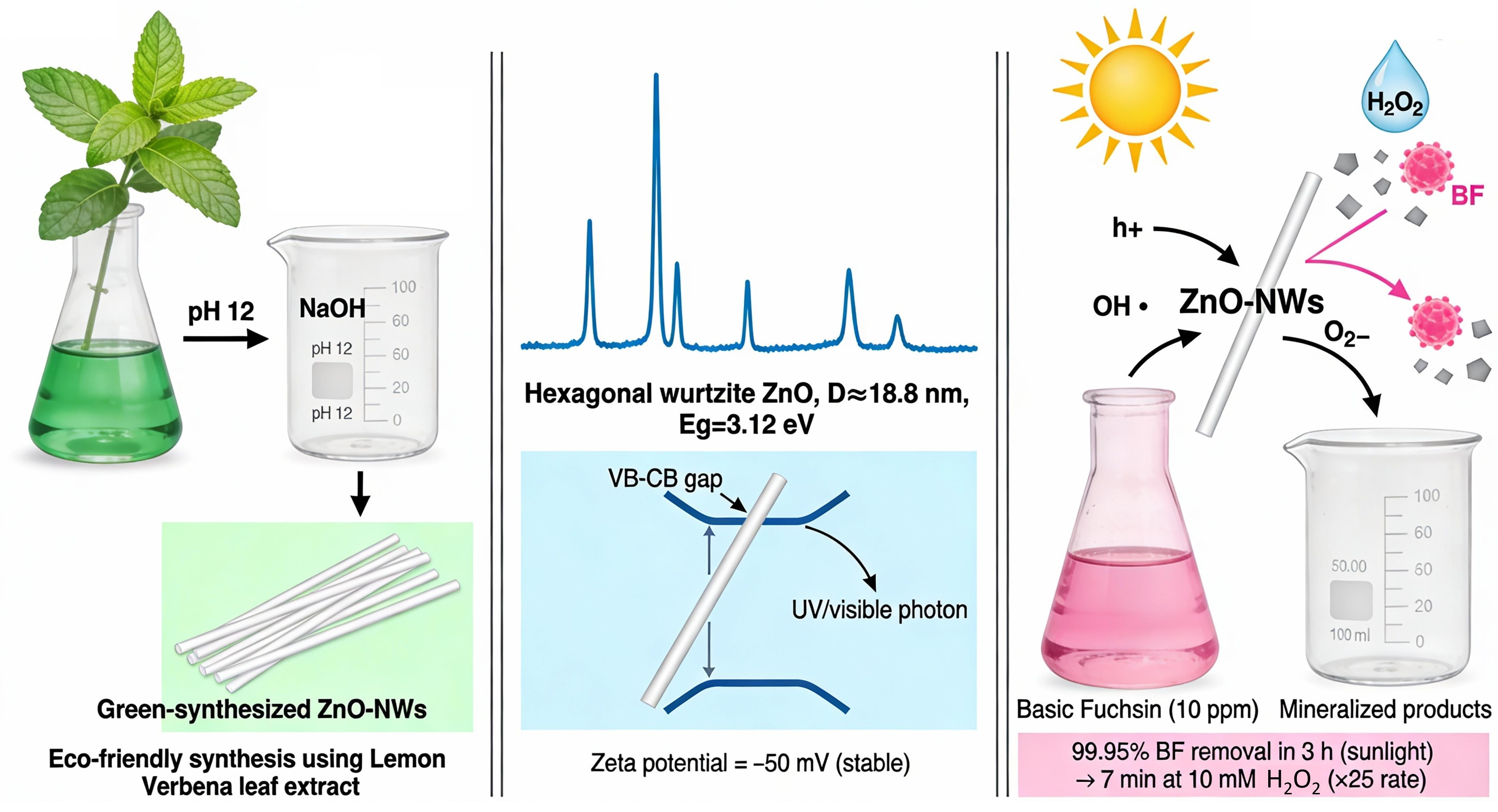
The efficient removal of toxic dyes from wastewater remains a major environmental challenge. In this study, we report a green and facile one-pot synthesis of zinc oxide nanowires (ZnO-NWs) using lemon verbena leaf extract as a sustainable capping and stabilizing agent. The extract played a vital role in directing the 1D growth of the wurtzite hexagonal structure. Characterization confirmed a band gap of 3.12 eV and the characteristic Zn-O stretching at 375 cm−1. Photocatalytic activity tests using 20 mg of biosynthesized ZnO-NWs demonstrated excellent degradation performance. A rate constant of 0.0067 min−1 was achieved, with 99.95% degradation of Basic Fuchsin under natural sunlight for 3 h. Active species analysis highlighted the crucial roles of holes (h+), superoxide radicals (O2), and hydroxyl radicals (•OH). Notably, the addition of 10 mM H2O2 produced a powerful synergistic effect, reducing the degradation time from 3 h to only 7 min and increasing the reaction rate by approximately 25-fold. These findings highlight the potential of biosynthesized ZnO-NWs as highly efficient, rapid, and sustainable photocatalysts for environmental remediation.

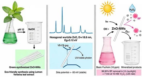
Graphical Abstract

1. Introduction

Synthetic dyes represent a major class of hazardous organic pollutants widely released into aquatic environments [1]. Among these pollutants, Basic Fuchsin (BF), also known as rosaniline hydrochloride or Basic Violet, is a cationic triarylmethane dye extensively used in textile dyeing, biological staining, and paper-printing applications [2]. However, BF exhibits high toxicity, poor biodegradability, and strong bioaccumulation potential and has been reported to induce genotoxic, neurotoxic, and carcinogenic effects [3]. Consequently, the efficient removal of BF from contaminated water is of significant environmental and public health importance. To address this challenge, various conventional wastewater-treatment methods, including coagulation–flocculation, membrane separation, adsorption, precipitation, ion exchange, and ozonation, have been developed [4]. Nevertheless, these techniques often suffer from several drawbacks, such as high operational costs, complex procedures, secondary pollution, and limited efficiency toward chemically stable dyes [5]. Therefore, the development of efficient, sustainable, and environmentally benign remediation strategies remains highly desirable.
Photocatalysis is a particularly promising advanced oxidation process (AOP) to overcome the drawbacks of conventional methods [6]. Photocatalytic nanomaterials represent a distinct class of nanostructured materials capable of initiating and accelerating chemical reactions under light irradiation, a phenomenon referred to as photocatalysis [7]. Because of its huge active surfaces, nanomaterials, with ranged diameters from 1 to 100 nanometers (nm), comprise a new field in nanotechnology and nanoscience that can offer solutions to the environmental and technological problems in the fields of water, medicine, solar energy conversion, and catalysis [8], regarding their unique properties, including size, composition, and morphology [9].
ZnO nanoparticles (ZnO NPs) have attracted extensive interest in electronics, sensors, environmental protection, and biomedical applications [10]. Their ability to form diverse nanostructures, including nanowires, nanorods, and nanosheets, enables the fine-tuning of surface and electronic properties. As a result, ZnO exhibits excellent photocatalytic activity, chemical stability, and strong resistance to matrix effects in complex aqueous environments [11,12].
Green synthesis is a relatively new area of nanotechnology that offers a substantial economic and minimal environmental effect [13]. It relies on non-toxic, renewable, and biocompatible substances [14], particularly plant-derived phytochemicals (flavonoids, tannins, alkaloids, proteins, amino acids) extracted using eco-friendly solvents such as distilled water [15]. The biosynthesis of ZnO NPs by plants such as olives leaf extract [16], Hibiscus sabdariffa L [17], Barleria buxifolia leaf extract [18], etc., has been reported in the literature [19].
In this work, lemon verbena (Aloysia citrodora) leaf extract was used as a reducing and stabilizing agent for the green synthesis of ZnO nanowires. This plant was selected due to its rich phytochemical composition, rapid growth, and high productivity (up to three harvests per year), making it a sustainable and renewable resource [20]. The extract is rich in bioactive compounds, including phenylpropanoids and glycosylated flavones [21], which can donate electrons to reduce Zn2+ ions into Zn(OH)2 intermediates. These intermediates subsequently undergo dehydration and oxidation to form ZnO nanowires upon thermal treatment. In addition to its reducing role, the extract acts as a stabilizing agent, preventing particle aggregation and controlling the morphology of the nanostructures [22].
ZnO exhibits strong photocatalytic activity, which can be further enhanced by the addition of H2O2 through the generation of extra •OH radicals that accelerate pollutant degradation [23]. Although the individual concepts of green-synthesized ZnO nanoparticles and ZnO/H2O2 photocatalysis are well documented, their rational integration into a sustainable, low-cost, and solar-driven system based on a medicinal plant extract remains scarcely explored. In particular, the use of lemon verbena extract for the controlled green synthesis of one-dimensional ZnO nanowires and the evaluation of their synergistic performance with low-dose H2O2 toward the degradation of highly toxic Basic Fuchsin dye have not yet been reported.
Therefore, the present work aims to develop a sustainable ZnO nanowire photocatalyst via a green synthesis route using lemon verbena leaf extract and to investigate the synergistic role of H2O2 in enhancing sunlight-driven photocatalytic degradation of the highly toxic Basic Fuchsin dye. To the best of our knowledge, no previous study has reported a green-engineered ZnO nanowire/H2O2 hybrid system capable of achieving such high photocatalytic efficiency under natural sunlight. Notably, the addition of an ultra-low H2O2 concentration (1 mM) leads to remarkable acceleration of the degradation kinetics, achieving rapid and nearly complete dye removal. This outstanding enhancement demonstrates the environmental relevance, energy efficiency, and practical applicability of the ZnO/H2O2 system as a promising alternative to conventional advanced oxidation processes for wastewater treatment.

2. Results and Discussion

2.1. Structural Properties

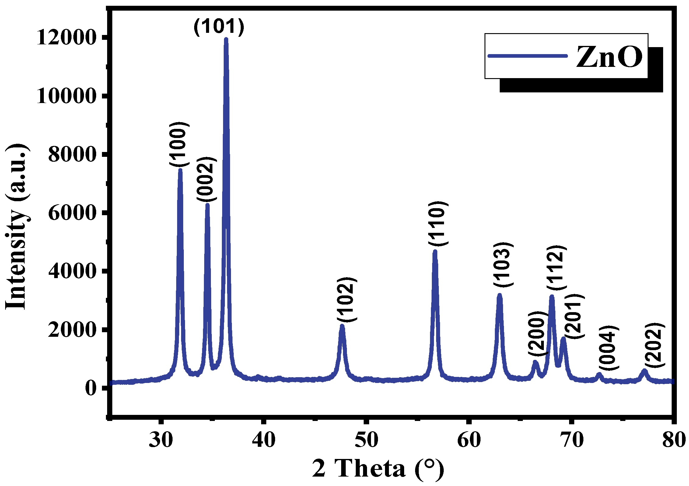
XRD was used to determine the crystalline structure of the synthesized materials. Figure 1 shows a typical XRD pattern of ZnO biosynthesized using plant extract and annealed at 600 °C. The diffraction was measured in the angular range of 2θ = 20–80°. The crystal unit cell was found to be a hexagonal wurtzite structure, according to the presence of the main peaks located at 2θ = 31.90, 34.50, 36.38, 47.64, 56.68, 62.93, 66.45, 68.04, 69.21, 72.71, and 77.20°, corresponding to the following (100), (002), (101), (102), (110), (103), (200), (112), (201), (004), and (202) planes, respectively. It is in good agreement with the ICSD-067848 for the Wurtzite structure (P 63 mc) of ZnO, which is the most thermodynamically stable at room temperature.
Furthermore, at the detection limit, no impurity peaks were observed, indicating the high purity of the prepared ZnO-NWs.
The lattice parameters a and c of the hexagonal ZnO structure were calculated using the interplanar spacings of several diffraction planes and the hexagonal wurtzite relation Equation (1). Specifically, the a parameter was determined from the (100) plane, while the c parameter was obtained from the (002) plane, using equation [24].
d h k l = 4 h 2 + h k + k 2 3 a 2 + l 2 c 2 1 2
where the interplanar spacing dhkl was obtained from Bragg’s law. The average experimental lattice parameters were found to be aexp = 0.324 nm, cexp = 0.519 nm, and a ratio cexp/aexp = 1.6. These values are in agreement with those of the wurtzite ZnO structure (a = 0.3249 nm and c = 0.5207 nm) [15].
Due to the high structural anisotropy of the nanowires, their physical dimensions (diameter and length) were further investigated using SEM analysis rather than XRD-based estimations to ensure better accuracy.

2.2. Morphological and Compositional Analysis

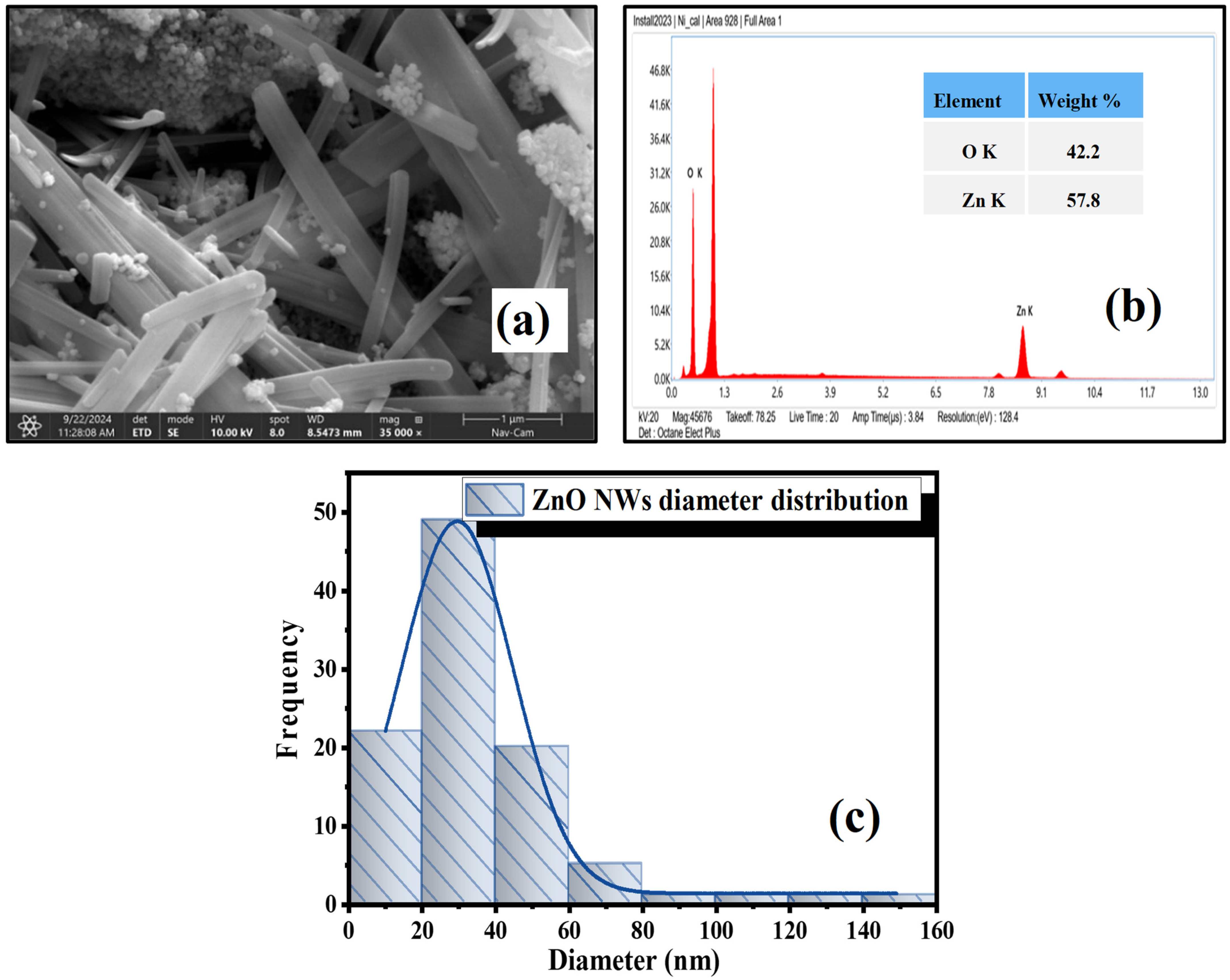
The surface morphology of the biosynthesized ZnO was analyzed using a scanning electron microscope (SEM) as shown in Figure 2a. The results clearly reveal that the as-prepared ZnO from an aqueous extract of lemon verbena leaf extracts obtained from an aqueous lemon verbena leaf extract exhibits an irregular morphology with both wire-like and spherical particles, with nanowires (NWs) being the dominant feature. Image J software (version 1.54g, National Institutes of Health, Bethesda, MD, USA) was used to determine the nanowires’ diameter, which ranges from 20 to 160 nm. The elemental composition of the nanostructures was verified via EDX analysis, as shown in Figure 2b. The EDX spectrum confirms the high purity of the synthesized ZnO-NWs, as zinc (Zn) and oxygen (O) are the dominant detected elements. The minor unlabeled peaks are mainly attributed to the carbon conductive tape used for sample mounting and instrumental background [25]. No additional peaks related to external chemical impurities were detected, confirming the successful green synthesis and high chemical purity of the prepared nanowires.
The average diameter of the as-synthesized ZnO nanostructures was estimated based on Gaussian fitting of the histogram data in Figure 2c and was found to be approximately 25.5 ± 10 nm. This value represents the most frequent diameter within the distribution, although the sample remains polydisperse. While both wires and spheres coexist, the XRD analysis (Figure 1) confirms the presence of a single hexagonal wurtzite phase, suggesting that both morphologies are chemically identical ZnO resulting from anisotropic growth.
As clearly seen, a large number of wire-like ZnO-NWs is observed with lengths reaching several micrometers. The smaller nanoparticles within the sample provide a large surface-area-to-volume ratio, which is highly beneficial for surface-dependent applications such as catalysis. The one-dimensional morphology of the ZnO nanowires is intrinsically related to the hexagonal wurtzite crystal structure, which promotes anisotropic growth along the polar c-axis [26].

2.3. UV-Visible Absorption Studies

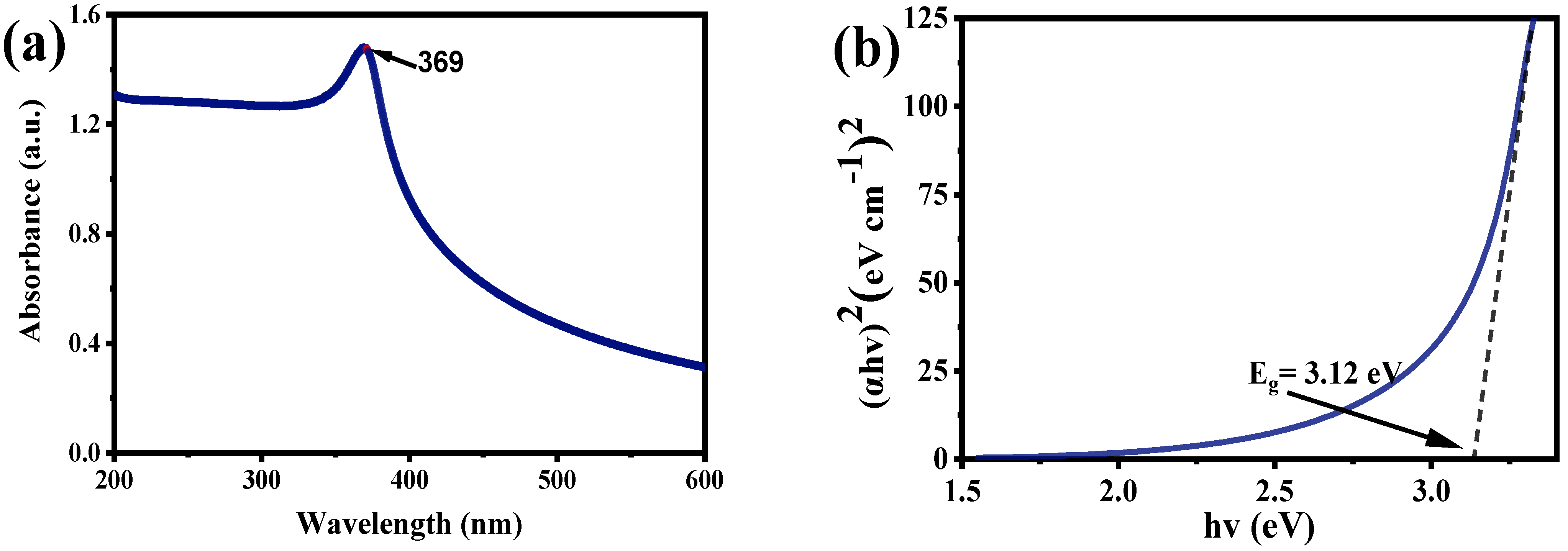
UV-Vis spectroscopy was used to determine the optical characteristics of the prepared samples. Before performing the UV characterization, 10 mg of the prepared nano-powder was initially dispersed in 50 mL of distilled water to produce a monodispersed solution. Following this, the solution was sonicated for 30 min. According to Figure 3, the UV-Vis spectrum of the zinc oxide NWs revealed a single peak at 369 nm, For ZnO NPs, the absorption peak maxima typically falls between 300 and 380 nm [27], which confirmed the synthesis of ZnO NPs via the green route (see Figure 3a). It matches well with the characteristic band of ZnO NPs in the literature [28,29], suggesting the high purity of the obtained monodisperse solution [30].
As described in research [27] the Tauc plot method was used to estimate the sample’s band gap energy (Eg). Its value was deduced using an extrapolation of the straight line to the hv axis (i.e., (αhv = 0) see Figure 3b):
α h v m = A h v       E g
where h is Planck’s constant, v is the frequency, A is the proportionality constant, α is the absorption coefficient, and m is taken equal to 2 due the direct band gap nature of the ZnO semiconductor.
The calculated bandgap was found to be 3.12 eV. The band gap values of pure ZnO nanoparticles are roughly 3.27 eV, according to the literature [31]. A decrease in Eg is expected as a result of using the plant extract since certain components of the extract cover or alter the surface and reduce the band-gap of the nanoparticles [32]. Similar results were observed with Rythrina abyssinica, in agreement with the previously reported ZnO nanoparticle band gap range of 3.10–3.39 eV [33].

2.4. Fourier Transformed Infra-Red Spectroscopy (FTIR)

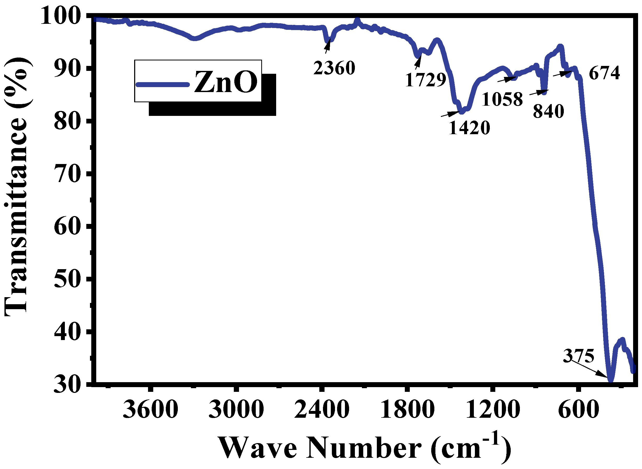
To confirm the production of the ZnO-NWs and identify the type of functional groups in the active compounds responsible for their synthesis, FTIR spectroscopy was performed, and spectra were recorded in the range of 200–4000 cm−1. ZnO NPs exhibited vibrations at 374, 674, 1058, 840, 1420, 1729, and 2360 cm−1 as shown in Figure 4. The absorption band at ~2360 cm−1 is attributed to the asymmetric stretching vibration of CO2, probably originating from atmospheric CO2, in agreement with previous reports [34], where the vibrations at 1420 cm−1 are attributed to aliphatic C-H vibrations. The band at 1058 cm−1 corresponded to the C-O stretching vibrations of the plant extracts [35]. The peak at 674 cm−1 is attributed to O=C=O bending vibration, which may be due to the absorption of CO2 from the atmosphere resulting from the preparation [36], whereas the peak that occurs at 375 cm−1 is qualified to Zn-O absorption (stretching vibration) [37].

2.5. Zeta Potential (ZP) Analysis

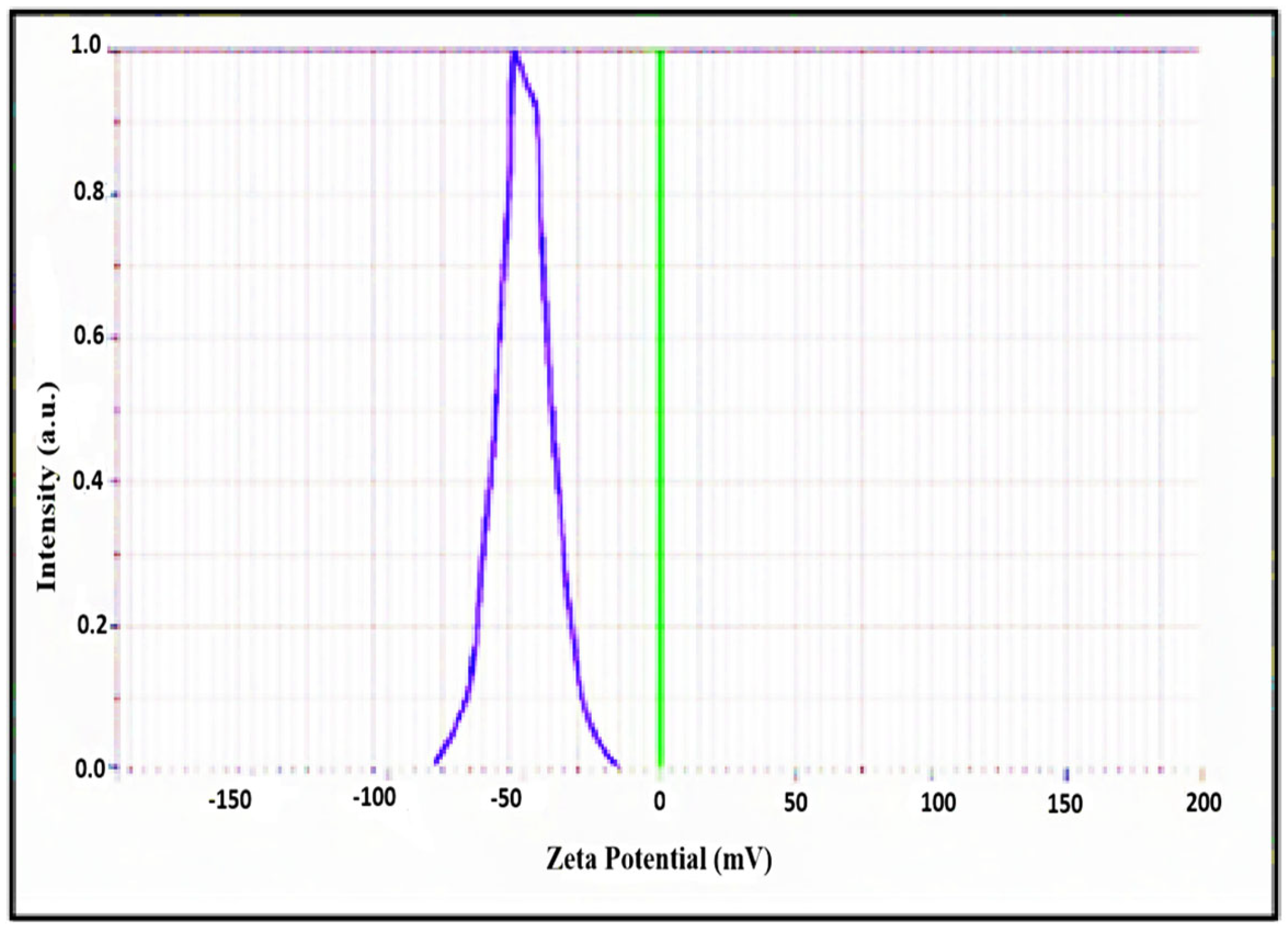
Zeta potential (ZP) analysis was carried out to determine the surface charges acquired by zinc oxide nanoparticles; this can be applied to evaluate the synthesized colloidal ZnO-NW stability. The stability of the nanoparticles in the solution is correlated with their ZP. If the zeta potential values of the particles in a suspension are strong, either positive or negative, they will reject each other and stop the nanoparticles from aggregating. As it is known, ZP values greater than (+30 mV) or less than (−30 mV) are generally thought to result in stable suspensions [38,39]. The zeta potential, ZP, of the biosynthesized ZnO NPs was measured using water as a dispersant. The ZP of ZnO NWs was determined and found to be −50 mV, as shown in Figure 5 and reported in [32]. Such result confirms that the majority of the capping biomolecules on the biosynthesized ZnO-NWs were negatively charged groups [40]. The biogenic ZnO NPs, having a negative charge, demonstrated the electrostatic repulsion between the produced nanoparticles [41,42].

2.6. Photocatalytic Degradation of Basic Fuchsin (BF) Dye

The photocatalytic performance of the green-synthesized ZnO-NW sample was evaluated through the degradation of Basic Fuchsin (BF) under natural sunlight irradiation (outside temperature: 35 °C). The pH of the BF solution was monitored in the dark, before and after complete BF degradation, as the pH significantly influences molecule adsorption and the generation of highly reactive OH⋅ radicals [12].

2.6.1. Catalyst Dose Effect

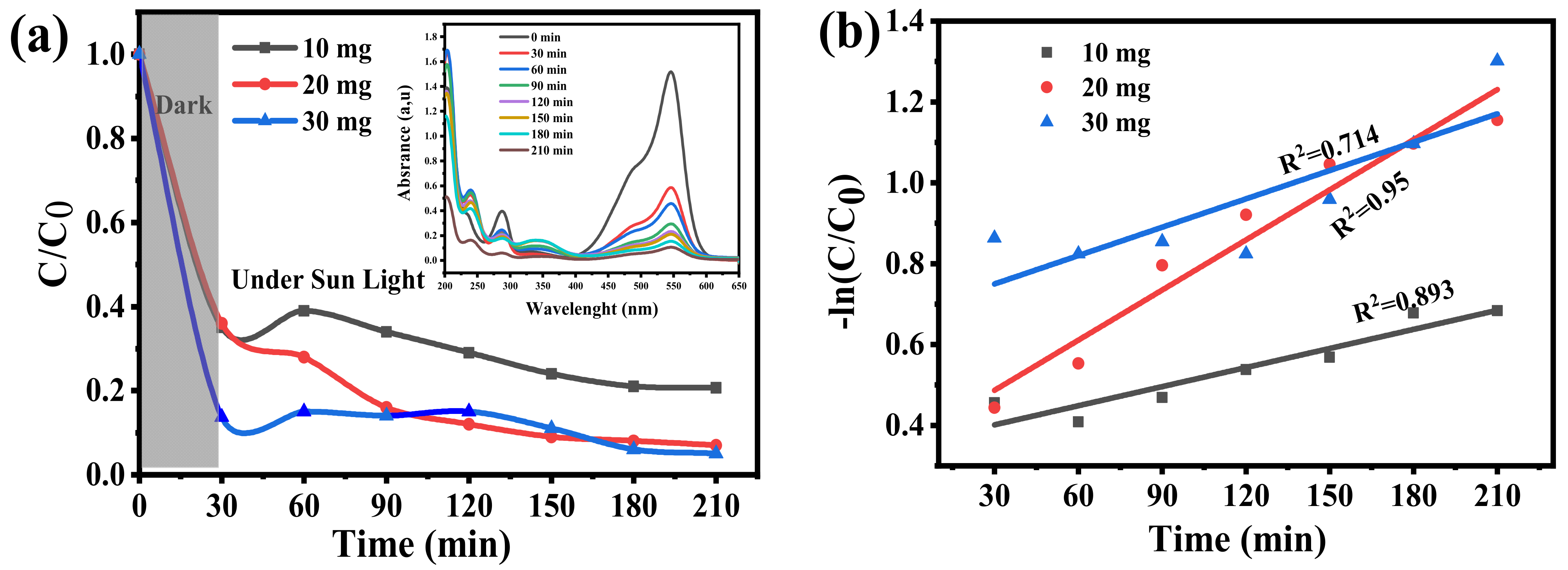
This study assessed the impact of different catalyst dosages (10, 20, and 30 mg) in 50 mL of Basic Fuchsin (10 ppm) for wastewater-treatment applications, as shown in Figure 6a. To create an adsorption–desorption equilibrium, the dye solution was left in the dark for half an hour before exposing it to sunlight irradiation for a further three hours (see inset of Figure 6a). In order to track the photocatalytic degradation process during the sun exposition, aliquots of the suspension were taken out every 30 min and analyzed. For this, Equation (1) was utilized in order to determine the deterioration efficiency (% Y). It was found that after three hours of exposure to the sunshine, the final degradation of catalysts containing 20 mg and 30 mg was almost equal. It can be concluded that 20 mg is the ideal catalyst loading, according to the degradation profile analysis, since raising the catalyst dosage above that level had no discernible impact on photocatalytic activity.
Based on the pseudo-first-order model where a linear plot of −ln(C/C0) versus irradiation time for the photocatalytic degradation of the dye was applied, as exhibited in Figure 6b, the kinetic analysis revealed that the apparent rate constants and correlation coefficients were k = 0.0016 min−1 (R2 = 0.893) for 10 mg, k = 0.0041 min−1 (R2 = 0.95) for 20 mg, and k = 0.0023 min−1 (R2 = 0.714) for 30 mg of catalyst. The best photocatalytic performance was obtained with 20 mg of catalyst (k = 0.0041 min−1, R2 = 0.95), indicating that this dosage offers the optimal balance between the number of active sites and light penetration, resulting in the most efficient degradation of BF. At higher amounts (30 mg), the sharp decrease in the correlation coefficient (R2 = 0.714) confirms that the reaction kinetics are severely hindered by particle agglomeration and light scattering, decreasing the effective surface area and hindering photon absorption [43].

2.6.2. Dye Concentration Effect

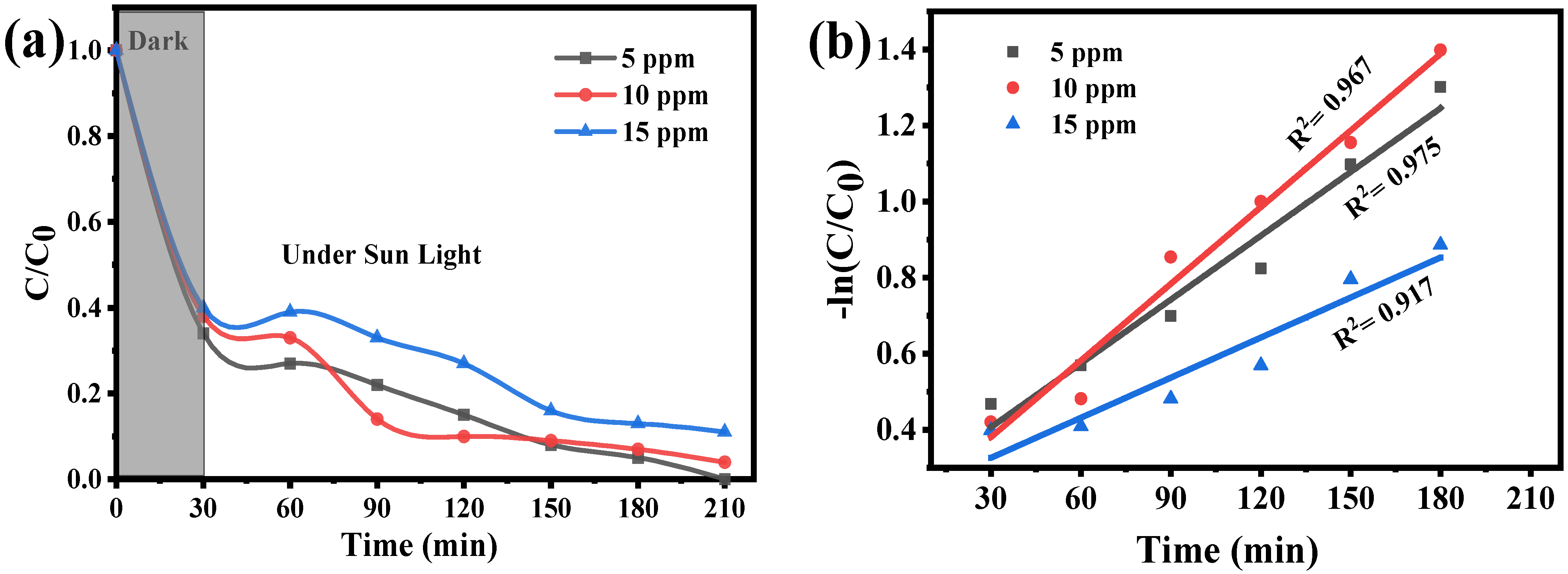
The impact of the initial pollutant concentration on the photocatalytic breakdown of Basic Fuchsin was investigated in this step of investigation. Initial dye concentrations were set at 5, 10, and 15 parts per million, while the catalyst dosage remained constant at 15 mg in 50 mL of solution at natural pH. The results have been illustrated in Figure 7a; it was observed that, even with the same amount of catalyst, approximately the same end degrading efficiency (nearly 100%) was obtained for 5 ppm and 10 ppm, suggesting that 10 ppm is more beneficial than 5 ppm for real-world applications. The degradation efficiency dropped to almost 89% at 15 ppm, demonstrating how increased pollutant loading affects photocatalytic efficacy.
Figure 7b presents the linear plots of −ln(C/C0) versus irradiation time for the photocatalytic degradation of the dye at different initial concentrations (5, 10, and 15 ppm). The linearity of these plots (R2 > 0.91) confirms that the degradation follows a pseudo-first-order kinetic model, which is an established simplification of the Langmuir–Hinshelwood model at low substrate concentrations described by Equation (3) [44]:
ln C C 0 = k × t
The apparent rate constants (k) were found to be 0.0056 min−1 (R2 = 0.9674) for 5 ppm, 0.0067 min−1 (R2 = 0.9754) for 10 ppm, and 0.0035 min−1 (R2 = 0.9171) for 15 ppm. The highest rate constant was obtained at 10 ppm, indicating that this concentration provided the most favorable conditions for photocatalytic degradation. A similar value has been reported by Mousa et al. [45], who found an apparent rate constant on the order of ~0.007 min−1 for a similar ZnO-based photocatalytic system, confirming that the k value obtained here (0.0067 min−1) lies within the expected range. This behavior can be explained by an optimal balance between sufficient light penetration and adequate dye adsorption on the catalyst surface. At lower concentrations (5 ppm), fewer dye molecules are available to interact with photogenerated charge carriers, slightly reducing the reaction probability. At higher pollutant concentrations (15 ppm), the photocatalytic efficiency was markedly decreased due to light attenuation and surface saturation effects. According to the Beer–Lambert law, the increased dye concentration enhances the absorption of incident light by the solution, which limits the photon flux reaching the catalyst surface and consequently reduces charge carrier generation, as demonstrated in several studies [44]. In addition, the excess dye molecules tend to cover the catalyst surface and compete for active sites, further restricting the degradation process. This surface screening effect suppresses the formation of reactive oxygen species (ROS), leading to a slower overall reaction rate and lower photocatalytic efficiency [46].

2.6.3. pH Effect Study

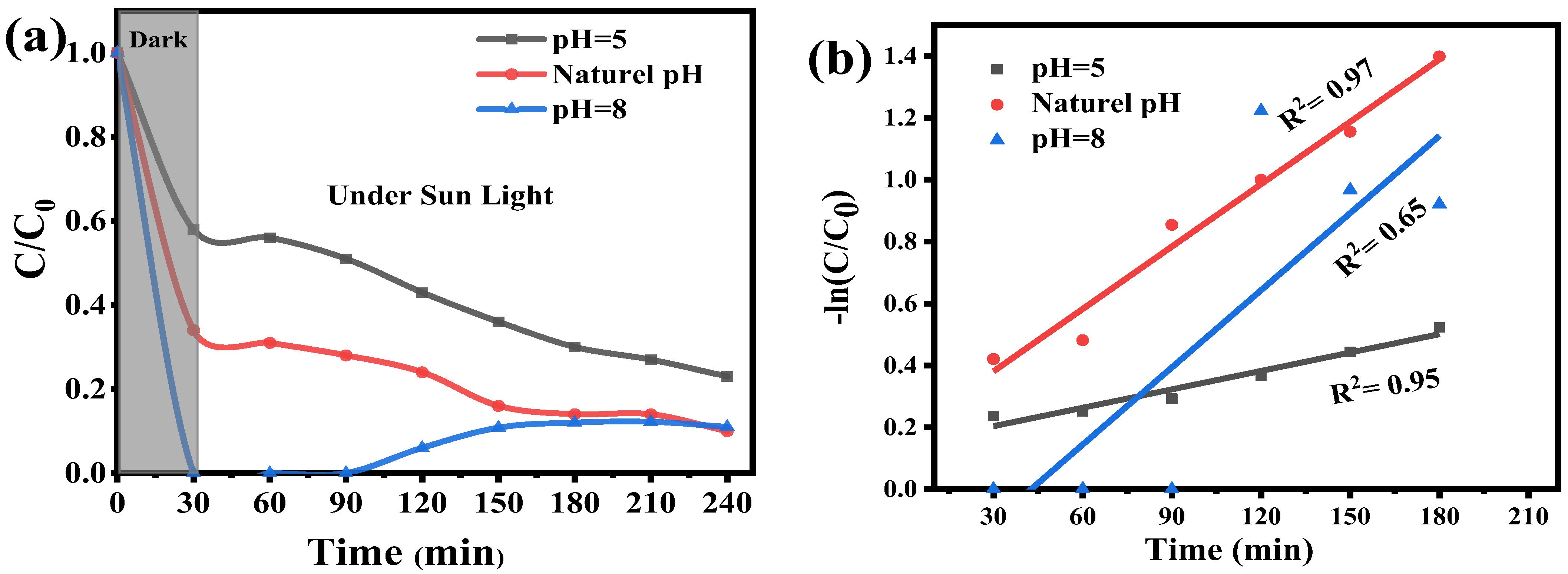
The pH impact of the initial solution on the photocatalytic degradation of BF was investigated using a constant dye concentration of 10 ppm. A catalyst dosage of 20 mg in 50 mL of solution was used to test the impact of pH on BF photocatalytic degradation at pH values of 5, 6.5 (natural pH), and 8. To track the degradation process, under sunlight, during the three hours, aliquots were taken every 30 min, and measurements were taken on it. The results, as shown in Figure 8a, confirmed that a maximum degradation efficiency was achieved at natural pH, which is in agreement with similar studies [43,47]. It is worth noting that after being exposed to sunlight irradiation for first 90 min at pH 8, the solution became almost colorless; however, the BF color gradually returned. This phenomenon could be linked to Basic Fuchsin’s pH-dependent reversible transition from its colorful cationic form to the colorless leuco form, as reported in previous studies [48].
Based on the pseudo-first-order model, the kinetic analysis of the degradation process, as it is displayed in Figure 8b, the latter clearly highlights a strong dependence of photocatalytic efficiency on the pH solution. High linearity was observed at pH 5 and 6.5 (R2 ≈ 0.95–0.97), unequivocally confirming first-order kinetics under these conditions. The apparent rate constant (k) reached its maximum at natural pH (k max = 0.0067 min−1), significantly higher than the rate observed at pH 5 (k = 0.0019 min−1), reflecting the most favorable surface charge interactions between the ZnO catalyst and the cationic dye at this optimal pH. Conversely, at a basic pH of 8, the degradation pattern exhibits a marked deviation from the expected pseudo-first-order kinetics, as evidenced by the significantly low R2 value (0.65). Although the calculated apparent rate constant was numerically high (k = 0.008 min−1), this k value lacks physical significance because the poor fit indicates that the model is statistically invalid under these conditions [44]. This is consistent with the observed reversible transition to the colorless leuco form, which interfered with the photocatalytic process. This decline in the model’s fitness at basic pH further confirms the detrimental effect of hydroxide ions on the reaction, primarily by hindering the formation of reactive radicals and altering the dye’s chemical structure [43].

2.6.4. Scavenger Study

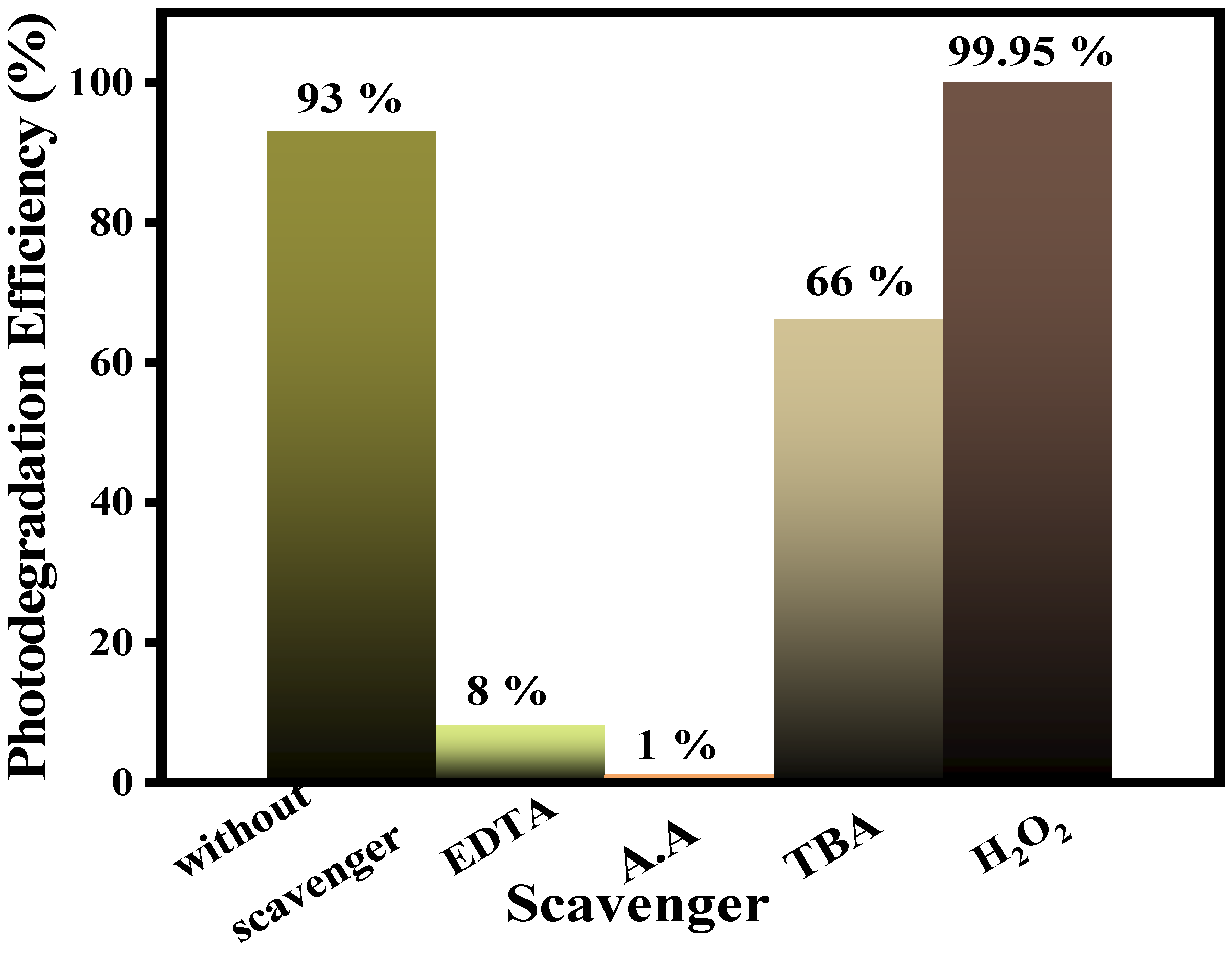
The roles of several oxidative species generated during the photocatalytic process, such as hydroxyl radicals (•OH), superoxide ions (O2), and singlet oxygen (O2), which are essential for the mineralization process, can be well understood through the use of radical scavenging tests [49]. Various scavengers were used independently in the BF dye solution (10 ppm) in this investigation to detect the active radicals during the photocatalytic experiments: ascorbic acid (A.A.) as a scavenger of superoxide radicals (O2), analogous to p-benzoquinone [50], tert-butanol (TBA) for hydroxyl radicals (•OH); and EDTA-Na2 as a scavenger of holes (h+) [51] have been sought. Such scavengers specifically target reactive molecules, and the contribution of them, in the photocatalytic breakdown of BF, can be easily identified.
Results obtained from Figure 9 show that while BF dye was entirely broken down in the absence of a scavenger, all scavengers significantly decreased the degradation effectiveness. Specifically, the presence of EDTA, A.A., and TBA reduced the degradation of BF to 1%, 8%, and 66%, respectively. This outcome demonstrates that holes (h+) and superoxide radicals (O2) are the main reactive species responsible for BF degradation in the absence of an accelerator, with h+ showing the greatest contribution (causing a 99% drop in efficiency). Conversely, the limited impact of TBA confirms that •OH radicals play a minor role under native conditions, as in [52]. Based on these results, the reactive species’ contribution to BF degradation follows the following order: h+ > O2 > •OH (under native conditions). The scavenging investigation confirms the synergistic functions of these species and establishes the benchmark for •OH activity before H2O2 is introduced.

2.6.5. H2O2 Concentration Effect on Degradation Rate

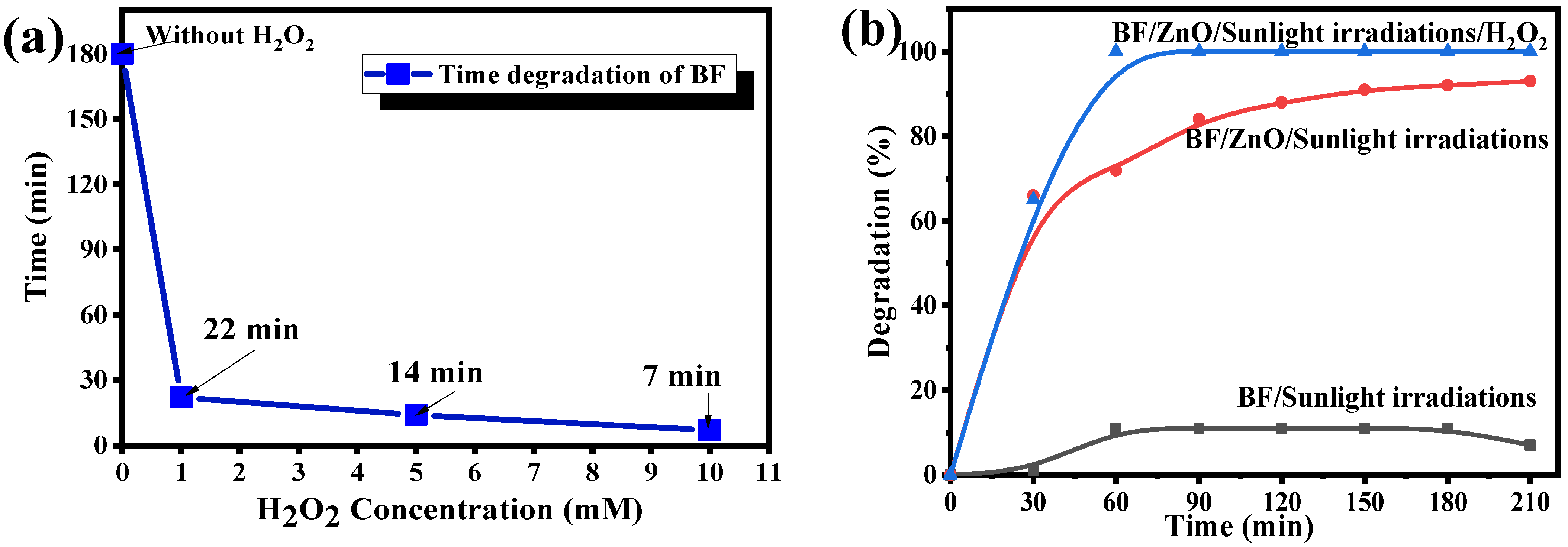
The effect of the hydrogen peroxide concentration on the degradation rate was investigated using H2O2 values of 1, 5, and 10 mM, following the approach outlined in research [53]. The maximum concentration was set at 10 mM to align with the principles of green chemistry, which prioritize high efficiency using minimal chemical dosages to reduce treatment costs and minimize residual chemical secondary pollution. Furthermore, this limit was chosen to avoid the well-known scavenging effect. At higher concentrations, excess H2O2 reacts with •OH radicals to produce hydroperoxyl radicals, which have a significantly lower oxidation potential, thereby reducing the overall photocatalytic performance [54].
The results show that increasing the H2O2 concentration significantly accelerates the reaction: the time required to reach 100% degradation decreased from 22 min at 1 mM, to 14 min at 5 mM, and to only 7 min at 10 mM, compared to about 3 h in the absence of H2O2. Figure 10 displays these results. The remarkable enhancement in efficiency observed in the presence of H2O2 (a potent electron scavenger) demonstrates that H2O2 boosts the effect of the dual structure by significantly reducing the recombination rate and providing an extra supply of the hydroxyl radical •OH [55]. Thus, the relation between morphology and performance is clearly established; the composite structure is the key to efficiency, as it optimizes both the surface area for interaction and the quantum efficiency of the photocatalytic process. This synergistic approach is consistent with previous studies on ZnO composites where H2O2 considerably boosts the photocatalytic rate by minimizing charge recombination [56].
Numerous investigations have shown that adding H2O2 can greatly improve the photocatalytic activity of various catalysts. For example, monochlorobenzene degrades in a TiO2 system in 120 min with H2O2 as opposed to 240 min without it, thus doubling the reaction rate [57], and the photocatalytic degradation of methyl blue (MB) in 60 min with 0.1 mol/L of H2O2 [53]. In another study, the degradation time of paracetamol with TiO2 decreased from 150 to 90 min (a 40% reduction) upon the addition of H2O2 [58].
Several studies have demonstrated that the addition of H2O2 enhances the photocatalytic activity of various materials. For instance, TiO2-based systems reported in the literature degrade triphenylmethane dyes such as Gentian Violet within 60–120 min under UV irradiation [59], while commercial P25-TiO2 decolorizes Congo Red in about 30 min in the presence of 20 mM H2O2 [60].
In addition, hybrid advanced oxidation processes have been reported using higher doses of H2O2 and various catalysts such as ZnO, TiO2, and SnO2 [61], targeting different organic pollutants, including azo and triphenylmethane dyes.
In comparison, the biosynthesized ZnO nanowires (ZnO-NWs) prepared in this study achieved nearly complete degradation of Basic Fuchsin in only 7 min under similar conditions, highlighting a markedly higher photocatalytic efficiency. This superior performance, combined with the green and low-cost synthesis route, suggests that ZnO-NWs are highly promising candidates for sustainable wastewater-treatment applications.
  • Photocatalytic degradation Mechanism of Basic Fuchsin (BF)
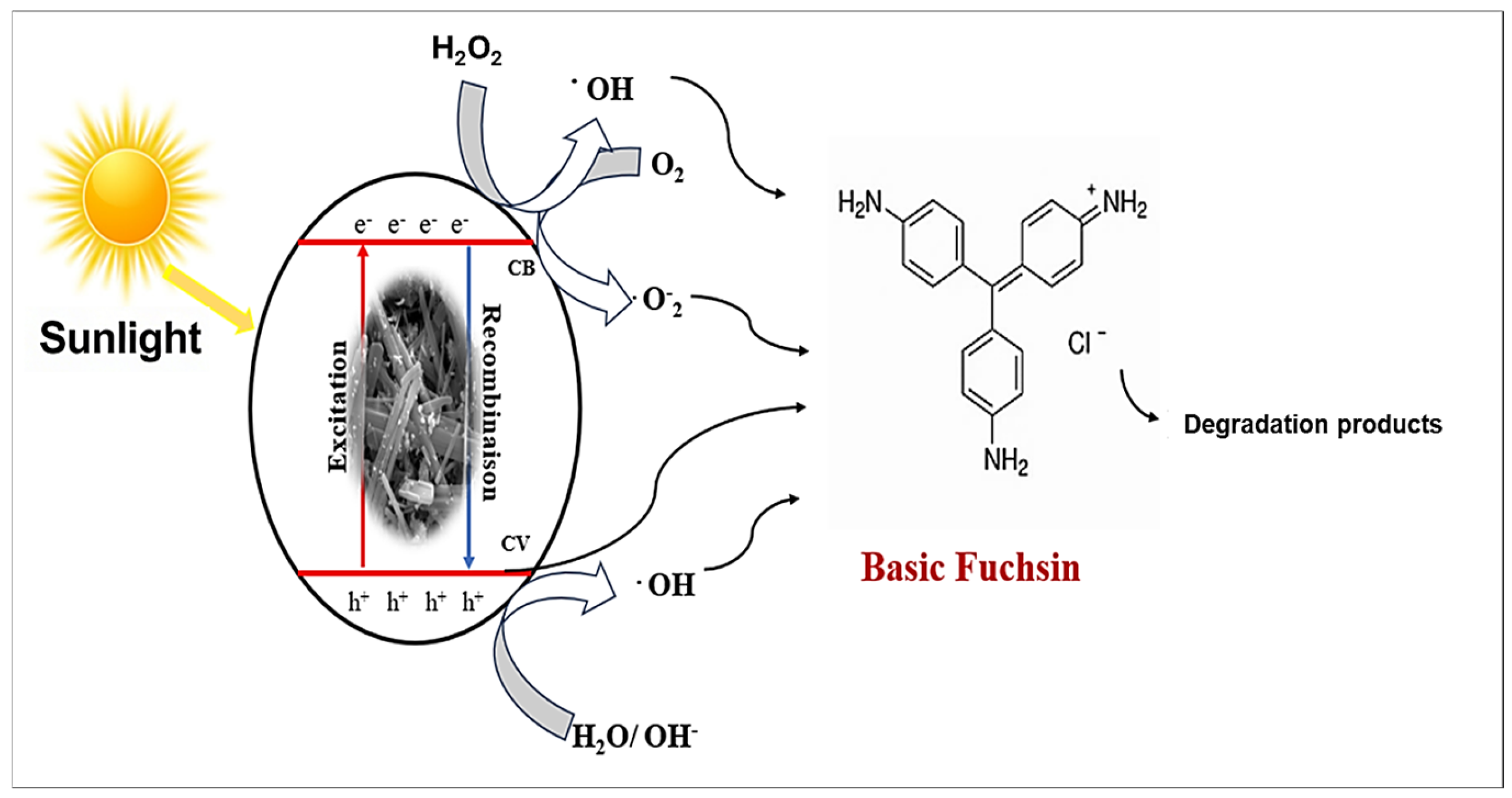
The proposed photocatalytic mechanism is directly supported by scavenger experiments, which demonstrate that photogenerated holes (h+) are the primary oxidative species responsible for BF degradation. In particular, the addition of EDTA-Na2 resulted in the almost complete suppression of photocatalytic activity (~99% inhibition), providing strong experimental evidence for the dominant role of holes (h+). The expected photocatalytic mechanism of the ZnO-NWs toward the BF dye is illustrated in Figure 11. When the ZnO semiconductor is illuminated by sunlight (hν), electrons (e) are excited from the valence band (VB) to the conduction band (CB) leaving a positive hole (h+) in the valence band (VB), as in Equation (4):
S u n l i g h t   ( h ν )   +   Z n O   e C B +   h V B +
The photogenerated h+ can directly oxidize adsorbed BF or, more typically, react with adsorbed water (H2O) or hydroxide ions (OH) to produce the highly oxidative hydroxyl radical (•OH), as in Equation (5). Simultaneously, the e in the CB reduces the dissolved oxygen (O2) to form superoxide radicals (O2), as in Equation (6) [51].
h V B + + H 2 O / O H   O H  
O 2 + e C B     O 2
  • Synergistic Role of H2O2
The incorporation of hydrogen peroxide (H2O2) significantly enhances the photocatalytic rate by serving two crucial functions: it acts as an electron scavenger, reducing electron-hole recombination [55], a behavior consistent with recent reports emphasizing the critical role of rapid interfacial charge-transfer pathways in boosting photocatalytic reactions [62], and it serves as an additional source of •OH radicals [23]. H2O2 traps the conduction-band electrons, as in Equation (7), and reacts with the generated superoxide radicals, as in Equation (8), thereby increasing the overall density of oxidizing species.
e C B + H 2 O 2     O H + O H
H 2 O 2 + O 2 O H + O H   + O 2
These highly reactive species (•OH, h+, O2) efficiently attack the BF molecules under solar irradiation, accelerating the degradation process and transforming the pollutant into degradation products, as in Equation (9) [63].
O H ,   h + ,   O 2 + B F   ( s o l a r   i r r a d i a t i o n )     D e g r a d a t i o n   p r o d u c t s
  • Comparative Studies
Several studies have reported the photocatalytic degradation of organic pollutants using ZnO-based nanomaterials under UV or solar irradiation. Although a direct comparison is challenging due to variations in experimental parameters, a qualitative evaluation remains meaningful to assess the relative performance of different systems.
The studies selected in Table 1 were chosen based on their use of green synthesis approaches, comparable organic pollutants, and similar irradiation conditions. As summarized in Table 1, most reported ZnO photocatalysts require prolonged irradiation times (105–180 min) to achieve high degradation efficiencies. In contrast, the ZnO nanowires developed in the present work exhibit remarkably enhanced performance, achieving 99.95% degradation of Basic Fuchsin within only 7 min under sunlight in the presence of H2O2.
This exceptional activity can be attributed to the one-dimensional nanowire morphology, which promotes efficient charge transport and suppresses electron–hole recombination, as well as to the synergistic effect between ZnO and H2O2, leading to the accelerated generation of reactive oxygen species. Consequently, the present system demonstrates superior photocatalytic efficiency and a significantly reduced reaction time, highlighting its strong potential for practical wastewater treatment applications.

3. Materials and Method

3.1. Chemicals and Reagents

Zinc acetate dihydrate (Zn(CH3COO)2·2H2O, with purity ≥ 99%, Analar Normapur, Prolabo, Paris, France, M.W = 219.50 g/mol), sodium hydroxide (NaOH, pellets, purity ≥ 99%, Merck, Darmstadt, Germany, M.W. = 40.00 g/mol), Basic Fuchsin dye (C20H20N3·HCl, dye content ≥ 95%, Sigma-Aldrich, St. Louis, MO, USA, M.W = 337.85 g/mol), disodium ethylenediaminetetraacetate (EDTA-Na2, C10H14N2Na2O8, ≥99%, Sigma-Aldrich, M.W = 372.24 g/mol), tert-Butanol (C4H10O, ≥99.5%, Merck, M.W. = 74.12 g/mol), ascorbic acid (C6H8O6, ≥99%, Sigma-Aldrich, M.W. = 176.12 g/mol), and hydrogen peroxide (H2O2, 30 wt.% in water, Biochem, M.W. = 34.01 g/mol) and distilled water are the products and reagents utilized in this work. All the chemical reagents used were of analytical grade and did not need to be further purified.

3.2. Preparation of Aqueous Leaf Extract of Lemon Verbena (Aloysia citrodora)

The leaves of fresh lemon verbena have been collected from a home garden. They were ground into a fine powder and allowed to air dry at room temperature in the shade. To produce a 1% (w/v) aqueous leaf extract (1 g of plant powder in 100 mL of water), this fine powder was steeped in hot sterile distilled water at a 1:100 (w/v) ratio (g/mL) with continuous stirring for 15 min. The solution was aged at room temperature for forty-five minutes. Whatman paper was used to filter the plant aqueous solution. The obtained plant extract was then stored and kept at 4 °C in a refrigerator.

3.3. Synthesis of Pure ZnO

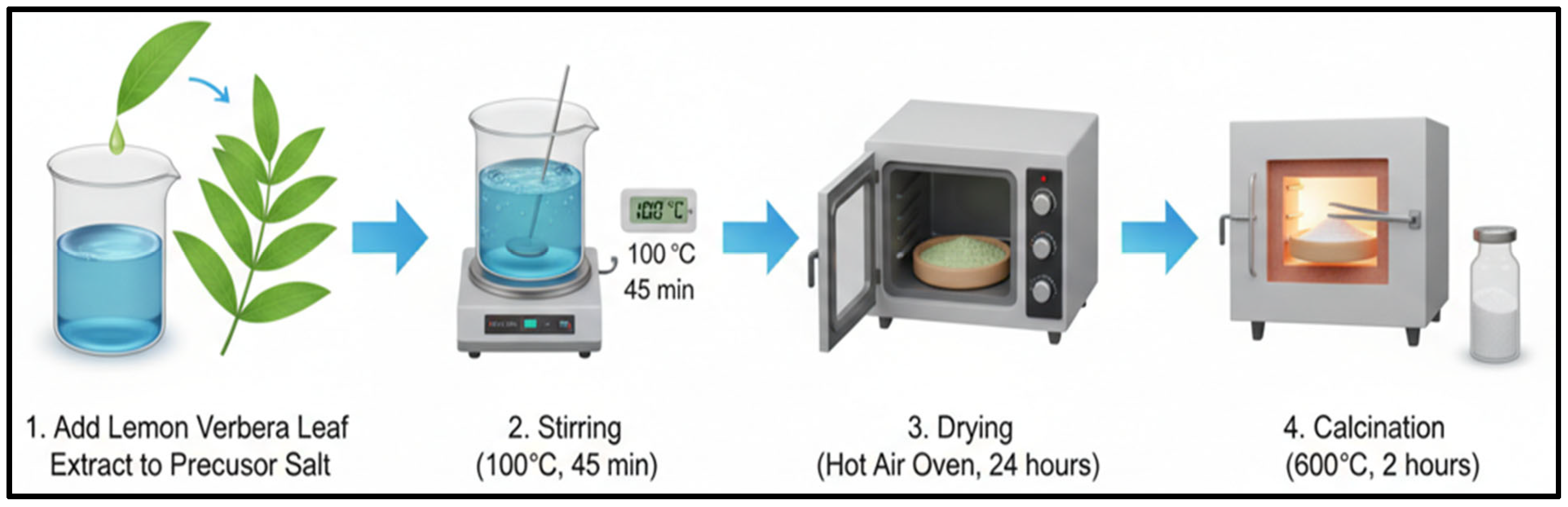
Green-synthesized ZnO-NWs were prepared using the following co-precipitation method, as shown in Figure 12. The latter was described in [68] with certain modifications in this context. Lemon verbena leaf extract was used as a reducing agent. A volume of 50 mL of lemon verbena leaf extract was added to 100 mL of 0.1M (Zn (CH3COO)2, 2H2O). The mixture was stirred at room temperature for 15 min. Subsequently, the mixture was heated at approximately 100 °C under magnetic stirring for 45 min; then, the pH of this mixture was adjusted to reach 12 via the dropwise addition of NaOH solution (0.1 M), which catalyzes the precipitation of Zn2+ as Zn(OH)2, which is a necessary step for the creation of well-crystallized ZnO [69].
The solution turned yellowish during this process, indicating the formation of Zn(OH)2 intermediates and the nucleation of ZnO nanoparticles. The precipitate was dried at 120 °C for 24 h and subsequently calcined at 600 °C for 2 h, resulting in fully crystallized ZnO nanowires, which appear white. These color changes reflect the chemical transformations during nanoparticle formation and thermal treatment [70].
The synthesis was repeated at least three times under identical conditions, yielding reproducible structural, optical, and photocatalytic results.

3.4. Characterization

The structure of the produced ZnO-NWs was investigated via X-ray diffraction using a Bruker model D8 ADVANCE (Bruker AXS GmbH, Karlsruhe, Germany), operating with Cu-Kα = 1.54184 Å radiation and a Bragg–Brentano configuration. Its functional group was verified using a Fourier Transformation Infrared Spectrometer (Bruker-INVENIO-R). The as-prepared ZnO-NW shape and morphology were surveyed using a Thermo Fisher Apreo 2 C (Thermo Fisher Scientific, Waltham, MA, USA) with a FEG-Schottky type electrical source. A UV-Vis Spectrophotometer (SHIMADZU 1900i, Shimadzu Corp, Kyoto, Japan) was utilized to investigate both the optical characteristics of the products and the photocatalytic degradation behavior of the Basic Fuchsin dye from water, whereas the zeta potential of ZnO-NWs was detected with a Nano Particle Analyzer (HORIBA Scientific SZ-100 instrument, Horiba Ltd., Kyoto, Japan).

3.5. Photocatalytic Degradation

At the Laboratory for Research in Medicine and Sustainable Development (ReMeDD), Faculty of Process Engineering, university of Salah Boubnider Constantine Algeria 36°16′35.1″ N 6°35′24.4″ E, it should be noted that all photocatalytic experiments were conducted under direct sunlight during the same time window (11:00 a.m.–3:00 p.m.) in July 2024, under similar weather conditions and with a very high UV index, ensuring a nearly constant solar intensity.
It was considered ideal irradiation conditions for the degradation process. The photocatalytic activity of these ZnO-NW products to break down Basic Fuchsin (BF) dye under sunlight was assessed. It is worth noting that photocatalysis is a chemical process that uses light energy to catalyze a reaction, and it is usually involving a photocatalyst. When the photocatalyst absorbs light, electron-hole pairs are consequently generated and involved in the chemical processes [71]. The green-synthesized sample’s photocatalytic activity was evaluated by photocatalytic degrading (BF) at different intervals of time through its exposition to sunlight at ambient temperature (35 °C). The photocatalytic process was thoroughly studied in relation to the impacts of the ZnO dosage, dye concentration, pH, scavengers, and H2O2 concentration; for keeping the adsorption–desorption equilibrium, the solution was placed in the dark for 30 min, before being exposed to sunlight, as preliminary adsorption experiments confirmed that the adsorption–desorption equilibrium was reached after 30 min in the dark.
Before being exposed to sunlight and for measuring the dye degradation, a testing sample was taken from the solution every 30 min. A UV-visible spectrophotometer was used to examine the dye degradation after centrifugation occurred at 10,000 rpm for five minutes to extract the catalyst from the samples. The following formula was used to determine the degraded dye’s percentage of degradation:
D e g r a d a t i o n     y i e l d   Y   % = C o C C o   × 100 %
where Co is the initial concentration of dye and C is the final concentration of the dye. The BF dye solution has a maximum absorption wavelength of 546 nm. The degradation mechanism was sketched in the figure below.

4. Conclusions

In this study, ZnO nanowires were successfully synthesized using an eco-friendly green chemistry approach based on lemon verbena leaf extract. Structural and optical analyses confirmed the formation of crystalline wurtzite ZnO nanowires and a band gap energy of 3.12 eV, consistent with typical ZnO nanostructures. The presence of phytochemicals involved in the transfer of metallic ions to ZnO-NWs was verified via FTIR spectroscopy, which indicates the Zn-O absorption mode, with a peak that occurs at 375 cm−1. Morphological characterization via SEM/EDX revealed a dominant one-dimensional nanowire morphology with an average diameter of 25.5 ± 10 nm, where the elemental analysis confirmed the absence of chemical impurities.
Under natural sunlight irradiation, the synthesized ZnO-NWs demonstrated excellent degradation efficiency toward the highly toxic Basic Fuchsin dye, achieving more than 99% within 3 h under natural sunlight, indicating their promising photocatalytic activity in the breakdown of BF dye. Photogenerated holes (h+), superoxide radicals (O2) and hydroxyl radicals (•OH) were found to be key reactive species in the degradation process, via active species analysis. Crucially, the addition of hydrogen peroxide played a decisive synergistic role in boosting the photocatalytic performance, reducing the degradation time to a remarkably short time of 7 min, and increasing the reaction rate by 25-fold at optimal concentrations of 20 mg of ZnO and 10 mM of H2O2. This synergistic coupled system consequently offers the potential to minimize the required catalyst loading.
These ZnO-NWs are therefore highly promising and cost-effective photocatalysts for large-scale, solar-driven wastewater-treatment applications. Although no direct experimental comparison with commercial ZnO was performed, literature benchmarking indicates that the nanowire morphology combined with low-dose H2O2 results in significantly faster kinetics than most reported ZnO- and TiO2-based systems.

Author Contributions

A.H.: Conceptualization, methodology, formal analysis, data curation, and writing—original draft preparation; C.B.: Methodology, validation, and supervision; S.I.: Supervision; B.B.: Writing—review and editing; F.F.: Validation, writing—review and editing, and supervision; A.A.A.: Conceptualization, methodology, supervision, project administration, and funding acquisition; A.A.: supervision, Validation; F.A.A.A.: Investigation and data curation; J.Z.: Formal analysis and validation; M.K.: Resources and data curation; H.T.: Investigation and visualization. All authors have read and agreed to the published version of the manuscript.

Funding

This work was supported and funded by the Deanship of Scientific Research at Imam Mohammad Ibn Saud Islamic University (IMSIU) (grant number IMSIU-DDRSP2602).

Data Availability Statement

The original contributions presented in this study are included in the article. Further inquiries can be directed to the corresponding authors.

Conflicts of Interest

The authors declare no conflicts of interest.

References

  1. Swaminaathan, P.; Saravanan, A.; Yaashikaa, P.R.; Vickram, A.S. Recent advances in photocatalytic degradation of persistent organic pollutants: Mechanisms, challenges, and modification strategies. Sustain. Chem. Environ. 2024, 8, 100171. [Google Scholar] [CrossRef]
  2. Ben Aissa, M.A.; Khezami, L.; Taha, K.; Elamin, N.; Mustafa, B.; Al-Ayed, A.S.; Modwi, A. Yttrium oxide-doped ZnO for effective adsorption of basic fuchsin dye: Equilibrium, kinetics, and mechanism studies. Int. J. Environ. Sci. Technol. 2022, 19, 9901–9914. [Google Scholar] [CrossRef]
  3. Abdelrahman, E.A. Efficient adsorption of basic fuchsin dye using thermally engineered novel smart nanocomposites. Sci. Rep. 2025, 15, 26232. [Google Scholar] [CrossRef] [PubMed]
  4. Ahmad Aftab, R.; Yusuf, M.; Ahmad, F.; Danish, M.; Zaidi, S.; Vo, D.V.N.; Nguyen, A.-T.; Rahman, M.M.; Ibrahim, H. Green Technology Approach Towards the Removal of Heavy Metals, Dyes, and Phenols from Water Using Agro-based Adsorbents: A Review. Chem. Asian J. 2024, 19, e202400154. [Google Scholar] [CrossRef]
  5. Kamati, S.N.; Yan, J.; Fan, J. A review on progresses in reactive dye-containing wastewater treatment. Water Pract. Technol. 2024, 19, 2712–2733. [Google Scholar] [CrossRef]
  6. Chalatsi-Diamanti, P.; Isari, E.A.; Grilla, E.; Kokkinos, P.; Kalavrouziotis, I.K. Recent prospects, challenges and advancements of photocatalysis as a wastewater treatment method. Water Emerg. Contam. Nanoplast. 2025, 4, 12. [Google Scholar] [CrossRef]
  7. Katre, S.; Baghmare, P.; Giri, A.S. Photocatalytic nanomaterials and their implications towards biomass conversion for renewable chemical and fuel production. Nanoscale Adv. 2024, 6, 5258–5284. [Google Scholar] [CrossRef]
  8. Gupta, D.; Boora, A.; Thakur, A.; Gupta, T.K. Green and sustainable synthesis of nanomaterials: Recent advancements and limitations. Environ. Res. 2023, 231, 116316. [Google Scholar] [CrossRef]
  9. Gayathri, T.; Logesh Kumar, S.; Sangavi, S.; Yudhika, M.; Swathy, M. Green synthesis of copper oxide nanoparticles using Carica papaya and their antimicrobial activity. Mater. Today Proc. 2023; in press. [Google Scholar] [CrossRef]
  10. Bala, N.; Saha, S.; Chakraborty, M.; Maiti, M.; Das, S.; Basu, R.; Nandy, P. Green synthesis of zinc oxide nanoparticles using Hibiscus subdariffa leaf extract: Effect of temperature on synthesis, anti-bacterial activity and anti-diabetic activity. RSC Adv. 2014, 5, 4993–5003. [Google Scholar] [CrossRef]
  11. Náfrádi, M.; Alapi, T.; Veres, B.; Farkas, L.; Bencsik, G.; Janáky, C. Comparison of TiO2 and ZnO for Heterogeneous Photocatalytic Activation of the Peroxydisulfate Ion in Trimethoprim Degradation. Materials 2023, 16, 5920. [Google Scholar] [CrossRef] [PubMed]
  12. Cervantes-Gaxiola, M.E.; Vázquez-González, F.A.; Rios-Iribe, E.Y.; Méndez-Herrera, P.F.; Leyva-Inzunza, C. Effect of pH on the green synthesis of ZnO nanoparticles using Sorghum bicolor seed extract and their application in photocatalytic dye degradation. Mater. Lett. 2024, 372, 136982. [Google Scholar] [CrossRef]
  13. Saberi Riseh, R.; Gholizadeh Vazvani, M. Green synthesis of metal nanoparticles using plant growth promoting rhizobacteria and application in agriculture. Plant Nano Biol. 2024, 10, 100111. [Google Scholar] [CrossRef]
  14. Cruz, D.; Falé, P.L.; Mourato, A.; Vaz, P.D.; Luisa Serralheiro, M.; Lino, A.R.L. Preparation and physicochemical characterization of Ag nanoparticles biosynthesized by Lippia citriodora (Lemon verbena). Colloids Surf. B Biointerfaces 2010, 81, 67–73. [Google Scholar] [CrossRef]
  15. Aldeen, T.S.; Ahmed Mohamed, H.E.; Maaza, M. ZnO nanoparticles prepared via a green synthesis approach: Physical properties, photocatalytic and antibacterial activity. J. Phys. Chem. Solids 2022, 160, 110313. [Google Scholar] [CrossRef]
  16. EL Machtani Idrissi, D.; Essate, A.; Kouzi, Y.; Achiou, B.; Beqqour, D.; Aaddane, A.; Reghai, A.-E.; Alami Younssi, S.; Ouammou, M. Green synthesis of ZnO using olives leaf extract: Parameters optimization and its application on adsorption and photocatalytic degradation of direct red 80. Ceram. Int. 2025, 51, 13152–13163. [Google Scholar] [CrossRef]
  17. Hussein, H.; Ibrahim, S.S.; Khairy, S.A. Green synthesis of ZnO nanoparticles using Hibiscus sabdariffa L: Rapid Pb2+ ion removal, photocatalytic degradation of methylene blue, and biomedical applications. J. Water Process Eng. 2025, 69, 106649. [Google Scholar] [CrossRef]
  18. Alalawy, A.I.; Saleh, F.M.; Saeedi, N.H.; Panneerselvam, C.; Sakran, M.I.; Khasim, S.; Parveen, H.; Mohammedsaleh, Z.M.; Mukhtar, S.; Faridi, U.; et al. Green synthesis of Bb-ZnO NPs using Barleria buxifolia leaf extract: Investigations into their physicochemical, biological and anti-cancer properties. Polyhedron 2025, 267, 117366. [Google Scholar] [CrossRef]
  19. Elumalai, K.; Velmurugan, S. Green synthesis, characterization and antimicrobial activities of zinc oxide nanoparticles from the leaf extract of Azadirachta indica (L.). Appl. Surf. Sci. 2015, 345, 329–336. [Google Scholar] [CrossRef]
  20. Bahramsoltani, R.; Rostamiasrabadi, P.; Shahpiri, Z.; Marques, A.M.; Rahimi, R.; Farzaei, M.H. Aloysia citrodora Paláu (Lemon verbena): A review of phytochemistry and pharmacology. J. Ethnopharmacol. 2018, 222, 34–51. [Google Scholar] [CrossRef]
  21. Hassanisaadi, M.; Bonjar, A.H.S.; Rahdar, A.; Varma, R.S.; Ajalli, N.; Pandey, S. Eco-friendly biosynthesis of silver nanoparticles using Aloysia citrodora leaf extract and evaluations of their bioactivities. Mater. Today Commun. 2022, 33, 104183. [Google Scholar] [CrossRef]
  22. Faisal, S.; Jan, H.; Shah, S.A.; Shah, S.; Khan, A.; Akbar, M.T.; Rizwan, M.; Jan, F.; Wajidullah; Akhtar, N.; et al. Green Synthesis of Zinc Oxide (ZnO) Nanoparticles Using Aqueous Fruit Extracts of Myristica fragrans: Their Characterizations and Biological and Environmental Applications. ACS Omega 2021, 6, 9709–9722. [Google Scholar] [CrossRef]
  23. González-Crisostomo, J.C.; López-Juárez, R.; Petranovskii, V. Photocatalytic Degradation of Rhodamine B Dye in Aqueous Suspension by ZnO and M-ZnO (M = La3+, Ce3+, Pr3+ and Nd3+) Nanoparticles in the Presence of UV/H2O2. Processes 2021, 9, 1736. [Google Scholar] [CrossRef]
  24. Nizar, B.M.; Lajnef, M.; Chaste, J.; Chtourou, R.; Herth, E. Highly C-oriented (002) plane ZnO nanowires synthesis. RSC Adv. 2023, 13, 15077–15085. [Google Scholar] [CrossRef] [PubMed]
  25. Mohamed, H.E.A.; Afridi, S.; Khalil, A.T.; Zia, D.; Shinwari, Z.K.; Dhlamini, M.S.; Maaza, M. Structural, Morphological and Biological Features of ZnO Nanoparticles Using Hyphaene thebaica (L.) Mart. Fruits. J. Inorg. Organomet. Polym. Mater. 2020, 30, 3241–3254. [Google Scholar] [CrossRef]
  26. Consonni, V. ZnO Nanowires: Growth, Properties, and Energy Applications. Nanomaterials 2023, 13, 2519. [Google Scholar] [CrossRef] [PubMed]
  27. Barzinjy, A.A.; Azeez, H.H. Green synthesis and characterization of zinc oxide nanoparticles using Eucalyptus globulus Labill. leaf extract and zinc nitrate hexahydrate salt. SN Appl. Sci. 2020, 2, 991. [Google Scholar] [CrossRef]
  28. Al-Khaial, M.Q.; Chan, S.Y.; Abu-Zurayk, R.A.; Alnairat, N. Biosynthesis and Characterization of Zinc Oxide Nanoparticles (ZnO-NPs) Utilizing Banana Peel Extract. Inorganics 2024, 12, 121. [Google Scholar] [CrossRef]
  29. Saeed, S.; Nawaz, S.; Nisar, A.; Mehmood, T.; Tayyab, M.; Nawaz, M.; Firyal, S.; Bilal, M.; Mohy-ud-Din, A.; Ullah, A. Effective fabrication of zinc-oxide (ZnO) nanoparticles using Achyranthes aspera leaf extract and their potent biological activities against the bacterial poultry pathogens. Mater. Res. Express 2021, 8, 035004. [Google Scholar] [CrossRef]
  30. Udayagiri, H.; Sana, S.S.; Dogiparthi, L.K.; Vadde, R.; Varma, R.S.; Koduru, J.R.; Ghodake, G.S.; Somala, A.R.; Boya, V.K.N.; Kim, S.-C.; et al. Phytochemical fabrication of ZnO nanoparticles and their antibacterial and anti-biofilm activity. Sci. Rep. 2024, 14, 19714. [Google Scholar] [CrossRef]
  31. Medina-Acosta, M.; Chinchillas-Chinchillas, M.J.; Garrafa-Gálvez, H.E.; Garcia-Maro, C.A.; Rosas-Casarez, C.A.; Lugo-Medina, E.; Luque-Morales, P.A.; Soto-Robles, C.A. Photocatalytic Degradation of Four Organic Dyes Present in Water Using ZnO Nanoparticles Synthesized with Green Synthesis Using Ambrosia ambrosioides Leaf and Root Extract. Processes 2024, 12, 2456. [Google Scholar] [CrossRef]
  32. Singh, D.; Pandey, D.; Singh, D. A study of nanosized zinc oxide and its nanofluid. Pramana 2012, 78, 759–766. [Google Scholar] [CrossRef]
  33. Balabye, S.; Mushebo, E.; Nasejje, S.; Moses, E.; Mukhokosi, E.P. Green synthesis of ZnO nanoparticles for DSSC photoanode: A joint experimental and density functional theory study. Mater. Res. Express 2025, 12, 105004. [Google Scholar] [CrossRef]
  34. Yamakawa, K.; Sato, Y.; Fukutani, K. Asymmetric and symmetric absorption peaks observed in infrared spectra of CO2 adsorbed on TiO2 nanotubes. J. Chem. Phys. 2016, 144, 154703. [Google Scholar] [CrossRef] [PubMed]
  35. Zenkin, K.; Durmuş, S.; Emre, D.; Bilici, A.; Yılmaz, S. Salvia officinalis leaf extract-stabilized NiO NPs, ZnO NPs, and NiO@ZnO nanocomposite: Green hydrothermal synthesis, characterization and supercapacitor application. Biomass Conv. Bioref. 2024, 15, 10149–10164. [Google Scholar] [CrossRef]
  36. Lushchikova, O.V.; Szalay, M.; Tahmasbi, H.; Juurlink, L.B.F.; Meyer, J.; Höltzl, T.; Bakker, J.M. IR spectroscopic characterization of the co-adsorption of CO2 and H2 onto cationic Cun+ clusters. Phys. Chem. Chem. Phys. 2021, 23, 26661–26673. [Google Scholar] [CrossRef]
  37. Al-Hada, N.M.; Saion, E.B.; Shaari, A.H.; Kamarudin, M.A.; Flaifel, M.H.; Ahmad, S.H.; Gene, S.A. A Facile Thermal-Treatment Route to Synthesize ZnO Nanosheets and Effect of Calcination Temperature. PLoS ONE 2014, 9, e103134. [Google Scholar] [CrossRef]
  38. Abdelbaky, A.S.; Mohamed, A.M.H.A.; Sharaky, M.; Mohamed, N.A.; Diab, Y.M. Green approach for the synthesis of ZnO nanoparticles using Cymbopogon citratus aqueous leaf extract: Characterization and evaluation of their biological activities. Chem. Biol. Technol. Agric. 2023, 10, 63. [Google Scholar] [CrossRef]
  39. Yedurkar, S.; Maurya, C.; Mahanwar, P. Biosynthesis of Zinc Oxide Nanoparticles Using Ixora Coccinea Leaf Extract—A Green Approach. Open J. Synth. Theory Appl. 2016, 5, 1–14. [Google Scholar] [CrossRef]
  40. El-Hawary, S.; Almaksoud, H.; Saber, F.; Elimam, H.; Sayed, A.; El-Raey, M.; Abdelmohsen, U. Green-synthesized zinc oxide nanoparticles, anti-Alzheimer potential and the metabolic profiling of Sabal blackburniana grown in Egypt supported by molecular modelling. RSC Adv. 2021, 11, 18009–18025. [Google Scholar] [CrossRef]
  41. Yassin, M.T.; Mostafa, A.A.-F.; Al-Askar, A.A.; Al-Otibi, F.O. Facile Green Synthesis of Zinc Oxide Nanoparticles with Potential Synergistic Activity with Common Antifungal Agents against Multidrug-Resistant Candidal Strains. Crystals 2022, 12, 774. [Google Scholar] [CrossRef]
  42. Liaqat, N.; Jahan, N.; Khalil-Ur-Rahman; Anwar, T.; Qureshi, H. Green synthesized silver nanoparticles: Optimization, characterization, antimicrobial activity, and cytotoxicity study by hemolysis assay. Front. Chem. 2022, 10, 952006. [Google Scholar] [CrossRef]
  43. Khammar, F.; Boukerche, S.; Djaber, S.; Boublia, A.; Messabhia, A.; Gharbi, A.; Ferkous, H.; Gomez, C.V.; Bellucci, S.; Albrahim, M.; et al. Synthesis, characterization, and photocatalytic efficiency of Mg-doped ZnO nanoparticles for basic Fuchsin dye degradation: Experimental and theoretical insights. Inorg. Chem. Commun. 2025, 176, 114274. [Google Scholar] [CrossRef]
  44. Tran, H.D.; Nguyen, D.Q.; Do, P.T.; Tran, U.N.P. Kinetics of photocatalytic degradation of organic compounds: A mini-review and new approach. RSC Adv. 2023, 13, 16915–16925. [Google Scholar] [CrossRef] [PubMed]
  45. Mousa, S.A.; Wissa, D.A.; Hassan, H.H.; Ebnalwaled, A.A.; Khairy, S.A. Enhanced photocatalytic activity of green synthesized zinc oxide nanoparticles using low-cost plant extracts. Sci. Rep. 2024, 14, 16713. [Google Scholar] [CrossRef] [PubMed]
  46. Langa, C.; Mabuba, N.; Mahlaule-Glory, L.M.; Motaung, D.E.; Tetana, Z.; Hintsho-Mbita, N.C. Enhanced photocatalytic degradation of dyes and pharmaceutical pollutants using Fe/TiO2-carbon nanospheres from Sutherlandia frutescence. Int. J. Environ. Anal. Chem. 2025, 106, 790–820. [Google Scholar] [CrossRef]
  47. Kumawat, P.; Joshi, M.; Ameta, R.; Ameta, S.C. Photocatalytic degradation of basic Fuchsin over quaternary oxide iron zinc cuprate (FeZn2cu3O6.5). Adv. Appl. Sci. Res. 2015, 6, 209–215. [Google Scholar]
  48. Ullah, S.; Ahmad, I.; Alias, Y.; Yusoff, I.; Ashraf, M.A. Characterization of Triphenylamino-Based Polymethine Dyes. J. Chem. 2013, 2013, 136908. [Google Scholar] [CrossRef]
  49. Boutamine, Z.; Boukerche, I.; Ayachi, A.; Mignot, M.; Zerouali, F.; Khraimech, S.; Abdelouahed, L. The investigation of the basic fuchsin dye photocatalytic degradation in aqueous media using stabilised Span 80 and synthesized ultrasonically CuO Nanoparticles (NPs). React. Kinet. Mech. Catal. 2025, 138, 1813–1832. [Google Scholar] [CrossRef]
  50. Sagita, C.P.; Yuliati, L. Improved Visible Light Activity of Copper Oxide/Carbon Nitride Photocatalysts Prepared by Photodeposition for Phenol Degradation. IOP Conf. Ser. Mater. Sci. Eng. 2021, 1143, 012075. [Google Scholar] [CrossRef]
  51. Chihi, A. Synthesis of Sb2 S3:Eu thin films as a catalyst for the efficient photocatalytic degradation of rhodamine B (RhB) dye under visible light. RSC Adv. 2025, 15, 5681–5697. [Google Scholar] [CrossRef] [PubMed]
  52. Tessema, A.; Wu, C.; Motora, K. Highly Efficient Solar Light Driven g-C3N4@Cs0.33WO3 Heterojunction for the Photodegradation of Colorless Antibiotics. ACS Omega 2022, 7, 38475–38486. [Google Scholar] [CrossRef]
  53. Aydin, E.B. Preparation, Characterization and Immobilization of Ag-Doped ZnO-Nanorods into Ca and Cu Alginate Beads and Their Application in the Photodegradation of Methylene Blue. ChemistrySelect 2021, 6, 11653–11663. [Google Scholar] [CrossRef]
  54. Villota, N.; Duoandicoechea, U.; Echevarria, B.; de Luis, A.M. Impact of Hydrogen Peroxide Concentration on Diclofenac Degradation by UV/H2O2: Kinetic Modeling for Water Treatment Applications. Separations 2025, 12, 125. [Google Scholar] [CrossRef]
  55. Bouarioua, A. High photocatalytic performance of coated TiO2 layers assisted by H2O2 oxidizing agent to remove an azo dye from water via the synergistic effect of optimum conditions. Desalination Water Treat. 2020, 184, 354–366. [Google Scholar] [CrossRef]
  56. Shahzad, R.; Muneer, M.; Khalid, R.; Amin, H.M.A.; Shahzad, R.; Muneer, M.; Khalid, R.; Amin, H.M.A. ZnO-Bi2O3 Heterostructured Composite for the Photocatalytic Degradation of Orange 16 Reactive Dye: Synergistic Effect of UV Irradiation and Hydrogen Peroxide. Catalysts 2023, 13, 1328. [Google Scholar] [CrossRef]
  57. Wen, H.; Huang, S.; Meng, X.; Xian, X.; Zhao, J.; Roy, V.A.L. Recent progress in the design of photocatalytic H2O2 synthesis system. Front. Chem. 2022, 10, 1098209. [Google Scholar] [CrossRef]
  58. Khoiriah, K.; Putri, R.A.; Ratnawati, R.; Sari, M.P. Investigating the Effect of Hydrogen Peroxide on Photocatalytic Degradation of Commercial Paracetamol Using TiO2. J. Chem. Technol. Met. 2024, 59, 539–548. [Google Scholar] [CrossRef]
  59. Bendjabeur, S.; Zouaghi, R.; Kaabeche, O.N.H.; Sehili, T. Parameters Affecting Adsorption and Photocatalytic Degradation Behavior of Gentian Violet under UV Irradiation with Several Kinds of TiO2 as a Photocatalyst. Int. J. Chem. React. Eng. 2017, 15, 20160206. [Google Scholar] [CrossRef]
  60. Mironyuk, I.; Danyliuk, N.; Tatarchuk, T.; Mykytyn, I.; Kotsyubynsky, V. Photocatalytic degradation of Congo red dye using Fe-doped TiO2 nanocatalysts. Phys. Chem. Solid. St. 2021, 22, 697–710. [Google Scholar] [CrossRef]
  61. Iqbal, A.; Bokhari, T.H.; Usman, M.; Yusaf, A.; Mansha, A.; Saeed, M.; Khan, S.; Iqbal, M.; Bhatti, M.A.; Shafqat, S.S.; et al. Methodological optimization for efficient degradation of Acid Violet 49 using advanced oxidation processes and varied photocatalyst combinations. Sci. Rep. 2024, 14, 30339. [Google Scholar] [CrossRef] [PubMed]
  62. Zou, J.; Li, L.; Tan, N.; Zhang, K.; Wang, L.; Chen, Z. Construction of S-scheme heterojunction with interfacial chemical bonds for enhanced photocatalytic CO2 reduction. Appl. Surf. Sci. 2026, 719, 165001. [Google Scholar] [CrossRef]
  63. Makota, O.; Dutková, E.; Briančin, J.; Bednarcik, J.; Lisnichuk, M.; Yevchuk, I.; Melnyk, I. Advanced Photodegradation of Azo Dye Methyl Orange Using H2O2-Activated Fe3O4@SiO2@ZnO Composite under UV Treatment. Molecules 2024, 29, 1190. [Google Scholar] [CrossRef] [PubMed]
  64. Venkatesan, S.; Suresh, S.; Arumugam, J.; Ramu, P.; Pugazhenthiran, N.; Jothilakshmi, R.; Prabu, K.M. Sunlight assisted degradation of methylene blue dye by zinc oxide nanoparticles green synthesized using Vitex negundo plant leaf extract. Results Chem. 2024, 7, 101315. [Google Scholar] [CrossRef]
  65. El Golli, A.; Contreras, S.; Dridi, C. Bio-synthesized ZnO nanoparticles and sunlight-driven photocatalysis for environmentally-friendly and sustainable route of synthetic petroleum refinery wastewater treatment. Sci. Rep. 2023, 13, 20809. [Google Scholar] [CrossRef]
  66. Tanuj; Kumar, R.; Kumar, S.; Kalra, N.; Sharma, S.; Singh, A. Green synthesis of zinc oxide nanoparticles from Rhododendron arboreum extract and their potential applications in photocatalytic degradation of cationic dyes malachite green and Fuchsin basic dye. Chem. Pap. 2023, 77, 6583–6604. [Google Scholar] [CrossRef]
  67. Hussaini, A.A.; Mirza, S.; Ulukuş, D.; Öztürk, T.; Paşayeva, L.; Tugay, O.; Yıldırım, M. Green synthesis of ZnO nanorods from Allium bourgeaui subsp. bourgeaui extract: Functional applications. Biomass Conv. Bioref. 2024, 15, 16675–16690. [Google Scholar] [CrossRef]
  68. López Miranda, J.; España-Sánchez, B.L.; Esparza, R.; Estevez, M. Self-assembly of ZnO nanoflowers synthesized by a green approach with enhanced catalytic, and antibacterial properties. Mater. Chem. Phys. 2022, 289, 126453. [Google Scholar] [CrossRef]
  69. Shaba, E.Y.; Jacob, J.O.; Tijani, J.O.; Suleiman, M.A.T. A critical review of synthesis parameters affecting the properties of zinc oxide nanoparticle and its application in wastewater treatment. Appl. Water Sci. 2021, 11, 48. [Google Scholar] [CrossRef]
  70. Salah, A.; Ali, T.; Babalghith, A.; Selim, S.; Mohamed, A. Green Synthesis and Characterization of ZnO Nanoparticles Using Pelargonium odoratissimum (L.) Aqueous Leaf Extract and Their Antioxidant, Antibacterial and Anti-inflammatory Activities. Antioxidants 2022, 11, 1444. [Google Scholar] [CrossRef]
  71. Ouni, S.; Yahia, F.; BelHaj Mohamed, N.; Bouzidi, M.; Alshammari, A.S.; Abdulaziz, F.; Bonilla-Petriciolet, A.; Mohamed, M.; Khan, Z.R.; Chaaben, N.; et al. Effective removal of textile dye via synergy of adsorption and photocatalysis over ZnS nanoparticles: Synthesis, modeling, and mechanism. Heliyon 2024, 10, e36949. [Google Scholar] [CrossRef]
Figure 1. XRD pattern of green-synthetized ZnO-NWs.
Catalysts 16 00193 g001
Figure 2. Surface morphology of the biosynthesized ZnO-NWs, (a) SEM images, (b) EDX elemental composition of the nanoparticles, and (c) diameter estimation of as-synthesized ZnO-NWs using Gaussian fitting of the histogram.
Catalysts 16 00193 g002
Figure 3. UV-Vis analysis of ZnO NWs: (a) absorbance of the sample and (b) Tauc plot showing band gap estimation via extrapolation of the straight line to the hv axis.
Catalysts 16 00193 g003
Figure 4. FTIR image of biosynthesized ZnO NWs.
Catalysts 16 00193 g004
Figure 5. Zeta potential analysis of biosynthesized ZnO-NWs (Purple curve: zeta distribution; Green line: 0 mV reference).
Catalysts 16 00193 g005
Figure 6. Degradation of BF under sunlight. (a) Effect of catalyst dosage on the photocatalytic degradation of BF and (b) pseudo-first-order kinetic plots (−ln(C/C0) vs. time) for BF degradation at different catalyst dosages.
Catalysts 16 00193 g006
Figure 7. Photocatalytic degradation of BF under sunlight. (a) Effect of initial dye concentration. (b) Pseudo-first-order kinetic plots (−ln(C/C0) vs. time) for BF degradation at different initial concentrations.
Catalysts 16 00193 g007
Figure 8. pH effect on the photocatalytic degradation of BF dye (a) under sunlight. (b) Pseudo-first-order kinetic plots. (−ln(C/C0) vs. time) for BF degradation at different pH values.
Catalysts 16 00193 g008
Figure 9. Radical scavenger study for photocatalytic degradation of BF dye employing ZnO-NWs under sunlight irradiation.
Catalysts 16 00193 g009
Figure 10. H2O2 effect on the photocatalytic degradation of BF dye. (a) Effect of H2O2 concentration on the photocatalytic degradation of BF; (b) decolorization of BF using ZnO-NWs.
Catalysts 16 00193 g010
Figure 11. Diagram of BF decomposition mechanism on the ZnO-NW surface.
Catalysts 16 00193 g011
Figure 12. Green synthesis of ZnO-NWs using lemon verbena leaf extract.
Catalysts 16 00193 g012
Table 1. Comparison of achieved results in this study with published ones in point-of-view photocatalytic capacities of ZnO against hard-to-degrade pollutants.
Plant Extract
Green Method
ZnO
Morphology
Pollutant
Concentration
Catalyst
Dose (g/L)
Efficiency (%)/
Conditions
Ref.
V. negundo
leaves
SphericalMethylene Blue
3.2 ppm
0.698.5%/Sunlight[64]
Moringa
oleifera Leaves
SphericalPhenol, O-Cresol,
Toluene, and Xylene
0.2551–93%/Sunlight[65]
Rhododendron arboreumSphericalBasic Fuchsin
25 ppm
199.27%/120 min/UV light[66]
Allium bourgeaui SubspNanorodsMethylene BlueNR92.38%/120 min/UV light[67]
P. roebelenii
leaves extract
SphericalMethylene BlueNR98%/105 min/UV light[15]
Lemon verbena
leaves extract
NanowiresBasic Fuchsin
10 ppm
0.499.95% in 3 h (Sunlight);
Reduced to 7 min (10 mM H2O2)
Present work
Note: NR: not reported.
Disclaimer/Publisher’s Note: The statements, opinions and data contained in all publications are solely those of the individual author(s) and contributor(s) and not of MDPI and/or the editor(s). MDPI and/or the editor(s) disclaim responsibility for any injury to people or property resulting from any ideas, methods, instructions or products referred to in the content.