
Article

Density Functional Theory Study of the Photocatalytic Degradation of Penicillin by Nanocrystalline TiO2

by
Corneliu I. Oprea
1,
Robert M. Solomon
2 and
Mihai A. Gîrțu
1,*
1
Faculty of Applied Sciences and Engineering, Ovidius University of Constanța, 900527 Constanța, Romania
2
Faculty of Physics-Doctoral School, University of Bucharest, 077125 Bucharest, Romania
*
Author to whom correspondence should be addressed.
Catalysts 2026, 16(2), 171; https://doi.org/10.3390/catal16020171
Submission received: 27 January 2026 / Revised: 30 January 2026 / Accepted: 3 February 2026 / Published: 5 February 2026
(This article belongs to the Special Issue Advances in Photocatalytic Degradation, 2nd Edition)

Abstract

A promising route for removing antibiotics such as penicillin from wastewater is photocatalytic degradation under UV irradiation using TiO2 nanoparticles. However, the microscopic mechanisms governing the initial degradation steps remain poorly understood. In particular, it is still unclear whether degradation preferentially occurs in solution or upon adsorption on the oxide surface, and which molecular sites are most vulnerable to attack in solution compared to those activated on the catalyst. In this work, we introduce a unified density functional theory approach that treats penicillin V (phenoxymethylpenicillin) consistently, both isolated in solution and adsorbed on an anatase TiO2 nanocluster, enabling a direct comparison between solution-phase and surface-mediated degradation pathways. Within this framework, we analyze the adsorption configurations, energy-level alignment, charge-transfer pathways, UV-Vis absorption properties, local reactivity descriptors, and the initial steps leading to bond breaking. The results show that the direct photoexcitation of PenV followed by electron transfer to the oxide is less likely, due to the high energy of the pollutant’s excited states. In contrast, degradation initiated by the transfer of photogenerated holes from the catalyst to the adsorbed antibiotic appears more probable, driven by the smaller energetic offset and by the hybridization between molecular and oxide states. Overall, adsorption on the oxide surface appears to be more conducive to degradation, with the carbon atom in the β-lactam ring consistently identified as a susceptible site for attack across different environments.

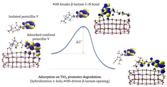
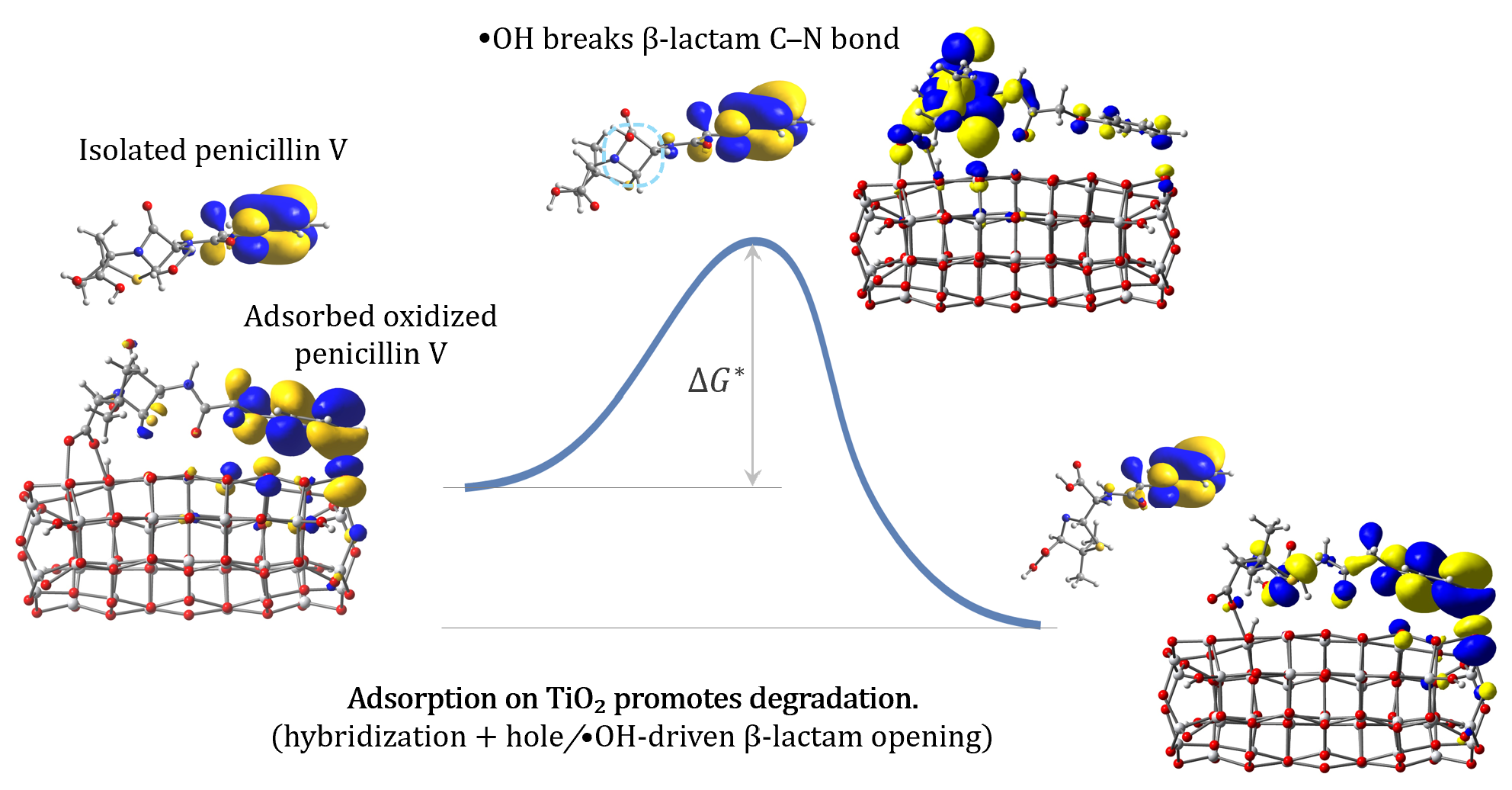
Graphical Abstract

1. Introduction

Urban wastewater was found to be the dominant emission pathway for pharmaceuticals at a global level, whereas emissions from industrial production, hospitals, agriculture, and aquaculture were found to be more significant locally [1,2]. Ecological disorders due to the appearance of antibiotics in water resources have led to an increased interest in their removal [3,4]. Being an extensively used antibiotic [5,6], penicillin is a common pollutant in wastewater [7]. To remove penicillin from wastewater, researchers use different methods, of which advanced oxidation techniques are among the preferred ones, due to the operational difficulties and high cost of conventional approaches [8].
Advanced oxidation processes involve the release of reactive oxygen species, such as hydroxyl radicals, which have a high redox potential and the ability to attack contaminants via electron transfer or hydrogen abstraction. The goal is to mineralize contaminants into harmless byproducts by breaking down complex pollutants into simpler, less toxic substances or making them biodegradable [9]. The advanced oxidation processes are very diverse and can be classified into homogeneous and heterogeneous, distinguishing between those that operate without an external input of energy and those that need radiant, ultrasonic, or electrical energy [10,11]. Among these techniques, the photocatalytic degradation of pollutants is based on the chemical reaction induced by the absorption of photons by a solid material, the photocatalyst [12].
Titanium dioxide is, probably, the most widely used and studied photocatalyst in advanced oxidation processes [13], primarily due to its favorable combination of non-toxicity and environmental friendliness, high chemical and optical stability, strong oxidizing ability, efficiency in mineralization, versatility in use, cost-effectiveness, and availability [14,15,16,17,18]. Photocatalytic degradation with TiO2 is most often interpreted in terms of direct semiconductor excitation, whereby UV absorption by the oxide generates electron–hole pairs that produce reactive oxygen species (ROS, such as •OH, •O2, etc.), responsible for pollutant oxidation [19,20,21,22]. However, increasing experimental evidence indicates that, for strongly adsorbed molecules, alternative pathways involving pollutant photoexcitation and interfacial electron transfer to TiO2 may also contribute to the overall degradation process [20,23,24,25].
The role of energy level alignment in the photocatalytic degradation of organic pollutants (antibiotics included) on TiO2 nanoparticles has been recognized from the early studies [20,26,27] and revisited and confirmed over the years [28,29,30,31,32], with some caveats related to pH level, surface-induced dipoles, etc. [33]. The analysis has been focused on the requirement that the valence band position of the oxide is sufficiently positive to enable oxidation either directly via photogenerated holes or indirectly through the formation of hydroxyl radicals, while the conduction band is marginally negative enough to allow for oxygen reduction to superoxide. The relevance of the pollutant’s ground and excited electronic states remains debated; a general understanding is still needed, notwithstanding the large number of studies [25,29,30].
Despite the tremendous progress in the study of the photodegradation of antibiotics, there are still some unresolved issues, particularly regarding that of penicillin on nanocrystalline TiO2. Although recent studies have demonstrated that anatase TiO2 P25 nanoparticles can achieve degradation efficiencies exceeding 90% for penicillin G under UV irradiation, most experimental investigations focus on optimizing operational parameters such as pH, catalyst loading, and irradiation time [34,35]. Consequently, while a general phenomenological understanding of the photodegradation process exists, the site-specific molecular attack by reactive species and the corresponding elementary steps remain insufficiently explored at the theoretical level.
Even within the prevailing interpretation that the direct UV excitation of TiO2 is followed by ROS generation [14,36], fundamental questions remain regarding whether the initial attack on the antibiotic occurs at the surface or within the interfacial water layer [37]. In parallel, the increasingly discussed alternative route, invoking light absorption in the complex oxide-pollutant system and subsequent electron injection into TiO2, requires clarification in terms of adsorption geometries, electronic coupling, charge-transfer efficiency, and the identification of the most vulnerable molecular sites [38].
Therefore, the fundamental question is whether the observed activity under broadband irradiation is governed primarily by intrinsic TiO2 excitation or includes a significant adsorbate-mediated charge-transfer contribution. Furthermore, for the two mechanisms, it is of interest to more rigorously identify the molecular sites most susceptible to initial degradation [39]. In particular, it remains unclear whether degradation initiates through the early opening of the β-lactam ring or via preferential attack on atoms of the backbone, and how the electronic structure of the thiazolidine and β-lactam moieties governs their relative reactivity [40]. Other questions requiring more careful theoretical investigation regard the binding configuration of the pollutant on the oxide [41,42,43], whether a deprotonation takes place during adsorption and whether it has an impact on reactivity, whether the primary attack is driven by hole h+ oxidation or •OH attack [36,44,45,46], etc.
Under these circumstances, there is a clear need for computational studies to disentangle light adsorption, interfacial charge transfer, and radical-driven bond-breaking processes [47] that cannot be resolved unambiguously from experiments alone. Here, we address these issues using density functional theory (DFT) calculations [48,49], which have been proven to provide atomistic insight into adsorption geometries, electronic structure modifications, energy level alignment, and charge-transfer energetics at the TiO2-penicillin interface [24,50,51,52].
Building upon our previous studies on the photocatalytic degradation of colorless aromatic pollutants [53] and antibiotics [54], we report density functional theory calculations aimed at elucidating the processes governing the photocatalytic degradation of penicillin V (phenoxymethylpenicillin, PenV) on anatase TiO2 nanoclusters. In particular, we investigate whether degradation preferentially occurs in solution or upon adsorption on the oxide surface and identify the molecular sites most vulnerable to attack in each case. The novelty of the present approach lies in the combined DFT treatment of penicillin, both free in aqueous solution and adsorbed on a TiO2 nanocluster. Within this framework, we analyze adsorption configurations, energy-level alignment, charge-transfer pathways, UV-Vis absorption properties, local reactivity through condensed Fukui functions, and the initial reaction pathways leading to bond breaking.
The present DFT investigation provides a unified mechanistic picture of the photocatalytic degradation of penicillin V by explicitly comparing solution-phase and surface-mediated pathways within a consistent theoretical framework, using the same descriptors and conceptual tools for isolated PenV in solution, neutral PenV adsorbed on TiO2, and oxidized PenV adsorbed on TiO2. Throughout this work, mechanistic conclusions are drawn from the convergence of multiple electronic, structural, spectroscopic, and reactivity descriptors evaluated within the same theoretical framework, rather than from any single energetic or kinetic criterion.

2. Results

This section is divided into five parts. The first subsection describes the optimized geometry and optical properties of penicillin V (PenV), the second focuses on the titania nanocluster, whereas the third focuses on the structure and properties of the antibiotic binding to the cluster. The last two subsections report DFT calculations exploring two photocatalytic degradation mechanisms for PenV, one in solution, the other adsorbed on the catalyst.

2.1. Optimized Geometry and Electronic Structure of Penicillin V

Despite the widespread environmental relevance of penicillin V and the interest in its structure, conformational stability, and vibrational properties [55,56], a DFT study to address both its reactivity [57] and its photocatalytic degradation is still needed. The first step in the investigation of the photocatalytic degradation of PenV is the characterization of its optimized molecular geometry and electronic structure in the isolated state. To establish a reference framework for interpreting the subsequent results on adsorption onto TiO2 nanoclusters and photocatalytic degradation pathways in solution and at the catalyst surface, we optimized the geometry of PenV in its neutral form, as well as in a deprotonated (carboxylate) state (Figure 1 and Table 1).
The DFT-optimized geometry of PenV exhibits a compact molecular shape with a clear distinction between structurally rigid and flexible regions. The structural parameters indicate, in agreement with previous reports [55,56], that the two fused rings—the four-membered β-lactam and the five-membered thiazolidine—together form a relatively rigid core. Their mutual orientation is largely preserved, as reflected by the characteristic dihedral angle ∠(N1–C4–S–C5), which changes only modestly, decreasing by 2.5° upon carboxylate deprotonation.
The thiazolidine ring itself shows only minor internal distortions. Upon –COOH deprotonation, the ∠(N1–C4–S–C5) dihedral decreases by 2.5° and the C3–S bond length increases by just 0.007 Å. The tighter C4–S bond exhibits slightly larger variations, increasing by 0.032 Å upon deprotonation.
The β-lactam ring also remains geometrically constrained, with only small changes in its internal angles. For example, ∠(N1–C4–C5) decreases by 0.4° upon deprotonation, while the dihedral ∠(N1–C4–C5–C6) changes by −1.4°. The N1–C6 bond, which corresponds to the strained amide linkage of the β-lactam ring, is sensitive to deprotonation, decreasing from 1.453 Å to 1.399 Å. These trends identify the β-lactam N–C bond as a key mechanistically relevant descriptor for subsequent adsorption and degradation processes.
In contrast to the rigid fused-ring core, the terminal carboxyl group undergoes substantial electronic and geometric reorganization upon deprotonation. The strongly asymmetric C–O bond lengths of the neutral –COOH group (1.365 and 1.222 Å) become nearly equal in the carboxylate (1.269 and 1.270 Å), while the O–C–O angle increases from 122.8° to 131.2°, reflecting resonance delocalization and electrostatic opening of the group. This transformation is accompanied by a large rotation of the O1–C1–C2–N1 dihedral from −14.4° to 23.6°, demonstrating the high conformational adaptability of the carboxylate moiety.
The flexibility of the phenoxyacetyl side chain is similarly reflected in its dihedral angles. Both ∠(C7–C8–O5–C9) and ∠(C8–O5–C9–C10) show only small variations for free PenV but change significantly upon binding to the TiO2 surface, as anticipated from the data in Table 1.
Together, these quantitative trends confirm that PenV consists of a rigid β-lactam–thiazolidine core surrounded by highly adaptable peripheral functional groups that can readily reorient to maximize electrostatic stabilization and surface coordination.
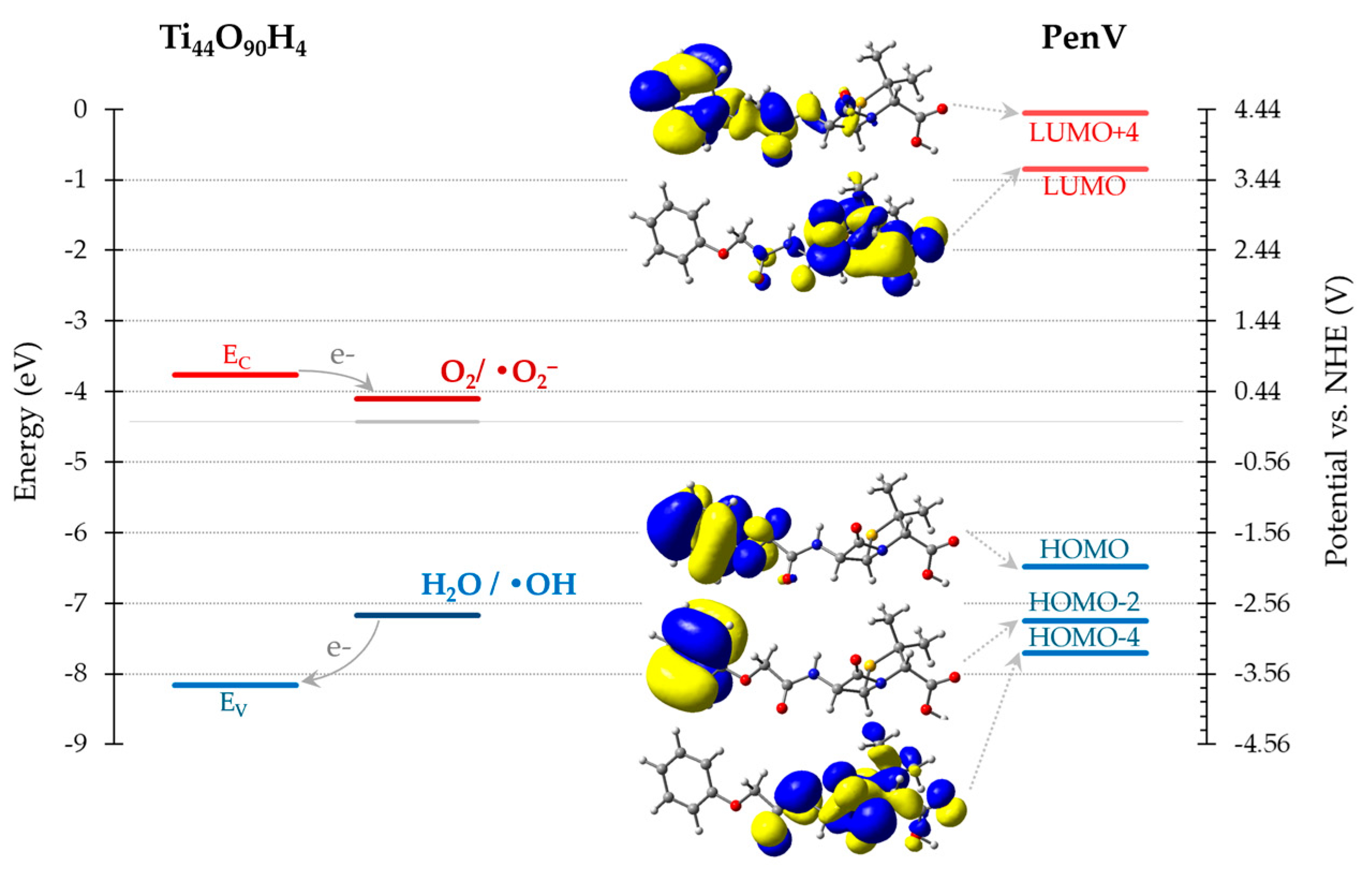
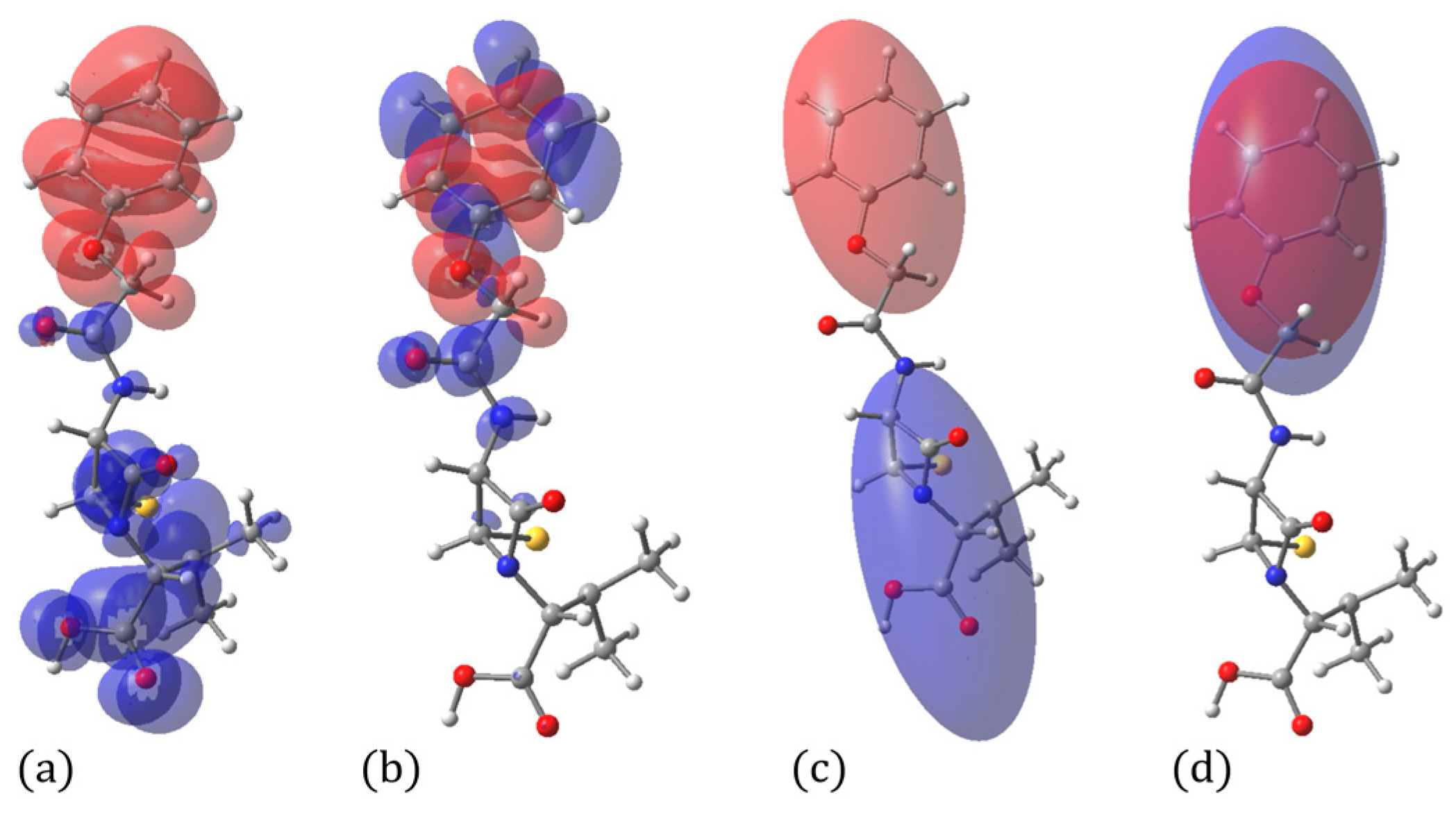
Analysis of the electronic structure shows that the frontier molecular orbitals are spatially separated over the PenV molecule. The highest occupied molecular orbital (HOMO) is mainly localized on the phenoxyacetyl moiety and sulfur-containing regions, whereas the lowest unoccupied molecular orbital (LUMO) has significant contributions from carbonyl-containing groups, including the β-lactam ring (Figure 2). This distribution indicates an intrinsic intramolecular charge-transfer character, a push→pull effect, which promotes electron density displacement toward the carboxyl group that can easily bind to the oxide.
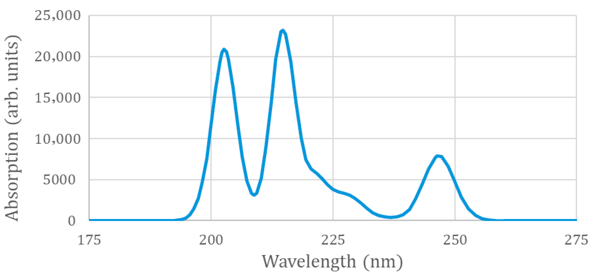
Importantly, the PenV LUMO lies more than 2.9 eV above the TiO2 conduction band edge. Although electron injection from photoexcited PenV into the oxide is therefore thermodynamically allowed, the large energetic offset makes this process comparatively unlikely and suggests inefficient electronic coupling in the absence of strong hybridization. In addition, direct photoexcitation of PenV is itself unlikely, owing to the large energy gap between its ground and excited states (5.63 eV, consistent with previous reports on penicillin [56]), which requires high-energy UV irradiation, as shown in Figure 3. Taken together, these considerations argue against a degradation mechanism driven by direct light absorption by PenV followed by charge transfer to the oxide.
The calculated electronic absorption spectra of PenV in aqueous solution are shown in Figure 3. The UV-Vis spectrum indicates that the lowest-energy electronic transitions occur at approximately 246 nm and are mainly associated with a HOMO → LUMO+2 excitation. In contrast, the HOMO → LUMO transition exhibits very low oscillator strength, which can be attributed to the near orthogonality of the involved orbitals arising from their spatial separation. In particular, the HOMO is localized predominantly on the phenoxyacetyl moiety, whereas the LUMO is mainly distributed over the β-lactam–thiazolidine core and the carboxyl anchoring group, as shown in Figure 2. The strongest absorption band is observed around 214 nm and can be assigned predominantly to a HOMO → LUMO+4 transition. An intense peak at approximately 202 nm originates from two closely spaced excitations, which are mainly characterized as HOMO−2 → LUMO and HOMO−4 → LUMO+1 transitions, respectively.
Overall, the optimized geometry and electronic structure of PenV highlight the complementary roles of molecular rigidity and flexibility in governing adsorption behavior and reactivity. These features provide a rational basis for the adsorption configurations and photocatalytic degradation pathways examined in the following subsections.
A charge density difference analysis was performed to elucidate how the electron density redistributes upon electronic excitation from the HOMO to the LUMO and to the LUMO+4 states. This approach enables the direct visualization of the regions of electron depletion and accumulation associated with each transition, as shown in Figure 4. To further quantify the extent of charge separation, the spatial separation between the centroids of the electron-depleted (hole) and electron-rich regions was also evaluated for both excitations.
Both analyses reveal a clear tendency for electron density to redistribute toward the anchoring region of the molecule upon excitation. Quantitatively, the centroid analysis indicates a pronounced charge separation of 10.118 Å for the HOMO → LUMO transition, whereas a much smaller separation of 0.053 Å is obtained for the HOMO → LUMO+4 transition.
This marked difference reflects the strong spatial separation of the orbitals involved in the lowest-energy excitation, in contrast to the more localized character of the higher-energy transition.

2.2. Optimized Geometry and Electronic Structure of TiO2 Nanoclusters

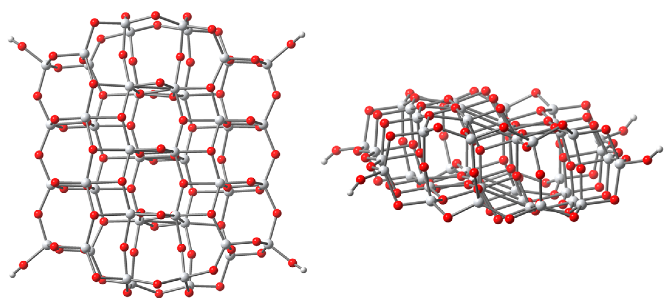
To gain insight into the dye–oxide interactions, we need to consider first the structure and properties of the TiO2 cluster on which the dye is adsorbed. We modeled TiO2 anatase nanoparticles by means of a Ti44O90H4 cluster, as an optimum size system [58] for describing the interface effects with the relatively small but flexible antibiotic molecule. In an earlier DFT study of PenV adsorbed on TiO2, we showed that when using increasingly larger nanoclusters, the energy of the system can be lowered substantially by distorting PenV [58]. The bending and twisting of the backbone allowed for energy minimization due to extra bonds and π-π interactions, indicating the cluster with 44 Ti atoms as an optimum compromise between rigor and computational effort [58].
The starting geometry for the structural optimization of the cluster originated in the experimental structure of the (101) and (001) anatase titania surfaces [59,60]. To avoid the problem of the surface states in the gap, we performed a geometry optimization of the model clusters with a slight deviation from the TiO2 stoichiometry, introducing four hydrogen atoms to terminate the dangling bonds at the periphery. This approach resulted in compact structures with 4-, 5-, and 6-fold coordinated Ti ions, together with 2- and 3-fold coordinated oxygen atoms [58].
Following the geometry optimization, the structure is slightly distorted from the bulk to minimize the surface stresses (Figure 5). The average Ti-O distance for the cluster is 1.896 Å, smaller than the 1.950 Å observed experimentally for the bulk. Additionally, the distribution of Ti-O distances in the cluster is wider in the cluster than in the bulk, with a standard deviation of 0.103 Å for the former, larger than the 0.022 Å for the latter. The deformation of the structure changes angles and distances such that the larger distances tend to be compensated for by smaller ones; the relative variation in the cluster distances compared to the values in the bulk is around 5%.
The geometry relaxation leads to a band gap of ~4.4 eV between the frontier orbitals, larger than the experimental value of ~3.2 eV for anatase titania [61], an overestimation that was observed in many DFT calculations of model clusters, particularly when using hybrid DFT functionals [60,62].
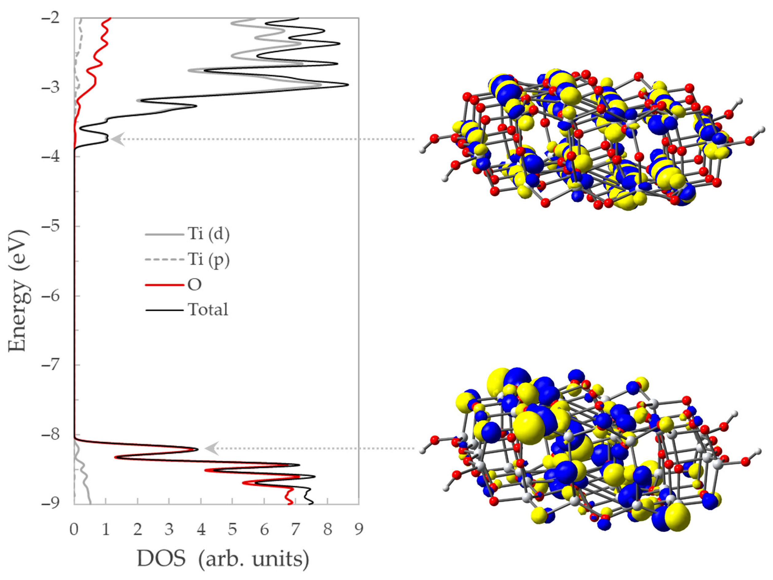
The nature of the states in the valence and conduction bands can be better understood from the density of states (DOSs), displayed in Figure 6. The valence band is indeed dominated by the 2p atomic orbitals of O with little contribution from Ti. The dominant contribution in the conduction band comes from the titanium 3d orbitals, the involvement of the oxygen atoms being small, even for the p orbitals. The hydrogen atoms, which are used to end four dangling bonds and remove the surface states from the gap, have a negligible contribution to the DOSs, typically less than 0.5%.
The corresponding band gap of the isolated cluster is ~4.46 eV, between edge states at about −8.21 eV and −3.75 eV, for the valence and conduction bands, respectively, consistent with previous DFT studies of anatase-like TiO2 clusters [60].
To visually illustrate the nature of the frontier orbitals, we also displayed in Figure 5 the electronic density of the states situated at the edges of the valence and conduction bands. The highest occupied orbital of the oxide has a predominantly p-character, with the charge localized especially on O atoms, whereas the lowest unoccupied orbital has a mostly d-character with the main contribution from Ti atoms. It is important to note the high degree of delocalization of the charge at the conduction band edge. While at the valence band edge, the charge is localized mostly on certain oxygen atoms, situated at the surface, in the conduction band, the electron is shared by most Ti atoms, particularly the peripheral ones.

2.3. Optimized Geometry and Electronic Structure of Penicillin V Adsorbed on TiO2 Nanoclusters

Understanding the photocatalytic oxidation of pollutants adsorbed on TiO2 surfaces necessarily begins with their binding to the oxide. Despite extensive investigation, the binding configurations of organic molecules on the reactive anatase TiO2 (101) surface remain the subject of ongoing debate, even for relatively simple adsorbates, such as formic acid [63,64]. Several studies have reported bidentate bridging configurations for a variety of dyes adsorbed on TiO2 surfaces [65,66,67]. However, alternative binding modes have been identified for molecules possessing multiple functional groups, as in the case of salicylic acid [53].
For penicillin, earlier theoretical and experimental reports [54,58] suggested adsorption predominantly via a bidentate carboxylate configuration, accompanied by deformation of the molecular backbone upon binding. Nevertheless, other studies have proposed monodentate or chelating coordination modes for carboxyl-containing molecules on TiO2 surfaces [41,42,43], highlighting that adsorption geometry can depend sensitively on molecular structure, surface model, and computational or experimental conditions.
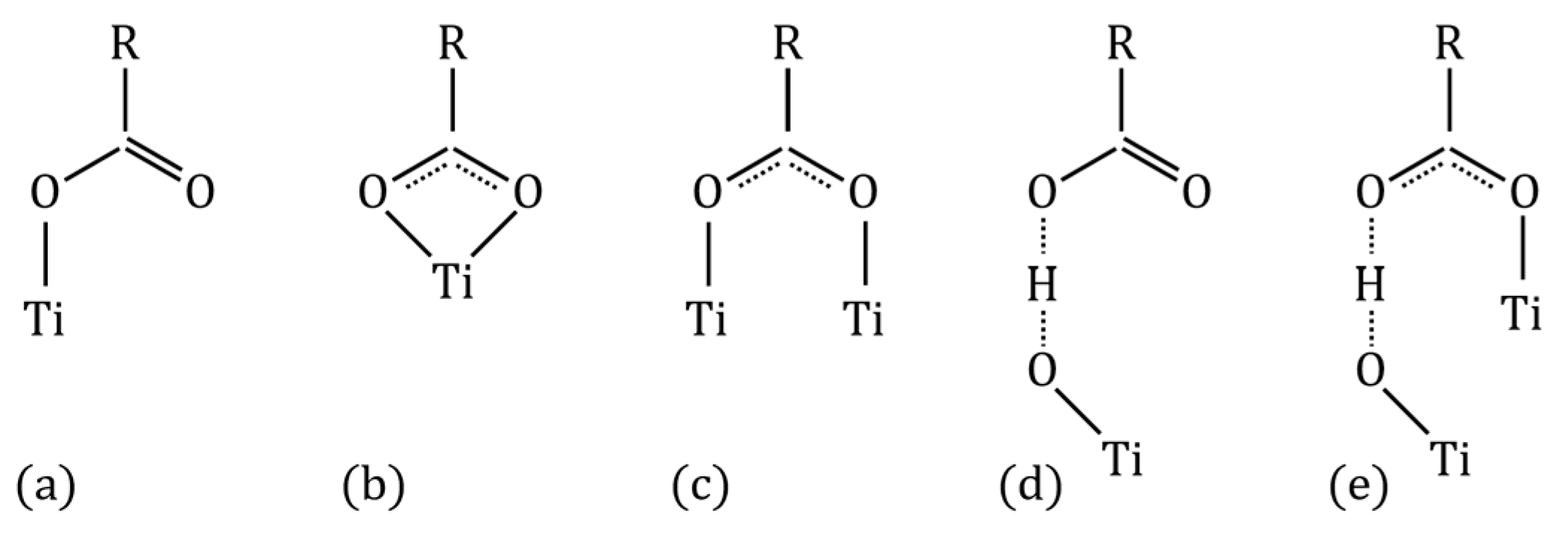
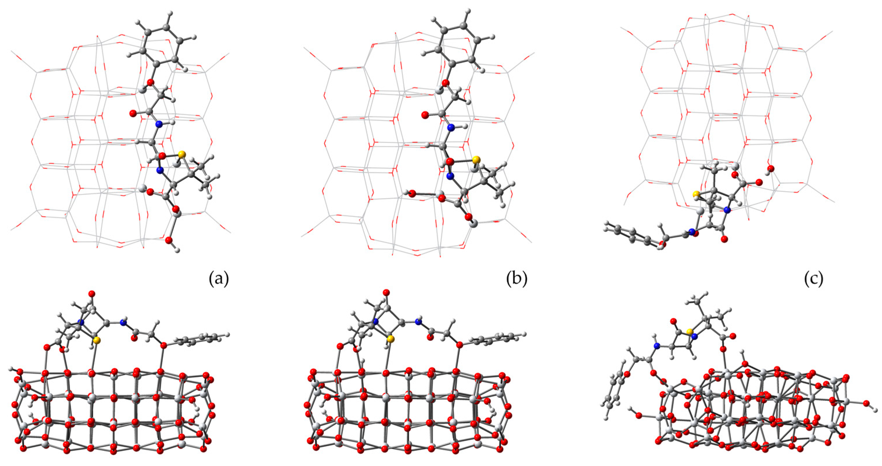
To clarify the preferred binding configuration of PenV on TiO2, we performed a series of DFT calculations exploring the most common adsorption motifs. A priori, carboxylate binding on TiO2 may occur in monodentate, bidentate-chelating, or bidentate-bridging modes, as well as through hydrogen-bonded interactions with surface oxygen atoms, which involve one or two surface Ti sites, as schematically illustrated in Figure 7.
The calculations were performed starting from the neutral molecule placed near the cluster surface in several initial orientations corresponding to the binding motifs illustrated in Figure 7. Upon full structural optimization, the low-energy configurations converged toward a bidentate-bridging geometry, displayed in Figure 8a, in which the carboxyl group transfers its proton to a nearby surface oxygen atom, leading to anchoring through both carboxylate oxygens to the adjacent Ti sites. In this configuration, the C1–O bond lengths become nearly equal (1.285 Å for C1–O1 and 1.293 Å for C1–O2), while the corresponding Ti–O distances are similar, measuring 2.061 Å for O1–Ti1 and 2.096 Å for O2–Ti2.
Adsorption is accompanied by significant molecular deformation. In particular, PenV bends toward the surface, bringing into closer proximity with the surface Ti sites both the sulfur atom (S–Ti distance of 2.688 Å) of the thiazolidine ring and the oxygen atom of the phenoxyacetyl moiety (O5–Ti distance of 2.342 Å). At the same time, the phenoxyacetyl chain undergoes a torsional rearrangement that reorients the phenyl ring roughly parallel to the oxide surface. These structural adjustments enable the effective accommodation of the molecule at the interface while preserving the integrity of the β-lactam-thiazolidine core.
A single optimization converged to a structure featuring one Ti–O bond and one hydrogen-bond interaction, shown in Figure 8c, corresponding to configuration (e) in Figure 7. In this case, the C1–O1 bond length is 1.305 Å, with a short O1–Ti1 distance of 1.961 Å. Deprotonation also occurs in this case, as the hydrogen atom lies closer to the oxygen atom on the catalyst surface (1.080 Å) than to the oxygen of the carboxyl group of the pollutant (1.464 Å). This binding motif requires substantially stronger bending of the PenV backbone toward the side of the cluster, leading to an additional interaction between O4 and a surface Ti atom at a distance of 2.051 Å. As a result of this increased deformation, the configuration is energetically disfavored, lying approximately 1.6 eV higher in energy than the bidentate-bridging structure.
Finally, additional calculations were performed starting from deprotonated PenV in the presence of a surface hydrogen atom on nearby oxygen, as displayed in Figure 8b. After optimization, the resulting energy was higher by only ~0.1 eV, and the adsorption geometry and structural features were found to be essentially identical to those obtained from the neutral starting configurations, indicating that proton transfer to the surface occurs spontaneously upon adsorption and does not affect the final structure of the system.
The lowest-energy bidentate-bridging configuration was selected for detailed analysis. Setting the ground for the study of the degradation mechanism, we start by comparing the structures of isolated deprotonated PenV with surface-bound PenV and oxidized PenV (see Table 1).
Comparison of the optimized PenV geometry in vacuum with the surface-bound structure shows that adsorption-induced deformation is highly localized. The dominant changes occur at the terminal carboxyl group, which reorients to enable bidentate coordination to Ti sites, with Ti–O distances of 2.042 and 2.117 Å. This coordination is accompanied by marked C–O bond-length equalization (1.365 → 1.299 Å for O1–C1 and 1.222 → 1.280 Å for O2–C1) and by a large rotation of the carboxyl-bearing fragment, as reflected by the dihedral angle ∠(Ti–O1–C1–C2), which shifts from approximately 0° to about 130°. A smaller adjustment follows in the adjacent dihedral ∠(O1–C1–C2–N1), which changes from −14.35° to −0.84°.
The clearest adsorption-driven deformation is not observed within the β-lactam–thiazolidine framework itself, but rather in the C1–C2–S linkage that couples the anchoring group to the fused rings. A reduction of approximately 14° is found for ∠(C1–C2–S), from 111.17° to 97.51°, and for the dihedral ∠(C1–C2–C3–S), from 92.49° to 77.88°. These changes indicate bending of the molecular backbone near the junction with the fused-ring core. In contrast, internal metrics of the β-lactam–thiazolidine scaffold (N1–C6, C6–O3, and ∠(N1–C4–S–C5)) remain nearly unchanged upon adsorption, supporting its role as a rigid structural unit. One noticeable, though still moderate, variation is observed for the C4–S bond, which increases by 0.037 Å.
The phenoxyacetyl side chain responds primarily through torsional rearrangement. This is evidenced by a decrease of approximately 26° in ∠(C7–C8–O5–C9), from 178.12° to 151.71°, and, more prominently, by an increase of about 46° in ∠(C8–O5–C9–C10), from 1.20° to 46.95°. These changes reflect a conformational accommodation to the surface environment, with the phenoxy ring adopting an orientation roughly parallel to the oxide surface, a configuration commonly associated with enhanced dispersive and π–surface interactions.
The most consistent trend upon adsorption is the balancing effect on the carboxyl group, which exhibits near-equal C–O bond lengths, consistent with carboxylate resonance delocalization, and the bending that occurs both at the anchor and along the phenoxyacetyl chain, as the phenyl group reorients roughly parallel to the surface.
Moving to oxidized PenV, the largest difference is observed for the linker angles ∠ (C1-C2-S) and ∠(C1-C2-C3-S), which decrease by almost 15°. Next, variations of about 10° and 6° are seen for the phenoxy side of the chain, for ∠(C8-O5-C9-C10) and ∠(C7-C8-O5-C9), respectively, and smaller changes for ∠(N1-C4-C5-C6) and ∠(O3-C6-C5-N2).
In summary, the adsorption on TiO2 has only a minor effect on the relatively rigid β-lactam–thiazolidine core, while inducing substantially larger structural adjustments in the flexible peripheral groups, namely the carboxylate anchoring moiety and the phenoxyacetyl side chain. In addition to bidentate carboxylate anchoring, secondary interactions contribute to stabilization, including close contacts between the sulfur atom of the thiazolidine ring and surface Ti sites, as well as between the ether oxygen along the side chain and the oxide surface. Furthermore, the reorientation of the phenoxyacetyl group aligns the benzene ring approximately parallel to the surface, favoring π–surface and dispersive interactions that enhance overall adsorption stability.
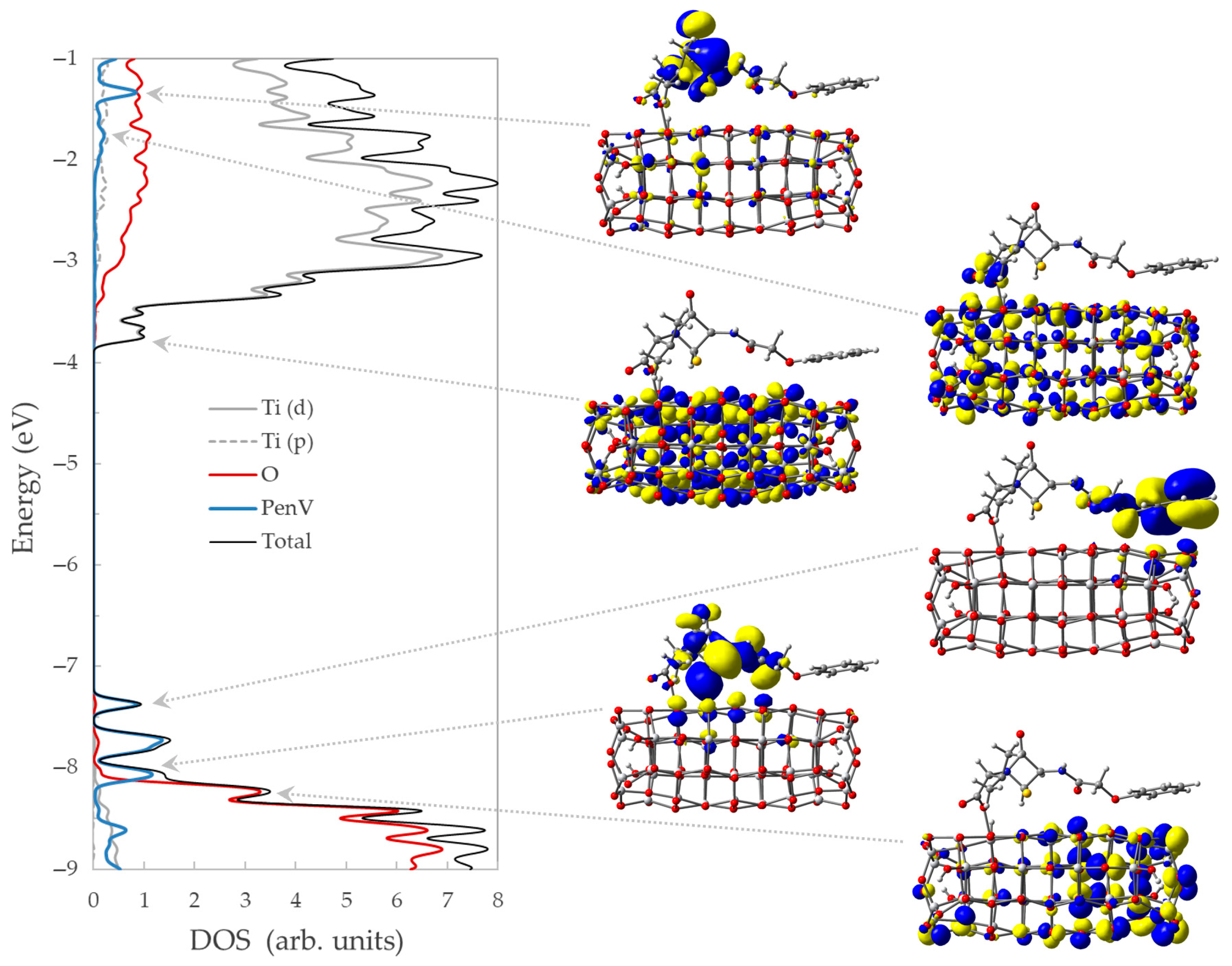
The projected density of states (DOSs) of PenV adsorbed on the TiO2 nanocluster provides direct insight into the electronic coupling between the molecule and the oxide (Figure 9). Upon adsorption of PenV, the overall shape and energetic position of the oxide-derived band edges remain essentially unchanged, indicating that the TiO2 electronic framework is not significantly perturbed by molecular binding. In particular, the onset of the Ti 3d-dominated conduction band and the upper edge of the O 2p-derived valence band occur at nearly the same energies as in the bare cluster.
In contrast, additional features appear in the DOSs of the hybrid system that are absent in the bare TiO2 cluster. Specifically, the PenV-derived states emerge as localized peaks within the oxide band gap, at about −8.03 eV, −7.73 eV, and 7.386 eV, and as discrete contributions overlapping with the conduction band, at −1.74 eV and at −1.34 eV.
On the occupied side, PenV contributes substantially to electronic states located within the band gap of the TiO2 cluster, with these states showing minor but non-negligible mixing with O 2p orbitals of the oxide, as evidenced by the projected DOSs. At the opposite end of the gap, near the conduction band edge, the lowest unoccupied states are dominated by Ti 3d character with negligible contribution from PenV-derived orbitals.
The hybridization between molecular and oxide states across the band gap introduces finite transition dipole moments between occupied PenV-derived gap states and Ti 3d-dominated conduction-band states. As a result, optical transitions that would be symmetry- or selection-rule-forbidden in the isolated subsystems become weakly allowed in the hybrid system. Due to the limited degree of electronic mixing, these transitions have low oscillator strength, but they occur at lower energies than the intrinsic TiO2 excitations, leading to a red shift in the UV-Vis absorption spectrum (see Figure 10).
Furthermore, on the vacant side, a sizeable hybridization between excited PenV states and TiO2-derived conduction-band states occurs only at high energies, affecting the likelihood of the interfacial charge transfer from a photoexcited antibiotic to the catalyst. In contrast, occupied PenV states lying close to the valence band edge are better positioned to participate in hole-mediated oxidation processes.
The key molecular orbitals of the PenV/TiO2 system are also illustrated in Figure 9 together with their corresponding positions in the density of states. The figure displays the representative orbitals associated with the valence band edge of the oxide, PenV-derived ground state located within the band gap, the conduction band edge, a mixed character state, and the PenV-derived excited state. The highest occupied states exhibit a predominantly p-character, with electron density largely localized on the oxygen atoms of the oxide and the molecular adsorbate, reminiscent of the π-interaction at the surface. At the opposite end, the lowest unoccupied state is mainly characterized by Ti 3d orbitals, whereas the illustrated mixed states have the charge delocalized over the oxide and over the carboxyl anchor or on the β-lactam–thiazolidine core.
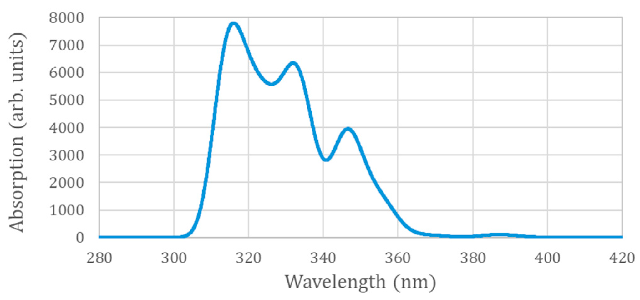
The calculated electronic absorption spectra of PenV in aqueous solution are shown in Figure 10. The UV-Vis spectrum indicates that the lowest-energy electronic transitions are low intensity, occur at approximately 387 nm, and are mainly associated with a HOMO → LUMO excitation, with the charge moving from PenV to the oxide. Similar transitions from the lower states of the antibiotic to the conduction band are found at 355 nm, with higher intensities at 347, 335, 333, and 317 nm. The transition to the excited state of PenV is at much higher energies, and it is not among the first 50 singlet transitions.
Overall, the electronic absorption spectrum of the PenV/TiO2 complex exhibits a clear red shift relative to the isolated antibiotic. While isolated PenV shows its lowest-energy allowed transitions in the deep-UV region (around 240–250 nm), adsorption on TiO2 leads to the appearance of lower-energy absorption features extending toward longer wavelengths. This red shift originates from the hybridization between PenV-derived occupied states located in the band gap and Ti 3d-dominated conduction-band states of the oxide, as evidenced by the density-of-state analysis. The resulting mixed molecular–oxide character enables the weakly allowed optical transitions that are absent in the isolated molecule, thereby reducing the effective excitation energy of the system.
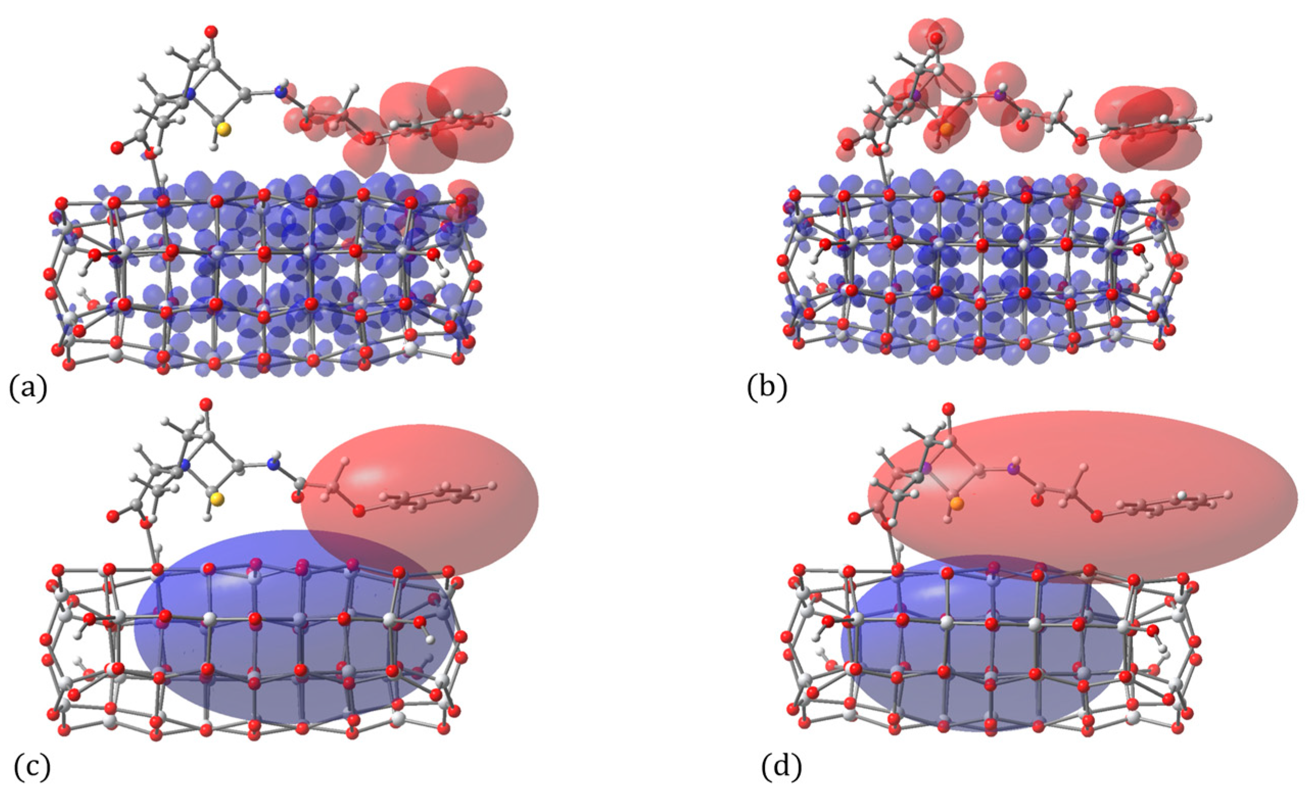
For the longest-wavelength transition at 387 nm (HOMO → LUMO) and for the intense transition at 333 nm, arising mainly from HOMO−1 → LUMO and HOMO−2 → LUMO excitations, charge density difference analyses were performed. In addition, the spatial separation between the centroids of the electron-depleted (hole) and electron-rich regions was evaluated for both transitions, as illustrated in Figure 11. In both cases, the charge density difference maps reveal a clear redistribution of electron density from the pollutant toward the catalyst upon excitation.
Quantitatively, the centroid analysis indicates a larger charge separation of 8.193 Å for the longest-wavelength transition, compared to 6.962 Å for the more intense transition. This difference reflects the distinct degree of charge delocalization in the initial states involved, with the HOMO exhibiting a stronger spatial separation from the LUMO than the lower-lying occupied orbitals contributing to the higher-intensity excitation.

2.4. Photocatalytic Degradation of Penicillin V in Solution

In this subsection, we address the initial steps of the photocatalytic degradation of PenV occurring in solution, where the antibiotic interacts with reactive oxygen species generated by the TiO2 catalyst but remains spatially separated from the oxide surface. On the basis of electrostatic potential maps, Fukui functions, and atom-resolved Fukui coefficients, we first analyze the intrinsic vulnerability of PenV in an aqueous environment toward the attack by reactive oxygen species. We then focus explicitly on the hydroxyl radical (•OH) and examine its interaction with the C6 carbon atom of the β-lactam ring, identified as the key reactive site, in order to assess the energetic barriers associated with bond weakening and to elucidate the most likely initiation step of the degradation process. Hydroxyl radicals are considered here as a representative and highly aggressive oxidative species [45,46], allowing for a consistent comparison of intrinsic molecular vulnerability across different environments, rather than as an exclusive reactive pathway.
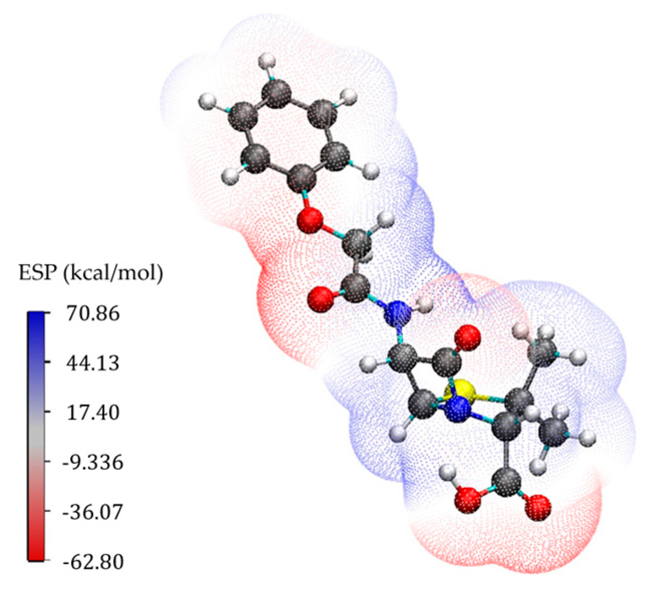
The electrostatic potential (ESP) mapped onto the electron density surface of PenV is shown in Figure 12. Regions of negative electrostatic potential are primarily localized on the carboxylate group and on the carbonyl oxygens of the β-lactam moiety, whereas positive electrostatic potential is found near hydrogen atoms and electron-deficient regions of the molecular framework, including the amide and heterocyclic nitrogen sites.
The Fukui function analysis provides a spatially resolved description of how the electron density of PenV responds to different types of electronic perturbations (Figure 13). In addition, the atom-resolved Fukui coefficients displayed in Table 2 further quantify the trends observed in the Fukui isodensity surfaces.
For both radical (f0) and nucleophilic (f+) perturbations, Fukui functions show significant contributions to the β-lactam–thiazolidine core. The largest coefficients are associated with atoms belonging to the β-lactam ring, O3 and C6 consistently appearing among the most responsive atoms in both f0 (0.1031 and 0.0997, respectively) and f+ (0.2022 and 0.1975), indicating that this region dominates the electronic response. Additional contributions common to both functions are found on heteroatoms such as O5, O2, and N1, as well as on carbon atoms adjacent to the β-lactam ring.
At the same time, some differences are observed in the spatial distribution of the two functions. The radical Fukui function (f0) shows comparatively larger contributions on atoms along the side chain, including C12, O5, and C9, reflecting a more distributed radical sensitivity. In contrast, the nucleophilic Fukui function (f+) is more strongly concentrated on heteroatoms and electrophilic centers, with secondary contributions on S, O2, and N1.
The electrophilic Fukui function (f) displays a distinct pattern, with its largest coefficients localized on carbon atoms of the phenoxyacetyl chain (C12, C9, C14, and C10) as well as on O5, indicating that electron removal primarily affects the aromatic and side-chain framework rather than the β-lactam core.
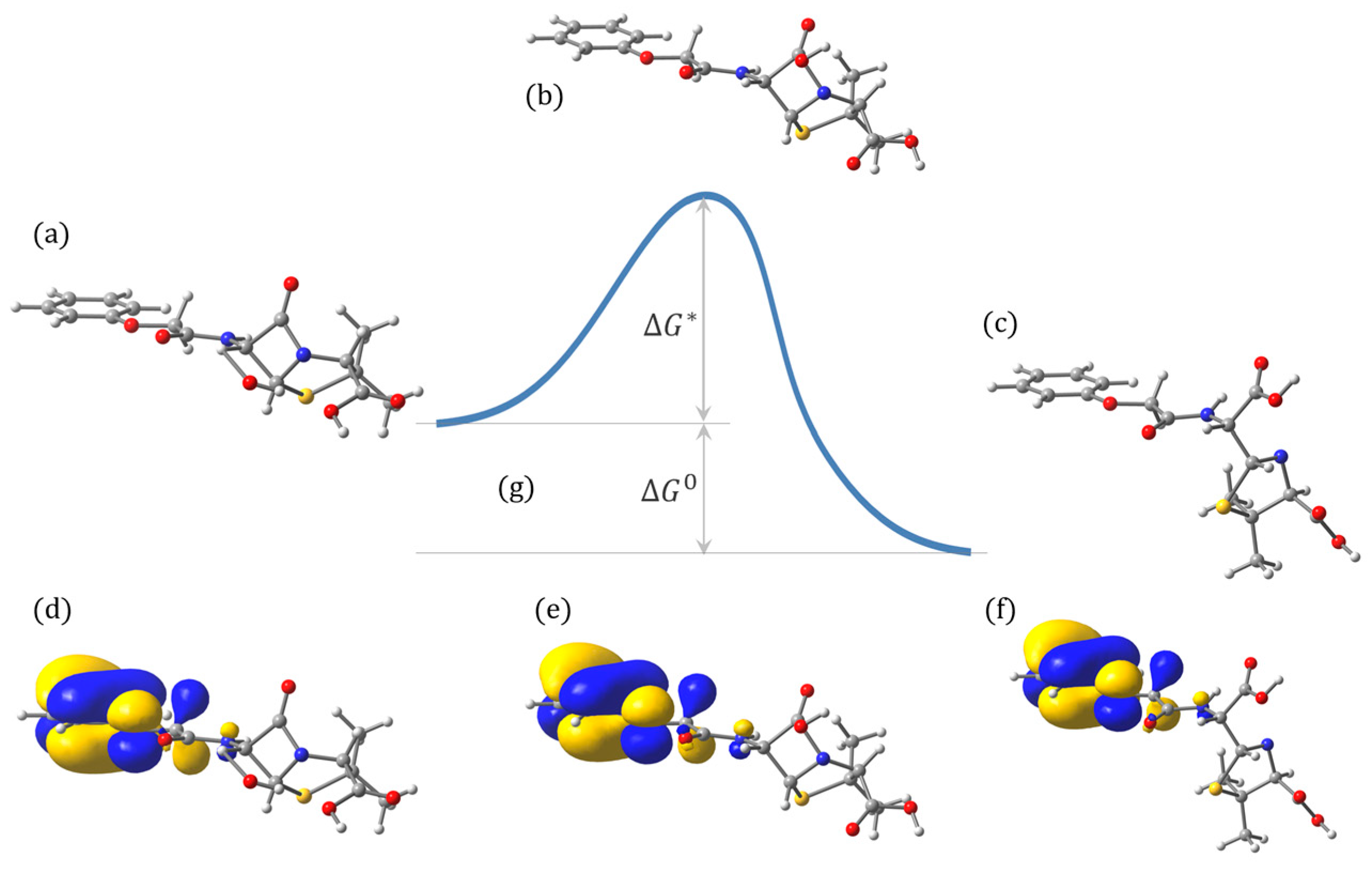
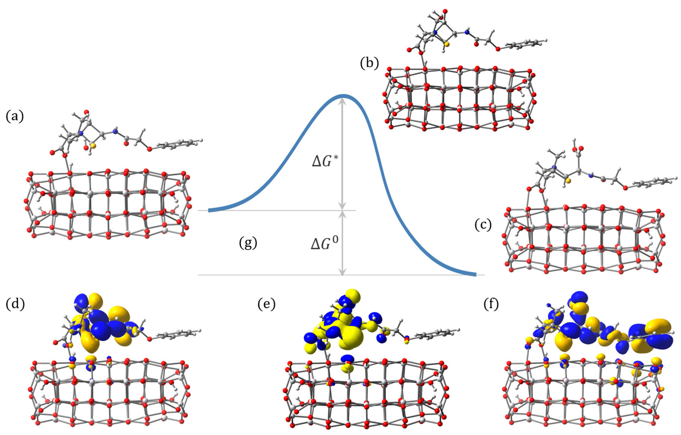
Further, to probe the initial step of photocatalytic degradation, we chose the hydroxyl radical (•OH), as it is generally considered the most reactive and aggressive oxidative species generated under TiO2 photocatalytic conditions and is expected to play a dominant role in initiating the degradation of β-lactam antibiotics [68,69]. A series of constrained geometry optimizations was performed by progressively bringing a hydroxyl radical toward the C6 carbon atom of the β-lactam ring, identified as the most vulnerable site from the reactivity analyses. For each system considered, the O···C6 distance was systematically reduced from 2.00 Å in steps of 0.01 Å, and the structure was optimized at each step while monitoring the evolution of the C6–N1 bond length. This procedure allows for a qualitative assessment of the energetics associated with bond weakening and provides an estimate of the distance at which rupture of the β-lactam C–N bond occurs, signaling the onset of molecular degradation.
It should be noted that the reaction profiles discussed below are obtained from constrained geometry scans and therefore provide semi-quantitative estimates of the energetics associated with β-lactam ring opening, as the barrier heights depend on the level of theory, basis sets used, model details, etc. However, the relative trends and the consistently low-barrier regime observed across different environments are robust and sufficient for assessing the kinetic accessibility of the initial degradation step.
Along these reaction scans, optimized geometries were examined both before and after C–N bond cleavage. Bond breaking is preceded by a gradual elongation of the C6–N bond, followed by a rapid structural reorganization of the β-lactam ring once a critical O···C6 separation is reached, as shown in Figure 14. These scans therefore provide a consistent structural marker for degradation initiation and enable a comparative analysis of solution-phase and surface-mediated pathways.
For PenV in aqueous solution, the approach of the hydroxyl radical toward C6 leads to progressive weakening of the β-lactam C–N bond, which ultimately breaks at 1.68 Å when the radical reaches close proximity to the ring carbon. Inspection of the optimized structures before and after bond cleavage shows that the degradation process is highly localized, with the main structural changes confined to the β-lactam moiety, while the phenoxyacetyl chain remains largely intact.
Analysis of the electronic structure along the reaction coordinate shows that the highest occupied molecular orbital retains a similar spatial distribution throughout the process. In particular, the HOMO remains predominantly localized on the phenoxyacetyl portion of the molecule both before and after C–N bond cleavage. This observation indicates that the initial degradation step does not involve substantial redistribution of the charge density toward the reacting β-lactam unit.
The energetic profiles obtained from the hydroxyl radical approach scans toward the C6 atom of the β-lactam ring provide semi-quantitative insight into the initial bond-breaking step of PenV degradation in solution. The calculated free-energy barriers and reaction driving forces are summarized in Table 3 for two basis sets and for both vacuum and solvent environments. For the smaller 3-21G(d) basis set, the activation barrier is low, with ΔG* values of 0.17 eV in vacuum and 0.14 eV in solution, corresponding to approximately 4.0 and 3.3 kcal·mol−1, respectively. The larger DZVP basis set yields higher but still moderate barriers, with ΔG* values of 0.37 eV (vacuum) and 0.35 eV (solvent). In all cases, the reaction is energetically favorable, with ΔG0 ranging between ~1.48 and 1.88 eV (34–43 kcal·mol−1), reflecting the energetic cost associated with opening the strained β-lactam ring. Because these barriers are obtained from constrained distance scans, the reported barrier values should be regarded only as semi-quantitative. Nevertheless, the consistently low barriers (of about 3–8 kcal/mol, depending on basis set) robustly indicate that •OH-induced β-lactam opening is kinetically accessible. In addition, these calculated values are consistent with the experimental ones reported earlier for β-lactam antibiotics [70,71].
Importantly, the inclusion of solvent effects has only a modest influence on both the activation barrier and the reaction driving force, indicating that the initial C6–N1 bond cleavage induced by •OH attack is largely governed by local electronic interactions rather than by long-range solvation effects. Analysis of the HOMO isodensity surfaces along the reaction coordinate (Figure 14d–f) further shows that the spatial distribution of the HOMO remains largely unchanged during the process, remaining localized predominantly on the phenoxyacetyl moiety before and after bond breaking. This persistence of the frontier orbital character indicates that β-lactam ring opening proceeds without substantial redistribution of the hole density toward the reacting bond.

2.5. Photocatalytic Degradation of Penicillin V on Catalyst

In this subsection, we address the initial steps of the photocatalytic degradation of PenV occurring at the TiO2 surface, where direct interaction between the antibiotic and the catalyst may allow for interfacial charge transfer.
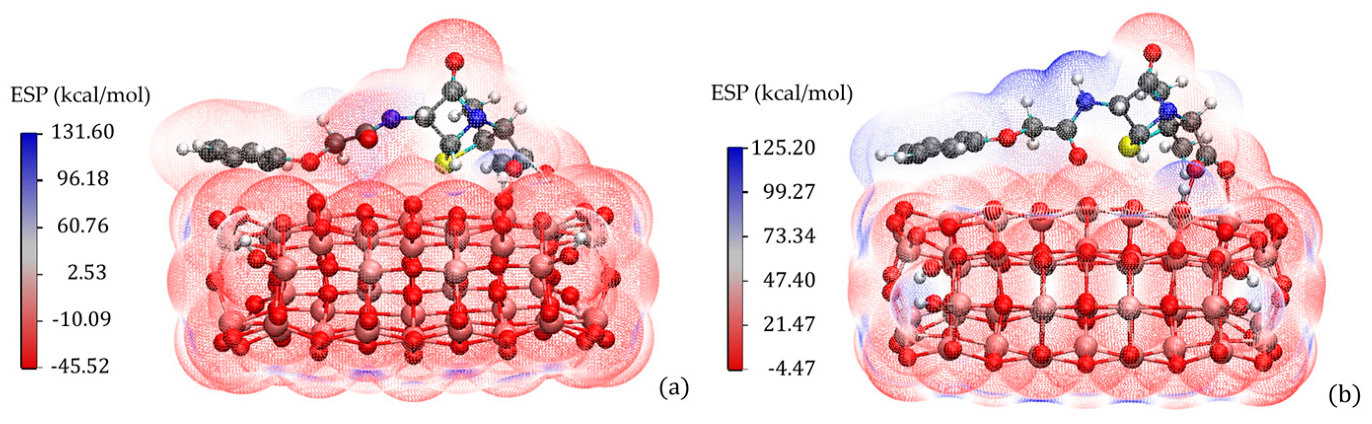
The electrostatic potential mapped onto the electron density surface of PenV adsorbed on the TiO2 nanocluster is shown in Figure 15. The ESP distribution reflects the combined charge characteristics of the molecular adsorbate and the oxide surface. Regions of negative electrostatic potential are predominantly localized on the carboxylate anchoring group and on oxygen atoms of the TiO2 cluster, while positive electrostatic potential is observed near hydrogen atoms and electron-deficient regions of the PenV framework. Compared to the isolated molecule, the ESP of the adsorbed system shows a redistribution of electrostatic potential in the vicinity of the anchoring region and at the molecule–oxide interface.
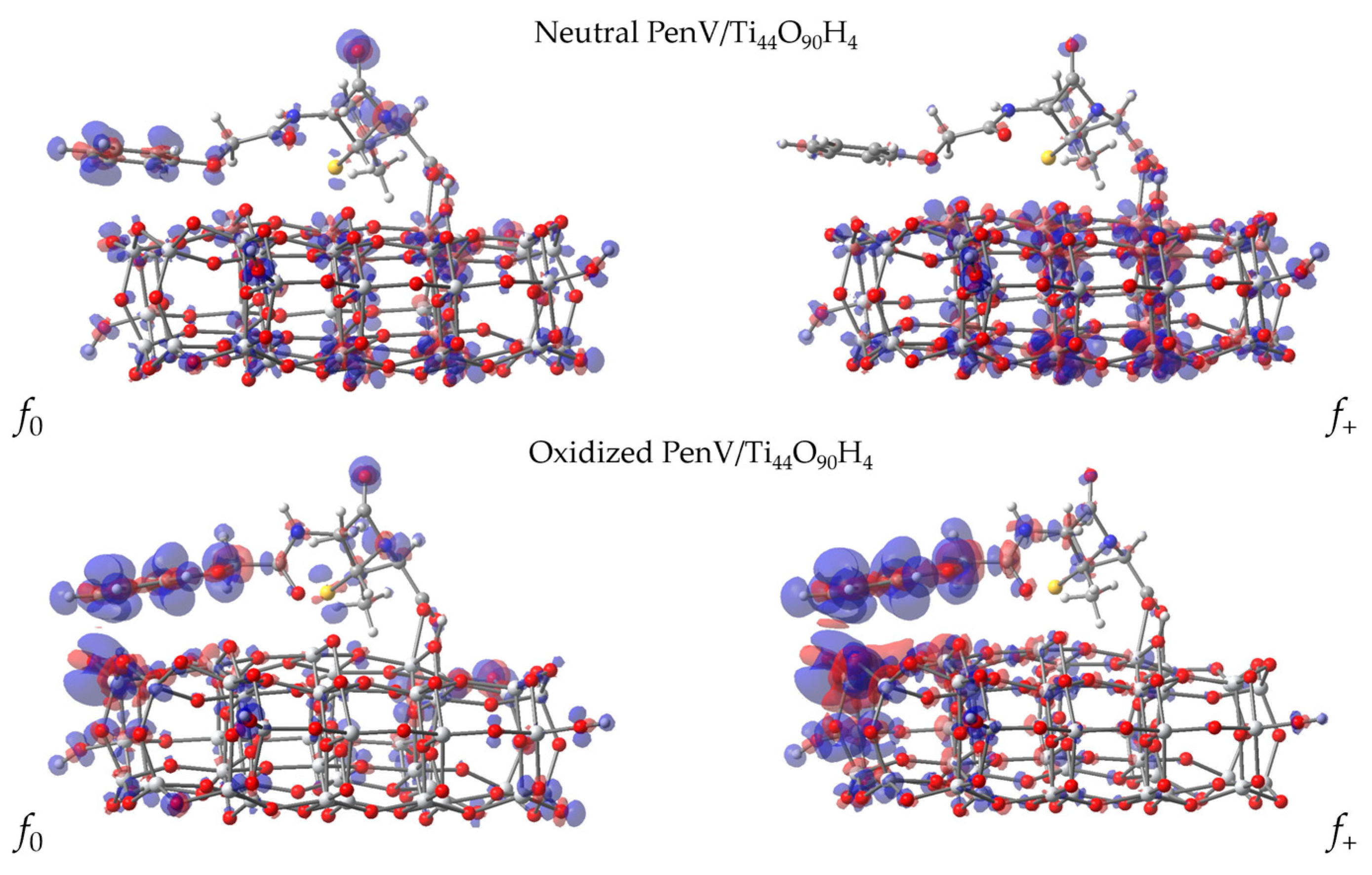
For neutral PenV adsorbed on the TiO2 nanocluster, the Fukui isodensity surfaces for radical (f0) and nucleophilic (f+) perturbations reveal an electronic response that extends across the molecule–surface interface. As shown in Figure 16, both descriptors display significant density on the oxide atoms, particularly near the adsorption region, with a higher presence on the antibiotic in the case of the radical Fukui function. In any case, the results suggest a coupled electronic behavior between the adsorbate and substrate, particularly on surface oxygen atoms beneath the phenoxyacetyl moiety.
For the adsorbed systems, the absolute values of the condensed Fukui coefficients associated with PenV atoms are systematically smaller than in the isolated molecule, reflecting the charge redistribution on both pollutant and catalyst. When summing the Fukui coefficients across the atoms of the pollutant only, the total value amounts to approximately 0.254, 0.0707, and 0.437, for f0, f+, and f, respectively, indicating that a significant fraction of the response resides on the oxide. To facilitate direct comparison with the isolated-molecule results, the Fukui coefficients reported in Table 4 were normalized by the inverse of those values: 3.937, 14.15, and 2.287, for f0, f+, and f, respectively. This rescaling does not affect the relative ordering of reactive sites within the molecule and therefore preserves the qualitative selectivity trends inferred from the Fukui analysis.
The rescaled atom-resolved Fukui coefficients reported in Table 4 provide a quantitative measure of these trends. For the radical Fukui function (f0), the largest contributions are associated with O3 (0.1067), C12 (0.0831), followed by C11, N1, C14, and C9, indicating the participation of both the β-lactam region and the phenoxyacetyl chain. The nucleophilic Fukui function (f+) exhibits smaller absolute values overall and is dominated by O3 (0.1471) and C12 (0.0750), with secondary contributions from O2, C11, and C1, reflecting a more localized response to electron addition. In contrast, the electrophilic Fukui function (f) shows the largest coefficients on O3 (0.1002), C12 (0.0841), and C11 (0.0691), followed by N1 and C14, indicating that electron removal involves both heteroatoms and carbon centers distributed over the adsorbed molecule.
Comparing the Fukui coefficients of the adsorbed antibiotic with those of the isolated pollutant, we note some key similarities, particularly the persistence of O3 as the dominant reactive site, indicating that the β-lactam carbonyl remains the most electronically responsive site regardless of environment, confirming the intrinsic vulnerability of the β-lactam core. Other similarities are revealed by the consistent contribution of the phenoxyacetyl chain (particularly C12 and C11) for radical and electrophilic perturbations and the participation of the β-lactam nitrogen N1N1 across environments, the latter indicating that the integrity of the β-lactam ring is electronically probed in both isolated and surface-bound configurations.
One of the most important differences is the reduced dominance of C6 upon adsorption, although the β-lactam ring is still vulnerable. It is important to note the enhanced role of the phenoxyacetyl chain in the adsorbed system, reflecting the increased electronic involvement of the aromatic side chain upon adsorption, consistent with the structural distortion and π–surface interactions. In addition, we mention the attenuation and redistribution of nucleophilic reactivity (f+) upon adsorption, being more evenly distributed among O3, C12, C11, and O2, suggesting that molecule–surface coupling spreads the electronic response and reduces site specificity. Finally, on adsorption, there is a shift in electrophilic response (f) from several carbons of the side chain (C12, C9, and C10) to O3, C12, C11, and N1. This indicates that oxidation-induced charge removal becomes more localized on heteroatoms and the β-lactam framework when PenV is bound to the surface.
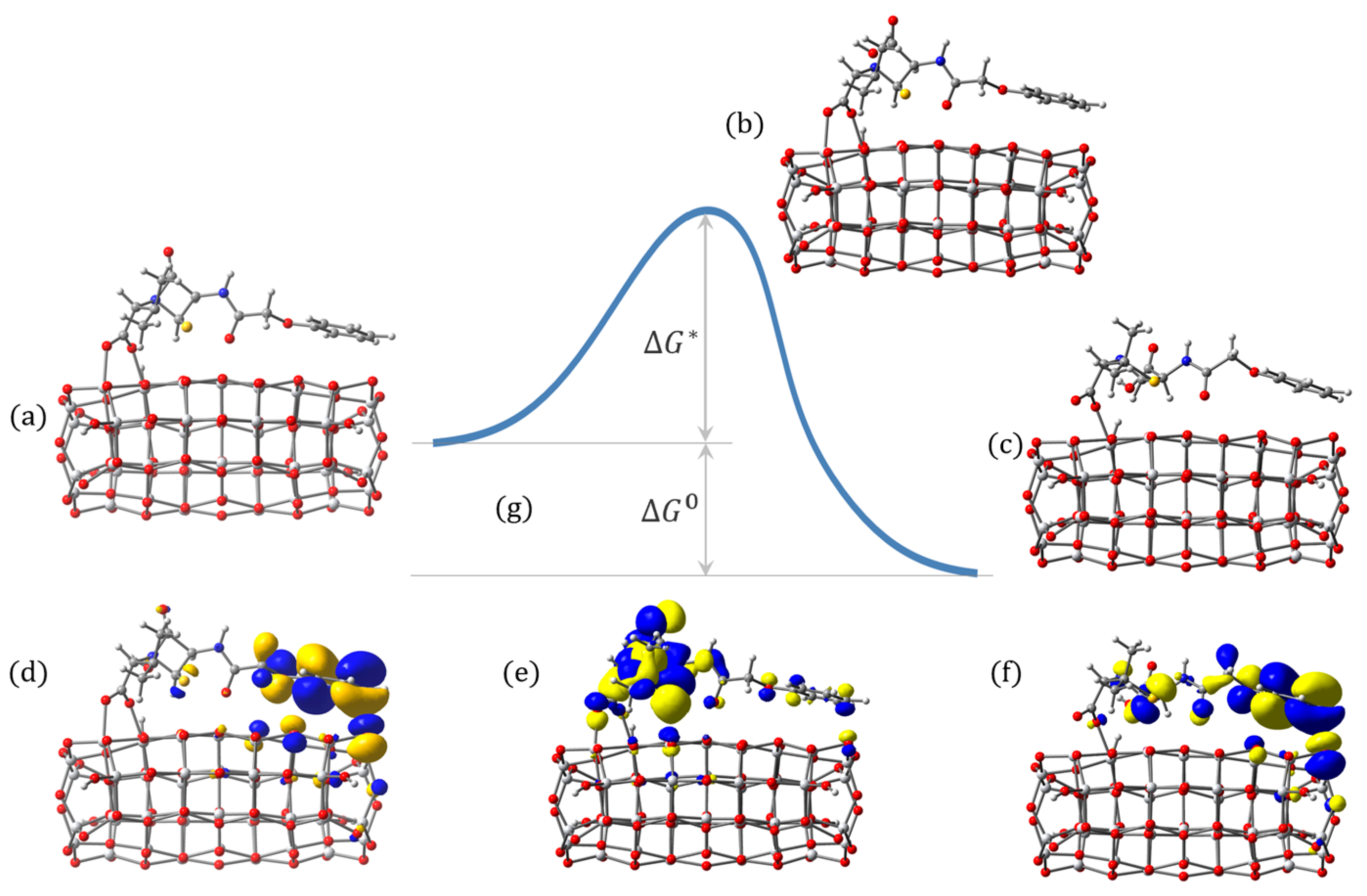
Next, to probe the initial step of photocatalytic degradation, we performed constrained geometry optimizations of the hydroxyl radical toward the C6 carbon atom of the β-lactam ring (Figure 17). For PenV adsorbed on the TiO2 nanocluster, the scans were performed starting from the optimized bidentate-bridging adsorption geometry. As in solution, progressive shortening of the O···C6 distance from 2.00 Å in steps of 0.01 Å leads to elongation and eventual rupture of the β-lactam C–N bond. The structural response of PenV in the adsorbed system is influenced by the interface interactions, with bond breaking occurring in a geometry that preserves the carboxylate anchoring and the π-interaction between the phenoxyacetyl moiety and the oxide surface.
The C–N bond cleavage occurs at an O···C6 distance of 1.62 Å, slightly shorter than in the case of the isolated molecule in solution. The barrier ΔG* is ~0.30 eV (6.89 kcal/mol), whereas the driving force ΔG0 is 1.82 eV (42.0 kcal/mol).
For oxidized PenV adsorbed on the TiO2 nanocluster, the Fukui isodensity surfaces for radical (f0) and nucleophilic (f+) perturbations reveal a markedly enhanced and redistributed electronic response compared to the neutral adsorbed system. As shown in Figure 16, both Fukui descriptors exhibit strong density not only on the PenV framework but also across the oxide surface near the adsorption region, indicating increased electronic coupling following oxidation. In particular, pronounced Fukui density is observed on atoms of the phenoxyacetyl chain and on surface oxygen atoms beneath this moiety, while the β-lactam–thiazolidine core remains electronically active.
In this case, the condensed Fukui coefficients associated with PenV atoms add up to 0.361, 0.476, and 0.246, for f0, f+, and f, respectively, the rest of the response residing on the oxide. To simplify the comparison with the previous cases, the Fukui coefficients reported in Table 5 were rescaled by 2.772, 2.103, and 4.067, representing the inverse of those values.
The most striking change induced by oxidation is the clear relocation of the dominant Fukui response from the β-lactam core toward the phenoxyacetyl moiety. For oxidized PenV/TiO2, the largest f0 and f+ coefficients are associated with C9, C12, and C11, all belonging to the aromatic side chain, whereas in neutral PenV/TiO2, the highest coefficients are shared between O3 (β-lactam carbonyl) and C12/C11. This indicates that oxidation enhances the electronic activity of the phenoxyacetyl chain.
It is also important to note the enhanced role of O3, S, and N1 under electrophilic conditions, indicating that charge removal preferentially affects heteroatoms involved in surface coupling. In neutral PenV/TiO2, f is more evenly distributed among O3, C12, C11, and N1.
Relative to isolated PenV, oxidized PenV/TiO2 exhibits a much stronger delocalization of reactivity toward the phenoxyacetyl chain. In the isolated molecule, f+ and f0 are dominated by O3 and C6, indicating a highly focused reactivity on the β-lactam core. Upon adsorption and oxidation, the electronic response becomes distributed over the aromatic framework (C9, C11, C12, and C13) and surface-interacting heteroatoms. Moreover, the persistence of O3 as a major f contributor across all three systems (isolated, neutral adsorbed, and oxidized adsorbed) highlights the intrinsic sensitivity of the β-lactam carbonyl to oxidation, while the relative increase in side-chain carbons upon adsorption and oxidation reflects the growing importance of molecule–surface and π–surface interactions.
Next, despite the differences in reactivity, to facilitate the comparison with the case of the neutral pollutant on the catalyst, we performed similar constrained geometry optimizations of hydroxyl radical toward the C6 carbon atom of the β-lactam ring (Figure 18). The scans provided the cleavage bond length of 1.75 Å, a barrier ΔG* of ~0.17 eV (4.03 kcal/mol), and a driving force ΔG0 of 1.43 eV (33.0 kcal/mol).

3. Discussion

The present DFT investigation provides a unified mechanistic picture of the photocatalytic degradation of penicillin V by explicitly comparing solution-phase and surface-mediated pathways within a consistent theoretical framework. This approach allows us to address two central questions [39,72,73]: where degradation is most likely to initiate, and how adsorption on TiO2 reshapes the electronic and structural vulnerability of the antibiotic [34,35].
In aqueous solution, hydroxyl radical attack on the β-lactam ring leads to the cleavage of the C6–N1 bond with a modest activation barrier, consistent with the high intrinsic reactivity of •OH. However, the process is energetically possible in the model considered, and the electronic structure analysis reveals that the frontier orbitals remain largely localized on the phenoxyacetyl moiety throughout the reaction. The persistence of the HOMO distribution before and after bond cleavage suggests that, in solution, β-lactam opening is primarily driven by direct radical attack rather than by hole-mediated electronic reorganization, consistent with the electrophilic character of •OH and its preference for high electron-density sites [44,45,74]. While such a pathway is kinetically accessible, these features indicate that solution-phase degradation may be less efficient than surface-mediated processes under photocatalytic conditions.
Adsorption on TiO2 fundamentally alters this picture by modifying both the geometry and the electronic structure of PenV. The optimized adsorption geometry is dominated by a bidentate-bridging configuration of the carboxylate group, accompanied by proton transfer to the surface and additional secondary interactions involving the sulfur atom of the thiazolidine ring and the oxygen atom of the phenoxyacetyl chain. This binding motif emerges consistently as the lowest-energy configuration among those explored. While earlier studies on carboxylate-containing molecules have debated the relative stability of chelating versus bridging coordination on anatase surfaces, and some reports on penicillin derivatives have proposed chelating geometries [41,42,43], the present results support bidentate bridging as the preferred configuration for PenV on the TiO2 nanocluster. The energetic penalty associated with alternative hydrogen-bonded or chelating motifs further reinforces this conclusion, aligning with established trends in dye–TiO2 anchoring studies where carboxylate bridging enhances electronic coupling [53,54].
Importantly, the adsorption-induced deformation of PenV is well localized. The β-lactam–thiazolidine core remains comparatively rigid, preserving the intrinsic strain and chemical identity of the β-lactam ring, while the carboxylate anchor and the phenoxyacetyl chain undergo significant reorientation. In particular, the aromatic ring aligns approximately parallel to the oxide surface, facilitating additional noncovalent interactions, an effect often noted for aromatic parasites of carboxylic acids on oxide surfaces where π–surface interactions contribute to adsorption stability [75]. These structural adjustments enable the effective accommodation of the molecule at the interface without compromising the integrity of the reactive core.
These geometric changes are accompanied by partial electronic hybridization between PenV and the TiO2 cluster. Density-of-state analysis reveals the presence of PenV-derived states within the oxide band gap and mixed molecular–oxide character at the conduction band edge. This hybridization renders weakly allowed optical transitions possible and provides a pathway for interfacial charge transfer that is absent in the isolated molecule. Consistent with this picture, direct photoexcitation of PenV followed by electron injection into the oxide is found to be unlikely due to the high energy of the molecular excited states, whereas hole transfer from photoexcited TiO2 to the adsorbed antibiotic is energetically more favorable. Importantly, the same adsorption-induced electronic hybridization that gives rise to red-shifted absorption features also lowers the energetic cost of hole transfer from TiO2 to the adsorbed antibiotic. These spectral, electronic, and reactivity trends therefore originate from a common interfacial electronic-structure modification induced by adsorption.
The evolution of local reactivity descriptors further highlights the impact of adsorption and oxidation. For isolated PenV, Fukui functions and coefficients consistently identify the β-lactam carbonyl region and the C6 carbon as the most reactive sites, in agreement with the known susceptibility of β-lactam antibiotics to oxidative degradation and prior computational reactivity analyses [57,67]. Upon adsorption, the Fukui response becomes more spatially extended and involves both molecular atoms and surface oxygen sites, reflecting partial delocalization of the charge perturbation onto the oxide. After normalization, the relative site selectivity reveals increased involvement of the phenoxyacetyl chain, consistent with its structural distortion and proximity to the surface.
The loss of an electron amplifies this redistribution. In oxidized PenV adsorbed on TiO2, radical and nucleophilic susceptibility shifts predominantly toward the aromatic side chain, while electrophilic response becomes concentrated on heteroatoms directly involved in surface coupling. Compared to both isolated PenV and neutral adsorbed PenV, the oxidized system exhibits a more delocalized and surface-coupled reactivity landscape. This suggests that oxidation stabilizes the β-lactam core electronically while activating alternative degradation channels associated with the peripheral moiety, potentially influencing subsequent reaction steps.
Finally, constrained hydroxyl-radical approach scans provide semi-quantitative insight into the energetics of β-lactam opening. While the absolute barrier heights depend on basis set and model details, the relatively small values obtained here are comparable to the typical uncertainty of DFT-based barrier calculations, particularly for radical-driven processes [49]. As a result, the reported ΔG* values should be regarded as semi-quantitative estimates rather than precise activation free energies [76]. However, the consistent order of magnitude of the barriers across different basis sets and environments supports the conclusion that β-lactam ring opening induced by •OH attack is kinetically accessible under photocatalytic conditions. Moreover, the calculated values for the barriers (about 0.15 to 0.35 eV) are consistent with values reported earlier for degradation of β-lactam antibiotics occurring on experimentally observed timescales under UV irradiation, where •OH concentrations are high, and reactions are diffusion-assisted [70,71].
The limited influence of solvent effects suggests that this initial step is governed primarily by local electronic interactions. Notably, the HOMO retains a similar spatial distribution before and after bond cleavage in both solution-phase and surface-bound systems, supporting a degradation mechanism driven by radical attack rather than by direct involvement of the frontier hole state.
These results indicate that adsorption on TiO2 not only enhances the likelihood of PenV degradation relative to solution-phase pathways, but also reshapes the electronic and structural determinants of reactivity. The combined effects of adsorption-induced deformation, electronic hybridization, and oxidation-driven redistribution of local reactivity provide a coherent mechanistic framework for understanding the photocatalytic degradation of PenV. Given the structural similarities shared by many β-lactam antibiotics [35,56,57], these insights are expected to be broadly relevant for rationalizing and optimizing photocatalytic degradation pathways on TiO2-based materials. Taken together, the consistent trends observed across adsorption energetics, electronic-structure analysis, optical response, local reactivity descriptors, and reaction scans provide a coherent and internally consistent mechanistic picture, despite the semi-quantitative nature of individual descriptors. The emphasis of the present study is placed on comparative trends obtained from a unified theoretical treatment, rather than on absolute energetic values.

4. Methods

All calculations were performed within the framework of density functional theory [48,49] using the hybrid B3LYP exchange–correlation functional [77,78], which constitutes a good balance between computational cost and accuracy in predicting reaction energies, generally providing a mean absolute error of 0.10–0.15 eV (about 2–3 kcal/mol) [79,80]. Although range-separated hybrids and dispersion-corrected functionals can improve absolute excitation energies, B3LYP reliably captures relative trends in adsorption energetics, charge localization, and reaction barriers, which are the primary focus of this comparative mechanistic study.
The basis sets used varied, depending on the size of the system. For isolated PenV in vacuum and aqueous solution, we optimized geometries using the double-ζ quality basis sets with polarization functions DGDZVP [81] for all atoms, and the conductor-like polarizable continuum model (CPCM) to account for solvent effects [82,83]. For the case of PenV adsorbed on the Ti44O90H4 cluster, the geometries were optimized using a smaller basis set of double-ζ quality with polarization functions 3-21G(d) [84], due to the limited computational resources available. For consistency of the comparison, the same basis set used was also applied to the isolated PenV in addition to the larger one, DGDZVP. We ran single-point calculations on key geometries, with titanium and sulfur atoms treated using the effective core potentials (ECP) and double-ζ quality basis functions for all atoms via LANL2DZ [85], which provide a reliable description of TiO2 clusters in photocatalytic studies. All optimized geometries of isolated PenV were verified by frequency calculations to ensure the absence of imaginary frequencies.
Adsorption geometries were generated by placing PenV near the TiO2 nanocluster in multiple initial orientations corresponding to commonly reported binding motifs for carboxylate-containing molecules, including monodentate, bidentate-chelating, bidentate-bridging, and hydrogen-bonded configurations.
The electronic properties of the isolated and adsorbed systems were analyzed through density-of-states (DOS) calculations and visualization of selected molecular orbitals. Projected density of states (PDOSs) were used to distinguish contributions from PenV and the TiO2 cluster.
Optical absorption spectra were computed using time-dependent DFT (TD-DFT) [86,87], with the wavelength and oscillator strength of the lowest 20 or 50 singlet-to-singlet electronic transitions for isolated PenV or adsorbed-on-TiO2 nanocluster, respectively.
All calculations were performed with the Gaussian09 quantum chemistry package [88].
The charge density difference maps [89] were obtained by subtracting ground-state densities from excited-state densities by means of the Multiwfn program [90,91]. Based on input densities from time-dependent DFT runs, Multiwfn allows for the construction of the charge density differences between two electronic states, identifying regions of electron accumulation and depletion. The electron–hole analysis [92] partitions the charge density differences into electron-rich and hole-rich contributions and computes their centroids as first moments of the respective distributions. The spatial separation between these centroids provides a quantitative descriptor of the degree of charge transfer, complementing the qualitative insights obtained from isodensity surface plots [90].
Electrostatic potential maps were computed by projecting the electrostatic potential onto the electron density surface with the Multiwfn 3.8 program and were visualized with VMD 1.9.4 (Visual Molecular Dynamics) [93]. Fukui functions [94,95] for radical (f0), nucleophilic (f+), and electrophilic (f) attack were evaluated using finite-difference schemes based on changes in electron density upon addition or removal of an electron. Atom-resolved Fukui coefficients were obtained by integrating the Fukui functions over atomic basins.
We acknowledge some intrinsic limitations of the adopted DFT methodology, as the absolute adsorption energies and reaction barriers depend on the choice of functional, basis set, and model size. Furthermore, radical-driven processes are known to be particularly sensitive to methodological details. In this context, the free-energy barriers reported for β-lactam ring opening should be regarded as semi-quantitative estimates rather than precise activation free energies. Nevertheless, the persistence of a low-barrier regime across different basis sets and environments, together with the consistent qualitative trends observed for adsorption geometries, electronic hybridization, and reactivity descriptors, lends confidence to the mechanistic conclusions drawn. The emphasis of this work is therefore placed on comparative trends and relative reactivity rather than on absolute energetic values.

5. Conclusions

In this work, density functional theory calculations were used to elucidate the initial steps of the photocatalytic degradation of penicillin V, explicitly comparing solution-phase and surface-mediated pathways within a unified theoretical framework. By treating PenV consistently both as an isolated molecule in aqueous solution and as an adsorbate on an anatase TiO2 nanocluster, this study provides direct insight into how adsorption reshapes the structural, electronic, and reactive properties of the antibiotic.
In aqueous solution, hydroxyl radical (•OH) attack can induce β-lactam ring opening via C6–N1 bond cleavage with a modest activation barrier. The process is energetically favorable in the models considered and proceeds without significant redistribution of the frontier electronic density, suggesting that solution-phase degradation may be kinetically accessible but relatively inefficient. In contrast, adsorption on TiO2 enhances susceptibility to oxidative PenV degradation. The preferred binding configuration involves bidentate-bridging coordination of the carboxylate group, accompanied by localized molecular deformation and additional stabilizing interactions that promote electronic coupling at the interface.
Electronic structure analysis reveals partial hybridization between PenV and TiO2 states, leading to molecular contributions within the oxide band gap and enabling interfacial charge-transfer pathways. Direct photoexcitation of PenV followed by electron injection into the oxide is unlikely due to the high energy of the molecular excited states. Instead, degradation is more plausibly initiated by hole transfer from photoexcited TiO2 and subsequent attack by reactive oxygen species, particularly •OH radicals.
Reactivity descriptors show that adsorption and oxidation substantially redistribute local vulnerability. While isolated PenV exhibits reactivity focused on the β-lactam core, adsorption leads to a more delocalized response involving both the molecule and the surface, and oxidation shifts radical and nucleophilic susceptibility toward the phenoxyacetyl side chain. Despite these changes, β-lactam opening remains kinetically accessible, as confirmed by scan-based barrier estimates. Although absolute barrier heights lie near the accuracy limits of DFT for radical-driven processes, the consistent low-barrier regime across models supports the qualitative conclusions.
Overall, these results indicate that adsorption on TiO2 reshapes the electronic and structural determinants of reactivity, enhancing the likelihood of PenV degradation relative to solution-phase pathways. The combined influence of binding configuration, electronic hybridization, and oxidation-driven reactivity redistribution provides a coherent mechanistic picture that is likely applicable to other β-lactam antibiotics and can inform the rational design of TiO2-based photocatalytic systems for antibiotic removal.

Supplementary Materials

The following supporting information can be downloaded at: https://www.mdpi.com/article/10.3390/catal16020171/s1. Table S1: Complete list of Fukui coefficients for PenV; Table S2: Complete list of Fukui coefficients for neutral PenV adsorbed on Ti44O90H4; Table S3: Complete list of Fukui coefficients for oxidized PenV adsorbed on Ti44O90H4.

Author Contributions

Conceptualization, C.I.O. and M.A.G.; methodology, C.I.O. and M.A.G.; software, C.I.O. and R.M.S.; validation, C.I.O. and M.A.G.; formal analysis, R.M.S.; investigation, C.I.O. and R.M.S.; resources, M.A.G.; data curation, C.I.O. and R.M.S.; writing—original draft preparation, C.I.O. and R.M.S.; writing—review and editing, M.A.G.; visualization, R.M.S.; supervision, M.A.G.; project administration, M.A.G.; funding acquisition, M.A.G. All authors have read and agreed to the published version of the manuscript.

Funding

This research received no external funding.

Data Availability Statement

Data is contained within the article. Optimized geometries of the systems studied here can be provided on request.

Conflicts of Interest

The authors declare no conflicts of interest.

Abbreviations

The following abbreviations are used in this manuscript:
ROSReactive oxygen species
DFTDensity functional theory
TD-DFTTime-dependent density functional theory
HOMOHighest occupied molecular orbital
LUMOLowest unoccupied molecular orbital
DOSsDensity of States
PenVPenicillin V

References

  1. Aus der Beek, T.; Weber, F.-A.; Bergmann, A.; Hickmann, S.; Ebert, I.; Hein, A.; Küster, A. Pharmaceuticals in the environement—Global occurrences and perspectives. Environ. Toxicol. Chem. 2014, 35, 823–835. [Google Scholar] [CrossRef] [Scilit]
  2. Klavarioti, M.; Mantzavinos, D.; Kassinos, D. Removal of residual pharmaceuticals from aqueous systems by advanced oxidation processes. Environ. Int. 2009, 35, 402–417. [Google Scholar] [CrossRef] [Scilit]
  3. Young, S.; Juhl, A.; O’Mullan, G.D. Antibiotic-resistant bacteria in the Hudson river estuary linked to wet weather sewage contamination. J. Water Health 2013, 11, 297–310. [Google Scholar] [CrossRef] [Scilit] [PubMed]
  4. Zhu, K.; Li, X.; Chen, Y.; Huang, Y.; Yang, Z.; Guan, G.; Yan, K. Recent advances on the spherical metal oxides for sustainable degradation of antibiotics. Coord. Chem. Rev. 2024, 510, 215813. [Google Scholar] [CrossRef] [Scilit]
  5. Abdipour, H.; Hemati, H. Sonocatalytic process of penicillin removal using-Fe2O3/effect of different parameters/degradation mechanism/kinetic study/optimisation with response surface model. Int. J. Environ. Anal. Chem. 2024, 104, 8617–8638. [Google Scholar] [CrossRef] [Scilit]
  6. Ighalo, J.O.; Igwegbe, C.A.; Aniagor, C.O.; Oba, S.M. A review of methods for the removal of penicillins from water. J. Water Process Eng. 2021, 39, 101886. [Google Scholar] [CrossRef] [Scilit]
  7. Gothwal, R.; Shashidhar, T. Antibiotic pollution in the environment: A review. Clean–Soil Air Water 2015, 43, 479–489. [Google Scholar] [CrossRef] [Scilit]
  8. Goodarzi, S.; Torabideh, M.; Parsaseresht, G.; Abdipour, H.; Kamani, H.; Zomorrodi Jangaee, T. Penicillin removal from the aqueous environment based on AOPs/challenges and outlook. A review. Appl. Water Sci. 2024, 14, 164. [Google Scholar] [CrossRef] [Scilit]
  9. Cuerda-Correa, E.M.; Alexandre-Franco, M.F.; Fernández-González, C. Advanced oxidation processes for the removal of antibiotics from water. An overview. Water 2020, 12, 102. [Google Scholar] [CrossRef] [Scilit]
  10. Pandis, P.K.; Kalogirou, C.; Kanellou, E.; Vaitsis, C.; Savvidou, M.G.; Sourkouni, G.; Zorpas, A.A.; Argirusis, C. Key Points of Advanced Oxidation Processes (AOPs) for Wastewater, Organic Pollutants and Pharmaceutical Waste Treatment: A Mini Review. ChemEngineering 2022, 6, 8. [Google Scholar] [CrossRef] [Scilit]
  11. Saravanan, A.; Deivayanai, V.C.; Senthil Kumar, P.; Rangasamy, G.; Hemavathy, R.V.; Harshana, T.; Gayathri, N.; Alagumalai, K. A detailed review on advanced oxidation process in treatment of wastewater: Mechanism, challenges and future outlook. Chemosphere 2022, 308, 136524. [Google Scholar] [CrossRef] [Scilit]
  12. Ohtani, B. Photocatalysis by inorganic solid materials: Revisiting its definition, concepts, and experimental procedures. Adv. Inorg. Chem. 2011, 63, 395–430. [Google Scholar] [CrossRef] [Scilit]
  13. Armaković, S.J.; Savanović, M.M.; Armaković, S. Titanium Dioxide as the Most Used Photocatalyst for Water Purification: An Overview. Catalysts 2023, 13, 26. [Google Scholar] [CrossRef] [Scilit]
  14. Tanos, F.; Razzouk, A.; Lesage, G.; Cretin, M.; Bechelany, M. A Comprehensive Review on Modification of Titanium Dioxide-Based Catalysts in Advanced Oxidation Processes for Water Treatment. ChemSusChem 2024, 17, e202301139. [Google Scholar] [CrossRef] [Scilit]
  15. Rueda-Marquez, J.J.; Levchuk, I.; Ibañez, P.F.; Sillanpää, M. A critical review on application of photocatalysis for toxicity reduction of real wastewaters. J. Clean. Prod. 2020, 258, 120694. [Google Scholar] [CrossRef] [Scilit]
  16. Hsu, C.-Y.; Mahmoud, Z.H.; Abdullaev, S.; Ali, F.K.; Naeem, Y.A.; Mizher, R.M.; Karim, M.M.; Abdulwahid, A.S.; Ahmadi, Z.; Habibzadeh, S.; et al. Nano titanium oxide (nano-TiO2): A review of synthesis methods, properties, and applications. Case Stud. Chem. Environ. Eng. 2024, 9, 100626. [Google Scholar] [CrossRef] [Scilit]
  17. El Mchaouri, M.; Mallah, S.; Abouhajjoub, D.; Boumya, W.; Elmoubarki, R.; Essadki, A.; Barka, N.; Elhalil, A. Engineering TiO2 photocatalysts for enhanced visible-light activity in wastewater treatment applications. Tetrahedron Green Chem. 2025, 6, 100084. [Google Scholar] [CrossRef] [Scilit]
  18. Karaolia, P.; Michael-Kordatou, I.; Hapeshi, E.; Drosou, C.; Bertakis, Y.; Christofilos, D.; Armatas, G.S.; Sygellou, L.; Schwartz, T.; Xekoukoulotakis, N.P.; et al. Removal of antibiotics, antibiotic-resistant bacteria and their associated genes by graphene-based TiO2 composite photocatalysts under solar radiation in urban wastewaters. Appl. Catal. B Environ. 2018, 224, 810–824. [Google Scholar] [CrossRef] [Scilit]
  19. Hoffmann, M.R.; Martin, S.T.; Choi, W.; Bahnemann, D.W. Environmental applications of semiconductor photocatalysis. Chem. Rev. 1995, 95, 69–96. [Google Scholar] [CrossRef] [Scilit]
  20. Fujishima, A.; Rao, T.N.; Tryk, D.A. Titanium dioxide photocatalysis. J. Photochem. Photobiol. C 2000, 1, 1–21. [Google Scholar] [CrossRef] [Scilit]
  21. Beranek, R. (Photo)electrochemical Methods for the Determination of the Band Edge Positions of TiO2-Based Nanomaterials. Adv. Phys. Chem. 2011, 2011, 786759. [Google Scholar] [CrossRef] [Scilit]
  22. Xia, Y.; Chen, S.; Zhu, X.; Ren, Z.; Feng, W. High-Efficiency S-Scheme Bi4O5IBr/Bi5O7I0.7Br0.3 Double Solid Solution Heterojunction Photocatalyst for Levofloxacin Degradation via Optimized Carrier Dynamics. Energy Environ. Mater. 2026, e70204. [Google Scholar] [CrossRef] [Scilit]
  23. Ohtani, B. Photocatalysis A to Z-What we know and what we do not know in a scientific sense. J. Photochem. Photobiol. C 2011, 11, 157–178. [Google Scholar] [CrossRef] [Scilit]
  24. Liu, W.; Li, Y.; Liu, F.; Jiang, W.; Zhang, D.; Liang, J. Visible-light-driven photocatalytic degradation of diclofenac by carbon quantum dots modified porous g-C3N4: Mechanisms, degradation pathway and DFT calculation. Water Res. 2019, 151, 8–19. [Google Scholar] [CrossRef] [Scilit] [PubMed]
  25. Litter, M.I.; Román, E.S.; Grela, M.A.; Meichtry, J.M.; Rodríguez, H.B. Sensitization of TiO2 by Dyes: A Way to Extend the Range of Photocatalytic Activity of TiO2 to the Visible Region. In Visible Light-Active Photocatalysis; Ghosh, S., Ed.; Wiley-VCH: Hoboken, NJ, USA, 2018; pp. 253–282. [Google Scholar]
  26. Bahnemann, D.; Bockelmann, D.; Goslich, R. Mechanistic studies of water detoxification in illuminated TiO2 suspensions. Sol. Energy Mater. 1991, 24, 564–583. [Google Scholar] [CrossRef] [Scilit]
  27. Mills, A.; Le Hunte, S. An overview of semiconductor photocatalysis. J. Photochem. Photobio. A 1997, 108, 1–35. [Google Scholar] [CrossRef] [Scilit]
  28. Shaham-Waldmann, N.; Paz, Y. Away from TiO2: A critical minireview on the developing of new photocatalysts for degradation of contaminants in water. Mater. Sci. Semic. Proc. 2016, 42, 72–80. [Google Scholar] [CrossRef] [Scilit]
  29. Kou, J.; Lu, C.; Wang, J.; Chen, Y.; Xu, Z.; Varma, R.S. Selectivity Enhancement in Heterogeneous Photocatalytic Transformations. Chem. Rev. 2017, 117, 1445–1514. [Google Scholar] [CrossRef] [Scilit]
  30. Zhang, Z.; Bai, L.; Li, Z.; Qu, Y.; Jing, L. Review of Strategies for the Fabrication of Heterojunctional Nanocomposites as Efficient Visible-Light Catalysts by Modulating Excited Electrons with Appropriate Thermodynamic Energy. J. Materi. Chem. A 2019, 7, 10879–10897. [Google Scholar] [CrossRef] [Scilit]
  31. Qin, K.; Zhao, Q.; Yu, H.; Xia, X.; Li, J.; He, S.; Wei, L.; An, T. A Review of Bismuth-Based Photocatalysts for Antibiotic Degradation: Insight into the Photocatalytic Degradation Performance, Pathways and Relevant Mechanisms. Environ. Res. 2021, 199, 111360. [Google Scholar] [CrossRef] [Scilit] [PubMed]
  32. Yang, D.; Xia, Y.; Xiao, T.; Xu, Z.; Lei, Y.; Jiao, Y.; Zhu, X.; Feng, W. Constructing Ag–TiO2-g-C3N4 S-scheme heterojunctions for photocatalytic degradation of malachite green. Opt. Mater. 2025, 159, 116652. [Google Scholar] [CrossRef] [Scilit]
  33. Nosaka, Y.; Nosaka, A.Y. Generation and Detection of Reactive Oxygen Species in Photocatalysis. Chem. Rev. 2017, 117, 11302–11336. [Google Scholar] [CrossRef] [Scilit] [PubMed]
  34. Mehralipour, J.; Bagheri, S.; Gholami, M. Synthesis and characterization of rGO/Fe0/Fe3O4/TiO2 nanocomposite and application of photocatalytic process in the decomposition of penicillin G from aqueous. Heliyon 2023, 9, e18172. [Google Scholar] [CrossRef] [Scilit]
  35. Silerio-Vázquez, F.J.; González-Burciaga, L.A.; Antileo, C.; Núñez-Núñez, C.M.; Proal-Nájera, J.B. Photocatalytic degradation of antibiotics in water via TiO2-x: Research needs for technological advancements. J. Hazard. Mater. Adv. 2024, 16, 100506. [Google Scholar] [CrossRef] [Scilit]
  36. Rengifo-Herrera, J.A.; Pulgarin, C. Why five decades of massive research on heterogeneous photocatalysis, especially on TiO2, has not yet driven to water disinfection and detoxification applications? Critical review of drawbacks and challenges. Chem. Eng. J. 2023, 477, 146875. [Google Scholar] [CrossRef] [Scilit]
  37. Nosaka, T.; Nosaka, A. Understanding Hydroxyl Radical (OH) Generation Processes in Photocatalysis. ACS Energy Lett. 2016, 1, 356–359. [Google Scholar] [CrossRef] [Scilit]
  38. Yin, M.; Li, Z.; Kou, J.; Zou, Z. Mechanism Investigation of Visible Light-Induced Degradation in a Heterogeneous TiO2/Eosin Y/Rhodamine B System. Environ. Sci. Technol. 2009, 43, 8361–8366. [Google Scholar] [CrossRef] [Scilit]
  39. Etacheri, V.; Di Valentin, C.; Schneider, J.; Bahnemann, D.; Pillai, S.C. Visible-light activation of TiO2 photocatalysts: Advances in theory and experiments. J. Photochem. Photobio. C 2015, 25, 1–29. [Google Scholar] [CrossRef] [Scilit]
  40. Giraldo-Aguirre, A.L.; Erazo-Erazo, E.D.; Flórez-Acosta, O.A.; Serna-Galvis, E.A.; Torres-Palma, R.A. TiO2 photocatalysis applied to the degradation and antimicrobial activity removal of oxacillin: Evaluation of matrix components, ex-perimental parameters, degradation pathways and identification of organics by-products. J. Photochem. Photobio. A Chem. 2015, 311, 95–103. [Google Scholar] [CrossRef] [Scilit]
  41. Chen, C.; Ma, W.; Zhao, J. Semiconductor-Mediated Photodegradation of Pollutants under Visible-Light Irradiation. Chem. Soc. Rev. 2010, 39, 4206–4219. [Google Scholar] [CrossRef] [Scilit]
  42. Zhang, G.; Kim, G.; Choi, W. Visible Light Driven Photocatalysis Mediated via Ligand-to-Metal Charge Transfer (LMCT): An Alternative Approach to Solar Activation of Titania. Energy Environ. Sci. 2014, 7, 954–966. [Google Scholar] [CrossRef] [Scilit]
  43. Lazic, V.; Nedeljkovic, J.M. Photocatalytic Reactions over TiO2-Based Interfacial Charge Transfer Complexes. Catalysts 2024, 14, 810. [Google Scholar] [CrossRef] [Scilit]
  44. Chen, P.; Liu, H.; Sun, Y.; Li, J.; Cui, W.; Wang, L.; Zhang, W.; Yuan, X.; Wang, Z.; Zhang, Y.; et al. Bi Metal Prevents the Deactivation of Oxygen Vacancies in Bi2O2CO3 for Stable and Efficient Photocatalytic NO Abatement. Appl. Catal. B Environ. 2020, 264, 118545. [Google Scholar] [CrossRef] [Scilit]
  45. Hu, X.; Hu, X.; Peng, Q.; Zhou, L.; Tan, X.; Jiang, L.; Tang, C.; Wang, H.; Liu, S.; Wang, Y.; et al. Mechanisms Underlying the Photocatalytic Degradation Pathway of Ciprofloxacin with Heterogeneous TiO2. Chem. Eng. J. 2020, 380, 122366. [Google Scholar] [CrossRef] [Scilit]
  46. Wang, S.; Liu, D.; Yu, J.; Zhang, X.; Zhao, P.; Ren, Z.; Sun, Y.; Li, M.; Han, S. Photocatalytic Penicillin Degradation Performance and the Mechanism of the Fragmented TiO2 Modified by CdS Quantum Dots. ACS Omega 2021, 6, 18178–18189. [Google Scholar] [CrossRef] [Scilit]
  47. Wang, Y.; Zhang, W.; Meng, H.; Duan, J.; Zhang, Y.; Xia, Z.; Wang, Y. Distinctive coordination configuration and interfacial water balance induced by anti-Kirkendall effect attain exceptional catalytic activity and selectivity. Angew. Chem. Int. Ed. 2026, 65, e13687. [Google Scholar] [CrossRef] [Scilit] [PubMed]
  48. Parr, R.G.; Yang, W. Density-Functional Theory of Atoms and Molecules; Oxford University Press: New York, NY, USA, 1989. [Google Scholar]
  49. Yu, H.S.; Li, S.L.; Truhlar, D.G. Perspective: Kohn-Sham density functional theory descending a staircase. J. Chem. Phys. 2016, 145, 130901. [Google Scholar] [CrossRef] [Scilit] [PubMed]
  50. Yi, X.-H.; Ji, H.; Wang, C.-C.; Li, Y.; Li, Y.-H.; Zhao, C.; Wang, A.; Fu, H.; Wang, P.; Zhao, X.; et al. Photocatalysis-activated SR-AOP over PDINH/MIL-88A(Fe) composites for boosted chloroquine phosphate degradation: Performance, mechanism, pathway and DFT calculations. Appl. Catal. B Environ. 2021, 293, 120229. [Google Scholar] [CrossRef] [Scilit]
  51. Jabbar, Z.H.; Graimed, B.H.; Ammar, S.H.; Sabit, D.A.; Najim, A.A.; Radeef, A.Y.; Taher, A.G. The latest progress in the design and application of semiconductor photocatalysis systems for degradation of environmental pollutants in wastewater: Mechanism insight and theoretical calculations. Mater. Sci. Semicond. Process. 2024, 173, 108153. [Google Scholar] [CrossRef] [Scilit]
  52. Kovačević, M.; Simić, M.; Živković, S.; Milović, M.; Tolić Stojadinović, L.; Relić, D.; Vasić Anićijević, D. Uncovering Metal-Decorated TiO2 Photocatalysts for Ciprofloxacin Degradation—A Combined Experimental and DFT Study. Int. J. Mol. Sci. 2024, 25, 11844. [Google Scholar] [CrossRef] [Scilit]
  53. Oprea, C.I.; Panait, P.; Gîrţu, M.A. DFT study of binding and electron transfer from colorless aromatic pollutants to a TiO2 nanocluster—Application to photocatalytic degradation under visible light irradiation. Beilstein J. Nanotechnol. 2014, 5, 1016–1030. [Google Scholar] [CrossRef] [Scilit]
  54. Oprea, C.I.; Petcu, L.C.; Gîrțu, M.A. DFT Study of Binding and Electron Transfer from Penicillin to a TiO2 Nanocluster: Applications to Photocatalytic Degradation. In Proceedings of the 2015 E-Health and Bioengineering Conference, Iași, Romania, 19–21 November 2015; pp. 1–4. [Google Scholar]
  55. Soriano-Correa, C.; Sánchez Ruiz, J.F.; Raya, A.; Esquivel, R.O. Electronic Structure and Physicochemical Properties of Selected Penicillins. Int. J. Quantum. Chem. 2006, 107, 628–636. [Google Scholar] [CrossRef] [Scilit]
  56. Teixeira, R.C.; Luiz, L.C.; Junqueira, G.M.A.; Bell, M.J.V.; Anjos, V.C. Detection of Antibiotic Residues in Cow’s Milk: A Theoretical and Experimental Vibrational Study. J. Mol. Struct. 2020, 1215, 128221. [Google Scholar] [CrossRef] [Scilit]
  57. Salari, A.A.; Talebi Tari, M.; Noei, M.; Tahan, A. The Ab Initio Study and NBO Interpretation of Solvent Effects on the Structural Stability and the Chemical Reactivity of Penicillin-V Conformations. Arabian J. Chem. 2017, 10, S2327–S2334. [Google Scholar] [CrossRef] [Scilit]
  58. Oprea, C.I.; Gîrțu, M.A. Structure and Electronic Properties of TiO2 Nanoclusters and Dye–Nanocluster Systems Appropriate to Model Hybrid Photovoltaic or Photocatalytic Applications. Nanomaterials 2019, 9, 357. [Google Scholar] [CrossRef] [Scilit]
  59. Wu, X.; Chen, Z.; Lu, G.Q.; Wang, L. Nanosized Anatase TiO2 Single Crystals with Tunable Exposed (001) Facets for Enhanced Energy Conversion Efficiency of Dye-Sensitized Solar Cells. Adv. Funct. Mater. 2011, 21, 4167–4172. [Google Scholar] [CrossRef] [Scilit]
  60. De Angelis, F.; Di Valentin, C.; Fantacci, S.; Vittadini, A.; Selloni, A. Theoretical Studies on Anatase and Less Common TiO2 Phases: Bulk, Surfaces, and Nanomaterials. Chem. Rev. 2014, 114, 9708–9753. [Google Scholar] [CrossRef] [Scilit] [PubMed]
  61. Grätzel, M. Photoelectrochemical cells. Nature 2001, 414, 338–344. [Google Scholar] [CrossRef] [Scilit]
  62. Zhang, Y.F.; Lin, W.; Li, Y.; Ding, K.N.; Li, J.Q. A Theoretical Study on the Electronic Structures of TiO2: Effect of Hartree−Fock Exchange. J. Phys. Chem. B 2005, 109, 19270–19277. [Google Scholar] [CrossRef] [Scilit]
  63. Wang, Y.; Wen, B.; Dahal, A.; Kimmel, G.A.; Rousseau, R.; Selloni, A.; Petrik, N.G.; Dohnálek, Z. Binding of Formic Acid on Anatase TiO2(101). J. Phys. Chem. C 2020, 124, 20228–20239. [Google Scholar] [CrossRef] [Scilit]
  64. Daldossi, C.; Di Valentin, C.; Selloni, A. Pathways of Photocatalytic Oxidation of Formic Acid on Dry and Hydrated Anatase TiO2 Surfaces. ACS Catal. 2025, 15, 11487–11501. [Google Scholar] [CrossRef] [Scilit] [PubMed]
  65. De Angelis, F.; Fantacci, S.; Mosconi, E.; Nazeeruddin, M.K.; Grätzel, M. Absorption Spectra and Excited State Energy Levels of the N719 Dye on TiO2 in Dye-Sensitized Solar Cell Models. J. Phys. Chem. C 2011, 115, 8825–8831. [Google Scholar] [CrossRef] [Scilit]
  66. Monti, S.; Pastore, M.; Li, C.; De Angelis, F.; Carravetta, V. Theoretical Investigation of Adsorption, Dynamics, Self-Aggregation, and Spectroscopic Properties of the D102 Indoline Dye on an Anatase (101) Substrate. J. Phys. Chem. C 2016, 120, 2787–2796. [Google Scholar] [CrossRef] [Scilit]
  67. Oprea, C.I.; Panait, P.; Lungu, J.; Stamate, D.; Dumbravă, A.; Cimpoesu, F.; Gîrţu, M.A. DFT Study of Binding and Electron Transfer from a Metal-Free Dye with Carboxyl, Hydroxyl, and Sulfonic Anchors to a Titanium Dioxide Nanocluster. Int. J. Photoenergy 2013, 2013, 1–15. [Google Scholar] [CrossRef] [Scilit]
  68. Dail, M.K.; Mezyk, S.P. Hydroxyl-Radical-Induced Degradative Oxidation of β-Lactam Antibiotics in Water: Absolute Rate Constant Measurements. J. Phys. Chem. A 2010, 114, 8391–8395. [Google Scholar] [CrossRef] [Scilit]
  69. Chen, X.; Wang, J. Degradation of Antibiotic Cephalosporin C in Different Water Matrices by Ionizing Radiation: Degradation Kinetics, Pathways, and Toxicity. Sci. Total Environ. 2021, 791, 148253. [Google Scholar] [CrossRef] [Scilit]
  70. Cohen, N.C. Beta.-Lactam Antibiotics: Geometrical Requirements for Antibacterial Activities. J. Med. Chem. 1983, 26, 259–264. [Google Scholar] [CrossRef] [Scilit]
  71. Buxton, G.V.; Greenstock, C.L.; Helman, W.P.; Ross, A.B. Critical Review of Rate Constants for Reactions of Hydrated Electrons, Hydrogen Atoms and Hydroxyl Radicals (-OH/-O in Aqueous Solution. J. Phys. Chem. Ref. Data 1988, 17, 513–886. [Google Scholar] [CrossRef] [Scilit]
  72. Montoya, J.F.; Peral, J.; Salvador, P. Comprehensive Kinetic and Mechanistic Analysis of TiO2 Photocatalytic Reactions According to the Direct–Indirect Model: (I) Theoretical Approach. J. Phys. Chem. C 2014, 118, 14266–14275. [Google Scholar] [CrossRef] [Scilit]
  73. Montoya, J.F.; Atitar, M.F.; Bahnemann, D.W.; Peral, J.; Salvador, P. Comprehensive Kinetic and Mechanistic Analysis of TiO2 Photocatalytic Reactions According to the Direct–Indirect Model: (II) Experimental Validation. J. Phys. Chem. C 2014, 118, 14276–14290. [Google Scholar] [CrossRef] [Scilit]
  74. Cinar, Z. The Role of Molecular Modeling in TiO2 Photocatalysis. Molecules 2017, 22, 556. [Google Scholar] [CrossRef] [Scilit]
  75. Mulay, M.R.; Martsinovich, N. Interaction of organic pollutants with TiO2: A density functional theory study of carboxylic acids on the anatase (101) surface. Mol. Phys. 2023, 121, e2165981. [Google Scholar] [CrossRef] [Scilit]
  76. Zhu, S.; Wan, D. Photocatalysis: Basic Principles, Diverse Forms of Implementations and Emerging Scientific Opportunities. Adv. Energy Mater. 2017, 7, 1700841. [Google Scholar] [CrossRef] [Scilit]
  77. Becke, A.D. Density-functional exchange-energy approximation with correct asymptotic behavior. Phys. Rev. A 1988, 38, 3098–3100. [Google Scholar] [CrossRef] [Scilit]
  78. Lee, C.; Yang, W.; Parr, R.G. Development of the colle-salvetti correlation-energy formula into a functional of the electron density. Phys. Rev. 1988, B37, 785–789. [Google Scholar] [CrossRef] [Scilit]
  79. Rabuck, A.D.; Scuseria, G.E. Assessment of recently developed density functionals for the calculation of enthalpies of formation in challenging cases. Chem. Phys. Lett. 1999, 309, 450–456. [Google Scholar] [CrossRef] [Scilit]
  80. Tirado-Rivers, J.; Jorgensen, W.L. Performance of B3LYP Density Functional Methods for a Large Set of Organic Molecules. J. Chem. Theory Comput. 2008, 4, 297–306. [Google Scholar] [CrossRef] [Scilit]
  81. Godbout, N.; Salahub, D.R.; Andzelmen, J.; Wimmer, E. Optimization of Gaussian-type basis sets for local spin density functional calculations. Part I. Boron through neon, optimization technique and validation. Can. J. Chem. 1992, 70, 560–571. [Google Scholar] [CrossRef] [Scilit]
  82. Barone, V.; Cossi, M. Quantum calculation of molecular energies and energy gradients in solution by a conductor solvent model. J. Phys. Chem. A 1998, 102, 1995–2001. [Google Scholar] [CrossRef] [Scilit]
  83. Tomasi, J.; Mennucci, B.; Cammi, R. Quantum mechanical continuum solvation models. Chem. Rev. 2005, 105, 2999–3093. [Google Scholar] [CrossRef] [Scilit]
  84. Dobbs, K.D.; Hehre, W.J. Molecular-orbital theory of the properties of inorganic and organometallic compounds. 4. Extended basis-sets for 3rd row and 4th row, main-group elements. J. Comp. Chem. 1986, 7, 359–378. [Google Scholar] [CrossRef] [Scilit]
  85. Hay, P.J.; Wadt, W.R. Ab initio effective core potentials for molecular calculations. Potentials for K to Au including the outermost core orbitals. J. Chem. Phys. 1985, 82, 299–311. [Google Scholar] [CrossRef] [Scilit]
  86. Gross, E.K.U.; Maitra, N.T. Introduction to TDDFT. In Fundamentals of Time-Dependent Density Functional Theory; Marques, M.A.L., Maitra, N.T., Nogueira, F.M.S., Gross, E.K.U., Eds.; Springer: Berlin/Heidelberg, Germany, 2012. [Google Scholar]
  87. Schirmer, J. Review of the foundations of time-dependent density-functional theory (TDDFT). Phys. Chem. Chem. Phys. 2025, 27, 4992–5005. [Google Scholar] [CrossRef] [Scilit] [PubMed]
  88. Frisch, M.J.; Trucks, G.W.; Schlegel, H.B.; Scuseria, G.E.; Robb, M.A.; Cheeseman, J.R.; Scalmani, G.; Barone, V.; Mennucci, B.; Petersson, G.A.; et al. Gaussian 09, Revision D.01; Gaussian, Inc.: Wallingford, CT, USA, 2009. [Google Scholar]
  89. Liu, Z.; Lu, T.; Chen, Q. An Sp-Hybridized All-Carboatomic Ring, Cyclo [18] Carbon: Electronic Structure, Electronic Spectrum, and Optical Nonlinearity. Carbon 2020, 165, 461–467. [Google Scholar] [CrossRef] [Scilit]
  90. Lu, T.; Chen, F. Multiwfn: A multifunctional wavefunction analyzer. J. Comput. Chem. 2012, 33, 580–592. [Google Scholar] [CrossRef] [Scilit]
  91. Lu, T. A comprehensive electron wavefunction analysis toolbox for chemists, Multiwfn. J. Chem. Phys. 2024, 161, 082503. [Google Scholar] [CrossRef] [Scilit]
  92. Le Bahers, T.; Adamo, C.; Ciofini, I. A Qualitative Index of Spatial Extent in Charge-Transfer Excitations. J. Chem. Theory Comput. 2011, 7, 2498–2506. [Google Scholar] [CrossRef] [Scilit]
  93. Humphrey, W.; Dalke, A.; Schulten, K. VMD: Visual Molecular Dynamics. J. Mol. Graph. 1996, 14, 33–38. [Google Scholar] [CrossRef] [Scilit]
  94. Yang, W.; Parr, R.G.; Pucci, R. Electron density, Kohn-Sham frontier orbitals, and Fukui functions. J. Chem. Phys. 1984, 81, 2862–2863. [Google Scholar] [CrossRef] [Scilit]
  95. Pucci, R.; Angilella, G.G.N. Density functional theory, chemical reactivity, and the Fukui functions. Found. Chem. 2022, 24, 59–71. [Google Scholar] [CrossRef] [Scilit]
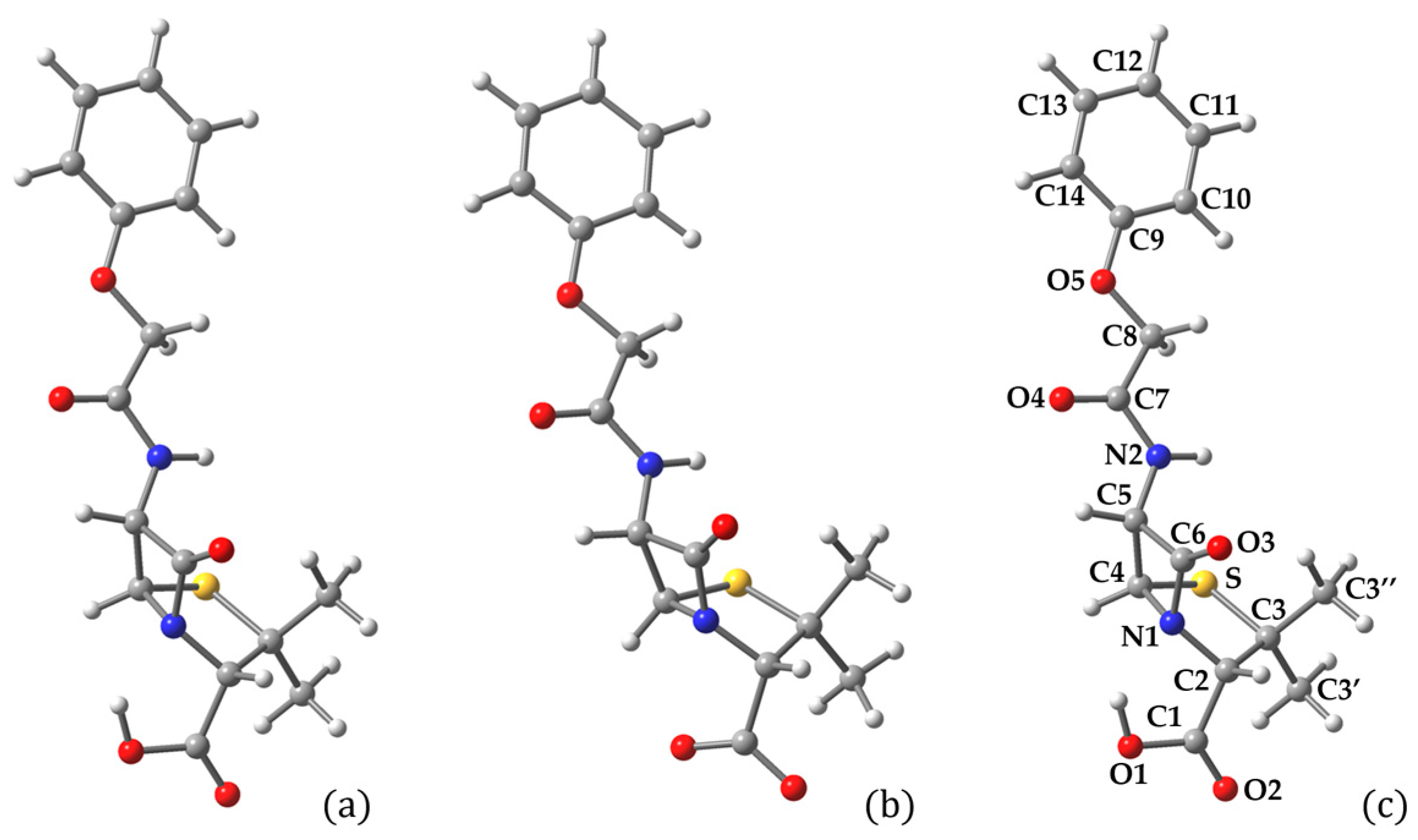
Figure 1. Optimized structure of (a) neutral and (b) deprotonated (carboxylate) PenV. Atom labeling is illustrated in (c). Atom colors: C—dark gray, O—red, S—yellow, N—blue, and H—light gray.
Catalysts 16 00171 g001
Figure 2. Isodensity surfaces of the key molecular orbitals of PenV, calculated in water solvent. Also represented are the conduction and valence band edges of the Ti44O90H4 cluster as well as the redox levels of the O2/•O2– and H2O/•OH couples. Atom colors: C—dark gray, O—red, S—yellow, N—blue, and H—light gray.
Catalysts 16 00171 g002
Figure 3. Simulated UV-Vis absorption spectra of PenV, calculated by TDDFT in water solvent. The spectral lines were convoluted with Gaussian distributions of 0.1 eV full width at half maximum.
Catalysts 16 00171 g003
Figure 4. Electron and hole density distributions (0.0004 e/bohr3) based on the charge difference analysis of PenV for the transitions from HOMO to (a) LUMO and (b) LUMO+4. Illustration of charge separation between the electron-depleted (red) and electron-rich (blue) centroids at the HOMO to (c) LUMO and (d) LUMO+4 transitions of PenV.
Catalysts 16 00171 g004
Figure 5. Top (left) and lateral (right) views of the optimized structures of the Ti44O90H4 anatase nanocluster. Atom colors: Ti—gray, O—red, and H—light gray.
Catalysts 16 00171 g005
Figure 6. (Left) DFT-calculated density of states of the Ti44O90H4 cluster in vacuum, showing, separately, the contribution of p and d orbitals on Ti and of p orbitals on O atoms. Energy levels were convoluted with Gaussian distributions with an FWHM of 0.1 eV. (Right) Isodensity surfaces of the frontier orbitals of the Ti44O90H4 cluster: conduction band edge (top) and valence band edge (bottom). Ti—gray, C—dark gray, O—red, S—yellow, N—blue, and H—light gray.
Catalysts 16 00171 g006
Figure 7. Schematic representation of typical binding configurations of a pollutant with a carboxyl anchoring group, such as Penicillin V, on a TiO2 surface: (a) monodentate binding via a single carboxylate oxygen; (b) bidentate-chelating binding to a single surface Ti site; (c) bidentate-bridging binding involving two adjacent surface Ti sites; (d) monodentate interaction mediated by hydrogen bonding with surface hydroxyl groups; and (e) mixed bidentate binding, involving one Ti–O bond and one hydrogen-bond interaction.
Catalysts 16 00171 g007
Figure 8. Top (upper) and side (lower) views of optimized PenV adsorption on the Ti44O90H4 nanocluster starting from different initial geometries. Bidentate bridging arises from (a) proton transfer during optimization and (b) initial pollutant deprotonation with proton attachment to adjacent surface oxygen. Mixed bidentate binding (c) involves one Ti–O bond and one hydrogen bond. Atom colors: Ti—gray, C—dark gray, O—red, S—yellow, N—blue, and H—light gray.
Catalysts 16 00171 g008
Figure 9. (Left) DFT-calculated density of states of PenV on the Ti44O90H4 cluster in vacuum, showing, separately, the contribution of p and d orbitals on Ti and of p orbitals on O atoms. Energy levels were convoluted with Gaussian distributions with a FWHM of 0.1 eV. (Right) Isodensity surfaces of the key orbitals of the PenV/Ti44O90H4 system, from bottom-up: valence band edge of TiO2 (HOMO−6), HOMO−4, ground state of PenV (HOMO), conduction band edge of TiO2 (LUMO), an orbital with mixed character (LUMO+105), and the excited state of PenV (LUMO+128). Atom colors: Ti—gray, C—dark gray, O—red, S—yellow, N—blue, and H—light gray.
Catalysts 16 00171 g009
Figure 10. Simulated UV-Vis absorption spectra of the PenV/Ti44O90H4 system, calculated by TDDFT. The spectral lines were convoluted with Gaussian distributions of 0.1 eV full width at half maximum.
Catalysts 16 00171 g010
Figure 11. Electron and hole density distributions based on the charge difference analysis of PenV for the transitions from HOMO to (a) LUMO and (b) LUMO+4. Illustration of charge separation between the electron-depleted (red) and electron-rich (blue) centroids at the HOMO to (c) LUMO and (d) LUMO+4 transition of PenV.
Catalysts 16 00171 g011
Figure 12. Electrostatic potential (ESP) mapped onto the electron density surface of PenV in solution, highlighting the spatial distribution of electron-rich (red) and electron-poor (blue) regions.
Catalysts 16 00171 g012
Figure 13. Isodensity surfaces of Fukui functions for radical (left) and nucleophilic (right) attack for PenV. Blue and red surfaces denote regions of higher and lower local reactivity toward the corresponding perturbation.
Catalysts 16 00171 g013
Figure 14. Illustration of the initial step of PenV degradation in solution induced by hydroxyl radical attack, highlighting localized β-lactam ring opening without significant redistribution of the HOMO. Optimized geometries of PenV in aqueous solution at the beginning of the scan (a), immediately before (b), and after (c) the cleavage of the C6–N1 bond of the β-lactam ring. Isodensity surfaces of the HOMO of PenV in solution at the beginning of the scan (d), just before (e), and after (f) C6–N1 bond breaking. Panel (g) provides a schematic representation of the free-energy barrier (ΔG*) and reaction driving force (ΔG0) associated with the bond-breaking step. Atom colors: C—dark gray, O—red, S—yellow, N—blue, and H—light gray.
Catalysts 16 00171 g014
Figure 15. Electrostatic potential (ESP) mapped onto the electron density surface of PenV adsorbed on the TiO2 nanocluster in neutral (a) and oxidized (b) forms, highlighting the spatial distribution of electron-rich (red) and electron-poor (blue) regions.
Catalysts 16 00171 g015
Figure 16. Isodensity surfaces of Fukui functions for radical (left) and nucleophilic (right) attack for PenV adsorbed on the Ti44O90H4 nanocluster in neutral (top) and oxidized (bottom) form. Blue and red surfaces denote regions of higher and lower local reactivity toward the corresponding perturbation.
Catalysts 16 00171 g016
Figure 17. Illustration of the initial step of PenV degradation in solution induced by hydroxyl radical attack, highlighting localized β-lactam ring opening. Optimized geometries of neutral PenV adsorbed on the Ti44O90H4 nanocluster in aqueous solution at the beginning of the scan (a), immediately before (b), and after (c) the cleavage of the C6–N1 bond of the β-lactam ring. Isodensity surfaces of the HOMO of PenV/Ti44O90H4 in solution at the beginning of the scan (d), just before (e), and after (f) C6–N1 bond breaking. Panel (g) provides a schematic representation of the free-energy barrier (ΔG*) and reaction driving force (ΔG0) associated with the bond-breaking step. Atom colors: Ti—gray, C—dark gray, O—red, S—yellow, N—blue, and H—light gray.
Catalysts 16 00171 g017
Figure 18. Illustration of the initial step of oxidized PenV degradation on the catalyst, induced by hydroxyl radical attack, highlighting localized β-lactam ring opening. Optimized geometries of oxidized PenV adsorbed on the Ti44O90H4 nanocluster in aqueous solution at the beginning of the scan (a), immediately before (b), and after (c) the cleavage of the C6–N1 bond of the β-lactam ring. Isodensity surfaces of the HOMO of PenV/Ti44O90H4 in solution at the beginning of the scan (d), just before (e), and after (f) C6–N1 bond breaking. Panel (g) provides a schematic representation of the free-energy barrier (ΔG*) and reaction driving force (ΔG0) associated with the bond-breaking step. Atom colors: Ti—gray, C—dark gray, O—red, S—yellow, N—blue, and H—light gray.
Catalysts 16 00171 g018
Table 1. Structural parameters of neutral and carboxylate-deprotonated PenV, calculated in vacuum and/or water solvent, and of neutral and oxidized PenV adsorbed on Ti44O90H4, in vacuum. Distances are expressed in Å and angles in degrees. The atom labeling is presented in Figure 1c.
ParameterPenV
(Vacuum)
PenV
(Water)
PenV Deprot.
(Vacuum)
PenV/TiO2
(Vacuum)
PenV-e-/TiO2
(Vacuum)
r(O1-C1)1.3651.3551.2691.2991.298
r(O2-C1)1.2221.2131.2701.2801.278
∠(O1-C1-O2)122.82123.20131.19122.62122.88
r(O1-H/Ti1)1.0080.978-2.0422.064
r(O2-Ti2)---2.1172.075
∠(H/Ti-O1-C1-C2)0.19−0.38-130.23130.26
∠(O1-C1-C2-N1)−14.35−15.1023.55−0.842.90
r(C3-S)1.8881.8871.8951.8891.898
r(C4-S)1.8351.8451.8671.8721.870
∠(C1-C2-S)111.17114.64102.9397.5195.65
∠(C1-C2-C3-S)92.49101.2586.3477.8875.55
r(N1-C6)1.4531.4061.3991.4491.429
∠(N1-C4-C5)88.2088.3887.8089.4688.22
∠(N1-C4-C5-C6)11.7210.9210.3013.0013.50
∠(N1-C4-S-C5)−96.26−96.75−93.73−96.55−95.58
r(C6-O3)1.2121.2081.2251.2111.212
∠(O3-C6-C5-N2)42.2543.5648.3246.1648.19
r(C7-O4)1.2311.2221.2361.2371.264
∠(C7-C8-O5-C9)178.12173.79176.65151.71172.81
∠(C8-O5-C9-C10)1.2032.521.4446.959.48
Table 2. Atom-resolved Fukui coefficients for PenV, corresponding to radical (f0), nucleophilic (f+), and electrophilic (f) perturbations. Only the largest contributions are shown in descending order; the complete list is available in the Supplementary Material. Atom labels correspond to the numbering scheme in Figure 1.
f0Atomf+AtomfAtom
0.1031O30.2022O30.1374C12
0.0997C60.1975C60.1320O5
0.0695C120.0524S0.0967C9
0.0671O50.0488O20.0948C14
0.0487C90.0449N10.0777C10
Table 3. Free-energy barrier (ΔG*) and reaction driving force (ΔG0) associated with the breaking of the C6–N1 bond of the β-lactam ring during the PenV degradation induced by •OH radical attack.
Basis Set3-21G(d)DZVP
VacuumSolventVacuumSolvent
ΔG0 (eV)1.481.481.881.79
ΔG* (eV)0.170.140.370.35
ΔG0 (kcal/mol)34.134.243.341.4
ΔG* (kcal/mol)4.023.288.508.09
Table 4. Rescaled atom-resolved Fukui coefficients for PenV adsorbed on Ti44O90H4 nanocluster, corresponding to radical (f0), nucleophilic (f+), and electrophilic (f) perturbations. Only the largest contributions are shown in descending order; the complete list is available in the Supplementary Material. Atom labels correspond to the numbering scheme in Figure 1.
f0Atomf+AtomfAtom
0.1067O30.1471O30.1002O3
0.0831C120.0750C120.0841C12
0.0673C110.0721O20.0691C11
0.0551N10.0566C110.0640N1
0.0512C140.0439C10.0613C14
0.0382C90.0439C130.0494C9
0.0323C130.0368C3″0.0366O4
0.0315S0.0354N20.0357S
0.0311C60.0311C60.0311C6
0.0303O20.0255C20.0306O5
Table 5. Atom-resolved Fukui coefficients for oxidized PenV adsorbed on Ti44O90H4 nanocluster, corresponding to radical (f0), nucleophilic (f+), and electrophilic (f) perturbations. Only the largest contributions are shown in descending order; the complete list is available in the Supplementary Material. Atom labels correspond to the numbering scheme in Figure 1.
f0Atomf+AtomfAtom
0.0837C90.1161C90.1316O3
0.0815C120.1030C120.1243S
0.0743C110.0984C110.0818N1
0.0673O50.0926O50.0398C12
0.0650C130.0851C130.0390C6
0.0563O30.0755C140.0278C11
0.0558C140.0572C100.0261C13
0.0423C100.0190C80.0256C5
0.0323N10.0173O30.0248O2
0.0317S0.0134N20.0210C9
0.0152C60.0078O40.0193C3″
Disclaimer/Publisher’s Note: The statements, opinions and data contained in all publications are solely those of the individual author(s) and contributor(s) and not of MDPI and/or the editor(s). MDPI and/or the editor(s) disclaim responsibility for any injury to people or property resulting from any ideas, methods, instructions or products referred to in the content.