Article

Hollow CuFe2O4/MgFe2O4 Heterojunction Boost Photocatalytic Oxidation Activity for Organic Pollutants

1
Department of Chemical Engineering and Technology, School of Chemistry and Chemical Engineering, Nanjing University of Science and Technology, Nanjing 210094, China
2
Department of Environmental Science and Technology, School of Environmental and Biological Engineering, Nanjing University of Science and Technology, Nanjing 210094, China
*
Authors to whom correspondence should be addressed.
Catalysts 2022, 12(8), 910; https://doi.org/10.3390/catal12080910
Submission received: 29 July 2022 / Revised: 13 August 2022 / Accepted: 17 August 2022 / Published: 18 August 2022

Abstract

:
P-n heterojunction-structured CuFe2O4/MgFe2O4 hollow spheres with a diameter of 250 nm were synthesized using a template-free solvothermal method, and time-dependent morphological studies were carried out to investigate the hollow formation mechanism. The CuFe2O4/MgFe2O4 with a molar ratio of 1:2 (Cu:Mg) had the highest degradation efficiency with the model organic dye Acid Orange 7, with a degradation rate of 91.96% over 60 min. The synthesized CuFe2O4/MgFe2O4 nanocomposites were characterized by XRD, TEM, HRTEM, UV-vis spectroscopy, Mott–Schottky, and EIS. Due to the synthesis of the p-n heterojunction, CuFe2O4/MgFe2O4 has efficient photogenerated carriers, and the hollow structure has a higher specific surface area and stronger adsorption capacity, which is significantly better than that of CuFe2O4 and MgFe2O4 in terms of photocatalytic performance. The outstanding performance shows that the p-n heterostructure of CuFe2O4/MgFe2O4 has potential for application in wastewater degradation.

1. Introduction

In today’s world, many chemical organic pollutants (dyes) are released into water bodies every year, which are difficult to degrade completely [1,2,3,4,5]. Advanced photocatalytic oxidation technology with a strong oxidizing ability and high efficiency is considered as a very promising means of wastewater treatment for the degradation of organic pollutants [6,7,8]. Light-induced photocatalytic activity and magnetic separation have potential as low-cost [9], high-efficiency [10,11,12,13], and high-stability [14,15] methods of water remediation. The hollow microsphere structure is one of the areas of focus in the field of catalysis at present, because the hollow microsphere structure has better structural properties than the solid sphere structure, according to the following considerations: (1) compared with solid microspheres, the hollow microsphere structure has a larger specific surface area and stronger adsorption capacity [16]; (2) the cavity structure can confine the catalytic reaction to the reactants, improving the stability of the reaction and the efficiency of the catalytic reaction [17,18]; (3) the photon can remain in the shell structure for a longer time, with a higher utilization rate [17,18]; and (4) the shell structure can also be beneficial for the diffusion of reactant molecules in the hollow microspheres and increase the contact area between the reactant and the catalyst [16,18].
In order to further improve their photocatalytic degradation efficiency, researchers have further promoted the separation of photogenerated carriers by designing a heterojunction structure, which improves the conversion efficiency of the solar energy [19,20]. Heterojunction photocatalysts are formed by coupling two semiconductor photocatalysts with complementary properties, relative band positions, and differences in fermi levels [21]. This method facilitates the separation of the electron-hole pairs [19,22,23], expands the absorbable spectral range [19], and enhances the redox capacity [20], thereby compensating for the shortcomings of a single semiconductor [23]. Yin et al. [15] prepared an all-solid-state hybrid photocatalyst, Au@CdS/U-WO3, with a unique sea urchin-like micro-nano heterostructure, without adding additives by the continuous photo deposition of Au and CdS on the WO3 supports, which significantly improved the photocatalytic hydrogen evolution activity. Zhu et al. [13] successfully synthesized a series of MA3Bi2Br9–COFs nanocomposites with a variety of B sites and different ratios of PNCs via a facile in situ growth approach. The MA3Bi2Br9–COFs nanocomposites efficiently photocatalyzed the polymerization of various functional monomers with the assistance of a large scope of co-initiators via both electron and hole transfer mechanisms in both aqueous and organic media, achieving a considerable conversion rate of up to 97.5%. Ferrites are one of the most efficient solid metal oxide photocatalysts and are commonly used to synthesize heterojunction-structured catalysts [24,25,26,27]. The homogeneous and heterogeneous ferrite composites CaFe2O4/MgFe2O4 [28] and ZnFe2O4/MoS2 [29] have been reported to exhibit enhanced visible-light photocatalysis. Spinel ferrite CuFe2O4 is a structurally stable heterogeneous catalytic material, which has many advantages, such as good magnetic properties, a small band gap, high chemical stability, low price, safety and environmentally protective properties [30,31]. The cycle activity of Cu2+/Cu+ is higher than that of Fe3+/Fe2+, and it can catalyze the production of ·OH from H2O2. Shen et al. [32] successfully prepared CuFe2O4 microspheres with a particle size of 116 nm using a solvothermal method, which exhibited good photocatalytic properties. Sharma et al. [33] experimentally verified that CuFe2O4 has the best photocatalytic performance compared to the spinel ferrite MFe2O4 (M = Cu, Zn, Ni, Co) nanoparticles. MgFe2O4 nanoparticles have attracted extensive attention due to their chemical properties, reproducibility, and magnetic properties. Due to these unique properties, they have a wide range of applications, including magnetic technology and heterogeneous catalysts [26,34]. CuFe2O4 and MgFe2O4 have similar crystalline features. However, they have different band structures. CuFe2O4 is a p-type semiconductor with a narrow band gap (Eg) of 1.32~1.69 eV [32,35,36], while MgFe2O4 is an n-type semiconductor with a wide band gap (Eg) of 1.9~2.56 eV [28,29,37]. Forming a p-n heterojunction is an ideal method for preventing the recombination of charges, and consequently enhancing the photocatalytic ability of materials [38,39,40,41]. Here, CuFe2O4 and MgFe2O4 were chosen for the fabrication of photocatalysts with p–n heterojunctions to enhance their photocatalytic activity.
Herein, the polyethylene glycol-20000 (PEG-20000)-assisted hydrothermal method was successfully developed to prepare hollow spheres of the homo ferrite composite of CuFe2O4/MgFe2O4. The physicochemical properties of the products were characterized, and the photocatalytic activities were evaluated by degrading the model organic dye Acid Orange 7 (AO7) under visible-light irradiation. In addition, the active species in the photocatalytic process were discussed, and the possible mechanism was proposed.

2. Results and Discussion

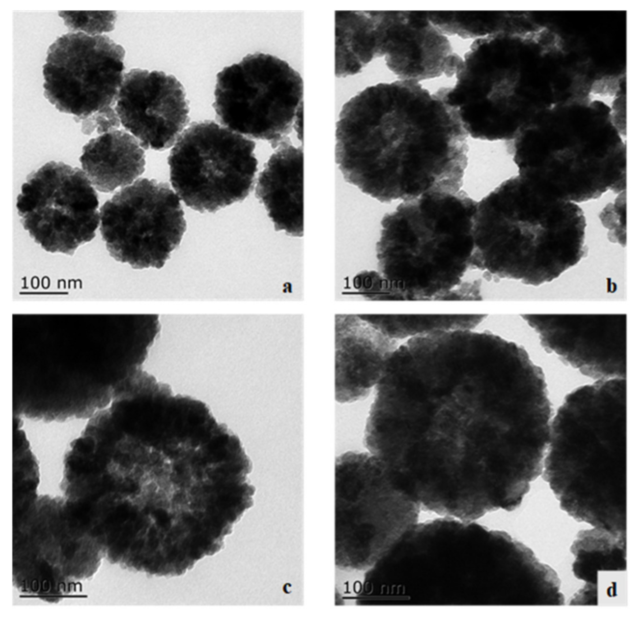
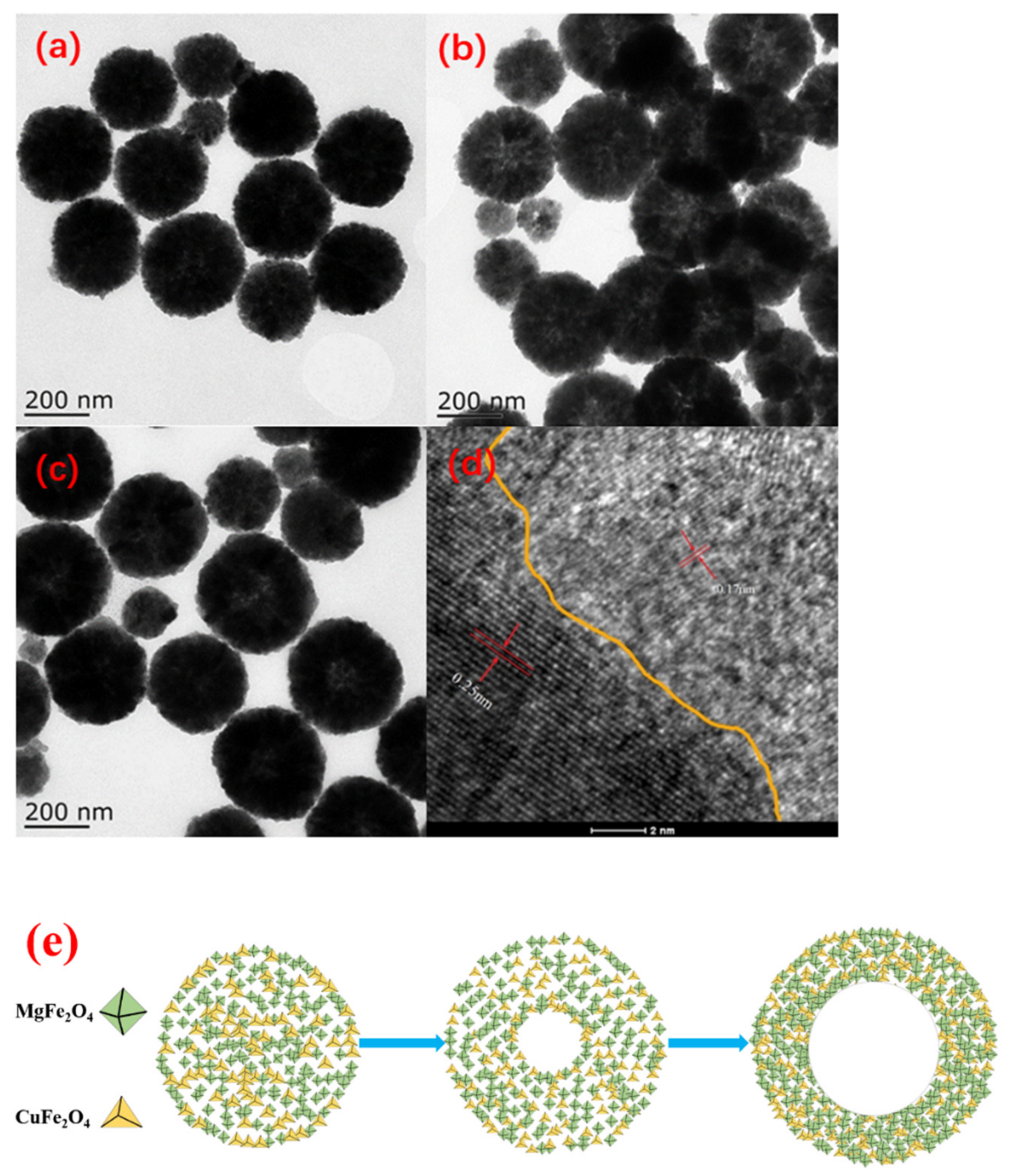
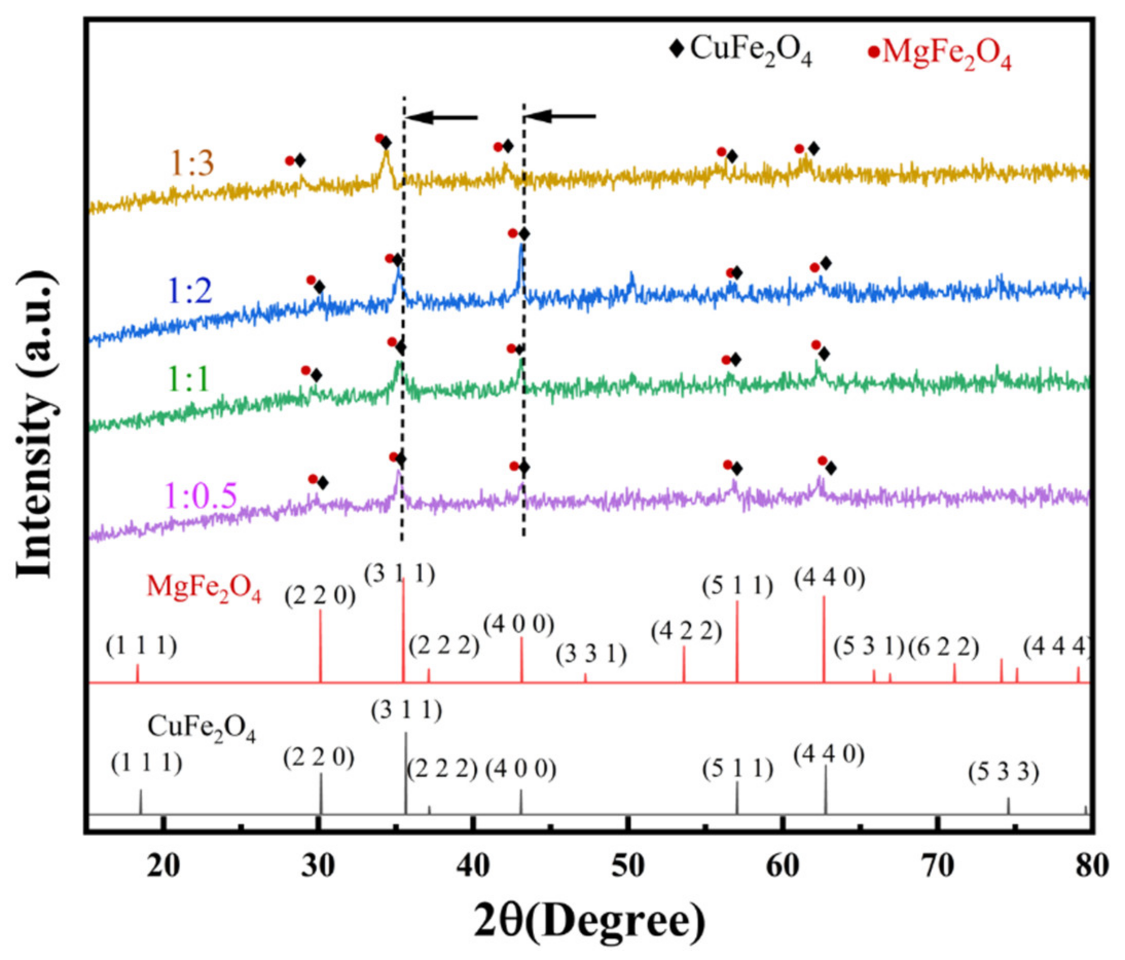
2.1. Fabrication of Hollow CuFe2O4/MgFe2O4 and Its Structure

The TEM images of CuFe2O4/MgFe2O4 with different Cu/Mg ratios in the precursors are shown in Figure 1. It can be seen that all samples formed hollow spherical structures with rough surfaces and wormholes between the fine grains. This kind of structure can contribute to the higher surface area [42], more active sites, easier mass diffusion and transport, lighter penetration and higher quantum efficiency of the photocatalytic reaction in the application process [16]. The diameter of the microspheres was 200–300 nm and the agglomeration of the hollow spherical structures can be attributed to the magnetic characteristics of the particles [43]. The cavity size gradually increased with an increasing Mg ratio, and when the ratio of CuFe2O4/MgFe2O4 reached 1:2, the cavity size no longer varied and reached about 100 nm. It can be seen from Table S1 that the Cu/Mg molar ratio in CuFe2O4/MgFe2O4 was close to the feeding ratio. Although the catalyst particle size was different, as seen from the TEM images, it had little effect on the photo reaction performance and reproducibility, as shown in Figure S2.
The structures of the hollow CuFe2O4/MgFe2O4 at different preparation times are shown in Figure 2a–c. A comparison of the different plots shows that the microstructure of the CuFe2O4/MgFe2O4 composite gradually expanded from a smaller solid spherical structure to a hollow spherical structure with a larger diameter, and the spherical surface gradually became smooth with an increasing preparation time under the same preparation conditions. However, when the reaction time reached 12 h, the smaller particles covered the surface of the larger particles, resulting in the observed hollow structure becoming blurred. Here, the introduction of PEG-20000 provided nucleation sites for the ferrite growth, and the hollowing process was similar to Ostwald ripening [16]. Therefore, 8 h of PEG-assisted hydrothermal synthesis was chosen as the time for the preparation of the hollow and homogeneous ferrite composites. The high-resolution TEM (HRTEM) image of CuFe2O4/MgFe2O4 (Figure 2d) clearly shows that CuFe2O4 had successfully formed a heterojunction structure with MgFe2O4. Clear lattice fringes with lattice spacings of 0.25 and 0.17 nm can be observed in the (311) crystal plane of CuFe2O4 and the (422) crystallographic plane of MgFe2O4, respectively [32,44,45]. It has also been observed that a close relationship exists between CuFe2O4 and MgFe2O4, which is expected to favor the formation of heterojunctions between CuFe2O4 and MgFe2O4, leading to a better interfacial charge transfer [46]. From this, it can be inferred that p-n junctions are formed when p-type CuFe2O4 and n-type MgFe2O4 are in close contact. At the same time, an internal electric field is established on their contact surfaces [47].
Based on the appearance of the hollow microspheres observed in the TEM images at different times, a possible mechanism for the formation of CuFe2O4/MgFe2O4 hollow nanospheres can be proposed, as shown in Figure 2e. In the present experiments, PEG-20000 played a role in inducing the crystallization of metallic elements. In the early stage of the reaction, as shown in Figure 2a, many smaller microcrystals were tightly packed in the CuFe2O4/MgFe2O4 spheres, forming a solid microsphere structure of solid spheres with a high external surface energy but minimal internal surface energy. As the reaction time increased, the inner central region of the solid spheres started to dissolve and evacuate to the outer surface, and the Ostwald ripening process occurred. The gradual dissolution of the internal small crystals and the continuous external diffusion resulting in recrystallization led to the formation of hollow cavities and the emergence of CuFe2O4/MgFe2O4 hollow nanosphere structures. When the reaction time continued to increase, as shown in Figure 2b, the surface energy of the outer surface of the hollow nanospheres gradually stabilized and the cavity structure no longer became larger. At this time, the cavity diameter reached about 100 nm; thus, 8 h should be identified as the optimal solvent heat time.
The XRD patterns of the complexes with different molar ratios of CuFe2O4 and MgFe2O4 are shown in Figure 3. All samples had obvious characteristic peaks at 2θ of 30.1°, 35.6°, 43.4°, 56.9° and 63.6°, ascribed to the (220), (311), (400), (551) and (440) lattice planes of CuFe2O4 (JCPDS No. 25-0283) and MgFe2O4 (JCPDS No. 88-1936). CuFe2O4 (Figure S1a) had a cube phase (Lattice parameters; α = 119.559°, β = 119.557°, γ = 90.763°) and MgFe2O4 (Figure S1b) had an orthorhombic phase (Lattice parameters; α = 120.114°, β = 119.940°, γ = 89.953°) [28]. Due to the fact that the cell volume of MgFe2O4 (154.342 Å3) is larger than that of CuFe2O4 (153.730 Å3), with the addition of the Mg content, the characteristic diffraction peaks of the composite shifted to lower angles, which means that the crystal spacing of the composites increased. Similar results have also been reported in regard to the CaFe2O4/MgFe2O4 heterojunction [28].

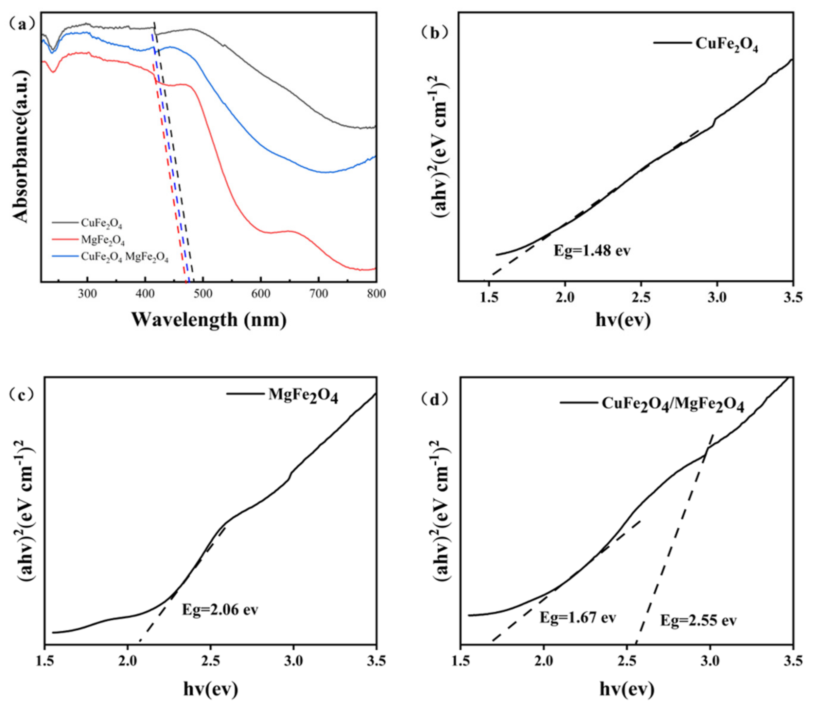
2.2. Optical Properties of the Hollow CuFe2O4/MgFe2O4

The effect of the heterogeneous binding of n-MgFe2O4 and p-CuFe2O4 on the visible light capture ability was investigated by UV-vis diffuse reflectance spectroscopy, as shown in Figure 4a. The absorption edges of MgFe2O4 and CuFe2O4 in the visible light absorption region were 471 nm and 486 nm, respectively. Moreover, the absorption edge of CuFe2O4/MgFe2O4 had a significant red shift increase compared to MgFe2O4, which indicates that the p-n CuFe2O4/MgFe2O4 photocatalyst had a higher visible light absorption ability. The band gaps of CuFe2O4/MgFe2O4 were obtained from Equation (8).
As shown in Figure 4b,c, the band gaps of n-MgFe2O4 and p-CuFe2O4 were 1.48 eV and 2.06 eV, respectively. However, for the CuFe2O4/MgFe2O4 composite, as illustrated in Figure 4d, the band positions of both CuFe2O4 and MgFe2O4 were shifted to ensure Fermi levels reached the new balanced state, respectively, which resulted in the final electronic structure of the p-n heterojunction [48].
As shown in Figure 5a, the impedance of the CuFe2O4/MgFe2O4 heterojunction structure was significantly lower than that of MgFe2O4 and CuFe2O4, indicating that the synthesis of the p-n junctions accelerated the process of interfacial charge transfer. This result indicates that the p-n junction structure promoted the separation and electron transfer of photogenerated carriers. To determine the position of the energy band edges of the obtained samples and to verify the p-n heterojunctions, Mott–Schottky measurements were performed on CuFe2O4, MgFe2O4, and CuFe2O4/MgFe2O4 samples. As shown in Figure 5b–d, the Mott–Schottky plot of CuFe2O4 has a negative slope, corresponding to a typical p-type with a flat-band voltage of 0.72 eV (vs. SCE). The Mott–Schottky plot of MgFe2O4 has a positive slope and corresponds to a typical n-type with a flat-band voltage of −0.49 eV (vs. SCE). As a characteristic of p-type semiconductors, the flat band potential of CuFe2O4 can be attributed to the valence band maximum (VBM), while the associated value of MgFe2O4 can be attributed to the conduction band minimum (CBM) [48].
The potential of the normal hydrogen electrode (NHE) of CuFe2O4/MgFe2O4 was calculated using Equation (9). The result was calculated as 0.961 eV for the VBM edge position of CuFe2O4 and −0.249 eV for the CBM edge position of MgFe2O4 with respect to the NHE. The Mott–Schottky plot of the CuFe2O4/MgFe2O4 composite (Figure 5d) shows an inverted V-shape with two regions distinguished, which is characteristic of the p-n junction formation between CuFe2O4 and MgFe2O4 [49]. Thus, the VBM of CuFe2O4 was located at 0.971 eV (vs. NHE) and the CBM was −0.699 eV, while the CBM of MgFe2O4 was located at −0.219 eV (vs. NHE) and the VBM was 2.331 eV.

2.3. Photocatalytic Experiments of the CuFe2O4/MgFe2O4 Composites

The degradation rates of AO7 by the hollow composite photocatalysts with different Cu:Mg ratios (1:0.5, 1:1, 1:2, 1:3) are shown in Figure 6a, and the maximum efficiency in degrading the AO7 over 60 min was 91.96% when the Cu:Mg ratio was 1:2, and the degradation efficiency was better than that of the other photocatalysts, shown in Table S2, over the same time. The degradation rate of 40% for the 1:2 Cu:Mg ratio under UV light was only slightly higher than that of 28% in the dark, indicating that the visible light-driven catalyzed AO7 degradation was the dominant reaction for CuFe2O4/MgFe2O4. The decrease in the AO7 concentration under dark conditions was due to the specific adsorption performance of the catalyst itself. As shown in Figure 6b,c, the light irradiation of AO7 in aqueous solution containing (Cu:Mg = 1:2) CuFe2O4/MgFe2O4 suspension caused the absorbance, at 485 nm, to decrease with time and, finally, nearly disappear, indicating the destruction of its chromophore structure, with an azo and hydrazone form. The absorbance at 228 and 310 nm, attributed to the benzoic and naphthalene rings, respectively [50,51,52], decreased simultaneously, and no new absorption bands appeared. These results indicated the complete photocatalytic degradation of AO7 during the reaction [53]. The linear relationship curves of the CuFe2O4/MgFe2O4 photocatalysts in the degradation of the AO7 concentration with the time and quasi primary reaction kinetics are shown in Figure 6d, where C0 and Ct are the initial concentration of AO7 and the concentration of AO7 during the reaction, respectively. The degradation rate of the CuFe2O4/MgFe2O4 photocatalyst in the AO7 solution reached 94%. The calculated rate constant k for the CuFe2O4/MgFe2O4 photocatalyst was 0.0371 min−1.
The stability and reusability of catalysts are important for their practical applications. The (Cu:Mg = 1:2) CuFe2O4/MgFe2O4 was reused to catalyze the degradation of AO7 over five cycles. The catalyst was magnetically separated after the reaction and washed with alcohol and dried at 60 °C for every cycle. An alcohol–water system proved to be effective in the desorption of AO7 in our prior work [54]. Figure 6d shows that the degradation rates for five cycles were 91.96%, 87%, 88%, 82% and 79%, respectively. The conversion rate decreased after the recycling due to the loss of the sample during the recycling process. The value was 79% after five cycles, demonstrating the good recyclability of the designed photocatalyst.

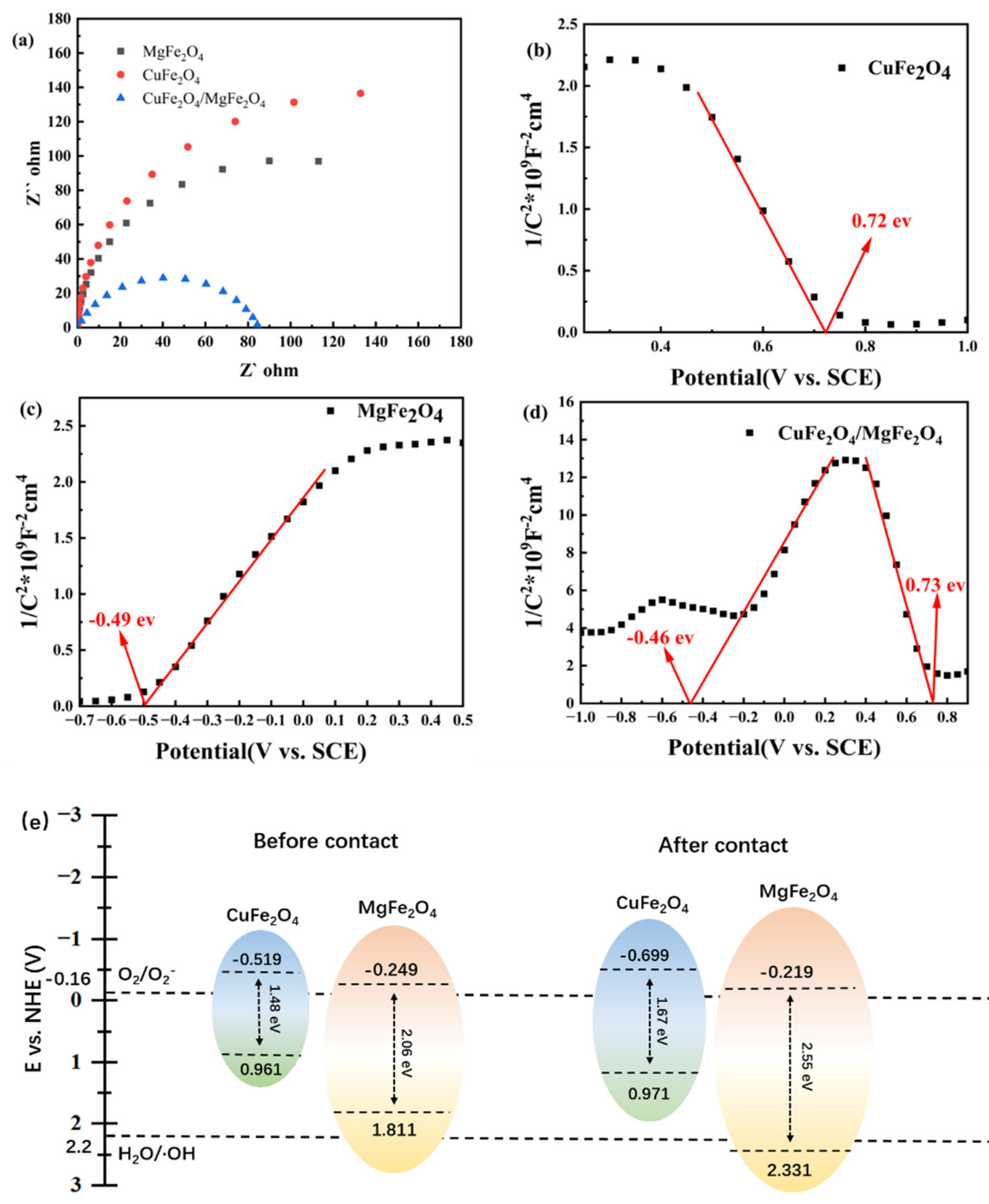
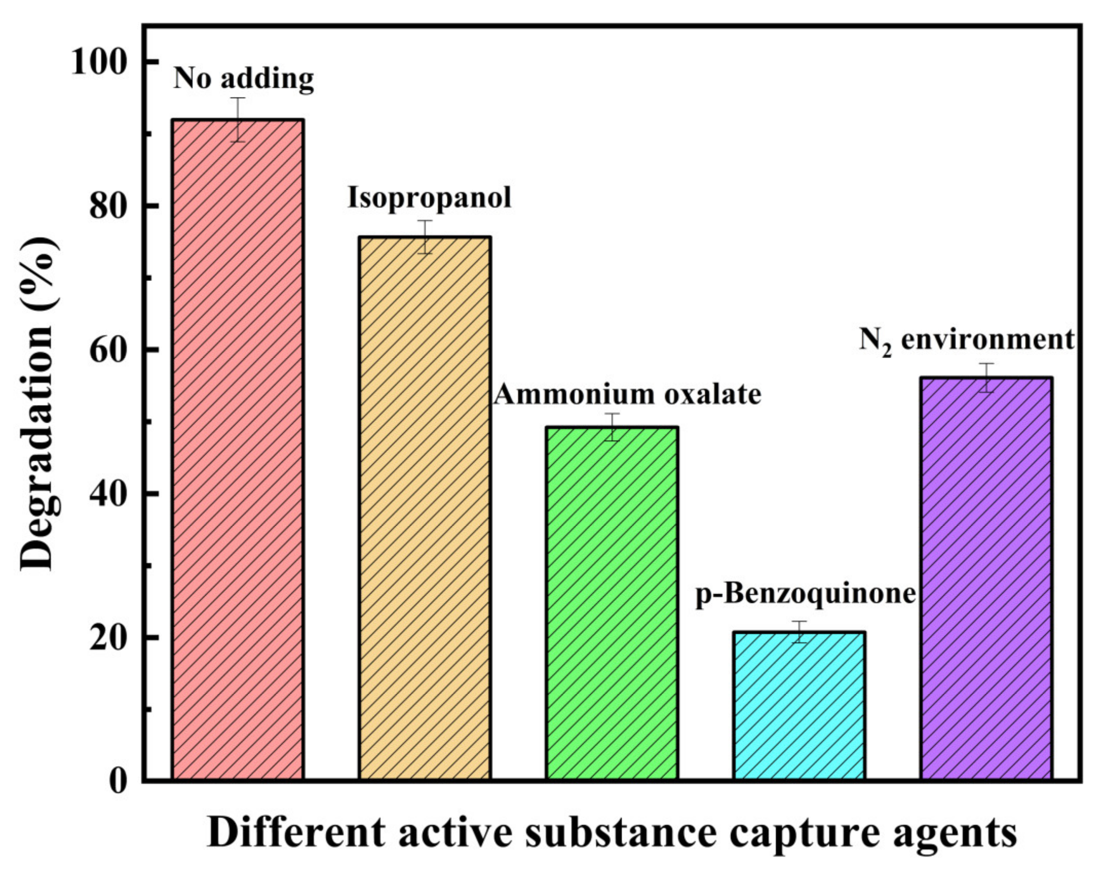
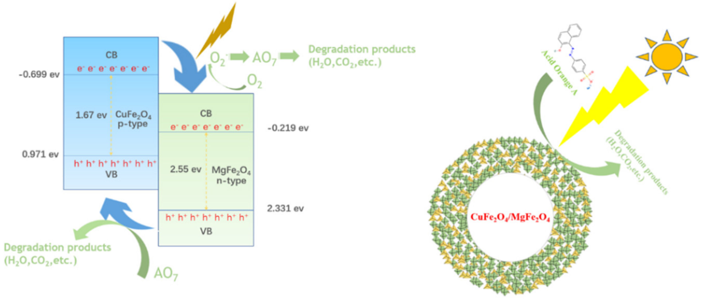
2.4. Mechanism of the Photocatalytic Reaction

The photocatalytic mechanism of the degradation of AO7 by the CuFe2O4/MgFe2O4 was explained in detail using active species trapping experiments (Figure 7). After the addition of O2 [55] scavenger p-benzoquinone, h+ scavenger ammonium oxalate [56,57] and OH [29,56,57] propanol, the degradation rates of AO7 were 20.8%, 49.2% and 16.3% respectively, implying that the photocatalytic mechanism was mainly controlled by the ·O2 radicals and holes (h+). In aqueous media, the redox potential of the dissolved oxygen/superoxide couple (O2(aq.)/O2) was −0.16 eV (vs. NHE) and for the H2O/·OH couple was 2.2 eV [58]. Thus, the photogenerated electrons, which are required to form superoxide, must have a potential less than −0.16 eV and photogenerated holes, needed to form ·OH, must have had a potential greater than 2.2 eV. In the case of the CuFe2O4/MgFe2O4 composite, the CuFe2O4 had the more negative balance band edge (ECB = −0.699 eV (vs. NHE) [59]) than O2(aq.)/·O2; thus, the reaction of e in the conduction band with the oxygen in the aqueous solution to form ·O2 was thermodynamically favorable. In order to verify the role of O2 in the photocatalytic experiments, pure N2 was continuously bubbled into the reaction solution with a flow rate of 30 mL min−1 in light or dark conditions. This was followed by blowing nitrogen for 30 min in the dark to remove the O2 from the reaction unit. The degradation rate of AO7 was reduced to 56.11% in the presence of N2, which proves that O2 is one of the main conditions required for this reaction to occur. The photogenerated h+ in the VB of MgFe2O4 activated H2O to generate ·OH, and e in the CB of MgFe2O4 reduced O2 to ·O2 radicals. Holes were retained on the VB and directly oxidative the pollutants as active species. The ·OH had a minor contribution to the oxidative degradation of AO7 in the CuFe2O4/MgFe2O4 visible light system.
The CB of CuFe2O4 was −0.699 eV (vs. NHE), and the calculated VB was 0.971 eV, based on Figure 5b–d. In a similar manner, the VB and CB of MgFe2O4 were measured as 2.331 eV and −0.219 eV, respectively. After absorbing the photons, the electrons of CuFe2O4/MgFe2O4 in the composite were able to be excited to the conduction band and leave holes in the valance band. The photogenerated e in the CB of CuFe2O4 was easily transferred to the CB of MgFe2O4 with the assistance of the internal electric field. This made the charge separation more effective and, hence, the electrons and holes migrated toward the surface of the respective particles and participated in the redox reaction. Simultaneously, plenty of holes gathered on the VB of CuFe2O4 participated in the photocatalytic reaction.
On the other hand, the dye molecules adsorbed on the photocatalyst surface could be excited by visible light photons, and the excited AO7 had the oxidation potential of −1.24 VNHE (AO7*/AO7+) [60], which was more negative than the potential of the conduction band of the catalyst. Thus, from a thermodynamic point of view, electrons can be injected from the excited AO7 to the conduction bands of catalyst. Here, the catalyst plays the crucial role as the electron carrier for the separation of the injected electrons and dye cation radicals. As a result, a high concentration of free electrons was formed in the conduction band of MgFe2O4. These electrons reduced the dissolved oxygen into superoxide radicals (·O2), and the generated ·O2 could form H2O2 and ·OH under acidic conditions (pH = 5.4) [61]. The oxidative species could react with the AO7 molecules and result in the degradation of the dyes. The mechanism of the degradation of AO7 by CuFe2O4/MgFe2O4 under visible light is shown in Equations (1)–(5) and Figure 8.
CuFe 2 O 4 + hv CuFe 2 O 4 h + + e
CuFe 2 O 4 e + MgFe 2 O 4 CuFe 2 O 4 + MgFe 2 O 4 e
MgFe 2 O 4 e + O 2 · O 2 + MgFe 2 O 4
O 2 + AO 7 CO 2 + H 2 O + Product
h + + AO 7 CO 2 + H 2 O + Product

3. Materials and Methods

3.1. Synthesis of CuFe2O4/MgFe2O4

The CuFe2O4/MgFe2O4 composites were fabricated via a solvothermal method with the surfactant of PEG-20000. Taking the synthesis of CuFe2O4/MgFe2O4 with the molar ratio of Cu:Mg = 1:1 in the precursors as an example, stoichiometric amounts of FeCl3·6H2O (5.0 mM), CuCl2·2H2O (1.25 mM) and MgCl2·6H2O (1.25 mM) were dissolved in 40 mL ethylene glycol under vigorous stirring. Under this condition, 1 g PEG-20000 and 3.6 g sodium acetate were added and kept stirring for 30 min. Then the dark-green solution was transferred into autoclaves and continued at 200 °C for 8 h. The products were harvested by centrifugation, washed with distilled water and ethanol to remove unexpected ions, and dried at 60 °C in air. The thus-prepared composite was denoted as (Cu:Mg = 1:1) CuFe2O4/MgFe2O4. The different samples were prepared with varying Cu:Mg ratios = 1:0.5, 1:1, 1:2 and 1:3, respectively, while keeping the total moles of cupric chloride and magnesium chloride at 2.5 mM.

3.2. Catalyst Characterizations

X-ray diffraction (XRD) patterns were recorded with a powder diffraction system using Cu Kα radiation (λ = 1.5406 Å) in the 2θ range of 10°–90°. Transmission electron microscopy (TEM) images were collected on a FEI Techai 20 microscope (Hillsboro, OR, USA). High-resolution transmission electron microscopy (HRTEM) images were collected on a JEM-2100 microscope (JEOL, Tokyo, Japan) operating at an acceleration voltage of 200 kV. The diffusion reflectance spectra (DRS) were measured on an ultraviolet–visible spectrophotometer (JASCO, Tokyo, Japan, V-530). Transient photocurrents and Mott–Schottky plots were measured on an electrochemical workstation (CHI 760E, three-electrode system in 0.5 mol/L Na2SO4 aqueous solution as an electrolyte). The working electrode was fabricated using a drop-casting method. A total of 5 mg of the photocatalysts was dispersed in 1 mL ethanol containing 40 μL nafion solution (5%) by sonication for 30 min. Then, the dispersive suspension was drop-cast onto a pretreated fluorine-doped tin oxide (FTO) glass substrate, and dried at 60 °C in air for 2 h to improve the adhesion. The optical properties were studied via diffuse reflectance spectroscopy in the wavelength range from 220 nm to 800 nm (DRS, Shimadzu UV-2600, Shimadzu, Kyoto, Japan).

3.3. Photocatalytic Experiments

The photocatalytic performances of the prepared ferrite composites were evaluated by the degradation of AO7 in aqueous solution at 25 °C under simulated visible-light irradiation. A 150 W Xe lamp with 96,000 Lux was used as the light source. In a typical experiment, 0.8 g/L of the ferrite composite was dispersed in 0.10 mM AO7 aqueous solution. Prior to irradiation, the suspension was magnetically stirred in the dark for 60 min to ensure the adsorption–desorption equilibrium. Afterwards, the solution was exposed to visible light with continuous aeration. At given time intervals, the solution was sampled and the residual concentration of AO7 was determined by a UV-vis spectroscopy (JASCO, V-530) at 485 nm, and the degradation rate of AO7 was determined by the following formula:
Q t = C 0 C t C 0 × 100 %
where Q t is the degradation rate at time t , and C 0 and C t are the initial concentration and concentration at time t , respectively.
The photocatalytic degradation reaction of AO7 can be described by the pseudo-first-order kinetic, as shown in Equation (7) [62]:
ln C t C 0 = kt
where the k is the apparent reaction rate constant.
The band gap can be estimated according to the energy dependence relation of [32]:
ahv = hv Eg 1 / 2
where a and Eg are the absorption coefficient and the energy gap of the semiconductor, respectively. Both the CuFe2O4 [32,36] and MgFe2O4 [63] have direct band absorption.
The potential of the normal hydrogen electrode (NHE) can be converted from the obtained data by using the energy Ernst Equation (9) [64]:
E NHE = E SCE + 0.241
where E NHE corresponds to the potential of the normal hydrogen electrode after conversion, and E SCE electrode indicates the collected value of the working electrode.

4. Conclusions

In summary, the magnetically recyclable CuFe2O4/MgFe2O4 heterojunction hollow sphere photocatalysts were prepared using a solvothermal method. The photocatalytic activity of (Cu:Mg = 1:2) CuFe2O4/MgFe2O4 was better than that of CuFe2O4 and MgFe2O4, which was due to the formation of the p-n heterojunction between CuFe2O4 and MgFe2O4. The synthesis of CuFe2O4/MgFe2O4 was demonstrated by XRD and XPS. The absorption edge of CuFe2O4/MgFe2O4 showed an obvious red shift increase following UV-Vis DRS, showing that p-n CuFe2O4/MgFe2O4 had a higher visible light absorption ability. The impedance of the CuFe2O4/MgFe2O4 heterojunction structure was lower than that of MgFe2O4 and CuFe2O4, showing that the p-n structure promotes the separation of photogenerated carriers, and the CuFe2O4/MgFe2O4 material had a better photocatalytic performance. The corresponding hollow formation mechanism is proposed: ·O2 and h+ are the two main active radicals involved in the photodegradation of organic matter in the visible light CuFe2O4/MgFe2O4 system. Here, an efficient method for constructing hollow heterojunction-structured nanomaterials is provided for efficient photocatalytic degradation.

Supplementary Materials

The following supporting information can be downloaded at: www.mdpi.com/article/10.3390/catal12080910/s1, Figure S1: (a). The cell structure diagram of CuFe2O4, (b). MgFe2O4; Figure S2: (Cu:Mg =1:2) CuFe2O4/MgFe2O4 on the degradation of AO7; Table S1: Element mole content table of CuFe2O4/MgFe2O4 by XPS; Table S2: Comparison of different reported photocatalysts for AO7 degradation. References [65,66,67,68,69,70] are cited in the Supplementary Materials.

Author Contributions

H.X.: conceptualization, methodology, supervision and writing—original draft; Z.Z.: investigation, data curation and writing—original draft; W.C.: soft, investigation and data curation; S.R.: investigation and data curation; H.Q.: conceptualization and funding acquisition. All authors have read and agreed to the published version of the manuscript.

Funding

This work was supported by the National Natural Science Foundation of China (grant number: 51778296).

Data Availability Statement

Not applicable.

Acknowledgments

This research was funded by the National Natural Science Foundation of China grant number: 51778296.

Conflicts of Interest

The authors declare no conflict of interest or any personal circumstances or interests that may be perceived as inappropriately influencing the representation or interpretation of the reported research results. The funders had no role in the design of the study or in the collection, analysis or interpretation of the data.

References

  1. Lee, M.-Y.; Wang, W.-L.; Du, Y.; Wu, Q.-Y.; Huang, N.; Xu, Z.-B.; Hu, H.-Y. Comparison of UV/H2O2 and UV/PS processes for the treatment of reverse osmosis concentrate from municipal wastewater reclamation. Chem. Eng. J. 2020, 388, 79–88. [Google Scholar] [CrossRef]
  2. Fu, J.; Xu, Q.; Low, J.; Jiang, C.; Yu, J. Ultrathin 2D/2D WO3/g-C3N4 step-scheme H2-production photocatalyst. Appl. Catal. B-Environ. 2019, 243, 556–565. [Google Scholar] [CrossRef]
  3. Masuda, Y. Bio-inspired mineralization of nanostructured TiO2 on PET and FTO films with high surface area and high photocatalytic activity. Sci. Rep. 2020, 10, 13499–13504. [Google Scholar] [CrossRef] [PubMed]
  4. Takanabe, K. Photocatalytic Water Splitting: Quantitative approaches toward photocatalyst by design. ACS Catal. 2017, 7, 8006–8022. [Google Scholar] [CrossRef]
  5. Alshorifi, F.T.; Alswat, A.A.; Salama, R.S. Gold-selenide quantum dots supported onto cesium ferrite nanocomposites for the efficient degradation of rhodamine B. Heliyon 2022, 8, e09652. [Google Scholar] [CrossRef]
  6. Ma, Y.; Ma, M.; Yin, X.; Shao, Q.; Lu, N.; Feng, Y.; Lu, Y.; Wujcik, E.K.; Mai, X.; Wang, C.; et al. Tuning polyaniline nanostructures via end group substitutions and their morphology dependent electrochemical performances. Polymer 2018, 156, 128–135. [Google Scholar] [CrossRef]
  7. Zhang, G.; Zhang, X.; Meng, Y.; Pan, G.; Ni, Z.; Xia, S. Layered double hydroxides-based photocatalysts and visible-light driven photodegradation of organic pollutants: A review. Chem. Eng. J. 2020, 392, 123684–123690. [Google Scholar] [CrossRef]
  8. Tobbala, D.E.; Rashed, A.S.; Salama, R.S.; Ahmed, T.I. Performance enhancement of reinforced concrete exposed to electrochemical magnesium chloride using nano-ferrite zinc-rich epoxy. J. Build. Eng. 2022, 57, 104869–104873. [Google Scholar] [CrossRef]
  9. Umar, M.; Mahmood, N.; Awan, S.U.; Fatima, S.; Mahmood, A.; Rizwan, S. Rationally designed La and Se co-doped bismuth ferrites with controlled bandgap for visible light photocatalysis. RSC Adv. 2019, 9, 17148–17156. [Google Scholar] [CrossRef]
  10. An, J.; Zhu, L.; Wang, N.; Song, Z.; Yang, Z.; Du, D.; Tang, H. Photo-Fenton like degradation of tetrabromobisphenol A with grapheneBiFeO3 composite as a catalyst. Chem. Eng. J. 2013, 219, 225–237. [Google Scholar] [CrossRef]
  11. Li, Z.; Shen, Y.; Yang, C.; Lei, Y.; Guan, Y.; Lin, Y.; Liu, D.; Nan, C.-W. Significant enhancement in the visible light photocatalytic properties of BiFeO3–graphene nanohybrids. J. Mater. Chem. A 2013, 1, 823–829. [Google Scholar] [CrossRef]
  12. Kazachenko, A.S.; Vasilieva, N.Y.; Fetisova, O.Y.; Sychev, V.V.; Elsuf’ev, E.V.; Malyar, Y.N.; Issaoui, N.; Miroshnikova, A.V.; Borovkova, V.S.; Kazachenko, A.S.; et al. New reactions of betulin with sulfamic acid and ammonium sulfamate in the presence of solid catalysts. Biomass Convers. Biorefinery 2022, 23, 02587–02591. [Google Scholar] [CrossRef]
  13. Zhu, Y.; Liu, Y.; Ai, Q.; Gao, G.; Yuan, L.; Fang, Q.; Tian, X.; Zhang, X.; Egap, E.; Ajayan, P.M.; et al. In situ synthesis of lead-free halide perovskite–COF nanocomposites as photocatalysts for photoinduced polymerization in both organic and aqueous phases. ACS Mater. Lett. 2022, 4, 464–471. [Google Scholar] [CrossRef]
  14. Bajpai, O.P.; Mandal, S.; Ananthakrishnan, R.; Mandal, P.; Khastgir, D.; Chattopadhyay, S. Structural features, magnetic properties and photocatalytic activity of bismuth ferrite nanoparticles grafted on graphene nanosheets. New J. Chem. 2018, 42, 10712–10723. [Google Scholar] [CrossRef]
  15. Yin, X.L.; Liu, J.; Jiang, W.J.; Zhang, X.; Hu, J.S.; Wan, L.J. Urchin-like Au@CdS/WO3 micro/nano heterostructure as a visible-light driven photocatalyst for efficient hydrogen generation. Chem. Commun. 2015, 51, 13842–13845. [Google Scholar] [CrossRef]
  16. Chakraborty, I.; Rakshit, R.; Mandal, K. Synthesis and functionalization of MnFe2O4 nano–hollow spheres for novel optical and catalytic properties. Surf. Interfaces 2017, 7, 106–112. [Google Scholar] [CrossRef]
  17. Cheng, C.; Zhang, H.; Ren, W.; Dong, W.; Sun, Y. Three dimensional urchin-like ordered hollow TiO2/ZnO nanorods structure as efficient photoelectrochemical anode. Nano Energy 2013, 2, 779–786. [Google Scholar] [CrossRef]
  18. Cui, X.; Jiang, G.; Zhu, M.; Zhao, Z.; Du, L.; Weng, Y.; Xu, C.; Zhang, D.; Zhang, Q.; Wei, Y.; et al. TiO2/CdS composite hollow spheres with controlled synthesis of platinum on the internal wall for the efficient hydrogen evolution. Int. J. Hydrogen Energy 2013, 38, 9065–9073. [Google Scholar] [CrossRef]
  19. Amuthan, T.; Sanjeevi, R.; Kannan, G.R.; Sridevi, A. A novel p-CuFe2O4/n-ZnS heterojunction photocatalyst: Co-precipitation synthesis, characterization and improved visible-light driven photocatalytic activity for removal of MB and CV dyes. Phys. B Condens. Matter 2022, 638, 413842–413848. [Google Scholar] [CrossRef]
  20. Mousavi, M.; Habibi, M.M.; Zhang, G.; Pourhakkak, P.; Moradian, S.; Ghasemi, J.B. In-situ construction of ZnO/Sb2MoO6 nano-heterostructure for efficient visible-light photocatalytic conversion of N2 to NH3. Surf. Interfaces 2022, 30, 101844. [Google Scholar] [CrossRef]
  21. Wang, Z.; Lin, Z.; Shen, S.; Zhong, W.; Cao, S. Advances in designing heterojunction photocatalytic materials. Chin. J. Catal. 2021, 42, 710–730. [Google Scholar] [CrossRef]
  22. Jang, J.S.; Kim, H.G.; Lee, J.S. Heterojunction semiconductors: A strategy to develop efficient photocatalytic materials for visible light water splitting. Catal. Today 2012, 185, 270–277. [Google Scholar] [CrossRef]
  23. Yan, Y.; Zeng, Z.; Huang, M.; Chen, P. Van der waals heterojunctions for catalysis. Mater. Today Adv. 2020, 6, 100059–100063. [Google Scholar] [CrossRef]
  24. Arimi, A.; Megatif, L.; Granone, L.I.; Dillert, R.; Bahnemann, D.W. Visible-light photocatalytic activity of zinc ferrites. J. Photochem. Photobiol. A-Chem. 2018, 366, 118–126. [Google Scholar] [CrossRef]
  25. Lenin, N.; Kanna, R.R.; Sakthipandi, K.; Kumar, A.S. Structural, electrical and magnetic properties of NiLaxFe2-xO4 nanoferrites. Mater. Chem. Phys. 2018, 212, 385–393. [Google Scholar] [CrossRef]
  26. Alshorifi, F.T.; Ali, S.L.; Salama, R.S. Promotional synergistic effect of Cs–Au NPs on the performance of Cs–Au/MgFe2O4 catalysts in catalysis 3,4-Dihydropyrimidin-2(1H)-Ones and degradation of RhB Dye. J. Inorg. Organomet. Polym. Mater. 2022, 8, 02389–02401. [Google Scholar] [CrossRef]
  27. Kumar, G.M.; Cho, H.D.; Lee, D.J.; Kumar, J.R.; Siva, C.; Ilanchezhiyan, P.; Kim, D.Y.; Kang, T.W. Elevating the charge separation of MgFe2O4 nanostructures by Zn ions for enhanced photocatalytic and photoelectrochemical water splitting. Chemosphere 2021, 283, 131134–131145. [Google Scholar] [CrossRef]
  28. Kim, H.G.; Borse, P.H.; Jang, J.S.; Jeong, E.D.; Jung, O.-S.; Suh, Y.J.; Lee, J.S. Fabrication of CaFe2O4/MgFe2O4 bulk heterojunction for enhanced visible light photocatalysis. Chem. Commun. 2009, 279, 5889–5891. [Google Scholar] [CrossRef]
  29. Fan, W.; Li, M.; Bai, H.; Xu, D.; Chen, C.; Li, C.; Ge, Y.; Shi, W. Fabrication of MgFe2O4/MoS2 heterostructure nanowires for photoelectrochemical catalysis. Langmuir 2016, 32, 1629–1636. [Google Scholar] [CrossRef]
  30. Liu, X.; An, S.; Shi, W.; Yang, Q.; Zhang, L. Microwave-induced catalytic oxidation of malachite green under magnetic Cu-ferrites: New insight into the degradation mechanism and pathway. J. Mol. Catal. A Chem. 2014, 395, 243–250. [Google Scholar] [CrossRef]
  31. Wang, Y.; Zhao, H.; Li, M.; Fan, J.; Zhao, G. Magnetic ordered mesoporous copper ferrite as a heterogeneous Fenton catalyst for the degradation of imidacloprid. Appl. Catal. B-Environ. 2014, 147, 534–545. [Google Scholar] [CrossRef]
  32. Shen, Y.; Wu, Y.; Xu, H.; Fu, J.; Li, X.; Zhao, Q.; Hou, Y. Facile preparation of sphere-like copper ferrite nanostructures and their enhanced visible-light-induced photocatalytic conversion of benzene. Mater. Res. Bull. 2013, 48, 4216–4222. [Google Scholar] [CrossRef]
  33. Sharma, R.; Kumar, V.; Bansal, S.; Singhal, S. Assortment of magnetic nanospinels for activation of distinct inorganic oxidants in photo-Fenton’s process. J. Mol. Catal. A-Chem. 2015, 402, 53–63. [Google Scholar] [CrossRef]
  34. Ilhan, S.; Izotova, S.G.; Komlev, A.A. Synthesis and characterization of MgFe2O4 nanoparticles prepared by hydrothermal decomposition of co-precipitated magnesium and iron hydroxides. Ceram. Int. 2015, 41, 577–585. [Google Scholar] [CrossRef]
  35. Derbal, A.; Omeiri, S.; Bouguelia, A.; Trari, M. Characterization of new heterosystem CuFeO2/SnO2 application to visible-light induced hydrogen evolution. Int. J. Hydrogen Energy 2008, 33, 4274–4282. [Google Scholar] [CrossRef]
  36. Fedailaine, M.; Bellal, B.; Berkani, S.; Trari, M.; Abdi, A. Photo-electrochemical characterization of the spinel CuFe2O4: Application to Ni2+ removal under solar light. Environ. Process. 2016, 3, 387–396. [Google Scholar] [CrossRef]
  37. Dom, R.; Subasri, R.; Radha, K.; Borse, P.H. Synthesis of solar active nanocrystalline ferrite, MFe2O4 (M: Ca, Zn, Mg) photocatalyst by microwave irradiation. Solid State Commun. 2011, 151, 470–473. [Google Scholar] [CrossRef]
  38. Feizpoor, S.; Habibi-Yangjeh, A.; Vadivel, S. Novel TiO2/Ag2CrO4 nanocomposites: Efficient visible-light-driven photocatalysts with n-n heterojunctions. J. Photochem. Photobiol. A-Chem. 2017, 341, 57–68. [Google Scholar] [CrossRef]
  39. Gu, X.; Li, C.; Yuan, S.; Ma, M.; Qiang, Y.; Zhu, J. ZnO based heterojunctions and their application in environmental photocatalysis. Nanotechnology 2016, 27, 402001–402013. [Google Scholar] [CrossRef]
  40. Yan, Y.; Guan, H.; Liu, S.; Jiang, R. Ag3PO4/Fe2O3 composite photocatalysts with an n-n heterojunction semiconductor structure under visible-light irradiation. Ceram. Int. 2014, 40, 9095–9100. [Google Scholar] [CrossRef]
  41. Zou, X.; Dong, Y.; Zhang, X.; Cui, Y.; Ou, X.; Qi, X. The highly enhanced visible light photocatalytic degradation of gaseous odichlorobenzene through fabricating like-flowers BiPO4/BiOBr p-n heterojunction composites. Appl. Surf. Sci. 2017, 391, 525–534. [Google Scholar] [CrossRef]
  42. Wang, X.; Feng, J.; Bai, Y.; Zhang, Q.; Yin, Y. Synthesis, Properties, and Applications of Hollow Micro-/Nanostructures. Chem. Rev. 2016, 116, 10983–11060. [Google Scholar] [CrossRef]
  43. Gabal, M.A.; Al-luhaibi, R.S.; Al Angari, Y.M. Mn-Zn nano-crystalline ferrites synthesized from spent Zn-C batteries using novel gelatin method. J. Hazard. Mater. 2013, 246, 227–233. [Google Scholar] [CrossRef]
  44. Sharma, R.; Bansal, S.; Singhal, S. Tailoring the photo-Fenton activity of spinel ferrites (MFe2O4) by incorporating different cations (M = Cu, Zn, Ni and Co) in the structure. RSC Adv. 2015, 5, 6006–6018. [Google Scholar] [CrossRef]
  45. Yin, Y.; Liu, W.; Huo, N.; Yang, S. High rate capability and long cycle stability of Fe2O3/MgFe2O4 anode material synthesized by gelcast processing. Chem. Eng. J. 2017, 307, 999–1007. [Google Scholar] [CrossRef]
  46. Zhang, L.; Ghimire, P.; Phuriragpitikhon, J.; Jiang, B.; Goncalves, A.A.S.; Jaroniec, M. Facile formation of metallic bismuth/bismuth oxide heterojunction on porous carbon with enhanced photocatalytic activity. J. Colloid Interface Sci. 2018, 513, 82–91. [Google Scholar] [CrossRef]
  47. Wen, X.-J.; Niu, C.-G.; Zhang, L.; Liang, C.; Zeng, G.-M. An in depth mechanism insight of the degradation of multiple refractory pollutants via a novel SrTiO3/BiOI heterojunction photocatalysts. J. Catal. 2017, 356, 283–299. [Google Scholar] [CrossRef]
  48. Huu, H.T.; Thi, M.D.N.; Nguyen, V.P.; Thi, L.N.; Phan, T.T.T.; Hoang, Q.D.; Luc, H.H.; Kim, S.J.; Vo, V. One-pot synthesis of S-scheme MoS2/g-C3N4 heterojunction as effective visible light photocatalyst. Sci. Rep. 2021, 11, 14787. [Google Scholar] [CrossRef]
  49. Wen, X.-J.; Niu, C.-G.; Zhang, L.; Liang, C.; Zeng, G.-M. A novel Ag2O/CeO2 heterojunction photocatalysts for photocatalytic degradation of enrofloxacin: Possible degradation pathways, mineralization activity and an in depth mechanism insight. Appl. Catal. B-Environ. 2018, 221, 701–714. [Google Scholar] [CrossRef]
  50. Barros, V.P.; Assis, M.D. Iron porphyrins as biomimetical models for disperse azo dye oxidation. J. Braz. Chem. Soc. 2013, 24, 830–836. [Google Scholar] [CrossRef]
  51. Wu, F.; Deng, N.S.; Hua, H.L. Degradation mechanism of azo dye C.I. reactive red 2 by iron powder reduction and photooxidation in aqueous solutions. Chemosphere 2000, 41, 1233–1238. [Google Scholar] [CrossRef]
  52. Zhang, S.J.; Yu, H.Q.; Li, Q.R. Radiolytic degradation of Acid Orange 7: A mechanistic study. Chemosphere 2005, 61, 1003–1011. [Google Scholar] [CrossRef]
  53. Li, X.; Hou, Y.; Zhao, Q.; Wang, L. A general, one-step and template-free synthesis of sphere-like zinc ferrite nanostructures with enhanced photocatalytic activity for dye degradation. J. Colloid Interface Sci. 2011, 358, 102–108. [Google Scholar] [CrossRef]
  54. Xing, X.; Qu, H.; Shao, R.; Wang, Q.; Xie, H. Mechanism and kinetics of dye desorption from dye-loaded carbon (XC-72) with alcohol-water system as desorbent. Water Sci. Technol. 2017, 76, 1243–1250. [Google Scholar] [CrossRef]
  55. Stylidi, M.; Kondarides, D.I.; Verykios, X.E. Visible light-induced photocatalytic degradation of Acid Orange 7 in aqueous TiO2 suspensions. Appl. Catal. B-Environ. 2004, 47, 189–201. [Google Scholar] [CrossRef]
  56. Liang, S.; Zhu, S.; Chen, Y.; Wu, W.; Wang, X.; Wu, L. Rapid template-free synthesis and photocatalytic performance of visible light-activated SnNb2O6 nanosheets. J. Mater. Chem. 2012, 22, 2670–2678. [Google Scholar] [CrossRef]
  57. Su, M.; He, C.; Sharma, V.K.; Abou Asi, M.; Xia, D.; Li, X.-Z.; Deng, H.; Xiong, Y. Mesoporous zinc ferrite: Synthesis, characterization, and photocatalytic activity with H2O2/visible light. J. Hazard. Mater. 2012, 211, 95–103. [Google Scholar] [CrossRef]
  58. Li, Y.; Zhang, W.; Niu, J.; Chen, Y. Mechanism of photogenerated reactive oxygen species and correlation with the antibacterial properties of engineered metal-oxide nanoparticles. ACS Nano 2012, 6, 5164–5173. [Google Scholar] [CrossRef]
  59. Hussain, S.; Hussain, S.; Waleed, A.; Tavakoli, M.M.; Wang, Z.; Yang, S.; Fan, Z.; Nadeem, M.A. Fabrication of CuFe2O4/alpha-Fe2O3 composite thin films on FTO coated glass and 3-D nanospike structures for efficient photoelectrochemical water splitting. ACS Appl. Mater. Interfaces 2016, 8, 35315–35322. [Google Scholar] [CrossRef]
  60. Nasr, C.; Vinodgopal, K.; Fisher, L.; Hotchandani, S.; Chattopadhyay, A.K.; Kamat, P.V. Environmental photochemistry on semiconductor surfaces. Visible light induced degradation of a textile diazo dye, naphthol blue black, on TiO2 nanoparticles. J. Phys. Chem. 1996, 100, 8436–8442. [Google Scholar] [CrossRef]
  61. Wen, X.-J.; Niu, C.-G.; Huang, D.-W.; Zhang, L.; Liang, C.; Zeng, G.-M. Study of the photocatalytic degradation pathway of norfloxacin and mineralization activity using a novel ternary Ag/AgCl-CeO2 photocatalyst. J. Catal. 2017, 355, 73–86. [Google Scholar] [CrossRef]
  62. Bechambi, O.; Jlaiel, L.; Najjar, W.; Sayadi, S. Photocatalytic degradation of bisphenol A in the presence of Ce-ZnO: Evolution of kinetics, toxicity and photodegradation mechanism. Mater. Chem. Phys. 2016, 173, 95–105. [Google Scholar] [CrossRef]
  63. Zhang, J.; Yan, M.; Sun, G.; Li, X.; Liu, K. Visible-light photo-Fenton catalytic MgFe2O4 spinel: Reaction sintering synthesis and DFT study. J. Alloy. Compd. 2021, 889, 161673–161684. [Google Scholar] [CrossRef]
  64. Hao, X.; Hu, Z.; Xiang, D.; Jin, Z. Construction of CdS@Cu2-xS core–shell p-n heterojunction with enhanced charge separation for wide spectrum photocatalytic H2 evolution. Mol. Catal. 2022, 528, 112417–112431. [Google Scholar] [CrossRef]
  65. Huang, T.; Mao, S.; Yu, J.; Wen, Z.; Lu, G.; Chen, J. Effects of N and F doping on structure and photocatalytic properties of anatase TiO2 nanoparticles. RSC Adv. 2013, 3, 16657–16664. [Google Scholar] [CrossRef]
  66. Baliarsingh, N.; Parida, K.M.; Pradhan, G.C. Effects of Co, Ni, Cu, and Zn on Photophysical and Photocatalytic Properties of Carbonate Intercalated MII/Cr LDHs for Enhanced Photodegradation of Methyl Orange. Ind. Eng. Chem. Res. 2014, 53, 3834–3841. [Google Scholar] [CrossRef]
  67. Yan, S.; Wang, B.; Shi, Y.; Yang, F.; Hu, D.; Xu, X.; Wu, J. Hydrothermal synthesis of CdS nanoparticle/functionalized graphene sheet nanocomposites for visible-light photocatalytic degradation of methyl orange. Appl. Surf. Sci. 2013, 285, 840–845. [Google Scholar] [CrossRef]
  68. Chen, Q.; Wu, L.; Zhao, X.; Yang, X.-J. Fabrication of Zn-Ti layered double oxide nanosheets with ZnO/ZnTiO3 heterojunction for enhanced photocatalytic degradation of MO, RhB and MB. J. Mol. Liq. 2022, 353, 118794. [Google Scholar] [CrossRef]
  69. Xu, M.L.; Jiang, X.J.; Li, J.R.; Wang, F.J.; Li, K.; Cheng, X. Self-Assembly of a 3D Hollow BiOBr@Bi-MOF Heterostructure with Enhanced Photocatalytic Degradation of Dyes. ACS Appl. Mater. Interfaces 2021, 13, 56171–56180. [Google Scholar] [CrossRef]
  70. Ma, M.; Yang, Y.; Chen, Y.; Jiang, J.; Ma, Y.; Wang, Z.; Huang, W.; Wang, S.; Liu, M.; Ma, D.; et al. Fabrication of hollow flower-like magnetic Fe3O4/C/MnO2/C3N4 composite with enhanced photocatalytic activity. Sci. Rep. 2021, 11, 2597. [Google Scholar] [CrossRef]
Figure 1. TEM images of CuFe2O4/MgFe2O4: (a) Cu:Mg = 1:0.5; (b) Cu:Mg = 1:1; (c) Cu:Mg = 1:2; (d) Cu:Mg = 1:3.
Catalysts 12 00910 g001
Figure 2. TEM images of (1:2) CuFe2O4/MgFe2O4: (a) 4 h with PEG; (b) 8 h with PEG; (c) 12 h with PEG; (d) HRTEM image; (e) schematic illustration (downward views) of the ripening process of the hollow structures.
Catalysts 12 00910 g002
Figure 3. XRD patterns of all CuFe2O4/MgFe2O4 samples.
Catalysts 12 00910 g003
Figure 4. (a) UV–vis DRS of the pure CuFe2O4, MgFe2O4 and CuFe2O4/MgFe2O4 composites; (bd) plots of (αhv)2 versus photon energy (hv) for CuFe2O4, MgFe2O4 and CuFe2O4/MgFe2O4.
Catalysts 12 00910 g004
Figure 5. (a) EIS curves of the CuFe2O4, MgFe2O4 and CuFe2O4/MgFe2O4 composites; (bd) Mott–Schottky plots of CuFe2O4, MgFe2O4 and CuFe2O4/MgFe2O4; (e) interfacial energy band diagram of the CuFe2O4/MgFe2O4 heterojunction.
Catalysts 12 00910 g005
Figure 6. (a) Effects of different Cu:Mg ratios on the degradation of AO7; (b) time-dependent UV–vis spectra of the AO7 solution for (Cu:Mg = 1:2) CuFe2O4/MgFe2O4; (c) dark adsorption and photodegradation curves of the AO7 solution and pseudo-first-order reaction kinetic linear relationship curve (insert; catalyst 0.8 g/L, AO7 = 0.10 mM); (d) reusability of (Cu:Mg = 1:2) CuFe2O4/MgFe2O4 for 5 successive cycles.
Catalysts 12 00910 g006
Figure 7. Effects of different scavengers on the AO7 degradation for (Cu:Mg = 1:2) CuFe2O4/MgFe2O4 (catalyst 0.8 g/L, AO7 = 0.10 mM, scavenger 0.1 mM).
Catalysts 12 00910 g007
Figure 8. Schematic presentation of the electron–hole transfer by the CuFe2O4/MgFe2O4 photocatalyst.
Catalysts 12 00910 g008
Publisher’s Note: MDPI stays neutral with regard to jurisdictional claims in published maps and institutional affiliations.

