Review

Promotion of Carbon Dioxide Biofixation through Metabolic and Enzyme Engineering

1
National Key Laboratory of Biochemical Engineering, Institute of Process Engineering, Chinese Academy of Sciences, Beijing 100190, China
2
School of Chemical Engineering, University of Chinese Academy of Sciences, Beijing 100049, China
*
Author to whom correspondence should be addressed.
Catalysts 2022, 12(4), 399; https://doi.org/10.3390/catal12040399
Submission received: 25 January 2022 / Revised: 30 March 2022 / Accepted: 1 April 2022 / Published: 3 April 2022
(This article belongs to the Special Issue Biocatalysis and Biotransformation of Extremozymes)

Abstract

:
Carbon dioxide is a major greenhouse gas, and its fixation and transformation are receiving increasing attention. Biofixation of CO2 is an eco–friendly and efficient way to reduce CO2, and six natural CO2 fixation pathways have been identified in microorganisms and plants. In this review, the six pathways along with the most recent identified variant pathway were firstly comparatively characterized. The key metabolic process and enzymes of the CO2 fixation pathways were also summarized. Next, the enzymes of Rubiscos, biotin-dependent carboxylases, CO dehydrogenase/acetyl-CoA synthase, and 2-oxoacid:ferredoxin oxidoreductases, for transforming inorganic carbon (CO2, CO, and bicarbonate) to organic chemicals, were specially analyzed. Then, the factors including enzyme properties, CO2 concentrating, energy, and reducing power requirements that affect the efficiency of CO2 fixation were discussed. Recent progress in improving CO2 fixation through enzyme and metabolic engineering was then summarized. The artificial CO2 fixation pathways with thermodynamical and/or energetical advantages or benefits and their applications in biosynthesis were included as well. The challenges and prospects of CO2 biofixation and conversion are discussed.

1. Introduction

Carbon dioxide (CO2) is the main greenhouse gas emitted from human activities such as burning coal, oil, and natural gas, and is considered one of the causes of global warming and climate change. Efforts are being made worldwide to reduce CO2 in the atmosphere and ultimately achieve carbon neutrality. Besides reducing the emission of CO2, the capture, sequestration, and utilization of CO2 is an effective way to decrease the atmospheric CO2 concentration [1,2]. In general, CO2 is captured from industrial emissions and sequestrated in a storage site such as the exploited oil fields, ocean, and abandoned coal mining areas, and then utilized as substrate for chemicals production [3]. To date, an increasing number of methods have been developed to capture and sequestrate CO2 [4,5,6]. Compared with CO2 capture and sequestration, the transformation of CO2 into chemicals is a more challenging step. Based on the physical and chemical properties of CO2, methods including chemical catalysis, photocatalysis, electrocatalysis, enzymatic and microbial catalysis have been explored for CO2 transformation [1,3,7,8,9,10,11]. However, most CO2 conversion methods have issues of poor selectivity, low yields, costly catalysts, and environmental pollution, which limits their application.
Biological carbon fixation is the process of converting inorganic carbon, including CO, CO2, and bicarbonate, into organic chemicals by plants, algae, and autotrophic microorganisms, which is an eco-friendly, highly selective, and promising approach compared with chemical catalysis. Biological fixation of CO2 is a key step in global carbon cycle and fixes 1017 g of CO2 annually [12]. The fixation of CO2 in plants and some microorganisms is through the reductive pentose phosphate (Calvin–Benson–Bassham (CBB)) cycle, which accounts for more than 90% of CO2 fixation in nature. [13]. In addition to the CBB cycle, chemoautotrophs and facultative chemoautotrophs have evolved multiple pathways for CO2 fixation. Both aerobic and obligate anaerobic bacteria are able to utilize CO2 as the sole carbon source for growth, some of which have been engineered to produce various products such as biofuel, bioplastic, and biochemical [14,15,16,17,18,19]. In addition, in vitro multi-enzymatic catalysis has been applied for biofixation and transformation of CO2 to value-added products [10,20].
In the present review, the pathways and key enzymes for CO2 fixation were firstly comparatively analyzed. Next, the latest research on metabolic engineering and enzyme engineering to improve CO2 fixation efficiency was introduced. Finally, key issues hindering CO2 biofixation, as well as potential methods and prospects for improving the efficiency, were discussed.

2. Comparative Analysis of Pathways for Carbon Dioxide Biofixation

Besides the CBB cycle, another five CO2 fixation pathways including the reductive acetyl-CoA cycle, reductive tricarboxylic cycle, 3-hydroxypropionate bi-cycle, 3-hydroxypropionate/4-hydroxybutyrate cycle, and dicarboxylate/4-hydroxybutyrate cycle have been discovered in autotrophic and facultative autotrophic microorganisms [21]. In the five pathways, CO2 is converted to central metabolites such as pyruvate or acetyl-CoA which are different from the CBB pivoting on carbohydrates interconversion. The natural CO2 biofixation pathways with the intermediates and products ranging from C2, C3, C4 organic acids to longer chain carbohydrates, providing valuable platforms for the production of value-added products from CO2.

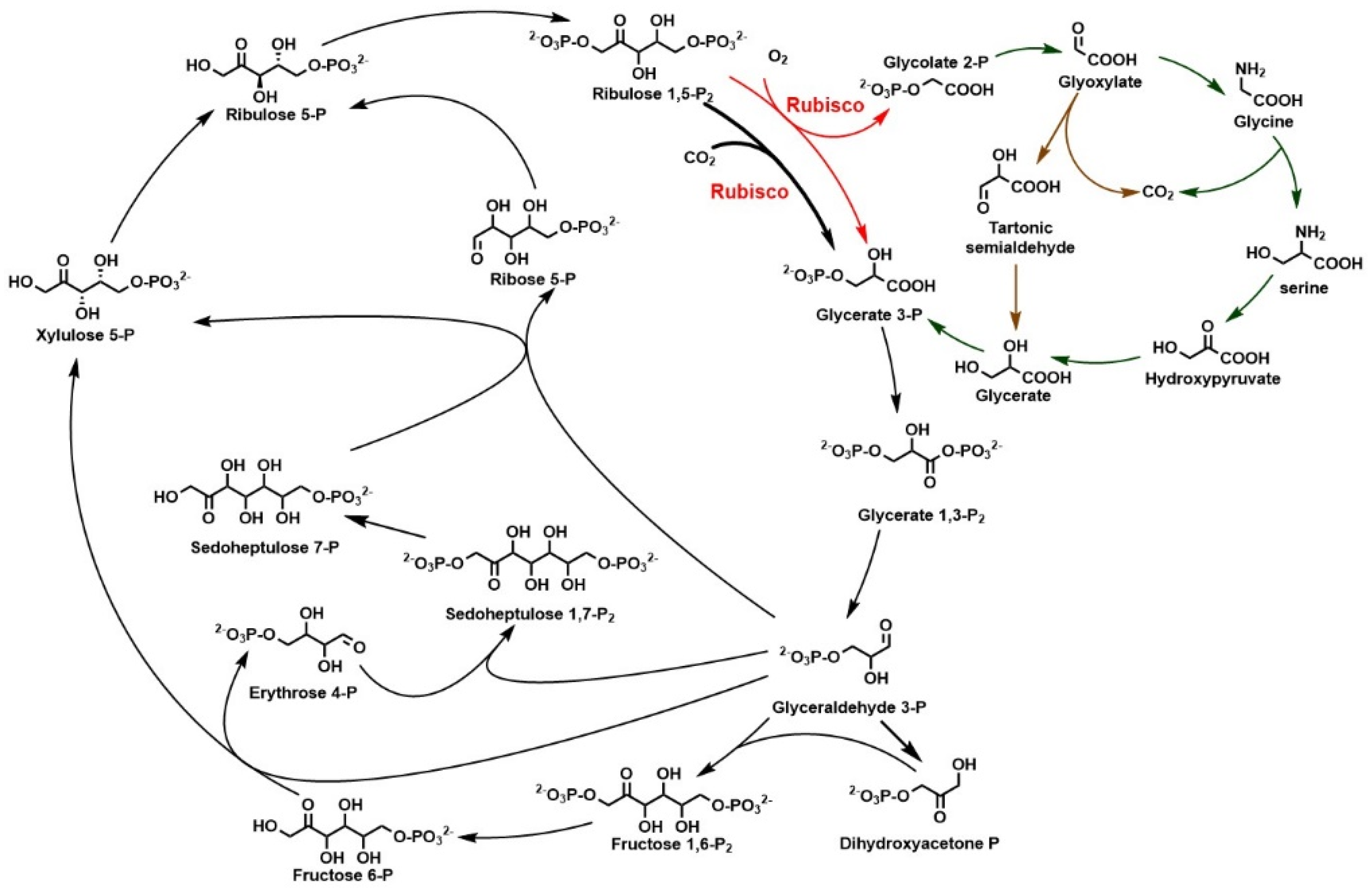
2.1. Calvin–Benson–Basham Cycle and Rubisco

Calvin–Benson–Basham (CBB) cycle, also known as the reductive pentose phosphate cycle, is the first discovered CO2 biofixation pathway and the predominant way of carbon fixation in nature. CBB cycle is widely distributed in plants, algae, cyanobacteria, and some chemoautotrophic microorganisms such as Cupriavidus necator, Rhodobacter, Nitrosomonas, and Acidithiobacillus [21,22,23]. In the CBB cycle, ribose-1,5-diphosphate carboxylase/oxygenase (Rubisco) catalyzes the electrophilic addition of CO2 to ribose-1,5-diphosphate (RuBP) and produces 2 molecules of 3-phosphoglycerate (Figure 1). Subsequently, 3-phosphoglycerate is transformed into glyceraldehyde-3-phosphate by the enzymes of gluconeogenic pathway. Some glyceraldehyde-3-phosphate branches enter the central carbon metabolism, and the others are used for RuBP regeneration, which is then used for CO2 fixation as a receptor.
Besides the carboxylation of CO2 fixation, Rubiscos also catalyze an oxygenation side reaction of converting RuBP and O2 to 3-phosphoglycerate and 2-phosphoglycolate (2PG). The 2PG is a non-CBB intermediate, which is recycled through photorespiration pathway generally known as phosphoglycolate salvage pathway [24,25]. In the photorespiration of plants, 2PG is converted to 3-phosphoglycerate through a pathway involving glycine and serine intermediates (Figure 1, dark green arrow). In some chemoautotrophic bacteria, the conversion can also be accomplished via the pathway involving tartonate semialdehyde intermediate (Figure 1, brown arrow) [24]. A molecule of CO2 is released in the process of RuBP oxygenation, which reduces the CO2 fixation efficiency of plant. It is estimated that about 20% of the CO2 fixed by plants is consumed by photorespiration under atmospheric conditions [26,27].
Different from the CBB cycle based on sugar-phosphate interconversion, the other CO2 fixation pathways are centered on acetyl-CoA and succinyl-CoA and share similarities in a modular feature of carbon fixation and receptor regeneration routines (Figure 2). The following sections will discuss the CO2 fixation pathways identified in different microorganisms and their latest developments.

2.2. The Reductive Tricarboxylic Cycle and Its Variant roTCA Cycle

The reductive tricarboxylic (rTCA) cycle or the reductive citric acid cycle (Figure 2, Routine 2 and 6), first found in the anaerobic green sulfur bacterium Chlorobium limicola, is mainly distributed in some strictly anaerobic or microaerobic autotrophic eubacteria [21,28,29]. The rTCA cycle is the oxidative TCA cycle operated in the opposite direction by employing different enzymes to catalyze the irreversible reactions in TCA cycle, including fumarate reductase, 2-oxoglutarate: ferredoxin oxidoreductase (OGOR, or ferredoxin-dependent 2-oxolutarate synthase), and ATP-citrate lyase [21]. Another two key carboxylases, pyruvate:ferredoxin oxidoreductase (PFOR, or ferredoxin-dependent 2-oxolutarate synthase) and PEP or pyruvate carboxylase, are also involved in rTCA cycle.
The first step of the rTCA cycle is the reductive carboxylation of acetyl-CoA to pyruvate by PFOR with the low redox potential reduced ferredoxin as electron donor, which is oxygen-sensitive and requires a reduced environment [30,31,32]. Pyruvate is further phosphorylated to PEP, and the generation of oxaloacetate is catalyzed by biotin-dependent pyruvate carboxylase or PEP carboxylase. Subsequently, oxaloacetate is converted to succinyl-CoA via the reverse TCA cycle and further reductively carboxylated to 2-oxoglutarate by OGOR. The followed step is the reversible carboxylation of 2-oxoglutarate to isocitrate by isocitrate dehydrogenase. Finally, isocitrate is converted to citrate, followed by cleavage to generate acetyl-CoA and oxaloacetate, completing one rTCA cycle.
In the conventional rTCA cycle, 2-oxoglutarate is converted isocitrate by the reversible isocitrate dehydrogenase. In some thermophilic species growing at 70 °C or above, such as Hydrogenobacter thermophilus, the reaction was catalyzed by an irreversible biotin-dependent ATP-consuming 2-oxoglutarate carboxylase and a non-decarboxylating isocitrate dehydrogenase [33,34,35,36]. These enzymes were found to work with an OGOR with low Km values for succinyl-CoA and to avoid the accumulation of heat–labile intermediate succinyl-CoA [21].
A new variant of rTCA cycle, termed the reverse oxidative tricarboxylic acid (roTCA) cycle, was reported recently [37]. The roTCA cycle is essentially the same as the rTCA cycle, except that the enzyme catalyzes the cleavage of citrate. Using citrate synthase instead of ATP–citrate lyase in the roTCA cycle, ATP input is reduced making the roTCA cycle an energy–efficient pathway with only one ATP input for one acetyl-CoA molecule synthesis. However, the citrate cleavage process in roTCA is highly thermodynamically unfavorable and requires extremely high levels of citrate synthases (over 7% of total proteins) and high CO2 partial pressure to drive the cycle.

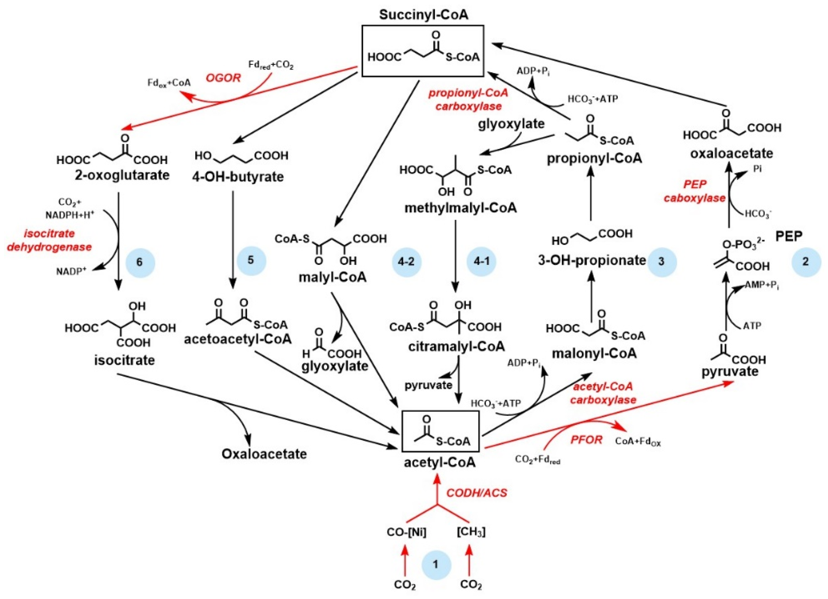
2.3. The Reductive Acetyl–CoA Pathway Is the Most Ancient and Energy–Efficient CO2 Fixation Pathway

The reductive acetyl–CoA pathway (rAC, or Wood–Ljungdahl pathway) is the only non-cyclic and energy-conserving CO2 biofixation pathway mainly distributed in obligate acetogenic bacteria and methanogenic archaea [21,38]. The rAC pathway is considered to be one of the most ancient anaerobic carbon fixation pathways, responsible for biomass accumulation and energy generation, and functions close to thermodynamic limit [39]. Notably, the rAC pathway is the most energy-efficient natural CO2 fixation pathway, with less than one ATP input for the synthesis of one acetyl-CoA or pyruvate molecule [21]. In the rAC pathway, two CO2 molecules are converted to acetyl-CoA. One CO2 was reduced to the methyl level mediated by different carriers, cofactors, and enzymes in acetogens and methanogens, and the other was reduced to CO bounding to nickel of enzyme active sites (Figure 2, Routine 1) [29,40,41]. The reduction of CO2 to CO is catalyzed by the bifunctional CO dehydrogenase/acetyl-CoA synthase, which also accounts for the condensation of coenzyme A, methyl group, and CO to form acetyl-CoA. Further assimilation of acetyl-CoA requires the same pathways of rTCA cycle (Figure 2, Routine 2). In the energy generation process of microbes with rAC pathway, acetate or methane is the end product. In addition to CO2 fixation, the rAC pathway can also act in reverse, producing reducing equivalents by oxidizing organic compounds such as acetate [42,43]. Moreover, the rAC pathway can also be used for the assimilation of various C1 compounds, including CO, formate, formaldehyde, methanol, and even methyl groups of aromatic O-methyl ethers/esters in lignin, giving it potential for industrial application [21,41].

2.4. The 3-Hydroxypropionate Bi-Cycle Producing Pyruvate from CO2

The 3-hydroxypropionate (3HP) bi-cycle was discovered in the green non-sulfur bacteria Chloroflexus aurantiacus [44]. In the first cycle (Routine 3 and 4–2 in Figure 2), acetyl-CoA is carboxylated to malonyl-CoA by the biotin-dependent acetyl-CoA carboxylase, and reduced to propionyl-CoA via 3-hydroxypropionate, which is one of the characteristic intermediates of this cycle [21,45]. Propionyl-CoA is further carboxylated to (S)-methylmalonyl-CoA by the biotin-dependent propionyl CoA carboxylase, which is then rearranged and reduced to (S)-malyl-CoA, which is another characteristic intermediate of the cycle. Cleavage of (S)-malyl-CoA yields an acetyl-CoA molecule and a glyoxylate molecule, and the latter is further assimilated in the second cycle (Routine 3 and 4–1 in Figure 2). Glyoxylate is condensed with propionyl-CoA and finally converted to citramalyl-CoA, which is cleaved into pyruvate and acetyl-CoA, thus completing the second cycle and yielding one net pyruvate [46].
Interestingly, the whole bicycle in C. aurantiacus involves 19 steps catalyzed by 13 enzymes, some of which are multifunctional [46]. Reduction of 1 mole of CO2 to pyruvate requires approximately 2.3 mol ATP in both 3HP bicycle and CBB cycle, while the activity of biotin-dependent carboxylase of the 3HP bicycle is higher than that of Rubisco in CBB cycle [30]. The presence of the B12-dependent methylmalonyl-CoA mutase of the 3HP bicycle makes it less oxygen tolerant and prevents it from functioning in oxygenic photoautotrophs [21].

2.5. The HP/HB Cycle and DC/HB Cycle Are the Most Recently Discovered CO2 Fixation Pathway

The most recently discovered CO2 fixation pathways are the 3-hydroxypropionate/3-hydroxybutyrate (HP/HB) cycle and the dicarboxylate/4-hydroxybutyrate (DC/HB) cycle. The two cycles share similarities in carbon acceptor regeneration, whereas the CO2 fixation enzymes differ in their oxygen sensitivity [21,47,48]. In the HP/HB cycle, two molecules of inorganic bicarbonate are fixed by the oxygen-tolerant biotin-dependent acetyl-CoA carboxylase and propionyl-CoA carboxylase, and subsequently converted to succinyl-CoA. The catalytic process is the same as that of 3HP bi-cycle (Figure 2, Routine 3), though non-homologous enzymes are used in HP/HB cycle, suggesting the independent evolution of related enzymes [29]. In the DC/HB cycle, the carboxylation reactions are catalyzed by the oxygen-sensitive PFOR and the biotin-dependent PEP carboxylase, followed by an incomplete rTCA cycle to produce succinyl-CoA (Figure 2, Routine 2). The intermediate succinyl-CoA in HP/HB cycle and DC/HB cycle is reduced to 4-hydroxybutyryl-CoA and further converted to 3-hydroxybutyryl-CoA, which is further oxidized to acetoacetyl-CoA and cleaved to acetyl-CoA. One net acetyl-CoA is produced from bicarbonate and CO2 in the DC/HB cycle (Figure 2, Routine 5).
Generally, the anaerobic carbon fixation pathways are more energetically efficient than their aerobic counterparts by using ferredoxin-dependent carbon fixation enzymes. For example, the DC/HB cycle requires approximately 1.6 mol ATP for the reduction of one mol CO2 to pyruvate, while the HP/HB cycle requires 3 mol ATP in the same process [30]. Ferredoxin-dependent carboxylases are almost restricted in strictly anaerobic microorganisms because they contain iron-sulfur clusters which are highly sensitive to oxygen. In contrast, the CBB cycle, the 3HP bicycle, and the DC/HB cycle have a wider distribution due to their oxygen tolerance.

3. Enzymes Catalyzing CO2 to Organic Compounds

Among the six natural biological carbon fixation pathways, the conversion of inorganic carbon to organic compounds mainly catalyzed by carboxylases is a key and rate-limiting step of CO2 fixation. These enzymes are present in all three domains of life and have distinct cofactor requirements, catalytic mechanisms, substrate specificities, and catalytic kinetics, and can be broadly clarified into four groups, including Rubiscos, biotin-dependent carboxylases, CO dehydrogenase/acetyl-CoA synthase, and 2-oxoacid:ferredoxin oxidoreductases. In addition to these enzymes involved in natural carbon fixation pathway, some alternative carboxylases implicated in artificial CO2 fixation pathways will be introduced as well.

3.1. Rubisco Is the Main Carboxylase for CO2 Fixation

Rubisco, the enzyme that catalyzes the carboxylation of RuBP in CBB cycle, is one of the most abundant enzymes on the earth [12]. Given its central role in CO2 fixation, Rubisco has been intensively studied. As a carboxylase, it is widely recognized that Rubisco has low catalytic efficiency, with an average turnover number (kcat,c) of 4.15 s−1 (Table 1), compared with the kcat values of approximately 80 s−1 of typical central metabolic enzymes [49]. In addition to the poor catalytic efficiency, Rubisco also catalyzes a non-productive oxidative side reaction, producing a toxic by-product 2-phosphoglycolate, which must be recycled through the phosphoglycolate salvage process mentioned above, thus leading to energy and carbon loss [24]. Furthermore, there exists a proposed tradeoff between substrate specificity and carboxylase activity [30,50]. In other words, Rubisco with higher catalytic efficiency has less differentiation between CO2 and O2, resulting in more oxidation reactions and more energy input for the phosphoglycolate salvage process. Most Rubiscos function in atmosphere with O2-abundant and CO2-scarce condition (21% O2, 0.04% CO2, corresponding to 0.012 mM CO2 and 0.26 mM bicarbonate in water (pH 7.4, 20 °C)), the carboxylation efficiency of which further decreases [29]. For example, the net carboxylation rate of Rubisco from the cyanobacterium Synechococcus elongatus PCC 7942 is estimated to be approximately 4% of its apparent kcat,c (14 s−1) under atmospheric conditions [50]. To date, four forms of Rubiscos have been found in nature [51,52]. Forms I to III are bona fide Rubiscos that catalyze the carboxylation or oxygenation of RuBP, while form IV Rubiscos termed Rubisco-like proteins (RLPs) are not involved in carbon fixation. Form I Rubiscos are widely distributed in plants, algae, cyanobacteria, and proteobacteria that rely on CBB cycle for carbon fixation. Form I Rubiscos are composed of eight large (L) and eight small (S) subunits organized in an L8S8 structure, representing the most structurally complex Rubiscos [51]. Small subunits were found to impact the conformation of the catalytic sites of large subunits dimers, and affect catalytic efficiency and substrate specificity [53]. A recently discovered clade of form I Rubiscos lacking small subunits exhibits a relatively low kcat,c, and average Km,c compared to form I enzymes [54]. In addition, certain form I Rubiscos are associated with CO2 concentrating mechanisms to improve carbon fixation efficiency (see below) [13]. Form II Rubiscos are mainly found in proteobacteria, archaea, and dinoflagellate algae, and also participate in CBB cycle [52]. Unlike form I Rubiscos, form II Rubiscos only consist of large subunits that form an L2 or L6 complex, exhibiting a simpler structure [51,55]. As a result, form II Rubiscos generally have a higher catalytic rate (kcat,c about 7 s−1) and lower substrate specificity (Km,c about 85 μM) compared with those of form I Rubiscos (Km,c less than 20 μM, kcat,c about 3 s−1) [50]. Form II Rubiscos typically exist in CO2-rich and/or anaerobic environments [21]. Form III Rubiscos are mainly found in archaea and participate in the ribonucleosides metabolism of pentose biphosphate pathway rather than the CBB cycle [52,56]. These archaea lack a complete pentose phosphate pathway, and the ribose moieties of nucleosides are important carbon sources and are linked to central metabolism through carboxylation, followed by cleavage catalyzed by form III enzymes. Form IV Rubiscos are not real Rubiscos and do not catalyze the key reaction of carboxylation, but share sequence similarity with Rubiscos despite lacking the conserved amino acid residues essential for carboxylation [13,51,57]. The catalytic mechanism of RLPs is similar to that of Rubiscos via the formation of an enolate intermediate, suggesting a divergent evolution.
The diversity of Rubiscos with different subunits, structural complexity, and kinetic parameters provides evidence for the evolution of Rubiscos [13]. It is believed that over a long period of evolution, Rubiscos have optimized substrate specificity and enzymatic activity for the hosts’ niche [64]. Rubiscos are not independent enzymes and require a series of proteins and enzymes to assist Rubiscos to function properly [65]. The natural evolution process of Rubiscos provides important clues for improving Rubiscos and CBB cycle. Considering the central role in CO2 fixation, there have been continuous efforts to improve the catalytic performance of Rubiscos. The latest research progress in improving the efficiency of Rubiscos for carboxylation is summarized below.

3.1.1. Screening and Recombinant Expression of Efficient Rubiscos to Enhance CO2 Fixation

The most direct way to enhance the carboxylation of CBB cycle is to introduce highly active Rubiscos. For example, a Rubisco with higher catalytic efficiency (kcat,c value about 14 s−1) from Synechococcus species was introduced into the facultative chemoautotroph Cupriavidus necator H16 in combination with the expression of endogenous GroES/EL chaperones, leading to an increased growth rate and biomass production of the engineered strain growing autotrophically [66]. Moreover, simultaneous overexpression of RcbX chaperone and hydrogenases resulted in a 93.4% increase in final biomass and a 99.7% increase in polyhydroxybutyrate (PHB) production. This result demonstrated that efficient Rubisco, along with appropriate CO2 supply, can sustain rapid growth and carbon fixation of autotrophic microorganisms.
Since effective Rubiscos have a significant effect on the growth rate of autotrophic microorganisms, screening Rubiscos with a high carboxylation rate is important to improve CO2 fixation efficiency. Through systematical mining of genomic information, the most catalytically efficient Rubisco was identified from Gallionella genus [55]. The Rubisco from Gallionella belongs to form II and has a kcat,c of 22 s−1 and a relatively low affinity for CO2 (Km,c = 276 μM). Importantly, the Gallionella Rubisco displays as a homodimer, and the simple structure may be beneficial for heterologous expression. Another form II Rubisco with a relatively high kcat,c of 16.4 s−1 was discovered from the endosymbiont of a deep-sea tubeworm Riftia pachyptila (RPE Rubisco) [58]. RPE Rubisco can be readily expressed in E. coli BL21(DE3) without additional expression of chaperones and showed high carboxylation efficiency. However, the low specificity for CO2 (Km,c = 172.4 μM) prevented the RPE Rubisco from functioning properly under ambient atmospheric conditions. Collectively, these results suggested that more catalytically efficient Rubiscos can be found in form II Rubiscos, although they are less specific and require an elevated CO2 concentration.

3.1.2. Enhancing CO2 Fixation through Constructing Synthetic Phosphoglycolate Salvage Pathway

The oxygenation catalyzed by Rubisco produces by-products, which are usually recycled by carbon loss photorespiration. It is an alternative strategy to recycle the by-products through non-carbon loss or carbon fixation processes, by which CO2 fixation efficiency of CBB cycle will be improved. The synthetic malyl-CoA-glycerate (MCG) cycle, relying on PEP carboxylase and malyl-CoA lyase to catalyze the cleavage of malyl-CoA to acetyl-CoA and glyoxylate, has been shown to enhance bicarbonate assimilation in cyanobacteria by approximately two-fold [67]. The PEP carboxylase is one of the most active oxygen-tolerant carboxylases with specific activity up to 150 μmol/min/mg, enabling fast carboxylation. Notably, glyoxylate is assimilated to C3 metabolites via tartronate semialdehyde without net carbon loss in the MCG pathway. Meanwhile, in the MCG cycle, the C3 metabolites produced in the CBB cycle were converted to acetyl-CoA through additional carbon fixation catalyzed by PEP carboxylase, which favors the generation of acetyl-CoA from CBB cycle products.
Through rational design, high-throughput microfluidics, and microplate screening, a novel glycolyl-CoA carboxylase with a kcat,c of 5.6 s−1 was designed recently based on the propionyl-CoA carboxylase from Methylorubrum extorquens [68]. The engineered enzyme displays comparable catalytic properties to natural carboxylases in CO2 fixation. By the catalysis of the engineered glycolyl-CoA synthetase, a tartronyl-CoA reductase from Chloroflexus aurantiacus, and a semialdehyde reductase, glyoxylate produced in the oxygenation of Rubisco can be converted to glycerate in the so-called tartronyl-CoA pathway [68]. In the synthetic pathway, an additional carboxylation is catalyzed by the engineered glycolyl-CoA carboxylase, thus connecting the by-products of CBB cycle with the central carbon metabolism. Notably, when using the tartronyl-CoA pathway to recycle 2-phosphoglycolate, additional CO2 is fixed rather than released, thus increasing the carbon efficiency of 2-phosphoglycolate recovery from 75% to 150% and decreasing ATP consumption by 21%. Moreover, the new pathway can be easily integrated into the CBB cycle for by-product recycling by the expression of three additional enzymes.

3.1.3. Concentrating CO2 for Fixation by Enzyme Engineering

The low concentration of CO2 in the atmosphere is not sufficient to sustain rapid carboxylation reactions of Rubiscos, especially those with less specificity. Carbon concentration mechanisms (CCM) exist in many photo-/chemo-autotrophic microorganisms and plants, providing Rubiscos with CO2-rich conditions that promote carboxylation and inhibit deleterious oxygenation of RuBP [69]. Carbonic anhydrases (CAs) can generate CO2 in cell by catalyzing the hydrolysis of bicarbonate and are key to the autotrophic growth of microorganisms [70,71]. Similarly, a biogenic polyamine has been reported to capture atmospheric CO2, forming carbamates and enriching CO2 for Rubiscos [72]. Furthermore, CO2 is concentrated through a biophysical CCM in Synechococcus species. A bicarbonate uptake system is coupled with a virus-like proteinaceous shell named carboxysome that encapsulates Rubiscos, CAs, and other accessory proteins in a confined microcompartment, which can exclude O2 and provide a CO2-rich condition for Rubiscos to promote carboxylation and inhibit deleterious side reaction [73]. In addition, a biochemical CCM which relies on PEP carboxylase was found in C4 plants. PEP carboxylase catalyzes CO2 to malate, which is then oxidatively decarboxylated to concentrate CO2 for Rubiscos [74]. As a stable carbon carrier, malate is suitable for long-distance transportation in multicellular plants. It can be speculated that transplantation of CCM from C4 to C3 plants such as rice is expected to increase photosynthesis and grain yield [75,76].
A functional carboxysome was recently constructed in a Rubisco-dependent E. coli strain. An engineered E. coli strain harboring form I Rubisco from Halothiobacillus neapolitanus and a prk gene from Synechococcus elongatus PCC 7942 was constructed, which can grow autotrophically with 10% CO2. The engineered E. coli was able to grow in ambient atmosphere when the CCM of H. neapolitanus was co-expressed [77]. It wasproved that the large subunit of Rubisco determines whether the Rubisco complex can be encapsulated in the H. neapolitanus carboxysome [78]. The CsoS2 protein wasproposed as a scaffold protein that acts as an interaction hub to encapsulate Rubisco of H. neapolitanus [79]. These results suggested that the interaction between Rubisco and scaffold protein is essential for the formation of functional carboxysome, and foreign Rubiscos might not be incorporated into the carboxysome. Nonetheless, expression of a functional carboxysome in E. coli demonstrates the potential to increase carboxylation efficiency in microorganisms without CCM.
Increasing carboxylation capacity of Rubisco is key to improving photosynthesis, as it is the main entry for inorganic carbon into biosphere. In addition to strategies described above, the approaches such as direct evolution of Rubiscos, engineering large or small subunits of Rubisco, and changing expression and activation of Rubiscos can also improve CO2 fixation [80].

3.2. Biotin-Dependent Carboxylase

Biotin-dependent carboxylases are an ancient group of carboxylases that carboxylate a variety of substrates, including acetyl-CoA, propionyl-CoA, pyruvate, and 2-oxoglutarate. These carboxylases play an important role in many essential metabolic pathways, including synthesis and degradation of fatty acids, degradation of certain amino acids, anaplerosis of TCA cycle intermediates, gluconeogenesis, and autotrophic fixation of CO2 [81]. Acetyl-CoA carboxylase, propionyl-CoA carboxylase, pyruvate carboxylase, and 2-oxoglutarate carboxylase are involved in four of the above discussed six CO2 fixation pathways. These enzymes are composed of at least three functional components: biotin carboxyl carrier protein (BCCP), biotin carboxylase (BC) domain, and carboxyl transferase (CT) domain, and share a common catalytic mechanism to catalyze a two-step reaction [82]. The BC domain catalyzes the ATP-dependent carboxylation of the biotin moiety of BCCP using bicarbonate as a carbon source, followed by the transfer of the carboxyl group from the carboxylated biotin at BCCP domain to the substrates determined by the CT domain.
The biotin-dependent carboxylases have several advantages over Rubiscos. Firstly, biotin-dependent carboxylases exhibit a higher carboxylation rate than Rubiscos (Table 1). In addition, O2 does not interfere with the carboxylation by biotin-dependent carboxylases, and no oxygenation reaction would occur to impair the carboxylation efficiency. Moreover, considering substrate availability for biotin-dependent carboxylases, bicarbonate is more available than CO2 in water, which is important for maintaining rapid carboxylation.
Efficient biotin-dependent carboxylases are promising for CO2 fixation and production of valuable products. Recently, simultaneous -carbon fixation and succinate production was achieved in a recombinant E. coli through partially introducing 3HP bi-cycle [83]. The succinate synthetic pathway is centered on acetyl-CoA carboxylase and propionyl-CoA carboxylase, the carboxyltransferase domain of which was mutated. Through directed evolution, the kcat,c value of the mutant enzyme is 13.3 s−1, and the overall catalytic efficiency is improved by 94 times based on kcat,c/Km,c. As a result, the highest production of succinate reached 2.66 g/L with a CO2 fixation rate of 0.94 mmol/L/h. However, the carboxylation and the subsequent reduction step require a substantial amount of ATP and NADPH (one succinate from acetyl-CoA requires 3 ATP and 3 NADPH), which will restrict the succinate yield [83,84]. In addition to the incomplete 3HP bi-cycle, pyruvate carboxylase, which plays an important role in anaplerotic generation of TCA intermediates, is also used for the production of dicarboxylate. Overexpression of pyruvate carboxylase from Rhizopus oryzae (RoPYC) was reported to enhance fumarate production in Saccharomyces cerevisiae, and further enhancement was achieved by mutation P474N in RoPYC, resulting in a 14% increase in PYC activity, and the final fumaric acid yield reached 314.5 mg/L [85]. Furthermore, the R458P mutation, which affects the allosteric and biotin carboxylation domain in RoPYC, was reported to further increase fumaric acid production with a maximal titer of 465.5 mg/L [86].
A unique member of the biotin-dependent carboxylases family, 2-oxoglutarate carboxylase (OGC), is predominantly identified in thermophilic bacteria from the rTCA cycle-dependent phylum Aquificae [36]. OGC catalyzes the carboxylation of 2-oxoglutarate to oxalosuccinate, which is further reduced to isocitrate. However, this process is accomplished by a single enzyme isocitrate dehydrogenase (ICDH) in the conventional rTCA cycle. Interestingly, ICDH normally catalyzes the oxidative decarboxylation of isocitrate in the oxidative TCA cycle. The reaction catalyzed by ICDH is characterized by reversibility and high specific activity. The recombinant ICDH from Chlorobium limicola showed a carboxylation rate of 27.2 μmol/min/mg at pH 7 and a decarboxylation rate of 160 μmol/min/mg at pH 9 [87,88]. Therefore, the carboxylation and decarboxylation reactions can be controlled by changing pH, showing great application prospects in CO2 fixation and storage process.

3.3. Carbon Monoxide Dehydrogenase/acetyl-CoA Synthase and the Application for Biosynthesis

Bifunctional carbon monoxide dehydrogenase/acetyl-CoA synthase (CODH/ACS) is a key enzyme of the rAC pathway in acetogens and methanogens. These enzymes typically consist of two functional subunits, CODH and ACS, which are associated with a heterotetrameric complex [89]. The CODH subunit catalyzes the reversible reduction of CO2 to CO, which is delivered to the active sites of ACS through an internal gas channel, preventing the leak of toxic CO and confining it within CODH/ACS complex [89,90]. The ACS subunit catalyzes the reversible condensation of CO, a methyl moiety bound to a corrinoid iron-sulfur protein and coenzyme-A to acetyl-CoA. Some CODH/ACS variants have been reported to contain unsealed channels for CO oxidation to generate reducing equivalents [91]. The reduction of CO2 is quite thermodynamically unfavorable, with a standard redox potential as low as –520mV, requiring highly reduced ferredoxin as an electron donor [29,92]. Therefore, this enzyme is mainly restricted to anaerobic environments.
Acetogens, which depend on CODH/ACS and the rAC pathway for energy generation and biomass accumulation, produce acetate as the end product by using CO2, H2, and toxic CO. The rAC pathway is one of the most energy-efficient carbon fixation pathways, and it is estimated that acetogens produce over 1013 kg acetate annually, demonstrating its potential for biomanufacturing [41]. Various acetogens have been engineered for the production of valuable products including acetate, ethanol, butanol, 2,3-butanediol, and acetoin, from syngas composed of CO2, H2, and CO [93,94,95,96].
In a recent study, the tolerance of Eubacterium limosum ATCC 8486 to CO increased up to 80% through adaptive evolution. The resulting strain displayed a faster growth rate and produced 42.28 mM/gCDW acetate under 40% CO condition [96]. Moreover, this strain was engineered to produce acetoin with a production of 19.6 mM/gCDW, a 1.34–fold increase compared with the parental strain. An engineered E. limosum KIST612 overexpressing CODH, ACS, and a coenzyme CooC2 exhibited a 3.1-fold increase in the rate of CO oxidation and a 1.4-fold increase in acetate production to 13.9 g/gCDW acetate [97]. The results suggest that acetogens are promising for biosynthesis by utilizing CO and CO2 [98].

3.4. 2-Oxoacid:Ferredoxin Oxidoreductase and the Application in CO2 Fixation

Three of the six natural carbon fixation pathways use enzymes from 2-oxoacid (2-keto acid):ferredoxin oxidoreductase (OFOR) family to catalyze the reductive carboxylation. Two members of OFOR enzymes, PROF and OGOR, are involved in the carboxylation of acetyl-CoA and succinyl-CoA to pyruvate and 2-oxoglutarate, respectively. Ferredoxins are a group of small proteins containing iron-sulfur clusters with mid-potentials between −400 mv and −500 mV. They are required as the electron donor to drive the thermodynamically and energetically challenging reductive carboxylation of acetyl-CoA and succinyl-CoA, the standard redox potentials of which are about −500 mV [29,99]. OFOR reactions are typically reversible and can operate in both fixation and release of CO2, mainly depending on the redox potential of co-factors [100,101].
Diverse oligomeric states of OFOR enzymes have been discovered, including homodimers, heterotetramers, and heterohexamers [102,103]. OFOR enzymes share a similar domain arrangement despite different domains constitution and contain up to seven different domains, several of which are conserved for substrate and cofactor binding, and housing iron-sulfur cluster(s) for electron transfer [104]. Notably, most OFOR enzymes are highly oxygen-sensitive and rapidly and irreversibly inactivated upon exposure to air, constraining the corresponding autotrophic pathways in a strictly anaerobic or microaerobic environment [30]. In addition, to drive the thermodynamically and energetically challenging process, ferredoxins need to be kept highly reduced by sodium (or proton) motive force driven oxidoreductases or electron bifurcation mechanism [29,99].
The oxygen-tolerant PFOR from the extremophilic crenarchaeon Sulfolobus acidocaldarius was proposed to catalyze the decarboxylation of pyruvate. Interestingly, the PROF exhibited carboxylase activity in vitro when coupled with low-potential ferredoxins or methylviologen (with standard redox potentials of less than −500 mV and −446 mV, respectively) as electron donors [104]. Besides using ferredoxin or methylviologen as electron donors, cadmium sulfide nanorods were reported to act as electron donors to drive the reductive carboxylation of succinyl-CoA to 2-oxoglutarate catalyzed by OGOR from Magnetococcus marinus MC-1 [62]. Notably, cadmium sulfide nanorods can be reduced by photo-excitation and exhibited a standard redox potential of ≤−700 mV. Generally, OFOR from the three anaerobic carbon fixation pathways, rTCA, rAcCoA, and DC/HB, require fully reduced ferredoxin to catalyze reductive carboxylation. Even though reduced ferredoxin is difficult to achieve outside native cells, artificial electron donors can be used as an alternative in vitro, expanding their application for biosynthesis.

3.5. Artificial Pathways for CO2 Fixation and Biosynthesis

In addition to natural carbon fixation pathways, several artificial carbon pathways have been designed, which are proposed to be energetically and/or kinetically superior to their natural counterparts theoretically [105]. Schwander et al. constructed a CO2 fixation pathway named “crotonyl-CoA/ethylmalonyl-CoA/hydroxybutyryl-CoA” (CETCH) cycle, and the in vitro CO2 fixation rate is comparable to that of CBB cycle [106]. The cycle was designed based on an efficient crotonyl-CoA carboxylase/reductase, a member of enoyl-CoA carboxylase/reductase family, with the highest CO2 fixation rate of 130 μmol/min/mg, which is approximately 37 times of Rubisco. Compared with the natural cycles, the CETCH cycle is the most energy-efficient aerobic carbon fixation pathway with only one ATP input for glyoxylate production. However, its direct product glyoxylate is not a central metabolite, and can be coupled with the tartronyl-CoA pathway to improve CO2 fixation efficiency [68]. Recently, the CETCH cycle was coupled with an in vitro enzyme cascade to convert CO2 to terpenes and polyketides, showing its potential for CO2 fixation and production of value-added products [107]. Since the CETCH cycle consists of 12 enzymatic reactions which are not involved in central carbon metabolism, in vivo functional CETCH by metabolic engineering has not been reported. Taken together, the CETCH, MCG, and tartronyl-CoA pathways have demonstrated the potential of artificial pathways for CO2 fixation both in vitro and in vivo, which will facilitate CO2 fixation and transformation to value-added products.
In addition to these ingenious and intricate artificial carbon fixation pathways, some relatively simple carbon fixation pathways have been constructed in vitro with immobilized enzymes [10]. The enzymatic carbon dioxide fixation system showed potential for biotransformation of CO2. For instance, amine-functionalized metal-organic frameworks were used to adsorb CO2 and immobilize the enzyme cascade consisting of carbonic anhydrase, formate dehydrogenase (FDH), and glutamate dehydrogenase for the reduction of CO2 to formate, achieving 13.1-times increase of final production compared with free enzyme systems [108]. The enzymatic cascade of FDH, alcohol dehydrogenase (ADH), and formaldehyde dehydrogenase (FalDH) was used to convert CO2 to methanol [10]. The immobilized FDH-FalDH-ADH cascade and in situ separation by pervaporation membrane were combined for improving the transformation of CO2 to methanol. [109]. In some cases, immobilization of enzymes displayed enhanced performance. For example, the dimeric form II Rubisco exhibits higher stability against proteolysis, and the larger form I Rubisco exhibits improved CO2 selectivity after immobilization [110].

4. Conclusions and Prospects

Biological carbon fixation and converting CO2 into value-added bioproducts is a promising approach to mitigate the greenhouse effect. However, the low activity of CO2 fixation enzymes hinders the further improvement of carbon fixation efficiency and its application (Table 1). It has been suggested that the enzyme activity and specificity of Rubisco have reached Pareto optimality in the long-term evolution, so the activity of some carboxylases, including Rubisco, could not be further enhanced through enzyme engineering [13,64,111]. Screening natural carboxylases based on omics analysis or de novo designing non-natural counterparts through enzyme engineering are promising approaches [55,68,83]. For example, a novel form II Rubisco with a kcat,c of 22 s−1 was recently reported, promising to improve CO2 fixation capacity of CBB cycle. In addition to improving enzyme activity, artificial CO2 fixation pathways have been designed to improve the energetic efficiency and oxygen tolerance. Some artificial carbon fixation pathways, such as the MCG pathway and CETCH cycle, have been demonstrated to improve CO2 fixation both in vivo and in vitro. Encouragingly, the heterotrophs of E. coli and Pichia pastoris have been transformed to autotrophs, providing a new idea for CO2 fixation [112,113].
Another key factor affecting carbon biofixation efficiency is CO2 concentration. It is generally accepted that a higher CO2 concentration enhances photosynthetic assimilation [114]. A low concentration of atmospheric CO2 cannot support rapid carboxylation, so natural autotrophs have evolved CCMs to concentrate CO2 for Rubiscos [69]. It has been proved that constructing CCM such as carboxysomes in microorganisms through metabolic engineering is effective for CO2 concentrating. However, the assembly of carboxysomes containing non-native contents, such as the fast form II Rubisco, needs to be further studied [16,73,77]. Fermentation of autotrophic microorganisms in bioreactors with improved CO2 concentrations can also promote the catalytic performance of Rubisco [16].
The biofixation and reduction of CO2 require a substantial amount of NAD(P)H and ATP, which is primarily provided by solar energy in nature. However, the supply of NAD(P)H and ATP is expensive and challenging, especially in the case of in vitro catalysis with immobilized enzyme cascades. Interdisciplinary approaches such as photochemistry and bioelectrochemistry can provide solutions for NAD(P)H and ATP generation using sunlight and electricity. For example, a ferredoxin NADP+ reductase and a crotonyl-CoA carboxylase/reductase were co-immobilized within a redox-active hydrogel and were coupled with electrodes for NADPH regeneration which was used to drive the carboxylation reaction [115]. Furthermore, a chloroplast mimic was constructed to encapsulate photosynthetic membranes and CETCH enzyme cascades, and the ATP and NAD(P)H regenerated from sunlight in the system were used for carboxylation [116]. Remarkable progress has been made in CO2 biofixation and value-added products synthesis. The rapid development of synthetic biology has provided novel strategies for improving CO2 biofixation, such as the development of more efficient carboxylases and artificial CO2 pathways. It is expected that more efficient pathways and enzymes for CO2 biofixation and conversion can be developed through metabolic and enzyme engineering.

Author Contributions

Conceptualization, X.P. and Y.H.; investigation, X.P. and Y.H.; writing—original draft preparation, X.P.; writing—review and editing, Y.H.; funding acquisition, Y.H. All authors have read and agreed to the published version of the manuscript.

Funding

This research was funded by the National Key Research and Development Program of China (2021YFC2101603) and the National Natural Science Foundation of China (32170093).

Institutional Review Board Statement

Not applicable.

Informed Consent Statement

Not applicable.

Data Availability Statement

Not applicable.

Acknowledgments

We thank the support from the National Key Research and Development Program of China (2021YFC2101603) and the National Natural Science Foundation of China (32170093).

Conflicts of Interest

The authors declare no conflict of interest.

References

  1. Shi, J.; Jiang, Y.; Jiang, Z.; Wang, X.; Wang, X.; Zhang, S.; Han, P.; Yang, C. Enzymatic conversion of carbon dioxide. Chem. Soc. Rev. 2015, 44, 5981–6000. [Google Scholar] [CrossRef] [PubMed]
  2. Haszeldine, R.S. Carbon capture and storage: How green can black be? Science 2009, 325, 1647–1652. [Google Scholar] [CrossRef] [PubMed]
  3. Sumida, K.; Rogow, D.L.; Mason, J.A.; McDonald, T.M.; Bloch, E.D.; Herm, Z.R.; Bae, T.H.; Long, J.R. Carbon dioxide capture in metal–organic frameworks. Chem. Rev. 2012, 112, 724–781. [Google Scholar] [CrossRef] [PubMed]
  4. Zheng, J.; Chong, Z.R.; Qureshi, M.F.; Linga, P. Carbon Dioxide Sequestration via Gas Hydrates: A Potential Pathway toward Decarbonization. Energy Fuels 2020, 34, 10529–10546. [Google Scholar] [CrossRef]
  5. Chen, C.; Zhang, S.; Row, K.H.; Ahn, W.-S. Amine–silica composites for CO2 capture: A short review. J. Energy Chem. 2017, 26, 868–880. [Google Scholar] [CrossRef] [Green Version]
  6. Gomes, J.; Santos, S.; Bordado, J. Choosing amine–based absorbents for CO2 capture. Environ. Technol. 2015, 36, 19–25. [Google Scholar] [CrossRef]
  7. Yeung, C.S. Photoredox Catalysis as a Strategy for CO2 Incorporation: Direct Access to Carboxylic Acids from a Renewable Feedstock. Angew. Chem. Int. Ed. 2019, 58, 5492–5502. [Google Scholar] [CrossRef]
  8. Xu, S.; Carter, E.A. Theoretical Insights into Heterogeneous (Photo)electrochemical CO2 Reduction. Chem Rev. 2019, 119, 6631–6669. [Google Scholar] [CrossRef]
  9. Appel, A.M.; Bercaw, J.E.; Bocarsly, A.B.; Dobbek, H.; DuBois, D.L.; Dupuis, M.; Ferry, J.G.; Fujita, E.; Hille, R.; Kenis, P.J.A.; et al. Frontiers, Opportunities, and Challenges in Biochemical and Chemical Catalysis of CO2 Fixation. Chem. Rev. 2013, 113, 6621–6658. [Google Scholar] [CrossRef] [Green Version]
  10. Marques, C.G.C.N.; Andrade, L.H.; Toma, H.E. Carbon dioxide/methanol conversion cycle based on cascade enzymatic reactions supported on superparamagnetic nanoparticles. An. Acad. Bras. Cienc. 2018, 90, 593–606. [Google Scholar] [CrossRef] [Green Version]
  11. Peng, X.; Kelly, R.M.; Han, Y. Sequential processing with fermentative Caldicellulosiruptor kronotskyensis and chemolithoautotrophic Cupriavidus necator for converting rice straw and CO2 to polyhydroxybutyrate. Biotechnol. Bioeng. 2018, 115, 1624–1629. [Google Scholar] [CrossRef] [PubMed]
  12. Phillips, R.; Milo, R. A feeling for the numbers in biology. Proc. Natl. Acad. Sci. USA 2009, 106, 21465–21471. [Google Scholar] [CrossRef] [PubMed] [Green Version]
  13. Erb, T.J.; Zarzycki, J. A short history of RubisCO: The rise and fall (?) of Nature’s predominant CO2 fixing enzyme. Curr. Opin. Biotechnol. 2018, 49, 100–107. [Google Scholar] [CrossRef] [PubMed]
  14. Kopke, M.; Mihalcea, C.; Bromley, J.C.; Simpson, S.D. Fermentative production of ethanol from carbon monoxide. Curr. Opin. Biotechnol. 2011, 22, 320–325. [Google Scholar] [CrossRef] [PubMed]
  15. Angermayr, S.A.; Gorchs Rovira, A.; Hellingwerf, K.J. Metabolic engineering of cyanobacteria for the synthesis of commodity products. Trends Biotechnol. 2015, 33, 352–361. [Google Scholar] [CrossRef] [PubMed]
  16. Nybo, S.E.; Khan, N.E.; Woolston, B.M.; Curtis, W.R. Metabolic engineering in chemolithoautotrophic hosts for the production of fuels and chemicals. Metab. Eng. 2015, 30, 105–120. [Google Scholar] [CrossRef] [PubMed] [Green Version]
  17. Nangle, S.N.; Ziesack, M.; Buckley, S.; Trivedi, D.; Loh, D.M.; Nocera, D.G.; Silver, P.A. Valorization of CO2 through lithoautotrophic production of sustainable chemicals in Cupriavidus necator. Metab. Eng. 2020, 62, 207–220. [Google Scholar] [CrossRef]
  18. Tang, R.H.; Peng, X.W.; Weng, C.H.; Han, Y.J. The overexpression of phasin and regulator genes promoting the synthesis of polyhydroxybutyrate in Cupriavidus necator H16 under nonstress conditions. Appl. Environ. Microbiol. 2022, 88. [Google Scholar] [CrossRef]
  19. Tang, R.; Weng, C.; Peng, X.; Han, Y. Metabolic engineering of Cupriavidus necator H16 for improved chemoautotrophic growth and PHB production under oxygen–limiting conditions. Metab. Eng. 2020, 61, 11–23. [Google Scholar] [CrossRef]
  20. Lee, K.; Evans, J.P.; Satagopan, S.; Sun, Y.; Parquette, J.R.; Sundaresan, V.B.; Tabita, F.R.; Bakshi, B.R. Carbon Footprint of Biomimetic Carbon Fixation by Immobilizing Nature’s CO2–sequestering Enzyme and Regenerating Its Energy Carrier. ACS Sustain. Chem. Eng. 2020, 8, 16833–16841. [Google Scholar] [CrossRef]
  21. Berg, I.A. Ecological aspects of the distribution of different autotrophic CO2 fixation pathways. Appl. Environ. Microbiol. 2011, 77, 1925–1936. [Google Scholar] [CrossRef] [PubMed] [Green Version]
  22. Calvin, M.; Benson, A.A. The Path of Carbon in Photosynthesis. Science 1948, 107, 476–480. [Google Scholar] [CrossRef] [PubMed] [Green Version]
  23. Schlegel, H.G.; Gottschalk, G.; Bartha, R.V. Formation and Utilization of Poly–β–Hydroxybutyric Acid by Knallgas Bacteria (Hydrogenomonas). Nature 1961, 191, 463–465. [Google Scholar] [CrossRef]
  24. Claassens, N.J.; Scarinci, G.; Fischer, A.; Flamholz, A.I.; Newell, W.; Frielingsdorf, S.; Lenz, O.; Bar-Even, A. Phosphoglycolate salvage in a chemolithoautotroph using the Calvin cycle. Proc. Natl. Acad. Sci. USA 2020, 117, 22452–22461. [Google Scholar] [CrossRef] [PubMed]
  25. Tcherkez, G. The mechanism of Rubisco–catalysed oxygenation. Plant. Cell Environ. 2016, 39, 983–997. [Google Scholar] [CrossRef]
  26. Sharkey, T.D. Estimating the rate of photorespiration in leaves. Physiol. Plant. 1988, 73, 147–152. [Google Scholar] [CrossRef]
  27. Walker, B.J.; VanLoocke, A.; Bernacchi, C.J.; Ort, D.R. The Costs of Photorespiration to Food Production Now and in the Future. Annu. Rev. Plant. Biol. 2016, 67, 107–129. [Google Scholar] [CrossRef] [Green Version]
  28. Evans, M.C.; Buchanan, B.B.; Arnon, D.I. A new ferredoxin–dependent carbon reduction cycle in a photosynthetic bacterium. Proc. Natl. Acad. Sci. USA 1966, 55, 928–934. [Google Scholar] [CrossRef] [Green Version]
  29. Fuchs, G. Alternative Pathways of Carbon Dioxide Fixation: Insights into the Early Evolution of Life? Annu. Rev. Microbiol. 2011, 65, 631–658. [Google Scholar] [CrossRef]
  30. Erb, T.J. Carboxylases in Natural and Synthetic Microbial Pathways. Appl. Environ. Microbiol. 2011, 77, 8466–8477. [Google Scholar] [CrossRef] [Green Version]
  31. Moulis, J.M.; Davasse, V.; Meyer, J.; Gaillard, J. Molecular mechanism of pyruvate–ferredoxin oxidoreductases based on data obtained with the Clostridium pasteurianum enzyme. Febs. Lett. 1996, 380, 287–290. [Google Scholar] [CrossRef] [Green Version]
  32. Bock, A.K.; Kunow, J.; Glasemacher, J.; Schonheit, P. Catalytic properties, molecular composition and sequence alignments of pyruvate:ferredoxin oxidoreductase from the methanogenic archaeon Methanosarcina barkeri (strain Fusaro). Eur. J. Biochem. 1996, 237, 35–44. [Google Scholar] [CrossRef] [PubMed]
  33. Lebedeva, N.V.; Malinina, N.V.; Ivanovsky, R.N. A comparative study of the isocitrate dehydrogenases of Chlorobium limicola forma thiosulfatophilum and Rhodopseudomonas palustris. Microbiology 2002, 71, 657–661. [Google Scholar] [CrossRef]
  34. Aoshima, M.; Igarashi, Y. Nondecarboxylating and decarboxylating isocitrate dehydrogenases: Oxalosuccinate reductase as an ancestral form of isocitrate dehydrogenase. J. Bacteriol. 2008, 190, 2050–2055. [Google Scholar] [CrossRef] [PubMed] [Green Version]
  35. Aoshima, M.; Igarashi, Y. A novel oxalosuccinate–forming enzyme involved in the reductive carboxylation of 2–oxoglutarate in Hydrogenobacter thermophilus TK–6. Mol. Microbiol. 2006, 62, 748–759. [Google Scholar] [CrossRef]
  36. Aoshima, M.; Ishii, M.; Igarashi, Y. A novel biotin protein required for reductive carboxylation of 2–oxoglutarate by isocitrate dehydrogenase in Hydrogenobacter thermophilus TK–6. Mol. Microbiol. 2004, 51, 791–798. [Google Scholar] [CrossRef]
  37. Nunoura, T.; Chikaraishi, Y.; Izaki, R.; Suwa, T.; Sato, T.; Harada, T.; Mori, K.; Kato, Y.; Miyazaki, M.; Shimamura, S.; et al. A primordial and reversible TCA cycle in a facultatively chemolithoautotrophic thermophile. Science 2018, 359, 559–562. [Google Scholar] [CrossRef] [Green Version]
  38. Schulman, M.; Parker, D.; Ljungdahl, L.G.; Wood, H.G. Total Synthesis of Acetate from CO2 V. Determination by Mass Analysis of the Different Types of Acetate Formed from 13CO2 by Heterotrophic Bacteria. J. Bacteriol. 1972, 109, 633–644. [Google Scholar] [CrossRef] [Green Version]
  39. Martin, W.F. Hydrogen, metals, bifurcating electrons, and proton gradients: The early evolution of biological energy conservation. FEBS Lett. 2012, 586, 485–493. [Google Scholar] [CrossRef] [Green Version]
  40. Ragsdale, S.W.; Pierce, E. Acetogenesis and the Wood–Ljungdahl pathway of CO2 fixation. Biochim. Biophys. Acta–Proteins Proteom. 2008, 1784, 1873–1898. [Google Scholar] [CrossRef] [Green Version]
  41. Ragsdale, S.W. Enzymology of the Wood–Ljungdahl pathway of acetogenesis. Ann. N. Y. Acad. Sci. 2008, 1125, 129–136. [Google Scholar] [CrossRef] [PubMed] [Green Version]
  42. Spormann, A.M.; Thauer, R.K. Anaerobic acetate oxidation to CO2 by Desulfotomaculum acetoxidans. Arch. Microbiol. 1988, 150, 374–380. [Google Scholar] [CrossRef]
  43. Schauder, R.; Preuß, A.; Jetten, M.; Fuchs, G. Oxidative and reductive acetyl CoA/carbon monoxide dehydrogenase pathway in Desulfobacterium autotrophicum. Arch. Microbiol. 1988, 151, 84–89. [Google Scholar] [CrossRef]
  44. Strauss, G.; Fuchs, G. Enzymes of a novel autotrophic CO2 fixation pathway in the phototrophic bacterium Chloroflexus aurantiacus, the 3–hydroxypropionate cycle. Eur. J. Biochem. 1993, 215, 633–643. [Google Scholar] [CrossRef] [PubMed]
  45. Alber, B.E.; Fuchs, G. Propionyl–coenzyme A synthase from Chloroflexus aurantiacus, a key enzyme of the 3–hydroxypropionate cycle for autotrophic CO2 fixation. J. Biol. Chem. 2002, 277, 12137–12143. [Google Scholar] [CrossRef] [Green Version]
  46. Zarzycki, J.; Brecht, V.; Mueller, M.; Fuchs, G. Identifying the missing steps of the autotrophic 3–hydroxypropionate CO2 fixation cycle in Chloroflexus aurantiacus. Proc. Natl. Acad. Sci. USA 2009, 106, 21317–21322. [Google Scholar] [CrossRef] [PubMed] [Green Version]
  47. Huber, H.; Gallenberger, M.; Jahn, U.; Eylert, E.; Berg, I.A.; Kockelkorn, D.; Eisenreich, W.; Fuchs, G. A dicarboxylate/4–hydroxybutyrate autotrophic carbon assimilation cycle in the hyperthermophilic Archaeum Ignicoccus hospitalis. Proc. Natl. Acad. Sci. USA 2008, 105, 7851–7856. [Google Scholar] [CrossRef] [Green Version]
  48. Berg, I.A.; Kockelkorn, D.; Buckel, W.; Fuchs, G. A 3–Hydroxypropionate/4–Hydroxybutyrate Autotrophic Carbon Dioxide Assimilation Pathway in Archaea. Science 2007, 318, 1782–1786. [Google Scholar] [CrossRef] [Green Version]
  49. Bar-Even, A.; Noor, E.; Savir, Y.; Liebermeister, W.; Davidi, D.; Tawfik, D.S.; Milo, R. The moderately efficient enzyme: Evolutionary and physicochemical trends shaping enzyme parameters. Biochemistry 2011, 50, 4402–4410. [Google Scholar] [CrossRef]
  50. Flamholz, A.I.; Prywes, N.; Moran, U.; Davidi, D.; Bar-On, Y.M.; Oltrogge, L.M.; Alves, R.; Savage, D.; Milo, R. Revisiting Trade–offs between Rubisco Kinetic Parameters. Biochemistry 2019, 58, 3365–3376. [Google Scholar] [CrossRef] [Green Version]
  51. Tabita, F.R.; Satagopan, S.; Hanson, T.E.; Kreel, N.E.; Scott, S.S. Distinct form I, II, III, and IV Rubisco proteins from the three kingdoms of life provide clues about Rubisco evolution and structure/function relationships. J. Exp. Bot. 2008, 59, 1515–1524. [Google Scholar] [CrossRef] [PubMed]
  52. Badger, M.R.; Bek, E.J. Multiple Rubisco forms in proteobacteria: Their functional significance in relation to CO2 acquisition by the CBB cycle. J. Exp. Bot. 2008, 59, 1525–1541. [Google Scholar] [CrossRef] [PubMed] [Green Version]
  53. Spreitzer, R.J. Role of the small subunit in ribulose–1,5–bisphosphate carboxylase/oxygenase. Arch. Biochem. Biophys. 2003, 414, 141–149. [Google Scholar] [CrossRef]
  54. Banda, D.M.; Pereira, J.H.; Liu, A.K.; Orr, D.J.; Hammel, M.; He, C.; Parry, M.A.J.; Carmo-Silva, E.; Adams, P.D.; Banfield, J.F.; et al. Novel bacterial clade reveals origin of form I Rubisco. Nat. Plants 2020, 6, 1158–1166. [Google Scholar] [CrossRef]
  55. Davidi, D.; Shamshoum, M.; Guo, Z.; Bar-On, Y.M.; Prywes, N.; Oz, A.; Jablonska, J.; Flamholz, A.; Wernick, D.G.; Antonovsky, N.; et al. Highly active rubiscos discovered by systematic interrogation of natural sequence diversity. EMBO J. 2020, 39, e104081. [Google Scholar] [CrossRef]
  56. Aono, R.; Sato, T.; Imanaka, T.; Atomi, H. A pentose bisphosphate pathway for nucleoside degradation in Archaea. Nat. Chem. Biol. 2015, 11, 355–360. [Google Scholar] [CrossRef] [PubMed]
  57. Hanson, T.E.; Tabita, F.R. A ribulose–1,5–bisphosphate carboxylase/oxygenase (RubisCO)–like protein from Chlorobium tepidum that is involved with sulfur metabolism and the response to oxidative stress. Proc. Natl. Acad. Sci. USA 2001, 98, 4397–4402. [Google Scholar] [CrossRef] [Green Version]
  58. Zhang, J.; Liu, G.; Carvajal, A.I.; Wilson, R.H.; Cai, Z.; Li, Y. Discovery of a readily heterologously expressed Rubisco from the deep sea with potential for CO2 capture. Bioresour. Bioprocess. 2021, 8, 86. [Google Scholar] [CrossRef]
  59. Cotton, C.A.R.; Edlich-Muth, C.; Bar-Even, A. Reinforcing carbon fixation: CO2 reduction replacing and supporting carboxylation. Curr. Opin. Biotechnol. 2018, 49, 49–56. [Google Scholar] [CrossRef]
  60. Chang, A.; Jeske, L.; Ulbrich, S.; Hofmann, J.; Koblitz, J.; Schomburg, I.; Neumann-Schaal, M.; Jahn, D.; Schomburg, D. BRENDA, the ELIXIR core data resource in 2021: New developments and updates. Nucleic Acids Res. 2021, 49, D498–D508. [Google Scholar] [CrossRef]
  61. Furdui, C.; Ragsdale, S.W. The role of pyruvate ferredoxin oxidoreductase in pyruvate synthesis during autotrophic growth by the Wood–Ljungdahl pathway. J. Biol. Chem. 2000, 275, 28494–28499. [Google Scholar] [CrossRef] [PubMed] [Green Version]
  62. Hamby, H.; Li, B.; Shinopoulos, K.E.; Keller, H.R.; Elliott, S.J.; Dukovic, G. Light–driven carbon–carbon bond formation via CO2 reduction catalyzed by complexes of CdS nanorods and a 2–oxoacid oxidoreductase. Proc. Natl. Acad. Sci. USA 2020, 117, 135–140. [Google Scholar] [CrossRef] [PubMed]
  63. Yamamoto, M.; Ikeda, T.; Arai, H.; Ishii, M.; Igarashi, Y. Carboxylation reaction catalyzed by 2–oxoglutarate:ferredoxin oxidoreductases from Hydrogenobacter thermophilus. Extremophiles 2010, 14, 79–85. [Google Scholar] [CrossRef] [PubMed]
  64. Tcherkez, G.G.B.; Farquhar, G.D.; Andrews, T.J. Despite slow catalysis and confused substrate specificity, all ribulose bisphosphate carboxylases may be nearly perfectly optimized. Proc. Natl. Acad. Sci. USA 2006, 103, 7246–7251. [Google Scholar] [CrossRef] [Green Version]
  65. Bracher, A.; Whitney, S.M.; Hartl, F.U.; Hayer-Hartl, M. Biogenesis and Metabolic Maintenance of Rubisco. Annu. Rev. Plant Biol. 2017, 68, 29–60. [Google Scholar] [CrossRef]
  66. Li, Z.; Xin, X.; Xiong, B.; Zhao, D.; Zhang, X.; Bi, C. Engineering the Calvin–Benson–Bassham cycle and hydrogen utilization pathway of Ralstonia eutropha for improved autotrophic growth and polyhydroxybutyrate production. Microb. Cell Fact. 2020, 19, 228. [Google Scholar] [CrossRef]
  67. Yu, H.; Li, X.; Duchoud, F.; Chuang, D.S.; Liao, J.C. Augmenting the Calvin–Benson–Bassham cycle by a synthetic malyl–CoA–glycerate carbon fixation pathway. Nat. Commun. 2018, 9, 1–10. [Google Scholar] [CrossRef] [Green Version]
  68. Scheffen, M.; Marchal, D.G.; Beneyton, T.; Schuller, S.K.; Klose, M.; Diehl, C.; Lehmann, J.; Pfister, P.; Carrillo, M.; He, H.; et al. A new–to–nature carboxylation module to improve natural and synthetic CO2 fixation. Nat. Catal. 2021, 4, 105–115. [Google Scholar] [CrossRef]
  69. Flamholz, A.; Shih, P.M. Cell biology of photosynthesis over geologic time. Curr. Biol. 2020, 30, R490–R494. [Google Scholar] [CrossRef]
  70. Ignatova, L.; Rudenko, N.; Zhurikova, E.; Borisova-Mubarakshina, M.; Ivanov, B. Carbonic Anhydrases in Photosynthesizing Cells of C3 Higher Plants. Metabolites 2019, 9, 73. [Google Scholar] [CrossRef] [Green Version]
  71. Lu, J.; Brigham, C.J.; Li, S.; Sinskey, A.J. Ralstonia eutropha H16 as a Platform for the Production of Biofuels, Biodegradable Plastics, and Fine Chemicals from Diverse Carbon Resources. In Biotechnology for Biofuel Production and Optimization; Elsevier: Amsterdam, The Netherlands, 2016; pp. 325–351. [Google Scholar] [CrossRef]
  72. Yasumoto, K.; Sakata, T.; Yasumoto, J.; Yasumoto-Hirose, M.; Sato, S.-I.; Mori-Yasumoto, K.; Jimbo, M.; Kusumi, T.; Watabe, S. Atmospheric CO2 captured by biogenic polyamines is transferred as a possible substrate to Rubisco for the carboxylation reaction. Sci. Rep. 2018, 8, 1–10. [Google Scholar] [CrossRef] [PubMed]
  73. Borden, J.S.; Savage, D.F. New discoveries expand possibilities for carboxysome engineering. Curr. Opin. Microbiol. 2021, 61, 58–66. [Google Scholar] [CrossRef] [PubMed]
  74. Sage, R.F.; Sage, T.L.; Kocacinar, F. Photorespiration and the evolution of C4 photosynthesis. Annu. Rev. Plant. Biol. 2012, 63, 19–47. [Google Scholar] [CrossRef] [PubMed]
  75. Ermakova, M.; Arrivault, S.; Giuliani, R.; Danila, F.; Alonso-Cantabrana, H.; Vlad, D.; Ishihara, H.; Feil, R.; Guenther, M.; Borghi, G.L.; et al. Installation of C4 photosynthetic pathway enzymes in rice using a single construct. Plant. Biotechnol. J. 2021, 19, 575–588. [Google Scholar] [CrossRef] [PubMed]
  76. Lin, H.; Arrivault, S.; Coe, R.A.; Karki, S.; Covshoff, S.; Bagunu, E.; Lunn, J.E.; Stitt, M.; Furbank, R.T.; Hibberd, J.M.; et al. A Partial C–4 Photosynthetic Biochemical Pathway in Rice. Front. Plant. Sci. 2020, 11, 564463. [Google Scholar] [CrossRef] [PubMed]
  77. Flamholz, A.I.; Dugan, E.; Blikstad, C.; Gleizer, S.; Ben-Nissan, R.; Amram, S.; Antonovsky, N.; Ravishankar, S.; Noor, E.; Bar-Even, A.; et al. Functional reconstitution of a bacterial CO2 concentrating mechanism in Escherichia coli. eLife 2020, 9, e59882. [Google Scholar] [CrossRef] [PubMed]
  78. Menon, B.B.; Dou, Z.; Heinhorst, S.; Shively, J.M.; Cannon, G.C. Halothiobacillus neapolitanus carboxysomes sequester heterologous and chimeric RubisCO species. PLoS ONE 2008, 3, e3570. [Google Scholar] [CrossRef] [Green Version]
  79. Oltrogge, L.M.; Chaijarasphong, T.; Chen, A.W.; Bolin, E.R.; Marqusee, S.; Savage, D.F. Multivalent interactions between CsoS2 and Rubisco mediate alpha–carboxysome formation. Nat. Struct. Mol. Biol. 2020, 27, 281–287. [Google Scholar] [CrossRef]
  80. Iniguez, C.; Aguilo-Nicolau, P.; Galmes, J. Improving photosynthesis through the enhancement of Rubisco carboxylation capacity. Biochem. Soc. Trans. 2021, 49, 2007–2019. [Google Scholar] [CrossRef]
  81. Lombard, J.; Moreira, D. Early evolution of the biotin–dependent carboxylase family. BMC Evol. Biol. 2011, 11, 1–22. [Google Scholar] [CrossRef] [Green Version]
  82. Jitrapakdee, S.; Wallace, J.C. The biotin enzyme family: Conserved structural motifs and domain rearrangements. Curr. Protein Pept. Sci. 2003, 4, 217–229. [Google Scholar] [CrossRef] [PubMed]
  83. Liu, X.; Feng, X.; Ding, Y.; Gao, W.; Xian, M.; Wang, J.; Zhao, G. Characterization and directed evolution of propionyl–CoA carboxylase and its application in succinate biosynthetic pathway with two CO2 fixation reactions. Metab. Eng. 2020, 62, 42–50. [Google Scholar] [CrossRef] [PubMed]
  84. Liu, Y.; Jiang, H. Directed Evolution of Propionyl–CoA Carboxylase for Succinate Biosynthesis. Trends Biotechnol. 2021, 39, 330–331. [Google Scholar] [CrossRef]
  85. Xu, G.; Wu, M.; Jiang, L. Site–saturation engineering of proline 474 in pyruvate carboxylase from Rhizopus oryzae to elevate fumaric acid production in engineered Saccharomyces cerevisiae cells. Biochem. Eng. J. 2017, 117, 36–42. [Google Scholar] [CrossRef]
  86. Xu, G.; Shi, X.; Gao, Y.; Wang, J.; Cheng, H.; Liu, Y.; Chen, Y.; Li, J.; Xu, X.; Zha, J.; et al. Semi–rational evolution of pyruvate carboxylase from Rhizopus oryzae for elevated fumaric acid synthesis in Saccharomyces cerevisiae. Biochem. Eng. J. 2022, 177, 108238. [Google Scholar] [CrossRef]
  87. Xia, S.; Veony, E. Gas Phase Carbon Dioxide as an Optimum Substrate for Isocitrate Dehydrogenase Reaction. In Proceedings of the International Conference on Green Energy and Environment Engineering (CGEEE), Seisa Dohto University, Kitahiroshima, Japan, 27–29 August 2019. [Google Scholar]
  88. Xia, S.; Zhao, X.; Frigo-Vaz, B.; Zheng, W.; Kim, J.; Wang, P. Cascade enzymatic reactions for efficient carbon sequestration. Bioresour. Technol. 2015, 182, 368–372. [Google Scholar] [CrossRef] [PubMed]
  89. Cohen, S.E.; Can, M.; Wittenborn, E.C.; Hendrickson, R.A.; Ragsdale, S.W.; Drennan, C.L. Crystallographic Characterization of the Carbonylated A–Cluster in Carbon Monoxide Dehydrogenase/Acetyl–CoA Synthase. ACS Catal. 2020, 10, 9741–9746. [Google Scholar] [CrossRef]
  90. Cohen, S.E.; Brignole, E.J.; Wittenborn, E.C.; Can, M.; Thompson, S.; Ragsdale, S.W.; Drennan, C.L. Negative–Stain Electron Microscopy Reveals Dramatic Structural Rearrangements in Ni–Fe–S–Dependent Carbon Monoxide Dehydrogenase/Acetyl–CoA Synthase. Structure 2021, 29, 43–49.e3. [Google Scholar] [CrossRef]
  91. Lemaire, O.N.; Wagner, T. Gas channel rerouting in a primordial enzyme: Structural insights of the carbon–monoxide dehydrogenase/acetyl–CoA synthase complex from the acetogen Clostridium autoethanogenum. Biochim. Biophys. Acta–Bioenerg. 2021, 1862, 148330. [Google Scholar] [CrossRef]
  92. Jain, S.; Katsyv, A.; Basen, M.; Mueller, V. The monofunctional CO dehydrogenase CooS is essential for growth of Thermoanaerobacter kivui on carbon monoxide. Extremophiles 2022, 26, 1–12. [Google Scholar] [CrossRef]
  93. Mock, J.; Zheng, Y.; Mueller, A.P.; Ly, S.; Tran, L.; Segovia, S.; Nagaraju, S.; Kopke, M.; Durre, P.; Thauer, R.K. Energy Conservation Associated with Ethanol Formation from H2 and CO2 in Clostridium autoethanogenum Involving Electron Bifurcation. J. Bacteriol. 2015, 197, 2965–2980. [Google Scholar] [CrossRef] [PubMed] [Green Version]
  94. Kopke, M.; Mihalcea, C.; Liew, F.; Tizard, J.H.; Ali, M.S.; Conolly, J.J.; Al-Sinawi, B.; Simpson, S.D. 2,3–butanediol production by acetogenic bacteria, an alternative route to chemical synthesis, using industrial waste gas. Appl. Environ. Microbiol. 2011, 77, 5467–5475. [Google Scholar] [CrossRef] [PubMed] [Green Version]
  95. Kopke, M.; Gerth, M.L.; Maddock, D.J.; Mueller, A.P.; Liew, F.; Simpson, S.D.; Patrick, W.M. Reconstruction of an acetogenic 2,3–butanediol pathway involving a novel NADPH–dependent primary–secondary alcohol dehydrogenase. Appl. Environ. Microbiol. 2014, 80, 3394–3403. [Google Scholar] [CrossRef] [Green Version]
  96. Kang, S.; Song, Y.; Jin, S.; Shin, J.; Bae, J.; Kim, D.R.; Lee, J.-K.; Kim, S.C.; Cho, S.; Cho, B.-K. Adaptive Laboratory Evolution of Eubacterium limosum ATCC 8486 on Carbon Monoxide. Front. Microbiol. 2020, 11, 402. [Google Scholar] [CrossRef] [PubMed]
  97. Kang, H.; Park, B.; Oh, S.; Pathiraja, D.; Kim, J.-Y.; Jung, S.; Jeong, J.; Cha, M.; Park, Z.-Y.; Choi, I.-G.; et al. Metabolism perturbation Caused by the overexpression of carbon monoxide dehydrogenase/Acetyl–CoA synthase gene complex accelerated gas to acetate conversion rate of Eubacterium limosum KIST612. Bioresour. Technol. 2021, 341, 125879. [Google Scholar] [CrossRef] [PubMed]
  98. Woo, J.E.; Jang, Y.-S. Metabolic engineering of microorganisms for the production of ethanol and butanol from oxides of carbon. Appl. Microbiol. Biotechnol. 2019, 103, 8283–8292. [Google Scholar] [CrossRef] [PubMed]
  99. Peters, J.W.; Miller, A.-F.; Jones, A.K.; King, P.W.; Adams, M.W.W. Electron bifurcation. Curr. Opin. Chem. Biol. 2016, 31, 146–152. [Google Scholar] [CrossRef] [Green Version]
  100. Xiao, L.; Liu, G.; Gong, F.; Cai, Z.; Li, Y. The reductive carboxylation activity of heterotetrameric pyruvate synthases from hyperthermophilic archaea. Biochem. Biophys. Res. Commun. 2021, 572, 151–156. [Google Scholar] [CrossRef]
  101. Li, B.; Elliott, S.J. The Catalytic Bias of 2–Oxoacid:ferredoxin Oxidoreductase in CO2: Evolution and reduction through a ferredoxin–mediated electrocatalytic assay. Electrochim. Acta 2016, 199, 349–356. [Google Scholar] [CrossRef] [Green Version]
  102. Yan, Z.; Maruyama, A.; Arakawa, T.; Fushinobu, S.; Wakagi, T. Crystal structures of archaeal 2–oxoacid: Ferredoxin oxidoreductases from Sulfolobus tokodaii. Sci. Rep. 2016, 6, 1–13. [Google Scholar] [CrossRef] [Green Version]
  103. Chen, P.Y.-T.; Li, B.; Drennan, C.L.; Elliott, S.J. A Reverse TCA Cycle 2–Oxoacid: Ferredoxin Oxidoreductase that Makes C–C Bonds from CO2. Joule 2019, 3, 595–611. [Google Scholar] [CrossRef] [PubMed] [Green Version]
  104. Witt, A.; Pozzi, R.; Diesch, S.; Haedicke, O.; Grammel, H. New light on ancient enzymes—In vitro CO2 Fixation by Pyruvate Synthase of Desulfovibrio africanus and Sulfolobus acidocaldarius. Febs. J. 2019, 286, 4494–4508. [Google Scholar] [CrossRef] [PubMed]
  105. Bar-Even, A.; Noor, E.; Lewis, N.E.; Milo, R. Design and analysis of synthetic carbon fixation pathways. Proc. Natl. Acad. Sci. USA 2010, 107, 8889–8894. [Google Scholar] [CrossRef] [PubMed] [Green Version]
  106. Schwander, T.; Borzyskowski, L.S.v.; Burgener, S.; Cortina, N.S.; Erb, T.J. A synthetic pathway for the fixation of carbon dioxide in vitro. Science 2016, 354, 900–904. [Google Scholar] [CrossRef] [Green Version]
  107. Sundaram, S.; Diehl, C.; Cortina, N.S.; Bamberger, J.; Paczia, N.; Erb, T.J. A Modular In Vitro Platform for the Production of Terpenes and Polyketides from CO2. Angew. Chem. Int. Ed. Engl. 2021, 60, 16420–16425. [Google Scholar] [CrossRef]
  108. Li, Y.; Wen, L.; Tan, T.; Lv, Y. Sequential Co–immobilization of Enzymes in Metal–Organic Frameworks for Efficient Biocatalytic Conversion of Adsorbed CO2 to Formate. Front. Bioeng. Biotechnol. 2019, 7, 394. [Google Scholar] [CrossRef] [Green Version]
  109. Zhu, D.; Ao, S.; Deng, H.; Wang, M.; Qin, C.; Zhang, J.; Jia, Y.; Ye, P.; Ni, H. Ordered Coimmobilization of a Multienzyme Cascade System with a Metal Organic Framework in a Membrane: Reduction of CO2 to Methanol. ACS Appl. Mater. Interfaces 2019, 11, 33581–33588. [Google Scholar] [CrossRef]
  110. Satagopan, S.; Sun, Y.; Parquette, J.R.; Tabita, F.R. Synthetic CO2–fixation enzyme cascades immobilized on self–assembled nanostructures that enhance CO2/O2 selectivity of RubisCO. Biotechnol. Biofuels 2017, 10, 1–14. [Google Scholar] [CrossRef]
  111. Bathellier, C.; Tcherkez, G.; Lorimer, G.H.; Farquhar, G.D. Rubisco is not really so bad. Plant Cell Environ. 2018, 41, 705–716. [Google Scholar] [CrossRef]
  112. Gassler, T.; Sauer, M.; Gasser, B.; Egermeier, M.; Troyer, C.; Causon, T.; Hann, S.; Mattanovich, D.; Steiger, M.G. The industrial yeast Pichia pastoris is converted from a heterotroph into an autotroph capable of growth on CO2. Nat. Biotechnol. 2020, 38, 210–216. [Google Scholar] [CrossRef]
  113. Gleizer, S.; Ben-Nissan, R.; Bar-On, Y.M.; Antonovsky, N.; Noor, E.; Zohar, Y.; Jona, G.; Krieger, E.; Shamshoum, M.; Bar-Even, A.; et al. Conversion of Escherichia coli to Generate All Biomass Carbon from CO2. Cell 2019, 179, 1255–1263.e1212. [Google Scholar] [CrossRef] [PubMed] [Green Version]
  114. Leakey, A.D.B.; Ainsworth, E.A.; Bernacchi, C.J.; Rogers, A.; Long, S.P.; Ort, D.R. Elevated CO2 effects on plant carbon, nitrogen, and water relations: Six important lessons from FACE. J. Exp. Bot. 2009, 60, 2859–2876. [Google Scholar] [CrossRef] [PubMed]
  115. Castaneda-Losada, L.; Adam, D.; Paczia, N.; Buesen, D.; Steffler, F.; Sieber, V.; Erb, T.J.; Richter, M.; Plumere, N. Bioelectrocatalytic Cofactor Regeneration Coupled to CO2 Fixation in a Redox–Active Hydrogel for Stereoselective C–C Bond Formation. Angew. Chem. Int. Ed. Engl. 2021, 60, 21056–21061. [Google Scholar] [CrossRef] [PubMed]
  116. Miller, T.E.; Beneyton, T.; Schwander, T.; Diehl, C.; Girault, M.; McLean, R.; Chotel, T.; Claus, P.; Cortina, N.S.; Baret, J.-C.; et al. Light–powered CO2 fixation in a chloroplast mimic with natural and synthetic parts. Science 2020, 368, 649–654. [Google Scholar] [CrossRef] [PubMed]
Figure 1. The Calvin–Benson–Basham (reductive pentose phosphate) cycle coupled with phosphoglycolate salvage pathways. The carboxylation step catalyzed by Rubisco is highlighted in bold, and the oxygenation side reaction of Rubisco is indicated in red arrow. The canonical plant photorespiratory pathway is shown in dark green arrow and an alternative phosphoglycolate salvage pathway found in bacteria is displayed (brown arrow) as well.
Catalysts 12 00399 g001
Figure 2. Schematic illustration of the other five carbon dioxide fixation pathways. The involved carbon dioxide fixation enzymes are indicated in red italics and oxygen-sensitive carbon fixation reactions are shown in red arrow. Routine numbers are enclosed in light blue circles. The reductive acetyl-CoA (rAC) pathway is composed of Routine 1 and Routine 2. The reductive tricarboxylic (rTCA) cycle consists of Routine 2 and Routine 6. Routine 2 and Routine 5 represent the anaerobic dicarboxylate/4-hydroxybutyrate (DC/HB) cycle. The aerobic carbon dioxide fixation pathways include the 3-hydroxypropionate (3HP) bi-cycle, which consists of Routine 3, Routine 4–1 and 4–2, and the 3-hydroxypropionate/4-hydroxybutyrate (HP/HB) cycle, which is the combination of Routine 3 and Routine 5.
Catalysts 12 00399 g002
Table 1. Carbon dioxide fixation enzymes in natural carbon fixation pathways.
Enzymes
(EC Number)
Carbon SpeciesOxygen SensitivityKm,C for Carbon Species (mM) akcat,c for Carbon Fixation (s−1) akcat,c/Km,C (×105 M−1 s−1) aSpecific Activity (μmol/min/mg) aCo-Factor RequirementsReferrence
Rubisco (4.1.1.39)CO2No but with side reaction0.05 (0.002–2.35)4.15 (0.2–22.2)1.22 (0.12–4.11)2.23 (0.023–9.9)/[55,58,59,60]
PFOR (1.2.7.1)CO2Yes2 b3.2 b0.016 b1.6 bFerredoxin(red)[61]
OGOR (1.2.7.3)CO2Yes3 b3 (1.7–4.3)/1.5 (0.45–2.5)Ferredoxin(red)[62,63]
CO dehydrogenase/acetyl–CoA synthase (2.3.1.169)CO, CO2Yes////Ferredoxin(red)
isocitrate dehydrogenase (1.1.1.41–42)CO2No4.3 (0.02–13.82)33.4 (1.5–96.6)0.38 (0.003–0.69)18.98 (2–38)NAD(P)H[59,60]
2–oxoglutarate carboxylase (6.4.1.7)HCO3No/30.6b/14.6 bATP[36]
PEP carboxylase (4.1.1.31)HCO3No0.99 (0.02–7.6)43.8 (6.1–150)10.7 (1.7–15)32.7 (1.5–150)/[59,60]
pyruvate carboxylase (6.4.1.1)HCO3No3.96 (0.22–29.9)55.7 (11.6–89.8)0.68 (0.25–1.8)22.0 (1.03–47.8)ATP[59,60]
acetyl–CoA/propionyl–CoA carboxylase (6.4.1.2/6.4.1.3)HCO3No2.69 (0.3–12.8)18.6 (2.9–42.7)0.17 (0.059–0.37)12.5 (0.3–52.4)ATP[59,60]
a: Values are collected from literatures and the BRENDA database (https://www.brenda–enzymes.org, accessed on 24 January 2022) by Antje Chang et al. [60], and the averages of collected values with their ranges marked in parentheses are shown in the table. b: Data from only one paper.
Publisher’s Note: MDPI stays neutral with regard to jurisdictional claims in published maps and institutional affiliations.

