
PSMA-PET Guided Treatment in Prostate Cancer Patients with Oligorecurrent Progression after Previous Salvage Treatment

 ,
,  , , , , , ,
  ,
,  ,
,
Abstract
Simple Summary
Abstract
1. Introduction
2. Materials and Methods
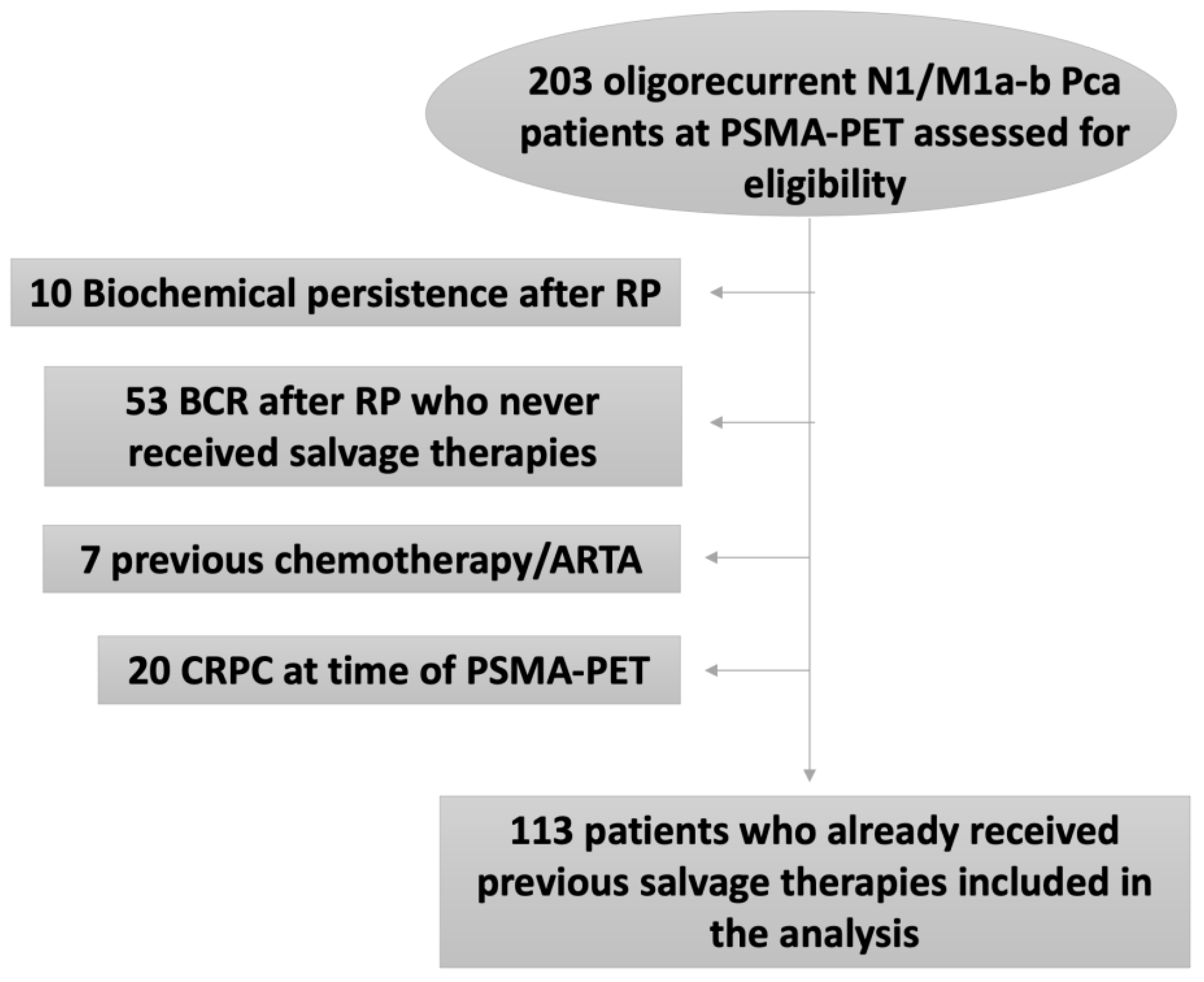
2.1. Study Design and Population Characteristics
2.2. PSMA-PET Procedure and Interpretation Criteria
2.3. Patient Management and Treatments
2.4. Outcomes Measurement
2.5. Statistical Analyses
3. Results
4. Discussion
Limitations
5. Conclusions
Author Contributions
Funding
Institutional Review Board Statement
Informed Consent Statement
Data Availability Statement
Conflicts of Interest
References
- Ost, P.; Reynders, D.; Decaestecker, K.; Fonteyne, V.; Lumen, N.; De Bruycker, A.; Lambert, A.; Delrue, L.; Bultijnck, R.; Claeys, T.; et al. Surveillance or Metastasis-Directed Therapy for Oligometastatic Prostate Cancer Recurrence: A Prospective, Randomized, Multicenter Phase II Trial. J. Clin. Oncol. Off. J. Am. Soc. Clin. Oncol. 2018, 36, 446–453. [Google Scholar] [CrossRef]
- Calais, J.; Ceci, F.; Eiber, M.; AHope, T.; SHofman, M.; Rischpler, C.; Bach-Gansmo, T.; Nanni, C.; Savir-Baruch, B.; Elashoff, D.; et al. (18)F-fluciclovine PET-CT and (68)Ga-PSMA-11 PET-CT in patients with early biochemical recurrence after prostatectomy: A prospective, single-centre, single-arm, comparative imaging trial. Lancet Oncol. 2019, 20, 1286–1294. [Google Scholar] [CrossRef] [PubMed]
- Perera, M.; Papa, N.; Christidis, D.; Wetherell, D.; Hofman, M.S.; Murphy, D.G.; Bolton, D.; Lawrentschuk, N. Sensitivity, Specificity, and Predictors of Positive 68Ga-Prostate-specific Membrane Antigen Positron Emission Tomography in Advanced Prostate Cancer: A Systematic Review and Meta-analysis. Eur. Urol. 2016, 70, 926–937. [Google Scholar] [CrossRef]
- Farolfi, A.; Ceci, F.; Castellucci, P.; Graziani, T.; Siepe, G.; Lambertini, A.; Schiavina, R.; Lodi, F.; Morganti, A.G.; Fanti, S.; et al. (68)Ga-PSMA-11 PET/CT in prostate cancer patients with biochemical recurrence after radical prostatectomy and PSA <0.5 ng/ml. Efficacy and impact on treatment strategy. Eur. J. Nucl. Med. Mol. Imaging 2019, 46, 11–19. [Google Scholar] [CrossRef] [PubMed]
- Gundem, G.; Van Loo, P.; Kremeyer, B.; Alexandrov, L.B.; Tubio, J.M.C.; Papaemmanuil, E.; Brewer, D.S.; Kallio, H.M.L.; Högnäs, G.; Annala, M.; et al. The evolutionary history of lethal metastatic prostate cancer. Nature 2015, 520, 353–357. [Google Scholar] [CrossRef]
- Phillips, R.; Shi, W.Y.; Deek, M.; Radwan, N.; Lim, S.J.; Antonarakis, E.S.; Rowe, S.R.; Ross, A.E.; Gorin, M.A.; Deville, C.; et al. Outcomes of Observation vs Stereotactic Ablative Radiation for Oligometastatic Prostate Cancer: The ORIOLE Phase 2 Randomized Clinical Trial. JAMA Oncol. 2020, 6, 650–659. [Google Scholar] [CrossRef]
- Glicksman, R.M.; Metser, U.; Vines, D.; Radwan, N.; Lim, S.J.; Antonarakis, E.S.; Rowe, S.P.; Ross, A.E.; Gorin, M.A.; Deville, C.; et al. Curative-intent Metastasis-directed Therapies for Molecularly-defined Oligorecurrent Prostate Cancer: A Prospective Phase II Trial Testing the Oligometastasis Hypothesis. Eur. Urol. 2021, 80, 374–382. [Google Scholar] [CrossRef]
- Ceci, F.; Rovera, G.; Iorio, G.C.; Guarneri, A.; Chiofalo, V.; Passera, R.; Oderda, M.; Dall’Armellina, S.; Liberini, V.; Grimaldi, S.; et al. Event-free survival after (68) Ga-PSMA-11 PET/CT in recurrent hormone-sensitive prostate cancer (HSPC) patients eligible for salvage therapy. Eur. J. Nucl. Med. Mol. Imaging 2022, 49, 3257–3268. [Google Scholar] [CrossRef] [PubMed]
- Bianchi, L.; Ceci, F.; Costa, F.; Balestrazzi, E.; Droghetti, M.; Piazza, P.; Pissavini, A.; Mei, R.; Farolfi, A.; Castellucci, P.; et al. The Impact of PSMA-PET on Oncologic Control in Prostate Cancer Patients Who Experienced PSA Persistence or Recurrence. Cancers 2022, 15, 247. [Google Scholar] [CrossRef]
- De Bruycker, A.; Spiessens, A.; Dirix, P.; Koutsouvelis, N.; Semac, I.; Liefhooghe, N.; Gomez-Iturriaga, A.; Everaerts, W.; Otte, F.; Papachristofilou, A.; et al. PEACE V—Salvage Treatment of OligoRecurrent nodal prostate cancer Metastases (STORM): A study protocol for a randomized controlled phase II trial. BMC Cancer 2020, 20, 406. [Google Scholar] [CrossRef]
- Sweeney, C.J.; Chen, Y.-H.; Carducci, M.; Liu, G.; Jarrard, D.F.; Eisenberger, M.; Wong, Y.-N.; Hahn, N.; Kohli, M.; Cooney, M.M.; et al. Chemohormonal Therapy in Metastatic Hormone-Sensitive Prostate Cancer. N. Engl. J. Med. 2015, 373, 737–746. [Google Scholar] [CrossRef] [PubMed]
- Calais, J.; Fendler, W.P.; Eiber, M.; Gartmann, J.; Chu, F.-I.; Nickols, N.G.; Reiter, R.E.; Rettig, M.B.; Marks, L.S.; Ahlering, T.E.; et al. Impact of (68)Ga-PSMA-11 PET/CT on the Management of Prostate Cancer Patients with Biochemical Recurrence. J. Nucl. Med. 2018, 59, 434–441. [Google Scholar] [CrossRef] [PubMed]
- Ceci, F.; Bianchi, L.; Borghesi, M.; Polverari, G.; Farolfi, A.; Briganti, A.; Schiavina, R.; Brunocilla, E.; Castellucci, P.; Fanti, S. Prediction nomogram for 68Ga-PSMA-11 PET/CT in different clinical settings of PSA failure after radical treatment for prostate cancer. Eur. J. Nucl. Med. Mol. Imaging 2020, 47, 136–146. [Google Scholar] [CrossRef] [PubMed]
- Bianchi, L.; Castellucci, P.; Farolfi, A.; Droghetti, M.; Artigas, C.; Leite, J.; Paola Corona, P.; Shagera, Q.A.; Moreira, R.; González, C.; et al. Multicenter External Validation of a Nomogram for Predicting Positive Prostate-specific Membrane Antigen/Positron Emission Tomography Scan in Patients with Prostate Cancer Recurrence. Eur. Urol. Oncol. 2021, 6, 41–48. [Google Scholar] [CrossRef]
- Rauscher, I.; Düwel, C.; Haller, B.; Rischpler, C.; Heck, M.M.; Gschwend, J.E.; Schwaiger, M.; Maurer, T.; Eiber, M. Efficacy, Predictive Factors, and Prediction Nomograms for (68)Ga-labeled Prostate-specific Membrane Antigen-ligand Positron-emission Tomography/Computed Tomography in Early Biochemical Recurrent Prostate Cancer After Radical Prostatectomy. Eur. Urol. 2018, 73, 656–661. [Google Scholar] [CrossRef]
- Calais, J.; Armstrong, W.R.; Kishan, A.U.; Booker, K.M.; Hope, T.A.; Fendler, W.P.; Elashoff, D.; Nickols, N.G.; Czernin, J. Update from PSMA-SRT Trial NCT03582774: A Randomized Phase 3 Imaging Trial of Prostate-specific Membrane Antigen Positron Emission Tomography for Salvage Radiation Therapy for Prostate Cancer Recurrence Powered for Clinical Outcome. Eur. Urol. Focus 2021, 7, 238–240. [Google Scholar] [CrossRef]
- Fanti, S.; Minozzi, S.; Morigi, J.J.; Booker, K.M.; Hope, T.A.; Fendler, W.P.; Elashoff, D.; Nickols, N.G.; Czernin, J. Development of standardized image interpretation for 68Ga-PSMA PET/CT to detect prostate cancer recurrent lesions. Eur. J. Nucl. Med. Mol. Imaging 2017, 44, 1622–1635. [Google Scholar] [CrossRef]
- Fendler, W.P.; Eiber, M.; Beheshti, M.; Bomanji, J.; Ceci, F.; Cho, S.; Giesel, F.; Haberkorn, U.; Hope, T.A.; Kopka, K.; et al. (68)Ga-PSMA PET/CT: Joint EANM and SNMMI procedure guideline for prostate cancer imaging: Version 1.0. Eur. J. Nucl. Med. Mol. Imaging 2017, 44, 1014–1024. [Google Scholar] [CrossRef]
- Mottet, N.; Cornford, P.; van den Bergh, R.C.N.; Briers, E.; Eberli, D.; de Meerleer, G.; de Santis, M.; Gillessen, S.; Grummet, J.; Henry, A.M.; et al. Guidelines on Prostate Cancer; EAU Guidelines Office: Arnhem, The Netherlands, 2022. [Google Scholar]
- Bianchi, L.; Schiavina, R.; Borghesi, M.; Casablanca, C.; Chessa, F.; Mineo Bianchi, F.; Pultrone, C.; Vagnoni, V.; Ercolino, A.; Dababneh, H.; et al. Patterns of positive surgical margins after open radical prostatectomy and their association with clinical recurrence. Minerva Urol. Nefrol. 2020, 72, 464–473. [Google Scholar] [CrossRef]
- Eiber, M.; Herrmann, K.; Calais, J.; Hadaschik, B.; Giesel, F.L.; Hartenbach, M.; Hope, T.; Reiter, R.; Maurer, T.; Weber, W.A.; et al. Prostate Cancer Molecular Imaging Standardized Evaluation (PROMISE): Proposed miTNMClassification for the Interpretation of PSMA-Ligand, PET/CT. J. Nucl. Med. 2018, 59, 469–478. [Google Scholar] [CrossRef]
- Perera, M.; Papa, N.; Roberts, M.; Williams, M.; Udovicich, C.; Vela, I.; Christidis, D.; Bolton, D.; Hofman, M.S.; Lawrentschuk, N.; et al. Gallium-68 Prostate-specific Membrane Antigen Positron Emission Tomography in Advanced Prostate Cancer-Updated Diagnostic Utility, Sensitivity, Specificity, and Distribution of Prostate-specific Membrane Antigen-avid Lesions: A Systematic Review and Meta-analysis. Eur. Urol. 2020, 77, 403–417. [Google Scholar] [CrossRef] [PubMed]
- Alongi, P.; Laudicella, R.; Lanzafame, H.; Farolfi, A.; Mapelli, P.; Picchio, M.; Burger, I.A.; Iagaru, A.; Minutoli, F.; Evangelista, L. PSMA and Choline PET for the Assessment of Response to Therapy and Survival Outcomes in Prostate Cancer Patients: A Systematic Review from the Literature. Cancers 2022, 14, 1770. [Google Scholar] [CrossRef] [PubMed]
- Vetrone, L.; Cuzzani, G.; Mei, R.; Zanoni, L.; Bertaccini, A.; Bianchi, L.; Castellucci, P.; Gaudiano, C.; Cappelli, A.; Giunchi, F.; et al. Case report: PSMAPET/CT addresses the correct diagnosis in a patient with metastatic prostate cancer despite negative core biopsies, m.p.M.R.I. A diagnostic challenge. Front. Oncol. 2023, 13, 1101221. [Google Scholar] [CrossRef]
- Decaestecker, K.; De Meerleer, G.; Ameye, F.; Fonteyne, V.; Lambert, B.; Joniau, S.; Delrue, L.; Billiet, I.; Duthoy, W.; Junius, S.; et al. Surveillance or metastasis-directed Therapy for OligoMetastatic Prostate cancer recurrence (STOMP): Study protocol for a randomized phase II trial. BMC Cancer 2014, 14, 671. [Google Scholar] [CrossRef] [PubMed]
- Bianchi, L.; Schiavina, R.; Borghesi, M.; Ceci, F.; Angiolini, A.; Chessa, F.; Droghetti, M.; Bertaccini, A.; Manferrari, F.; Marcelli, E.; et al. How does 68Ga-prostate-specific membrane antigen positron emission tomography/computed tomography impact the management of patients with prostate cancer recurrence after surgery? Int. J. Urol. 2019, 26, 804–811. [Google Scholar] [CrossRef]
- Bianchi, L.; Borghesi, M.; Schiavina, R.; Castellucci, P.; Ercolino, A.; Mineo Bianchi, F.; Barbaresi, U.; Polverari, G.; Brunocilla, E.; Fanti, S.; et al. Predictive accuracy and clinical benefit of a nomogram aimed to predict 68Ga-PSMA PET/CT positivity in patients with prostate cancer recurrence and PSA < 1 ng/ml external validation on a single institution database. Eur. J. Nucl. Med. Mol. Imaging 2020, 47, 2100–2105. [Google Scholar] [CrossRef]
- Hölscher, T.; Baumann, M.; Kotzerke, J.; Zöphel, K.; Paulsen, F.; Müller, A.-C.; Zips, D.; Koi, L.; Thomas, C.; Löck, S.; et al. Toxicity and Efficacy of Local Ablative, Image-guided Radiotherapy in Gallium-68 Prostate-specific Membrane Antigen Targeted Positron Emission Tomography-staged, Castration-sensitive Oligometastatic Prostate Cancer: The OLI-P Phase 2 Clinical Trial. Eur. Urol. Oncol. 2022, 5, 44–51. [Google Scholar] [CrossRef]
- Bravi, C.A.; Fossati, N.; Gandaglia, G.; Suardi, N.; Mazzone, E.; Robesti, D.; Osmonov, D.; Juenemann, K.-P.; Boeri, L.; Karnes, R.J.; et al. Long-term Outcomes of Salvage Lymph Node Dissection for Nodal Recurrence of Prostate Cancer After Radical Prostatectomy: Not as Good as Previously Thought. Eur. Urol. 2020, 78, 661–669. [Google Scholar] [CrossRef] [PubMed]
- Vetrone, L.; Mei, R.; Bianchi, L.; Giunchi, F.; Farolfi, A.; Castellucci, P.; Droghetti, M.; Presutti, M.; Degiovanni, A.; Schiavina, R.; et al. Histology and PSMA Expression on Immunohistochemistry in High-Risk Prostate Cancer Patients: Comparison with 68Ga-PSMA PET/CT Features in Primary Staging. Cancers 2023, 15, 1716. [Google Scholar] [CrossRef]


| Overall | |
|---|---|
| Patients, n (%) | 113 (100) |
| Age | |
| Median (IQR) | 61 (56–66) |
| pT stage, n (%) | |
| pT2 | 38 (33.6) |
| pT3a | 37 (32.7) |
| pT3b–pT4 | 38 (33.6) |
| pN stage, n (%) | |
| pNx | 30 (26.5) |
| pN0 | 58 (51.3) |
| pN1 | 25 (22.1) |
| Pathologic ISUP grade, n (%) | |
| 1–3 | 61 (54) |
| 4–5 | 52 (46) |
| Salvage therapies, n (%) | |
| Prostate Bed RT | 42 (37.2) |
| Whole Pelvis RT | 52 (46) |
| Whole Pelvis RT + ADT | 3 (2.7) |
| ADT | 16 (14.2) |
| PSA level at PSMA-PET, ng/mL | |
| Median (IQR) | 0.62 (0.29–1.27) |
| Time from RP to PSMA-PET, months | |
| Median (IQR) | 52 (30–94) |
| Follow-up after PSMA-PET, months | |
| Median (IQR) | 31 (19–42) |
| Overall | MDT Approach | Conventional Approach | p Value | |
|---|---|---|---|---|
| Patients, n (%) | 113 (100) | 91 (80) | 22 (20) | - |
| Number of positive lesions at PSMA-PET, n | 0.04 | |||
| Median (IQR) | 1 (1–2) | 1 (1–2) | 2 (1–2) | |
| Site of positive PSMA-PET, n (%) | 0.1 | |||
| LND | 77 (68.1) | 65 (71.4) | 12 (54.5) | |
| Bones | 24 (21.2) | 19 (20.9) | 5 (22.7) | |
| LND and Bones | 12 (10.6) | 7 (6.2) | 5 (22.7) | |
| mi stage at PSMA-PET, n (%) | 0.06 | |||
| N1 | 66 (58.4) | 57 (62.6) | 9 (40.9) | |
| M1a-b | 47 (41.6) | 34 (37.4) | 13 (59.1) | |
| Progression after PSMA-PET guided treatment, n (%) | 0.3 | |||
| No | 45 (39.8) | 34 (37.4) | 11 (50) | |
| Yes | 68 (60.2) | 57 (62.6) | 11 (50) | |
| Time to progression, months | 0.012 | |||
| Median (IQR) | 16 (6–26) | 13 (6–25) | 18 (15–29) | |
| PSA recurrence after PSMA-PET guided treatment, n (%) | 0.5 | |||
| No | 54 (47.8) | 42 (46.2) | 12 (54.5) | |
| Yes | 59 (52.2) | 49 (53.8) | 10 (45.5) | |
| PSA at recurrence after PSMA-PET guided treatment, ng/mL | 0.8 | |||
| Median (IQR) | 3.18 (0.7–14) | 3.4 (0.69–1.5) | 2.54 (0.95–16.25) | |
| Metastases after PSMA-PET guided treatment, n (%) | 0.1 | |||
| No | 81 (71.7) | 68 (74.7) | 13 (59.1) | |
| Yes | 32 (28.3) | 23 (25.3) | 9 (40.9) | |
| Time to Metastases, months | 0.8 | |||
| Median (IQR) | 19 (13–26) | 19 (12–26) | 18 (12–26) | |
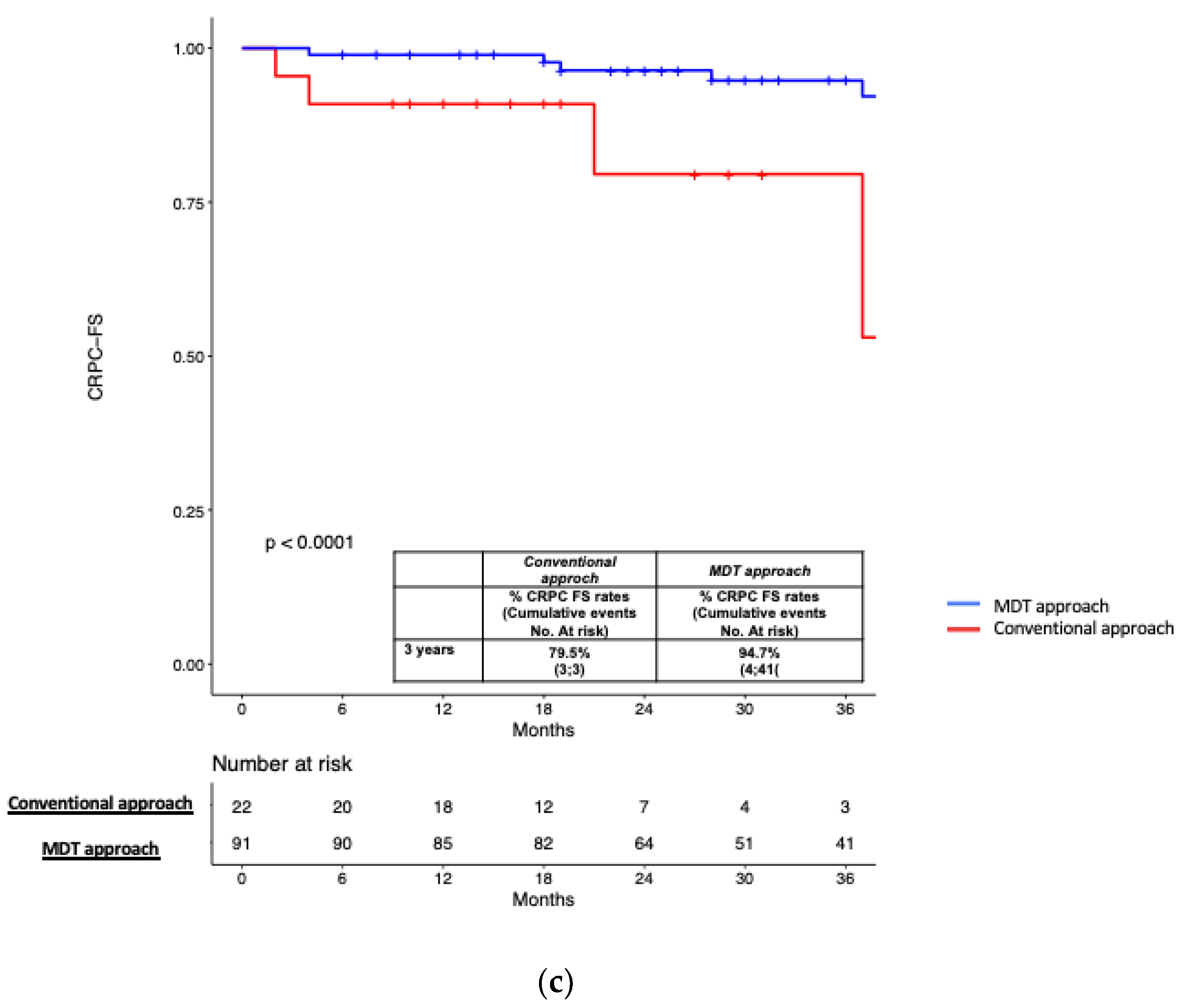
| CRPC after PSMA-PET guided treatment, n (%) | 0.03 | |||
| No | 98 (86.7) | 82 (90.1) | 16 (72.7) | |
| Yes | 15 (13.3) | 9 (9.9) | 6 (27.3) | |
| Time to CRPC, months | ≤0.001 | |||
| Median (IQR) | 37 (18–43) | 37 (19–42) | 29 (4–43) | |
| Overall Mortality, n (%) | 0.3 | |||
| No | 108 (95.6) | 86 (94.5) | 22 (100) | |
| Yes | 5 (4.4) | 5 (5.5) | 0 (0) | |
| Cancer specific mortality, n (%) | 0.5 | |||
| No | 111 (98.2) | 89 (97.8) | 22 (100) | |
| Yes | 2 (1.8) | 2 (2.2) | 0 (0) |
| Variables | Progression | Metastasis | ||
|---|---|---|---|---|
| HR (95% C.I.) | p Value | HR (95% C.I.) | p Value | |
| Age (years) | 0.96 (0.92–0.99) | 0.04 | - | - |
| Pathologic ISUP group | - | - | ||
| ISUP 1–3 | 1–0 (Ref) | 0.9 | ||
| ISUP 4–5 | 1.04 (0.57–1.87) | |||
| miTNM stage | ||||
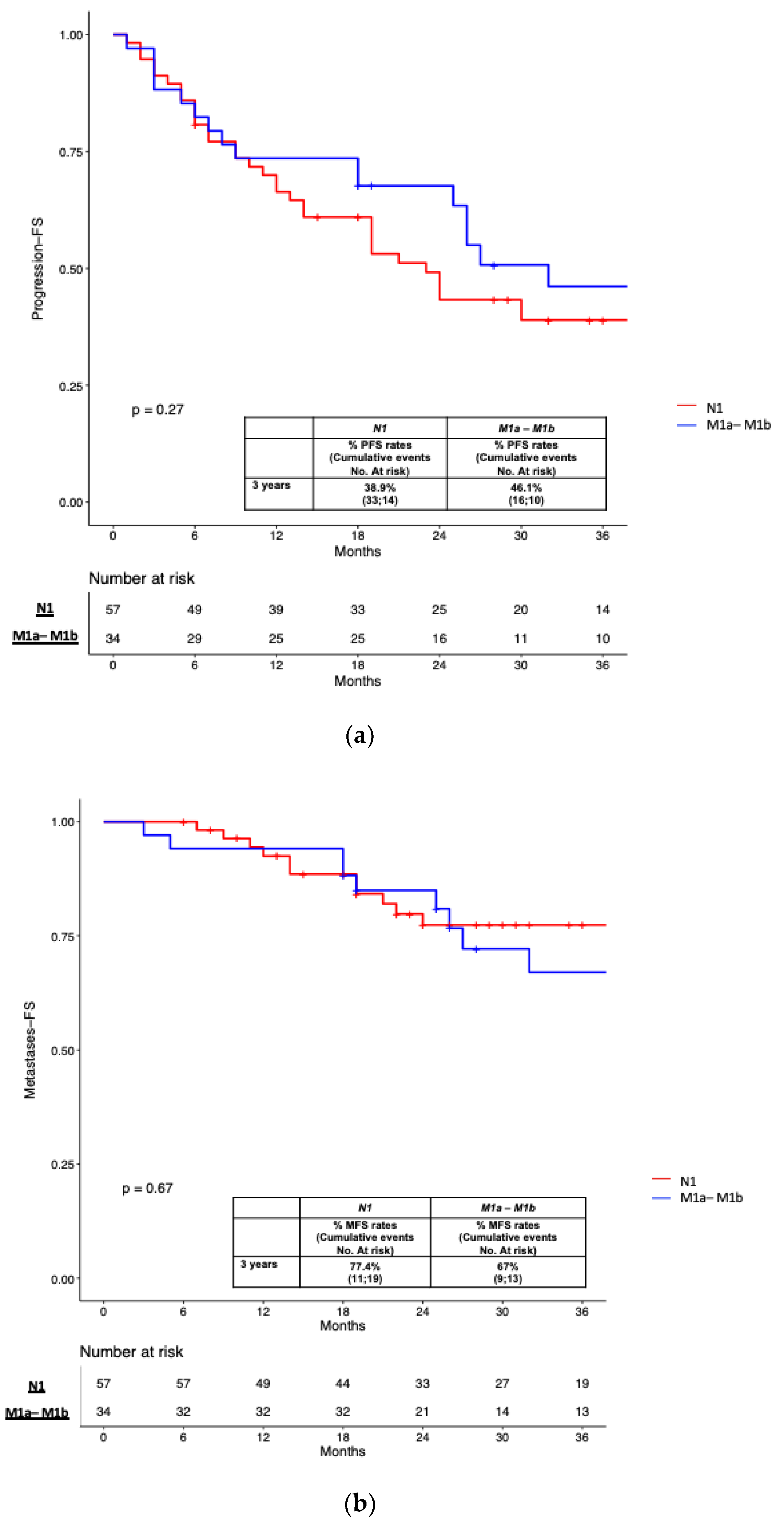
| N1 | 1–0 (Ref) | 0.8 | 1–0 (Ref) | 0.9 |
| M1a-M1b | 1.06 (0.55–2.06) | 0.97 (0.43–2.20) | ||
| Number of positive lesions at PSMA-PET | 0.98 (0.67–1.439) | 0.9 | 1.03 (0.60–1.80) | 0.9 |
| ADT during second line salvage treatment | ||||
| No | 1–0 (Ref) | 1.0 (Ref) | ||
| Yes | 0.50 (0.27–0.93) | 0.03 | 1.95 (0.82–4.62) | 0.1 |
| Type of salvage treatment after PSMA-PET | ||||
| Conventional approach | 1.0 (Ref) | 1.0 (Ref) | ||
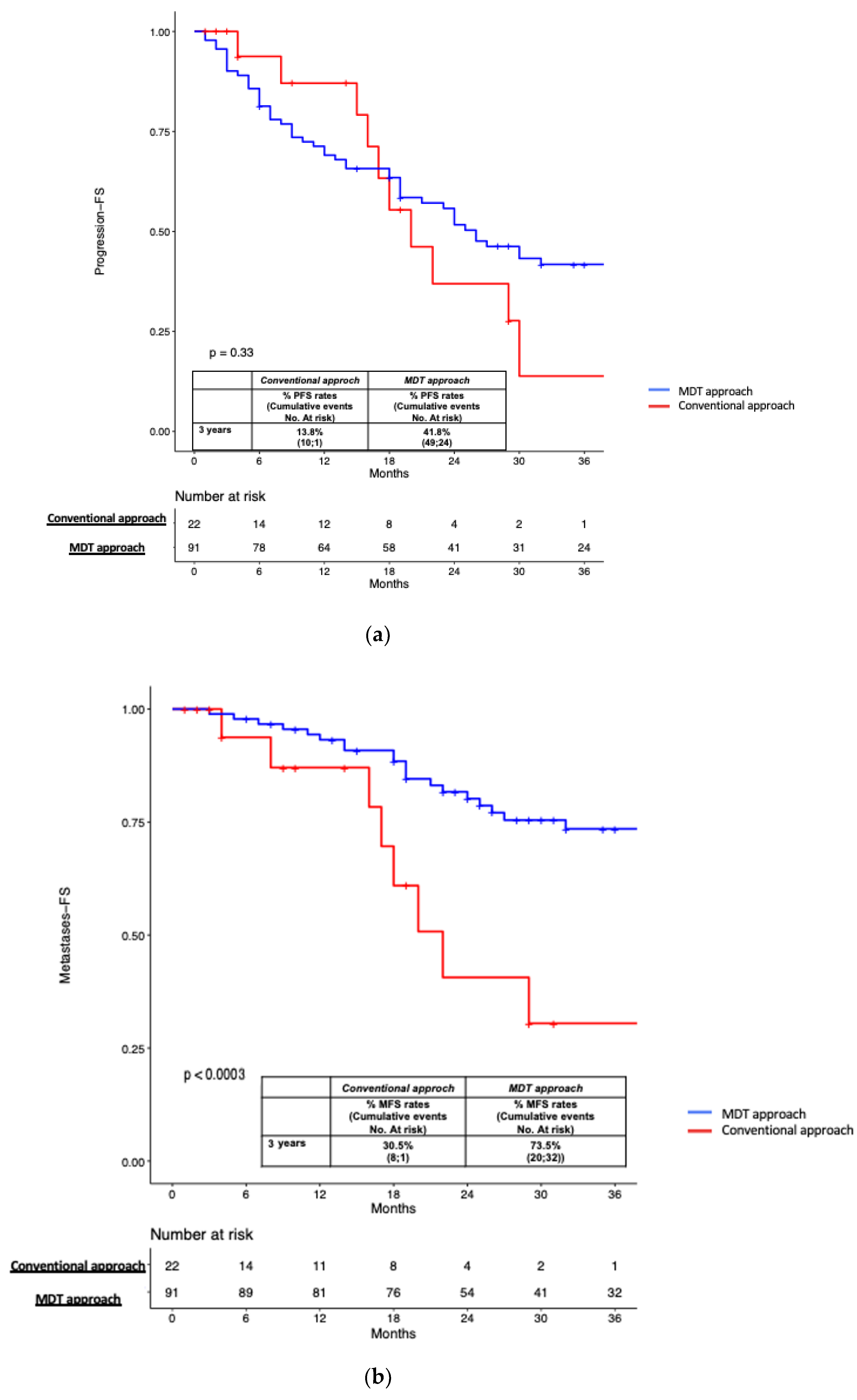
| MDT | 0.49 (0.20–1.25) | 0.1 | 0.27 (0.10–0.69) | 0.006 |
Disclaimer/Publisher’s Note: The statements, opinions and data contained in all publications are solely those of the individual author(s) and contributor(s) and not of MDPI and/or the editor(s). MDPI and/or the editor(s) disclaim responsibility for any injury to people or property resulting from any ideas, methods, instructions or products referred to in the content. |
© 2023 by the authors. Licensee MDPI, Basel, Switzerland. This article is an open access article distributed under the terms and conditions of the Creative Commons Attribution (CC BY) license (https://creativecommons.org/licenses/by/4.0/).
Share and Cite
Bianchi, L.; Ceci, F.; Balestrazzi, E.; Costa, F.; Droghetti, M.; Piazza, P.; Pissavini, A.; Presutti, M.; Farolfi, A.; Mei, R.; et al. PSMA-PET Guided Treatment in Prostate Cancer Patients with Oligorecurrent Progression after Previous Salvage Treatment. Cancers 2023, 15, 2027. https://doi.org/10.3390/cancers15072027
Bianchi L, Ceci F, Balestrazzi E, Costa F, Droghetti M, Piazza P, Pissavini A, Presutti M, Farolfi A, Mei R, et al. PSMA-PET Guided Treatment in Prostate Cancer Patients with Oligorecurrent Progression after Previous Salvage Treatment. Cancers. 2023; 15(7):2027. https://doi.org/10.3390/cancers15072027
Chicago/Turabian StyleBianchi, Lorenzo, Francesco Ceci, Eleonora Balestrazzi, Francesco Costa, Matteo Droghetti, Pietro Piazza, Alessandro Pissavini, Massimiliano Presutti, Andrea Farolfi, Riccardo Mei, and et al. 2023. "PSMA-PET Guided Treatment in Prostate Cancer Patients with Oligorecurrent Progression after Previous Salvage Treatment" Cancers 15, no. 7: 2027. https://doi.org/10.3390/cancers15072027
APA StyleBianchi, L., Ceci, F., Balestrazzi, E., Costa, F., Droghetti, M., Piazza, P., Pissavini, A., Presutti, M., Farolfi, A., Mei, R., Castellucci, P., Gandaglia, G., Larcher, A., Robesti, D., Mottrie, A., Briganti, A., Morganti, A. G., Fanti, S., Montorsi, F., ... Brunocilla, E. (2023). PSMA-PET Guided Treatment in Prostate Cancer Patients with Oligorecurrent Progression after Previous Salvage Treatment. Cancers, 15(7), 2027. https://doi.org/10.3390/cancers15072027