Review

Exploring the Therapeutic Potential of Royal Jelly in Metabolic Disorders and Gastrointestinal Diseases

by
Hesham R. El-Seedi
1,2,3,*,
Suzy Salama
4,
Aida A. Abd El-Wahed
5,
Zhiming Guo
2,
Alessandro Di Minno
6,7,
Maria Daglia
6,7,
Chuan Li
8,
Xiao Guan
9,10,
Daniele Giuseppe Buccato
6,
Shaden A. M. Khalifa
11,* and
Kai Wang
12,*
1
Pharmacognosy Group, Department of Pharmaceutical Biosciences, Uppsala University, Biomedical Centre, P.O. Box 591, SE-751 24 Uppsala, Sweden
2
School of Food and Biological Engineering, Jiangsu University, Zhenjiang 212013, China
3
International Joint Research Laboratory of Intelligent Agriculture and Agri-Products Processing, Jiangsu University, Zhenjiang 210024, China
4
Indigenous Knowledge and Heritage Center, Ghibaish College of Science and Technology, Ghibaish 51111, Sudan
5
Department of Bee Research, Plant Protection Research Institute, Agricultural Research Centre, Giza 12627, Egypt
6
Department of Pharmacy, University of Napoli Federico II, Via D. Montesano 49, 80131 Naples, Italy
7
CEINGE-Biotecnologie Avanzate, Via Gaetano Salvatore 486, 80145 Naples, Italy
8
School of Food Science and Engineering, Hainan University, Haikou 570228, China
9
School of Health Science and Engineering, University of Shanghai for Science and Technology, Shanghai 200093, China
10
National Grain Industry (Urban Grain and Oil Security) Technology Innovation Center, Shanghai 200093, China
11
Psychiatry and Neurology Department, Capio Saint Göran’s Hospital, Sankt Göransplan 1, 112 19 Stockholm, Sweden
12
State Key Laboratory of Resource Insects, Institute of Apicultural Research, Chinese Academy of Agricultural Sciences, Beijing 100093, China
*
Authors to whom correspondence should be addressed.
Nutrients 2024, 16(3), 393; https://doi.org/10.3390/nu16030393
Submission received: 30 November 2023 / Revised: 7 January 2024 / Accepted: 23 January 2024 / Published: 29 January 2024
(This article belongs to the Special Issue Diet and Nutrition: Metabolic Diseases)

Abstract

:
Metabolic disorders, encompassing diabetes mellitus, cardiovascular diseases, gastrointestinal disorders, etc., pose a substantial global health threat, with rising morbidity and mortality rates. Addressing these disorders is crucial, as conventional drugs often come with high costs and adverse effects. This review explores the potential of royal jelly (RJ), a natural bee product rich in bioactive components, as an alternative strategy for managing metabolic diseases. RJ exhibits diverse therapeutic properties, including antimicrobial, estrogen-like, anti-inflammatory, hypotensive, anticancer, and antioxidant effects. This review’s focus is on investigating how RJ and its components impact conditions like diabetes mellitus, cardiovascular disease, and gastrointestinal illnesses. Evidence suggests that RJ serves as a complementary treatment for various health issues, notably demonstrating cholesterol- and glucose-lowering effects in diabetic rats. Specific RJ-derived metabolites, such as 10-hydroxy-2-decenoic acid (10-HDA), also known as the “Queen bee acid,” show promise in reducing insulin resistance and hyperglycemia. Recent research highlights RJ’s role in modulating immune responses, enhancing anti-inflammatory cytokines, and suppressing key inflammatory mediators. Despite these promising findings, further research is needed to comprehensively understand the mechanisms underlying RJ’s therapeutic effects.

1. Introduction

Obesity, insulin resistance, hypertension, and heart disease are groups of diseases categorized as metabolic disorders [1]. Metabolic disorders are caused by the disruption of regular metabolic functions triggered by oxidative stress and chronic inflammation. The characteristics of chronic inflammation include elevated levels of inflammatory mediators, such as chemokines and cytokines, which confirm the promotion of the development of metabolic disorders [2]. These chronic inflammations also contribute to the development of major chronic diseases such as non-alcoholic fatty liver disease and obesity. Type 2 diabetes and metabolic disorders now rank among the main risks to human health [3,4]. The significant rise in the prevalence of various metabolic diseases is related to aging, environmental factors, changes in lifestyle, and genetics [5]. Metabolic disorders have been estimated to affect 25% of the population globally [6].
Metformin and Glimepiride are the two synthetic drugs that are currently validated and available for the management of Type 2 diabetes mellitus (T2DM) [7,8]. Metformin is the first-line treatment and has been used for many decades to reduce blood sugar [9]. One of the alternate methods for treating metabolic disorders, and therefore reducing health risks, is utilizing natural product resources. Berberine, derived from the root of Berberis vulgaris L. and taken as a typical example, demonstrates significant potential in fighting T2DM [10].
Royal jelly (RJ) is a “rich source of nutrients” that nurse bees produce and feed to worker larvae and queen bees. RJ supplementation is beneficial for a variety of disorders, including diabetes [11,12], gastrointestinal diseases [13,14], and cardiovascular diseases [15,16]. The active ingredients of RJ, including its proteins, carbohydrates, and fats, as well as its minerals, amino acids, vitamins, enzymes, hormones, and polyphenols, are what provide its biological properties [17].
The current review intends to emphasize the protective properties of RJ and/or its components against metabolic disorders such as diabetes, gastrointestinal ailments, and cardiovascular diseases as a part of our ongoing project studying honeybee products [18,19,20].

2. Methodology

Sci-finder, PubMed, Google Scholar, Web of Science, ScienceDirect, Microsoft Academic Search, Core, and Scopus were all accessed to conduct a literature search. The following keywords were used to address the search terms: “royal jelly”, “diabetes mellitus”, “gastrointestinal diseases”, and “cardiovascular diseases”. The search included published studies, and therefore only articles in English were selected. Studies that explored the functions of RJ or its active components with the co-administration of prescription products were chosen. The search approach identified 310 specific studies, out of which 87 were disregarded due to the irrelevance of the study scope. Finally, the studies were further analyzed to offer an insightful overview of the field’s progress. The authors, year of publication, RJ utilization, dosage form or percentage, category of investigation, main points, pathway of interaction, and techniques used were all data items.

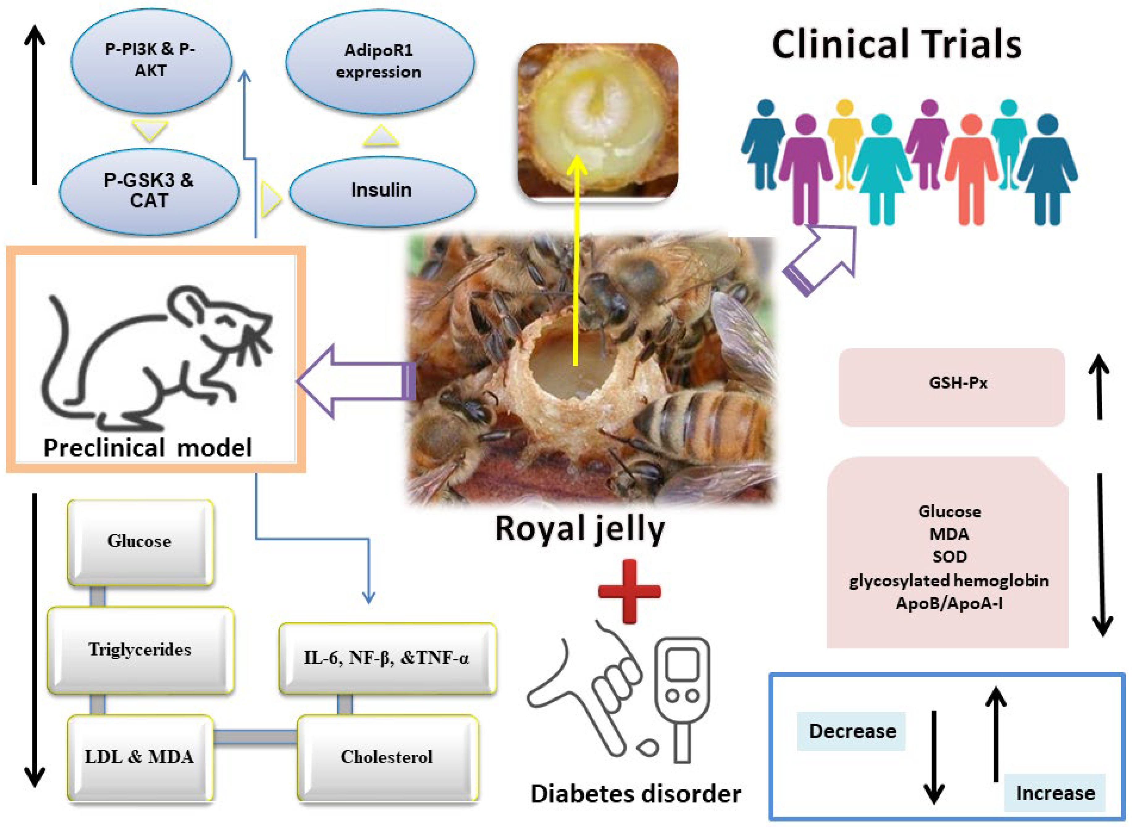
3. Royal Jelly in Diabetes

Diabetes mellitus (DM) is a worldwide metabolic disorder. According to Saeedi et al., it affected 463 million individuals globally in 2019, with that figure expected to climb to 578 million by 2030 and 700 million by 2045 [21]. DM is anticipated to become more prevalent in the world, and thus health concerns are only liable to increase [22]. Diabetes is a prevalent health issue that affects both sexes equally and impairs sexual function (e.g., sexual disinclination, negative pregnancy outcomes, infertility, loss of penile erection, and diminished clitoral sensitivity). The male reproductive system is impacted by DM in a variety of anatomical and functional aspects, with reduced sperm parameters being an example of the secondary complications of diabetes [23]. Clinically, T2DM, which accounts for 90% of cases of diabetes, is characterized by hyperglycemia and insulin insufficiency caused by cell dysfunction and insulin resistance (IR) in target organs, including the liver, heart, skeletal muscle, and adipose tissue. Atherosclerosis, coronary heart disease, and kidney disease are all chronic consequences that are highly likely to develop in T2DM patients [24]. The two most common causes of morbidity in people with DM are infections and foot ulcers [25]. Up to 60% of non-traumatic lower-limb amputations in diabetics result from diabetic foot ulcers (DFU), the leading risk factor. Given the high expense of diabetic medical treatments, it is critical to explore alternative entities that can be cost-effective. These choices should not only manage blood glucose levels, but also reduce the probability of complications [26]. Thus, the race to find a sustainable and economically viable solution for diabetes remains ongoing. As demonstrated below, RJ treatments have shown therapeutic potential in both rodent and human diabetic models and are effective against hypercholesterolemia and diabetes, as mentioned in Figure 1 [12,27].

3.1. Preclinical Studies

In preclinical studies, RJ was given orally to KK-Ay mice at a dose of 10 mg/kg body weight (BW). In obese/diabetic KK-Ay mice, RJ treatment improves hyperglycemia and partially lowers BW. RJ administration activates the expression of adiponectin (AdipoQ) and adiponectin receptor-1 (AdioR1), which then activate the expression of phosphorylated AMP-activated protein kinase (pAMPK). Additionally, RJ treatment that increases adiponectin receptor-1 (AdipoR1) expression also boosts Ppara and Pgc1a expression, which improves lipid utilization and causes a reduction in BW in KK-Ay mice [28]. Adult male Wistar rats were divided into four groups: diabetic, RJ, diabetic treated with RJ, and control. To induce diabetes, streptozotocin-induced diabetes (STZ)was administered intravenously at a dosage of 60 mg/kgBW. RJ was then administered via gavage at a dosage of 100 mg/kg BW for six weeks. Testicular weight, viability, sperm count, deformity, motility, chromatin quality, DNA integrity, testicular tissue malondialdehyde (MDA) levels, and serum testosterone were all enhanced via RJ in diabetic mice [33]. A total of 28 adult Wistar rats were randomized and divided into four groups: control, RJ, diabetic, and hyperglycemic treated with RJ. To induce diabetes, a single intraperitoneal injection of STZ at a dose of 50 mg/kg BW was used. The rats were then administered RJ (100 mg/kg BW) orally each day for a duration of six weeks. The treatment with RJ resulted in improved levels of catalase (CAT) and ferric reducing antioxidant power (FRAP) when compared to other groups [29]. RJ was given orally to the diabetic rats at a dosage of 100 mg/kg for 42 days following STZ. RJ improved the serum levels of aspartate aminotransferase (AST), alkaline phosphatase (ALP), high-density lipoprotein cholesterol (HDL-c), alanine aminotransferase (ALT), total protein (TP), fasting blood glucose (FBG) levels, insulin, and albumin. RJ dramatically lowered MDA levels in the liver and pancreatic tissues and simultaneously normalized the levels of CAT and FRAP [30]. In a similar experimental study, diabetes was induced in rats through an intraperitoneal injection of STZ (60 mg/kg BW). Then, RJ was administered via gavage for three days at doses of 100 and 200 mg/kg. This treatment notably decreased the levels of cholesterol, glucose, low-density lipoprotein (LDL), and triglycerides in the diabetic rats. Interestingly, the rats that received RJ treatment exhibited significantly elevated HDL levels compared to the untreated diabetic rats [11]. The 18 adult Wistar albino rats were grouped into three groups: control, STZ-induced diabetes, and STZ-induced diabetes plus RJ at a dosage of 400 mg/kg/day for a month. To induce diabetes, STZ (60 mg/kg) was injected intraperitoneally once. Both the RJ-treated and untreated diabetic rats exhibited lower body and testicular weights compared to the control group. Rats treated with STZ had significantly more degenerative alterations in their spermatogenesis and seminiferous tubules, according to the histological analysis. The RJ treatment group, on the other hand, revealed nearly normal morphology, in addition to more intense immunohistochemistry staining for Ki67-positive cells [34]. To induce diabetes, STZ was administered intravenously to rats once at a dosage of 75 mg/kg BW. The rats were categorized into four distinct groups: a healthy control group and three treatment groups. Three groups of rats; untreated diabetic group, 100 mg/kg/daily of metformin group, and one group received a honey–RJ (H–RJ) combination, containing 2% RJ and 98% honey. H–RJ was given daily to this rat group (100 mg/kg BW). The H–RJ treatment significantly lowered the levels of very low-density lipoprotein (VLDL) in the blood, compared to both the control therapy and metformin treatment. Rats with diabetes can effectively present with lower blood sugar when given H–RJ. This combination can also successfully lower triglycerides and VLDL-C lipids (TGs) [35]. Likewise, C57BL/6J mice were subjected to a high-fat diet (HFD) and administered a 5% RJ diet. Alloxan-induced diabetes in male Albino Wistar rats was discussed, as well as the hypoglycemic effect of composite formulations of Moringa oleifera seed oil extract and RJ. When compared to pure M. oleifera or pure RJ, the medication containing 20% RJ mixed with M. oleifera seed extract was found to be more effective in reducing blood sugar levels in treated mice [36]. Taken together, this dietary intervention with RJ effectively mitigated diet-induced obesity, hyperglycemia, and hepatic steatosis in mice by stimulating metabolic thermogenesis in brown adipose tissue (BAT) [37].
Fatty acid 10-hydroxy-2-decenoic acid (10H2DA) is a component found in RJ (Figure 2, Table 1). Female KK-Ay mice received 10H2DA orally at a dosage of 3 mg/kg BW via gavage for four weeks. It greatly reduced insulin resistance and hyperglycemia. In skeletal muscles, 10H2DA elevated the expression of the pAMPK protein; however, this expression was unrelated to elevated glucose transporter 4 (GLUT4) translocation. Adiponectin receptor mRNA expression was not improved by 10H2DA, and the liver’s glycogen synthetase kinase-3β (GSK-3β) phosphorylation was not triggered by the insulin signaling cascade [38]. The main active component of RJ is 10-hydroxydecanoic acid (10-HDA) (Figure 2). Recent research findings indicate that 10-HDA may possess anti-T2DM properties. When administered orally at a dosage of 100 mg/kg BW, it stopped liver degeneration in the diabetic rats and boosted insulin levels while decreasing fasting blood glucose. Additionally, 10-HDA intervention improved lipid peroxidation, reduced liver NF-β nuclear translocation, reduced interlukin-6 (IL-6) and tumor necrosis factor (TNF-α) content, and elevated P-PI3K, phosphorylated Akt (p-AKT), and glycogen synthase kinase 3β (p-GSK3β( protein levels. It also enhanced glutathione peroxidase (GPx), superoxide dismutase (SOD), and CA activity in diabetic mouse livers. Through the PI3K/AKT/GSK3 signaling pathway, 10-HDA clearly exhibited hypoglycemic effects on diabetic mice [39].Growth factor deficiency and bacterial infection are two of the main factors causing non-healing wounds in diabetics [40]. 8-Bromoadenosine-3′, 5′-cyclic monophosphate (8Br-cAMP) and antimicrobial peptide Jelleine-1 (J-1) (Figure 2) were combined to form a hydrogel without the use of any other gelators or chemical crosslinkers. This hydrogel demonstrated remarkable antibacterial action in a wound model in diabetic rats infected with methicillin-resistant Staphylococcus aureus (MRSA) [41].

3.2. Clinical Studies

A double-blind, placebo-controlled trial including 50 T2DM patients was carried out, where either 1000 mg of RJ or a placebo were administered to subjects three times per day for eight weeks. The groups were assigned to the RJ or placebo groups at intervals. Baseline characteristics and food intake between groups did not differ significantly. In the RJ group, the mean glucose level decreased (−9.4 mg/dL vs. 4 mg/dL), the mean ApoA-I concentration increased (34.4 mg/dL vs. −1.08 mg/dL), and there was a significant decrease in the mean apolipoprotein B (ApoB) /apolipoprotein A-I (ApoA-I), (0.008 vs. 0.13; p < 0.044, respectively) when comparing the RJ group to the placebo group [31].
For eight weeks, 50 female T2DM volunteers were divided into two groups and given either a daily dose of 1000 mg RJ (soft gel) or a placebo. In the RJ group, the average fasting blood sugar dropped significantly to 149.68 mg/dL after supplementation with RJ. The mean serum levels of glycosylated hemoglobin also significantly decreased to 7.05%, and the mean insulin concentration significantly decreased through RJ supplementation to 27.5 pmol/L. MDA levels declined, and GPx and erythrocyte superoxidase dismutase activity was dramatically elevated [32]. A daily dose of a 1000 mg soft gel of RJ or a placebo was administered to 50 female T2DM volunteers, divided into two respective groups, in a randomized clinical trial for 8 weeks. RJ supplementation reduced the daily total energy and carbohydrate intake as well as mean BW (72.45 vs. 71.00 kg) when compared to the control group [12]. Another randomized controlled trial comprised 46 T2DM patients aged 25–65 years with a hemoglobin A1c (HbA1c) of 6–8%. For eight weeks, the patients were randomized to take 1000 mg of RJ supplement or a placebo three times each day. In the RJ group, the insulin resistance index (HOMA-IR) decreased (1.98 vs. 3.13) while the serum total antioxidant capacity increased (907.63 vs. 765.69 mol/L) [57].
The effectiveness of topical RJ for treating diabetic foot ulcers has also been studied [58]. The trial design was randomized, controlled, and open-label, with a 12-week average follow-up time. After conservative debridement of necrotic tissue and irrigation with warm normal saline, 189 eligible patients with diabetic foot wounds from three outpatient clinics in Egypt were randomized to receive a local application of either RJ + Panthenol (PedyPhar® Ointment) or Panthenol ointment underdressing. The purpose of the research was to look at the use of PedyPhar® Ointment in the treatment of individuals suffering from limb-threatening diabetic foot infections [59]. At the end of the 12-week follow-up period, PedyPhar® revealed a greater degree (32.4%) of full healing of limb-threatening wounds in the target population, versus 12% in the Panthenol-treated (control) group [60].
Adiponectin is an adipokine released from adipose tissue that has a role in insulin sensitivity [61], and has been reported for its inhibitory effect on the glucogenesis process in the liver via down-expression of phosphoenolpyruvate carboxykinase and glucose-6-phosphatase genes, which play crucial enzymatic roles in glucose production by the liver [62]. Furthermore, research has shown that giving RJ to diabetic rats at a dosage of 10 mg/kg/day for a month controls hyperglycemia via the acceleration of adiponectin secretion [28]. The possible mechanism of RJ in diabetes was reported by Maleki et al. (2019), through the activation of the AMP-activated protein kinase pathway through increasing the production of adiponectin in skeletal muscle and liver cells [63]. Joshi et al. (2019) noted that the activation of the AMPK protein increases the cells’ uptake of glucose, while reducing intracellular secretion of glucose. Therefore, the AMPK signaling pathway is the target in controlling diabetes [64].

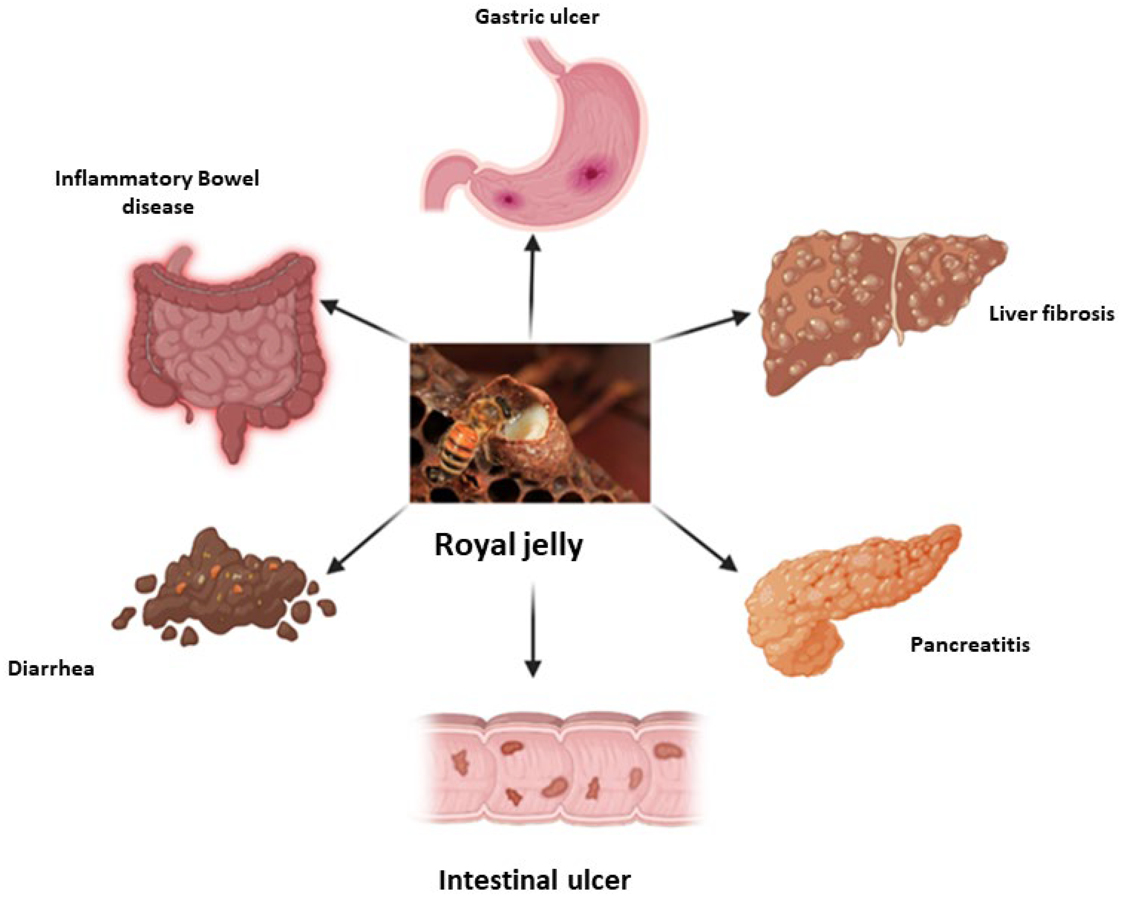
4. Royal Jelly in Gastrointestinal Diseases

Gastrointestinal disorders are common illnesses, including irritable bowel syndrome, peptic ulcers, liver diseases, pancreatitis, gallstones, and Crohn’s disease, and are often found in tropical regions [65]. Sperber et al. (2021) estimated that 40% of the people in 33 countries across six continents have functional gastrointestinal problems [66]. A strong link between gastrointestinal diseases and the diet of individuals in at-risk groups has been found. The dietary habits of people in the different areas contribute to the composition of their individual gut microbiota, and this is particularly noticeable when people move from urban to rural areas [67]. According to Rizello et al. (2019), a westernized diet rich in carbohydrates and animal proteins is a main contributor to the development and progression of chronic inflammatory bowel disease [68]. Due to involvement of the gastrointestinal tract in the absorption of nutrients, as well as its role in immune response, the risk of developing an inflammatory, autoimmune, chronic disease is inevitably increasing [69].
RJ is considered one of the most important super foods, having displayed much biological activity in preclinical and clinical studies [70]. As a honey bee product, it has been documented for its active potential against many disorders, including inflammation, liver disease, hypercholesterolemia, oxidative stress, and immune disease [71]. RJ contains many bioactive compounds such as proteins, vitamins, phenolics, and flavonoids. Additional pharmaceutical studies have revealed that the bioactive, major protein constituents of RJ (MRJPs) are considered the main therapeutic compounds of those tested [72,73].

4.1. Inflammatory Bowel Diseases

Ulcerative colitis and Crohn’s disease are both chronic, inflammatory bowel illnesses. Inflammatory bowel disease describes a persistent, non-infectious inflammation with uncertain causes, affecting one or more locations in the digestive system. According to global estimations, the prevalence of inflammatory bowel disease accounted for approximately 7 million people in 2017 [74]. Hence, the exploration of natural product remedies would pave the way to natural and complementary tools for healing. For instance, the experimental induction of colon inflammation using 2,4,6-trinitrobenzene sulphonic acid was found to be significantly inhibited by the administration of RJ in mice at a dosage of 250 mg/kg/day for a week via the inhibition of pro-inflammatory cytokines, TNF-α, and interlukin-1β (IL-1β) along with the elevation of the anti-inflammatory cytokine interlukin-10 (IL-10) [75]. Another study revealed that daily administration of RJ (150 mg/kg) considerably ameliorated the damage caused by acetic acid in rats with induced colitis, manifesting as decreased lesion areas in the colon where the intestinal mast cells were also involved in inflammation [76]. Likewise, similar doses of RJ were found to reduce the proliferation of T-lymphocytes involved in the intestinal inflammation induced by acetic acid in rats [77]. According to a recent study, synergism between RJ and selenium exhibits significant anti-inflammatory activity in inflammatory bowel disease in mice and promotes intestinal health through the improvement of the gut microbiota [78].
The mechanism of RJ in treating inflammatory bowel syndrome has been reported by Guo et al. (2022) as shown on Figure 3. The investigation revealed that RJ boosted the activity of the anti-inflammatory cytokine IL-10 and the intracellular antioxidant enzyme GPx. Additionally, RJ decreased the number of CD3+, CD5+, CD8+, and CD45+ T-cells, the release of TNF-αand the pro-inflammatory cytokines IL-1β, the nuclear factor Kappa-B (NF-κB), and cyclogenase-2 (COX-2) and tumor necrosis factor-induced injury in rats with colitis induced by 2,4,6-trinitrobenzene sulfonic acid [79].

4.2. Lactose Intolerance

Lactose intolerance is a gastrointestinal disorder that results from a lack of β-galactosidase, resulting in the maldigestion of lactose from milk and milk products. Patients with lactose intolerance present with symptoms such as pain in the abdomen, diarrhea, and flatulence, which appear after the intake of lactose-containing foods [80]. Recently, researchers have found that the synergism between RJ and probiotic yogurt has potent activity in treating lactose intolerance [81].
There is growing evidence that lactose intolerance symptoms can be treated with probiotic bacteria found in fermented and unfermented milk products [82]. The mechanism of RJ in reducing lactose intolerance relies on the activity of probiotics delivered via fermented milk products, which have been found to play an important role in health benefits, as reported by Hassan et al. (2022). The fermentation of milk with 1% RJ displayed the presence of abundant probiotics, namely Lactobacillus helveticus, which results in boosting the bioactive properties of fermented milk [83].

4.3. Chronic Diarrhea and Constipation

The symptoms of chronic constipation include uncomfortable defecation, marked by straining and difficulty along with extended time in stool passage [84]. Constipation in children is estimated to affect from 1% to 30% of the young generation worldwide [85]. Compared to standard antiviral medication, honey has been shown to reduce the incidence and duration of viral diarrhea [86]. As documented by Miyauchi-Wakuda et al. (2019), under in vitro circumstances, acetylcholine in RJ induced contractions of the smooth muscle of the mouse’s ilium via the muscarinic acetylcholine receptor, which was independent of nicotinic acetylcholine activity. The intake of royal jelly does not result in severe symptoms like diarrhea in normal situations [14,87]. Further, the anti-diarrheal potency of RJ could be attributed to the antimicrobial activity of its peptide constituents, royalisin and royalactin [88,89]. Even though RJ has a high concentration of acetylcholine, only one oral dose of RJ was not enough to boost intestinal motility or alleviate constipation.

4.4. Gastrointestinal Ulcer Disease

Gastric and intestinal ulcers induced by diclofenac (50 mg/kg) have been normalized using RJ at a dose of 150 mg/kg or 300 mg/kg via the increase of prostaglandin-2 (PGE-2) and COX-2 in the stomach tissues of mice, as well as reducing myeloperoxidase (MPO) and inducible nitric oxide synthase (iNOS) [71]. Another study revealed that acetic acid-induced peptic ulcers in rats could be treated significantly using a daily dose of RJ (200 mg/kg), in comparison to the commonly used anti-ulcer drug omeprazole (20 mg/kg), for 14 days of treatment [90]. Furthermore, when RJ was administered to rats at a dose of 250 mg/kg, it protected them from ulcers in the stomach caused by ethanol. This was relative to the ulcer-preventing medication, lansoprazole, given at a dose of 30 mg/kg. The mechanism of gastroprotection has been claimed to be due to the attenuation of pro-inflammatory cytokines, TNF-α, lipid peroxidation, and IL-1β in addition to the augmentation of the endogenous antioxidant enzyme SOD and CAT [13]. El-Naeem and Fareed (2022) reported the positive effects of 30-day administration of RJ (300 mg/kg) on ameliorating the gastric mucosal histopathological changes that were caused in rats by intra-peritoneal injection of 0.5 mg nicotine tartarate [91].

4.5. Liver Disease

4.5.1. Preclinical Studies

Synergistic, daily treatment of mice with the drug diclofenac (50 mg/kg) for seven days, following which RJ was given orally at dosages of 150/or 300 mg/kg for a month, was found to alleviate the hepato-renal toxicity of the drug through over-expression of PGE-2 and COX-2 in the animals’ liver and stomach tissues [71]. The hepatic toxicity triggered by the immunosuppressive drug azathioprine was found to be altered by oral administration of RJ (200 mg/kg) in rats through the attenuation of the high levels of serum hepatic enzymes caused by an intra-peritoneal injection of 50 mg/kg dose of azathioprine [92]. According to a recent study, feeding diabetic rats 300 mg/kg of RJ for 16 weeks, development of non-alcoholic fatty liver disease (NAFLD) was reported. The study concluded that RJ has anti-inflammatory and antioxidant properties that protect against NAFLD, while also regulating the metabolism of fatty acids such as arachidonic acid and linoleic acid as well as the production of unsaturated fatty acids [93]. The study revealed that RJ treatment significantly raised the serum levels of adiponectin and concurrently raised the hepatic phosphorylation of 5’ adenosine monophosphate-activated protein kinase (AMPK). Where the hypolipidemic effect of RJ is mediated mainly by regulating AMPK, these effects have been noticed in rats that were fed a high-fat diet and subsequently developed NAFLD. RJ suggests a novel, independent mode of action by promoting fatty acid oxidation via activation of hepatic AMPK signaling and by suppressing cholesterol formation via sterol regulatory element-binding proteins SREBP1/2 without altering the production of adiponectin, the enzyme responsible for fatty acid oxidation, and lowering the synthesis of triglycerides and cholesterol [93,94].

4.5.2. Clinical Studies

A further investigation found that administration of 1 g/day of RJ for 30 days to patients with chronic hepatitis B could influence the immunological responses of patients by inhibiting the protein responsible for initiating inflammatory responses, NLRP1. Additionally, RJ up-regulated the functions that the inflammasome adaptor speck-like protein (ASC) performs in modulating immune responses [95]. Collectively, RJ was documented for its potent activity in treating gastrointestinal diseases as per in vitro and in vivo studies. The possible impact of RJ on various digestive tract illnesses is summarized in Figure 4.

5. Royal Jelly in Cardiovascular Disease

Among all disorders, cardiovascular conditions pose the greatest threat to human health and cause the greatest number of fatalities (more than 17 million/year) [96]. The latest mortality records in Europe have estimated there to be more than 3.5 million deaths per year due to cardiovascular diseases, ranking them first in the world for modern mortality causes [97]. Aslan et al. (2021) have reported that, in the rat model, the administration of RJ to drinking water at doses of 50 or 100 mg/kg for a month demonstrated cardioprotective activity against fluoride-induced heart injury through the down-expression of Bcl-2 protein and the enhanced expression of Bcl-2-associated X proteins (Bax) in the heart tissues and the caspase family (caspase-3, 6 and 9). Additionally, RJ revealed a significant reduction in the expression of cardiac glycogen synthase kinase-3 (Gsk-3) and Nf-κB proteins [98].

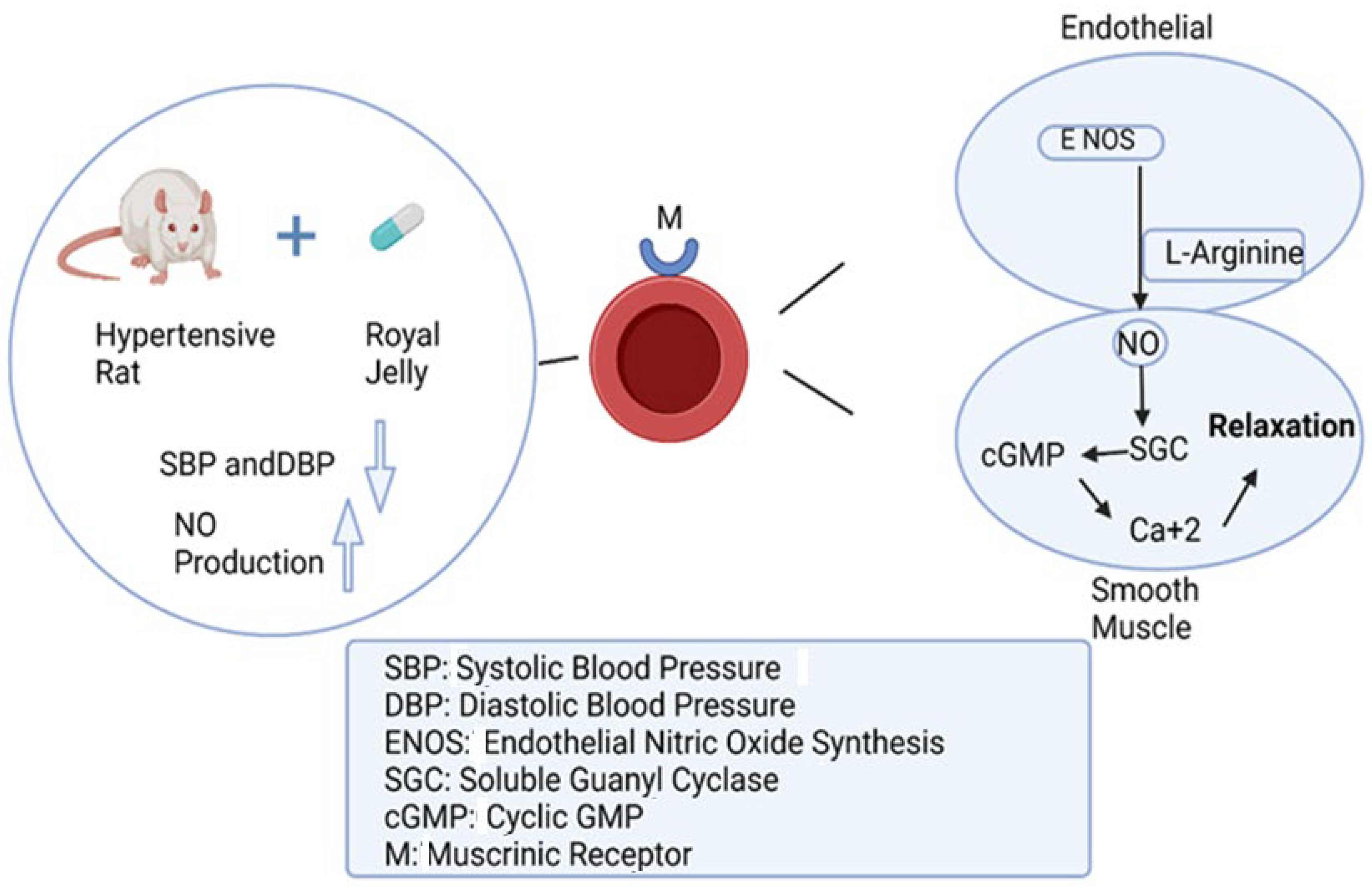
5.1. Antihypertensive Activity of Royal Jelly

The biggest risk factor for cardiovascular disease around the globe is hypertension, which is caused by a disturbance in the contractile or proliferative function of the vascular smooth muscle cells of blood vessels [99].

5.1.1. Preclinical Studies

Previous research has demonstrated that peptides isolated from RJ at doses of 1 g/kg considerably reduced the high blood pressure of hypertensive rats after 10 weeks of administration. The evidential effect was explained by the down-expression of angiotensin-1-converting enzyme, the main regulator of blood pressure [100]. The protein content of RJ represents half of its dry weight, while a major RJ protein, namely MRJP1-9, represents 80% of its total protein content [101]. A recent in vitro study revealed that incubation of aortic vascular smooth muscle cell lines from mice with MRJP1 showed a significant reduction in the cellular α-smooth muscle actin protein, the marker responsible for hypertension [102]. A similar in vivo study conducted on experimentally induced hypertension in Wistar rats using angiotensin-converting enzyme revealed that administration of RJ at a dosage of 15 mg/kg every day for four weeks to hypertensive rats suppressed their increases in blood pressure [103]. Another preclinical study on rats and rabbits showed that RJ exhibited a hypotensive impact via increased nitric oxide production. The study also demonstrated vasodilation effects through the suppression of the cyclic guanosine monophosphate pathway that mediates the contraction and relaxation of vascular smooth muscle cells [104]. Other studies suggested that a muscarinic receptor agonist, like acetylcholine, might be one of the vasodilators in RJ. In fact, it has already been noted that RJ contains more than 900 µg/g of acetylcholine-like substances. Additionally, acetylcholine stimulates the release of endothelium-derived relaxing factors (EDRFs), including nitric oxide (NO), prostacyclin (PGI2), and endothelium-derived hyperpolarizing factor (EDHF), by the vascular endothelial cells. They are the primary mediators of the vasorelaxant effects that are endothelium dependent [104,105].

5.1.2. Clinical Studies

At the clinical level, a randomized, placebo-controlled study demonstrated that a daily intake of RJ tablets (690 mg) for four weeks significantly improved the vascular endothelial activity of the participants’ blood vessels, suggesting that RJ may exert anti-atherogenic activity [106]. Another clinical study reported that the treatment of renal failure patients suffering from cardiovascular disease with a daily dosage of RJ (3600 mg) for a year considerably attenuated the progression of atherosclerosis in hemodialysis patients [15]. Figure 5 illustrates the mechanism of the antihypertensive activity of RJ.

5.2. Hypo-Cholesterolemic Activity of Royal Jelly

Cholesterol plays an important role in several physiological activities inside the body; nevertheless, increased levels in serum cause serious health problems, including cardiovascular disorders [107]. Previous studies reported that providing a diet containing 5% RJ to mice for seven days markedly reduced cholesterol levels in the blood through down-expression of squalene epoxidase, which is essential for the biosynthesis of cholesterol [108]. Animal experimentation proved that MRJP1 has anti-cholesterolemic potential through its interaction with bile acids that increase cholesterol catabolism in the liver and excretion of cholesterol in feces, compared to a β-sitosterol drug [109].
In a placebo-controlled trial, daily use of RJ capsules containing 350 mg RJ for three months was found to significantly alter low-density lipoprotein and total cholesterol levels [110]. The study’s findings led the authors to the conclusion that RJ consumption significantly increased the levels of dehydroepiandrosterone sulphate (DHEA-S). By significantly raising the concentration of DHEA-S over the course of three months, nine RJ capsules taken daily might significantly reduce the levels of LDL-c and total cholesterol (TC) in the serum. In addition, the investigations revealed that DHEA-S can affect the activity of glycerol 3-phosphate dehydrogenase and glucose 6-phosphate dehydrogenase, which in turn can stop the creation of NADPH and hence prevent the biosynthesis of fatty acids, phospholipids, and cholesterol. The hypo-cholesterolemic potential of RJ has been confirmed by a meta-analysis study that supported the notion that RJ reduces total cholesterol levels while also increasing high-density lipoproteins and accordingly regulating the lipid profile [16]. According to Balan et al. (2020), atherosclerosis and cardiovascular diseases related to post-menopausal symptoms in women were found to be inhibited with the regular intake of RJ [111].

6. Conclusions and Future Prospects

The beneficial impact of RJ on diabetes, gastrointestinal ailments, and cardiovascular disease is well-documented in the research articles reviewed. These findings, derived from both preclinical and clinical studies, highlight the potential of RJ as a promising intervention in the field of metabolic health. Furthermore, RJ may enhance the treatment of these disorders by mitigating the adverse effects linked with the drugs used for managing diabetes, gastrointestinal, and cardiovascular conditions. However, despite these promising findings, the specific mechanisms through which RJ exerts its therapeutic effects remain a subject of ongoing research. This underscores the need for further scientific investigations to validate the therapeutic efficacy of RJ and to fully understand its role in disease management. As we move forward, it is crucial to continue exploring the potential of RJ in treating metabolic disorders, with a focus on elucidating its mechanisms of action and potential applications in personalized medicine.

Author Contributions

Conceptualization, H.R.E.-S. and S.A.M.K.; methodology, S.S., A.A.A.E.-W., H.R.E.-S. and S.A.M.K.; software, A.A.A.E.-W. and S.A.M.K.; writing of the original draft, S.S., A.A.A.E.-W. and S.A.M.K.; revision and editing of the final version, S.A.M.K., Z.G., A.D.M., M.D., C.L., X.G., D.G.B., H.R.E.-S. and K.W.; supervision, H.R.E.-S. and K.W.; project administration, H.R.E.-S. All authors have read and agreed to the published version of the manuscript.

Funding

This research received no external funding.

Acknowledgments

H.R. El-Seedi thanks Jiangsu University for the Adjunct Professor fellowship.

Conflicts of Interest

The authors declare that the research was conducted in the absence of any commercial or financial relationships that could be construed as a potential conflict of interest.

References

  1. Han, T.S.; Lean, M.E.J. Metabolic Syndrome. Medicine 2011, 39, 24–31. [Google Scholar] [CrossRef]
  2. Robbins, G.R.; Wen, H.; Ting, J.P.Y. Inflammasomes and Metabolic Disorders: Old Genes in Modern Diseases. Mol. Cell 2014, 54, 297–308. [Google Scholar] [CrossRef] [PubMed]
  3. Chait, A.; den Hartigh, L.J. Adipose Tissue Distribution, Inflammation and Its Metabolic Consequences, Including Diabetes and Cardiovascular Disease. Front. Cardiovasc. Med. 2020, 7, 181–191. [Google Scholar] [CrossRef] [PubMed]
  4. Rinaldi, L.; Pafundi, P.C.; Galiero, R.; Caturano, A.; Morone, M.V.; Silvestri, C.; Giordano, M.; Salvatore, T.; Sasso, F.C. Mechanisms of Non-Alcoholic Fatty Liver Disease in the Metabolic Syndrome. A Narrative Review. Antioxidants 2021, 10, 270. [Google Scholar] [CrossRef] [PubMed]
  5. Heindel, J.J.; Blumberg, B.; Cave, M.; Machtinger, R.; Mantovani, A.; Mendez, M.A.; Nadal, A.; Palanza, P.; Panzica, G.; Sargis, R.; et al. Metabolism Disrupting Chemicals and Metabolic Disorders. Reprod. Toxicol. 2017, 68, 3–33. [Google Scholar] [CrossRef] [PubMed]
  6. Korivi, M.; Liu, B.R. New Strategies from Natural Materials to Fight against Diet-Induced Metabolic Disorders (Part–II). Curr. Pharm. Des. 2021, 27, 761–762. [Google Scholar] [CrossRef] [PubMed]
  7. Burant, C.F.; Viswanathan, P.; Marcinak, J.; Cao, C.; Vakilynejad, M.; Xie, B.; Leifke, E. TAK-875 versus Placebo or Glimepiride in Type 2 Diabetes Mellitus: A Phase 2, Randomised, Double-Blind, Placebo-Controlled Trial. Lancet 2012, 379, 1403–1411. [Google Scholar] [CrossRef] [PubMed]
  8. Tabatabaei-Malazy, O.; Larijani, B.; Abdollahi, M. Targeting Metabolic Disorders by Natural Products. J. Diabetes Metab. Disord. 2015, 14, 57–77. [Google Scholar] [CrossRef] [PubMed]
  9. Wang, H.; Zhu, C.; Ying, Y.; Luo, L.; Huang, D.; Luo, Z. Metformin and Berberine, Two Versatile Drugs in Treatment of Common Metabolic Diseases. Oncotarget 2018, 9, 10135–10146. [Google Scholar] [CrossRef]
  10. Zhao, J.D.; Yu, C.J.; Li, J.Y.; Wang, S.H.; Yang, D.; Guo, C.L.; Du, X.; Zhang, W.J.; Cheng, R.D.; Diao, X.C.; et al. Effect of Berberine on Hyperglycaemia and Gut Microbiota Composition in Type 2 Diabetic Goto-Kakizaki Rats. World J. Gastroenterol. 2021, 27, 708–724. [Google Scholar] [CrossRef]
  11. Asgari, M.; Asle-Rousta, M.; Sofiabadi, M. Effect of Royal Jelly on Blood Glucose and Lipids in Streptozotocin Induced Type 1 Diabetic Rats. Arak Med. Univ. J. 2017, 20, 48–56. [Google Scholar]
  12. Pourmoradian, S.; Mahdavi, R.; Mobasseri, M.; Faramarzi, E.; Mobasseri, M. Effects of Royal Jelly Supplementation on Body Weight and Dietary Intake in Type 2 Diabetic Females. Health Promot. Perspect. 2012, 2, 231–235. [Google Scholar] [CrossRef]
  13. Duran, Y.; Karaboğa, İ.; Polat, F.R.; Polat, E.; Erboğa, Z.F.; Ovalı, M.A.; Öztopuz, R.Ö.; Çelikkol, A.; Yılmaz, A. Royal Jelly Attenuates Gastric Mucosal Injury in a Rat Ethanol-Induced Gastric Injury Model. Mol. Biol. Rep. 2020, 47, 8867–8879. [Google Scholar] [CrossRef]
  14. Miyauchi-Wakuda, S.; Kagota, S.; Maruyama-Fumoto, K.; Wakuda, H.; Yamada, S.; Shinozuka, K. Effect of Royal Jelly on Mouse Isolated Ileum and Gastrointestinal Motility. J. Med. Food 2019, 22, 789–796. [Google Scholar] [CrossRef]
  15. Ohba, K.; Miyata, Y.; Shinzato, T.; Funakoshi, S.; Maeda, K.; Matsuo, T.; Mitsunari, K.; Mochizuki, Y.; Nishino, T.; Sakai, H. Effect of Oral Intake of Royal Jelly on Endothelium Function in Hemodialysis Patients: Study Protocol for Multicenter, Double-Blind, Randomized Control Trial. Trials 2021, 22, 950–957. [Google Scholar] [CrossRef]
  16. Hadi, A.; Najafgholizadeh, A.; Aydenlu, E.S.; Shafiei, Z.; Pirivand, F.; Golpour, S.; Pourmasoumi, M. Royal Jelly Is an Effective and Relatively Safe Alternative Approach to Blood Lipid Modulation: A Meta-Analysis. J. Funct. Foods 2018, 41, 202–209. [Google Scholar] [CrossRef]
  17. Bagameri, L.; Baci, G.-M.; Dezmirean, D.S. Royal Jelly as a Nutraceutical Natural Product with a Focus on Its Antibacterial Activity. Pharmaceutics 2022, 14, 1142. [Google Scholar] [CrossRef]
  18. Khalifa, S.A.M.; Elashal, M.; Kieliszek, M.; Ghazala, N.E.; Farag, M.A.; Saeed, A.; Sabir, J.S.M.; Battino, M.; Xiao, J.; Zou, X.; et al. Recent Insights into Chemical and Pharmacological Studies of Bee Bread. Trends Food Sci. Technol. 2020, 97, 300–316. [Google Scholar] [CrossRef]
  19. El-Seedi, H.; El-Wahed, A.A.; Yosri, N.; Musharraf, S.G.; Chen, L.; Moustafa, M.; Zou, X.; Al-Mousawi, S.; Guo, Z.; Khatib, A.; et al. Antimicrobial Properties of Apis Mellifera’s Bee Venom. Toxins 2020, 12, 451. [Google Scholar] [CrossRef] [PubMed]
  20. El-Wahed, A.A.A.; Farag, M.A.; Eraqi, W.A.; Mersal, G.A.M.; Zhao, C.; Khalifa, S.A.M.; El-Seedi, H.R. Unravelling the Beehive Air Volatiles Profile as Analysed via Solid-Phase Microextraction (SPME) and Chemometrics. J. King Saud Univ. Sci. 2021, 33, 101449–101456. [Google Scholar] [CrossRef]
  21. Saeedi, P.; Petersohn, I.; Salpea, P.; Malanda, B.; Karuranga, S.; Unwin, N.; Colagiuri, S.; Guariguata, L.; Motala, A.A.; Ogurtsova, K.; et al. Global and Regional Diabetes Prevalence Estimates for 2019 and Projections for 2030 and 2045: Results from the International Diabetes Federation Diabetes Atlas, 9th Edition. Diabetes Res. Clin. Pract. 2019, 157, 107843. [Google Scholar] [CrossRef]
  22. Heron, M. Deaths: Leading Causes for 2016. Natl. Vital Stat. Rep. 2018, 67, 1–77. [Google Scholar]
  23. Van Cauwenberghe, J.; Enzlin, P.; Nefs, G.; Ruige, J.; Hendrieckx, C.; De Block, C.; Pouwer, F. Prevalence of and Risk Factors for Sexual Dysfunctions in Adults with Type 1 or Type 2 Diabetes: Results from Diabetes MILES–Flanders. Diabet. Med. 2022, 39, e14676. [Google Scholar] [CrossRef]
  24. Bin Rakhis, S.A.; AlDuwayhis, N.M.; Aleid, N.; AlBarrak, A.N.; Aloraini, A.A. Glycemic Control for Type 2 Diabetes Mellitus Patients: A Systematic Review. Cureus 2022, 14, 6–13. [Google Scholar] [CrossRef] [PubMed]
  25. Chamberlain, R.C.; Fleetwood, K.; Wild, S.H.; Colhoun, H.M.; Lindsay, R.S.; Petrie, J.R.; McCrimmon, R.J.; Gibb, F.; Philip, S.; Sattar, N.; et al. Foot Ulcer and Risk of LowerLimb Amputation or Death in People with Diabetes: A National Population-Based Retrospective Cohort Study. Diabetes Care 2022, 45, 83–91. [Google Scholar] [CrossRef]
  26. McEwan, P.; Morgan, A.R.; Boyce, R.; Bergenheim, K.; Gause-Nilsson, I.A.M.; Bhatt, D.L.; Leiter, L.A.; Johansson, P.A.; Mosenzon, O.; Cahn, A.; et al. The Cost-Effectiveness of Dapagliflozin in Treating High-Risk Patients with Type 2 Diabetes Mellitus: An Economic Evaluation Using Data from the DECLARE-TIMI 58 Trial. Diabetes Obes. Metab. 2021, 23, 1020–1029. [Google Scholar] [CrossRef] [PubMed]
  27. Switanek, M.; Crailsheim, K.; Truhetz, H.; Brodschneider, R. Modelling Seasonal Effects of Temperature and Precipitation on Honey Bee Winter Mortality in a Temperate Climate. Sci. Total Environ. 2017, 579, 1581–1587. [Google Scholar] [CrossRef] [PubMed]
  28. Yoshida, M.; Hayashi, K.; Watadani, R.; Okano, Y.; Tanimura, K.; Kotoh, J.; Sasaki, D.; Matsumoto, K.; Maeda, A. Royal Jelly Improves Hyperglycemia in Obese/Diabetic KK-Ay Mice. J. Vet. Med. Sci. 2017, 79, 299–307. [Google Scholar] [CrossRef] [PubMed]
  29. Ghanbari, E.; Nejati, V.; Khazaei, M. Antioxidant and Protective Effects of Royal Jelly on Histopathological Changes in Testis of Diabetic Rats. Int. J. Reprod. Biomed. 2016, 14, 511–518. [Google Scholar] [CrossRef]
  30. Ghanbari, E.; Nejati, V.; Khazaei, M. Improvement in Serum Biochemical Alterations and Oxidative Stress of Liver and Pancreas Following Use of Royal Jelly in Streptozotocin-Induced Diabetic Rat. Cell J. 2016, 18, 362–370. [Google Scholar] [PubMed]
  31. Khoshpey, B.; Djazayeri, S.; Amiri, F.; Malek, M.; Hosseini, A.F.; Hosseini, S.; Shidfar, S.; Shidfar, F. Effect of Royal Jelly Intake on Serum Glucose, Apolipoprotein A-I (ApoA-I), Apolipoprotein B (ApoB) and ApoB/ApoA-I Ratios in Patients with Type 2 Diabetes: A Randomized, Double-Blind Clinical Trial Study. Can. J. Diabetes 2016, 40, 324–328. [Google Scholar] [CrossRef]
  32. Pourmoradian, S.; Mahdavi, R.; Mobasseri, M.; Faramarzi, E.; Mobasseri, M. Effects of Royal Jelly Supplementation on Glycemic Control and Oxidative Stress Factors in Type 2 Diabetic Female: A Randomized Clinical Trial. Chin. J. Integr. Med. 2014, 20, 347–352. [Google Scholar] [CrossRef]
  33. Ghanbari, E.; Nejati, V.; Najafi, G.; Khazaei, M.; Babaei, M. Study on the Effect of Royal Jelly on Reproductive Parameters in Streptozotocin-Induced Diabetic Rats. Int. J. Fertil. Steril. 2015, 9, 113–120. [Google Scholar]
  34. Karaca, T.; Demirtaş, S.; Karaboğa, İ.; Ayvaz, S. Protective Effects of Royal Jelly against Testicular Damage in Streptozotocin-Induced Diabetic Rats. Turk. J. Med. Sci. 2015, 45, 27–32. [Google Scholar] [CrossRef]
  35. Nohair, S.F. Al Antidiabetic Efficacy of a Honey-Royal Jelly Mixture: Biochemical Study in Rats. Int. J. Health Sci. 2021, 15, 4–9. [Google Scholar]
  36. Macadangdang, R.R., Jr.; Orodio, A.L.T.; Calabio, A.G.T.; Miciano, K.B.V.; Coleen, A.L.T. Hypoglycemic Effect of Moringa Oliefera Lam. Seed Oil Extract and Royal Jelly Composite Mixture in Alloxan-Induced Diabetes in Male Albino Wistar Rats. Asian, J. Biol. Life Sci. 2021, 10, 159–164. [Google Scholar] [CrossRef]
  37. Yoneshiro, T.; Kaede, R.; Nagaya, K.; Aoyama, J.; Saito, M.; Okamatsu-Ogura, Y.; Kimura, K.; Terao, A. Royal Jelly Ameliorates Diet-Induced Obesity and Glucose Intolerance by Promoting Brown Adipose Tissue Thermogenesis in Mice. Obes. Res. Clin. Pract. 2016, 10, 127–137. [Google Scholar] [CrossRef] [PubMed]
  38. Watadani, R.; Kotoh, J.; Sasaki, D.; Someya, A.; Matsumoto, K.; Maeda, A. 10-Hydroxy-2-Decenoic Acid, a Natural Product, Improves Hyperglycemia and Insulin Resistance in Obese/Diabetic KK-Ay Mice, but Does Not Prevent Obesity. J. Vet. Med. Sci. 2017, 79, 1596–1602. [Google Scholar] [CrossRef]
  39. Hu, X.; Liu, Z.; Lu, Y.; Chi, X.; Han, K.; Wang, H.; Wang, Y.; Ma, L.; Xu, B. Glucose Metabolism Enhancement by 10-Hydroxy- 2-Decenoic Acid via the PI3K/AKT Signaling Pathway in High-Fat-Diet/Streptozotocin Induced Type 2 Diabetic Mice. Food Funct. 2022, 13, 9931–9946. [Google Scholar] [CrossRef]
  40. You, S.; Xiang, Y.; Qi, X.; Mao, R.; Cai, E.; Lan, Y.; Lu, H.; Shen, J.; Deng, H. Harnessing a Biopolymer Hydrogel Reinforced by Copper / Tannic Acid Nanosheets for Treating Bacteria-Infected Diabetic Wounds. Mater. Today Adv. 2022, 15, 100271–100286. [Google Scholar] [CrossRef]
  41. Zhou, J.; Wang, Z.; Yang, C.; Zhang, H.; Yan, W.; Wang, K. A Carrier-Free, Dual-Functional Hydrogel Constructed of Antimicrobial Peptide Jelleine-1 and 8Br-CAMP for MRSA Infected Diabetic Wound Healing. Acta Biomater. J. 2022, 151, 223–234. [Google Scholar] [CrossRef]
  42. Jayaraman, R.; Subramani, S.; Sheik Abdullah, S.H.; Udaiyar, M. Antihyperglycemic Effect of Hesperetin, a Citrus Flavonoid, Extenuates Hyperglycemia and Exploring the Potential Role in Antioxidant and Antihyperlidemic in Streptozotocin-Induced Diabetic Rats. Biomed. Pharmacother. 2018, 97, 98–106. [Google Scholar] [CrossRef]
  43. López-Gutiérrez, N.; Aguilera-Luiz, M.D.M.; Romero-González, R.; Vidal, J.L.M.; Garrido Frenich, A. Fast Analysis of Polyphenols in Royal Jelly Products Using Automated TurboFlowTM-Liquid Chromatography-Orbitrap High Resolution Mass Spectrometry. J. Chromatogr. B Anal. Technol. Biomed. Life Sci. 2014, 973, 17–28. [Google Scholar] [CrossRef]
  44. Singh, P.; Bansal, S.; Kuhad, A.; Kumar, A.; Chopra, K. Naringenin Ameliorates Diabetic Neuropathic Pain by Modulation of Oxidative-Nitrosative Stress, Cytokines and MMP-9 Levels. Food Funct. 2020, 11, 4548–4560. [Google Scholar] [CrossRef] [PubMed]
  45. Wojnar, W.; Zych, M.; Kaczmarczyk-Sedlak, I. Antioxidative Effect of Flavonoid Naringenin in the Lenses of Type 1 Diabetic Rats. Biomed. Pharmacother. 2018, 108, 974–984. [Google Scholar] [CrossRef] [PubMed]
  46. Ortiz-Andrade, R.R.; Sánchez-Salgado, J.C.; Navarrete-Vázquez, G.; Webster, S.P.; Binnie, M.; García-Jiménez, S.; León-Rivera, I.; Cigarroa-Vázquez, P.; Villalobos-Molina, R.; Estrada-Soto, S. Antidiabetic and Toxicological Evaluations of Naringenin in Normoglycaemic and NIDDM Rat Models and Its Implications on Extra-Pancreatic Glucose Regulation. Diabetes Obes. Metab. 2008, 10, 1097–1104. [Google Scholar] [CrossRef]
  47. Kumar, B.; Gupta, S.K.; Srinivasan, B.P.; Nag, T.C.; Srivastava, S.; Saxena, R. Hesperetin Ameliorates Hyperglycemia Induced Retinal Vasculopathy via Anti-Angiogenic Effects in Experimental Diabetic Rats. Vascul. Pharmacol. 2012, 57, 201–207. [Google Scholar] [CrossRef] [PubMed]
  48. Yang, R.; Jia, Q.; Mehmood, S.; Ma, S.; Liu, X. Genistein Ameliorates Inflammation and Insulin Resistance through Mediation of Gut Microbiota Composition in Type 2 Diabetic Mice. Eur. J. Nutr. 2021, 60, 2155–2168. [Google Scholar] [CrossRef]
  49. Fu, Z.; Zhang, W.; Zhen, W.; Lum, H.; Nadler, J.; Bassaganya-Riera, J.; Jia, Z.; Wang, Y.; Misra, H.; Liu, D. Genistein Induces Pancreatic β-Cell Proliferation through Activation of Multiple Signaling Pathways and Prevents Insulin-Deficient Diabetes in Mice. Endocrinology 2010, 151, 3026–3037. [Google Scholar] [CrossRef]
  50. Li, R.Z.; Ding, X.W.; Geetha, T.; Al-Nakkash, L.; Broderick, T.L.; Babu, J.R. Beneficial Effect of Genistein on Diabetes-Induced Brain Damage in the OB/OB Mouse Model. Drug Des. Devel. Ther. 2020, 14, 3325–3336. [Google Scholar] [CrossRef]
  51. Qnais, E.; Alqudah, A.; Wedyan, M.; Gammoh, O.; Alkhateeb, H.; Alnoaimi, M. Formononetin Suppresses Hyperglycaemia through Activation of GLUT4-AMPK Pathway. Pharmacia 2023, 70, 527–536. [Google Scholar] [CrossRef]
  52. Oza, M.J.; Kulkarni, Y.A. Formononetin Ameliorates Diabetic Neuropathy by Increasing Expression of SIRT1 and NGF. Chem. Biodivers. 2020, 17, e2000162. [Google Scholar] [CrossRef] [PubMed]
  53. Zywno, H.; Bzdega, W.; Kolakowski, A.; Kurzyna, P.; Harasim-Symbor, E.; Sztolsztener, K.; Chabowski, A.; Konstantynowicz-Nowicka, K. The Influence of Coumestrol on Sphingolipid Signaling Pathway and Insulin Resistance Development in Primary Rat Hepatocytes. Biomolecules 2021, 11, 268. [Google Scholar] [CrossRef] [PubMed]
  54. Kim, S.M.; Imm, J.Y. The Effect of Chrysin-Loaded Phytosomes on Insulin Resistance and Blood Sugar Control in Type 2 Diabetic Db/Db Mice. Molecules 2020, 25, 5503. [Google Scholar] [CrossRef] [PubMed]
  55. Salama, A.; Asaad, G.; Shaheen, A. Chrysin Ameliorates STZ-Induced Diabetes in Rats: Possible Impact of Modulation of TLR4/NF-Κβ Pathway. Res. Pharm. Sci. 2022, 17, 1–11. [Google Scholar] [CrossRef]
  56. Honda, Y.; Araki, Y.; Hata, T.; Ichihara, K.; Ito, M.; Tanaka, M.; Honda, S. 10-Hydroxy-2-Decenoic Acid, the Major Lipid Component of Royal Jelly, Extends the Lifespan of Caenorhabditis Elegans through Dietary Restriction and Target of Rapamycin Signaling. J. Aging Res. 2015, 2015, 425261. [Google Scholar] [CrossRef]
  57. Shidfar, F.; Jazayeri, S.; Mousavi, S.N.; Malek, M.; Hosseini, A.F.; Khoshpey, B. Does Supplementation with Royal Jelly Improve Oxidative Stress and Insulin Resistance in Type 2 Diabetic Patients? Iran. J. Public Health 2015, 44, 797–803. [Google Scholar]
  58. Siavash, M.; Shokri, S.; Haghighi, S.; Mohammadi, M.; Shahtalebi, M.A.; Farajzadehgan, Z. The Efficacy of Topical Royal Jelly on Diabetic Foot Ulcers Healing: A Case Series. J. Res. Med. Sci. 2011, 16, 904–909. [Google Scholar]
  59. Boukraĝ, L.; Meslem, A.; Benhanifia, M.; Hammoudi, S.M. Synergistic Effect of Starch and Royal Jelly against Staphylococcus Aureus and Escherichia Coli. J. Altern. Complement. Med. 2009, 15, 755–757. [Google Scholar] [CrossRef]
  60. Yakoot, M.; Abdelatif, M.; Helmy, S. Efficacy of a New Local Limb Salvage Treatment for Limb-Threatening Diabetic Foot Wounds–A Randomized Controlled Study. Diabetes Metab. Syndr. Obes. Targets Ther. 2019, 12, 1659–1665. [Google Scholar] [CrossRef]
  61. Aljafary, M.A.; Al-suhaimi, E.A. Adiponectin System (Rescue Hormone): The Missing Link between Metabolic and Cardiovascular Diseases. Pharmaceutics 2022, 14, 1430. [Google Scholar] [CrossRef]
  62. Geagea, A.G.; Mallat, S.; Matar, C.F.; Zerbe, R.; Filfili, E.; Francis, M.; Haidar, H.; Jurjus, A. Adiponectin and Inflammation in Health and Disease: An Update. Open Med. J. 2018, 5, 20–32. [Google Scholar] [CrossRef]
  63. Maleki, V.; Jafari-Vayghan, H.; Saleh-Ghadimi, S.; Adibian, M.; Kheirouri, S.; Alizadeh, M. Effects of Royal Jelly on Metabolic Variables in Diabetes Mellitus: A Systematic Review. Complement. Ther. Med. 2019, 43, 20–27. [Google Scholar] [CrossRef] [PubMed]
  64. Joshi, T.; Singh, A.K.; Haratipour, P.; Sah, A.N.; Pandey, A.K.; Naseri, R.; Juyal, V.; Farzaei, M.H. Targeting AMPK Signaling Pathway by Natural Products for Treatment of Diabetes Mellitus and Its Complications. J. Cell. Physiol. 2019, 234, 17212–17231. [Google Scholar] [CrossRef] [PubMed]
  65. Ananthakrishnan, A.N.; Xavier, R.J. Gastrointestinal Diseases. In Hunter’s Tropical Medicine and Emerging Infectious Diseases; Elsevier: Amsterdam, The Netherlands, 2020; pp. 16–26. [Google Scholar]
  66. Sperber, A.D.; Bangdiwala, S.I.; Drossman, D.A.; Ghoshal, U.C.; Simren, M.; Tack, J.; Whitehead, W.E.; Dumitrascu, D.L.; Fang, X.; Fukudo, S.; et al. Worldwide Prevalence and Burden of Functional Gastrointestinal Disorders, Results of Rome Foundation Global Study. Gastroenterology 2021, 160, 99–114.e3. [Google Scholar] [CrossRef] [PubMed]
  67. De Filippo, C.; Di Paola, M.; Ramazzotti, M.; Albanese, D.; Pieraccini, G.; Banci, E.; Miglietta, F.; Cavalieri, D.; Lionetti, P. Diet, Environments, and Gut Microbiota. A Preliminary Investigation in Children Living in Rural and Urban Burkina Faso and Italy. Front. Microbiol. 2017, 8, 1979–1983. [Google Scholar] [CrossRef] [PubMed]
  68. Rizzello, F.; Spisni, E.; Giovanardi, E.; Imbesi, V.; Salice, M.; Alvisi, P.; Valerii, M.C.; Gionchetti, P. Implications of the Westernized Diet in the Onset and Progression of IBD. Nutrients 2019, 11, 1033. [Google Scholar] [CrossRef]
  69. Vona, R.; Pallotta, L.; Cappelletti, M.; Severi, C.; Matarrese, P. The Impact of Oxidative Stress in Human Pathology: Focus on Gastrointestinal Disorders. Antioxidants 2021, 10, 201. [Google Scholar] [CrossRef]
  70. Proestos, C. Superfoods: Recent Data on Their Role in the Prevention of Diseases. Curr. Res. Nutr. Food Sci. 2018, 6, 576–593. [Google Scholar] [CrossRef]
  71. Mostafa, R.E.; El-Marasy, S.A.; Abdel Jaleel, G.A.; Bakeer, R.M. Protective Effect of Royal Jelly against Diclofenac-Induced Hepato-Renal Damage and Gastrointestinal Ulcerations in Rats. Heliyon 2020, 6, e03330. [Google Scholar] [CrossRef]
  72. Ahmad, S.; Campos, M.G.; Fratini, F.; Altaye, S.Z.; Li, J. New Insights into the Biological and Pharmaceutical Properties of Royal Jelly. Int. J. Mol. Sci. 2020, 21, 382. [Google Scholar] [CrossRef]
  73. Li, S.; Tao, L.; Yu, X.; Zheng, H.; Wu, J.H.F. Royal Jelly Proteins and Their Derived Peptides: Preparation, Properties, and Biological Activities. J. Agric. Food Chem. 2021, 69, 14415–14427. [Google Scholar] [CrossRef]
  74. Jairath, V.; Feagan, B.G. Global Burden of Inflammatory Bowel Disease. Lancet Gastroenterol. Hepatol. 2020, 5, 2–3. [Google Scholar] [CrossRef]
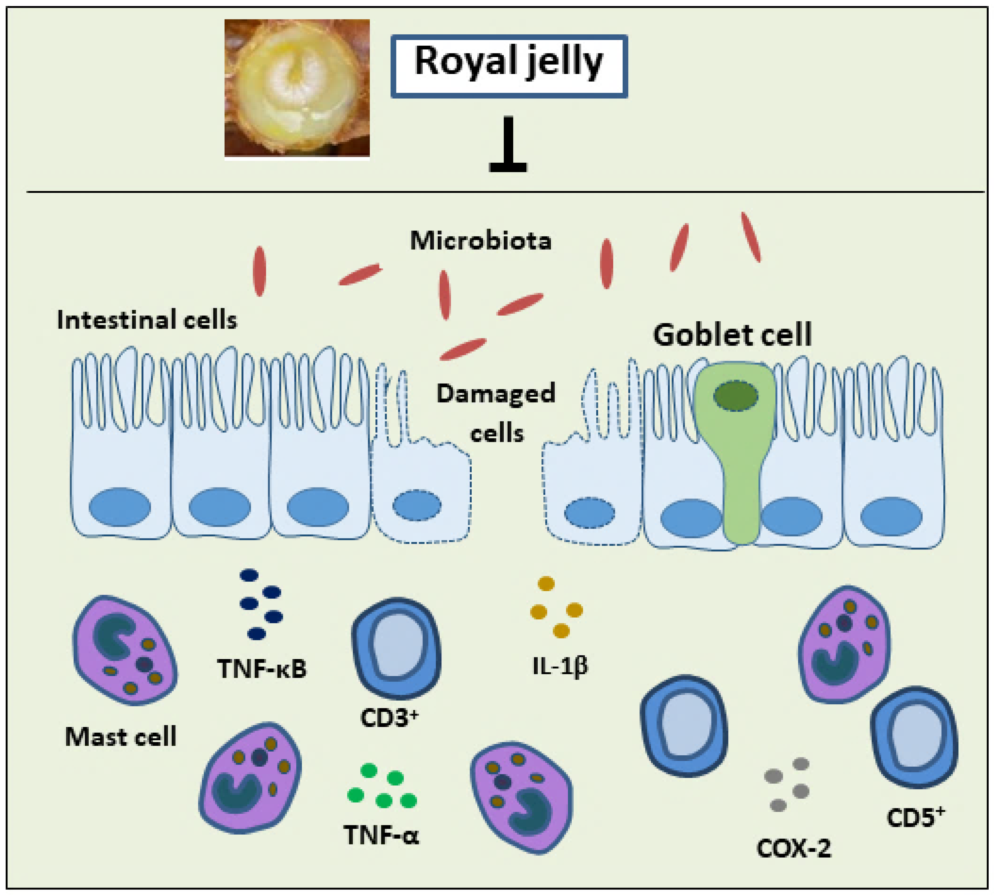
  75. Karaca, T.; Uz, Y.H.; Demirtas, S.; Karaboga, I.; Can, G. Protective Effect of Royal Jelly in 2,4,6 Trinitrobenzene Sulfonic Acid-Induced Colitis in Rats. Iran. J. Basic Med. Sci. 2015, 18, 370–379. [Google Scholar]
  76. Karaca, T.; Bayiroglu, F.; Yoruk, M.; Kaya, M.S.; Uslu, S.; Comba, B.; Mis, L. Effect of Royal Jelly on Experimental Colitis Induced by Acetic Acid and Alteration of Mast Cell Distribution in the Colon of Rats. Eur. J. Histochem. 2010, 54, 158–161. [Google Scholar] [CrossRef]
  77. Karaca, T.; Şimşek, N.; Uslu, S.; Kalkan, Y.; Can, I.; Kara, A.; Yörük, M. The Effect of Royal Jelly on CD3+, CD5+, CD45+ T-Cell and CD68+ Cell Distribution in the Colon of Rats with Acetic Acid-Induced Colitis. Allergol. Immunopathol. 2012, 40, 357–361. [Google Scholar] [CrossRef]
  78. Chi, X.; Liu, Z.; Wang, H.; Wang, Y.; Xu, B.; Wei, W. Regulation of a New Type of Selenium-Rich Royal Jelly on Gut Microbiota Profile in Mice. Biol. Trace Elem. Res. 2022, 200, 1763–1775. [Google Scholar] [CrossRef] [PubMed]
  79. Guo, J.; Ma, B.; Wang, Z.; Chen, Y.; Tian, W.; Dong, Y. Royal Jelly Protected against Dextran-Sulfate-Sodium-Induced Colitis by Improving the Colonic Mucosal Barrier and Gut Microbiota. Nutrients 2022, 14, 2069. [Google Scholar] [CrossRef] [PubMed]
  80. Li, J.; Zhang, W.; Wang, C.; Yu, Q.; Dai, R.; Pei, X. Lactococcus Lactis Expressing Food-Grade β-Galactosidase Alleviates Lactose Intolerance Symptoms in Post-Weaning Balb/c Mice. Appl. Microbiol. Biotechnol. 2012, 96, 1499–1506. [Google Scholar] [CrossRef] [PubMed]
  81. Kavas, N. Functional Probiotic Yoghurt Production with Royal Jelly Fortification and Determination of Some Properties. Int. J. Gastron. Food Sci. 2022, 28, 100519. [Google Scholar] [CrossRef]
  82. Oak, S.J.; Jha, R. The Effects of Probiotics in Lactose Intolerance: A Systematic Review–The Effects of Probiotics in Lactose Intolerance: A Systematic Review. Crit. Rev. Food Sci. Nutr. 2018, 8398, 1425977. [Google Scholar] [CrossRef]
  83. Hassan, A.A.M.; Elenany, Y.E.; Nassrallah, A.; Cheng, W.; Abd El-Maksoud, A.A. Royal Jelly Improves the Physicochemical Properties and Biological Activities of Fermented Milk with Enhanced Probiotic Viability. LWT 2022, 155, 112912. [Google Scholar] [CrossRef]
  84. Vazhacharickal, J.P. A Review on Health Benefits and Biological Action of Honey, Propolis and Royal Jelly. J. Med. Plants Stud. 2021, 9, 1–13. [Google Scholar]
  85. Ali, M.W.; Sabir, A.M.; Gadour, M.O. Gum Arabic in Treatment of Functional Constipation in Children in Sudan. Sudan JMS 2012, 8, 73–76. [Google Scholar]
  86. Postali, E.; Peroukidou, P.; Giaouris, E.; Papachristoforou, A. Investigating Possible Synergism in the Antioxidant and Antibacterial Actions of Honey and Propolis from the Greek Island of Samothrace through Their Combined Application. Foods 2022, 11, 2041. [Google Scholar] [CrossRef]
  87. Guo, J.; Wang, Z.; Chen, Y.; Cao, J.; Tian, W.; Ma, B.; Dong, Y. Active Components and Biological Functions of Royal Jelly. J. Funct. Foods 2021, 82, 104514. [Google Scholar] [CrossRef]
  88. Bílikova, K.; Huang, S.-C.; Lin, I.-P.; Šimuth, J.; Peng, C.-C. Structure and Antimicrobial Activity Relationship of Royalisin, an Antimicrobial Peptide from Royal Jelly of Apis Mellifera. Peptides 2015, 86, 190–196. [Google Scholar] [CrossRef]
  89. Chantawannakul, P. From Entomophagy to Entomotherapy. Front. Biosci. 2020, 25, 179–200. [Google Scholar] [CrossRef]
  90. Sofiabadi, M.; Samiee-Rad, F. Royal Jelly Accelerates Healing of Acetate Induced Gastric Ulcers in Male Rats. Gastroenterol. Hepatol. Bed Bench 2020, 13, 18–22. [Google Scholar] [CrossRef]
  91. Abd El-Naeem, A. Effect of Nicotine on the Structure of Gastric Mucosa of Adult Male Albino Rats and the Possible Protective Effect of Royal Jelly (Light and Scanning Electron Microscopic Study). Egypt. J. Histol. 2021, 45, 404–415. [Google Scholar] [CrossRef]
  92. Ahmed, W.M.S.; Khalaf, A.A.; Moselhy, W.A.; Safwat, G.M. Royal Jelly Attenuates Azathioprine Induced Toxicity in Rats. Environ. Toxicol. Pharmacol. 2014, 37, 431–437. [Google Scholar] [CrossRef] [PubMed]
  93. Felemban, A.H.; Alshammari, G.M.; Yagoub, A.E.G.A.; Al-Harbi, L.N.; Alhussain, M.H.; Yahya, M.A. Activation of AMPK Entails the Protective Effect of Royal Jelly against High-Fat-Diet-Induced Hyperglycemia, Hyperlipidemia, and Non-Alcoholic Fatty Liver Disease in Rats. Nutrients 2023, 15, 1471. [Google Scholar] [CrossRef] [PubMed]
  94. Fang, C.; Pan, J.; Qu, N.; Lei, Y.; Han, J.; Zhang, J.; Han, D. The AMPK Pathway in Fatty Liver Disease. Front. Physiol. 2022, 13, 970292. [Google Scholar] [CrossRef]
  95. Bahaaldin-Beygi, M.; Kariminik, A.; Arababadi, M.K. Royal Jelly Significantly Alters Inflammasome Pathways in Patients with Chronic Hepatitis B. Indian J. Exp. Biol. 2022, 60, 875–879. [Google Scholar]
  96. Ezzati, M.; Obermeyer, Z.; Tzoulaki, I.; Mayosi, B.M.; Elliott, P.; Leon, D.A. Contributions of Risk Factors and Medical Care to Cardiovascular Mortality Trends. Nat. Rev. Cardiol. 2015, 12, 508–530. [Google Scholar] [CrossRef] [PubMed]
  97. Townsend, N.; Kazakiewicz, D.; Lucy Wright, F.; Timmis, A.; Huculeci, R.; Torbica, A.; Gale, C.P.; Achenbach, S.; Weidinger, F.; Vardas, P. Epidemiology of Cardiovascular Disease in Europe. Nat. Rev. Cardiol. 2022, 19, 133–143. [Google Scholar] [CrossRef]
  98. Aslan, A.; Beyaz, S.; Gok, O.; Can, M.I.; Parlak, G.; Ozercan, I.H.; Gundogdu, R. Royal Jelly Abrogates Flouride-Induced Oxidative Damage in Rat Heart Tissue by Activating of the Nrf-2/NF-ΚB and Bcl-2/Bax Pathway. Toxicol. Mech. Methods 2021, 31, 644–654. [Google Scholar] [CrossRef]
  99. Fan, P.; Han, B.; Feng, M.; Fang, Y.; Zhang, L.; Hu, H.; Hao, Y.; Qi, Y.; Zhang, X.; Li, J. Functional and Proteomic Investigations Reveal Major Royal Jelly Protein 1 Associated with Anti-Hypertension Activity in Mouse Vascular Smooth Muscle Cells. Sci. Rep. 2016, 6, 30230–30241. [Google Scholar] [CrossRef]
  100. Matsui, T.; Yukiyoshi, A.; Doi, S.; Sugimoto, H.; Yamada, H.; Matsumoto, K. Gastrointestinal Enzyme Production of Bioactive Peptides from Royal Jelly Protein and Their Antihypertensive Ability in SHR. J. Nutr. Biochem. 2002, 13, 80–86. [Google Scholar] [CrossRef]
  101. Lin, N.; Chen, S.; Zhang, H.; Li, J.; Fu, L. Quantification of Major Royal Jelly Protein 1 in Fresh Royal Jelly by Ultraperformance Liquid Chromatography-Tandem Mass Spectrometry. J. Agric. Food Chem. 2018, 66, 1270–1278. [Google Scholar] [CrossRef]
  102. Feng, M.; Fang, Y.; Han, B.; Xu, X.; Fan, P.; Hao, Y.; Qi, Y.; Hu, H.; Huo, X.; Meng, L.; et al. In-Depth N-Glycosylation Reveals Species-Specific Modifications and Functions of the Royal Jelly Protein from Western (Apis mellifera) and Eastern Honeybees (Apis cerana). J. Proteome Res. 2015, 14, 5327–5340. [Google Scholar] [CrossRef]
  103. Escamilla, K.I.A.; Ordóñez, Y.B.M.; Sandoval-Peraza, V.M.; Fernández, J.J.A.; Ancona, D.A.B. Anti-Hypertensive Activity in Vitro and in Vivo on Royal Jelly Produced by Different Diets. Emir. J. Food Agric. 2022, 34, 9–15. [Google Scholar] [CrossRef]
  104. Pan, Y.; Rong, Y.; You, M.; Ma, Q.; Chen, M.; Hu, F. Royal Jelly Causes Hypotension and Vasodilation Induced by Increasing Nitric Oxide Production. Food Sci. Nutr. 2019, 7, 1361–1370. [Google Scholar] [CrossRef]
  105. Wei, W.; Wei, M.; Kang, X.; Deng, H.; Lu, Z. A Novel Method Developed for Acetylcholine Detection in Royal Jelly by Using Capillary Electrophoresis Coupled with Electrogenerated Chemiluminescence Based on a Simple Reaction. Electrophoresis 2009, 30, 1949–1952. [Google Scholar] [CrossRef]
  106. Fujisue, K.; Yamamoto, E.; Sueta, D.; Arima, Y.; Hirakawa, K.; Tabata, N.; Ishii, M.; Ito, M.; Yamanaga, K.; Hanatani, S.; et al. A Randomized, Double-Blind Comparison Study of Royal Jelly to Augment Vascular Endothelial Function in Healthy Volunteers. J. Atheroscler. Thromb. 2022, 29, 1285–1294. [Google Scholar] [CrossRef] [PubMed]
  107. Roman, W.A.; Piato, A.L.; Conterato, G.M.M.; Wildner, S.M.; Marcon, M.; Mocelin, R.; Emanuelli, M.P.; Emanuelli, T.; Nepel, A.; Barison, A.; et al. Hypolipidemic Effects of Solidago Chilensis Hydroalcoholic Extract and Its Major Isolated Constituent Quercetrin in Cholesterol-Fed Rats. Pharm. Biol. 2015, 53, 1488–1495. [Google Scholar] [CrossRef] [PubMed]
  108. Kamakura, M.; Moriyama, T.; Sakaki, T. Changes in Hepatic Gene Expression Associated with the Hypocholesterolaemic Activity of Royal Jelly. J. Pharm. Pharmacol. 2010, 58, 1683–1689. [Google Scholar] [CrossRef]
  109. Kashima, Y.; Kanematsu, S.; Asai, S.; Kusada, M.; Watanabe, S.; Kawashima, T.; Nakamura, T.; Shimada, M.; Goto, T.; Nagaoka, S. Identification of a Novel Hypocholesterolemic Protein, Major Royal Jelly Protein 1, Derived from Royal Jelly. PLoS ONE 2014, 9, e105073–e105084. [Google Scholar] [CrossRef] [PubMed]
  110. Chiu, H.F.; Chen, B.K.; Lu, Y.Y.; Han, Y.C.; Shen, Y.C.; Venkatakrishnan, K.; Golovinskaia, O.; Wang, C.K. Hypocholesterolemic Efficacy of Royal Jelly in Healthy Mild Hypercholesterolemic Adults. Pharm. Biol. 2017, 55, 497–502. [Google Scholar] [CrossRef] [PubMed]
  111. Bălan, A.; Moga, M.A.; Dima, L.; Toma, S.; Neculau, A.E.; Anastasiu, C.V. Royal Jelly–A Traditional and Natural Remedy for Postmenopausal Symptoms and Aging-Related Pathologies. Molecules 2020, 25, 3291. [Google Scholar] [CrossRef]
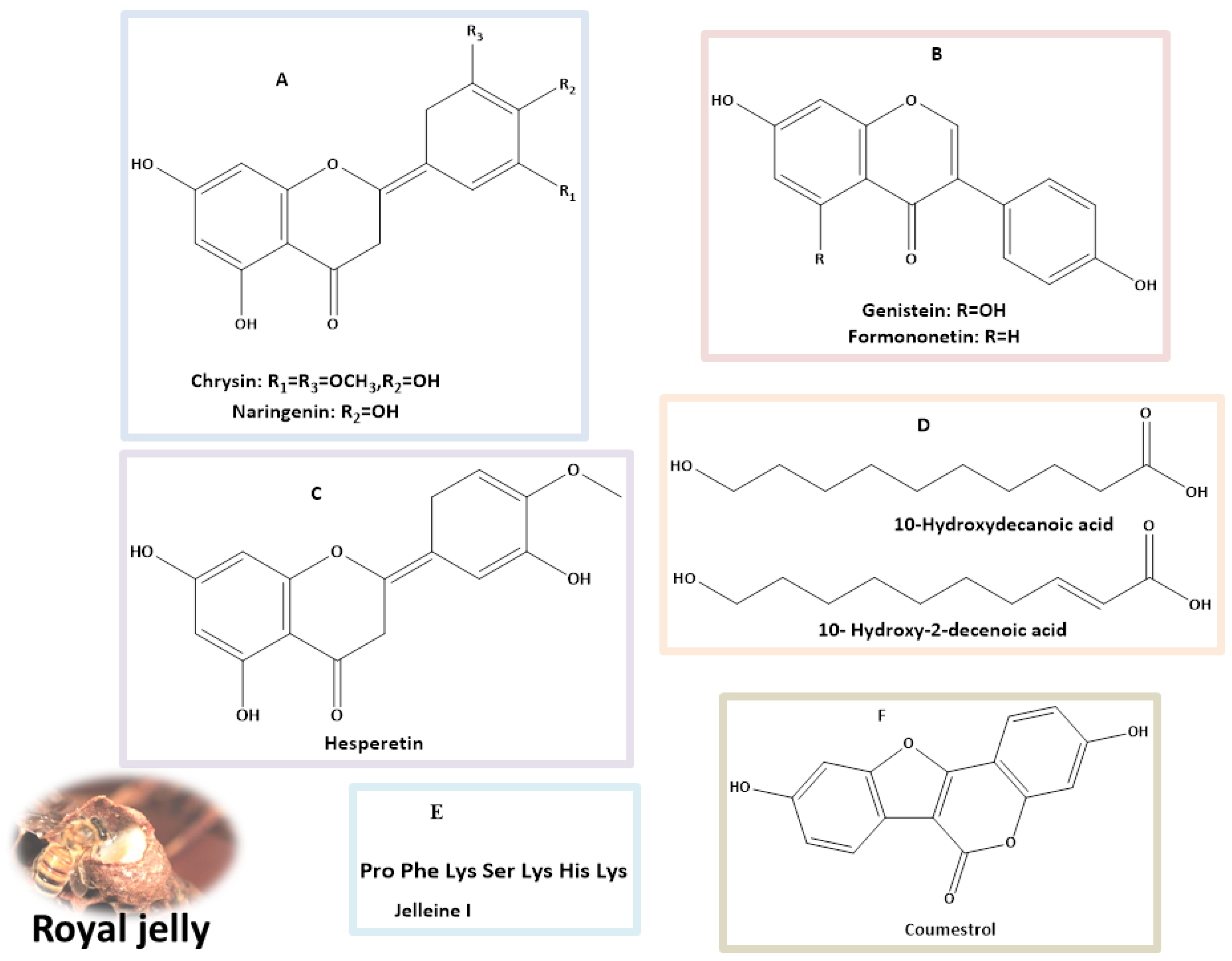
Figure 1. Mechanism of action of royal jelly in diabetes mellitus (DM), preclinical and clinical models [28,29,30,31,32]. LDL: low-density lipoprotein; SOD: superoxide dismutase, IL-6: interlukin-6; MDA: malondialdehyde; GSH-Px: glutathione peroxidase; ApoB/ApoA-I: apolipoprotein B/apolipoprotein A-I; AdipoR1: adiponectin receptor-1; CAT: catalase; p-GSK3β: glycogen synthase kinase 3β; p-AKT: phosphorylated Akt; PI3K: phosphoinositide 3-kinase; IL-1β: interleukin-1β; TNF-α: tumor necrosis factor-α; NF-κB: nuclear factor kappa-B; COX-2: cyclooxygenase-2.
Nutrients 16 00393 g001
Figure 2. Major classes of natural products (AC) flavonoids; (D) fatty acids; (E) peptide; and (F) coumestans identified in royal jelly with potential anti-diabetes properties.
Nutrients 16 00393 g002
Figure 3. Treatment with royal jelly suppressed the rise of CD3+, CD5+, CD8+ and CD45+ T-cells, pro-inflammatory cytokines, IL-1β, TNF–α, and the expression of major inflammatory mediators (COX-2 and NF-κB) in the colon of rats with colitis. IL-1β: interleukin-1β; TNF-α: tumor necrosis factor-α; NF-κB: nuclear factor kappa-B; COX-2: cyclooxygenase-2.
Nutrients 16 00393 g003
Figure 4. Potential activity of royal jelly in the most common gastrointestinal diseases.
Nutrients 16 00393 g004
Figure 5. The mechanism of the antihypertensive activity of royal jelly.
Nutrients 16 00393 g005
Table 1. Bioactive compounds identified from royal jelly as antidiabetic agents.
Identified CompoundsDosageBiological Activity (In Vitro/In Vivo)References
Hesperetin40 mg/kg body weight (BW) for 45 daysReduces high blood sugar and lipid levels by enhancing insulin secretion (in vivo). [42,43]
Naringenin(25, 50, 100 mg/kg) for 4 weeksThe therapy significantly enhanced the control of blood glucose levels and also contributed to the recovery of BWin diabetic rats, in contrast to those that received a vehicle treatment (in vivo).[43,44]
50 mg/kg for 4 weeksAdequate to mitigate the alterations in the lenses caused by diabetes-related oxidative stress (in vivo).[45]
50 mg/kg/day for 5 daysSignificant decrease in blood glucose and triglyceride levels in diabetic rats (in vivo).[46]
100 mg/kg BW /day for 4 weeksRestored the serum insulin and C-peptide levels, replenished liver glycogen, and reduced glucose-6-phosphatase and glycogen phosphorylase activity in the liver. Additionally, it improved the serum lipid profile and strengthened the liver’s antioxidant defense system (in vivo).[47]
Genistein(20 and 40 mg/kg) for 8 weeks.Improved glucose tolerance, blood glucose levels, insulin, glucagon, lipid profiles, and pro-inflammatory factors. It also improved liver function, reduced inflammation in the liver and colon, and positively altered gut microbiota composition (in vivo).[43,48]
25–200 mg/dayImproved hyperglycemia, glucose tolerance, and blood insulin levels, along with enhancing islet beta-cell proliferation, survival, and mass (in vivo).[49]
600 mg/kg for 4 weeksEnhanced insulin sensitivity and increased expression of neurotrophic factors, such as nerve growth factor (NGF) and brain-derived neurotrophic factors (BDNF) (in vivo).[50]
Formononetin20 mg/kg for 28 days Reduced serum glucose levels and increased serum insulin compared to the control group. It also decreased insulin resistance and reduced fasting glucose (C57BL/6 mice, in vivo).[43,51]
40 mg/kg/day for 16 weeksDecreased insulin resistance and regulated hypoglycemia in male rats with diabetes (in vivo).[52]
Coumestrol50 µMImproved hepatic insulin resistance in primary at hepatocyte (in vivo).[43,53]
Chrysin 100 mg/kgIt resulted in a reduction of fasting blood glucose and insulin levels in db/db mice when compared to the control group (in vivo).[43,54]
80 mg/kg BW for 10 daysAnti-diabetic effects via increasing insulin levels, reducing oxidative stress, and regulating the inflammatory pathway (in vivo).[55]
10-Hydroxy-2-decenoic acid100 mg per kg BW/Daily for 4 weeks.Decreased fasting blood glucose and increased insulin levels in diabetic mice. Enhanced activity of crucial antioxidants in the livers of diabetic mice, such as superoxide dismutase, catalase, and glutathione peroxidase (in vivo).[39,56]
Disclaimer/Publisher’s Note: The statements, opinions and data contained in all publications are solely those of the individual author(s) and contributor(s) and not of MDPI and/or the editor(s). MDPI and/or the editor(s) disclaim responsibility for any injury to people or property resulting from any ideas, methods, instructions or products referred to in the content.

