Review

A Link between Chronic Kidney Disease and Gut Microbiota in Immunological and Nutritional Aspects

by
Paulina Mertowska
1,
Sebastian Mertowski
1,
Julia Wojnicka
2,
Izabela Korona-Głowniak
3,*,
Ewelina Grywalska
1,
Anna Błażewicz
2 and
Wojciech Załuska
4
1
Department of Experimental Immunology, Medical University of Lublin, 4a Chodzki Street, 20-093 Lublin, Poland
2
Department of Pathobiochemistry and Interdisciplinary Applications of Ion Chromatography, Medical University of Lublin, 1 Chodzki Street, 20-093 Lublin, Poland
3
Department of Pharmaceutical Microbiology, Medical University of Lublin, 1 Chodzki Street, 20-093 Lublin, Poland
4
Department of Nephrology, Medical University of Lublin, 8 Jaczewskiego Street, 20-954 Lublin, Poland
*
Author to whom correspondence should be addressed.
Nutrients 2021, 13(10), 3637; https://doi.org/10.3390/nu13103637
Submission received: 17 September 2021 / Revised: 14 October 2021 / Accepted: 15 October 2021 / Published: 17 October 2021
(This article belongs to the Special Issue Eating Habits, Nutrition and Chronic Kidney Disease)

Abstract

:
Chronic kidney disease (CKD) is generally progressive and irreversible, structural or functional renal impairment for 3 or more months affecting multiple metabolic pathways. Recently, the composition, dynamics, and stability of a patient’s microbiota has been noted to play a significant role during disease onset or progression. Increasing urea concentration during CKD can lead to an acceleration of the process of kidney injury leading to alterations in the intestinal microbiota that can increase the production of gut-derived toxins and alter the intestinal epithelial barrier. A detailed analysis of the relationship between the role of intestinal microbiota and the development of inflammation within the symbiotic and dysbiotic intestinal microbiota showed significant changes in kidney dysfunction. Several recent studies have determined that dietary factors can significantly influence the activation of immune cells and their mediators. Moreover, dietary changes can profoundly affect the balance of gut microbiota. The aim of this review is to present the importance and factors influencing the differentiation of the human microbiota in the progression of kidney diseases, such as CKD, IgA nephropathy, idiopatic nephropathy, and diabetic kidney disease, with particular emphasis on the role of the immune system. Moreover, the effects of nutrients, bioactive compounds on the immune system in development of chronic kidney disease were reviewed.

Graphical Abstract

1. Introduction

Social development, economic and socio-geographical factors contribute to the observation of an increased number of kidney diseases. Currently, in addition to independent kidney disease entities, such as chronic kidney disease (CKD) and acute kidney injury (AKI), the participation of these organs in complications of other diseases is noted. The importance of the kidneys for the proper functioning of the human body does not need to be mentioned [1]. The kidneys play a key role in such processes as removing waste products from the blood and maintaining the correct concentration of electrolytes and water in the body. Depending on the body weight, 4 to 6 L of blood can circulate in the human body, which means that every day around 1500 L of blood can flow through the kidneys, being purified by means of nearly a million small filters in the form of nephrons [2]. Despite performing such important functions, the kidneys are one of the most neglected organs. It is related not only to the lack of adequate knowledge and actions in the field of prophylaxis, but also due to the fact that most kidney diseases in the initial stages are asymptomatic. As a result, patients see doctors too late, and the kidney dysfunction is so great that it significantly affects the functioning of other organs in the body [3]. Literature data estimate that CKD occurs in 1 in 10 inhabitants of the globe, while in Poland this problem may affect about 4 million people [4]. The diagnosis of kidney disease is also extremely limited, as in most cases there are no sensitive and specific molecular markers indicative of the development of a particular disease entity. Therefore, new methods and diagnostic tools are sought increasingly often, are aimed at discovering new disease markers that will not only allow for more accurate and early diagnosis, but also to predict risk, increase prognosis and select the appropriate personalized treatment [5]. One of the factors with increasing diagnostic potential is the analysis of the composition of the human microbiota. Understanding the composition, dynamics, and stability of a patient’s microbiota in different areas of the body and identifying changes that occur during disease onset or progression can aid in the development of personalized microbiota-based therapies. Although the literature abound with reports on the role of the human microbiota in the progression of diseases such as obesity, diabetes, and cancer, their importance in the development of kidney diseases is a subject that has not been explored so far [4,6].
The aim of this study is to present the importance and factors influencing the differentiation of the human microbiota in the progression of kidney diseases, such as CKD, IgA nephropathy, idiopathic nephropathy, and diabetic kidney disease, with particular emphasis on the role of the immune system. Moreover, evaluation of the effects of nutrients, bioactive compounds and both conventional and functional foods on the immune system in development of chronic kidney disease were reviewed.

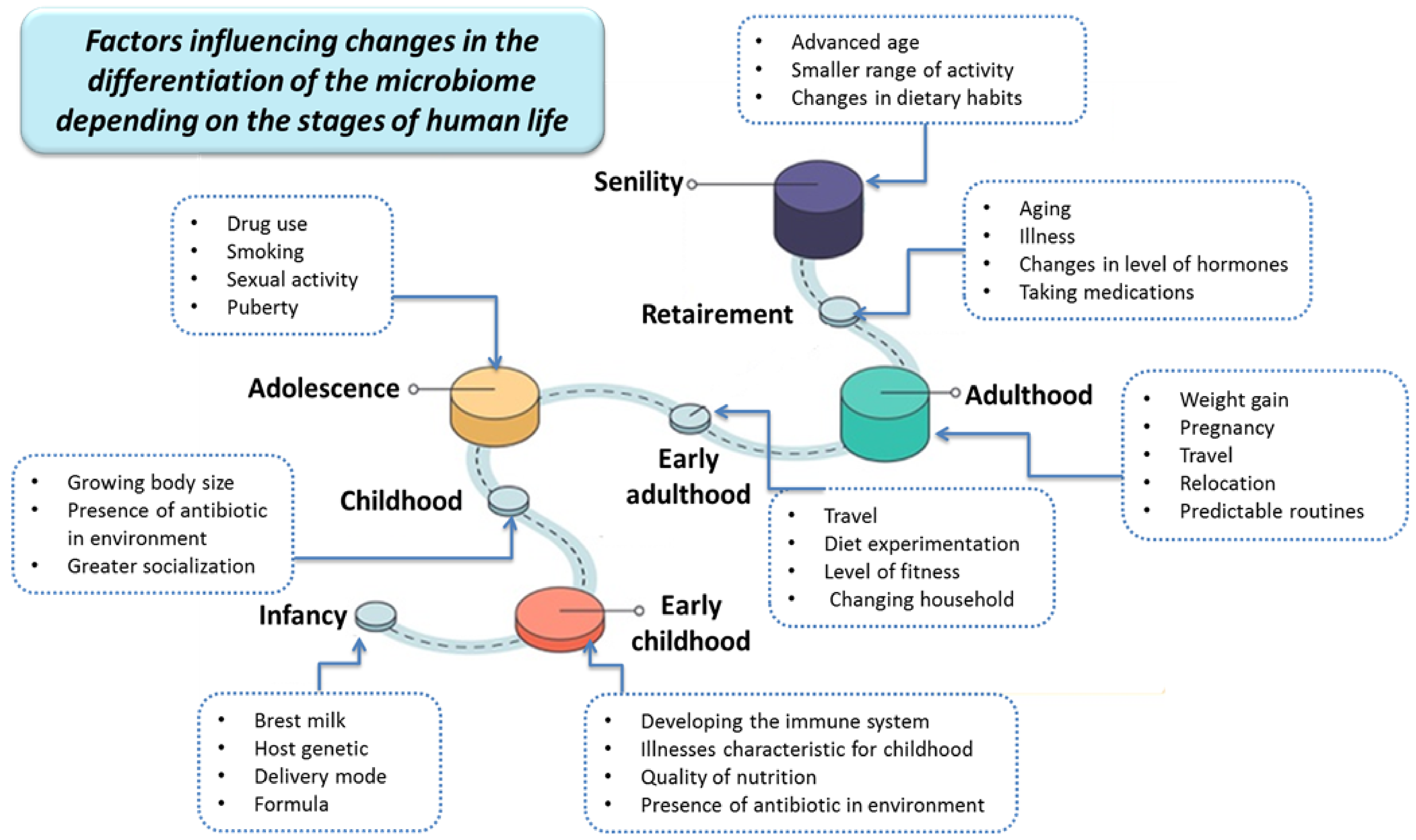
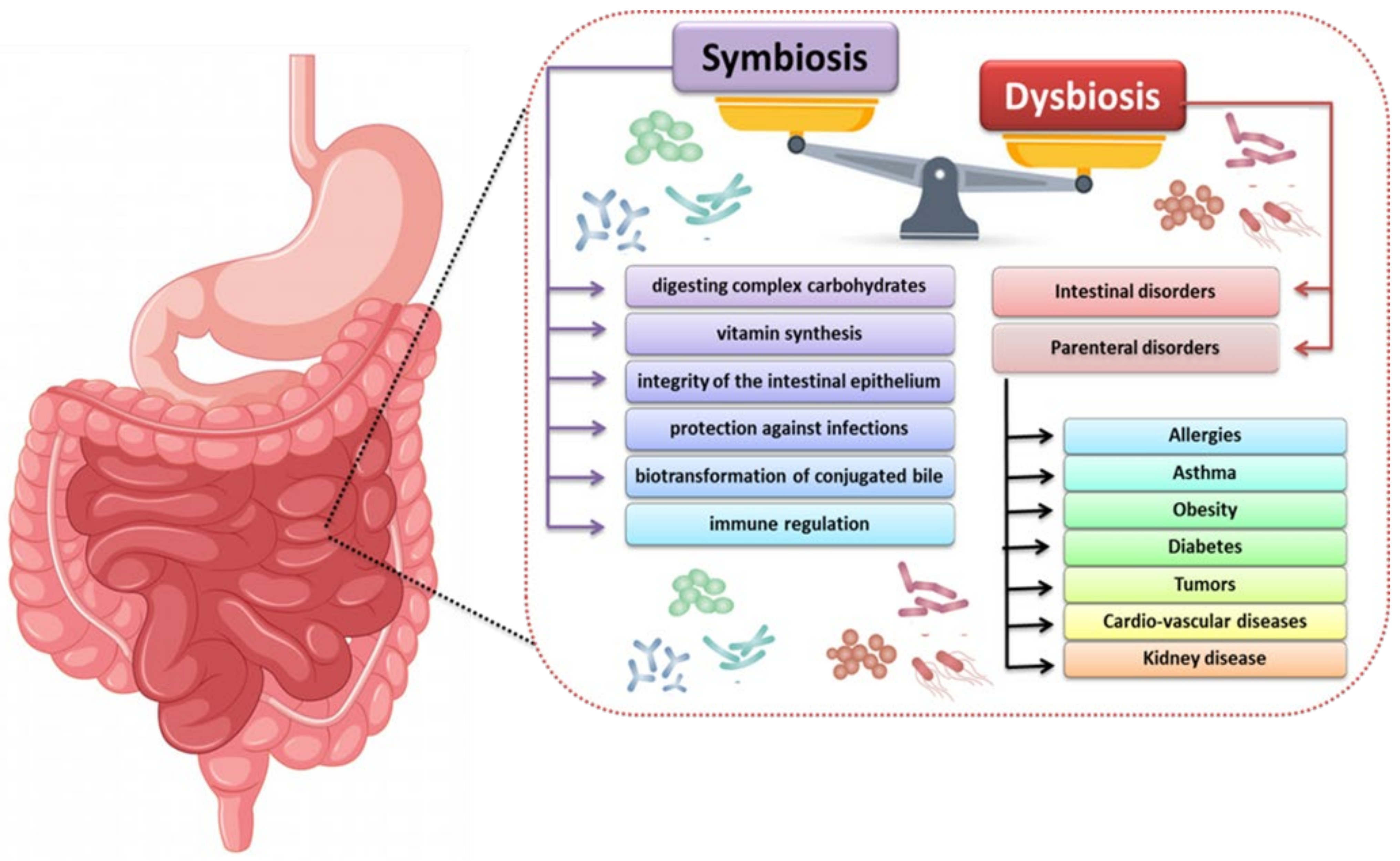
2. The Importance of the Human Microbiota

The definition of the human microbiota covers all the microorganisms inhabiting the human body, which consists of three main domains of life: bacteria, archea and eukaryotes. The development of molecular analysis techniques, including genomics and proteomics, has shown that each person has his own unique microbiota pattern in terms of quantitative and qualitative composition, which plays an important role in maintaining health and the occurrence of diseases [7,8,9]. With age, the composition of the basic human microbiota, including firmicutes (60% of the total intestinal microbiota), bacteroidetes (15% of the total intestinal microbiota), actinobacteria and proteobacteria, changes. The stages of human life and the physiological changes occurring during them, as well as environmental factors like ethnicity and geographic location are strongly correlated with the diversity of the human gut microbiota (Figure 1) [10,11,12,13,14].
Under the conditions of homeostasis, the intestinal microbiome performs a number of important functions aimed at supporting the human body, especially in terms of supplementing metabolic dysfunctions of the digestive system. Commensal intestinal microbiota acting as symbionts are responsible for such processes as digestion of complex carbohydrates, vitamin synthesis, maintaining the intestinal epithelium, protection against infections by pathogenic microorganisms, and immune regulation (Figure 2) [15]. With the proper functioning of the organism (understood as the absence of pathogenic symptoms), intestinal microorganisms form communities called enterotypes, which have a completely different effect on the intestine. It should be noted that the enterotypes possessed by a given person are not constant and are subject to dynamic changes conditioned by a number of factors such as diet, lifestyle or environmental stress (Table 1) [16,17,18].
All quantitative and qualitative changes occurring in the gut microbiota are termed dysbiosis and lead to cellular and metabolic disorders that affect the emergence or progression of disease states. The most common cause of dysbiosis is the development of allergies, asthma, diabetes, obesity and kidney disease (Figure 2). In the case of the last group, the causes of intestinal dysbiosis may be iatrogenic factors or uremia, which cause renal dysfunction. Reduction or loss of the filtration capacity of the kidneys causes the secretion of urea into the gastrointestinal tract, which, due to the enzyme urease produced by some microorganisms, undergoes hydrolysis and produces large amounts of ammonia. The presence of ammonia significantly influences the development of the commensal bacteria living in the human intestines and thus the quantitative and qualitative disturbance of the microbiota. Literature data indicate that other factors are also involved in the process of intestinal dysbiosis, such as consumption of medications (antibiotics, orally administered iron), changes in diet (lowering the amount of consumed diet, vitamin K deficiency), metabolic changes (metabolic acidosis, slowing down the passage of the intestinal epithelium) [21,22,23].

3. The Process of Immune Modulation by Human Gut Microbiota

Increasingly frequent scientific and experimental studies indicate the important role of intestinal microbiota not only in maintaining the proper homeostasis of the human body, but also in modulating the immune system. In the literature, it can be found that the intestinal microbiota is compared to a separate organ in the human body, the metabolic capacity of which exceeds the range of biochemical reactions taking place in the liver [24]. The importance of the intestinal microbiota is also demonstrated by its quantity. Research shows that people who eat a typical Western diet have 1010–1011 cfu/g, which in terms of weight means that the human caecum and colon are inhabited by 250 to 750 g of bacteria. They take into account that bacterial biomass may constitute from 40 to 55% of the solid mass of feces, which means that an average person excretes approximately 15 g of bacterial mass per day. A detailed analysis showed that almost 50% of the bacteria excreted despite aerobic conditions (the intestine is anaerobic and the presence of oxygen negatively affects the survival of some species of bacteria) are still alive [24,25,26].

3.1. The Role and Importance of Bacterial Metabolites and Components in the Human Body

3.1.1. The Role of Short-Chain Fatty Acids Which Are Products of Intestinal Bacterial Metabolism in the Human Body

Due to the anaerobic conditions in the distal gastrointestinal tract, most biochemical reactions occurring there are based on the fermentation process, which is responsible for the hydrolysis of nutrients in the diet. This process mainly affects polysaccharides, oligosaccharides and disaccharides, which are broken down into simple sugars, which are easily digestible energy compounds for microorganisms. The process of the fermentation of carbohydrate compounds itself leads to the production of short-chain fatty acids (SCFA), H2 and CO2, while in the case of amino acids and proteins, branched fatty acids are formed [27]. In terms of chemicals, SCFA includes organic acids composed of 1 to 6 carbon atoms in an aliphatic chain, which means that this group includes compounds such as acetic, propionic, butyric, valeric or caproic acid [28,29]. Additional studies have shown that the molar ratios of acetate, propionate and butyrate are also variable. In the colon, this ratio is 60:25:15, respectively, and it varies in individual sections of the intestine depending on factors such as diet, age and disease. The importance of SCFA in the human body may be proven by the fact that 95% of these compounds are absorbed by intestinal epithelial cells, and only 5% is excreted from the body with the feces [30].
The intestinal microbiota, using a number of metabolic processes, are responsible for the production of SCFA in the human body, which occurs at three sites:
  • In colonic epithelial cells where butyrate is the main substrate (which is the energy source for colonocytes);
  • In liver cells, where the acetate produced in the gluconeogenesis process is metabolized, as well as butyrate and propionate; and
  • In muscles, where the process of generating energy takes place due to the oxidation of acetate [31,32,33].
SCFAs also play a very important role in the protection of the human body. This applies to two aspects: the first is the inhibition of histone deacetylase (HDAC) activity, and the second is the participation in signaling by the complex of free fatty acid receptors coupled with G proteins (GPRs) [34,35].
Histone deacetylase is the enzyme responsible for removing the acetyl group from the ε-N-acetyl lysine found in histone. Such a process enables better wrapping of histones by DNA, which influences gene expression (only hyperacetylated chromatin is transcriptionally active). Literature data indicate that inhibition of HDAC by SCFA depends on many factors, including the type of acid and the type of cells and tissues in which this process occurs [36]. One of the most potent HDAC inhibitors is butyric acid, which, although produced in smaller amounts, plays the most important role in regulating this process. The next two places are propionic acid and acetic acid. There are two mechanisms of HDAC activity inhibition: direct (through the binding of two butyrate molecules to the enzyme pocket) and indirect (through GPR41, GPR43 and GPR109 receptors) [37,38]. Studies have shown that inhibition of HDAC activity by SCFA takes place in all cells of the immune system, both innate and acquired.
The second protective aspect concerns signal transmission by the G protein-coupled free fatty acid receptor complexes. We distinguish two types of complexes: free fatty acid receptor 2 coupled with G proteins—FFAR2/GPR43, which is responsible for the binding of acetates, butyrate, valerate and caproate; and receptor free fatty acids 3 coupled to G proteins—FFAR3/GPR41, which has an affinity for acetate and propionate and little for butyrate, valerate and caproate. The first type of receptor can be found in almost the entire digestive tract (secretory cells in the ileum, colon, colonocytes and enterocytes in the small and large intestine) and cells of the immune (on eosinophils, basophils, neutrophils, monocytes, dendritic cells and mast cells), and nervous systems. Studies have shown that by inducing the secretion of the YY peptide (PYY) and the glucagon-like peptide-1 (GLP-1), SCFAs can influence weight change and reduce the amount of food consumed by humans [39,40,41]. The second type of receptor, is expressed, inter alia, in adipose tissue and in the peripheral nervous system. Activation of GPR41 by SCFA improves glucose tolerance by inducing intestinal gluconeogenesis. Additionally, their presence has been demonstrated in the pancreas on Langerhans cells, in the spleen, and on peripheral blood mononuclear cells (PBMC), but their role in these organs has not been described to date [42,43,44].
By analyzing the effect of SCFAs on the human immune system, it was shown that they are involved in the process of maintaining the balance of anti-inflammatory and pro-inflammatory responses. Thanks to this, SCFAs become a kind of communication channel between the naturally occurring commensal intestinal microbiota and the immune system itself. Numerous studies have shown that SCFAs are directly involved in the differentiation of IL-17, IFN-γ and IL-10-secreting T cells through HDAC inhibition and are indirectly dependent on GPR41 and GPR43 receptors. As a result, these compounds can stimulate the process of T cell differentiation into effector and regulatory cells and can participate in the regulation of pro-inflammatory and anti-inflammatory responses [45,46].

3.1.2. The Role and Importance of Indole

Eating tryptophan-rich foods has a significant effect on intestinal microorganisms. This aromatic amino acid is broken down by bacterial tryptophanase (systentized by many intestinal bacteria, including E. coli) to indole. The concentration of this compound in the human colon is not fully known. Studies show that E. coli strains (both commensal and pathogenic) produce about 500 μM of indole [47] in laboratory conditions. In addition, studies conducted by Karlin et al. and Zuccato et al. have indicated that the concentration of indole in the human stool may vary from 250 to 1000 μM [48,49]. As for the functions of indole in the human body, it is a compound responsible for intercellular signaling, involved in processes such as increasing the expression of genes of intestinal epithelial cell connections or pro and anti-inflammatory factors in intestinal epithelial cells. This makes this compound responsible for maintaining host–microbiota homeostasis on the mucosa surface [50,51]. It should be mentioned that the produced indole is absorbed into the blood from the intestine and is metabolized to indoxyl sulphate in the liver; its residues are excreted in the urine in the case of properly functioning kidneys. This means that the production of indole by the gut microbiota and its uptake by the host cells suggests that there may be an indole concentration gradient in the gut. Excessive production of this compound by bacteria and its conversion into uremic toxin (which may occur due to the individual specificity of the composition of the intestinal microflora or as a result of dysbiosis) may result in impaired proper functioning of the kidneys [52]. Over 600 different compounds belonging to the indole group have been detected in the human body, of which indole acetic acid (IAA) seems to be extremely important. Studies have shown that this compound is only partially removed by hemodialysis in patients with CKD, and that its accumulation in the patient’s body leads to glomerular sclerosis and interstitial fibrosis, which may lead to the progression of CKD [53].

3.1.3. The Role and Importance of Aryl Hydrocarbon Receptor

The aryl hydrocarbon receptor (AhR) has been discovered to mediate toxic reactions induced by halogenated aromatic hydrocarbons and polycyclic aromatic hydrocarbons (e.g., such as 2,3,7,8-tetrachlorodibenzo-p-dioxin (TCDD)) [54]. The inactive form of these receptors is located in the cytoplasm as a complex with chaperones such as HSP90, P23 and XAP2. The ligands for this type of receptor are numerous compounds of both endo- (lipoxin A4, bilirubin and lipopolysaccharides) and exogenous origin, including dietary components, the host metabolism, the intestinal microbiome (mainly derived from tryptophan metabolism) or compounds of environmental origin that correspond to induce AhR conformational changes. These receptors play extremely important functions in the human body, including inducing the expression of genes of pro-inflammatory factors, the metabolism of xenobiotics (CYP1A1, CYP1A2, CYP1B1 and COX-2), or inducing selective protein degradation. Literature data also showed that AhR receptors are also correlated with CKD. It turns out that many uremic toxins which are products of the metabolism of the intestinal microflora have been classified as AhR antagonists. A recent study found that AhR activation in patients with CKD stages 3 to 5 correlates strongly with eGFR and IS levels, and the expression of AHR target genes in the blood (CYP1A1 and AhRR) was found to be increased in patients with CKD, compared with healthy controls [55,56].

3.1.4. The Role and Importance of Polyamines

Another group of compounds are polyamines, which include spermine (involved in cellular metabolism and a growth factor for some intestinal bacteria), putrescine (resulting from the breakdown of proteins by anaerobic bacteria), as well as polyamine oxidase and acrolein. Studies in animal models have shown that these compounds are involved in the development of CKD. Changes in polyamine metabolism resulted from changes in the metabolism of intestinal microorganisms, which resulted in the development of intestinal dysbiosis and thus intensified the progression of CKD. A study in patients diagnosed with CKD showed a decrease in spermine and an increase in plasma putrescine, polyamine oxidase and acrolein, which may suggest that these compounds may act similarly to uremic toxins. To date, scientific and clinical studies have shown that blood creatinine levels are effectively used as a marker for CKD. However, creatinine is not a toxic compound, and a number of studies on the concentration of acrolein (which is a toxin) correlate with kidney damage, and more specifically with the acceleration of the process of kidney fibrosis. Therefore, researchers have postulated using acrolein together with the determination of creatinine levels as a new diagnostic marker of CKD progression [57].

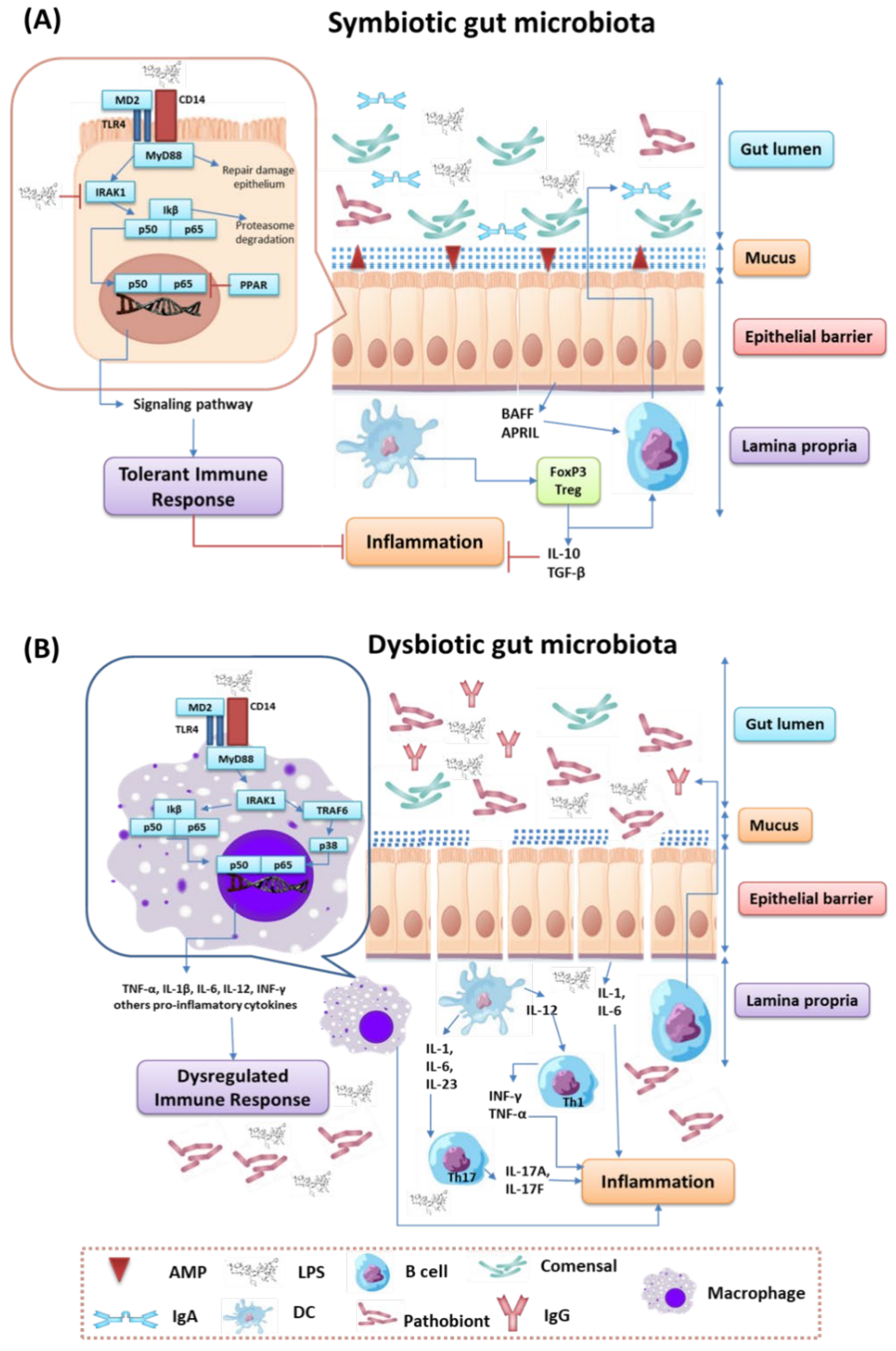
3.2. Regulation of the Immune Response by Gut Microbiota

In the intestinal environment, the intestinal epithelium performs the main nutritional and protective functions. It is responsible for the absorption of nutrients and as a protective barrier that often prevents pathogens and antigens from entering. The intestinal epithelium, composed of single layers of cylindrical cells tightly connected with each other, separates the intestinal lumen from the lamina propria, constituting a kind of seal. Consortia of commensal microorganisms found in the gastrointestinal tract are responsible for establishing and/or maintaining homeostasis in the intestinal environment through the process of immunomodulation and the development of a number of mechanisms responsible for maintaining the functional integrity of the intestine [58]. Such mechanisms include participation in the maintenance of the structure of tight junction proteins (claudins, occludins, junctional adhesion molecules (JAMs, belonging to the immunoglobulin subfamily) and tricellulins), the induction of epithelial heat shock proteins, i ncreasing the expression of mucin genes, secretion of antimicrobial peptides and competition with pathogenic bacteria. This means that the gut microbiota is involved in many functions in the field of metabolic (the ability of flora to break down undigested food debris by fermentation, SCFA), trophic (competitive inhibition for biotope and nutrients, and prevention of harmful colonization and multiplication of pathogenic bacteria), and immunological activity [58,59]. The implementation of the latter group of functions is related to the process of eliminating harmful antigens by molecular means by combining TLR receptors and NOD domains (nucleotide oligomerisation domain) with the structures of bacterial cells such as lipopolysaccharide or teichoic acid, which will lead to the induction of the signaling cascade responsible for the secretion of inflammatory mediators. In addition, microorganisms promote cell survival via the phosphatidylinositol 3-kinase or kinase B via the MyD88 factor, which allows the building of a protective barrier against damage caused by stress factors. It has also been shown that the signaling process of intestinal microorganisms by TLRs found in the intestinal mucosa is required to maintain not only the homeostasis of the entire epithelium, but also its repair [60,61].
Detailed analysis of the relationship between the role of intestinal microorganisms and the development of inflammation within the symbiotic and dysbiotic intestinal microbiota showed a number of significant changes. Under conditions of a symbiotic gut microbiota, we can see an increase in commensal bacteria and the maintenance of gut epithelial integrity. The first line of defense is the mucus layer, which is composed of two integral parts: the outer (rich in antibacterial peptides produced by Paneth cells and immunoglobulin A, synthesized by plasma cells) and the inner (responsible for hydration, regeneration processes and protection against the action of digestive enzymes of epithelial cells). This keeps microorganisms away from intestinal epithelial cells, which leads to increased tolerance of the immune system to the commensal microorganisms residing there [62,63]. When the mucous protective layer is compromised, the intestinal epithelial cells use signaling cascades using TLRs to detect microbes. In the case of Gram-negative bacteria, the signaling molecule will be LPS, which will be taken up by TLR4, while in Gram-positive bacteria, teichoic acids will be taken up by TLR2. Upon ligation of the signal molecule to the appropriate TLR, MyD88 is recruited, which activates the NFκβ pathway and leads to the production of antimicrobial proteins and pro-inflammatory cytokines. Under normal microbiota conditions, intestinal epithelial cells are desensitized by continuous exposure to the same LPS derived from commensal bacteria or may be weakened [64,65]. There are three mechanisms involved in this process. The first one concerns the downregulation of IL-1 receptor related kinase 1 (IRAK1), which acts as an activator of the NF-κβ cascade. The second involves the induction of the G receptor, activated by the proliferators of PPAR peroxisomes (which are transcription factors that regulate the expression of genes related to carbohydrate, fat and protein metabolism, as well as cell proliferation and inflammation), which can divert NF-κβ from the nucleus. The third mechanism is based on the inhibition of polyubiquitilation and degradation of I κβ (nuclear factor kappa β inhibitor), which inactivates NF- κβ. Exposure to LPS or teichoic acid induces epithelial cells to secrete TGF-β (transforming growth factor beta), BAFF (TNF family B-cell activating factor) and APRIL (proliferation-inducing ligand), which are responsible for the development of immune cells tolerating the inhabiting microbiota. This process also involves dendritic cells that support the development of IL-10 and TGF-β secreting Tregs and stimulates the production of an IgA specific for commensal microorganisms [66,67] (Figure 3A).
In the case of dysbiosis of the intestinal microbiota, the number of commensal microorganisms is reduced in favor of pathogenic microorganisms, the accumulation of toxins (mainly urea and ammonia), and the integrity of the intestinal epithelium is disrupted, which in turn leads to the development of inflammation. When the continuity of the intestinal epithelium is interrupted, bacteriaand bacterial cell components are translocated. This leads to a situation where the gut’s immune system directs a pro-inflammatory response to remove pathogenic bacteria. This is possible through the secretion of IL-1 and IL-6 from intestinal epithelial cells, by the promotion of the Th1 and Th2 responses by dendritic cells and macrophages, and by the production of higher levels of specific IgG by B cells. When a bacterial factor such as LPS binds to the receptor complex (CD14-MD2-TLR4) within macrophages, it results in the activation of the signaling cascade with the activation of p38 MAPK (mitogen-activated protein kinases), leading to the production of significant amounts of inflammatory cytokines, such as INF -β, INF-γ, IL-1b, IL-6, TNF-α and IL-12. The presence of toxic compounds called uremic toxins is one of the causes of inflammation affecting the development of kidney disease [68,69] (Figure 3B).

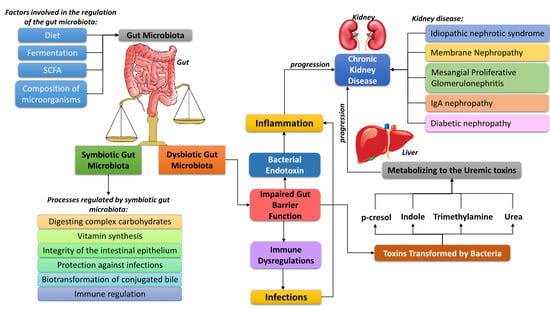
4. Influence of the Intestinal Microbiota on the Development of Kidney Diseases, with Particular Emphasis on the Role of the Immune System

Any abnormalities of the intestinal microbiota resulting in its dysregulation may lead to inflammation and thus cause a number of diseases, including kidney dysfunction. Currently, the changes observed in the intestinal microbiota affecting renal dysfunction include:
  • Reduced diversity and number of microorganisms, with a predominance of proteolytic bacteria;
  • A phenomenon of the translocation of the microorganisms associated with the colonization of regions of the gastrointestinal tract that have been much less populated so far, and changes in the ratio of aerobic and anaerobic bacteria;
  • The intestinal epithelial barrier is disrupted; and
  • The production of uremic toxins [70,71].
In many cases of people with kidney diseases, including extreme cases of kidney failure, the synthesis of many toxic compounds in the human body is accelerated, which leads to an increase in the concentration of uremic toxins in the plasma and the progression of kidney diseases. A normal gut microbiota produces compounds that are normally excreted by the kidneys, but which can also be considered potentially toxic. This is the case with the bacterial fermentation of the tyrosine amino acids, which are obtained from the diet through the consumption of meat and dairy products, to a compound in the form of p-cresol. This is also the case with the fermentation of tryptophan to indole. After absorption in the colon, these compounds are metabolized in the liver and converted into the toxic forms of p-cresyl sulphate and p-indoxyl sulphate [70,71,72,73]. Both of these compounds have an affinity for albumin, which means that they can exist in the human body in two forms: the free fraction and the serum-bound fraction. The third type of harmful compounds formed are amines. More precisely, they are compounds of choline and phosphatidylcholine fermentation by intestinal bacteria to trimethylamine, which are converted in the liver into trimethylamine N-oxide (TMAO) (Table 2). All of these toxins are mainly eliminated from the body by the kidneys, and more specifically by the kidney tubules, and an excessive quantity of these toxins leads to damage to the kidney function. As a result of breaking the continuity of the intestinal barrier, these compounds may also enter the systemic circulation and influence the development of cardiovascular diseases as well as symptoms of the central nervous system [74,75,76,77].

4.1. Chronic Kidney Disease

The importance of gut microbiota in the progression of chronic kidney disease requires an understanding of many interrelated aspects, including the composition, dynamics, stability, and interactions between bacteria and the human body. The development of CKD is largely related to the accumulation of uremic toxins like indoxyl sulfate, p-cresol sulphate and trimethylamine N-oxide.
The first toxin is p-indoxyl sulphate, which is an indole derivative produced by metabolism in the liver. This compound is a ligand for the acrylic hydrocarbon receptor and acts as a transcriptional regulator. In patients with CKD, this compound is not excreted in the urine and accumulates in the body. Literature data indicate that the elimination of this toxin from the body is extremely difficult, and its reduction ratio with regular hemodialysis is only 31.8% [78]. Scientific studies in animal models have also shown that p-indoxyl sulphate can damage renal tubular cells and mediate changes in the expression of the TGF-β1 gene and the tissue metalloproteinase inhibitor associated with tubulointerstitial fibrosis. In addition, studies conducted by Ichii’s team on mouse models showed the effect of this uremic toxin on the altered pro-inflammatory phenotype of podocytes, which was accompanied by decreased expression of genes specific for these cells, as well as their reduced viability [79]. Moreover, it has also been shown that an excessive amount of indoxyl sulfate also affects the activation of the renin-angiotensin-aldosterone system in the kidneys of mice [79,80].
Another uremic toxin which is a product of the breakdown of the amino acids tyrosine and phenylalanine is p-cresol sulphate produced in the liver. In the case of fully functional kidneys, this compound is excreted in the urine, which depends on tubular secretion by specific transporters. In patients with CKD, these transporters are impaired and the toxin accumulates in the body [81,82]. Literature data estimate that this toxin cannot be effectively removed by dialysis, and the reduction factor is only 29.1% with regular hemodialysis [78]. In animal models, this compound has been shown to lead to the increased expression of numerous transcription factors, such as fibronectin and L-actin in smooth muscle within proximal tubular cells. In addition, experiments in mice with partial nephrectomy showed that p-cresol sulfate was responsible for the activation of the intrarenal renin-angiotensin-aldosterone system, and also led to interstitial fibrosis and glomerulosclerosis [80]. Research data compiled by the Meijers team, which included nearly 500 patients, showed that there is a correlation between the level of p-cresol sulphate and the development of CKD. This mainly concerns the glomerular filtration rate, the values of which decreased with the increase in the concentration of the uremic toxin tested. A similar relationship was found in patients with end-stage renal failure treated with hemodialysis, where it was related to the increased risk of death in patients with increased levels of p-cresol sulphate [81]. The research team led by Lin showed that there is a negative correlation between serum levels of p-indoxyl sulphate and p-cresol and renal function in patients with CKD. Based on the obtained studies, it was found that both of these uremic toxins exert a number of negative effects on numerous cellular processes, such as stimulation of oxidative stress, fibrosis and inflammatory reactions. Additionally, it has been established that high plasma levels of both of these toxins are correlated with progression to end-stage renal disease and increased mortality in patients with CKD [83].
TMAO, a breakdown product of choline, phosphatidylcholine, and dietary L-carnitine, is also considered an extremely important uremic toxin. Studies have shown that there is a positive correlation between the concentration of this compound in the blood and the presence of bacteria from the Clostridiaceae and Peptostreptococcacea families [84]. Experiments in animal models showed that the increased concentration of this compound significantly correlated with an increase in tubular interstitial fibrosis, collagen deposition and changes in the degree of phosphorylation of Smad3, which is an important regulator of this process. The studies conducted by the Tang team showed a correlation between the concentration of TMAO and the development and progression of CKD also in humans [85]. Their studies showed that elevated levels of this uremic toxin were associated with an almost 70% higher risk of death in CKD patients (even after adjusting for traditional risk factors and CRP protein). Due to all the above-mentioned changes caused by TMAO and the increased mortality of patients with CKD who have excessive levels of this uremic toxin, it is extremely important and warranted to continue further research that may allow for a detailed determination of the role of this compound in the progression of CKD [86]. Additionally, it has been shown that TMAO is involved in changes in cellular metabolism, that is, it affects the metabolism of cholesterol and bile acids, is responsible for the stimulation of the expression of scavenging receptors on macrophages and it modifies the sterol transporter in the liver and intestines [84,85,86,87,88].
A consequence of the accumulation of uremic toxins in CKD is also the very occurrence of the disease, and more specifically its impact on the intestinal microbiota. Patients diagnosed with CKD have poorer diets, especially when it comes to consuming dietary fiber, often use antibiotics and oral iron supplementation, and are at risk of lactic acidosis. In addition, such patients are characterized by slower passage of colon cells or changes in the intestines associated with volume overload with congestion of the intestinal walls, as well as intestinal edema. These changes are reflected in the composition of the intestinal microbiota. Among patients with CKD, a decrease in the number of Lactobacillus spp. and Prevotella ssp. bacteria, which belong to the normal microbiota of the colon, and nearly 100 times more bacteria from the Enterobacteriaceae and Enterococcaceae families, the number of which in the normal microbiota is significantly lower, are often found [5,89]. The observed dysbiosis of the gastrointestinal tract also has consequences for the human body itself. This is because the tight connections between the intestinal epithelial barrier and the translocation of bacteria and components of bacterial origin are loosened, which triggers an immune response inducing inflammation. The process of intestinal dysbiosis may also be influenced by other mechanisms related to the increased secretion of urea from the gastrointestinal tract. This means that urea hydrolysable microorganisms produce large amounts of ammonia, to which the commensals of gastrointestinal bacteria is sensitive. The most common causes of the development of chronic kidney disease include the presence and progression of primary and secondary glomeluropathy, diabetic nephropathy and hypertensive nephropathy [90,91].

4.2. Idiopathic Nephrotic Syndrome

Idiopathic nephrotic syndrome (INS) is one of the glomerular disorders characterized by edema, proteinuria and hypoalbulinemia. This disease causes gromeluropathy in children from one to 30% of cases in adults, and the pathogens themselves are still the subject of research by many scientists. Research has shown that the immune system is strongly involved in the pathomechanism of INS formation. The underlying mechanism is the disruption of the glomerular permeability barrier, which is caused by the stimulation of antigen presenting cells and B lymphocytes in response to the emergence of allergens or infections. As a result of such stimulation, T lymphocytes are also activated through antigen presentation and cytokine production. Literature data show numerous changes in the T cell population in INS patients. The most frequently observed changes include a decrease in the number of CD4 plus T helper lymphocytes (Th), which is associated with the presence of cytotoxic CD8 plus T lymphocytes, imbalance between Th2 and Th1 lymphocytes, and reduced frequency and function of regulatory T lymphocytes (Tregs), as opposed to increased Th17 cell activity [92].
Additionally, this unit has different histopathological types, including minimal change disease (MCN), membranous nephropathy (MN), focal segmental glomerulosclerosis (FSGS) and membrane proliferative glomerulonephritis (MPGN) [93]. Due to such a wide variety of the disease, researchers have investigated whether intestinal dysbiosis can lead to development (INS) and what potential factors may predispose to the development or progression of this disease. Detailed analysis of the intestinal microbiota at the level of quantitative and qualitative differentiation of taxa carried out by the He team in 2021 [94] showed that there are differences between the microbiota of healthy and INS patients. In patients with INS, fewer bacteria belonging to the following types: Acidobacteria, Firmicutes: especially Negativicutes class, Selenomonadales class, Veillonellaceae family, Clostridiaceae, and genera: Dialister, Rombousia, Ruminiclostridium, Lachnospira, Alloprevotella, Closstridium and Megamonas were detected in comparison to healthy individuals. There were also changes in the types of bacteria such as Parabacteroides spp., Bilophila spp., Enterococcus spp., Eubacterium spp., which were higher in INS patients than in controls. These observations resulted in the establishment of a clear specific type of bacterial pattern that decreased the amount of bacteria capable of producing SCFA in INS patients. In addition, significant relationships between selected clinical parameters and the presence of individual bacteria were demonstrated. There was a negative correlation between serum creatinine concentration and the occurrence of Burkholderiales, Barnesiella spp. or Alcaligenaceae, as well as a positive correlation between proteinuria and the occurrence of Coriobacteria, Nitrosomonadales, Verrucomicrobia and Blautia spp. However, the close interrelationships between clinical parameters and the amount and diversity of the gut microbiota requires much research to fully understand [95,96].

Membrane Nephropathy and Mesangial Proliferative Glomerulonephritis

Membrane nephropathy (MN) is also one type of primary glomerulonephritis. It is one of the most common causes of nephrotic syndrome development in adults all over the world. The disease is caused by immunological disorders related to the production of autoantibodies against PLA2R (phospholipase A2 receptor) and HSD7A (Thrombospondin Type 1 Domain Containing 7A) antigens, which belong to the IgG4 immunoglobulin subclass. The second disease is Mesangial proliferative glomerulonephritis (MPGN) which can also manifest as nephrotic syndrome. The most common symptoms are proteinuria, low blood protein levels, high cholesterol, high triglycerides, and edema. A characteristic feature of MPGN is the increased number of mesangial cells in the kidney glomeruli, leading to their damage. Due to the diversity of these diseases and their classification as histopathological subtypes of INS, some researchers have examined whether the development of these disease entities may be influenced by intestinal microorganisms [97,98]. Studies have shown that there are differences between the quantitative and qualitative scales of the gut microbiota of both studied disease entities. Higher amounts of bacteria belonging to Proteobacteria and Gammaproteobacteria have been shown in patients diagnosed with MN than with MPGN. This also concerned changes within the orders of Enterobacteriales, Erysipelotrichales, Enterobacteriaceae, Rikenellaceae, Ruminococcaceae, the Coriobacteriia class, or the Tyzzerella, Alistipes, Lachnospira, Odoribacter genera, which were dominant in patients with MN. In the case of MPGN, bacteria from the Rhodobacterales order, Phyllobacteriaceae families, and the Terrimonas and Mesorhizobium genera dominated. The presented differences in taxa may be used in the future as biological diagnostic markers to help distinguish MPGN from MN [94,97].

4.3. IgA Nephropathy

IgA nephropathy is one of the most common conditions associated with primary glomerulonephritis. The hallmark of this disease is glomerulonephritis accompanied by the deposition of the IgA1 subclass in these structures. The main IgA receptor is CD89, which is expressed on the surface of monocytes and plays an important role in the pathogenesis of the disease. As shown by the results of studies available in the literature, it can be considered a prognostic marker of disease development. IgAs are produced by Peyer’s patches in the lymphoid tissue of the gastrointestinal mucosa. This area is composed of lymph nodes rich in a large number of B lymphocytes, and between them (in the areas between the clumps) we can find T lymphocytes. The entire structure is covered with a layer of specialized M cells, which are responsible for the uptake of antigens (e.g., bacterial antigens) from the intestinal lumen, which are then transferred to macrophages or dendritic cells involved in the presentation of antigens to T lymphocytes. Due to this fact, under the influence of bacterial antigens of pathogenic origin or commensal Peyer’s patches capable of producing IgA1, an excess quantity is the first step in the development of nephropathy [13,96,99,100]. There is no doubt that the intestinal microbiota may affect the progression of this disease entity. Therefore, many researchers have documented these relationships. The team of De Angelis approached the subject in the most detail, in which the microbiota of IgAN patients were compared with not onlt the control group, but also patients with IgAN progressors and non-progressors. This study involved a very comprehensive approach, with the development of culture-dependent and independent methods, as well as the inclusion of metabolomics analysis. Eight types of bacteria were identified in all studied groups of patients, the dominant of which were representatives of Firmicutes, Bacteroidetes and Proteobacteria, constituting over 98% of all 16S rDNA and 16S rRNA. There were also differences between the 16S rDNA and 16S rRNA analysis, which indicated the types of metabolically active bacteria. In the case of Firmicutes bacteria, their metabolic activity increased in patients diagnosed with IgAN (both subtypes) compared to controls. On the other hand, an inverse relationship was noted for Bacteroidetes bacteria, which were more numerous in patients from the control group. In addition, it was observed that the number of both completely and metabolically active Proteobacteria was lower in healthy subjects than in IgAN patients, while the opposite relationship was found for Actinobacteria, which were more numerous in healthy subjects than those diagnosed with IgAN [96].
Other studies have shown that in patients diagnosed with IgAN there is an increase in the numbers of bacteria from families, such as Enterobacteriaceae, Ruminococcaceae, Streptoccaceae, Eubacteriaceae and Lachnospiraceae. There was also a decrease in such genera of bacteria as Clostridium, Lactobacillus, Enterococcus and Bifidobacterium. Although the latter type of bacteria is commonly considered to be a microorganism with a pro-health potential involved in a number of immunomodulatory mechanisms, inhibiting the development of pathogens or producing SCFA, it should be remembered that with intestinal dysbiosis it may increase its invasive potential. Their amount has been confirmed to be higher than that of healthy people in the case of ulcerative colitis, which may indicate that some species of bacteria are specific for the disease, and further studies on their influence on maintaining normal intestinal homeostasis are necessary [13,101,102,103,104]. Some scientists, similarly to MN and MGPN, showed significant relationships between clinical parameters and the presence of specific microorganisms in the course of IgAN. This concerns the correlation between high levels of albumin and the prevalence of Prevotella spp. bacteria, which has also been associated with improved glucose metabolism and insulin sensitivity. The researchers observed a negative correlation between albumin and bacteria of the genera Klebsiella, Citobacter and Fusobacterium. Additionally, the correlation between the occurrence of Klebsiella spp. and increased disintegration of intestinal epithelial cells at IgAN has also been demonstrated [97].

4.4. Diabetic Nephropathy

Diabetic nephropathy is one of the most important complications of diabetes worldwide, and as shown by numerous scientific studies, abnormal intestinal microbiota may be involved in its development. Many clinical trials have found an increased level of inflammatory markers in patients with diabetic nephropathy. The cause of inflammation itself is not fully understood, but it is estimated that it may be related to tissue damage, trauma, and patients’ susceptibility to infections. Then the immune cells of T lymphocytes, macrophages, and dendritic cells of both the innate and adaptive systems, as well as other metabolic signals that contribute to disease progression, are activated. Disturbances in the gut microbiota have been observed and documented for both type 1 and type 2 diabetes. Therefore, only detailed studies that can produce evidence of a better understanding of the interaction between the intestinal microbiota and diabetes can help in the development of effective treatments not only for diabetes itself, but also for its complications, such as diabetic nephropathy. For both subtypes of diabetes, there are several interrelationships between microorganisms and disease progression. This applies mainly to disturbances of the intestinal mucosa barrier, which is associated with an increased translocation of bacteria and bacterial components influencing the development of inflammation and insulin resistance. Additionally, studies of fecal transplantation carried out by scientists have shown that changes in the intestinal microbiota directly affect the course of both type 1 and type 2 diabetes [101]. The number and composition of the gut microorganisms also changes drastically during the development of diabetes. There is a decline in the number of bacteria, including Lactobacillus spp., Bifidobacterium spp., and Roseburia spp., which are involved in the immunomodulation process, SCFA production, and the process of supporting the integrity of the intestinal epithelium by producing tight junction proteins. In place of commensal microorganisms, bacteria with high pathogenic potential, such as Clostridium spp., Bacteroides spp., Betaproteoovibacter spp., Prevotella spp. or Desulfovibrio spp., multiply and increase the permeability of the intestinal mucosa barrier by producing toxins. In the case of type I diabetes, the growth of Leptotrichia googfellowii, which has an antigen on its surface that stimulates CD8+ T cells to attack pancreatic islets, has also been observed due to the phenomenon of molecular mimicry, which allows the development of diabetes. In addition, some of the intestinal microbiota (Lactobacillus spp., Bifidobacterium spp., Clostridium spp., Bacteroides spp. or butyrate-producing bacteria) may participate in the process of Treg cell differentiation, the number of which is reduced in type 1 and 2 diabetes [98,105].
Additionally, the development and progression of diabetes may be influenced by changes in the endocrine function of the intestines and the composition of metabolites produced by the intestinal microbiota. Butyrate-producing bacteria, as one example of SCFA (Lactobacillus spp., Bifidocacterium spp.), protect against the development of diabetes by inducing apoptosis in pancreatic islet macrophages. In addition, SCFA has the function of inducing the secretion of GLP-1 (glucagon-like peptide-1), which improves blood glucose levels and reduces insulin resistance in type I diabetes, while stimulating insulin secretion in type 2 diabetes. A complex relationship exists between the intestinal microbiota, the intestinal metabolism, the pathogenesis of diabetes and diabetic nephropathy [98,105,106].

5. The Importance of Diet in the Progression of CKD

In patients with CKD, body composition disorders are very common, resulting from excess body fat (leading to obesity) accompanied by muscle wasting. Both these factors not only influence the patients’ problems in everyday existence, but also significantly lower their prognosis. This is largely due to metabolic changes occurring during the course of the disease, i.e., as shown in the literature, to the imbalance in the metabolic balance of insulin-dependent tissues. This means that in the muscles of CKD patients there is an increase in catabolic processes (regulated by glucagon, glucocorticosteroids, catecholamines or pro-inflammatory cytokines), accompanied by an increase in anabolic processes in adipose tissue. Such changes are caused by decreased physical activity of the patients (with decreased muscle strength), the development of metabolic acidosis, and insulin resistance [107,108,109]. The process of disease progression is influenced by the nutritional status of the patient’s body, i.e., the balance between the consumption, absorption and use of nutrients by the body. Literature data estimate that malnutrition is present in approximately 20% of all patients diagnosed with CKD, which strongly correlates with the severity of the disease [110,111]. Therefore, in many regions of the world teams or committees of researchers, doctors and nutritionists are formed to prepare detailed recommendations regarding the diet used during the treatment of CKD, e.g., the National Kidney Foundation (NKF) [112], American Dietetic Association, Academy of Nutrition and Dietetics (ADA) [113], the International Society of Renal Nutrition and Metabolism (ISRNM) [114], and the European Dialysis and Transplant Nurses Association/European Renal Care Association (EDTNA/ERCA) [115]. The recommendations of these teams concern a number of factors, from recommendations regarding the energy values of the diet, through recommendations on the amount of consumed macronutrients (protein, phosphorus, potassium or sodium), vitamins (vitamins C, B12, D, folic acid), to the consumption of minerals. Very often, the selection of the appropriate proportions of the diet depends on the value of the glomerular filtration rate (eGFR), as well as the severity of comorbidities. However, not all countries around the world operate in the same way and include the establishment of teams of specialists to develop such guidelines. Additionally, it should be considered that reports prepared by expert teams from individual countries differ from each other. This is related to the diversity of food products consumed in a given region of the world, as well as their profile, e.g., the dominance of plant-derived products in the diet, but also the availability of specialized food for patients and the scope of financing health services that allow for the modification of the diet composition of CKD patients. Many literature data supported by research indicate that dietary regulation in patients with CKD associated with, among others, a reduction in the consumption of protein, fats, carbohydrates or antioxidants, affects therapeutic success. However, changes in nutrients and the macro- and microelements or minerals they contain may significantly affect the composition and proper functioning of the microbiomes of these patients and there may be accompanying changes in immunomodulatory processes. Thus, it is extremely important to ensure the proper homeostasis of patients’ nutrition and the functioning of their microbiomes in order to increase the prognosis and therapeutic success [111,116].

6. How to Restore the Symbiosis of the Gut Microbiota?

Many scientists discuss methods of effectively restoring the symbiosis of the gut microbiota in patients with renal dysfunction as one of the countermeasures in the process of disease progression. However, the studies conducted to date provide only limited evidence and contradictory information regarding the effectiveness of the actions taken by the researchers. The first obvious strategy is to change the diets of such patients. For chronic kidney disease, this diet is rather low in fiber, phosphorus and potassium. This, of course, translates into deficiencies of prebiotic compounds, including consuming the right quantities of dairy products rich in lactic acid bacteria or fiber-rich fruits and vegetables. Several studies concluded that the inclusion of high-fiber foods in the diet decreased the levels of uremic toxins in the body of patients with kidney disease [117,118,119,120]. Another approach is to include probiotics, prebiotics and even symbiotics in the diet (Table 3). Live microorganisms are considered probiotics, which, in an appropriately selected amount, have a positive effect on human health, this applies mainly to lactic acid bacteria, but also to some strains of yeast or mold. These microorganisms are characterized by the ability to colonize various environments in the human body, in particular the intestines, where they play a key role in stimulating the passage of intestinal epithelial cells and ensure the proper development of commensal microorganisms. Prebiotics are food ingredients that selectively influence the development of a specific group or type of microorganism with probiotic properties in the gastrointestinal tract [121,122,123].
Prebiotics can occur naturally in many plants or artificially as food additives or pharmaceutical preparations. One feature of prebiotics is the fact that they are not digested by enzymes in the human body and can only be used by specific microorganisms equipped with an enzymatic apparatus for their decomposition. The last group are symbiotic, i.e., a combination of pro and prebiotics, which together show a synergistic effect influencing the development of normal intestinal microbiota. In addition, it has been shown that they can participate in reducing the concentration of undesirable toxins or metabolites in the human body, and are also involved in the processes of preventing putrefactive reactions in the intestines and the formation of constipation or diarrhea [121,122,123].
Research with the use of probiotics, prebiotics or their combination in symbiotics for the progression of kidney diseases is relatively innovative. Several studies report that the use of probiotics reduced the concentration of uremic toxins, especially p-cresol sulphate and p-indoxyl sulphate in patients with chronic kidney disease and patients undergoing hemodialysis [124]. Research conducted by the Ranganathan team [125] and the Alatriste team [126] showed that the use of probiotics in non-dialyzed CKD patients reduced the level of urea in the serum. In addition, the analysis of individual components of the immune system conducted by the Wang team [127] showed that the use of probiotics for a period of 6 months allowed for the reduction of TNFα, IL-5 and IL-6 levels. In the case of the use of prebiotics, many research teams have shown a decrease in serum and plasma p-cresyl sulphate levels by nearly 20% [128,129,130] and a decrease in serum TMAO in patients diagnosed with CKD [131]. The use of prebiotics additionally affects the immune system by reducing the level of TNFα and IL-6, as was the case with the administration of probiotics [132].
Few studies also mention the use of adsorbent compounds, including AST-120, which is an orally administered carbon adsorbent involved in the removal of uremic toxins [133,134]. It has been shown that this compound adsorbs p-indoxyl sulphate and allows it to reduce the rate of decline in renal function or delay the start of dialysis treatment. However, despite advanced work on animal models, the use of this compound in humans has been approved only in a few Asian countries such as Japan, Korea and the Philippines [135]. Modern medicine is also increasingly searching for new, extremely innovative methods of restoring intestinal symbiosis. This applies to the use of so-called intelligent bacteria, whose genetic modification allows for the delivery of therapeutic agents to the body or the uptake of uremic toxins [24,136,137]. Intestinal microbiota transplant therapy, which has been used to treat chronic Clostridial-induced diarrhea, is also used, and its modifications in animal models allow for good results in restoring the balance of the intestinal microbiota in other diseases [138,139].
In addition to dietary considerations, significant attention should also be paid to the level of physical activity in patients with CKD. As mentioned above, in this group of patients the muscle metabolism is significantly impaired, and there is an excessive accumulation of fat in the body usually caused by restriction of movement or leading a sedentary lifestyle. Due to this fact, many comorbidities develop, such as cardiovascular diseases, hypertension and diabetes. Recent studies have shown that the activation of people with CKD allows them to maintain well-being and improve functional capacity (especially in the context of improving aerobic capacity, kidney function and reducing the risk of other comorbidities) [140].
Due to the increasing number of cases of the development of CKD among people, research has also begun to determine whether the progression of this disease is significantly influenced by other aspects of human life, including not only lifestyle but also addictions. One such example is a study of effect of smoking on the development of CKD. However, the obtained results are not unequivocal. In studies by Yacoub and Habib, smoking has been shown to significantly increase the risk of CKD compared to the control group, especially in patients diagnosed with hypertensive nephropathy. However, research conducted by Xia team based on the meta-analysis of available literature data showed that cigarette smoking is an independent risk factor for CKD. Further comprehensive research is needed to see if quitting smoking can reduce the incidence of CKD in the general adult population [141,142].

7. Conclusions

The pathogenesis of chronic kidney diseases involves not only immune dysregulation, but also the genetic susceptibility of people suffering from this type of disease. Additionally, a number of environmental factors are involved in the process of the progression of many kidney diseases, which may directly and indirectly affect the intestinal microbiota and its interactions with the human body. That is why research combining a comprehensive approach to the role of the intestinal microbiota at various stages of the occurrence of kidney disease becomes so important. However, this requires the involvement of genetic, immunological and dietary approaches in determining interactions in the gut-kidney axis. Only by comparing a large number of appropriately selected patients with specific kidney diseases, active and inactive microorganisms inhabiting the intestines and their participation in the immunomodulation process will it be possible not only to understand the prevalence and progression of the disease, but also to develop effective methods of diagnosis and treatment. The research should also consider dietary factors, which, as many studies have shown, have a very significant impact on the differentiation of microorganisms and the development of intestinal dysbiosis. Maintaining the intestinal microbiota in conditions of symbiosis is a challenge for a healthy person, and it is also a necessary aspect in the fight against any type of disease, and the occurrence of specific species, genera or the number ratios of individual bacterial families may in the future become potential diagnostic biomarkers and undoubtedly therapeutic goals that modern medicine is currently facing.

Author Contributions

Conceptualization, P.M., S.M., J.W., E.G. and I.K.-G.; writing—original draft preparation, P.M., S.M., J.W., E.G. and A.B.; writing—review and editing, I.K.-G. and W.Z. All authors have read and agreed to the published version of the manuscript.

Funding

This work was supported by Research Grant No. UMO-2016/23/B/NZ6/02844 of the Polish National Science Centre (NCN) and Research Grant No. DS460 of the Medical University of Lublin.

Conflicts of Interest

The authors declare no conflict of interest.

References

  1. Chronic Kidney Disease Basics/Chronic Kidney Disease Initiative/CDC. Available online: https://www.cdc.gov/kidneydisease/basics.html (accessed on 11 September 2021).
  2. Kaufman, D.P.; Basit, H.; Knohl, S.J. Physiology, Glomerular Filtration Rate; StatPearls Publishing: Treasure Island, FL, USA, 2021. [Google Scholar]
  3. Filipska, A.; Bohdan, B.; Wieczorek, P.P.; Hudz, N. Chronic kidney disease and dialysis therapy: Incidence and prevalence in the world. Pharmacia 2021, 68, 463–470. [Google Scholar] [CrossRef]
  4. Zdrojewski, Ł.; Zdrojewski, T.; Rutkowski, M.; Bandosz, P.; Król, E.; Wyrzykowski, B.; Rutkowski, B. Prevalence of chronic kidney disease in a representative sample of the Polish population: Results of the NATPOL 2011 survey. Nephrol. Dial. Transplant. 2016, 31, 433–439. [Google Scholar] [CrossRef] [PubMed] [Green Version]
  5. Hobby, G.P.; Karaduta, O.; Dusio, G.F.; Singh, M.; Zybailov, B.L.; Arthur, J.M. Chronic kidney disease and the gut microbiome. Am. J. Physiol. Renal. Physiol. 2019, 316, F1211–F1217. [Google Scholar] [CrossRef] [PubMed]
  6. Ren, Z.; Fan, Y.; Li, A.; Shen, Q.; Wu, J.; Ren, L.; Lu, H.; Ding, S.; Ren, H.; Liu, C.; et al. Alterations of the human gut microbiome in chronic kidney disease. Adv. Sci. 2020, 7, 2001936. [Google Scholar] [CrossRef] [PubMed]
  7. Ursell, L.K.; Metcalf, J.L.; Parfrey, L.W.; Knight, R. Defining the human microbiome. Nutr. Rev. 2012, 70, S38–S44. [Google Scholar] [CrossRef] [Green Version]
  8. Rackaityte, E.; Lynch, S.V. The human microbiome in the 21st century. Nat. Commun. 2020, 11, 5256. [Google Scholar] [CrossRef]
  9. Ogunrinola, G.A.; Oyewale, J.O.; Oshamika, O.O.; Olasehinde, G.I. The human microbiome and its impacts on health. Int. J. Microbiol. 2020, 2020, e8045646. [Google Scholar] [CrossRef]
  10. Hasan, N.; Yang, H. Factors affecting the composition of the gut microbiota, and its modulation. PeerJ 2019, 7, e7502. [Google Scholar] [CrossRef] [Green Version]
  11. Wen, L.; Duffy, A. Factors influencing the gut microbiota, inflammation, and type 2 diabetes. J. Nutr. 2017, 147, 1468S–1475S. [Google Scholar] [CrossRef] [Green Version]
  12. Kho, Z.Y.; Lal, S.K. The human gut microbiome—A potential controller of wellness and disease. Front. Microbiol. 2018, 9, 1835. [Google Scholar] [CrossRef] [Green Version]
  13. Turnbaugh, P.J.; Ley, R.E.; Hamady, M.; Fraser-Liggett, C.M.; Knight, R.; Gordon, J.I. The human microbiome project. Nature 2007, 449, 804–810. [Google Scholar] [CrossRef]
  14. Cahana, I.; Iraqi, F.A. Impact of host genetics on gut microbiome: Take-home lessons from human and mouse studies. Anim. Models Exp. Med. 2020, 3, 229–236. [Google Scholar] [CrossRef]
  15. Malard, F.; Dore, J.; Gaugler, B.; Mohty, M. Introduction to host microbiome symbiosis in health and disease. Mucosal. Immunol. 2021, 14, 547–554. [Google Scholar] [CrossRef]
  16. Sharma, A.; Im, S.-H. Special issue on the human microbiome: From symbiosis to therapy. Exp. Mol. Med. 2020, 52, 1361–1363. [Google Scholar] [CrossRef]
  17. Lee, L.-H.; Wong, S.H.; Chin, S.-F.; Singh, V.; Ab Mutalib, N.-S. Editorial: Human microbiome: Symbiosis to pathogenesis. Front. Microbiol. 2021, 12, 252. [Google Scholar] [CrossRef]
  18. Eloe-Fadrosh, E.A.; Rasko, D.A. The human microbiome: From symbiosis to pathogenesis. Annu. Rev. Med. 2013, 64, 145–163. [Google Scholar] [CrossRef] [Green Version]
  19. Henke, M.T.; Kenny, D.J.; Cassilly, C.D.; Vlamakis, H.; Xavier, R.J.; Clardy, J. Ruminococcus gnavus, a member of the human gut microbiome associated with Crohn′s disease, produces an inflammatory polysaccharide. Proc. Natl. Acad. Sci. USA 2019, 116, 12672–12677. [Google Scholar] [CrossRef] [Green Version]
  20. Caesar, R.; Tremaroli, V.; Kovatcheva-Datchary, P.; Cani, P.D.; Bäckhed, F. Crosstalk between gut microbiota and dietary lipids aggravates WAT inflammation through TLR signaling. Cell Metab. 2015, 22, 658–668. [Google Scholar] [CrossRef] [Green Version]
  21. Mao, L.; Franke, J. Symbiosis, dysbiosis, and rebiosis—The value of metaproteomics in human microbiome monitoring. Proteomics 2015, 15, 1142–1151. [Google Scholar] [CrossRef]
  22. Li, M.; Wang, B.; Zhang, M.; Rantalainen, M.; Wang, S.; Zhou, H.; Zhang, Y.; Shen, J.; Pang, X.; Zhang, M.; et al. Symbiotic gut microbes modulate human metabolic phenotypes. Proc. Natl. Acad. Sci. USA 2008, 105, 2117–2122. [Google Scholar] [CrossRef] [Green Version]
  23. Berg, G.; Rybakova, D.; Fischer, D.; Cernava, T.; Vergès, M.-C.C.; Charles, T.; Chen, X.; Cocolin, L.; Eversole, K.; Corral, G.H.; et al. Microbiome definition revisited: Old concepts and new challenges. Microbiome 2020, 8, 103. [Google Scholar] [CrossRef]
  24. Zheng, D.; Liwinski, T.; Elinav, E. Interaction between microbiota and immunity in health and disease. Cell Res. 2020, 30, 492–506. [Google Scholar] [CrossRef]
  25. Geva-Zatorsky, N.; Sefik, E.; Kua, L.; Pasman, L.; Tan, T.G.; Ortiz-Lopez, A.; Yanortsang, T.B.; Yang, L.; Jupp, R.; Mathis, D.; et al. Mining the human gut microbiota for immunomodulatory organisms. Cell 2017, 168, 928–943. [Google Scholar] [CrossRef] [Green Version]
  26. Lazar, V.; Ditu, L.-M.; Pircalabioru, G.G.; Gheorghe, I.; Curutiu, C.; Holban, A.M.; Picu, A.; Petcu, L.; Chifiriuc, M.C. Aspects of gut microbiota and immune system interactions in infectious diseases, immunopathology, and cancer. Front. Immunol. 2018, 9, 1830. [Google Scholar] [CrossRef] [PubMed] [Green Version]
  27. Silva, Y.P.; Bernardi, A.; Frozza, R.L. The role of short-chain fatty acids from gut microbiota in gut-brain communication. Front. Endocrinol. 2020, 11, 25. [Google Scholar] [CrossRef] [PubMed] [Green Version]
  28. Parada Venegas, D.; De la Fuente, M.K.; Landskron, G.; González, M.J.; Quera, R.; Dijkstra, G.; Harmsen, H.J.M.; Faber, K.N.; Hermoso, M.A. Short chain fatty acids (SCFAs)—Mediated gut epithelial and immune regulation and its relevance for inflammatory bowel diseases. Front. Immunol. 2019, 10, 277. [Google Scholar] [CrossRef] [PubMed] [Green Version]
  29. Blaak, E.E.; Canfora, E.E.; Theis, S.; Frost, G.; Groen, A.K.; Mithieux, G.; Nauta, A.; Scott, K.; Stahl, B.; van Harsselaar, J.; et al. Short chain fatty acids in human gut and metabolic health. Benef. Microbes 2020, 11, 411–455. [Google Scholar] [CrossRef]
  30. Jacobson, D.K.; Honap, T.P.; Ozga, A.T.; Meda, N.; Kagoné, T.S.; Carabin, H.; Spicer, P.; Tito, R.Y.; Obregon-Tito, A.J.; Reyes, L.M.; et al. Analysis of global human gut metagenomes shows that metabolic resilience potential for short-chain fatty acid production is strongly influenced by lifestyle. Sci. Rep. 2021, 11, 1724. [Google Scholar] [CrossRef]
  31. Rey, F.E.; Faith, J.J.; Bain, J.; Muehlbauer, M.J.; Stevens, R.D.; Newgard, C.B.; Gordon, J.I. Dissecting the in vivo metabolic potential of two human gut acetogens. J. Biol. Chem. 2010, 285, 22082–22090. [Google Scholar] [CrossRef] [Green Version]
  32. Morrison, D.J.; Preston, T. Formation of short chain fatty acids by the gut microbiota and their impact on human metabolism. Gut Microbes 2016, 7, 189–200. [Google Scholar] [CrossRef] [Green Version]
  33. Koh, A.; Vadder, F.D.; Kovatcheva-Datchary, P.; Bäckhed, F. From dietary fiber to host physiology: Short-chain fatty acids as key bacterial metabolites. Cell 2016, 165, 1332–1345. [Google Scholar] [CrossRef] [Green Version]
  34. Baxter, N.T.; Schmidt, A.W.; Venkataraman, A.; Kim, K.S.; Waldron, C.; Schmidt, T.M. Dynamics of human gut microbiota and short-chain fatty acids in response to dietary interventions with three fermentable fibers. mBio 2019, 10, e02566-18. [Google Scholar] [CrossRef] [Green Version]
  35. Nogal, A.; Valdes, A.M.; Menni, C. The role of short-chain fatty acids in the interplay between gut microbiota and diet in cardio-metabolic health. Gut Microbes 2021, 13, 1897212. [Google Scholar] [CrossRef]
  36. Fellows, R.; Denizot, J.; Stellato, C.; Cuomo, A.; Jain, P.; Stoyanova, E.; Balázsi, S.; Hajnády, Z.; Liebert, A.; Kazakevych, J.; et al. Microbiota derived short chain fatty acids promote histone crotonylation in the colon through histone deacetylases. Nat. Commun. 2018, 9, 105. [Google Scholar] [CrossRef]
  37. Yuille, S.; Reichardt, N.; Panda, S.; Dunbar, H.; Mulder, I.E. Human gut bacteria as potent class I histone deacetylase inhibitors in vitro through production of butyric acid and valeric acid. PLoS ONE 2018, 13, e0201073. [Google Scholar] [CrossRef] [Green Version]
  38. Licciardi, P.V.; Ververis, K.; Karagiannis, T.C. Histone deacetylase inhibition and dietary short-chain fatty acids. ISRN Allergy 2011, 2011, 869647. [Google Scholar] [CrossRef] [Green Version]
  39. Mishra, S.P.; Karunakar, P.; Taraphder, S.; Yadav, H. Free fatty acid receptors 2 and 3 as microbial metabolite sensors to shape host health: Pharmacophysiological view. Pharmacophysiol. View. Biomed. 2020, 8, 154. [Google Scholar] [CrossRef]
  40. Sivaprakasam, S.; Gurav, A.; Paschall, A.V.; Coe, G.L.; Chaudhary, K.; Cai, Y.; Kolhe, R.; Martin, P.; Browning, D.; Huang, L.; et al. An essential role of Ffar2 (Gpr43) in dietary fibre-mediated promotion of healthy composition of gut microbiota and suppression of intestinal carcinogenesis. Oncogenesis 2016, 5, e238. [Google Scholar] [CrossRef] [Green Version]
  41. Kimura, I.; Inoue, D.; Hirano, K.; Tsujimoto, G. The SCFA receptor GPR43 and energy metabolism. Front. Endocrinol. 2014, 5, 85. [Google Scholar] [CrossRef] [Green Version]
  42. Kasubuchi, M.; Hasegawa, S.; Hiramatsu, T.; Ichimura, A.; Kimura, I. Dietary gut microbial metabolites, short-chain fatty acids, and host metabolic regulation. Nutrients 2015, 7, 2839–2849. [Google Scholar] [CrossRef] [Green Version]
  43. Kim, S.; Kim, J.-H.; Park, B.O.; Kwak, Y.S. Perspectives on the therapeutic potential of short-chain fatty acid receptors. BMB Rep. 2014, 47, 173–178. [Google Scholar] [CrossRef] [Green Version]
  44. Kim, C.H. Control of lymphocyte functions by gut microbiota-derived short-chain fatty acids. Cell Mol. Immunol. 2021, 18, 1161–1171. [Google Scholar] [CrossRef]
  45. Dupraz, L.; Magniez, A.; Rolhion, N.; Richard, M.L.; Da Costa, G.; Touch, S.; Mayeur, C.; Planchais, J.; Agus, A.; Danne, C.; et al. Gut microbiota-derived short-chain fatty acids regulate IL-17 production by mouse and human intestinal Γδ T cells. Cell Rep. 2021, 36, 109332. [Google Scholar] [CrossRef]
  46. Luu, M.; Visekruna, A. Short-chain fatty acids: Bacterial messengers modulating the immunometabolism of T cells. Eur. J. Immunol. 2019, 49, 842–848. [Google Scholar] [CrossRef] [Green Version]
  47. Bansal, T.; Alaniz, R.C.; Wood, T.K.; Jayaraman, A. The bacterial signal indole increases epithelial-cell tight-junction resistance and attenuates indicators of inflammation. Proc. Natl. Acad. Sci. USA 2010, 107, 228–233. [Google Scholar] [CrossRef] [Green Version]
  48. Karlin, D.A.; Mastromarino, A.J.; Jones, R.D.; Stroehlein, J.R.; Lorentz, O. Fecal skatole and indole and breath methane and hydrogen in patients with large bowel polyps or cancer. J. Cancer Res. Clin. Oncol. 1985, 109, 135–141. [Google Scholar] [CrossRef]
  49. Zuccato, E.; Venturi, M.; Di Leo, G.; Colombo, L.; Bertolo, C.; Doldi, S.B.; Mussini, E. Role of bile acids and metabolic activity of colonic bacteria in increased risk of colon cancer after cholecystectomy. Digest. Dis. Sci. 1993, 38, 514–519. [Google Scholar] [CrossRef]
  50. Gryp, T.; De Paepe, K.; Vanholder, R.; Kerckhof, F.-M.; Van Biesen, W.; Van de Wiele, T.; Verbeke, F.; Speeckaert, M.; Joossens, M.; Couttenye, M.M.; et al. Gut microbiota generation of protein-bound uremic toxins and related metabolites is not altered at different stages of chronic kidney disease. Kidney Int. 2020, 97, 1230–1242. [Google Scholar] [CrossRef] [Green Version]
  51. Lee, J.-H.; Lee, J. Indole as an intercellular signal in microbial communities. FEMS Microbiol. Rev. 2010, 34, 426–444. [Google Scholar] [CrossRef]
  52. Shimada, Y.; Kinoshita, M.; Harada, K.; Mizutani, M.; Masahata, K.; Kayama, H.; Takeda, K. Commensal bacteria-dependent indole production enhances epithelial barrier function in the colon. PLoS ONE 2013, 8, e80604. [Google Scholar] [CrossRef] [Green Version]
  53. Satoh, M.; Hayashi, H.; Watanabe, M.; Ueda, K.; Yamato, H.; Yoshioka, T.; Motojima, M. Uremic toxins overload accelerates renal damage in a rat model of chronic renal failure. Nephron. Exp. Neurol. 2003, 95, e111–e118. [Google Scholar] [CrossRef] [PubMed]
  54. Brito, J.S.; Borges, N.A.; Esgalhado, M.; Magliano, D.C.; Soulage, C.O.; Mafra, D. Aryl hydrocarbon receptor activation in chronic kidney disease: Role of uremic toxins. Nephron 2017, 137, 1–7. [Google Scholar] [CrossRef] [PubMed]
  55. Carlson, E.A.; McCulloch, C.; Koganti, A.; Goodwin, S.B.; Sutter, T.R.; Silkworth, J.B. Divergent transcriptomic responses to aryl hydrocarbon receptor agonists between rat and human primary hepatocytes. Toxicol. Sci. 2009, 112, 257–272. [Google Scholar] [CrossRef] [PubMed] [Green Version]
  56. Safe, S.; Han, H.; Goldsby, J.; Mohankumar, K.; Chapkin, R.S. Aryl hydrocarbon receptor (AhR) ligands as selective AhR modulators: Genomic studies. Curr. Opin. Toxicol. 2018, 11, 10–20. [Google Scholar] [CrossRef]
  57. Feng, Y.-L.; Cao, G.; Chen, D.-Q.; Vaziri, N.D.; Chen, L.; Zhang, J.; Wang, M.; Guo, Y.; Zhao, Y.-Y. Microbiome—Metabolomics reveals gut microbiota associated with glycine-conjugated metabolites and polyamine metabolism in chronic kidney disease. Cell. Mol. Life Sci. 2019, 76, 4961–4978. [Google Scholar] [CrossRef] [Green Version]
  58. Ebnet, K.; Suzuki, A.; Ohno, S.; Vestweber, D. Junctional adhesion molecules (JAMs): More molecules with dual functions? J. Cell Sci. 2004, 117, 19–29. [Google Scholar] [CrossRef] [Green Version]
  59. Chelakkot, C.; Ghim, J.; Ryu, S.H. Mechanisms regulating intestinal barrier integrity and its pathological implications. Exp. Mol. Med. 2018, 50, e450. [Google Scholar] [CrossRef]
  60. Koch, B.E.V.; Yang, S.; Lamers, G.; Stougaard, J.; Spaink, H.P. Intestinal microbiome Adjusts the innate immune setpoint during colonization through negative regulation of MyD88. Nat. Commun. 2018, 9, 4099. [Google Scholar] [CrossRef]
  61. Karmarkar, D.; Rock, K.L. Microbiota signalling through MyD88 is necessary for a systemic neutrophilic inflammatory response. Immunology 2013, 140, 483–492. [Google Scholar] [CrossRef]
  62. Schoenborn, A.A.; von Furstenberg, R.J.; Valsaraj, S.; Hussain, F.S.; Stein, M.; Shanahan, M.T.; Henning, S.J.; Gulati, A.S. The enteric microbiota regulates jejunal paneth cell number and function without impacting intestinal stem cells. Gut Microbes 2019, 10, 45–58. [Google Scholar] [CrossRef] [Green Version]
  63. Lueschow, S.R.; McElroy, S.J. The paneth cell: The curator and defender of the immature small intestine. Front. Immunol. 2020, 11, 587. [Google Scholar] [CrossRef]
  64. Peng, C.; Ouyang, Y.; Lu, N.; Li, N. The NF-ΚB signaling pathway, the microbiota, and gastrointestinal tumorigenesis: Recent advances. Front. Immunol. 2020, 11, 1387. [Google Scholar] [CrossRef]
  65. Lakhdari, O.; Tap, J.; Béguet-Crespel, F.; Le Roux, K.; de Wouters, T.; Cultrone, A.; Nepelska, M.; Lefèvre, F.; Doré, J.; Blottière, H.M. Identification of NF-ΚB modulation capabilities within human intestinal commensal bacteria. J. Biomed. Biotechnol. 2011, 2011, e282356. [Google Scholar] [CrossRef] [Green Version]
  66. Kerperien, J.; Veening-Griffioen, D.; Wehkamp, T.; van Esch, B.C.A.M.; Hofman, G.A.; Cornelissen, P.; Boon, L.; Jeurink, P.V.; Garssen, J.; Knippels, L.M.J.; et al. IL-10 receptor or TGF-β neutralization abrogates the protective effect of a specific nondigestible oligosaccharide mixture in cow-milk-allergic mice. J. Nutr. 2018, 148, 1372–1379. [Google Scholar] [CrossRef]
  67. Pang, X.; Tang, Y.; Ren, X.; Chen, Q.; Tang, Y.; Liang, X. Microbiota, epithelium, inflammation, and TGF-β signaling: An intricate interaction in oncogenesis. Front. Microbiol. 2018, 9, 1353. [Google Scholar] [CrossRef]
  68. Wan, Y.-D.; Zhu, R.-X.; Bian, Z.-Z.; Pan, X.-T. Improvement of gut microbiota by inhibition of P38 mitogen-activated protein kinase (MAPK) signaling pathway in rats with severe acute pancreatitis. Med. Sci. Monit. 2019, 25, 4609–4616. [Google Scholar] [CrossRef]
  69. Budden, K.F.; Gellatly, S.L.; Wood, D.L.A.; Cooper, M.A.; Morrison, M.; Hugenholtz, P.; Hansbro, P.M. Emerging pathogenic links between microbiota and the gut–lung axis. Nat. Rev. Microbiol. 2017, 15, 55–63. [Google Scholar] [CrossRef]
  70. Jaglin, M.; Rhimi, M.; Philippe, C.; Pons, N.; Bruneau, A.; Goustard, B.; Daugé, V.; Maguin, E.; Naudon, L.; Rabot, S. Indole, a signaling molecule produced by the gut microbiota, negatively impacts emotional behaviors in rats. Front. Neurosci. 2018, 12, 216. [Google Scholar] [CrossRef]
  71. Alexeev, E.E.; Lanis, J.M.; Kao, D.J.; Campbell, E.L.; Kelly, C.J.; Battista, K.D.; Gerich, M.E.; Jenkins, B.R.; Walk, S.T.; Kominsky, D.J.; et al. Microbiota-derived indole metabolites promote human and murine intestinal homeostasis through regulation of interleukin-10 receptor. Am. J. Pathol. 2018, 188, 1183–1194. [Google Scholar] [CrossRef] [Green Version]
  72. Bermudez-Martin, P.; Becker, J.A.J.; Caramello, N.; Fernandez, S.P.; Costa-Campos, R.; Canaguier, J.; Barbosa, S.; Martinez-Gili, L.; Myridakis, A.; Dumas, M.-E.; et al. The microbial metabolite P-cresol induces autistic-like behaviors in mice by remodeling the gut microbiota. Microbiome 2021, 9, 157. [Google Scholar] [CrossRef]
  73. Al Hinai, E.A.; Kullamethee, P.; Rowland, I.R.; Swann, J.; Walton, G.E.; Commane, D.M. Modelling the role of microbial P-cresol in colorectal genotoxicity. Gut Microbes 2019, 10, 398–411. [Google Scholar] [CrossRef] [Green Version]
  74. Arias, N.; Arboleya, S.; Allison, J.; Kaliszewska, A.; Higarza, S.G.; Gueimonde, M.; Arias, J.L. The relationship between choline bioavailability from diet, intestinal microbiota composition, and its modulation of human diseases. Nutrients 2020, 12, 2340. [Google Scholar] [CrossRef]
  75. Romano, K.A.; Vivas, E.I.; Amador-Noguez, D.; Rey, F.E. Intestinal microbiota composition modulates choline bioavailability from diet and accumulation of the proatherogenic metabolite trimethylamine-N-oxide. mBio 2015, 6, e02481-14. [Google Scholar] [CrossRef] [Green Version]
  76. Liu, Y.; Dai, M. Trimethylamine N-oxide generated by the gut microbiota is associated with vascular inflammation: New insights into atherosclerosis. Mediators Inflamm. 2020, 2020, 4634172. [Google Scholar] [CrossRef] [Green Version]
  77. Yang, S.; Li, X.; Yang, F.; Zhao, R.; Pan, X.; Liang, J.; Tian, L.; Li, X.; Liu, L.; Xing, Y.; et al. Gut microbiota-dependent marker TMAO in promoting cardiovascular disease: Inflammation mechanism, clinical prognostic, and potential as a therapeutic target. Front. Pharmacol. 2019, 10, 1360. [Google Scholar] [CrossRef]
  78. Palmer, S.C.; Rabindranath, K.S.; Craig, J.C.; Roderick, P.J.; Locatelli, F.; Strippoli, G.F.M. High-flux versus low-flux membranes for end-stage kidney disease. Cochrane Database Syst. Rev. 2012, 9, CD005016. [Google Scholar] [CrossRef]
  79. Ichii, O.; Otsuka-Kanazawa, S.; Nakamura, T.; Ueno, M.; Kon, Y.; Chen, W.; Rosenberg, A.Z.; Kopp, J.B. Podocyte injury caused by indoxyl sulfate, a uremic toxin and aryl-hydrocarbon receptor ligand. PLoS ONE 2014, 9, e108448. [Google Scholar] [CrossRef] [Green Version]
  80. Sun, C.; Chang, S.; Wu, M. Uremic toxins induce kidney fibrosis by activating intrarenal renin-angiotensin-aldosterone system associated epithelial-to-mesenchymal transition. PLoS ONE 2012, 7, e34026. [Google Scholar] [CrossRef] [Green Version]
  81. Meijers, B.K.I.; Evenepoel, P. The gut-kidney axis: Indoxyl sulfate, p-cresyl sulfate and CKD progression. Nephrol. Dial. Transplant. 2011, 26, 759–761. [Google Scholar] [CrossRef]
  82. Poesen, R.; Viaene, L.; Verbeke, K.; Claes, K.; Bammens, B.; Sprangers, B.; Naesens, M.; Vanrenterghem, Y.; Kuypers, D.; Evenepoel, P.; et al. Renal clearance and intestinal generation of P-cresyl sulfate and indoxyl sulfate in CKD. Clin. J. Am. Soc. Nephrol. 2013, 8, 1508–1514. [Google Scholar] [CrossRef] [Green Version]
  83. Lin, C.-J.; Chuang, C.-K.; Jayakumar, T.; Liu, H.-L.; Pan, C.-F.; Wang, T.-J.; Chen, H.-H.; Wu, C.-J. Serum P-cresyl sulfate predicts cardiovascular disease and mortality in elderly hemodialysis patients. Arch. Med. Sci. 2013, 9, 662–668. [Google Scholar] [CrossRef] [PubMed]
  84. Wang, Z.; Klipfell, E.; Bennett, B.J.; Koeth, R.; Levison, B.S.; DuGar, B.; Feldstein, A.E.; Britt, E.B.; Fu, X.; Chung, Y.-M.; et al. Gut flora metabolism of phosphatidylcholine promotes cardiovascular disease. Nature 2011, 472, 57–63. [Google Scholar] [CrossRef] [PubMed] [Green Version]
  85. Tang, W.H.; Wang, Z.; Kennedy, D.J.; Wu, Y. Gut microbiota-dependent trimethylamine N-oxide (TMAO) pathway contributes to both development of renal insufficiency and mortality risk in chronic kidney disease. Circ. Res. 2015, 116, 448–455. [Google Scholar] [CrossRef] [PubMed] [Green Version]
  86. Tang, W.H.W.; Wang, Z.; Li, X.S.; Fan, Y.; Li, D.S.; Wu, Y.; Hazen, S.L. Increased trimethylamine N-oxide portends high mortality risk independent of glycemic control in patients with type 2 diabetes mellitus. Clin. Chem. 2017, 63, 297–306. [Google Scholar] [CrossRef] [Green Version]
  87. Koeth, R.A.; Wang, Z.; Levison, B.S.; Buffa, J.A.; Org, E.; Sheehy, B.T.; Britt, E.B.; Fu, X.; Wu, Y.; Li, L.; et al. Intestinal microbiota metabolism of L-carnitine, a nutrient in red meat, promotes atherosclerosis. Nat. Med. 2013, 19, 576–585. [Google Scholar] [CrossRef] [Green Version]
  88. Guldris, S.C.; Parra, E.G.; Amenós, A.C. Gut microbiota in chronic kidney disease. Nefrología 2017, 37, 9–19. [Google Scholar] [CrossRef]
  89. Stanford, J.; Charlton, K.; Stefoska-Needham, A.; Ibrahim, R.; Lambert, K. The gut microbiota profile of adults with kidney disease and kidney stones: A systematic review of the literature. BMC Nephrol. 2020, 21, 215. [Google Scholar] [CrossRef]
  90. Ramezani, A.; Raj, D.S. The gut microbiome, kidney disease, and targeted interventions. JASN 2014, 25, 657–670. [Google Scholar] [CrossRef] [Green Version]
  91. Li, F.; Wang, M.; Wang, J.; Li, R.; Zhang, Y. Alterations to the gut microbiota and their correlation with inflammatory factors in chronic kidney disease. Front. Cell. Infect. Microbiol. 2019, 9, 206. [Google Scholar] [CrossRef] [Green Version]
  92. Colucci, M.; Corpetti, G.; Emma, F.; Vivarelli, M. Immunology of idiopathic nephrotic syndrome. Pediatr. Nephrol. 2018, 33, 573–584. [Google Scholar] [CrossRef]
  93. Zhang, J.; Luo, D.; Lin, Z.; Zhou, W.; Rao, J.; Li, Y.; Wu, J.; Peng, H.; Lou, T. Dysbiosis of gut microbiota in adult idiopathic membranous nephropathy with nephrotic syndrome. Microb. Pathog. 2020, 147, 104359. [Google Scholar] [CrossRef]
  94. He, H.; Lin, M.; You, L.; Chen, T.; Liang, Z.; Li, D.; Xie, C.; Xiao, G.; Ye, P.; Kong, Y.; et al. Gut microbiota profile in adult patients with idiopathic nephrotic syndrome. BioMed Res. Int. 2021, 2021, e8854969. [Google Scholar] [CrossRef]
  95. Kaneko, K.; Tsuji, S.; Kimata, T. Role of gut microbiota in idiopathic nephrotic syndrome in children. Med. Hypotheses 2017, 108, 35–37. [Google Scholar] [CrossRef]
  96. Angelis, M.D.; Montemurno, E.; Piccolo, M.; Vannini, L.; Lauriero, G.; Maranzano, V.; Gozzi, G.; Serrazanetti, D.; Dalfino, G.; Gobbetti, M.; et al. Microbiota and metabolome associated with immunoglobulin a nephropathy (IgAN). PLoS ONE 2014, 9, e99006. [Google Scholar] [CrossRef] [Green Version]
  97. Dong, R.; Bai, M.; Zhao, J.; Wang, D.; Ning, X.; Sun, S. A comparative study of the gut microbiota associated with immunoglobulin a nephropathy and membranous nephropathy. Front. Cell. Infect. Microbiol. 2020, 10, 598. [Google Scholar] [CrossRef]
  98. Yu, W.; Shang, J.; Guo, R.; Zhang, F.; Zhang, W.; Zhang, Y.; Wu, F.; Ren, H.; Liu, C.; Xiao, J.; et al. The gut microbiome in differential diagnosis of diabetic kidney disease and membranous nephropathy. Ren. Fail. 2020, 42, 1100. [Google Scholar] [CrossRef]
  99. Shah, N.B.; Nigwekar, S.U.; Kalim, S.; Lelouvier, B.; Servant, F.; Dalal, M.; Krinsky, S.; Fasano, A.; Tolkoff-Rubin, N.; Allegretti, A.S. The gut and blood microbiome in IgA nephropathy and healthy controls. Kidney360 2021, 2, 1261–1274. [Google Scholar] [CrossRef]
  100. Coppo, R. The gut-kidney axis in IgA nephropathy: Role of microbiota and diet on genetic predisposition. Pediatr. Nephrol. 2018, 33, 53–61. [Google Scholar] [CrossRef]
  101. Kanbay, M.; Onal, E.M.; Afsar, B.; Dagel, T.; Yerlikaya, A.; Covic, A.; Vaziri, N.D. The crosstalk of gut microbiota and chronic kidney disease: Role of inflammation, proteinuria, hypertension, and diabetes mellitus. Int. Urol. Nephrol. 2018, 50, 1453–1466. [Google Scholar] [CrossRef] [Green Version]
  102. Sugurmar, A.N.K.; Mohd, R.; Shah, S.A.; Neoh, H.; Cader, R.A. Gut microbiota in immunoglobulin a nephropathy: A Malaysian perspective. BMC Nephrol. 2021, 22, 145. [Google Scholar] [CrossRef]
  103. Chi, M.; Ma, K.; Wang, J.; Ding, Z.; Li, Y.; Zhu, S.; Liang, X.; Zhang, Q.; Song, L.; Liu, C. The immunomodulatory effect of the gut microbiota in kidney disease. J. Immunol. Res. 2021, 2021, e5516035. [Google Scholar] [CrossRef] [PubMed]
  104. Al Khodor, S.; Shatat, I.F. Gut microbiome and kidney disease: A bidirectional relationship. Pediatr. Nephrol. 2017, 32, 921–931. [Google Scholar] [CrossRef] [Green Version]
  105. Chen, Z.; Zhu, S.; Xu, G. Targeting gut microbiota: A potential promising therapy for diabetic kidney disease. Am. J. Transl. Res. 2016, 8, 4009–4016. [Google Scholar] [PubMed]
  106. Mosterd, C.M.; Kanbay, M.; van den Born, B.J.H.; van Raalte, D.H.; Rampanelli, E. Intestinal microbiota and diabetic kidney diseases: The role of microbiota and derived metabolites inmodulation of renal inflammation and disease progression. Best Pract. Res. Clin. Endocrinol. Metab. 2021, 35, 101484. [Google Scholar] [CrossRef] [PubMed]
  107. Thurber, C.; Dugas, L.R.; Ocobock, C.; Carlson, B.; Speakman, J.R.; Pontzer, H. Extreme events reveal an alimentary limit on sustained maximal human energy expenditure. Sci. Adv. 2019, 5, eaaw0341. [Google Scholar] [CrossRef] [Green Version]
  108. Johansen, K.L.; Lee, C. Body composition in chronic kidney disease. Curr. Opin. Nephrol. Hypertens. 2015, 24, 268–275. [Google Scholar] [CrossRef] [Green Version]
  109. Weiner, I.D.; Mitch, W.E.; Sands, J.M. Urea and ammonia metabolism and the control of renal nitrogen excretion. Clin. J. Am. Soc. Nephrol. 2015, 10, 1444–1458. [Google Scholar] [CrossRef] [Green Version]
  110. Beddhu, S.; Wei, G.; Chen, X.; Boucher, R.; Kiani, R.; Raj, D.; Chonchol, M.; Greene, T.; Murtaugh, M.A. Associations of dietary protein and energy intakes with protein-energy wasting syndrome in hemodialysis patients. Kidney Int. Rep. 2017, 2, 821–830. [Google Scholar] [CrossRef] [Green Version]
  111. Carrero, J.J.; Stenvinkel, P.; Cuppari, L.; Ikizler, T.A.; Kalantar-Zadeh, K.; Kaysen, G.; Mitch, W.E.; Price, S.R.; Wanner, C.; Wang, A.Y.M.; et al. Etiology of the protein-energy wasting syndrome in chronic kidney disease: A consensus statement from the International Society of Renal Nutrition and Metabolism (ISRNM). J. Ren. Nutr. 2013, 23, 77–90. [Google Scholar] [CrossRef] [Green Version]
  112. Nutrition in CKD. Available online: www.kidney.org/professionals/guidelines/guidelines_commentaries/nutrition-ckd (accessed on 26 September 2021).
  113. Beto, J.A.; Ramirez, W.E.; Bansal, V.K. Medical nutrition therapy in adults with chronic kidney disease: Integrating evidence and consensus into practice for the generalist registered dietitian nutritionist. J. Acad. Nutr. Diet 2014, 114, 1077–1087. [Google Scholar] [CrossRef]
  114. Ikizler, T.A.; Cano, N.J.; Franch, H.; Fouque, D.; Himmelfarb, J.; Kalantar-Zadeh, K.; Kuhlmann, M.K.; Stenvinkel, P.; TerWee, P.; Teta, D.; et al. Prevention and treatment of protein energy wasting in chronic kidney disease patients: A consensus statement by the International Society of Renal Nutrition and Metabolism. Kidney Int. 2013, 84, 1096–1107. [Google Scholar] [CrossRef] [Green Version]
  115. James, G.; Jackson, H. European guidelines for the nutritional care of adult renal patients. EDTNA-ERCA J. 2003, 29, 23–43. [Google Scholar] [CrossRef]
  116. Piccoli, G.B.; Moio, M.R.; Fois, A.; Sofronie, A.; Gendrot, L.; Cabiddu, G.; D′Alessandro, C.; Cupisti, A. The diet and haemodialysis dyad: Three eras, four open questions and four paradoxes. A narrative review, towards a personalized, patient-centered approach. Nutrients 2017, 9, 372. [Google Scholar] [CrossRef] [Green Version]
  117. Nallu, A.; Sharma, S.; Ramezani, A.; Muralidharan, J.; Raj, D. Gut microbiome in CKD: Challenges and opportunities. Transl. Res. 2017, 179, 24–37. [Google Scholar] [CrossRef] [Green Version]
  118. Mafra, D.; Borges, N.; Alvarenga, L.; Esgalhado, M.; Cardozo, L.; Lindholm, B.; Stenvinkel, P. Dietary components that may influence the disturbed gut microbiota in chronic kidney disease. Nutrients 2019, 11, 496. [Google Scholar] [CrossRef] [Green Version]
  119. Meijers, B.; Evenepoel, P.; Anders, H.-J. Intestinal microbiome and fitness in kidney disease. Nat. Rev. Nephrol. 2019, 15, 531–545. [Google Scholar] [CrossRef]
  120. Montemurno, E.; Cosola, C.; Dalfino, G.; Daidone, G.; Angelis, M.D.; Gobbetti, M.; Gesualdo, L. What would you like to eat, Mr CKD microbiota? A Mediterranean diet, please! Kydney Blood Press. Res. 2014, 39, 114–123. [Google Scholar] [CrossRef]
  121. Ale, E.C.; Binetti, A.G. Role of probiotics, prebiotics, and synbiotics in the elderly: Insights into their applications. Front. Microbiol. 2021, 12, 19. [Google Scholar] [CrossRef]
  122. Duque, A.L.R.F.; Demarqui, F.M.; Santoni, M.M.; Zanelli, C.F.; Adorno, M.A.T.; Milenkovic, D.; Mesa, V.; Sivieri, K. Effect of probiotic, prebiotic, and synbiotic on the gut microbiota of autistic children using an in vitro gut microbiome model. Food Res. Int. 2021, 149, 110657. [Google Scholar] [CrossRef]
  123. Edwards, P.T.; Kashyap, P.C.; Preidis, G.A. Microbiota on biotics: Probiotics, prebiotics, and synbiotics to optimize growth and metabolism. Am. J. Physiol. Gastrointest. Liv. Physiol. 2020, 319, G382–G390. [Google Scholar] [CrossRef]
  124. Turroni, S.; Magnani, M.; Kc, P.; Lesnik, P.; Vidal, H.; Heer, M. Gut microbiome and space travelers′ health: State of the art and possible pro/prebiotic strategies for long-term space missions. Front. Physiol. 2020, 11, 1135. [Google Scholar] [CrossRef]
  125. Ranganathan, N.; Ranganathan, P.; Friedman, E.A.; Joseph, A.; Delano, B.; Goldfarb, D.S.; Tam, P.; Rao, A.V.; Anteyi, E.; Musso, C.G. Pilot study of probiotic dietary supplementation for promoting healthy kidney function in patients with chronic kidney disease. Adv. Ther. 2010, 27, 634–647. [Google Scholar] [CrossRef]
  126. Alatriste, P.V.; Urbina Arronte, R.; Gómez Espinosa, C.O.; Espinosa Cuevas, M.d.l.Á. Effect of probiotics on human blood urea levels in patients with chronic renal failure. Nutr. Hosp. 2014, 29, 582–590. [Google Scholar] [CrossRef]
  127. Wang, I.-K.; Wu, Y.-Y.; Yang, Y.-F.; Ting, I.-W.; Lin, C.-C.; Yen, T.-H.; Chen, J.-H.; Wang, C.-H.; Huang, C.-C.; Lin, H.-C. The effect of probiotics on serum levels of cytokine and endotoxin in peritoneal dialysis patients: A randomised, double-blind, placebo-controlled trial. Benef. Microbes 2015, 6, 423–430. [Google Scholar] [CrossRef]
  128. Sirich, T.L.; Plummer, N.S.; Gardner, C.D.; Hostetter, T.H.; Meyer, T.W. Effect of increasing dietary fiber on plasma levels of colon-derived solutes in hemodialysis patients. Clin. J. Am. Soc. Nephrol. 2014, 9, 1603–1610. [Google Scholar] [CrossRef] [Green Version]
  129. Salmean, Y.A.; Segal, M.S.; Palii, S.P.; Dahl, W.J. Fiber supplementation lowers plasma P-cresol in chronic kidney disease patients. J. Ren. Nutr. 2015, 25, 316–320. [Google Scholar] [CrossRef] [Green Version]
  130. Meijers, B.K.I.; De Preter, V.; Verbeke, K.; Vanrenterghem, Y.; Evenepoel, P. P-cresyl sulfate serum concentrations in haemodialysis patients are reduced by the prebiotic oligofructose-enriched inulin. Nephrol. Dial. Transplant. 2010, 25, 219–224. [Google Scholar] [CrossRef] [Green Version]
  131. Poesen, R.; Evenepoel, P.; de Loor, H.; Delcour, J.A.; Courtin, C.M.; Kuypers, D.; Augustijns, P.; Verbeke, K.; Meijers, B. The influence of prebiotic arabinoxylan oligosaccharides on microbiota derived uremic retention solutes in patients with chronic kidney disease: A randomized controlled trial. PLoS ONE 2016, 11, e0153893. [Google Scholar] [CrossRef] [PubMed]
  132. Tayebi Khosroshahi, H.; Vaziri, N.D.; Abedi, B.; Asl, B.H.; Ghojazadeh, M.; Jing, W.; Vatankhah, A.M. Effect of high amylose resistant starch (HAM-RS2) supplementation on biomarkers of inflammation and oxidative stress in hemodialysis patients: A randomized clinical trial. Hemodial. Int. 2018, 22, 492–500. [Google Scholar] [CrossRef] [PubMed]
  133. Schulman, G.; Vanholder, R.; Niwa, T. AST-120 for the management of progression of chronic kidney disease. Int. J. Nephrol. Renovasc. Dis. 2014, 7, 49–56. [Google Scholar] [CrossRef] [PubMed] [Green Version]
  134. Nakada, Y.; Onoue, K.; Nakano, T.; Ishihara, S.; Kumazawa, T.; Nakagawa, H.; Ueda, T.; Nishida, T.; Soeda, T.; Okayama, S.; et al. AST-120, an oral carbon absorbent, protects against the progression of atherosclerosis in a mouse chronic renal failure model by preserving SFlt-1 expression levels. Sci. Rep. 2019, 9, 15571. [Google Scholar] [CrossRef] [Green Version]
  135. Yamaguchi, J.; Tanaka, T.; Inagi, R. Effect of AST-120 in chronic kidney disease treatment: Still a controversy? Nephron 2017, 135, 201–206. [Google Scholar] [CrossRef]
  136. Sirisinha, S. The potential impact of gut microbiota on your health: Current status and future challenges. Asian Pac. J. Allergy Immunol. 2016, 34, 249–264. [Google Scholar] [CrossRef]
  137. Marcos-Zambrano, L.J.; Karaduzovic-Hadziabdic, K.; Loncar Turukalo, T.; Przymus, P.; Trajkovik, V.; Aasmets, O.; Berland, M.; Gruca, A.; Hasic, J.; Hron, K.; et al. Applications of machine learning in human microbiome studies: A review on feature selection, biomarker identification, disease prediction and treatment. Front. Microbiol. 2021, 12, 313. [Google Scholar] [CrossRef]
  138. Wang, Z.-K.; Yang, Y.-S.; Chen, Y.; Yuan, J.; Sun, G.; Peng, L.-H. Intestinal microbiota pathogenesis and fecal microbiota transplantation for inflammatory bowel disease. World J. Gastroenterol. 2014, 20, 14805–14820. [Google Scholar] [CrossRef]
  139. Burrello, C.; Garavaglia, F.; Cribiù, F.M.; Ercoli, G.; Lopez, G.; Troisi, J.; Colucci, A.; Guglietta, S.; Carloni, S.; Guglielmetti, S.; et al. Therapeutic faecal microbiota transplantation controls intestinal inflammation through IL10 secretion by immune cells. Nat. Commun. 2018, 9, 5184. [Google Scholar] [CrossRef]
  140. Aucella, F.; Valente, G.L.; Catizone, L. The role of physical activity in the CKD setting. Kidney Blood Press. Res. 2014, 39, 97–106. [Google Scholar] [CrossRef]
  141. Yacoub, R.; Habib, H.; Lahdo, A.; Al Ali, R.; Varjabedian, L.; Atalla, G.; Kassis Akl, N.; Aldakheel, S.; Alahdab, S.; Albitar, S. Association between smoking and chronic kidney disease: A case control study. BMC Public Health 2010, 10, 731. [Google Scholar] [CrossRef] [Green Version]
  142. Xia, J.; Wang, L.; Ma, Z.; Zhong, L.; Wang, Y.; Gao, Y.; He, L.; Su, X. Cigarette smoking and chronic kidney disease in the general population: A systematic review and meta-analysis of prospective cohort studies. Nephrol. Dial. Transplant. 2017, 32, 475–487. [Google Scholar] [CrossRef]
Figure 1. Factors influencing changes in the differentiation of the microbiome depending on the stages of human life (based on [11]).
Nutrients 13 03637 g001
Figure 2. The importance of symbiosis and dysbiosis in the human intestinal microbiota in maintaining homeostasis and pathological changes (based on [16,17,18]).
Nutrients 13 03637 g002
Figure 3. Changes in the symbiotic (A) and dysbiotic (B) intestinal microbiota in the context of inflammation (based on [64,65,66,67,68,69]).
Nutrients 13 03637 g003
Table 1. Diversity of enterotypes of intestinal microorganisms depending on the type of microorganisms, energy source, vitamin production capacity and dietary components (based on [16,17,18,19,20]).
Enterotype NameMicrobiological DiversityThe Main Source of EnergyProduction of VitaminsDiet or Diet Components
Enterotype IThe most common bacteria are Bacteroides spp.sugars and protein fermentationbiotin, riboflavin, panthenol, ascorbic acid and thiamine.meat and products such as mayonnaise, cream, cheese, and other products containing large amounts of saturated fat
Enterotype IIThe most common bacteria are Prevotella spp.high ability to break down glycoproteins, especially mucinsbiotin, riboflavin, panthenol, ascorbic acid and thiamine.simple sugars and in vegetarians, Mediterranean ones, rich in fruits and vegetables
Enterotype IIIThe most common bacteria are Ruminococcus and Akkermansia spp.protein fermentation, decomposition of mucin and simple sugarsbiotin, riboflavin, panthenol, ascorbic acid and thiamine, folic acidexcess of alcohol and products rich in polyunsaturated fatty acids
Table 2. Formation and health consequences of selected uremic toxins (based on [70,71,72,73,74,75,76,77]).
CompoundSource in the DietCompound by Microorganisms TransformationCompound by Liver MetabolismHealth Consequences
TyrosineTurkey, chicken, beef, brown rice, fish, milk, nuts, yogurt, eggs, cheese, fruit and vegetablesp-cresolp-cresol sulfateIncreased gene expression associated with tubular interstitial fibrosis, aorta and vascular calcification, endothelial cell damage. It lowers the production of erythropoietin and bone rotation
TryptophanBeef, poultry, pork, fish, milk, yoghurt, eggs, soy productsIndolep-indoxyl sulfateRenal fibrosis, oxidative stress, increased inflammation cytokines, mortality. Braking endothelial proliferation, increased endothelial permeability.
Phosphatidylcholine and cholinefish and seafood, meat and dairy productsTrimethylamineTrimethylamine N-oxide (TMNO)Associated with higher mortality
Protein and nitrogen compoundsDairy products, eggsUreaAmmoniaDamage to intestinal epithelial cells due to an increase in the pH of the intestinal environment
Table 3. Comparison of probiotics, prebiotics and symbiotics (based on [121,122,123].
FeaturesProbioticsPrebioticsSynbiotics
Examples of microorganismsLactobacillus spp., Streptococcus spp., Saccharomyces spp., Aspergillus spp.-Lactobacillus rhamnosus, Bifidobacterium lactis
Diet ingredients rich in these microorganisms or compoundsThey are found mainly in fermented products dairy, pickled vegetables and fruits, fermented sausages, sourdough cakes, sauerkraut, beer, wine and food silage as well as pharmaceutical preparationsNatural:
of plant origin, including in garlic (9–16%), chicory (13–20%), artichokes (15–20%), asparagus (10–15%), onions (2–6%), wheat (1–4%) and bananas (0.3–0.7%)
Artificial:
lactulose, galacto-oligosaccharides, fructo-oligosaccharides, malotoligosaccharides,
cyclodextrins, lactosucrose
Pharmaceutical preparations which contain selected strains of bacteria with additives that facilitate colonization, such as inulin or bean fibers, fermented milk drinks
Publisher’s Note: MDPI stays neutral with regard to jurisdictional claims in published maps and institutional affiliations.

