Article

Allium porrum Extract Decreases Effector Cell Degranulation and Modulates Airway Epithelial Cell Function

1
Departamento de Bioquímica y Biología Molecular, Facultad de Ciencias Químicas, Universidad Complutense de Madrid, 28040 Madrid, Spain
2
Cembio (Centro de Metabolómica y Bioanálisis), Departamento de Química y Bioquímica, Facultad de Farmacia, Universidad CEU San Pablo, 28668 Monteprincipe, Spain
*
Authors to whom correspondence should be addressed.
Current address: Instituto de Investigación en Ciencias de la Alimentación (CIAL, CSIC-UAM) Nicolás Cabrera 9, 28049 Madrid, Spain.
Nutrients 2019, 11(6), 1303; https://doi.org/10.3390/nu11061303
Submission received: 25 April 2019 / Revised: 3 June 2019 / Accepted: 4 June 2019 / Published: 8 June 2019
(This article belongs to the Special Issue Edible Fruits and Human Health)

Abstract

:
Allium genus plants, such as leek (Allium porrum), are rich sources of anti-inflammatory and anti-oxidant secondary metabolites; this is of interest because it demonstrates their suitability as pharmacological alternatives for inflammatory processes, including allergy treatment. The composition of methanolic leek extract (LE) was analyzed by GC–MS and LC–IT/MS, and the total phenolic content and antioxidant capacity were quantified by colorimetric methods. Its pharmacological potential was analyzed in human bronchial epithelial Calu-3 cells, human mast cells LAD2, and humanized rat basophiles RBL-2H3. LE exhibited a cytotoxic effect on Calu-3 cells and HumRBL-2H3 cells only at high concentrations and in a dose-dependent manner. Moreover, LE decreased the degranulation of LAD2 and HumRBL-2H3 cells. LE treatment also significantly prevented alterations in transepithelial electrical resistance values and mRNA levels of glutathione-S-transferase (GST), c-Jun, and NFκB after treatment with H2O2 in ALI-cultured Calu-3 cells. Finally, ALI-cultured Calu-3 cells treated with LE showed lower permeability to Ole e 1 compared to untreated cells. A reduction in IL-6 secretion in ALI-cultured Calu-3 cells treated with LE was also observed. In summary, the results obtained in this work suggest that A. porrum extract may have potential anti-allergic effects due to its antioxidant and anti-inflammatory properties. This study provides several important insights into how LE can protect against allergy.

1. Introduction

Plants have been used for medicinal purposes since prehistoric times. They synthetize a wide variety of active compounds, named phytochemicals, with a potential use in drug development. Over the last few years, there has been an increase in the demand and recognition of medicinal plants. In fact, since 1999, the World Health Organization (WHO) has published three monographs to provide scientific information on the safety, efficacy, and quality control of widely used medicinal plants [1].
To date, the World Allergy Organization (WAO) has estimated that between 10–40% of the global population is affected by some type of allergic diseases, and it is predicted that their prevalence will continue to increase worldwide, thus representing a major global public health issue [2]. These inflammatory diseases are characterized by an exacerbated T helper 2 (Th2)-type immune response and the production of high levels of serum immunoglobulin E (IgE) against substances (named allergens) usually harmless to most people. Mast cells are the main effector cells responsible for allergic symptoms as they release multiple pro-inflammatory mediators. Moreover, growing evidence has demonstrated the key role of epithelial cells in orchestrating and influencing allergic responses, and the association between epithelial barrier dysfunction and allergies. Thus, mast cells represent promising therapeutic targets for the treatment of allergic diseases. Corticosteroids are usually administered to control these pathological processes, however, long-term steroid treatment could cause side effects such as hypertension, cataracts, and osteoporosis [3]. Drugs such as ketotifen and sodium cromoglycate (a khellin derivative obtained from the Ammi visnaga plant [4]) are the most commonly used mast cell stabilizers for the treatment of bronchial asthma and allergic conjunctivitis [5].
The relative efficacy of these drugs, together with their side effects and high cost, raise the urgent need to look for new bioactive compounds. Searching for new stabilizers of mast cells that prevent their activation and the subsequent releasing of pro-inflammatory mediators contained in their granules is one of the main goals of allergy treatment. Recent advances towards the discovery of the next generation of mast cell stabilizers include a phytochemical study of plant extracts such as phenolic compound [4], while promising results have been obtained for magnolol and honokiol [6], resveratrol [7], and curcumin [8]. Furthermore, Kimata et al. showed that flavonoid luteolin inhibits the secretion of the pro-inflammatory cytokines TNF-α and IL-6 in mouse mast cells [9]. Among the phenolic compounds, it has been shown that ellagic acid from fruits and nuts and curcumin from turmeric attenuate the IgE-mediated allergic response in vitro and in vivo [8]. The L-amino acid theanine of green tea inhibits the secretion of pro-inflammatory cytokines in human mast cells by suppressing the activation of the nuclear transcription factor NFκB [10]. Finally, terpenoids [11], alkaloids [12], and phenols [6] from different plants, also exhibit properties suitable for allergy treatment.
Moreover, oxidative stress induced from environmental factors can contribute to allergic inflammatory response. In that case, antioxidant phytochemicals such as the flavonoids morin flavonoid—present in many herbs—or astragalin from green tea seeds and persimmon leaves may reduce airway inflammation by regulating reactive oxygen species (ROS) signaling [13]. Previous studies have reported that phytochemicals from Allium genus plants—in particular A. cepa (onion) and A. sativum (garlic)—exhibit beneficial properties for human health; anti-microbial, antioxidant, anti-tumorigenic, and immunomodulatory properties have been found [14,15,16,17,18]. Regarding allergic diseases, garlic extract protects mice against allergic inflammation of the airways [19].
Leek (A. porrum) is a hardy biennial crop that has been historically consumed since ancient Egypt, and today, it is widely used as a culinary ingredient in gastronomy throughout the world. A broad set of bioactive plant chemicals has been reported in leek [20,21]; thus, it is a promising plant in the search for new phytochemicals to treat allergy.
In this study, we investigated the effect of leek extract (LE) on both human bronchial epithelial and mast cells. Individual phytochemicals were identified in the methanolic LE and their antioxidant, anti-inflammatory, membrane permeability, and cytotoxic activities were assayed.

2. Materials and Methods

2.1. Chemicals

A collection of a set of 48 phenolic compounds meeting commercially available standards was purchased from Sigma Aldrich Chemicals. All of them were of an analytical grade and were used without further purifications. Methanol (mass spectrometry grade) was purchased from Fluka and ultrapure water was obtained from Milli-Q apparatus. The standard solutions (10 µg/mL) were prepared in methanol.

2.2. Plant Material

Leeks (Allium ampeloprasum var. porrum) were collected from the fields of Jaca (Spain) in autumn 2016. Long white stems (approximately 200 g) were skinned, cut longitudinally into thin strips, freeze-dried, and then ground into powder. Samples were stored in sealed plastic bottles at −80 °C until used. The absence of LPS in the samples was confirmed by using the transfected cell line THP1-XBlue™ and the QUANTI-Blue™ assay (InvitroGen), following manufacturer instructions.

2.3. Extract Preparation by Ultrasonication

Leek extract (LE) was prepared by ultrasonication of dried samples (10% w/v) in 100% methanol using a Brason Ultrasonic Bath Model 1200 (Branson, MO, USA) at room temperature for 30 min. After centrifugation at 5000× g for 15 min, samples were filtrated through a 0.4 µm filter, followed by a 0.2 µm filter. Three milliliters of liquid extract were used for yield determination. Solvent was removed in a Univapo 100H (Biotech S.L., Spain) vacuum centrifuge at room temperature. Dry extracts were stored at −80 °C to prevent oxidative damage until analysis.

2.4. Determination of Total Phenols Content

Total phenols were determined using Folin–Ciocalteu’s method by Radovanović et al. with minor modifications, using gallic acid as the standard [22]. Briefly, LE (100 µL) was mixed with 300 µL of Folin–Ciocalteu’s reagent and 600 µL of NaHCO3. After 1 h at room temperature, the absorbance was measured at 765 nm using a UV-VIS 1800 spectrophotometer (Shimadzu, Japan). Total phenols were expressed as mg of gallic acid equivalents (GAE) per g dry weight and presented as the mean ± standard error of mean (SEM) of triplicate samples.

2.5. Antioxidant Activity

2.5.1. Determination of Total Antioxidant Activity

The total antioxidant activity of the LE was determined by the phosphomolybdenum method [23] based on the reduction of Mo (VI) to Mo (V) by the antioxidants present in the sample. An aliquot of 100 μL of LE was incubated with 1 mL of reagent solution (H2SO4 0.2 M, NaH2PO4 9.3 mM, (NH4)6MO7O24 1.3 mM) for 90 min at 95 °C. After cooling to room temperature, the increase of absorbance by the subsequent formation of a green phosphate/Mo (V) complex was measured at 695 nm in a UV-VIS 1800 spectrophotometer. Total antioxidant capacity was expressed as mg of ascorbic acid equivalents (AAE) per g of dry weight. Data were the mean values ± SEM of triplicate experiments.

2.5.2. Determination of DPPH Free Radical Scavenging Activity

The radical scavenging activity (RSA) of the LE was determined by using the 2,2-Diphenyl-2-picrilhydrazil (DPPH) assay according to Brand-Williams et al., with some modifications [24]. Briefly, 100 μL aliquots of LE (0.01–1 mg/mL) were incubated with 100 μL of DPPH (80 μg/mL) in methanol for 30 min in the dark at room temperature. The decrease in the absorbance of the DPPH solution after the addition of an antioxidant was measured at 517 nm in an iMark plate reader (Biorad, Hercules, CA, USA). Ascorbic acid was used as reference. The percentage (%) of RSA was calculated according to the following equation:
%RSA = [(AbsControl − AbsSample)/AbsControl] × 100
where AbsControl and AbsSample are the absorbances of the DPPH radical in the absence and presence of LE, respectively. The effective concentration of the extract required to neutralize 50% (w/v) of DPPH free radicals (EC50) was expressed in μg/mL and interpolated from the percent inhibition plot against the extract concentration. Data were the mean values ± SEM of triplicate experiments.

2.6. Gas Chromatography–Mass Spectrometry (GC–MS) Analysis

The analysis of the LE by GC–MS was carried out at the mass spectrometry research support center at the Complutense University of Madrid (Spain) following a standard method. Briefly, samples were dried under nitrogen and derivatized with 60 μL of N,O-Bis(trimethylsilyl)trifluoroacetamide and trimethylchlorosilane (BSTFA: TMSC; 99:1). Then, the derivatized samples were analyzed on a Varian CP3800 GC system (Varian Inc., United States) with a ZB-5MS plus column (Phenomenex Inc., 30 m × 0.25 mm × 0.25 μm) coupled to a Saturn 2200 ion trap mass spectrometer (Varian Inc., Palo Alto, CA, USA), using helium as the starting mobile phase at 60 °C, gradually increasing to 220 °C at 16 min, and to 320 °C at 5 min, with 320 °C held for 6 min. The injection volume was 2 μL and the flow rate was set up to 1 mL/min. Compounds were identified by searching against the NIST library and considering an R-Match higher than 700.

2.7. Liquid Chromatography–Mass Spectrometry (LC–IT/MS)

Three biological samples of dry extract from A. porrum (obtained as previously described in Section 2.3) were prepared as follows: 300 µL of methanol were added to 30 mg of powder. The mixture was vortexed for 2 min, sonicated for 10 min at room temperature in an ultrasonic bath and finally, centrifuged at 10,000× g for 5 min at 4 °C. Supernatants of two replicates of each biological sample were then collected for direct analysis.
Phenolic acids and flavonoids were then analyzed by LC–IT/MS. The identification of compounds was performed using an Agilent 1100 HPLC system (Agilent Technologies, Germany) connected to a Bruker Daltonics esquire 3000plus Ion Trap (IT) Mass Spectrometer (Bruker Daltonics, Germany) with an Electrospray Interfase (ESI).
With regard to the LC analysis, separation was on a Zorbax Eclipse XDB-C18 column (4.6 × 50 mm, 1.8 µm, Agilent Technologies) running the following gradient of methanol (solvent B) versus 0.1% (v/v) aqueous formic acid (solvent A) as the starting mobile phase: 2% B (0–1 min), 2–50% B (1–13 min), 50–95% B (13–18 min), 95% B for 2 min (18–20 min), and returning to starting conditions 2% B in 1 min (20–21 min) to finally keep the re-equilibration with a total analysis time of 25 min [25]. The injection volume was 10 μL, the flow rate was 0.5 mL/min, and the column temperature was 60 °C. Peaks were detected by UV/visible light absorbance collecting chromatograms at 260 nm.
With regard to the MS analysis, the ESI–IT/MS was conducted in a positive and negative ionization mode and operated according to defined conditions: Nitrogen gas temperature was 350 °C; drying gas flow rate was 11.5 mL/min; capillary voltage was ± 4000 V and nebulizing pressure was 25 psi. Mass spectra were recorded using the full scan mode in the range of m/z 50–1300.
For the study, the data acquisition software employed was Esquire Control 5.2 and the data analysis was performed using the DataAnalysis 3.1 software (Bruker Daltonics).

2.8. Cell Culture

A human bronchial epithelial Calu-3 cell line (2.5 × 10⁵ cells/mL, ATCC No. HTB-55, Lot. 61449062) was grown in DMEM/F-12 (Dulbecco’s Modified Eagle Medium/F-12 Nutrient Mixture, Gibco) supplemented with 10% (v/v) fetal bovine serum, 100 IU/mL penicillin, 100 μg/mL streptomycin, and 2 mM L-glutamine, at 37 °C and 5% CO2 until confluence. Then, the cells (7.5 × 10⁵ cells/mL) were grown at an air-liquid interface (ALI) on inserts (24-well plates with a pore size of 0.4 μm and a surface of 0.33 cm2, Corning) in DMEM F-12 supplemented with 5% (v/v) fetal bovine serum, 100 IU/mL of penicillin, 100 μg/mL of streptomycin, and 2 mM of L-glutamine. Culturing cells at ALI led to the formation of polarized tight monolayers, which mimic a human respiratory epithelial barrier.
ALI-cultured Calu-3 cells were apically exposed to the LPS free-Ole e 1 (25 µg/mL) allergen or LE (5 μg/mL) for 16 h, or H2O2 1mM for 24 h, in a DMEM F-12 supplemented medium on days 2 or 7, which correspond to a non-differentiated and differentiated epithelium, respectively. Ole e 1 was purified from olive pollen (Olea europaea, Iberpolen, Spain) as previously described [26]. Epithelial barrier integrity was checked by measuring the transepithelial electrical resistance (TEER) using an EVOM2 device (WPI).
The human mast LAD2 cell line was kindly provided by Drs. Dean Metcalfe and Arnold Kirshenbaum (National Institute of Allergy and Infectious Diseases, NIH, Bethesda, MD, USA) [27]. Cells (0.25–5 × 10⁵ cells/mL) were grown in serum-free Stem Pro-34 media (Invitrogen, Carlsbad, CA, USA), containing nutrient supplements, 2 mM L-glutamine, 100 IU/mL penicillin, 100 µg/mL streptomycin, and 100 ng/mL recombinant human stem cell factor (Peprotech, London, UK) at 37 °C and 5% CO2. The culture medium was hemidepleted each week with a fresh medium.
Rat basophilic leukemia cells transfected with cDNA coding for the human high affinity IgE receptor chains (HumRBL-2H3) were kindly donated by Dr. Lothan Vogel (Division of Allergology, Paul-Ehrlich-Institut, Langen, Germany). The cells were cultured in MEM supplemented with 5% fetal bovine serum, 100 UI/mL penicillin, and 100 µg/mL streptomycin. The cells (5 × 10⁴ cells/mL) were grown in 75 cm2 tissue culture flasks at 37 °C and 5% CO2 as previously described [28].

2.9. Cytotoxicity Assay

Dry LE was dissolved at 10 mg/mL in a phosphate buffer with pH 7.4, and the possible cytotoxic effects on LAD2, HumRBL-2H3, and Calu-3 cell lines were investigated by conducting cell viability assays using two-fold serial dilutions. Cell viability was determined by the 3-(4,5-dimethylthiazol-2-yl)-2,5-diphenyltetrazolium bromide (MTT) assay [29]. Cells were seeded in 96-well microtiter plates for 24 h at 37 °C and 5% CO2 at the indicated density: HumRBL-2H3 cells, 5 × 10³ cells per well; LAD2 cells, 4.5 × 10⁴ cells per well; and Calu-3 cells, 3.0 × 10⁴ cells per well. Then, various concentrations of LE were added to the cells and incubated for an additional 72 h. Twenty μl of MTT solution (5 mg/mL PBS) were added to each well. Samples were incubated for a further 4 h in the dark. Then, media were removed, and 100 μL of dimethyl sulfoxide:methanol (1:1) were added to dissolve the purple insoluble MTT formazan; the absorbance was measured at 570 nm in an iMark plate reader (Biorad, Hercules, CA, USA) using a 650 nm filter as a reference. Cell viability was expressed as the percentage (%) of dead cells relative to the untreated control cells. All determinations were performed in triplicate.

2.10. Interleukin (IL) 6 Analysis

The determination of human IL-6 levels in the supernatants of the ALI-cultured Calu-3 cells after exposure to Ole e 1 with or without LE was performed using the ELISA kit BD OptEIA (BD Biosciences, San Jose, CA, USA), according to the manufacturer instructions.

2.11. Epithelial Permeability Analysis

The permeability of ALI-cultured Calu-3 cells to the Ole e 1 allergen, in the presence of absence of LE, was analyzed by a Western blot using an anti-Ole e 1 rabbit polyclonal antiserum (1:5000) generated by Dr. F. Vivanco’s laboratory (Fundación Jiménez Díaz, Madrid, Spain), as previously described [30]. For that purpose, cells were apically exposed to Ole e 1 (25 µg/mL), with or without LE (5 µg/mL), for 7 h on day 7 at ALI. Basolateral and apical supernatants were loaded onto SDS-PAGE (15% acrylamide gel) and then electro transferred onto a nitrocellulose membrane using a Trans-Blot SD Semi-Dry Transfer Cell (Biorad, Hercules, CA, USA). Detection was achieved by means of enhanced chemiluminescence with Pierce ECL Western Blotting Substrates (Thermo Scientific, Waltham, MA, USA) according to the manufacturer’s instructions. Images were acquired in a UVP ChemiDoc-It (Fisher Scientific, Hampton, NH, USA). Ole e 1 was purified from olive pollen as described by Villalba et al. [26].

2.12. Degranulation Assay

Sera were collected from Ole e 1-allergic patients in accordance with a protocol approved by the ethics committee of both Ciudad Real and Cordoba Hospital (Spain), and a written informed consent was obtained from all subjects. The inclusion criteria were a well-defined clinical history of allergy to olive pollen and specific IgE antibodies to the Ole e 1 allergen. Non-atopic individuals were included as negative controls.
The assay was performed as previously described, with minor modifications [31]. HumRBL-2H3 cells (0.45 × 10⁶ cells/mL) were incubated in 96 well plates for 24 h at 37 °C under 5% CO2. Passive sensitization was performed by overnight incubation with sera from Ole e 1-allergic patients and non-atopic individuals as control (optimal dilution). After washing them by centrifugation at 200× g for 10 min, cells were treated with LE (5 µg/mL) or PBS for another 5 h. Degranulation of cells was induced by incubation with Ole e 1 (25 µg/mL) in Tyrode´s buffer with 50% of D2O for 30 min. The released β-hexosaminidase enzyme was measured in the supernatants using p-nitrophenyl-N-acetyl-β-D-glucosaminide (3.5 mg/mL, Sigma-Aldrich, Germany) in 40 mM citric acid, pH 4.5. Optical density was measured in an ELISA reader at 405 nm. Data were expressed as the percentage (%) of total β-hexosaminidase content in the cells. The spontaneous release was subtracted from this total value.
For non-IgE mediated LAD2 activation, cells (0.2 × 10⁶) were seeded in 96 well plates and treated with LE (0.05–50 µg/mL) for 24 h. Then, cells were activated with compound 48/80 in Tyrode´s buffer at 5 μg/mL for 30 min and processed as described above.

2.13. RNA Extraction and Quantitative RT-PCR (qRT-PCR) Analysis

RNA was extracted from ALI-cultured Calu-3 cells using a Qiagen-RNeasy kit (Qiagen, CA, USA), according to the manufacturer’s instructions. RNA concentration and integrity were assessed in a 2100B Bioanalyzer (Agilent Technology). First-stranded cDNA was synthetized using the Superscript III first-strand synthesis System (Invitrogen, Carlsbad, CA, USA). RT–PCR was performed on a 7900HT fast real-time PCR detection system (Applied Biosystems), with the Power SYBR Green PCR Master Mix (Applied Biosystems) and the specific primers described in Table 1. Glyceraldehyde-3-phosphate dehydrogenase (GAPDH) was used as the housekeeping gene. Data were expressed asfold increase compared with levels measured in untreated cells by using a ∆∆CT threshold cycle method of calculation. All amplifications were carried out in triplicates.

2.14. Statistical Analysis

Statistical analyses were performed using GraphPad Prism software, version 7.0e (GraphPad). A two-tailed Student’s t test, 1-way ANOVA, or 2-way ANOVA were used for determining statistical significance (p < 0.05). Data were expressed as the mean ± SEM of three independent experiments.

3. Results

3.1. Characterization of Leek Extract

Leek extract was obtained using methanol extraction with an average yield of 45.5% ± 1.5 from three independent extracts, and qualitative composition was studied by GC–MS. Twelve organic acids, several carbohydrates, and L-amino acids were identified (Table 2).
The total phenol content in LE was 2.3 ± 0.2 mg/g dry weight, expressed as GAE (Table 3).
The determination of phenolic compounds of LE was carried out by LC–IT/MS (Table 4). Performing a detailed evaluation of the mass spectra of each peak, and based on the fragments observed in the source, we identified several flavonoids glycosides and steroidal saponins. The metabolites found and putatively identified in the samples are summarized in the Table 3. They corresponded to the m/z experimental values from the base peak chromatograms obtained in the positive ion mode as well as the fragments generated in the source.
Besides, the fragmentation patterns observed for some compounds were consistent with the loss of hexose units and the corresponding aglycons. Based on the MS fragmentation data in the ESI positive mode, kampherol di- and triglycosides were predicted to produce kaempherol aglycone (m/z 287) after the loss of glycosyl units, while the quercetin mono-, di-, and triglycosides were predicted to produce quercetin aglycone (m/z 303) after the loss of glycosyl units.
The flavonoids (-)-epicatechin, (+)-catechin, kaempferol-O-diglycoside and O-triglycoside derivatives, and quercetin-O-triglycoside, quercetin 3′,4′-O-diglycoside, and quercetin-O-monoglycoside derivatives, were also detected in the LE.

3.2. Antioxidant Activity of Leek Extract

Using the phosphomolybdenum method, the total antioxidant activity of methanolic LE, expressed as mg of AAE per gram of dry extract, was 60.2 ± 3.1 (Table 3). Another indicator of the antioxidant activity of the LE was its free high radical scavenging activity of 289.6 ± 6.1 µg/mL (expressed in IC50 values) determined using DPPH.

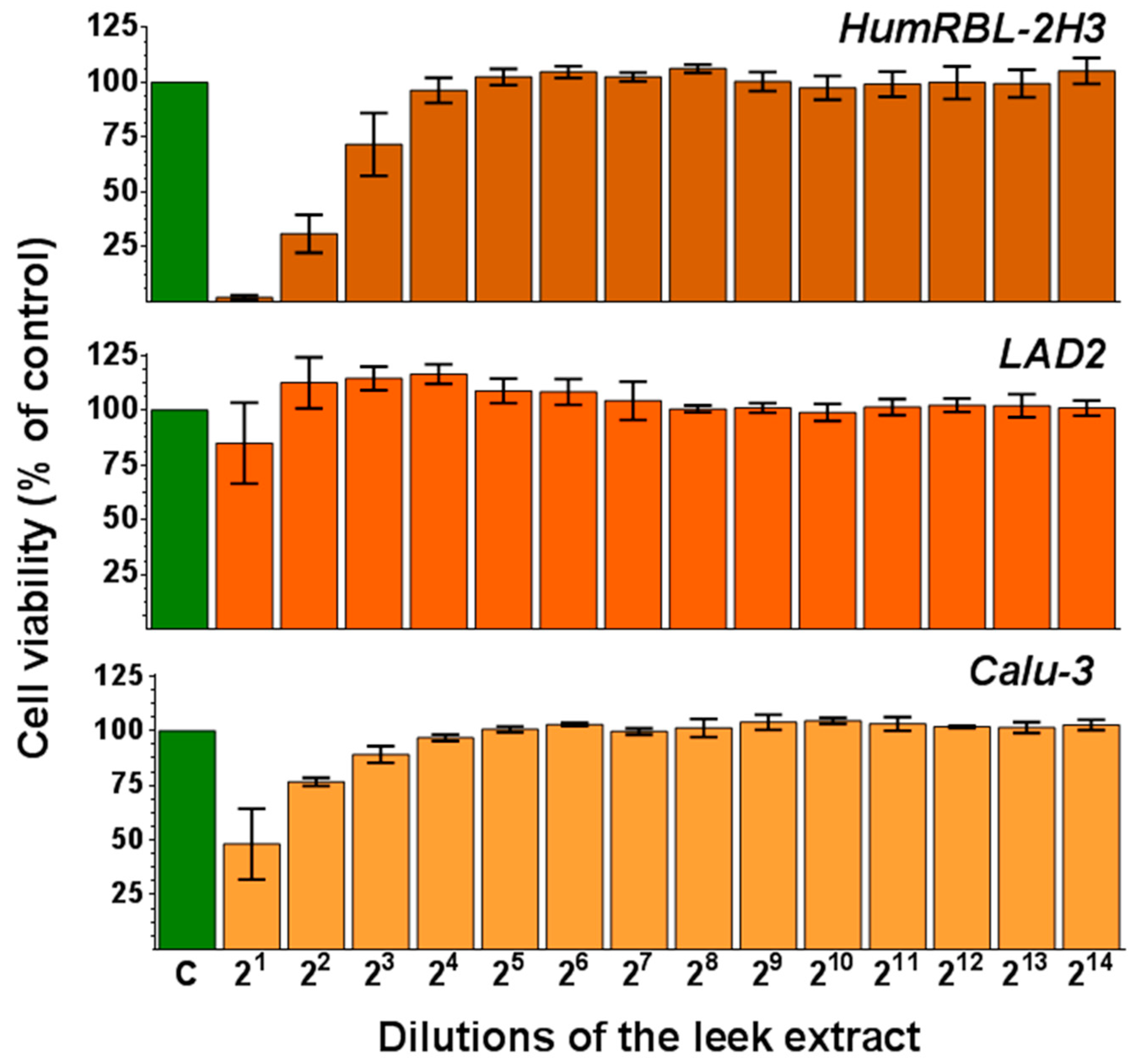
3.3. Leek Extract Exhibits Cytotoxic Activity in a Dose-Dependent Manner

In order to evaluate the cytotoxic effect of the methanolic LE, a cell viability assay was performed on three cell lines using MTT: Humanized rat basophils (HumRBL-2H3), human mast LAD2 cells, and human bronchial epithelial cells (Calu-3). The results are shown in Figure 1. No cytotoxic effect was observed for the LE on any of the tested cell lines at dilution ≥24. However, the cytotoxicity of the LE increased as the concentration did. At 22 dilution, LE showed the most cytotoxic activity on HumRBL-2H3 cells (cell viability was reduced to 25%), followed by Calu-3 (75% cell viability). No significant cytotoxic effect of LE was detected on the LAD2 cells at any of the tested concentrations.

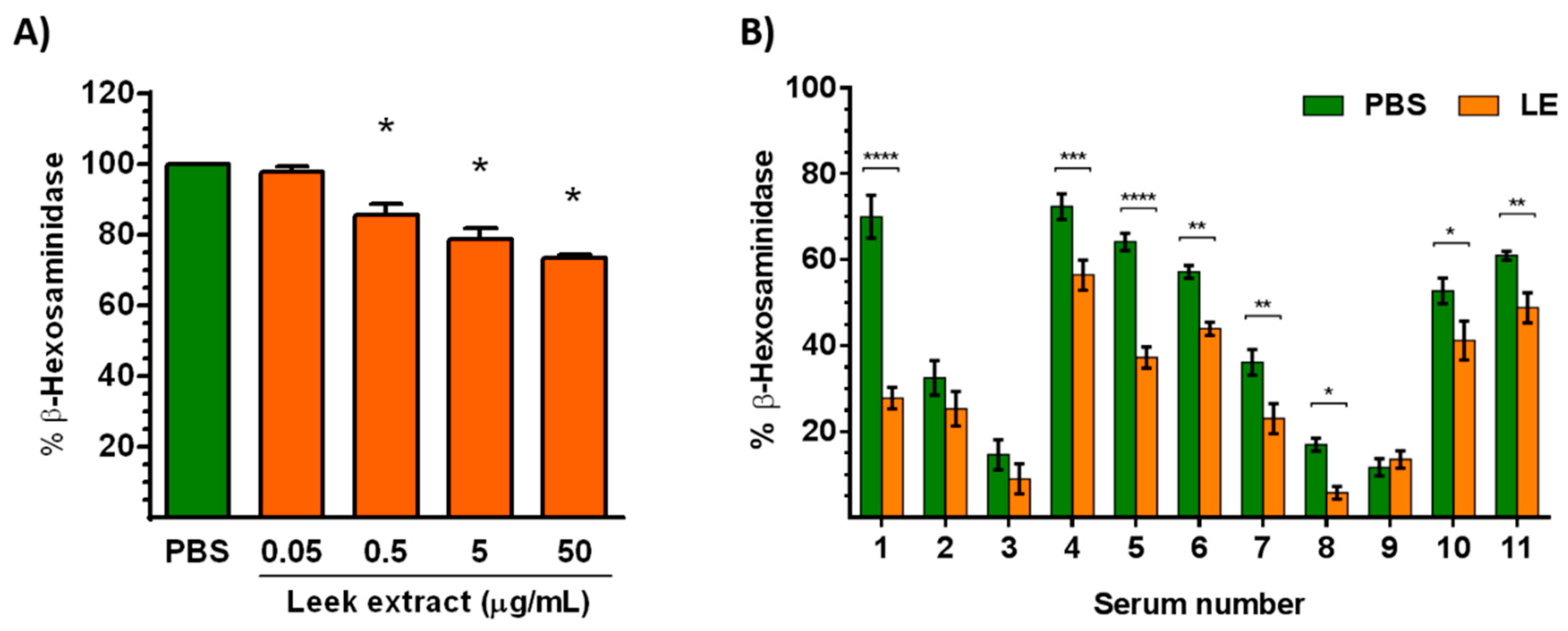
3.4. Leek Extract Decreases Degranulation of Mast Cells

To assay the effect of LE on degranulation of effector cells, the human mast cell line LAD2 was treated with LE for 48 h before addition of the component 48/80, a polymer that promotes the activation and degranulation of mast cells through an IgE-independent pathway. As shown in Figure 2A, LE decreased the degranulation of LAD2 cells, expressed as the percentage (%) of β-hexosaminidase release, in a dose-dependent manner.
Leek extract also decreased the IgE-mediated degranulation of HumRBL-2H3 cells promoted by the Ole e 1 allergen in eight out of eleven individual sera tested from olive pollen-allergic patients (Figure 2B).

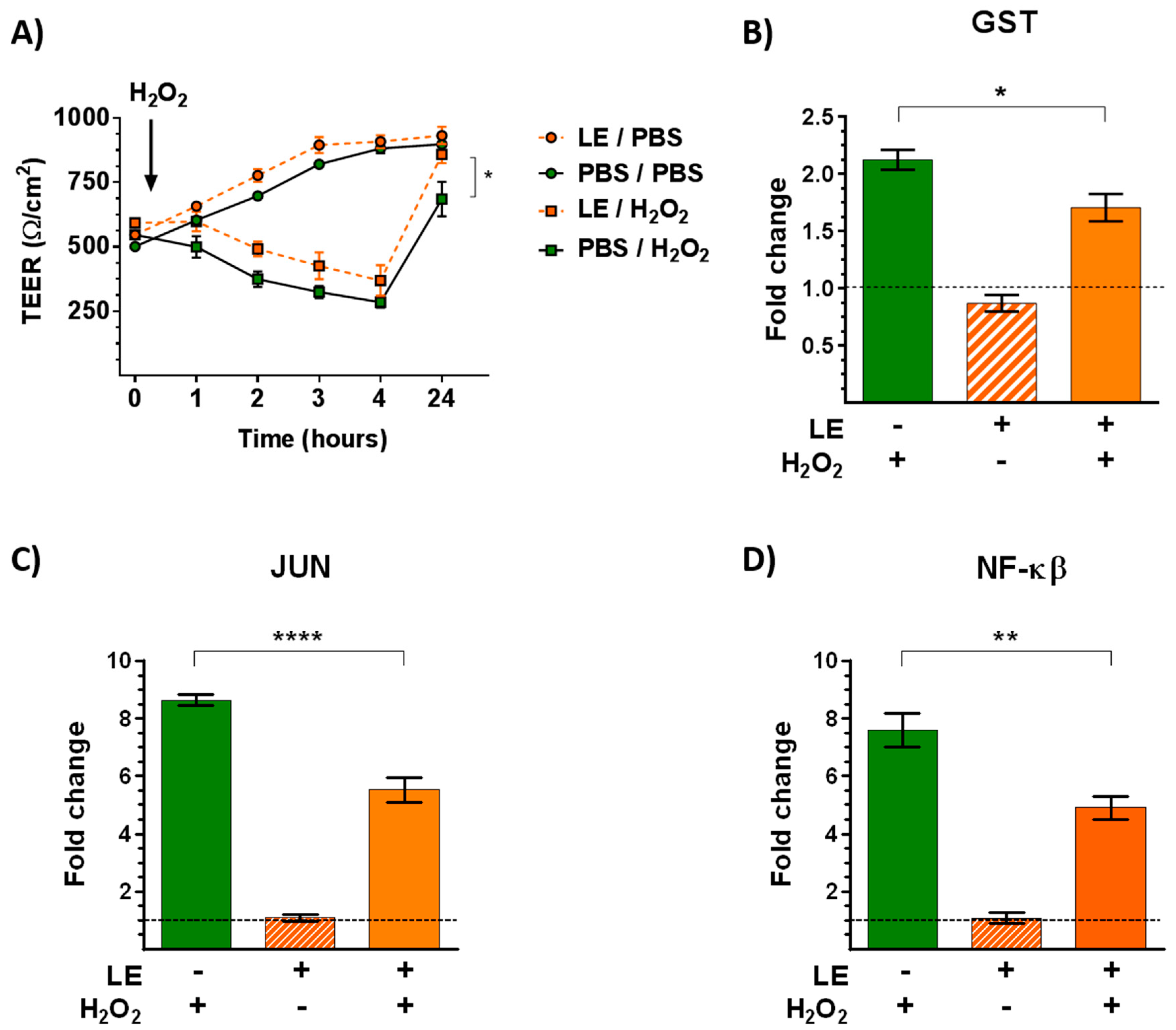
3.5. Leek Extract Prevents Both the Decrease of TEER and Gene Expression Induced by H2O2 Oxidative Stress/Inflammatory Stimulus

Pulmonary epithelium constitutes a protective physical and immunological barrier against exogenous substances. The effect of LE on epithelial barrier integrity was analyzed in ALI-culturedCalu-3 cells after exposure to H2O2 oxidative stress/inflammatory stimulus on day 7. Changes in TEER values (an indirect measure of intercellular apical junction formation) were monitored at different time-points post-treatment (Figure 3A). TEER values were not significantly higher on cells treated with LE compared to control cells. Treatment of cells with 1 mM H2O2 induced a decrease to 70% of control TEER values in a time-dependent manner. LE pretreatment protected against the deleterious effect of H2O2 on TEER values and seemed to promote epithelial barrier maturation.
To know more about the mechanism of action of LE, the expression of genes implicated in redox metabolism was analyzed by RT–PCR (Figure 3B–D). Leek extract treatment did not modify the expression levels of glutathione S-transferase (GST), c-Jun, and nuclear factor kappa B (NFĸB) mRNA in comparison to control cells. As expected, exposure to H2O2 increased mRNA levels of the three studied genes. However, this effect on mRNA levels was significantly prevented by LE, suggesting that LE can regulate the bronchial epithelial response induced by oxidative stress/inflammatory stimuli.

3.6. Leek Extract Decreases Epithelial Permeability to an Allergen

Epithelial permeability to Ole e 1, the main allergen of olive pollen, was analyzed to determine whether the positive effect of LE on the physical barrier was associated with a decrease in its permeability to the allergen. For this purpose, ALI-cultured Calu-3 cells were apically exposed to Ole e 1, in the presence or absence of LE, on day 2, which mimicked an impaired barrier as indicated by the low TEER values (Figure 4A). After 7 h, the presence of the allergen in the apical and basolateral media was detected by a Western blot using a specific anti-Ole e 1 polyclonal antiserum (Figure 4B). ALI-cultured Calu 3-cells displayed a disrupted physical barrier that exhibited permeability to Ole e 1. Interestingly, pretreatment with LE reduced permeability of Calu-3 cells to the allergen: No allergen was detected on the basolateral medium of treated cells. These results correlated with the significantly higher TEER values exhibited by cells treated with LE. Taken together, these data supported that LE has a protective effect on the bronchial physical barrier and thus may be useful in preventing the sensitization to allergens and the exacerbation of allergic diseases.

3.7. Leek Extract Decreases the Apical Release of IL-6 by Bronchial Epithelial Cells

We analyzed the effect of LE on the IL-6 level released by ALI-cultured Calu-3 cells into both apical and basolateral media after 24 h on day 2. The apical IL-6 level was significantly lower in cells treated with LE compared to control cells (Figure 4C). No differences were detected in basolateral media.

4. Discussion

Species from the Allium genus- including A. cepa (onion), A. sativum (garlic), A. ascalonicum (shallot), and A. porrum (leek) are rich sources of secondary metabolites [33,34]. Among them, phenolic compounds are one of the most abundant metabolites, which exhibit anti-inflammatory and antioxidant properties [35,36]. Several studies have suggested that this type of compounds has a therapeutic potential to treat different diseases, including allergy [37].
Leek extract was obtained using methanol extraction since it has been reported as one of the best solvents to obtain plant materials in terms of yield, total phenol content, and antioxidant activity [38]. The qualitative composition of LE included twelve organic acids such as boric acid, fumaric acid, stearic acid, and oleic acid, for which antioxidant activity has been previously reported [39,40,41,42]. Several types of sugar—such as D-glucose and D-fructose—have also been identified; these represent a fast energy source. The L-amino acids valine and proline were identified, and their immunostimulatory properties have been shown in previous studies [43,44]. In addition, it has been reported that many compounds from leek protect against various diseases through their antioxidant activity, being able to chelate metals or to neutralize electron-stealing reactions involving free radicals [45].
The total phenol content found in LE (2.3 ± 0.2 mg/g dry weight) was lower than previously reported by other authors. This difference can be explained in terms of genetic, environmental, and experimental factors such as plant variety, geographical region, climatic conditions or extraction procedure, among others. Bernaert et al. found that total phenol content varied from 5 to 15 mg GAE/g dry weight in the extract of the 30 leek varieties analyzed [36]. Moreover, Piluzza and Bullitta reported that the total phenol content of onion and garlic extracts—two species belonging to the Allium genus—were 3.80 ± 0.42 mg EAG/g dry weight and 3.18 ± 0, 16 mg EAG/g dry weight, respectively [46].
Several flavonoids glycosides and steroidal saponins were found in LE. Beneficial effects on human health have been reported for phenolic components attributed to their antioxidant activity due to their redox properties, which can play an important role in absorbing and neutralizing free radicals, quenching singlet and triplet oxygen, or decomposing peroxides [47,48,49].
We found a high antioxidant activity of methanolic LE. Numerous studies have reported both the total antioxidant and radical scavenging activities of members of the Allium genus, ranging from 14.5 to 128 mg AAE/g [22,50] and 11 to 15,000 μg/mL, respectively [33,51].
LE decreased the degranulation of mast cells through both an IgE-independent and -dependent pathway. The inhibitory effect of plant extracts on mast cell degranulation has been reported before. Yoo et al. demonstrated that aged black garlic extract inhibited the release of β-hexosaminidase on HumRBL-2H3 cells [52]. Moreover, Lee et al. showed that the Korean herbal medicine KOTMIN13 inhibited the degranulation of bone marrow mast cells derived [53]. Our results demonstrated that methanolic LE inhibits the degranulation of both HumRBL-2H3 and LAD2 cells in response to immunological and non-immunological stimuli, respectively. Therefore, LE could be useful as a mast cell stabilizer in allergy.
Since respiratory epithelial barrier disruption has been observed in allergic patients, and airborne pollutants are considered as important inductors of lung oxidative stress [54], resulting in altered lung function [55], LE pretreatment protected against the deleterious effect of H2O2 on TEER values and promoted epithelial barrier maturation. The protective effect of plant extracts against oxidative stress/inflammatory stimuli has been reported for other species, including Capsicum annuum (pepper) [56], Alpinia katsumadai [57], and Boswellia serrate [58].
Exposure to environmental pollutants induces oxidative stress via the activation of transcription factors such as NFĸB and c-Jun [59,60]. Moreover, the intracellular antioxidant defense system includes metabolic enzymes such as GST, which catalyzes the conjugation of the reduced form of glutathione to xenobiotic substrates [61]. We found that LE treatment of epithelial cells prevented the increase in the expression of genes implicated in redox metabolism.
IL-6 is a pro-inflammatory cytokine found at high levels in asthmatic patients [62], and we found significantly lower levels of this cytokine in epithelial cells treated with LE compared to control cells. Previous studies using extracts from other plants of the Allium genus have also shown their ability to reduce the production of pro-inflammatory cytokines in vivo. According to Oliveira et al., extracts of onion were able to reduce IL-4 and IL-5 levels in bronchoalveolar lavage of a murine model of asthma [63] and Kim et al. observed that extracts of A. hookeri decreased levels of TNF-α in nasal mucosa of a murine model of allergic rhinitis [64].

5. Conclusions

In summary, the results obtained in this work suggest that A. porrum extract may have potential anti-allergic effects due to its antioxidant and anti-inflammatory properties. More research is needed in order to elucidate the mechanisms and the bioactive compounds responsible for these actions.

Author Contributions

Conceptualization, S.B. and E.B.; Data curation, S.B. and A.G.; Formal analysis, S.B. and A.G.; Funding acquisition, M.V.; Investigation, S.B., A.G. and E.B.; Methodology, S.B., A.G. and E.B.; Project administration, E.B.; Supervision, S.B. and E.B.; Validation, S.B.; Visualization, S.B.; Writing—original draft, S.B.; Writing—review and editing, S.B., A.G., M.V. and E.B.

Funding

This work was supported by grants from the Spanish Ministry of Economy and Competitivity SAF2014-53209-R , SAF2017-86483-R, PI15/00107 and RIRAAF Network RD16/0006/0014 from the ISCIII. S.B. is supported by a Juan de la Cierva Formación contract from the Spanish Ministerio de Economía.

Acknowledgments

Authors thank Lothan Vogel for the HumRBL-2H3 cell line donation.

Conflicts of Interest

The authors have no financial conflicts of interest.

References

  1. World Health Organization. WHO Monographs On Selected Medicinal Plants; World Health Organization: Geneva, Switzerland, 1999. [Google Scholar]
  2. Pawankar, R.; Canonica, G.W.; Holgate, S.T.; Lockey, R.F.; Blaiss, M.S. The WAO White Book on Allergy: Update 2013; World Allergy Organization: Milwaukee, WI, USA, 2013. [Google Scholar]
  3. Durrani, S.R.; Viswanathan, R.K.; Busse, W.W. What effect does asthma treatment have on airway remodeling? Current perspectives. J. Allergy Clin. Immunol. 2011, 128, 439–448. [Google Scholar] [CrossRef] [PubMed]
  4. Finn, D.F.; Walsh, J.J. Twenty-first century mast cell stabilizers. Br. J. Pharmacol. 2013, 170, 23–37. [Google Scholar] [CrossRef] [PubMed] [Green Version]
  5. Castillo, M.; Scott, N.W.; Mustafa, M.Z.; Mustafa, M.S.; Azuara-Blanco, A. Topical antihistamines and mast cell stabilisers for treating seasonal and perennial allergic conjunctivitis. Cochrane Libr. 2015, 6, CD009566. [Google Scholar] [CrossRef] [PubMed]
  6. Han, S.J.; Bae, E.A.; Trinh, H.T.; Yang, J.H.; Youn, U.J.; Bae, K.H.; Kim, D.H. Magnolol and honokiol: Inhibitors against mouse passive cutaneous anaphylaxis reaction and scratching behaviors. Biol. Pharm. Bull. 2007, 30, 2201–2203. [Google Scholar] [CrossRef] [PubMed]
  7. Kang, O.H.; Jang, H.J.; Chae, H.S.; Oh, Y.C.; Choi, J.G.; Lee, Y.S.; Kim, J.H.; Kim, Y.C.; Sohn, D.H.; Park, H.; et al. Anti-inflammatory mechanisms of resveratrol in activated HMC-1 cells: Pivotal roles of NFκB and MAPK. Pharmacol. Res. 2009, 59, 330–337. [Google Scholar] [CrossRef] [PubMed]
  8. Lee, J.H.; Kim, J.W.; Ko, N.Y.; Mun, S.H.; Her, E.; Kim, B.K.; Han, J.W.; Lee, H.Y.; Beaven, M.A.; Kim, Y.M.; et al. Curcumin, a constituent of curry, suppresses IgE-mediated allergic response and mast cell activation at the level of Syk. J. Allergy Clin. Immunol. 2008, 121, 1225–1231. [Google Scholar] [CrossRef] [PubMed]
  9. Kimata, M.; Inagaki, N.; Nagai, H. Effects of luteolin and other flavonoids on IgE-mediated allergic reactions. Planta Med. 2000, 66, 25–29. [Google Scholar] [CrossRef] [PubMed]
  10. Kim, N.H.; Jeong, H.J.; Kim, H.M. Theanine is a candidate amino acid for pharmacological stabilization of mast cells. Amino Acids 2012, 42, 1609–1618. [Google Scholar] [CrossRef]
  11. Penissi, A.B.; Vera, M.E.; Mariani, M.L.; Rudolph, M.I.; Ceñal, J.P.; de Rosas, J.C.; Fogal, T.H.; Tonn, C.E.; Favier, L.S.; Giordano, O.S.; et al. Novel anti-ulcer α, β-unsaturated lactones inhibit compound 48/80-induced mast cell degranulation. Eur. J. Pharmacol. 2009, 612, 122–130. [Google Scholar] [CrossRef]
  12. Huang, F.; Yamaki, K.; Tong, X.; Fu, L.; Zhang, R.; Cai, Y.; Yanagisawa, R.; Inoue, K.; Takano, H.; Yoshino, S. Inhibition of the antigen-induced activation of RBL-2H3 cells by sinomenine. Int. Immunopharmacol. 2008, 8, 502–507. [Google Scholar] [CrossRef]
  13. Qu, J.; Li, Y.; Zhong, W.; Gao, P.; Hu, C. Recent developments in the role of reactive oxygen species in allergic asthma. J. Thorac. Dis. 2017, 9, E32. [Google Scholar] [CrossRef] [PubMed]
  14. Corzo-Martinez, M.; Corzo, N.; Villamiel, M. Biological properties of onions and garlic. Trends Food Sci. Technol. 2007, 18, 609–625. [Google Scholar] [CrossRef]
  15. Rizwani, G.H.; Shareef, H. Genus Allium: The potential nutritive and therapeutic source. JPANS 2011, 1, 158–165. [Google Scholar] [CrossRef]
  16. Lee, K.H.; Park, E.; Lee, H.J.; Kim, M.O.; Cha, Y.J.; Kim, J.M.; Lee, H.; Shin, M.J. Effects of daily quercetin-rich supplementation on cardiometabolic risks in male smokers. Nutr. Res. Pract. 2011, 5, 28–33. [Google Scholar] [CrossRef] [PubMed] [Green Version]
  17. Pal, C.B.T.; Jadeja, G.C. Deep eutectic solvent-based extraction of polyphenolic antioxidants from onion (Allium cepa L.) peel. J. Sci. Food Agric. 2019, 99, 1969–1979. [Google Scholar] [CrossRef] [PubMed]
  18. Jalalvand, A.R.; Zhaleh, M.; Goorani, S.; Zangeneh, M.M.; Seydi, N.; Zangeneh, A.; Moradi, R. Chemical characterization and antioxidant, cytotoxic, antibacterial, and antifungal properties of ethanolic extract of Allium Saralicum, R.M. Fritsch leaves rich in linolenic acid, methyl ester. J. Photochem. Photobiol. B 2019, 192, 103–112. [Google Scholar] [CrossRef]
  19. Zare, A.; Farzaneh, P.; Pourpak, Z.; Zahedi, F.; Moin, M.; Shahabi, S.; Hassan, Z.M. Purified aged garlic extract modulates allergic airway inflammation in BALB/c mice. Iran J. Allergy Asthma Immunol. 2008, 7, 133–141. [Google Scholar] [PubMed]
  20. Di Donna, L.; Mazzotti, F.; Taverna, D.; Napoli, A.; Sindona, G. Structural characterisation of malonyl flavonols in leek (Allium porrum L.) using high-performance liquid chromatography and mass spectrometry. Phytochem. Anal. 2014, 25, 207–212. [Google Scholar] [CrossRef]
  21. Soininen, T.H.; Jukarainen, N.; Soininen, P.; Auriola, S.O.K.; Julkunen-Tiitto, R.; Oleszek, W.; Stochmal, A.; Karjalainen, R.O.; Vepsalainen, J.J. Metabolite profiling of leek (Allium porrum L) cultivars by H-1 NMR and HPLC-MS. Phytochem. Anal. 2014, 25, 220–228. [Google Scholar] [CrossRef]
  22. Radovanović, B.; Mladenović, J.; Radovanović, A.; Pavlović, R.; Nikolić, V. Phenolic composition, antioxidant, antimicrobial and cytotoxic activities of Allium porrum L. (Serbia) extracts. J. Food Nutr. Res. 2015, 3, 564–569. [Google Scholar]
  23. Prieto, P.; Pineda, M.; Aguilar, M. Spectrophotometric quantitation of antioxidant capacity through the formation of a phosphomolybdenum complex: Specific application to the determination of vitamin E. Anal. Biochem. 1999, 269, 337–341. [Google Scholar] [CrossRef] [PubMed]
  24. Brand-Williams, W.; Cuvelier, M.E.; Berset, C.L.W.T. Use of a free radical method to evaluate antioxidant activity. LWT-Food Sci. Technol. 1995, 28, 25–30. [Google Scholar] [CrossRef]
  25. Acero, N.; Gradillas, A.; Beltran, M.; García, A.; Muñoz Mingarro, D. Comparison of phenolic compounds profile and antioxidant properties of different sweet cherry (Prunus avium L.) varieties. Food Chem. 2019, 279, 260–271. [Google Scholar] [CrossRef] [PubMed]
  26. Villalba, M.; Batanero, E.; López-Otín, C.; Sánchez, L.M.; Monsalve, R.I.; González de la Peña, M.A.; Lahoz, C.; Rodríguez, R. Amino acid sequence of Ole e 1, the major allergen from olive tree polen (Olea europea). Eur. J. Biochem. 1993, 103, 147–153. [Google Scholar]
  27. Kirshenbaum, A.S.; Akin, C.; Wu, Y.; Rottem, M.; Goff, J.P.; Beaven, M.A.; Rao, V.K.; Metcalfe, D.D. Characterization of novel stem cell factor responsive human mast cell lines LAD 1 and 2 established from a patient with mast cell sarcoma/leukemia; activation following aggregation of FcepsilonRI or FcgammaRI. Leuk. Res. 2003, 27, 677–682. [Google Scholar] [CrossRef]
  28. Gadermaier, G.; Jahn-Schmid, B.; Vogel, L.; Egger, M.; Himly, M.; Briza, P.; Ebner, C.; Vieths, S.; Bohle, B.; Ferreira, F. Targeting the cysteine-stabilized fold of Art v 1 for immunotherapy of Artemisia pollen allergy. Mol. Immunol. 2010, 47, 1292–1298. [Google Scholar] [CrossRef] [PubMed]
  29. Denizot, F.; Lang, R. Rapid colorimetric assay for cell growth and survival. Modifications to the tetrazolium dye procedure giving improved sensitivity and reliability. J. Immunol. Methods 1986, 89, 271–277. [Google Scholar] [CrossRef]
  30. Prado, N.; De Linares, C.; Sanz, M.L.; Gamboa, P.; Villalba, M.; Rodríguez, R.; Batanero, E. Pollensomes as natural vehicles for pollen allergens. J. Immunol. 2015, 195, 445–449. [Google Scholar] [CrossRef]
  31. Vogel, L.; Lüttkopf, D.; Hatahet, L.; Haustein, D.; Vieths, S. Development of a functional in vitro assay as a novel tool for the standardization of allergen extracts in the human system. Allergy 2005, 60, 1021–1028. [Google Scholar] [CrossRef]
  32. Kachlicki, P.; Piasecka, A.; Stobiecki, M.; Marczak, L. Structural Characterization of Flavonoid Glycoconjugates and Their Derivatives with Mass Spectrometric Techniques. Molecules 2016, 21, 1494. [Google Scholar] [CrossRef]
  33. García-Herrera, P.; Morales, P.; Fernández-Ruiz, V.; Sánchez-Mata, M.C.; Cámara, M.; Carvalho, A.M.; Ferreira, I.C.F.R.; Pardo-de-Santayana, M.; Molina, M.; Tardio, J. Nutrients, phytochemicals and antioxidant activity in wild populations of Allium ampeloprasum L., a valuable underutilized vegetable. Food Res. Int. 2014, 62, 272–279. [Google Scholar] [CrossRef]
  34. Bernaert, N.; De Paepe, D.; Bouten, C.; De Clercq, H.; Stewart, D.; Van Bockstaele, E.; De Loose, M.; Van Droogenbroeck, B. Antioxidant capacity, total phenolic and ascorbate content as a function of the genetic diversity of leek (Allium ampeloprasum var. porrum). Food Chem. 2012, 134, 669–677. [Google Scholar] [CrossRef] [PubMed]
  35. Chen, S.; Shen, X.; Cheng, S.; Li, P.; Du, J.; Chang, Y.; Meng, H. Evaluation of garlic cultivars for polyphenolic content and antioxidant properties. PLoS ONE 2013, 8, e79730. [Google Scholar] [CrossRef] [PubMed]
  36. Rogerio, A.P.; Kanashiro, A.; Fontanari, C.; Da Silva, E.V.G.; Lucisano-Valim, Y.M.; Soares, E.G.; Faccioli, L.H. Anti-inflammatory activity of quercetin and isoquercitrin in experimental murine allergic asthma. Inflamm. Res. 2007, 56, 402–408. [Google Scholar] [CrossRef]
  37. Kaiser, P.; Youssouf, M.S.; Tasduq, S.A.; Singh, S.; Sharma, S.C.; Singh, G.D.; Gupta, V.K.; Gupta, B.D.; Johri, R.K. Anti-allergic effects of herbal product from Allium cepa (bulb). J. Med. Food. 2009, 12, 374–382. [Google Scholar] [CrossRef] [PubMed]
  38. Scherer, R.; Godoy, H.T. Effects of extraction methods of phenolic compounds from Xanthium strumarium L. and their antioxidant activity. Rev. Bras. Plantas Med. 2014, 16, 41–46. [Google Scholar] [CrossRef]
  39. Yamada, K.E.; Eckhert, C.D. Boric acid activation of eIF2α and Nrf2 Is PERK dependent: A mechanism that explains how boron prevents DNA damage and enhances antioxidant status. Biol. Trace Elem. Res. 2019, 188, 2–10. [Google Scholar] [CrossRef]
  40. Linker, R.A.; Lee, D.H.; Ryan, S.; van Dam, A.M.; Conrad, R.; Bista, P.; Zeng, W.; Hronowsky, X.; Buko, A.; Chollate, S.; et al. Fumaric acid esters exert neuroprotective effects in neuroinflammation via activation of the Nrf2 antioxidant pathway. Brain 2011, 134, 678–692. [Google Scholar] [CrossRef] [Green Version]
  41. Wang, Z.J.; Liang, C.L.; Li, G.M.; Yu, C.Y.; Yin, M. Stearic acid protects primary cultured cortical neurons against oxidative stress. Acta Pharmacol. Sin. 2007, 28, 315–326. [Google Scholar] [CrossRef] [Green Version]
  42. Wei, C.C.; Yen, P.L.; Chang, S.T.; Cheng, P.L.; Lo, Y.C.; Liao, V.H. Antioxidative activities of both oleic acid and Camellia tenuifolia seed oil are regulated by the transcription factor DAF-16/FOXO in Caenorhabditis elegans. PLoS ONE 2016, 11, e0157195. [Google Scholar] [CrossRef]
  43. Luo, J.B.; Feng, L.; Jiang, W.D.; Liu, Y.; Wu, P.; Jiang, J.; Kuang, S.Y.; Tang, L.; Zhang, Y.A.; Zhou, X.Q. The impaired intestinal mucosal immune system by valine deficiency for young grass carp (Ctenopharyngodon idella) is associated with decreasing immune status and regulating tight junction proteins transcript abundance in the intestine. Fish Shellfish Immunol. 2014, 40, 197–207. [Google Scholar] [CrossRef]
  44. Ren, W.; Zou, L.; Ruan, Z.; Li, N.; Wang, Y.; Peng, Y.; Liu, G.; Yin, Y.; Li, T.; Hou, Y.; et al. Dietary L-proline supplementation confers immunostimulatory effects on inactivated Pasteurella Multocida vaccine immunized mice. Amino Acids 2013, 45, 555–561. [Google Scholar] [CrossRef]
  45. Seabra, R.M.; Andrade, P.B.; Valentão, P.; Fernandes, E.; Carvalho, F.; Bastos, M.L. Anti-oxidant compounds extracted from several plant materials. In Biomaterials from Aquatic and Terrestrial Organisms, 1st ed.; Fingerman, M., Ed.; CRC Press: Boca Raton, FL, USA, 2006; Chapter 4. [Google Scholar] [CrossRef]
  46. Piluzza, G.; Bullitta, S. Correlations between phenolic content and antioxidant properties in twenty-four plant species of traditional ethnoveterinary use in the Mediterranean area. Pharm. Biol. 2011, 49, 240–247. [Google Scholar] [CrossRef]
  47. Grzesik, M.; Naparło, K.; Bartosz, G.; Sadowska-Bartosz, I. Antioxidant properties of catechins: Comparison with other antioxidants. Food Chem. 2018, 241, 480–492. [Google Scholar] [CrossRef]
  48. Qu, W.; Fan, L.; Kim, Y.C.; Ishikawa, S.; Iguchi-Ariga, S.M.; Pu, X.P.; Ariga, H. Kaempferol derivatives prevent oxidative stress-induced cell death in a DJ-1-dependent manner. J. Pharmacol. Sci. 2009, 110, 191–200. [Google Scholar] [CrossRef]
  49. Nile, S.H.; Nile, A.S.; Keum, Y.S.; Sharma, K. Utilization of quercetin and quercetin glycosides from onion (Allium cepa L.) solid waste as an antioxidant, urease and xanthine oxidase inhibitors. Food Chem. 2017, 235, 119–126. [Google Scholar] [CrossRef]
  50. Gunathilake, K.D.P.P.; Ranaweera, K.K.D.S. Antioxidative properties of 34 green leafy vegetables. J. Funct. Foods 2016, 26, 176–186. [Google Scholar] [CrossRef] [Green Version]
  51. Assadpour, S.; Nabavi, S.M.; Nabavi, S.F.; Dehpour, A.A.; Ebrahimzadeh, M.A. In vitro antioxidant and antihemolytic effects of the essential oil and methanolic extract of Allium rotundum L. Eur. Rev. Med. Pharmacol. Sci. 2016, 20, 5210–5215. [Google Scholar]
  52. Yoo, J.M.; Sok, D.E.; Kim, M.R. Anti-allergic action of aged black garlic extract in RBL-2H3 cells and passive cutaneous anaphylaxis reaction in mice. J. Med. Food. 2014, 17, 92–102. [Google Scholar] [CrossRef]
  53. Lee, E.; Kim, S.G.; Park, N.Y.; Park, H.H.; Jeong, K.T.; Choi, J.; Lee, I.H.; Lee, H.; Kim, K.J.; Lee, E. KOTMIN13, a Korean herbal medicine alleviates allergic inflammation in vivo and in vitro. BMC Complement. Altern. Med. 2016, 16, 169. [Google Scholar] [CrossRef]
  54. Valavanidis, A.; Vlachogianni, T.; Fiotakis, K.; Loridas, S. Pulmonary oxidative stress, inflammation and cancer: Respirable particulate matter, fibrous dusts and ozone as major causes of lung carcinogenesis through reactive oxygen species mechanisms. Int. J. Environ. Res. Public Health 2013, 10, 3886–3907. [Google Scholar] [CrossRef]
  55. Evans, M.D.; Dizdaroglu, M.; Cooke, M.S. Oxidative DNA damage and disease: Induction, repair and significance. Mutat. Res. 2004, 567, 1–61. [Google Scholar] [CrossRef]
  56. Jang, H.Y.; Kim, S.M.; Yuk, J.E.; Kwon, O.K.; Oh, S.R.; Lee, H.K.; Jeong, H.; Ahn, K.S. Capsicum annuum L. methanolic extract inhibits ovalbumin-induced airway inflammation and oxidative stress in a mouse model of asthma. J. Med. Food 2011, 14, 1144–1151. [Google Scholar] [CrossRef]
  57. Lee, M.Y.; Lee, N.H.; Seo, C.S.; Lee, J.A.; Jung, D.; Kim, J.H.; Shin, H.K. Alpinia katsumadai seed extract attenuate oxidative stress and asthmatic activity in a mouse model of allergic asthma. Food Chem. Toxicol. 2010, 48, 1746–1752. [Google Scholar] [CrossRef]
  58. Catanzaro, D.; Rancan, S.; Orso, G.; Dall’Acqua, S.; Brun, P.; Giron, M.C.; Carrara, M.; Castagliuolo, I.; Ragazzi, E.; Caparrotta, L.; et al. Boswellia serrata preserves intestinal epithelial barrier from oxidative and inflammatory damage. PLoS ONE 2015, 10, e0125375. [Google Scholar] [CrossRef]
  59. Bhalla, D.K. Ozone-induced lung inflammation and mucosal barrier disruption: Toxicology, mechanisms, and implications. J. Toxicol. Environ. Health B Crit. Rev. 1999, 2, 31–86. [Google Scholar] [CrossRef]
  60. Janssen, Y.M.; Matalon, S.; Mossman, B.T. Differential induction of c-fos, c-jun, and apoptosis in lung epithelial cells exposed to ROS or RNS. Am. J. Physiol. 1997, 273, 789–796. [Google Scholar] [CrossRef]
  61. Oakley, A. Glutathione transferases: A structural perspective. Drug Metab. Rev. 2011, 43, 13–151. [Google Scholar] [CrossRef]
  62. Marini, M.; Vittori, E.; Hollemborg, J.; Mattoli, S. Expression of the potent inflammatory cytokines, granulocyte-macrophage-colony-stimulating factor and interleukin-6 and interleukin-8, in bronchial epithelial cells of patients with asthma. J. Allergy Clin. Immunol. 1992, 89, 1001–1009. [Google Scholar] [CrossRef]
  63. Oliveira, T.T.; Campos, K.M.; Cerqueira-Lima, A.T.; Carneiro, T.C.B.; da Silva Velozo, E.; Ribeiro Melo, I.C.; Figueiredo, E.A.; de Jesus Oliveira, E.; de Vasconcelos, D.F.; Pontes-de-Carvalho, L.C.; et al. Potential therapeutic effect of Allium cepa L. and quercetin in a murine model of Blomia tropicalis induced asthma. DARU J. Pharm. Sci. 2015, 23, 18. [Google Scholar] [CrossRef]
  64. Kim, H.Y.; Nam, S.Y.; Hong, S.W.; Kim, M.J.; Jeong, H.J.; Kim, H.M. Protective effects of rutin through regulation of vascular endothelial growth factor in allergic rhinitis. Am. J. Rhinol. Allergy 2015, 29, e87–e94. [Google Scholar] [CrossRef]
Figure 1. Cytotoxicity of methanolic LE at different concentrations on three cell lines: Humanized rat basophils (HumRBL-2H3), human mast cells (LAD2), and bronchial epithelial cells (Calu-3). The cell viability was assayed using the 3-(4,5-dimethylthiazol-2-yl)-2,5-diphenyltetrazolium bromide (MTT) method after 72 h of treatment. DMSO was used as positive control. Data are expressed as a percentage (%) of control (C) and shown as the mean ± SEM of three independent experiments.
Nutrients 11 01303 g001
Figure 2. Measurement of β-hexosaminidase after the activation of (A) human mast cells LAD2 with the compound 48/80 (5 μg/mL) and (B) HumRBL-2H3 with Ole e 1 (25 ng/mL) after treatment with leek extract (LE) −0.05–50 μg/mL (A) or 5 μg/mL (B) for 48 h. HumRBL-2H3 were previously sensitized with sera from patients allergic to Ole e 1. * p < 0.05, ** p < 0.01, *** p < 0.001, **** p < 0.0001 compared to control cells treated with PBS.
Nutrients 11 01303 g002
Figure 3. (A) Effect of leek extract on the response of epithelial cells to oxidative stress. On day 7, ALI-cultured Calu-3 cells were grown in the presence or absence of leek extract (LE, 5 μg/mL) for 48 h and then exposed to 1 mM H2O2. The time course of transepithelial electrical resistance (TEER) was measured. PBS, control cells. * p < 0.05 mRNA levels of GST (B), c-Jun (C) and NFκB (D) in ALI-cultured Calu-3 cells exposed to 1mM H2O2 for 48 h, after treatment with leek extract (5 μg/mL, LE) compared to control cells (PBS). Relative mRNA levels were determined by RT–PCR and expressed as fold increase of control values after normalization using GADPH as the housekeeping gene. Data are mean ± SEM of three different experiments. * p < 0.05, ** p < 0.01, **** p < 0.0001.
Nutrients 11 01303 g003
Figure 4. Effect of leek extract on the bronchial permeability to Ole e 1. (A) Time course of TEER values on day 2 ALI-cultured Calu-3 cells after treatment with leek extract (LE) compared to control cells (PBS). Data are the mean ± SD of triplicate determinations. * p < 0.05. (B) ALI-cultured Calu-3 cells were exposed to Ole e 1 (2.5 µg) on day 2 for 7 h after treatment with leek extract (LE, 5 μg/mL) for 24 h, and the presence of the allergen was determined in the medium using a rabbit polyclonal antibody. (C) ALI-cultured Calu-3 cells were treated with leek extract at 5 μg/mL (LE) for 24 h on day 2, and IL-6 levels in the medium were determined by sandwich ELISA. PBS, control cells. Data are the mean ± SD of triplicate determinations ** p < 0.01.
Nutrients 11 01303 g004
Table 1. Primers used for RT–PCR amplification of specific genes on Calu-3 cells at ALI.
GeneForward Sequence 5′-3′Reverse Primer 5′-3′
GAPDHAAAGGGTCATCATCTCTGGCTGTTGTCATACTTCTC
GSTCGGGCAACTGAAGCCTTTTGTCAGCGAAGGAGATCTGGTC
c-JunGCAAAGAACTTTCCCGGCTGGGAGAAGCCTAAGACGCAGG
NFκBTGAGGATGATGAGAATGGATCGGAACACAATGGCATAC
GAPDH, glyceraldehyde-3-phosphate dehydrogenase; GST, glutathione S-transferase; c-Jun, Jun proto-oncogene; NFκB, Nuclear factor kappa B subunit.
Table 2. Organic acids, carbohydrates, and L-amino acids identified in leek extract (LE) by Gas Chromatography–Mass Spectrometry (GC–MS).
CompoundRetention Time (min)Molecular FormulaMolecular Mass (g/mol)R-Match
Organic acids
Boric acid4.8H3BO361.8814
Propanoic acid5.8C3H6O274.1899
Acetic acid6.1C2H4O260.1849
Phosphoric acid8.9H3PO498.0869
Succinic acid9.5C4H6O4118.09918
Fumaric acid10.0C4H4O4116.1776
Nonanoic acid10.2C9H18O2158.2787
Malic acid11.8C4H6O5134.1859
Arabinonic acid13.5C5H10O6166.1729
Palmitic acid17.8C16H32O2256.4705
Stearic acid19.1C18H36O2284.5702
Oleic acid19.7C18H34O2282.5808
Carbohydrates
Arabinofuranose8.6C5H10O150.1777
Glucofuranoside15.4C6H12O6180.2772
D-fructose15.5C6H12O6180.2809
Mannofuranoside15.6C7H14O6194.2781
Glucose17.1C6H12O6180.2790
Galactopyranose17.1C6H12O6180.2825
Amino acids
L-valine6.3C5H11NO2117.2798
L-proline12.2C5H9NO2115.1850
Table 3. Total phenol content, antioxidant activity, and 2,2-diphenyl-1-picrylhydrazyl radical scavenging activity IC50 of LE.
Mean ± SD
Total phenol content2.3 ± 0.2 a
Total antioxidant activity60.2 ± 3.1 b
DPPH scavenging activity, IC50289.6 ± 6.1 c
SD, standard deviation. (a) mg GAE/g dry extract; (b) mg AAE/g dry extract; (c) μg/mL.
Table 4. Mass spectral data for identification of phenolic compounds in LE by Liquid Chromatography–Mass Spectrometry (LC–IT/MS).
Polyphenol
Subclass (Flavonoids)
Compound NameRetention Time (min) Positive Ions (m/z)
MW[M+H]+[M+Na]+[M+K]+In-Source Fragments
flavanols(-)-Epicatechin a10.0290291------
flavanols(+)-Catechin a12.3290291------
flavonolsKaempferol derivative b19.4742743765----
[Aglycone+H]+ = 287,
[Aglycone+H-H2O]+ = 269
flavonolsKaempferol derivative c19.8902903925--[(M-Rham)+H]+ = 757
[(M-Rham-Glc)+H]+ = 595
[(M-Rham-2Glc)+H]+ = 433
[Aglycone+H]+ = 287,
[Aglycone+H-H2O]+ = 269
flavonolsQuercetin derivative b18.8625627----[(M-Glc)+H]+ = 463
[Aglycone+H]+ = 303,
[Aglycone+H-H2O]+ = 285
flavonolsQuercetin derivative b20.5609611633649[Aglycone+H]+ = 303,
[Aglycone+H-H2O]+ = 285
m/z values for the base peak are given in bold type. (a) Identification by comparison with the pure standard. (b) Identification by in source-fragmentation and by searching in online databases such as FOODB (http://foodb.ca) and METLIN (http://metlin.scripps.edu). (c) Identification by comparison with bibliography: kaempherol-3-O-[rhamnosyl-glucosylglucoside]-7-O-rhamnoside [32].

