

Supernumerary Teeth in the Anterior Maxilla of Non-Syndromic Children and Adolescents: A Retrospective Study Based on Cone-Beam Computed Tomography Scans


 , , , and
Abstract
1. Introduction
2. Materials and Methods
2.1. Study Design, Protocol, and Ethics
2.2. Study Setting
2.3. Study Population
Eligibility Criteria
2.4. Study Outcomes
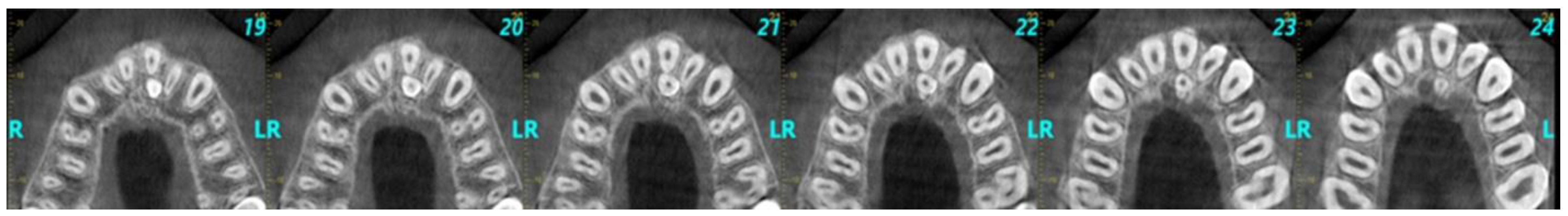
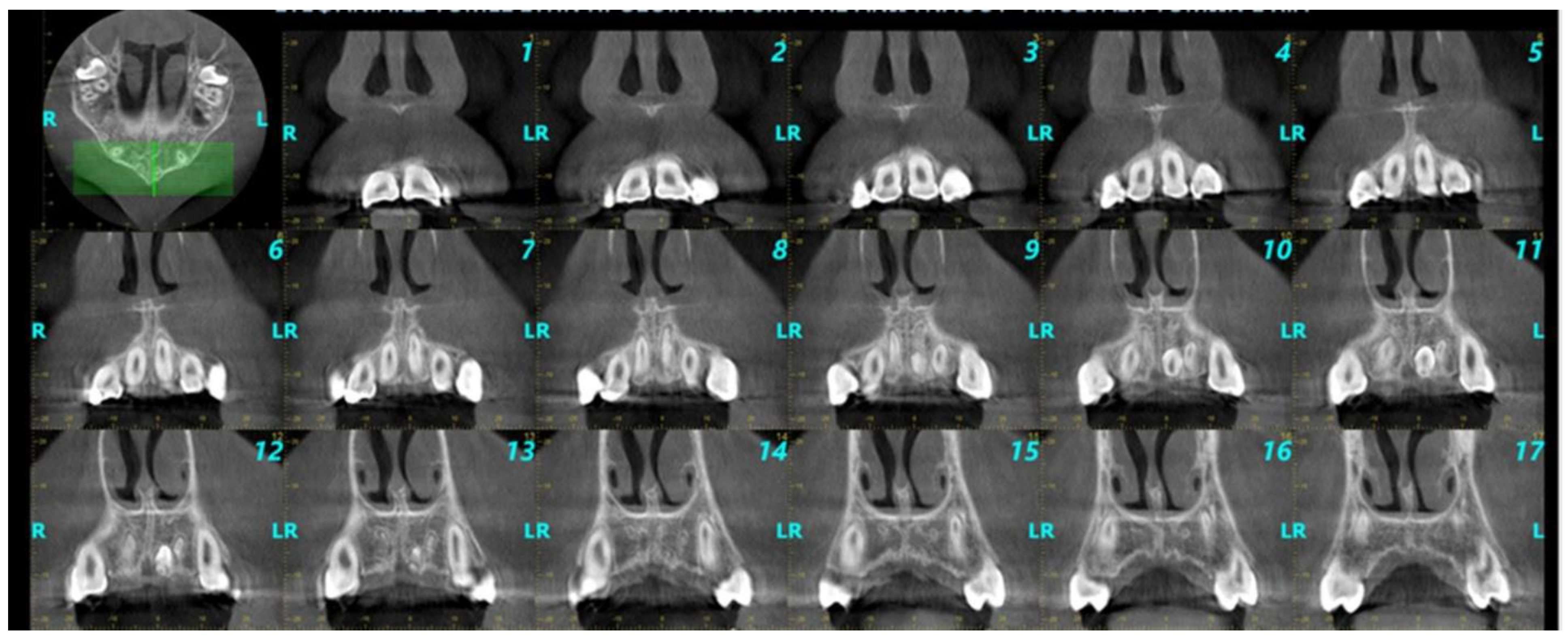
2.5. CBCT Scans’ Evaluation, Data Collection, and Extraction
- a.
- Sex and age (in years) of each subject.
- b.
- Imaging technique (modality) used to identify the supernumerary teeth.
- c.
- Number, size, shape, and location of the supernumerary teeth detected in the maxillary anterior area.
- d.
- Level of impaction of each supernumerary tooth (impacted, semi-impacted, fully erupted).
- e.
- Spatial orientation of supernumerary teeth in the sagittal plane.
- f.
- Relationship and proximity of supernumerary teeth to adjacent anatomical structures and adjacent teeth.
- g.
- Implications of supernumerary teeth on the dental arch.
2.6. Statistical Analysis
- a.
- To inquire about the differences in demographic features (gender and age) between children/adolescents of supernumerary teeth;
- b.
- To investigate the correlation of supernumerary teeth with both demographic features and CBCT findings.
3. Results
4. Discussion
4.1. Summary of Evidence
4.2. Limitations
4.3. Generalizability (External Validity)
4.4. Future Research
5. Conclusions
- There is higher prevalence in males.
- Supernumerary teeth appear more frequently as single and impacted, with a conical shape and normal spatial orientation, while their most frequent location is the middle line of the palate.
Author Contributions
Funding
Institutional Review Board Statement
Informed Consent Statement
Data Availability Statement
Conflicts of Interest
Abbreviations
| CBCT | Cone-beam computed tomography |
| FAP | Familial adenomatous polyposis |
| OFCD | Oculofaciocardiodental syndrome |
| ALARA | As low as reasonably achievable |
References
- Scheiner, M.A.; Sampson, W.J. Supernumerary Teeth: A Review of the Literature and Four Case Reports. Aust. Dent. J. 1997, 42, 160–165. [Google Scholar] [CrossRef]
- Omer, R.S.M.; Anthonappa, R.P.; King, N.M. Determination of the Optimum Time for Surgical Removal of Unerupted Anterior Supernumerary Teeth. Pediatr. Dent. 2010, 32, 14–20. [Google Scholar] [PubMed]
- Gravey, M.T.; Barry, H.J.; Blake, M. Supernumerary Teeth—An Overview of Classification, Diagnosis and Management. J. Can. Dent. Assoc. 1999, 65, 612–616. [Google Scholar]
- Hua, W.; Gan, Z.; Wu, Y.; Zhao, L. Identification of a Novel Missense Mutation in Non-Syndromic Familial Multiple Supernumerary Teeth. Arch. Oral Biol. 2022, 143, 105542. [Google Scholar] [CrossRef] [PubMed]
- Anthonappa, R.P.; Lee, C.K.; Yiu, C.K.Y.; King, N.M. Hypohyperdontia: Literature Review and Report of Seven Cases. Oral Surg. Oral Med. Oral Pathol. Oral Radiol. Endod. 2008, 106, e24–e30. [Google Scholar] [CrossRef]
- Cammarata-Scalisi, F.; Avendaño, A.; Callea, M. Main Genetic Entities Associated with Supernumerary Teeth. Arch. Argent. Pediatr. 2018, 116, 437–444. [Google Scholar] [CrossRef] [PubMed]
- De Oliveira Gomes, C.; Drummond, S.N.; Jham, B.C.; Abdo, E.N.; Mesquita, R.A. A Survey of 460 Supernumerary Teeth in Brazilian Children and Adolescents. Int. J. Paediatr. Dent. 2008, 18, 98–106. [Google Scholar] [CrossRef]
- Lubinsky, M.; Kantaputra, P.N. Syndromes with Supernumerary Teeth. Am. J. Med. Genet. Part A 2016, 170, 2611–2616. [Google Scholar] [CrossRef]
- Ranta, R.; Ylipaavalniemi, P. Development Course of Supernumerary Premolars in Childhood: Report of Two Cases. ASDC J. Dent. Child. 1981, 48, 385–388. [Google Scholar]
- Wang, X.P.; Fan, J. Molecular Genetics of Supernumerary Tooth Formation. Genesis 2011, 49, 261–277. [Google Scholar] [CrossRef]
- Liu, J. Characteristics of Premaxillary Supernumerary Teeth: A Survey of 112 Cases. ASDC J. Dent. Child. 1995, 62, 262–265. [Google Scholar] [PubMed]
- Salcido-García, J.F.; Ledesma-Montes, C.; Hernández-Flores, F.; Pérez, D.; Garcés-Ortíz, M. Frequency of Supernumerary Teeth in Mexican Population. Med. Oral Patol. Oral Cir. Bucal 2004, 9, 406–407. [Google Scholar]
- Leco Berrocal, M.I.; Martín Morales, J.F.; Martínez González, M.J. An Observational Study of the Frequency of Supernumerary Teeth in a Population of 2000 Patients. Med. Oral Patol. Oral Cir. Bucal 2007, 12, E134–E138. [Google Scholar] [PubMed]
- Gündüz, K.; Celenk, P.; Zengin, Z.; Sümer, P. Mesiodens: A Radiographic Study in Children. J. Oral Sci. 2008, 50, 287–291. [Google Scholar] [CrossRef]
- Yonezu, T.; Hayashi, Y.; Sasaki, J.; Machida, Y. Prevalence of Congenital Dental Anomalies of the Deciduous Dentition in Japanese Children. Bull. Tokyo Dent. Coll. 1997, 38, 27–32. [Google Scholar]
- Chen, K.C.; Huang, J.S.; Chen, M.Y.; Cheng, K.H.; Wong, T.Y.; Huang, T.T. Unusual Supernumerary Teeth and Treatment Outcomes Analyzed for Developing Improved Diagnosis and Management Plans. J. Oral Maxillofac. Surg. 2019, 77, 920–931. [Google Scholar] [CrossRef] [PubMed]
- Ata-Ali, F.; Ata-Ali, J.; Peñarrocha-Oltra, D.; Peñarrocha-Diago, M. Prevalence, Etiology, Diagnosis, Treatment and Complications of Supernumerary Teeth. J. Clin. Exp. Dent. 2014, 6, e414–e418. [Google Scholar] [CrossRef]
- Brook, A.H.; Jernvall, J.; Smith, R.N.; Hughes, T.E.; Townsend, G.C. The Dentition: The Outcomes of Morphogenesis Leading to Variations of Tooth Number, Size and Shape. Aust. Dent. J. 2014, 59 (Suppl. S1), 131–142. [Google Scholar] [CrossRef]
- Rajab, L.D.; Hamdan, M.A.M. Supernumerary Teeth: Review of the Literature and a Survey of 152 Cases. Int. J. Paediatr. Dent. 2002, 12, 244–254. [Google Scholar] [CrossRef]
- Fernández Montenegro, P.; Valmaseda Castellón, E.; Berini Aytés, L.; Gay Escoda, C. Retrospective Study of 145 Supernumerary Teeth. E340 Med. Oral Patol. Oral Cir. Bucal 2006, 11, 339–383. [Google Scholar]
- Sarne, O.; Shapira, Y.; Blumer, S.; Finkelstein, T.; Schonberger, S.; Bechor, N.; Shpack, N. Supernumerary Teeth in the Maxillary Anterior Region: The Dilemma of Early Versus Late Surgical Intervention. J. Clin. Pediatr. Dent. 2018, 42, 55–61. [Google Scholar] [CrossRef] [PubMed]
- Anthonappa, R.P.; King, N.M.; Rabie, A.B.M. Diagnostic Tools Used to Predict the Prevalence of Supernumerary Teeth: A Meta-Analysis. Dentomaxillofacial Radiol. 2012, 41, 444–449. [Google Scholar] [CrossRef]
- Katheria, B.C.; Kau, C.H.; Tate, R.; Chen, J.-W.; English, J.; Bouquot, J. Effectiveness of Impacted and Supernumerary Tooth Diagnosis from Traditional Radiography Versus Cone Beam Computed Tomography. Pediatr. Dent. 2010, 32, 304–309. [Google Scholar]
- Liu, J.; Wang, X.; Shan, P.; Hu, S.; Liu, D.; Ma, J.; Nie, X. A Randomized Controlled Trial: Evaluation of Efficiency and Safety of a Novel Surgical Guide in the Extraction of Deeply Impacted Supernumerary Teeth in the Anterior Maxilla. Ann. Transl. Med. 2022, 10, 292. [Google Scholar] [CrossRef] [PubMed]
- Bereket, C.; Çakir-Özkan, N.; Şener, I.; Bulut, E.; Baştan, A. Analyses of 1100 Supernumerary Teeth in a Nonsyndromic Turkish Population: A Retrospective Multicenter Study. Niger. J. Clin. Pract. 2015, 18, 731–738. [Google Scholar] [CrossRef] [PubMed]
- Vandenbroucke, J.P.; Von Elm, E.; Altman, D.G.; Gøtzsche, P.C.; Mulrow, C.D.; Pocock, S.J.; Poole, C.; Schlesselman, J.J.; Egger, M. Strengthening the Reporting of Observational Studies in Epidemiology (STROBE): Explanation and Elaboration. PLoS Med. 2007, 4, 1628–1654. [Google Scholar] [CrossRef]
- Reddy, G.S.P.; Reddy, G.V.; Krishna, I.V.; Regonda, S.K. Nonsyndromic Bilateral Multiple Impacted Supernumerary Mandibular Third Molars: A Rare and Unusual Case Report. Case Rep. Dent. 2013, 2013, 857147. [Google Scholar] [CrossRef]
- Chaudhary, S.; Chaitra, T.R.; Sultan, S.; Arora, R. Supernumerary Teeth in Primary Dentition. BMJ Case Rep. 2013, 2013, bcr2013200029. [Google Scholar] [CrossRef]
- Esenlik, E.; Sayin, M.Ö.; Atilla, A.O.; Özen, T.; Altun, C.; Başak, F. Supernumerary Teeth in a Turkish Population. Am. J. Orthod. Dentofac. Orthop. 2009, 136, 848–852. [Google Scholar] [CrossRef]
- Rallan, M.; Rallan, N.; Goswami, M.; Rawat, K. Surgical Management of Multiple Supernumerary Teeth and an Impacted Maxillary Permanent Central Incisor. BMJ Case Rep. 2013, 2013, bcr2013009995. [Google Scholar] [CrossRef]
- Mallineni, S.K.; Nuvvula, S.; Cheung, A.C.H.; Kunduru, R. A Comprehensive Review of the Literature and Data Analysis on Hypo-Hyperdontia. J. Oral Sci. 2014, 56, 295–302. [Google Scholar] [CrossRef]
- Zadurska, M.; Sieminska-Piekarczyk, B.; Maciejak, D.; Wyszomirska-Zdybel, B.; Kurol, J. Concomitant Hypodontia and Hyperodontia: An Analysis of Nine Patients. Acta Odontol. Scand. 2012, 70, 154–159. [Google Scholar] [CrossRef] [PubMed]
- Venkataraghavan, K.; Muralikrishnan, B.; Anantharaj, A. Mandibular Mesiodens with Agenesis of Central Incisors (Hypohyperdontia): A Case Report & Review. Int. J. Contemp. Dent. 2011, 2, 26–30. [Google Scholar]
- Tunis, T.S.; Sarne, O.; Hershkovitz, I.; Finkelstein, T.; Pavlidi, A.M.; Shapira, Y.; Davidovitch, M.; Shpack, N. Dental Anomalies’ Characteristics. Diagnostics 2021, 11, 1161. [Google Scholar] [CrossRef] [PubMed]
- Mallineni, S.K. Supernumerary Teeth: Review of the Literature with Recent Updates. Conf. Pap. Sci. 2014, 2014, 764050. [Google Scholar] [CrossRef]
- He, D.; Mei, L.; Wang, Y.; Li, J.; Li, H. Association between Maxillary Anterior Supernumerary Teeth and Impacted Incisors in Mixed Dentition. J. Am. Dent. Assoc. 2017, 148, 595–603. [Google Scholar] [CrossRef]
- Shekhar, M.G. Characteristics of Premaxillary Supernumerary Teeth in Primary and Mixed Dentitions: A Retrospective Analysis of 212 Cases. J. Investig. Clin. Dent. 2012, 3, 221–224. [Google Scholar] [CrossRef]
- Kim, Y.; Jeong, T.; Kim, J.; Shin, J.; Kim, S. Effects of Mesiodens on Adjacent Permanent Teeth: A Retrospective Study in Korean Children Based on Cone-Beam Computed Tomography. Int. J. Paediatr. Dent. 2018, 28, 161–169. [Google Scholar] [CrossRef]
- Kim, S.-G.; Lee, S.-H. Mesiodens: A Clinical and Radiographic Study. J. Dent. Child. 2003, 70, 58–60. [Google Scholar]
- Asaumi, J.I.; Shibata, Y.; Yanagi, Y.; Hisatomi, M.; Matsuzaki, H.; Konouchi, H.; Kishi, K. Radiographic Examination of Mesiodens and Their Associated Complications. Dentomaxillofacial Radiol. 2004, 33, 125–127. [Google Scholar] [CrossRef]
- Umweni, A.; Osunbor, G. Non-Syndrome Multiple Supernumerary Teeth in Nigerians. Odontostomatol. Trop. 2002, 25, 43–48. [Google Scholar]
- Tyrologou, S.; Koch, G.; Kurol, J. Location, Complications and Treatment of Mesiodentes—A Retrospective Study in Children. Swed. Dent. J. 2005, 29, 1–9. [Google Scholar]
- Mukhopadhyay, S. Mesiodens: A Clinical and Radiographic Study in Children. J. Indian Soc. Pedod. Prev. Dent. 2011, 29, 34–38. [Google Scholar] [CrossRef] [PubMed]
- Russell, K.; Folwarczna, M. Mesiodens--Diagnosis and Management of a Common Supernumerary Tooth. J. Can. Dent. Assoc. 2003, 69, 362–366. [Google Scholar]
- Anegundi, R.; Tegginmani, V.; Battepati, P.; Tavargeri, A.; Patil, S.; Trasad, V.; Jain, G. Prevalence and Characteristics of Supernumerary Teeth in a Non-Syndromic South Indian Pediatric Population. J. Indian Soc. Pedod. Prev. Dent. 2014, 32, 9–12. [Google Scholar] [CrossRef] [PubMed]
- Yassin, O.; Hamori, E. Characteristics, Clinical Features and Treatment of Supernumerary Teeth. J. Clin. Pediatr. Dent. 2009, 33, 247–250. [Google Scholar] [CrossRef]
- Khandelwal, V.; Nayak, A.U.; Naveen, R.B.; Ninawe, N.; Nayak, P.A.; Sai Prasad, S.V. Prevalence of Mesiodens among Six- to Seventeen-Year-Old School Going Children of Indore. J. Indian Soc. Pedod. Prev. Dent. 2011, 29, 288–293. [Google Scholar] [CrossRef]
- Gábris, K.; Fábián, G.; Kaán, M.; Rózsa, N.; Tarján, I. Prevalence of Hypodontia and Hyperdontia in Paedodontic and Orthodontic Patients in Budapest. Community Dent. Health 2006, 23, 80–82. [Google Scholar] [PubMed]
- Brook, A.H. Dental Anomalies of Number, Form and Size: Their Prevalence in British Schoolchildren. J. Int. Assoc. Dent. Child. 1974, 5, 37–53. [Google Scholar]
- Giancotti, A.; Grazzini, F.; De Dominicis, F.; Romanini, G.; Arcuri, C. Multidisciplinary Evaluation and Clinical Management of Mesiodens. J. Clin. Pediatr. Dent. 2002, 26, 233–237. [Google Scholar]
- Davis, P.J. Hypodontia and Hyperdontia of Permanent Teeth in Hong Kong Schoolchildren. Community Dent. Oral Epidemiol. 1987, 15, 218–220. [Google Scholar] [CrossRef] [PubMed]
- Ozan, F.; Kara, I.; Ay, S. Impacted Mandibular Permanent Incisors Associated With a Supernumerary Tooth: A Case Report. Eur. J. Dent. 2009, 03, 324–328. [Google Scholar] [CrossRef]
- Ma, X.; Jiang, Y.; Ge, H.; Yao, Y.; Wang, Y.; Mei, Y.; Wang, D. Epidemiological, Clinical, Radiographic Characterization of Non-Syndromic Supernumerary Teeth in Chinese Children and Adolescents. Oral Dis. 2021, 27, 981–992. [Google Scholar] [CrossRef] [PubMed]
- Tay, F.; Pang, A.; Yuen, S. Unerupted Maxillary Anterior Supernumerary Teeth: Report of 204 Cases. ASDC J. Dent. Child. 1984, 51, 289–294. [Google Scholar] [PubMed]
- Alberti, G.; Mondani, P.M.; Parodi, V. Eruption of Supernumerary Permanent Teeth in a Sample of Urban Primary School Population in Genoa, Italy. Eur. J. Paediatr. Dent. 2006, 7, 89–92. [Google Scholar]
- Van Buggenhout, G.; Bailleul-Forestier, I. Mesiodens. Eur. J. Med. Genet. 2008, 51, 178–181. [Google Scholar] [CrossRef]
- Ashkenazi, M.; Greenberg, B.P.; Chodik, G.; Rakocz, M. Postoperative Prognosis of Unerupted Teeth after Removal of Supernumerary Teeth or Odontomas. Am. J. Orthod. Dentofacial Orthop. 2007, 131, 614–619. [Google Scholar] [CrossRef]
- Kim, S.-D.; Lee, S.-H.; Lee, N.-Y.; Jeon, S.-Y. Three-Dimensional Evaluation of Impacted Mesiodens Using Dental Cone-Beam Computed Tomography in Korean Children and Adolescents. J. Korean Acad. Pediatr. Dent. 2013, 40, 149–158. [Google Scholar] [CrossRef]
- Liu, D.G.; Zhang, W.L.; Zhang, Z.Y.; Wu, Y.T.; Ma, X.C. Three-Dimensional Evaluations of Supernumerary Teeth Using Cone-Beam Computed Tomography for 487 Cases. Oral Surg. Oral Med. Oral Pathol. Oral Radiol. Endod. 2007, 103, 403–411. [Google Scholar] [CrossRef]
- Kazancı, F.; Celikoglu, M.; Miloglu, O.; Yıldırım, H.; Ceylan, I. The Frequency and Characteristics of Mesiodens in a Turkish Patient Population. Eur. J. Dent. 2011, 5, 361–365. [Google Scholar] [CrossRef]
- Lee, D.H.; Lee, J.S.; Yoon, S.J.; Kang, B.C. Three-Dimensional Evaluation of Impacted Mesiodens Using Dental Cone Beam CT. Korean J. Oral Maxillofac. Radiol. 2015, 40, 109–114. [Google Scholar] [CrossRef] [PubMed]


| Demographic Features | Supernumerary Teeth | p-Value | |
|---|---|---|---|
| Yes (n = 26) | No (n = 198) | ||
| Sex | n (%) | n (%) | |
| Males | 16 (61.5) | 99 (50.0) | 0.268 1 |
| Females | 10 (38.5) | 99 (50.0) | |
| Age (years: mean value, SD) | 14.6 (2.5) | 14.8 (2.6) | 0.712 2 |
| Demographic Features | Descriptive Statistical Measures |
|---|---|
| Sex | n (%) |
| Males | 16 (61.5) |
| Females | 10 (38.5) |
| Age (years: mean value, SD) | 14.6 (2.5) |
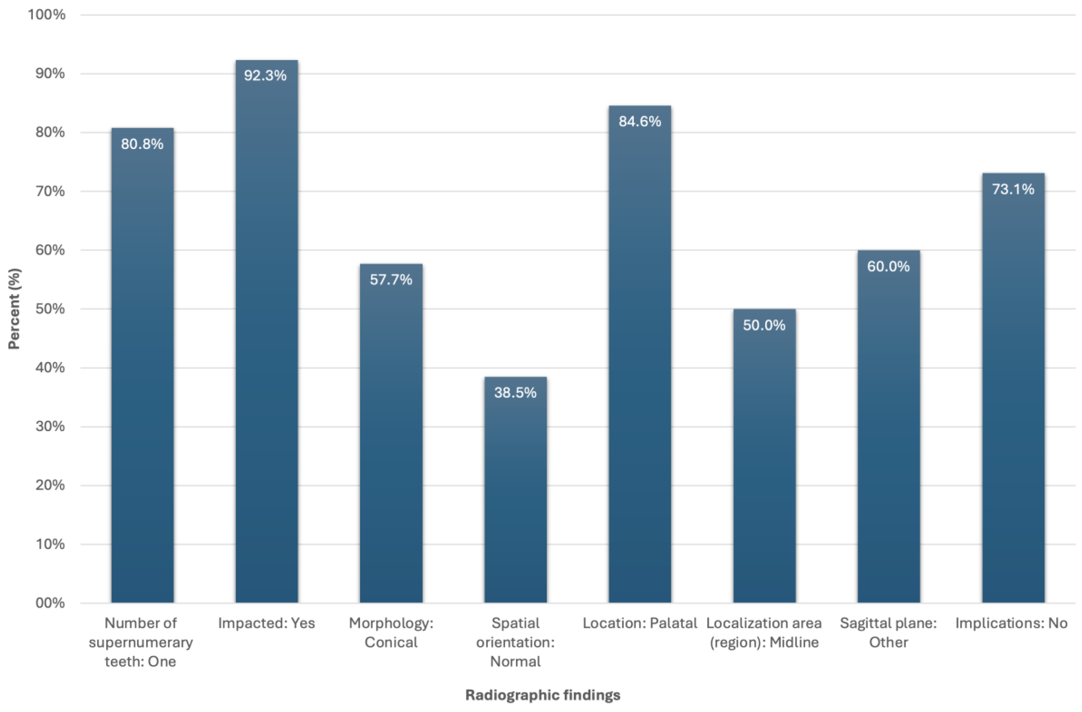
| Radiographic findings | |
| Number of supernumerary teeth | n (%) |
| 1 | 21 (80.8) |
| 2 | 5 (19.2) |
| Impacted | n (%) |
| No | 2 (7.7) |
| Yes | 24 (92.3) |
| Morphology | n (%) |
| Conical | 15 (57.7) |
| Other | 11 (42.3) |
| Spatial orientation | n (%) |
| Normal | 10 (38.5) |
| Inverted | 7 (26.9) |
| Horizontal | 5 (19.2) |
| Other | 4 (15.4) |
| Location | n (%) |
| Palatal | 22 (84.6) |
| No | 4 (15.4) |
| Localization area (region) | n (%) |
| Middle line | 13 (50.0) |
| No | 13 (50.0) |
| Sagittal plane | n (%) |
| Adjacent to tooth neck (cervix) | 10 (40.0) |
| Other | 15 (60.0) |
| Implications | n (%) |
| No | 19 (73.1) |
| Yes | 7 (26.9) |
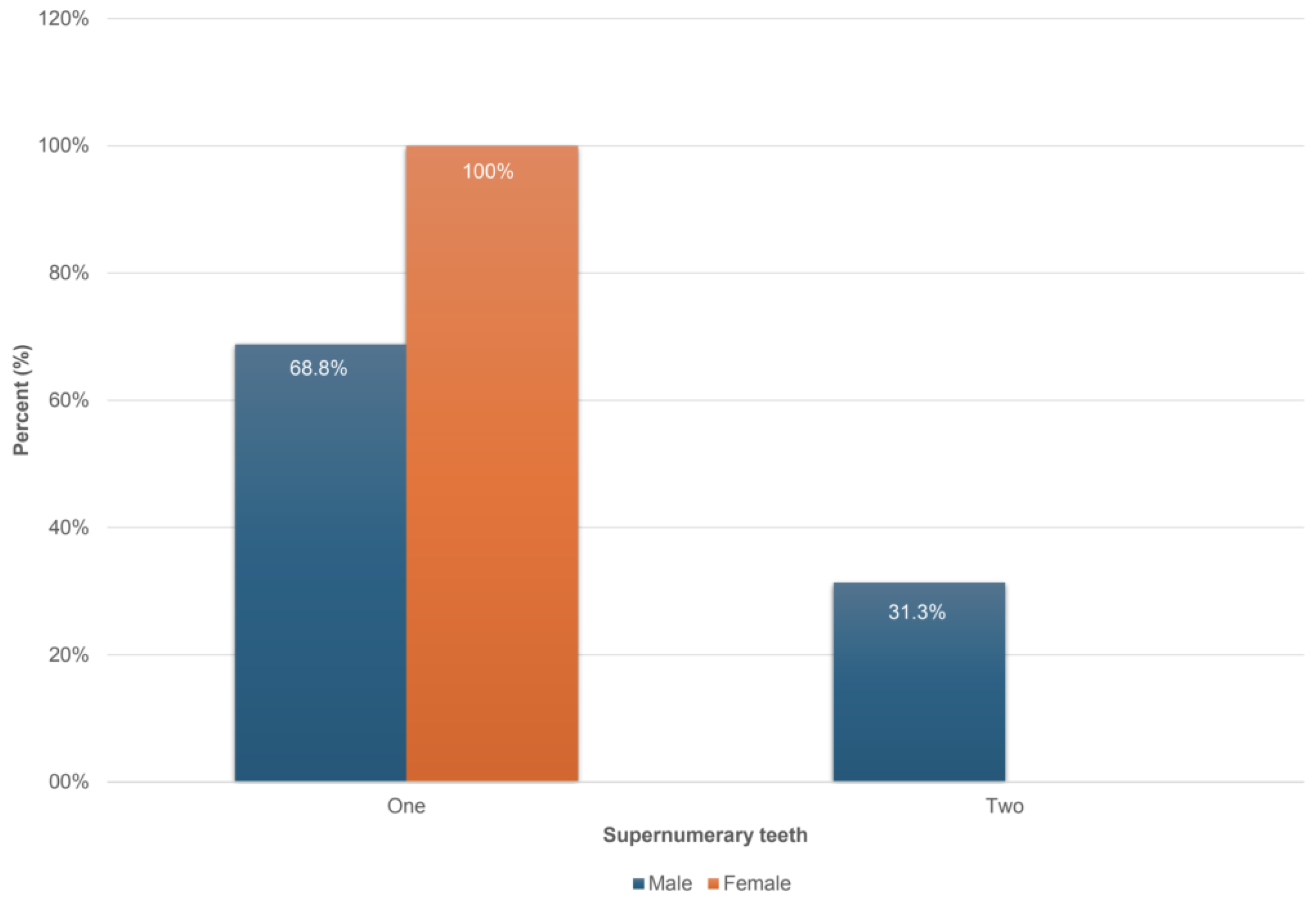
| Demographic Features | Number of Supernumerary Teeth | p-Value | |
|---|---|---|---|
| One (n = 21) | Two (n = 5) | ||
| Sex | n (%) | n (%) | |
| Males | 11 (68.8) | 5 (31.3) | 0.049 1,* |
| Females | 10 (100.0) | 0 (0.0) | |
| Age (years: mean value, SD) | 14.4 (2.6) | 15.2 (1.5) | 0.538 2 |
| Radiographic findings | |||
| Impacted | n (%) | n (%) | 0.646 3 |
| No | 2 (9.5) | 0 (0.0) | |
| Yes | 19 (90.5) | 5 (100.0) | |
| Morphology | n (%) | n (%) | |
| Conical | 12 (57.1) | 3 (60.0) | 0.907 1 |
| Other | 9 (42.9) | 2 (40.0) | |
| Spatial orientation | n (%) | n (%) | |
| Normal | 7 (33.3) | 3 (60.0) | 0.506 3 |
| Inverted | 5 (23.8) | 2 (40.0) | |
| Horizontal | 5 (23.8) | 0 (0.0) | |
| Other | 4 (19.0) | 0 (0.0) | |
| Location | n (%) | n (%) | |
| Palatal | 17 (81.0) | 5 (100.0) | 0.289 1 |
| No | 4 (19.0) | 0 (0.0) | |
| Localization area (region) | n (%) | n (%) | |
| Middle line | 9 (42.9) | 4 (80.0) | 0.1351 1 |
| No | 12 (57.1) | 1 (20.0) | |
| Sagittal plane | n (%) | n (%) | |
| Adjacent to tooth neck (cervix) | 7 (33.3) | 3 (75.0) | 0.119 1 |
| Other | 14 (66.7) | 1 (25.0) | |
| Implications | n (%) | n (%) | |
| No | 16 (76.2) | 3 (60.0) | 0.463 1 |
| Yes | 5 (23.8) | 2 (40.0) | |
Disclaimer/Publisher’s Note: The statements, opinions and data contained in all publications are solely those of the individual author(s) and contributor(s) and not of MDPI and/or the editor(s). MDPI and/or the editor(s) disclaim responsibility for any injury to people or property resulting from any ideas, methods, instructions or products referred to in the content. |
© 2025 by the authors. Licensee MDPI, Basel, Switzerland. This article is an open access article distributed under the terms and conditions of the Creative Commons Attribution (CC BY) license (https://creativecommons.org/licenses/by/4.0/).
Share and Cite
Lykousis, A.; Pouliezou, I.; Christoloukas, N.; Rontogianni, A.; Mitsea, A.; Angelopoulos, C. Supernumerary Teeth in the Anterior Maxilla of Non-Syndromic Children and Adolescents: A Retrospective Study Based on Cone-Beam Computed Tomography Scans. Pediatr. Rep. 2025, 17, 52. https://doi.org/10.3390/pediatric17030052
Lykousis A, Pouliezou I, Christoloukas N, Rontogianni A, Mitsea A, Angelopoulos C. Supernumerary Teeth in the Anterior Maxilla of Non-Syndromic Children and Adolescents: A Retrospective Study Based on Cone-Beam Computed Tomography Scans. Pediatric Reports. 2025; 17(3):52. https://doi.org/10.3390/pediatric17030052
Chicago/Turabian StyleLykousis, Antonis, Ioanna Pouliezou, Nikolaos Christoloukas, Aliki Rontogianni, Anastasia Mitsea, and Christos Angelopoulos. 2025. "Supernumerary Teeth in the Anterior Maxilla of Non-Syndromic Children and Adolescents: A Retrospective Study Based on Cone-Beam Computed Tomography Scans" Pediatric Reports 17, no. 3: 52. https://doi.org/10.3390/pediatric17030052
APA StyleLykousis, A., Pouliezou, I., Christoloukas, N., Rontogianni, A., Mitsea, A., & Angelopoulos, C. (2025). Supernumerary Teeth in the Anterior Maxilla of Non-Syndromic Children and Adolescents: A Retrospective Study Based on Cone-Beam Computed Tomography Scans. Pediatric Reports, 17(3), 52. https://doi.org/10.3390/pediatric17030052