Article

Monoxenic Root Organ Culture Enables High-Yield Production of Viable Indigenous Rhizophagus irregularis Inoculum for Arid Oasis Agroecosystems

1
Laboratory of Bioresources, Biotechnology, Ethnopharmacology and Health, Faculty of Sciences, Université Mohamed Premier, BV Mohammed VI BP 717, Oujda 60000, Morocco
2
Laboratory for the Improvement of Agricultural Production Biotechnology and Environment, Faculty of Science, University Mohammed First, Oujda 60000, Morocco
3
Laboratory of Water Analysis of Figuig (L.A.E.F), Administrative Centre, Municipality of Figuig, BP 121, Figuig 61000, Morocco
4
Department of Research in Drug Development, Faculty of Pharmacy, Université Libre de Bruxelles, Bvd du Triomphe, 1050 Brussels, Belgium
5
Laboratory of Plant Biotechnology, Université Libre de Bruxelles, 6041 Gosselies, Belgium
*
Author to whom correspondence should be addressed.
Microbiol. Res. 2026, 17(1), 28; https://doi.org/10.3390/microbiolres17010028
Submission received: 22 December 2025 / Revised: 12 January 2026 / Accepted: 19 January 2026 / Published: 22 January 2026
(This article belongs to the Special Issue Advances in Plant–Pathogen Interactions)

Abstract

Arbuscular mycorrhizal fungi (AMF) play a pivotal role in plant adaptation to arid ecosystems, yet their widespread agricultural use is constrained by the scarcity of high-quality, locally adapted inoculum. This study established a reliable monoxenic culture system for mass-producing an indigenous AMF isolate from the date palm (Phoenix dactylifera L.) rhizosphere in the Figuig oasis, southeastern Morocco. The isolate was identified as Rhizophagus irregularis based on spore morphology and Large Subunit ribosomal DNA (LSU rDNA) phylogeny. Two propagule types, surface-sterilized spores and mycorrhizal root fragments of Plantago lanceolata L., were compared for initiation of in vitro cultures on Ri T-DNA-transformed carrot (Daucus carota L.) hairy roots. By week 16, cultures initiated from mycorrhizal root fragments produced 1414 ± 65 spores per plate and showed significantly higher performance than spore-derived cultures in terms of propagule viability, root colonization, and hairy root growth. Propagule viability reached 84% and 68%, root colonization frequencies were 95% and 72%, and hairy root lengths averaged 81 and 63 cm in root fragment- and spore-derived cultures, respectively (p < 0.01). In a subsequent whole-plant assay using P. lanceolata, in vitro-produced spores induced markedly higher mycorrhizal colonization frequency (91.0 ± 1.6% compared with 74.8 ± 1.9%) and intensity (70.0 ± 1.6% compared with 55.0 ± 1.6%) than spores obtained from conventional trap cultures (p < 0.001). These results demonstrate that monoxenic root-organ culture using root fragments is a robust, reproducible method for generating abundant, contaminant-free, and functionally superior inoculum of native R. irregularis. This advance provides a solid platform for developing tailored bio-inoculants to enhance crop resilience and sustainability in arid and semi-arid agroecosystems.

1. Introduction

Ensuring global food security while preserving ecosystem integrity represents one of the greatest challenges of the 21st century. In the context of climate change, rising greenhouse gas emissions, and ongoing soil degradation, the adoption of sustainable agricultural practices has become a strategic imperative [1,2,3]. Arbuscular mycorrhizal fungi (AMF) have attracted considerable attention as effective bioinoculants due to their proven ability to enhance plant growth, improve nutrient acquisition, and increase tolerance to both abiotic and biotic stresses [4,5,6,7]. Although naturally widespread in most soils, the abundance and symbiotic efficiency of AMF are frequently limited by soil degradation, intensive farming practices, and the absence of compatible host plants [8,9,10]. To overcome these constraints, early inoculation of horticultural crops at the nursery stage has proven to be a highly effective strategy. This approach enables rapid establishment of the symbiosis prior to transplanting and promotes early, vigorous root colonization once plants are transferred to the field [5,11,12,13]. This early colonization is particularly critical in arid and semi-arid ecosystems, where the extraradical hyphal network of AMF plays an essential role in soil exploration and nutrient uptake [14,15,16,17].
Large-scale implementation of this strategy, however, depends on the availability of viable, locally adapted fungal inoculum produced in sufficient quantities to ensure consistent and successful colonization under field conditions. This requires the development of reliable, standardized methods for AMF production. In this regard, in vitro cultivation using transformed root organ cultures, most commonly carrot (Daucus carota L.) roots genetically modified with the Ri T-DNA of Agrobacterium rhizogenes (Riker et al. [18]), has emerged as the leading technique [19,20,21,22]. This monoxenic culture system provides a sterile environment that yields pure, contaminant-free, and abundant propagules, an outcome rarely achieved with traditional pot-based or hydroponic/aeroponic systems [23]. A substantial body of evidence shows that AMF intraradical structures (root fragments containing vesicles or arbuscules) and isolated spores can germinate and re-establish functional symbioses under in vitro conditions when associated with a susceptible hairy root host [19,20,22]. This method has enabled successful monoxenic cultivation of numerous AMF species, including Gigaspora margarita, Rhizophagus intraradices (syn. Glomus intraradices), Glomus etunicatum, Glomus proliferum, and others [24,25,26,27].
The Figuig oasis in southeastern Morocco exemplifies an arid agroecosystem in which date palm (Phoenix dactylifera L.) cultivation depends heavily on the integrity and functionality of soil microbial communities. This oasis is exposed to multiple abiotic stresses, including extreme drought, high salinity, limited water availability, and elevated temperatures, all of which impair soil quality and plant productivity [28,29,30,31]. In addition, severe biotic stress is posed by Fusarium wilt (Bayoud disease), caused by Fusarium oxysporum f. sp. albedinis, which seriously threatens date palm survival and yield stability [32,33,34]. These combined pressures jeopardize agricultural sustainability and highlight the need to maintain a healthy soil microbiome capable of enhancing plant resilience and productivity. Despite the clear agronomic potential of indigenous AMF in this region, their biology, propagation techniques, and mass-production protocols remain poorly documented [30,34]. Several native species have nevertheless demonstrated remarkable ecological adaptability and symbiotic efficiency under controlled conditions, making them promising candidates for bioinoculant development [35,36,37,38]. The present study aimed to establish a reliable propagation system for a native AMF strain adapted to oasis soils, thereby facilitating its use as a bioinoculant in field, nursery, and laboratory settings. This approach is particularly relevant for the mycorrhization of in vitro-produced date palm plantlets in nurseries prior to their final transplantation into the field, a practice that is rapidly expanding in Moroccan oasis environments. For this purpose, an indigenous AMF isolate identified as Rhizophagus irregularis, originating from the date palm rhizosphere, was used to inoculate transformed carrot hairy root cultures. Spore production, spore germination, and root colonization dynamics were monitored to evaluate the potential for large-scale inoculum production. Accordingly, the main objective of this study was to evaluate whether monoxenic root organ culture can serve as an efficient and reliable system for the large-scale production of viable indigenous R. irregularis inoculum adapted to arid oasis agroecosystems. Such inoculum is expected to improve agricultural productivity, support sustainable management of palm groves, and contribute to the restoration of degraded arid ecosystems.

2. Materials and Methods

2.1. Isolation of AMF and Establishment of Trap Cultures with Plantago lanceolata L.

The AMF propagules were isolated from the rhizosphere of date palm in the Figuig oasis, (32°06′00″ N, 1°14′00″ W), southeastern Morocco, at an altitude of approximately 850 m above sea level. The vegetation is characteristic of traditional oasis agroecosystems, dominated by date palm (P. dactylifera) forming the upper canopy, with an understory composed of cultivated crops and scattered native xerophytic species adapted to arid conditions. Soil samples were collected at 0–30 cm depth and 50 cm from the trunk, air-dried, and sieved (2 mm mesh). Trap cultures were established using P. lanceolata as host in autoclaved (121 °C, two 1 h cycles) Terragreen substrate (Oil-Dri, Wisbech, United Kingdom) composed of calcined attapulgite clay–quartz sand (0.4–0.8 mm)–coarse quartz sand (1–2 mm) at a 2:2:1 (v/v/v) ratio. Seedlings were grown in Sun-bags (Sigma-Aldrich, Poznań, Poland) under greenhouse conditions for six months. Spores were extracted by wet sieving and sucrose density-gradient centrifugation [39,40,41]. Morphologically similar spores were isolated under a stereomicroscope were used to initiate single-spore cultures on P. lanceolata in autoclaved Terragreen substrate. After an additional six-month multiplication cycle, the morphotype exhibiting the highest spore density and most abundant intraradical structures (hyphae, arbuscules, vesicles) was selected for taxonomic identification and monoxenic culture establishment.

2.2. Taxonomic Identification of the Selected AMF Isolate

Spore morphology was examined in polyvinyl-lacto-glycerol (PVLG) and PVLG + Melzer’s reagent [42], and compared with descriptions from the AMF Phylogeny database and INVAM. Molecular identification followed Krüger et al. [43]. DNA was extracted by crushing single spores in 5 µL nuclease-free water. Nested PCR targeting the D2 region of the LSU rDNA was performed with primer pairs SSUmAf–LSUmAr (first round) and SSUmCf–LSUmBr (second round) using Phusion High-Fidelity DNA Polymerase (Thermo Fisher Scientific, Waltham, MA, USA). Products were cloned into E. coli using the Zero Blunt TOPO PCR Cloning Kit (Invitrogen, Waltham, MA, USA) and sequenced bidirectionally using the Sanger sequencing method, and analysed with Sequencher v5.4.6. BLASTn searches were performed against the NCBI GenBank database using BLASTn (version 2.15.0+, NCBI), and phylogenetic analysis was conducted in MEGA11 using maximum likelihood with reference sequences from Krüger et al. [44]. The isolate was identified as R. irregularis.

2.3. Surface Disinfection of Mycorrhizal Root Fragments and Spores

Root fragments and spores were surface-disinfected under aseptic conditions following [45] with minor modifications. Roots were sequentially treated with 95% ethanol (10 s), 6% calcium hypochlorite (2 min), 2% chloramine-T + Tween 20 (10 min), and antibiotic solution (streptomycin 0.2 g L−1 + gentamicin 0.1 g L−1, 10 min), with three sterile water rinses after each step. Disinfected 1 cm root fragments were stored at 4 °C in antibiotic solution for ≤48 h. Spores were disinfected in a 45 µm filter holder under vacuum using the same sequence, omitting the ethanol step.

2.4. Establishment and Maintenance of Monoxenic Cultures

Ri T-DNA-transformed carrot (D. carota) hairy roots (kindly provided by the Mycology Laboratory, Université Catholique de Louvain, Belgium) were routinely subcultured on modified Strullu–Romand (MSR) medium [19] solidified with 5 g L−1 Gelzan at pH 5.5 and autoclaved at 121 °C for 15 min. Disinfected root fragments containing intraradical spores or single surface-sterilized spores were placed on MSR medium adjacent to an actively growing 7 cm carrot root segment. Five replicate plates were established per propagule type. Plates were incubated inverted at 27 °C in the dark. Following hyphal outgrowth, a 15 mm agar plug containing the germinated propagule and associated hyphae was transferred to fresh MSR medium. Spore production was monitored weekly under a stereomicroscope using a 10 × 10 mm grid drawn on the plate bottom [46].

2.5. Comparative Evaluation of In Vitro and In Vivo Produced Inoculum Using P. lanceolata as Host

The symbiotic performance of R. irregularis inoculum produced in vitro (spores from carrot hairy root cultures) and in vivo (spores from P. lanceolata trap cultures) was compared. Spore suspensions were adjusted to 500 viable spores per plant and inoculated at transplanting. P. lanceolata seedlings were grown individually in 7.5 × 7.5 × 16 cm pots containing autoclaved Terragreen substrate inside Sun-bags. The experiment was arranged in a completely randomized design with ten biological replicates per treatment, resulting in a total of 20 plants. Plants were maintained for three months in a growth chamber (25–30 °C, 50% RH, 16/8 h light/dark photoperiod).

2.6. Assessment of Mycorrhizal Colonization, Spore Viability, and Hairy Root Growth

Root colonization was evaluated after clearing and staining with 2% Parker blue ink (Parker Pen Company, Newhaven, UK) [47] and quantified according to Trouvelot et al. [48] under a compound microscope. Briefly, root samples were cleared in 10% KOH at 90 °C for 30 min, stained with 2% Parker blue ink in 5% acetic acid at 90 °C for 15 min, and destained in 5% acetic acid at room temperature for 10–15 min. For each treatment, 30 randomly selected root fragments (approximately 1 cm in length) were examined for the presence of AMF structures (hyphae, arbuscules, and vesicles). Colonization frequency (F%) was calculated as the percentage of root fragments showing any mycorrhizal structures relative to the total number of observed fragments, whereas colonization intensity (I%) represents the mean degree of fungal colonization within the root cortex and was estimated using a scoring scale from 0 to 5, where 0 indicates the absence of mycorrhizal structures and scores of 1 to 5 correspond to progressively increasing levels of colonization (≤1%, 1–10%, 10–50%, 50–90%, and >90%, respectively). The Mycocalc software (https://www2.dijon.inrae.fr/mychintec/Mycocalc-prg/download.html, accessed on 20 October 2025) was used to calculate the frequency (F%) and intensity (I%) of AMF colonization in the root system, according to the following equations: F% = (number of mycorrhizal root fragments/total number of observed root fragments) × 100 and I% = (95n5 + 70n4 + 30n3 + 5n2 + n1)/N, where n1–n5 represent the numbers of root fragments assigned to colonization classes 1 to 5, respectively, and N is the total number of observed root fragments. Spore germination rate was determined by transferring 20 propagules (in vitro spores or root fragments with one spore) to fresh MSR medium and recording hyphal regrowth after two weeks. Carrot hairy root fresh weight and total length were measured at harvest.

2.7. Statistical Analysis

All data were expressed as the mean ± standard deviation (SD) of independent biological replicates. Data normality was assessed using the Shapiro–Wilk test prior to statistical comparisons. Statistical differences between inoculum types were evaluated using Student’s t-test for normally distributed data or the Mann–Whitney U test for non-normal data. Pearson correlation analysis was used to assess relationships between mycorrhizal traits and plant growth parameters. All statistical analyses were performed using IBM SPSS Statistics v.26, and graphs were generated with Microsoft Excel 2016.

3. Results

3.1. Taxonomic Identification of the Isolated AMF

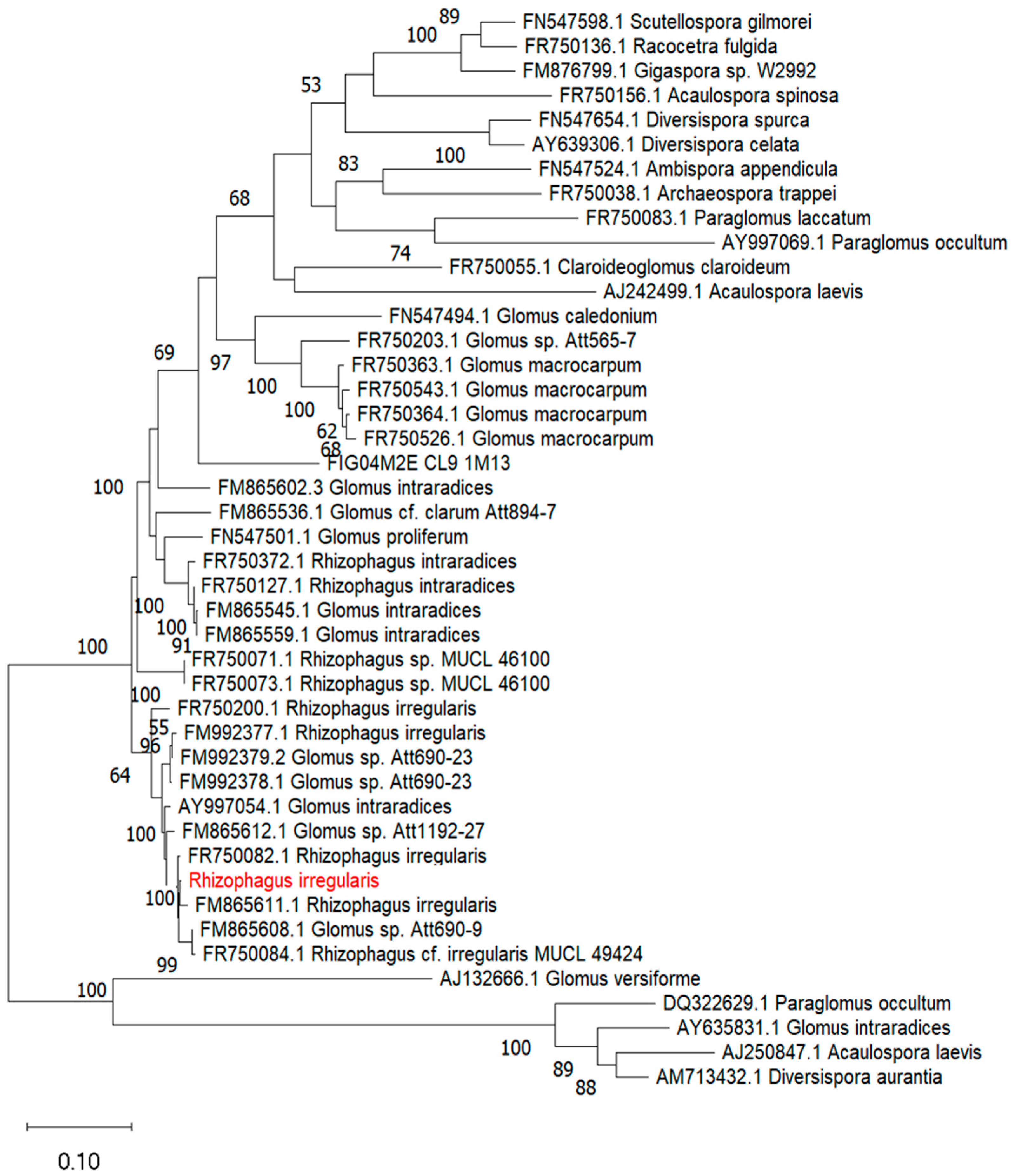
The dominant morphotype from trap cultures exhibited high spore density (Figure 1A). Spores were globose to subglobose (70–150 µm diameter), formed singly, hyaline to pale yellow, and possessed a three-layered wall with a laminated inner layer (Figure 1B,C). Subtending hyphae were straight to slightly recurved, constricted at the attachment point, with an open pore (Figure 1D). These traits matched reference descriptions of R. irregularis (AMF Phylogeny database; INVAM). Molecular identification confirmed the morphological assignment. Maximum-likelihood analysis of LSU rDNA sequences placed the isolate in a strongly supported clade (100% bootstrap) with authenticated R. irregularis sequences, including FR750082.1 and FM865611.1 (Figure 2). The isolate was therefore identified as R. irregularis, native to the date palm rhizosphere of Figuig oasis, Morocco.

3.2. Establishment of Monoxenic Cultures from Different Propagule Types

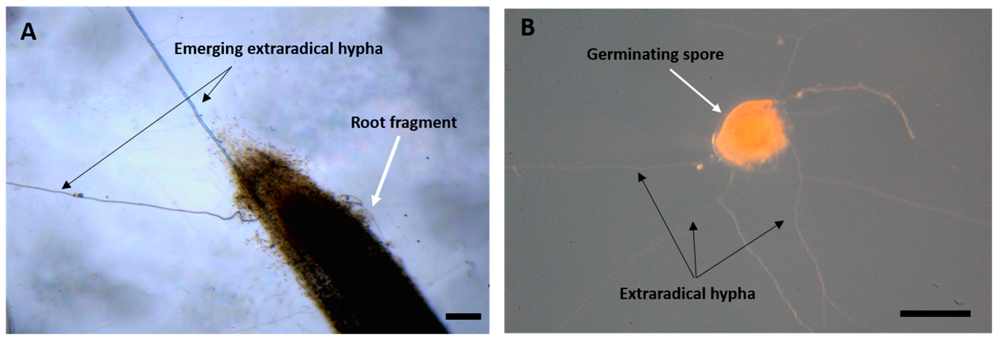
Monoxenic cultures were successfully initiated using either surface-sterilized spores or mycorrhizal root fragments of P. lanceolata. Both propagule types produced hyphal outgrowth within 7–10 days (Figure 3A,B). Root fragments generated multiple hyphae from cut ends and epidermal ruptures, whereas spores emitted single or multiple germ tubes. Contact with transformed carrot hairy roots occurred within one week, rapidly developing into an extensive extraradical mycelial network across the MSR medium.

3.3. Sporulation Dynamics in Monoxenic Culture

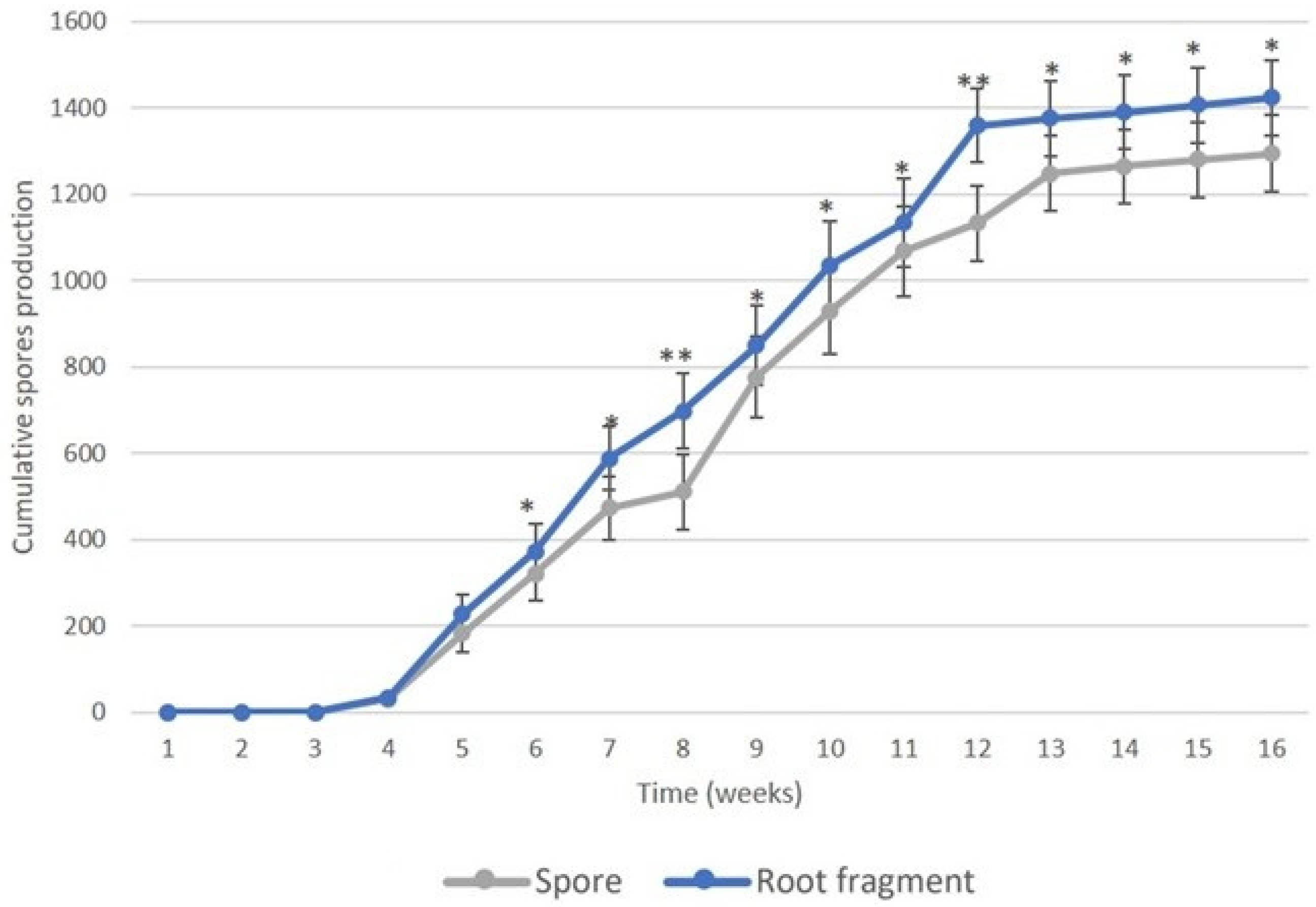
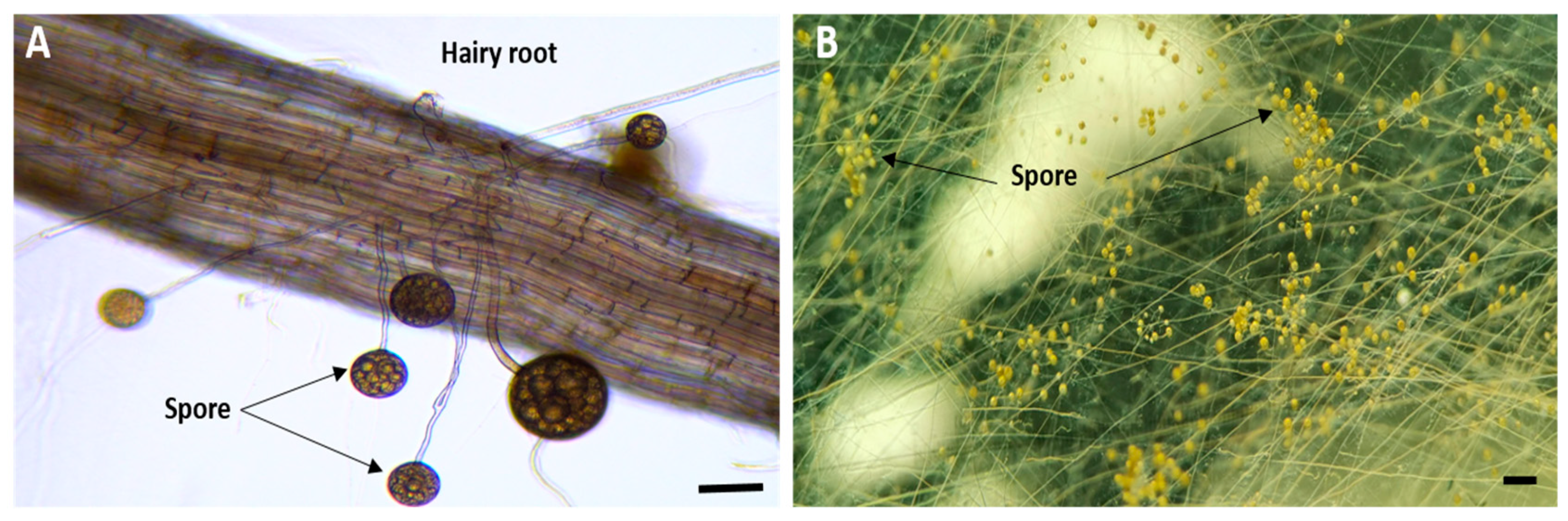
Sporulation commenced at week 5 in both treatments and followed parallel kinetics over 16 weeks (Figure 4). Cultures initiated from root fragments consistently yielded slightly higher spore numbers than those from isolated spores. Significant differences (p < 0.05) were observed from week 6 to week 16, culminating in 1414 ± 65 spores per plate (fragment-derived) versus 1295 ± 60 spores per plate (spore-derived) at week 16 (p < 0.05). At harvest, a dense layer of newly formed spores covered the mycelial network (Figure 5), demonstrating the suitability of both propagule types for high-density inoculum production.

3.4. Propagule Viability

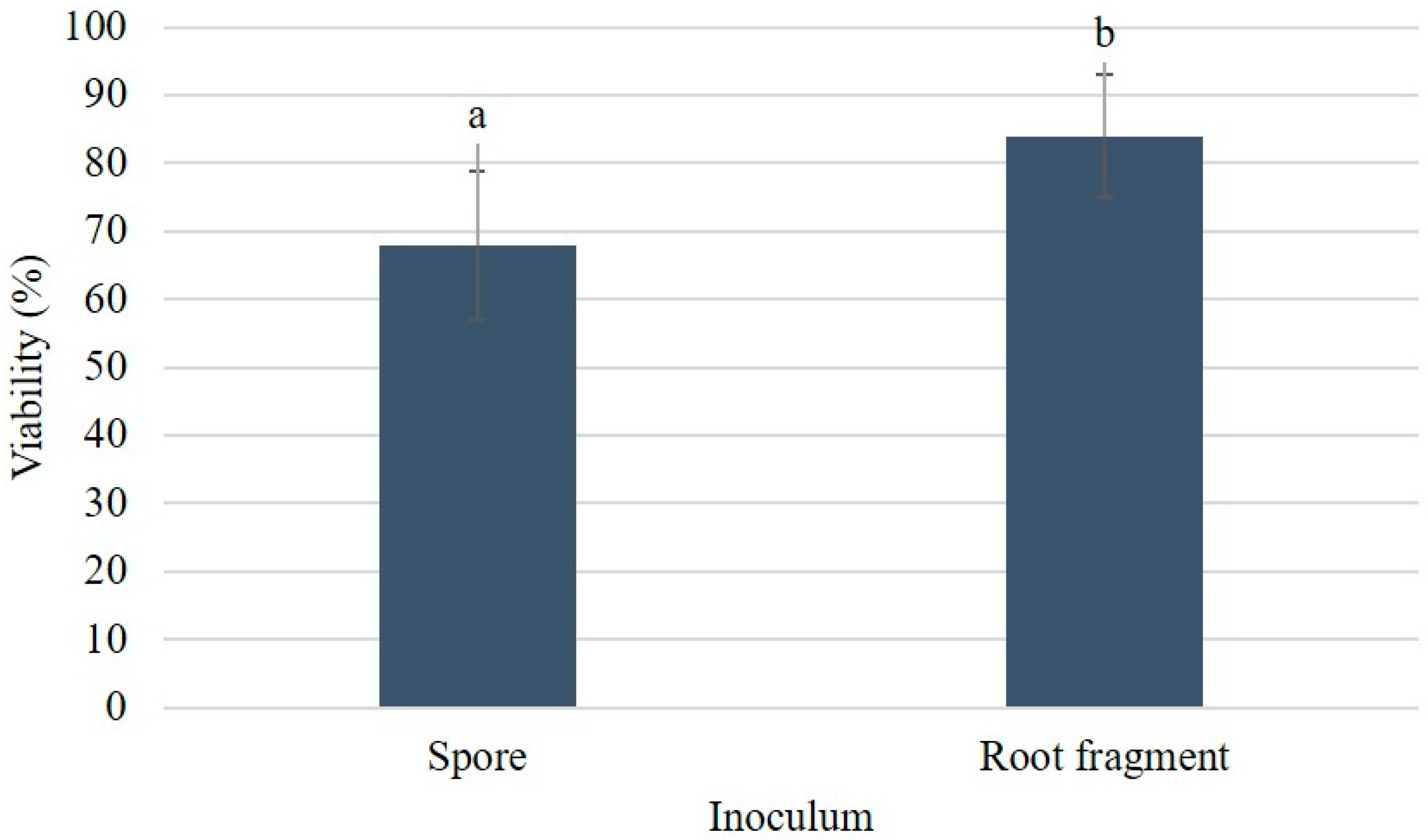
Root fragment-derived propagules exhibited significantly higher viability (84.0 ± 4.7%) than spore-derived propagules (68.0 ± 6.2%) after two weeks on fresh MSR medium (p < 0.01; Figure 6). Germination from fragments was more uniform and produced denser hyphal networks, indicating greater physiological vigor.

3.5. Mycorrhizal Colonization of Carrot Hairy Roots

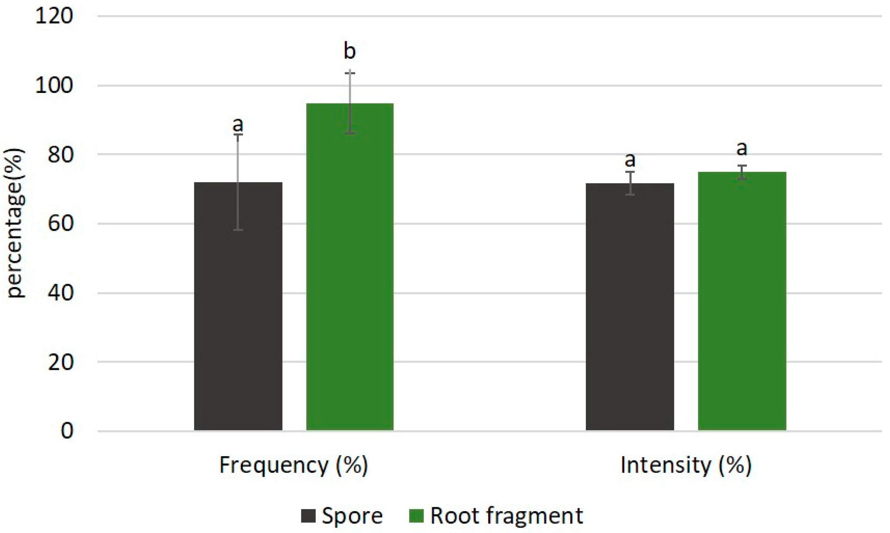
After 16 weeks, colonization frequency was significantly higher in cultures initiated from root fragments (94.7 ± 8.7%) than from spores (72.0 ± 13.7%) (p < 0.05; Figure 7). Colonization intensity did not differ significantly (74.9 ± 1.9% vs. 71.8 ± 3.3%; Figure 7).

3.6. Effect of Inoculum Source on Carrot Hairy Root Growth

Root fragments induced significantly greater total root length (80.9 ± 2.7 cm) than isolated spores (63.0 ± 3.1 cm) after 16 weeks (p < 0.01), whereas fresh weight remained comparable between treatments (Table 1).

3.7. Correlations Between Fungal and Host Root Parameters

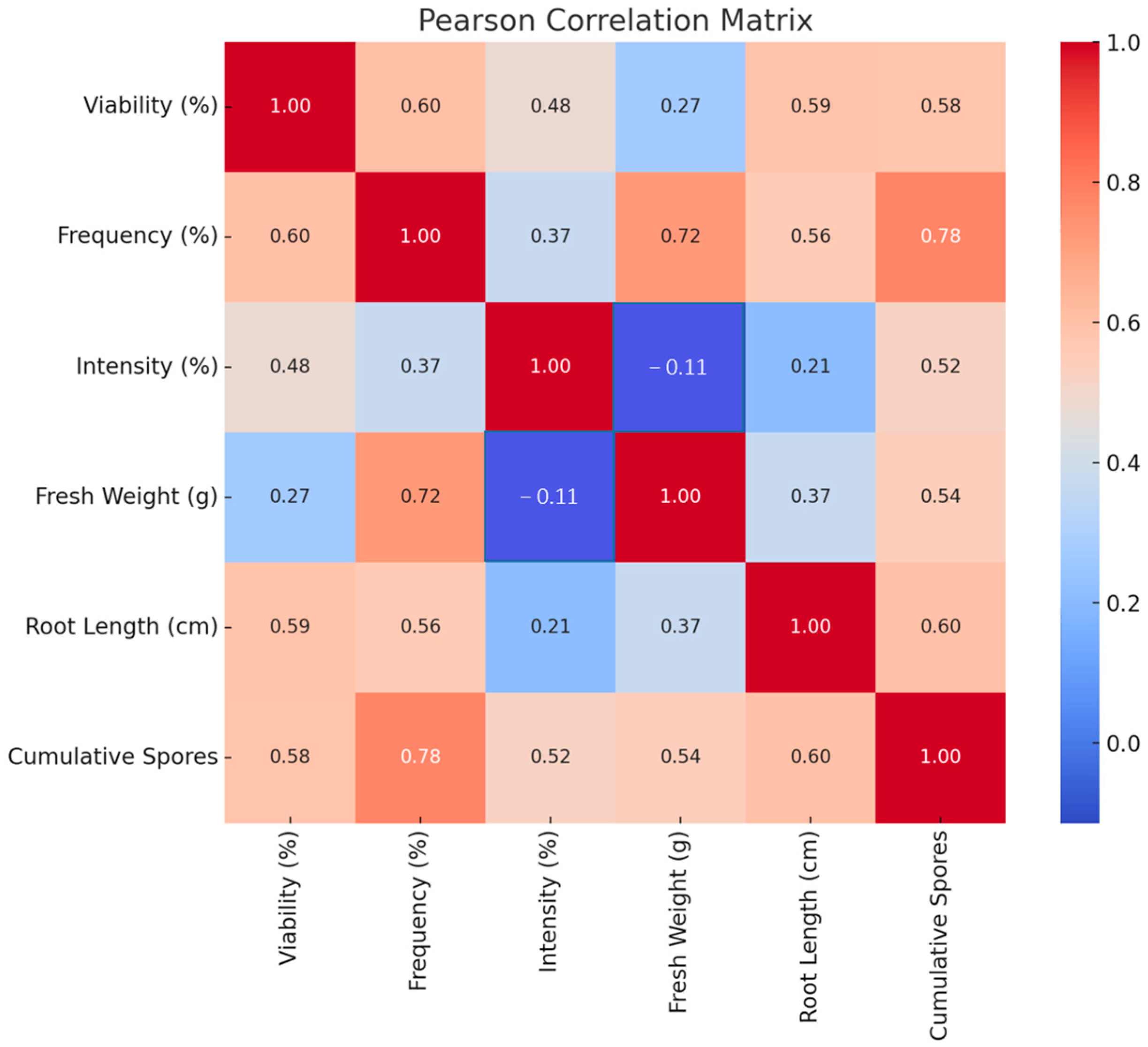
Pearson correlation analysis revealed strong positive relationships between cumulative spore production and both colonization frequency (r = 0.78, p < 0.01) and root length (r = 0.60, p < 0.05). Colonization frequency correlated positively with root fresh weight (r = 0.72, p < 0.01) and root length (r = 0.56, p < 0.05). Spore viability was moderately correlated with colonization frequency (r = 0.60) and root length (r = 0.59) (Figure 8).

3.8. Comparative Symbiotic Performance of R. irregularis Inoculum Produced In Vitro and In Vivo in P. lanceolata

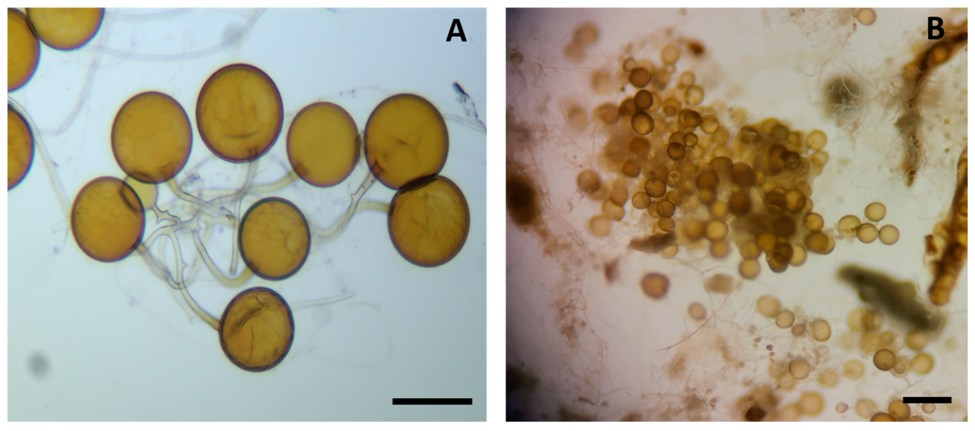
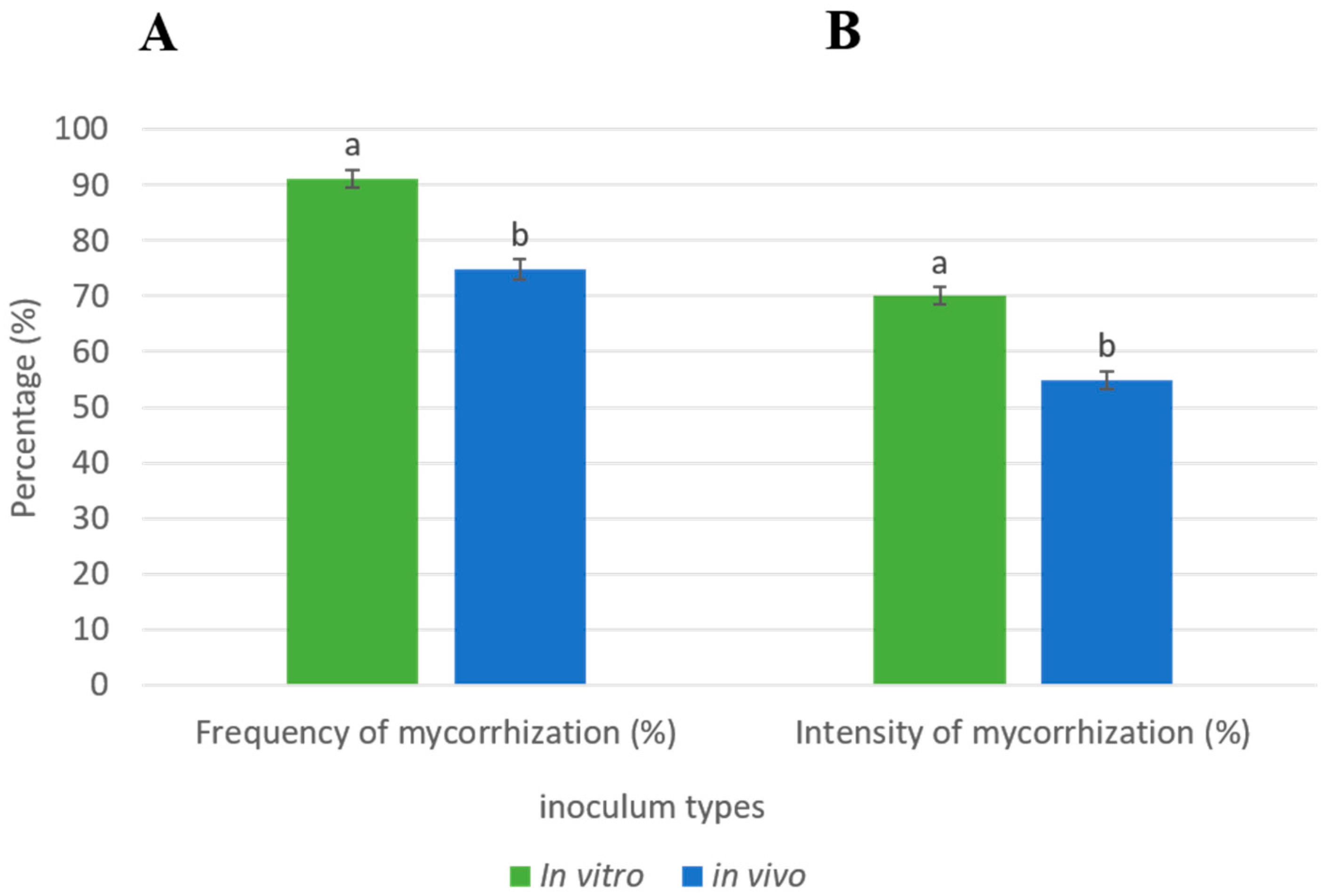
After three months, P. lanceolata plants inoculated with in vitro-produced spores (Figure 9A), showed significantly higher mycorrhizal frequency (91.0 ± 1.6% vs. 74.8 ± 1.9%; p < 0.001) and colonization intensity (70.0 ± 1.6% vs. 55.0 ± 1.6%; p < 0.001) than those receiving in vivo-produced spores (Figure 10).
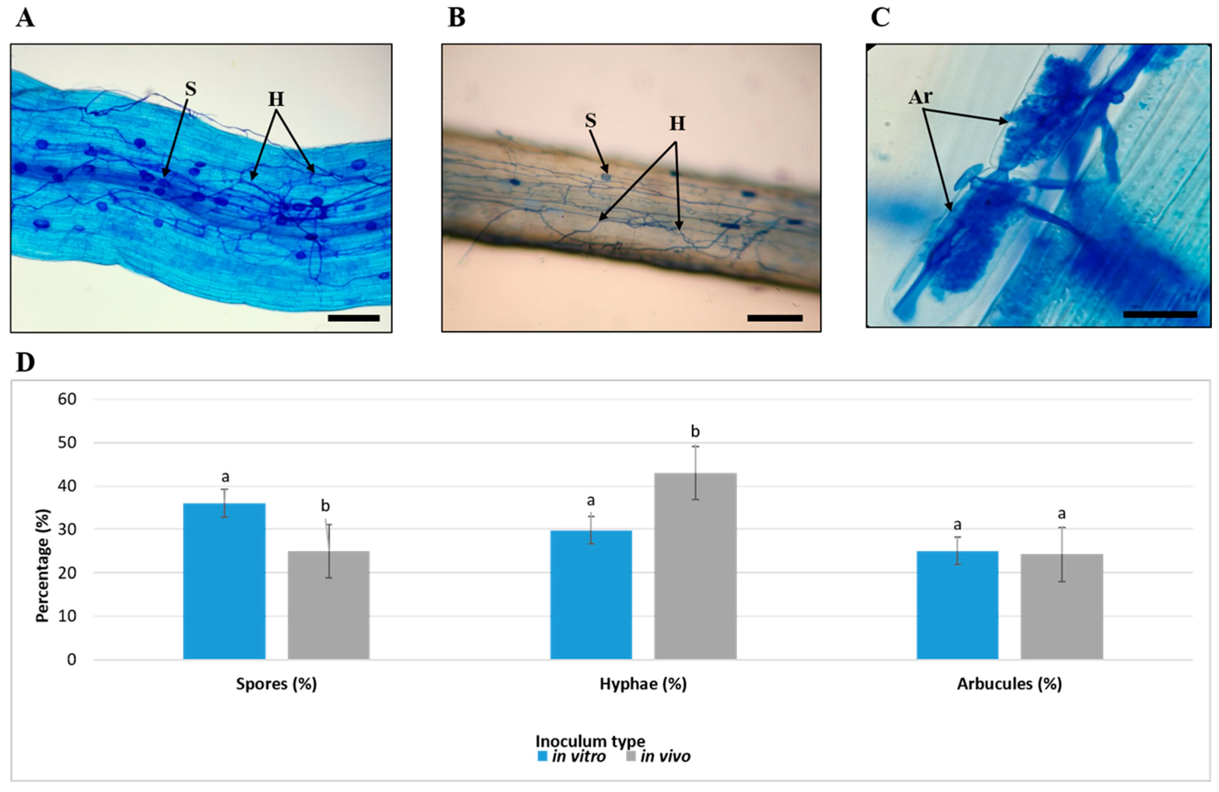
Microscopic examination confirmed more abundant intraradical spores and balanced with arbuscules and hyphae in roots colonized by in vitro inoculum (Figure 11A,D), whereas in vivo inoculum (Figure 9B), produced denser hyphal networks but fewer spores (Figure 11B). Arbuscules were abundant in both treatments (Figure 11C), confirming functional symbiosis. Quantitative assessment of intraradical structures showed higher spore (35.0 ± 1.6% vs. 25.0 ± 1.6%) and arbuscule proportions with in vitro inoculum, while hyphae dominated with in vivo inoculum (41.0 ± 1.6% vs. 30.4 ± 1.1%). These results demonstrate that in vitro monoxenic propagation using root fragments yields highly viable, vigorous R. irregularis inoculum capable of rapid, uniform, and functionally effective root colonization in both monoxenic and whole-plant systems.

4. Discussion

4.1. Robust Taxonomic Identification of an Indigenous R. irregularis Isolate

The combination of classical morphological traits and LSU rDNA phylogeny provided unequivocal identification of the Figuig oasis isolate as R. irregularis (Figure 1 and Figure 2). Although spore morphology remains essential in AMF taxonomy, phenotypic plasticity and overlapping characters among closely related taxa can compromise reliability, particularly in extreme environments [43,49]. Molecular confirmation is therefore indispensable for resolving cryptic species and ensuring accurate assignment [50,51]. Our integrative approach follows current best practice [52] and confirms the widespread occurrence of R. irregularis in arid and semi-arid agroecosystems across North Africa [53,54,55], the Arabian Peninsula [35,36,37], and analogous habitats worldwide. This cosmopolitan yet recurrent presence in stressful environments underscores the remarkable ecological amplitude of R. irregularis and its potential as a keystone symbiont for oasis agriculture. Beyond taxonomic confirmation, accurate identification of the indigenous isolate was a prerequisite for interpreting its biological performance under monoxenic culture conditions, which is addressed in the following sections.

4.2. Influence of Propagule Type on Symbiotic Establishment, Spore Production, and Propagule Quality

Both surface-sterilized spores and mycorrhizal root fragments successfully initiated monoxenic cultures, but root fragments consistently outperformed spores in nearly all measured parameters (Figure 4, Figure 5, Figure 6 and Figure 7; Table 1). Faster hyphal emergence, higher colonization frequency (95% vs. 72%), greater propagule viability (84% vs. 68%), and enhanced carrot hairy-root elongation all point to a clear physiological advantage of intraradical structures over isolated spores. These results align with earlier reports showing that vesicles and mycelial fragments bypass the energy-demanding spore-germination phase, enabling rapid symbiotic re-establishment [45,56,57]. Similar advantages of intraradical propagules have been reported in other monoxenic root organ culture systems, where root fragments promoted faster hyphal development and higher sporulation than isolated spores. Such patterns have been consistently observed across different AMF species and host plants, highlighting the robustness of intraradical propagules as starter material for in vitro AMF multiplication [58,59]. The sustained higher sporulation in fragment-initiated cultures further indicates that early and vigorous symbiosis supports greater carbon allocation to fungal reproduction. Consequently, root fragments containing intraradical spores and hyphae should be preferred as starter material when the objective is rapid, high-density production of viable R. irregularis inoculum.

4.3. Functional Significance of Colonization Parameters

Correlation analyses revealed strong positive relationships between spore production, colonization frequency, and host root growth, whereas colonization intensity showed only weak associations (Figure 8). These findings challenge the widespread assumption that higher percentage root-length colonization automatically translates into greater symbiotic benefit [60]. Instead, colonization frequency and propagule viability emerged as more reliable predictors of fungal performance and host response, consistent with evidence that the extent (frequency) and quality (arbuscule activity, hyphal connectivity) of symbiosis matter more than raw colonization density [61,62]. This nuance is particularly relevant for inoculum screening and quality control protocols. Similar observations have been reported in monoxenic culture studies, where high colonization rates and extensive extraradical mycelium development were not necessarily associated with immediate increases in host plant biomass, particularly under nutrient-sufficient in vitro conditions [59].

4.4. Superior Symbiotic Performance of In Vitro Produced Inoculum

When inoculation dose was standardized (500 viable spores plant−1), in vitro monoxenic spores induced significantly higher mycorrhizal frequency and intensity in P. lanceolata than traditional in vivo (pot-culture) spores (Figure 10). Roots colonized by in vitro inoculum also displayed more balanced development of arbuscules and intraradical spores (Figure 11), indicative of a functionally mature symbiosis. The greater homogeneity among replicates further highlights the reproducibility of monoxenic systems [45,63]. Comparable improvements in infectivity and functional performance of in vitro-produced AMF inoculum have been reported in previous studies using root organ culture systems. In these systems, in vitro inoculum often exhibited higher reproducibility, stable propagule quality, and enhanced colonization efficiency compared with traditional pot-based inoculum. Together, these findings support the view that controlled monoxenic systems preserve, and may even enhance, the symbiotic competence of AMF [58,59,64]. Contrary to occasional concerns that axenic cultivation might reduce infectivity, our data demonstrate that, contrary to occasional concerns that axenic cultivation might reduce infectivity, controlled and contaminant-free conditions preserve and can even enhance symbiotic competence.

4.5. Ecological and Agronomic Implications for Oasis Agroecosystems

By establishing a reliable monoxenic multiplication platform for a locally adapted R. irregularis strain, this study bridges a critical gap between AMF biodiversity surveys and practical application in arid-zone agriculture. Indigenous isolates are generally superior to commercial exotic strains under local edapho-climatic constraints [50,65], and R. irregularis has repeatedly proven effective at alleviating drought, salinity, and nutrient deficiency in date palm and other oasis crops [66]. The superior performance of in vitro inoculum documented here paves the way for nursery-scale mycorrhization of micropropagated date palm plantlets, a practice that is expanding rapidly in Morocco and neighbouring countries. More broadly, in vitro root organ culture has been widely recognized as a reliable and scalable platform for producing high-quality AMF inoculum, offering advantages in biosecurity, standardization, and consistency over conventional pot-based methods. Such systems are particularly relevant for nursery-stage mycorrhization, where uniformity and functional reliability of the inoculum are critical [58,59].

5. Conclusions and Perspectives

Root fragments containing intraradical structures are the preferred propagule type for initiating monoxenic cultures of R. irregularis, yielding faster symbiotic establishment, higher spore production, and more viable inoculum than isolated spores. Moreover, monoxenically produced propagules outperform conventional soil-based inoculum in colonization efficiency and reproducibility. These results validate transformed-root organ culture as a powerful tool for generating high-quality, autochthonous AMF inoculum at scale. Future research should focus on (i) upscaling production, (ii) developing stable inoculum formulations, and (iii) field validation with date palm under oasis conditions. Harnessing locally adapted R. irregularis strains offers a promising, environmentally sound strategy to enhance resilience and productivity of North African palm groves in the face of climate change and soil degradation.

Author Contributions

Conceptualization, E.G., A.H. and M.E.J.; methodology, E.G. and M.E.J.; software, H.E.Y. and E.G.; validation, A.H., M.E.J. and K.C.; formal analysis, E.G. and W.C.; investigation, E.G., M.A. and K.C.; resources, T.L.; data curation, H.E.Y. and W.C.; writing—original draft preparation, E.G. and M.E.J.; writing—review and editing, E.G., M.E.J., K.C. and A.H.; visualization, A.H., T.L. and K.C.; supervision, A.H. and M.E.J.; project administration, A.H. and M.E.J.; funding acquisition, A.H. and M.E.J. All authors have read and agreed to the published version of the manuscript.

Funding

This research was funded by the Academy of Research and Higher Education (ARES) through the Development Research Project PRD-2022-Maroc, entitled “A local and quality organic amendment to sustainably enrich Moroccan oasis soil”.

Institutional Review Board Statement

Not applicable.

Informed Consent Statement

Not applicable.

Data Availability Statement

The original contributions presented in this study are included in the article. Further inquiries can be directed to the corresponding author.

Acknowledgments

We are deeply grateful to the Academy of Research and Higher Education (ARES) in Belgium for their invaluable support. We also warmly thank our partners—Mohammed First University and the Municipality of Figuig—for their dedicated collaboration and valuable contributions to this project.

Conflicts of Interest

The authors declare no conflicts of interest.

References

  1. Foley, J.A.; Ramankutty, N.; Brauman, K.A.; Cassidy, E.S.; Gerber, J.S.; Johnston, M.; Mueller, N.D.; O’Connell, C.; Ray, D.K.; West, P.C.; et al. Solutions for a Cultivated Planet. Nature 2011, 478, 337–342. [Google Scholar] [CrossRef] [Scilit] [PubMed]
  2. FAO. The State of Food and Agriculture 2021: Making Agrifood Systems More Resilient to Shocks and Stresses; FAO: Rome, Italy, 2021; ISBN 92-5-134330-6. [Google Scholar]
  3. Pörtner, H.-O.; Roberts, D.; Tignor, M.; Poloczanska, E.; Mintenbeck, K.; Alegría, A.; Craig, M.; Langsdorf, S.; Löschke, S.; Möller, V.; et al. Climate Change 2022: Impacts, Adaptation and Vulnerability Working Group II Contribution to the Sixth Assessment Report of the Intergovernmental Panel on Climate Change; Intergovernmental Panel on Climate Change: Geneva, Switzerland, 2022. [Google Scholar]
  4. Igiehon, N.O.; Babalola, O.O. Biofertilizers and Sustainable Agriculture: Exploring Arbuscular Mycorrhizal Fungi. Appl. Microbiol. Biotechnol. 2017, 101, 4871–4881. [Google Scholar] [CrossRef] [Scilit]
  5. Begum, N.; Qin, C.; Ahanger, M.A.; Raza, S.; Khan, M.I.; Ashraf, M.; Ahmed, N.; Zhang, L. Role of Arbuscular Mycorrhizal Fungi in Plant Growth Regulation: Implications in Abiotic Stress Tolerance. Front. Plant Sci. 2019, 10, 1068. [Google Scholar] [CrossRef] [Scilit]
  6. Sarathambal, C.; Manimaran, B.; Peeran, M.F.; Srinivasan, V.; Praveena, R.; Gayathri, P.; Dilkush, F.; Abraham, A. Arbuscular Mycorrhizal Colonization Promotes Plant Growth and Regulates Biochemical and Molecular Defense Responses against Pythium myriotylum and Meloidogyne incognita in Ginger (Zingiber officinale Rosc.). Rhizosphere 2025, 34, 101071. [Google Scholar] [CrossRef] [Scilit]
  7. Sonbol, H.; Korany, S.M.; Nhs, M.; Abdi, I.; Maridueña-Zavala, M.G.; Alsherif, E.A.; Aldailami, D.A.; Elsheikh, S.Y.S. Exploring the Benefits of AMF Colonization for Improving Wheat Growth, Physiology and Metabolism, and Antimicrobial Activity under Biotic Stress from Aphid Infection. BMC Plant Biol. 2025, 25, 198. [Google Scholar] [CrossRef] [Scilit]
  8. Smith, S.E.; Read, D.J. Mycorrhizal Symbiosis; Academic Press: Cambridge, MA, USA, 2010; ISBN 0-08-055934-4. [Google Scholar]
  9. Verbruggen, E.; Veresoglou, S.D.; Anderson, I.C.; Caruso, T.; Hammer, E.C.; Kohler, J.; Rillig, M.C. Arbuscular Mycorrhizal Fungi–Short-Term Liability but Long-Term Benefits for Soil Carbon Storage? New Phytol. 2013, 197, 366–368. [Google Scholar] [CrossRef] [Scilit]
  10. Rillig, M.C.; Ryo, M.; Lehmann, A.; Aguilar-Trigueros, C.A.; Buchert, S.; Wulf, A.; Iwasaki, A.; Roy, J.; Yang, G. The Role of Multiple Global Change Factors in Driving Soil Functions and Microbial Biodiversity. Science 2019, 366, 886–890. [Google Scholar] [CrossRef] [Scilit]
  11. Sorensen, J.; Larsen, J.; Jakobsen, I. Pre-Inoculation with Arbuscular Mycorrhizal Fungi Increases Early Nutrient Concentration and Growth of Field-Grown Leeks under High Productivity Conditions. Plant Soil 2008, 307, 135–147. [Google Scholar] [CrossRef] [Scilit]
  12. Nzanza, B.; Marais, D.; Soundy, P. Yield and Nutrient Content of Tomato (Solanum lycopersicum L.) as Influenced by Trichoderma Harzianum and Glomus Mosseae Inoculation. Sci. Hortic. 2012, 144, 55–59. [Google Scholar] [CrossRef] [Scilit]
  13. Pandino, G.; Lombardo, S.; Lo Monaco, A.; Ruta, C.; Mauromicale, G. Mycorrhizal Inoculation Improves Plant Growth and Yield of Micropropagated Early Globe Artichoke under Field Conditions. Agriculture 2022, 12, 114. [Google Scholar] [CrossRef] [Scilit]
  14. Requena, N.; Jeffries, P.; Barea, J.M. Assessment of Natural Mycorrhizal Potential in a Desertified Semiarid Ecosystem. Appl. Environ. Microbiol. 1996, 62, 842–847. [Google Scholar] [CrossRef] [Scilit]
  15. Azcón-Aguilar, C.; Palenzuela, J.; Roldán, A.; Bautista, S.; Vallejo, R.; Barea, J. Analysis of the Mycorrhizal Potential in the Rhizosphere of Representative Plant Species from Desertification-Threatened Mediterranean Shrublands. Appl. Soil Ecol. 2003, 22, 29–37. [Google Scholar] [CrossRef] [Scilit]
  16. Emmett, B.D.; Lévesque-Tremblay, V.; Harrison, M.J. Conserved and Reproducible Bacterial Communities Associate with Extraradical Hyphae of Arbuscular Mycorrhizal Fungi. ISME J. 2021, 15, 2276–2288. [Google Scholar] [CrossRef] [Scilit]
  17. Nthebere, K.; Narasimha Yadav, M.B.; Hota, R.; Kumar, R.; Tyagi, J. Role of Arbuscular Mycorrhizal Fungi in Nutrient Attainment and Plant Growth Development. In Soil Health and Nutrition Management; CABI: Oxfordshire, UK, 2025; pp. 90–108. [Google Scholar]
  18. Riker, A.J.; Banfield, W.M.; Wright, W.H.; Keitt, G.W.; Sagen, H.E. Studies on Infectious Hairy Root of Nursery Apple Trees. J. Agric. Res. 1930, 41, 507–540. [Google Scholar]
  19. Bécard, G.; Fortin, J. Early Events of Vesicular–Arbuscular Mycorrhiza Formation on Ri T-DNA Transformed Roots. New Phytol. 1988, 108, 211–218. [Google Scholar] [CrossRef] [Scilit]
  20. Declerck, S.; Strullu, D.; Plenchette, C. In Vitro Mass-Production of the Arbuscular Mycorrhizal Fungus, Glomus versiforme, Associated with Ri T-DNA Transformed Carrot Roots. Mycol. Res. 1996, 100, 1237–1242. [Google Scholar] [CrossRef] [Scilit]
  21. Declerck, S.; D’or, D.; Cranenbrouck, S.; Boulengé, L.E. Modelling the Sporulation Dynamics of Arbuscular Mycorrhizal Fungi in Monoxenic Culture. Mycorrhiza 2001, 11, 225–230. [Google Scholar] [CrossRef] [Scilit]
  22. Dalpé, Y.; Cranenbrouck, S.; Séguin, S.; Declerck, S. The Monoxenic Culture of Arbuscular Mycorrhizal Fungi as a Tool for Systematics and Biodiversity. In In Vitro Culture of Mycorrhizas; Springer: Berlin/Heidelberg, Germany, 2005; pp. 31–48. [Google Scholar]
  23. Fortin, J.A.; Bécard, G.; Declerck, S.; Dalpé, Y.; St-Arnaud, M.; Coughlan, A.P.; Piché, Y. Arbuscular Mycorrhiza on Root-Organ Cultures. Can. J. Bot. 2002, 80, 1–20. [Google Scholar] [CrossRef] [Scilit]
  24. Pawlowska, T.E.; Douds, D.D.; Charvat, I. In Vitro Propagation and Life Cycle of the Arbuscular Mycorrhizal Fungus Glomus etunicatum. Mycol. Res. 1999, 103, 1549–1556. [Google Scholar] [CrossRef] [Scilit]
  25. Tiwari, P.; Adholeya, A. In Vitro Co-Culture of Two AMF Isolates Gigaspora margarita and Glomus intraradices on Ri T-DNA Transformed Roots. FEMS Microbiol. Lett. 2002, 206, 39–43. [Google Scholar] [CrossRef] [Scilit]
  26. Fonseca, H.M.; Berbara, R.L.; Pereira, M.L. Lunularia Cruciata, a Potential in Vitro Host for Glomus proliferum and G. intraradices. Mycorrhiza 2006, 16, 503–508. [Google Scholar] [CrossRef] [Scilit]
  27. Danesh, Y.R.; Najafı, S.; Demır, S. Using in Vitro Culturing Technique for Studying Life Cycle of Arbuscular Mycorrhizal Fungus (AMF) Glomus intraradices. Yuz. Yıl Univ. J. Agric. Sci. 2016, 26, 161–167. [Google Scholar]
  28. Jilali, A.; Zarhloule, Y.; Georgiadis, M. Vulnerability Mapping and Risk of Groundwater of the Oasis of Figuig, Morocco: Application of DRASTIC and AVI Methods. Arab. J. Geosci. 2015, 8, 1611–1621. [Google Scholar] [CrossRef] [Scilit]
  29. Jilali, A.; Fagel, N.; Amar, M.; Abbas, M.; Zarhloule, Y. Hydrogeochemical Processes Constrained by Multivariate Statistical Methods and Isotopic Evidence of Groundwater Recharge in the Aquifer of Figuig, Eastern High Atlas of Morocco. Arab. J. Geosci. 2016, 9, 42. [Google Scholar] [CrossRef] [Scilit]
  30. Gagou, E.; Chakroune, K.; Abbas, M.; Lamkami, T.; Hakkou, A. Evaluation of the Mycorrhizal Potential of Date Palm (Phoenix dactylifera L.) Rhizosphere Soils in the Figuig Oasis (Southeastern Morocco). J. Fungi 2023, 9, 931. [Google Scholar] [CrossRef] [Scilit]
  31. Kalukuta Mahina, L.; Gagou, E.; Chakroune, K.; Hakkou, A.; El Jaziri, M.; Lamkami, T.; Van Pottelsberghe De La Potterie, B. Turning Waste into Wealth: The Case of Date Palm Composting. Sustainability 2025, 17, 7980. [Google Scholar] [CrossRef] [Scilit]
  32. Hakkou, A.; Bouakka, M. Oasis de Figuig: État Actuel de La Palmeraie et Incidence de La Fusariose Vasculaire. Sci. Chang. Planétaires Sécheresse 2004, 15, 147–158. [Google Scholar]
  33. Moujaoui, N.; Hariri, E.; Elhoumaizi, M. Bayoud and Belaat Diseases of Date Palm (Phoenix dactylifera L.) in Figuig Oasis of Morocco. IOP Conf. Ser. Earth Environ. Sci 2021, 782, 042065. [Google Scholar]
  34. Gagou, E.; Bouchentouf, H.; Chakroune, K.; Abbas, M.; Lamkami, T.; El Jaziri, M.; Hakkou, A. Indigenous Arbuscular Mycorrhizal Fungi Consortium Enhances Growth and Protects Boufeggous Gharas Date Palm Against Fusarium oxysporum f. sp. albedinis Infection in Figuig Oasis (Morocco). Int. J. Plant Biol. 2025, 16, 20. [Google Scholar] [CrossRef] [Scilit]
  35. Al-Yahya’ei, M.N.; Oehl, F.; Vallino, M.; Lumini, E.; Redecker, D.; Wiemken, A.; Bonfante, P. Unique Arbuscular Mycorrhizal Fungal Communities Uncovered in Date Palm Plantations and Surrounding Desert Habitats of Southern Arabia. Mycorrhiza 2011, 21, 195–209. [Google Scholar] [CrossRef] [Scilit] [PubMed]
  36. Symanczik, S.; Błaszkowski, J.; Koegel, S.; Boller, T.; Wiemken, A.; Al-Yahya’Ei, M.N. Isolation and Identification of Desert Habituated Arbuscular Mycorrhizal Fungi Newly Reported from the Arabian Peninsula. J. Arid. Land 2014, 6, 488–497. [Google Scholar] [CrossRef] [Scilit]
  37. Symanczik, S.; Błaszkowski, J.; Chwat, G.; Boller, T.; Wiemken, A.; Al-Yahya’ei, M.N. Three New Species of Arbuscular Mycorrhizal Fungi Discovered at One Location in a Desert of Oman: Diversispora omaniana, Septoglomus nakheelum and Rhizophagus arabicus. Mycologia 2014, 106, 243–259. [Google Scholar] [CrossRef] [Scilit]
  38. Madouh, T.A.; Quoreshi, A.M. The Function of Arbuscular Mycorrhizal Fungi Associated with Drought Stress Resistance in Native Plants of Arid Desert Ecosystems: A Review. Diversity 2023, 15, 391. [Google Scholar] [CrossRef] [Scilit]
  39. Daniels, B.; Skipper, H. Methods for the Recovery and Quantitative Estimation of Propagules from Soil; CABI: Oxfordshire, UK, 1982. [Google Scholar]
  40. Sieverding, E.; Friedrichsen, J.; Suden, W. Vesicular-Arbuscular Mycorrhiza Management in Tropical Agrosystems; TZ-Verlagsgesellschaft: Hanau, Germany, 1991; ISBN 3-88085-462-9. [Google Scholar]
  41. Gagou, E.; Guérin, C.; Chakroune, K.; Abbas, M.; Lamkami, T.; El Jaziri, M.; Hakkou, A. Morphological and Molecular Characterization of Arbuscular Mycorrhizal Fungi from the Rhizosphere of Date Palm (Phoenix dactylifera L.) in the Oasis of Figuig, Morocco. Diversity 2025, 17, 710. [Google Scholar] [CrossRef] [Scilit]
  42. Koske, R.; Walker, C. Species of Gigaspora (Endogonaceae) with Roughened Outer Walls. Mycologia 1985, 77, 702–720. [Google Scholar] [CrossRef] [Scilit]
  43. Krüger, M.; Stockinger, H.; Krüger, C.; Schüßler, A. DNA-based Species Level Detection of Glomeromycota: One PCR Primer Set for All Arbuscular Mycorrhizal Fungi. New Phytol. 2009, 183, 212–223. [Google Scholar] [CrossRef] [Scilit]
  44. Krüger, M.; Krüger, C.; Walker, C.; Stockinger, H.; Schüßler, A. Phylogenetic Reference Data for Systematics and Phylotaxonomy of Arbuscular Mycorrhizal Fungi from Phylum to Species Level. New Phytol. 2012, 193, 970–984. [Google Scholar] [CrossRef] [Scilit]
  45. Cranenbrouck, S.; Voets, L.; Bivort, C.; Renard, L.; Strullu, D.-G.; Declerck, S. Methodologies for in Vitro Cultivation of Arbuscular Mycorrhizal Fungi with Root Organs. In In Vitro Culture of Mycorrhizas; Springer: Berlin/Heidelberg, Germany, 2005; pp. 341–375. [Google Scholar]
  46. Declerck, S.; D’Or, D.; Bivort, C.; de SOUZA, F.A. Development of Extraradical Mycelium of Scutellospora reticulata under Root-Organ Culture: Spore Production and Function of Auxiliary Cells. Mycol. Res. 2004, 108, 84–92. [Google Scholar] [CrossRef] [Scilit]
  47. Vierheilig, H.; Coughlan, A.P.; Wyss, U.; Piché, Y. Ink and Vinegar, a Simple Staining Technique for Arbuscular-Mycorrhizal Fungi. Appl. Environ. Microbiol. 1998, 64, 5004–5007. [Google Scholar] [CrossRef] [Scilit] [PubMed]
  48. Trouvelot, A. Measure Du Taux de Mycorrhization d’un Systeme Radiculaire. Recherche de Methods d’estimation Ayant Une Signification Fonctionnelle. In Physiological and Genetical Aspects of Mycorrhizae; INRA: Paris, France, 1986; pp. 217–221. [Google Scholar]
  49. Redecker, D.; Schüßler, A.; Stockinger, H.; Stürmer, S.L.; Morton, J.B.; Walker, C. An Evidence-Based Consensus for the Classification of Arbuscular Mycorrhizal Fungi (Glomeromycota). Mycorrhiza 2013, 23, 515–531. [Google Scholar] [CrossRef] [Scilit]
  50. Oehl, F.; Laczko, E.; Bogenrieder, A.; Stahr, K.; Bösch, R.; van der Heijden, M.; Sieverding, E. Soil Type and Land Use Intensity Determine the Composition of Arbuscular Mycorrhizal Fungal Communities. Soil Biol. Biochem. 2010, 42, 724–738. [Google Scholar] [CrossRef] [Scilit]
  51. Stockinger, H.; Peyret-Guzzon, M.; Koegel, S.; Bouffaud, M.-L.; Redecker, D. The Largest Subunit of RNA Polymerase II as a New Marker Gene to Study Assemblages of Arbuscular Mycorrhizal Fungi in the Field. PLoS ONE 2014, 9, e107783. [Google Scholar] [CrossRef] [Scilit]
  52. Akbar, K.; Yaseen, T.; Abbasi, B.A.; Iqbal, J.; Alharthi, B.; Fiaz, S.; Noureen, S.; Ijaz, S.; Aziz, E.; Iqbal, R. Morphological and Molecular Identification of Indigenous Arbuscular Mycorrhizal Fungi in the Rhizosphere of Chickpea (Cicer arietinum) and Their Role in Nutrient Uptake. Funct. Plant Biol. 2025, 52, FP24326. [Google Scholar] [CrossRef] [Scilit] [PubMed]
  53. Meddich, A.; Ait El Mokhtar, M.; Bourzik, W.; Mitsui, T.; Baslam, M.; Hafidi, M. Optimizing Growth and Tolerance of Date Palm (Phoenix dactylifera L.) to Drought, Salinity, and Vascular Fusarium-Induced Wilt (Fusarium oxysporum) by Application of Arbuscular Mycorrhizal Fungi (AMF). In Root Biology; Springer: Berlin/Heidelberg, Germany, 2018; pp. 239–258. [Google Scholar]
  54. Chebaane, A.; Symanczik, S.; Oehl, F.; Azri, R.; Gargouri, M.; Mäder, P.; Mliki, A.; Fki, L. Arbuscular Mycorrhizal Fungi Associated with Phoenix dactylifera L. Grown in Tunisian Sahara Oases of Different Salinity Levels. Symbiosis 2020, 81, 173–186. [Google Scholar] [CrossRef] [Scilit]
  55. El Hilali, R.; Symanczik, S.; El Kinany, S.; Oehl, F.; Ouahmane, L.; Bouamri, R. Cultivation, Identification, and Application of Arbuscular Mycorrhizal Fungi Associated with Date Palm Plants in Drâa-Tafilalet Oasis. Rhizosphere 2022, 22, 100521. [Google Scholar] [CrossRef] [Scilit]
  56. Declerck, S.; Strullu, D.G.; Plenchette, C. Monoxenic Culture of the Intraradical Forms of Glomus sp. Isolated from a Tropical Ecosystem: A Proposed Methodology for Germplasm Collection. Mycologia 1998, 90, 579–585. [Google Scholar] [CrossRef] [Scilit]
  57. IJdo, M.; Cranenbrouck, S.; Declerck, S. Methods for Large-Scale Production of AM Fungi: Past, Present, and Future. Mycorrhiza 2011, 21, 1–16. [Google Scholar] [CrossRef] [Scilit]
  58. Ellatif, S.A.; Ali, E.A.M.; Senousy, H.H.; Razik, E.S.A. Production of Arbuscular Mycorrhizal Fungi Using In Vitro Root Organ Culture and Phenolic Compounds. J. Pure Appl. Microbiol. 2019, 13, 1985–1994. [Google Scholar] [CrossRef] [Scilit]
  59. Romero-Ceciliano, M.; Andrade-Torres, A.; Artavia-Salazar, E.; Solís-Ramos, L.Y. Establishing Monoxenic Culture of Arbuscular Mycorrhizal Fungus Glomus sp. Through In Vitro Root Organ Culture and Swietenia Macrophylla King In Vitro Cultures. Agriculture 2025, 15, 673. [Google Scholar] [CrossRef] [Scilit]
  60. Janoušková, M.; Krak, K.; Wagg, C.; Štorchová, H.; Caklová, P.; Vosátka, M. Effects of Inoculum Additions in the Presence of a Preestablished Arbuscular Mycorrhizal Fungal Community. Appl. Environ. Microbiol. 2013, 79, 6507–6515. [Google Scholar] [CrossRef] [Scilit]
  61. Berruti, A.; Lumini, E.; Balestrini, R.; Bianciotto, V. Arbuscular Mycorrhizal Fungi as Natural Biofertilizers: Let’s Benefit from Past Successes. Front. Microbiol. 2016, 6, 1559. [Google Scholar] [CrossRef] [Scilit]
  62. Koch, A.M.; Antunes, P.M.; Maherali, H.; Hart, M.M.; Klironomos, J.N. Evolutionary Asymmetry in the Arbuscular Mycorrhizal Symbiosis: Conservatism in Fungal Morphology Does Not Predict Host Plant Growth. New Phytol. 2017, 214, 1330–1337. [Google Scholar] [CrossRef] [Scilit] [PubMed]
  63. Voets, L.; Dupré de Boulois, H.; Renard, L.; Strullu, D.-G.; Declerck, S. Development of an Autotrophic Culture System for the in Vitro Mycorrhization of Potato Plantlets. FEMS Microbiol. Lett. 2005, 248, 111–118. [Google Scholar] [CrossRef] [Scilit]
  64. Iakab, M.; Domokos, E.; Albert, C.; Buta, E.; Ilonka, Z.; Dulf, F.V. The Influence of Rhizophagus irregularis Arbuscular Mycorrhizal Fungus on Echinacea Purpurea Root Biomass and Bioactive Compounds. Not. Bot. Horti Agrobot. Cluj-Napoca 2025, 53, 14711. [Google Scholar] [CrossRef] [Scilit]
  65. Öpik, M.; Vanatoa, A.; Vanatoa, E.; Moora, M.; Davison, J.; Kalwij, J.; Reier, Ü.; Zobel, M. The Online Database MaarjAM Reveals Global and Ecosystemic Distribution Patterns in Arbuscular Mycorrhizal Fungi (Glomeromycota). New Phytol. 2010, 188, 223–241. [Google Scholar] [CrossRef] [Scilit] [PubMed]
  66. Evelin, H.; Devi, T.S.; Gupta, S.; Kapoor, R. Mitigation of Salinity Stress in Plants by Arbuscular Mycorrhizal Symbiosis: Current Understanding and New Challenges. Front. Plant Sci. 2019, 10, 470. [Google Scholar] [CrossRef] [Scilit] [PubMed]
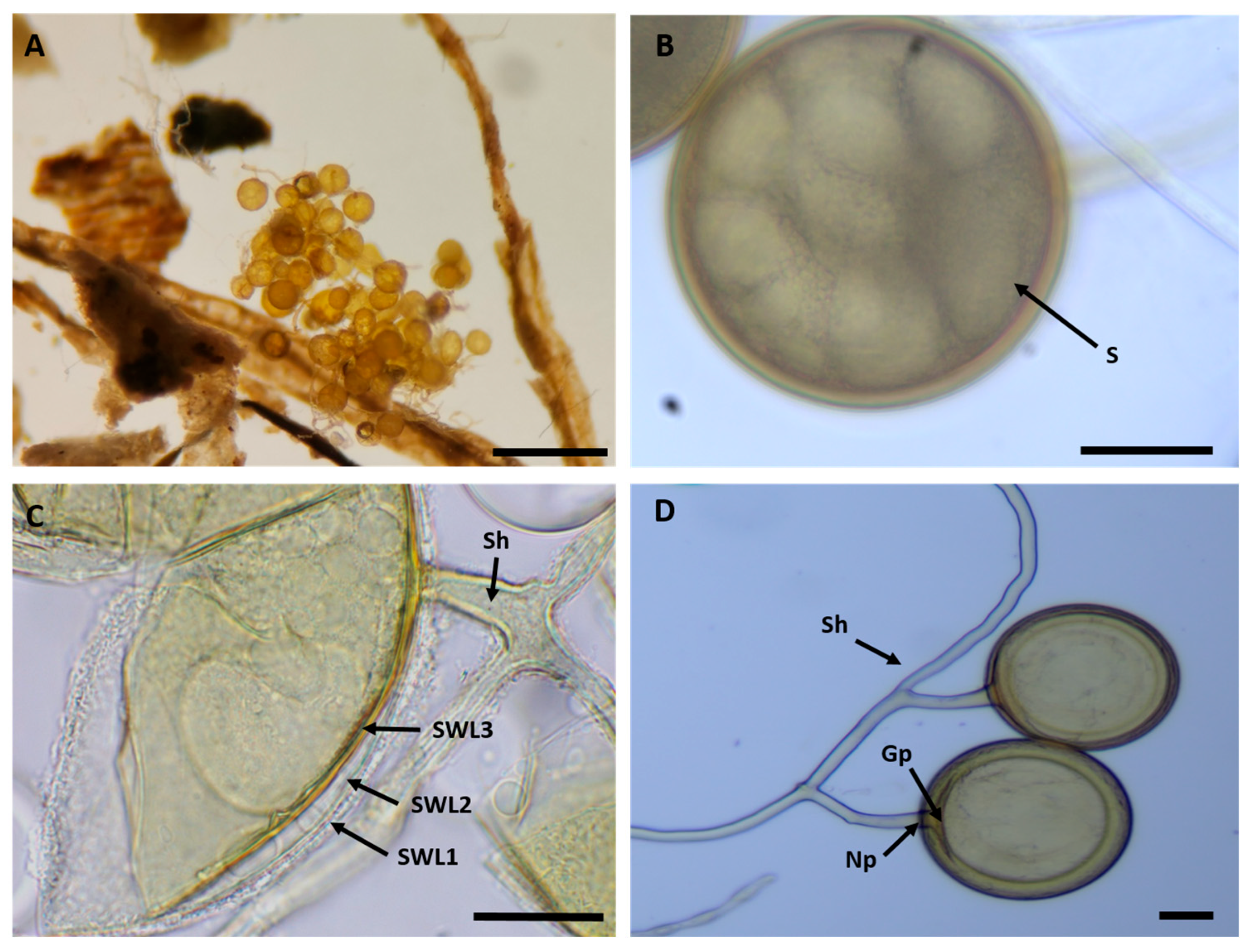
Figure 1. Morphological characteristics of the isolate R. irregularis isolate. (A). Spore cluster observed in P. lanceolata trap culture, attached to root fragments. (B). Mature isolated spore showing typical spherical morphology (S). (C). Mature spores mounted in PVLG + Melzer’s reagent, showing multilayered spore walls (SWL) and subtending hyphae (Sh). (D). Detailed view of subtending hypha (Sh), germ pore (Gp), and narrowing point at the hyphal attachment (Np). Scale bars: (A) = 500 µm; (BD) = 50 µm.
Microbiolres 17 00028 g001
Figure 2. Maximum-likelihood phylogenetic tree based on SSU–ITS–LSU rDNA sequences showing the placement of the R. irregularis isolate from Figuig (highlighted in red) within the R. irregularis clade. The tree includes reference sequences from Rhizophagus and closely related genera. Bootstrap values > 50% are indicated at nodes. Alignment and tree construction were performed using MEGA11.
Microbiolres 17 00028 g002
Figure 3. Hyphal emergence observed 2 weeks after monoxenic culture initiation from two types of inoculum. (A) Hyphal outgrowth from the cut end of a colonized root fragment (white arrow) and formation of extraradical hyphae (black arrows). (B) Germination of a single spore (white arrow) and subsequent emission of germ tubes and extraradical hyphae (black arrows). Images captured under inverted light microscopy; Scale bars: (A) = 100 µm; (B) = 200 µm.
Microbiolres 17 00028 g003
Figure 4. Spore formation rate of R. irregularis over a 16-week monoxenic culture on carrot hairy roots, using spore- and root fragment-derived inocula on MSR medium (n = 5). Values represent means ± SD. Asterisks indicate significant differences between inoculum types at the same week according to Student’s t-test (* p < 0.05; ** p < 0.01).
Microbiolres 17 00028 g004
Figure 5. Sporulation of R. irregularis in monoxenic carrot hairy root cultures initiated from a root-fragment inoculum at week 16. (A) Early sporulation stage (week 8), showing ornamented spores (≈90–120 µm) developing on subtending hyphae at the hairy root surface. (B) Late sporulation stage (week 16), profuse spore production associated with an extensive extraradical hyphal network covering the medium surface. Scale bars: (A) = 50 µm; (B) = 500 µm.
Microbiolres 17 00028 g005
Figure 6. Viability of R. irregularis Propagules from Different Inoculum Sources After 2 Weeks of Culture on MSR Medium (n = 20). Different letters above bars indicate significant differences between treatments according to the Mann–Whitney U test (p < 0.05).
Microbiolres 17 00028 g006
Figure 7. Root colonization frequency and intensity of R. irregularis after 16 weeks of monoxenic culture on carrot hairy roots, using spore-derived or root fragment-derived inocula (n = 10). Different letters indicate significant differences (p < 0.05) according to the Mann–Whitney test for frequency and Student’s t-test for intensity.
Microbiolres 17 00028 g007
Figure 8. Pearson correlation heatmap showing the relationships between mycorrhizal traits (spore viability, colonization frequency and intensity) and plant growth parameters (root length, fresh weight, and cumulative spore production) in monoxenic cultures of R. irregularis.
Microbiolres 17 00028 g008
Figure 9. Propagules used for inoculation. (A) In vitro spores produced under monoxenic culture on transformed carrot hairy roots. (B) In vivo spores recovered from pot-based trap cultures. Note the cleaner and more size-homogeneous spores in (A) compared with the more heterogeneous, debris-associated spores in (B). Scale bars: (A) = 100 µm; (B) = 500 µm.
Microbiolres 17 00028 g009
Figure 10. Comparative mycorrhizal colonization efficiency (frequency and intensity) in P. lanceolata following inoculation with R. irregularis propagated either in vitro or in vivo after 16 weeks of culture. (A) Frequency of mycorrhization (%), (B) Intensity of mycorrhization (%). Bars show mean ± SD (n = 10). Different letters above the bars indicate significant differences according to Student’s t-test (p < 0.05).
Microbiolres 17 00028 g010
Figure 11. Microscopic observations and quantitative assessment of intraradical structures formed by R. irregularis in P. lanceolata inoculated either with in vitro or in vivo derived propagules after 16 weeks of culture. (AC): Representative micrographs showing intraradical spores (S), hyphae (H), and arbuscules (Ar). (D): Quantitative distribution of intraradical structures (spores, hyphae, and arbuscules). Bars represent mean ± SD (n = 10). Different letters above the bars indicate significant differences according to Student’s t-test (p < 0.05). Scale bars: (A,B) = 200 µm; (C) = 20 µm.
Microbiolres 17 00028 g011
Table 1. Fresh weight and root length of carrot roots 16 weeks post-inoculation with R. irregularis spores or root fragments. Values are expressed as mean ± SD (n = 5 biological replicates per treatment) and were measured at the end of the 16-week monoxenic culture experiment.
TreatmentFresh Weight (g)Root Length (cm)
Root fragment0.678 ± 0.025 a80.90 ± 2.68 a
Spore0.600 ± 0.052 a62.96 ± 3.12 b
Different letters (a, b) within the same column indicate sig-nificant differences between treatments according to Student’s t-test (p < 0.05).
Disclaimer/Publisher’s Note: The statements, opinions and data contained in all publications are solely those of the individual author(s) and contributor(s) and not of MDPI and/or the editor(s). MDPI and/or the editor(s) disclaim responsibility for any injury to people or property resulting from any ideas, methods, instructions or products referred to in the content.