Article

Whole-Genome Sequencing and Genomic Features of Vagococcus sp. JNUCC 83 Isolated from Camellia japonica Flowers


Department of Chemistry and Cosmetics, Jeju National University, Jeju 63243, Republic of Korea
*
Author to whom correspondence should be addressed.
These authors contributed equally to this work.
Microbiol. Res. 2026, 17(1), 23; https://doi.org/10.3390/microbiolres17010023
Submission received: 17 December 2025 / Revised: 8 January 2026 / Accepted: 14 January 2026 / Published: 18 January 2026
(This article belongs to the Special Issue Advances in Plant–Pathogen Interactions)

Abstract

Vagococcus species have been isolated from diverse environments, including aquatic, terrestrial, food-associated, and clinical sources; however, plant- and flower-associated representatives remain poorly characterized at the genomic level. In this study, we report the complete genomic sequence and analysis of Vagococcus sp. JNUCC 83, isolated from flowers of Camellia japonica collected on Jeju Island, Republic of Korea. The genome comprises a single circular chromosome of 2,472,896 bp with a GC content of 33.5 mol% and was assembled at high depth (555.43×), resulting in a high-quality complete genome. Genome-based phylogenomic analysis using the Type (Strain) Genome Server (TYGS) showed that strain JNUCC 83 forms a distinct lineage within the genus Vagococcus. Digital DNA–DNA hybridization (dDDH) values were far below the 70% species threshold, and 16S rRNA gene-based phylogeny consistently supported its independent placement, suggesting that JNUCC 83 represents a previously undescribed genomic species. Functional annotation based on EggNOG/COG analysis indicated the enrichment of genes involved in core metabolism and genome maintenance, while antiSMASH analysis identified a terpene-precursor-type biosynthetic locus encoding a polyprenyl synthase. Overall, this study expands the genomic understanding of flower-associated Vagococcus lineages and provides a foundation for future investigations into their ecological roles and potential applications as plant-derived microbial resources.

1. Introduction

The genus Vagococcus belongs to the family Enterococcaceae within the order Lactobacillales [1,2] and comprises Gram-positive, catalase-negative cocci that typically occur singly, in pairs, or in short chains, although rod-like morphologies have also been reported in some strains [3,4]. Members of this genus are facultatively anaerobic and primarily produce L-lactic acid as the major end product of glucose fermentation [1,5]. Their cell wall peptidoglycans are Lys–D-Asp in type [2,4], and their genomic DNA G+C content generally ranges from approximately 33.6 to 44.5 mol% [2,6,7]. Colonies are usually grayish-white and convex on blood agar, exhibiting α-hemolytic or non-hemolytic phenotypes [4]. Motility varies among species, reflecting the historical origin of the genus name Vagococcus (from the Latin vagus, meaning “wandering”), although non-motile species are also common [1,3].
Since its proposal in 1989, the genus Vagococcus has expanded to include multiple recognized species, such as Vagococcus fluvialis (the type species) [1,5], V. salmoninarum [8], V. lutrae [9], V. fessus [10], V. carniphilus [11], V. elongatus [6], V. penaei [12], V. acidifermentans [13], V. bubulae [14], and V. vulneris [14]. These species have been isolated from a wide array of ecological niches, including aquatic environments (freshwater, fish, and marine mammals) [8,9,12], terrestrial settings (poultry litter and livestock manure) [1,6], food matrices (meat, shrimp, and fermented soybean products) [6,11], and clinical samples [8,9]. Due to phenotypic similarities with Lactococcus and Enterococcus, Vagococcus species are frequently misidentified in routine diagnostics, necessitating the use of molecular approaches such as 16S rRNA gene sequencing or MALDI-TOF mass spectrometry for accurate taxonomic assignment [15,16,17].
Clinically, Vagococcus has gained attention as an emerging opportunistic pathogen [5,17]. Several species are associated with streptococcosis-like infections in fish, often resulting in high mortality [18,19,20], and have also been reported from infections in aquatic mammals, livestock, and humans [10,17,21]. Human cases, although relatively rare, include skin and soft tissue infections, osteomyelitis, bacteremia, peritonitis, and meningitis [5,17,22,23]. Moreover, resistance to certain antibiotic classes, including fluoroquinolones and macrolides, has been documented in some strains [5,24], complicating empirical treatment and underscoring the importance of genome-level characterization to better understand pathogenic potential and resistance mechanisms [5,17].
Despite this pathogenic reputation, members of the genus Vagococcus also exhibit notable biotechnological potential [5,17]. Certain strains have been investigated as probiotics in aquaculture, where V. fluvialis has been shown to enhance host innate immunity and protect fish against Vibrio anguillarum infection, significantly improving survival rates [25,26]. In addition, V. carniphilus produces bacteriocins with inhibitory activity against foodborne pathogens such as Listeria and Staphylococcus, highlighting possible applications in food preservation [11,27]. Other Vagococcus strains have been reported to produce thermostable alkaline proteases—enzymes of interest for industrial and detergent applications due to their robustness and efficacy—from agricultural waste substrates [2,7]. Collectively, these findings suggest that the functional repertoire of Vagococcus extends beyond pathogenicity and warrants re-evaluation from a beneficial and application-oriented perspective [5,17].
In recent years, increasing attention has been directed toward the exploration of beneficial microorganisms derived from natural and minimally disturbed ecosystems for use as cosmetic and cosmeceutical ingredients [28,29,30]. In this context, Jeju Island, a volcanic island recognized for its unique climate, geological features, and high biodiversity, represents an exceptional reservoir of unexplored microbial resources [31,32,33]. Flower-associated microbiota, in particular, are highly specialized ecological niches shaped by plant secondary metabolites, fluctuating environmental conditions, and interactions with pollinators [34,35]. These selective pressures often give rise to microorganisms with enhanced stress tolerance and the capacity to produce bioactive compounds [27,36,37], traits that are highly relevant to cosmetic applications such as skin protection, antioxidant activity, and barrier-supporting functions [28,29,38].
Although Vagococcus species have been isolated from a broad spectrum of environments [3,39], plant- and flower-associated representatives remain poorly characterized at the genomic level [2,14]. This knowledge gap limits our understanding of their ecological roles, adaptive strategies, and potential as functional microbial resources [5,17]. From an industrial standpoint, systematic genomic evaluation is essential to distinguish strains with beneficial attributes from those with pathogenic risk, thereby enabling the safe development of Vagococcus-derived materials for cosmetic and biotechnological applications [5,25,26].
In this study, we report the complete genome sequence and comprehensive genomic analysis of Vagococcus sp. JNUCC 83, isolated from the flowers of Camellia japonica collected on Jeju Island, Republic of Korea. By integrating genomic features related to metabolism, stress adaptation, and potential functional traits, this work aims to reassess the genus Vagococcus from a plant-associated and application-oriented perspective. Our findings expand the genomic landscape of Vagococcus and provide a scientific foundation for future studies exploring flower-derived beneficial bacteria as novel resources for cosmetic and industrial applications.
Given that plant- and flower-associated members of the genus Vagococcus remain poorly characterized at the genomic level and that the genus includes opportunistic lineages with reported antimicrobial resistance, whole-genome sequencing is essential to resolve species boundaries and to provide a genomic foundation for evaluating ecological adaptation and potential safety risks of plant-derived isolates.

2. Materials and Methods

2.1. Bacterial Isolation and DNA Extraction

Vagococcus sp. JNUCC 83 was isolated from flowers of C. japonica collected at the Citrus Research Center, Jeju Island, Republic of Korea (33.302465° N, 126.612042° E), on 7 December 2023. Floral samples were aseptically collected, homogenized, and spread onto de Man–Rogosa–Sharpe (MRS) agar plates. The plates were incubated at 28 °C under anaerobic conditions for 48 h, after which individual colonies were selected and repeatedly streaked on fresh MRS agar plates to obtain a pure culture.
For genomic DNA extraction, the isolate was cultivated in MRS broth until the late exponential phase, and cells were harvested by centrifugation. Genomic DNA was extracted by CJ Bioscience Inc. (Seoul, Republic of Korea) using the Qiagen MagAttract HMW DNA Kit (Qiagen, Hilden, Germany) in accordance with the manufacturer’s instructions. Briefly, harvested cells were subjected to enzymatic lysis, followed by protein digestion and DNA purification according to the kit protocol. The concentration of the extracted genomic DNA was quantified using a Qubit 2.0 fluorometer (Invitrogen, Carlsbad, CA, USA). Potential contamination of the cultured strain was assessed by sequencing the 16S rRNA gene using an ABI 3730 DNA Analyzer (Applied Biosystems, Foster City, CA, USA). Only high-quality genomic DNA meeting the sequencing provider’s quality control criteria was used for library preparation and subsequent whole-genome sequencing).

2.2. Genome Sequencing, Assembly, and Quality Assessment

Whole-genome sequencing of Vagococcus sp. JNUCC 83 was performed by CJ Bioscience Inc. (Seoul, Republic of Korea) using a long-read-first hybrid sequencing strategy that integrated PacBio long-read and Illumina short-read platforms. PacBio long-read sequencing data were used for primary de novo genome assembly, enabling the construction of contiguous genome sequences.
Illumina short-read sequencing was conducted using the Illumina NovaSeq 6000 platform (NovaSeq Control Software v1.8 with a v1.5 reagent kit). Paired-end libraries with an average insert size of approximately 350 bp were constructed and sequenced. Raw Illumina reads were subjected to quality control procedures, including adapter trimming and removal of low-quality reads, prior to downstream analyses. Adapter trimming and removal of low-quality reads were performed using Trimmomatic v0.36, and contaminating PhiX sequences were removed using BBMap v38.32. The average sequencing depth across the assembled genome was approximately 555.43×, as calculated based on read mapping to the final assembly.
The initial genome assembly was generated using the Microbial Assembly protocol implemented in SMRT Link v25.2, which is optimized for long-read-based microbial genome assembly. The resulting assembly was subsequently polished using high-quality Illumina short reads to improve base-level accuracy. Circularization and rotation of circular contigs were performed based on the dnaA or predicted replication origin.
The final assembly consisted of six circular contigs, including one main chromosome and five small circular contigs, with a total genome size of approximately 2.50 Mbp. The average sequencing depth was calculated based on read mapping to the assembled genome [40].
Genome annotation was performed using the NCBI Prokaryotic Genome Annotation Pipeline (PGAP) v6.10. Gene prediction was conducted using the GeneMarkS-2+ algorithm [41,42] in combination with the best-placed reference protein set provided by the NCBI. The annotation process identified protein-coding sequences (CDSs), ribosomal RNA (rRNA) genes, transfer RNA (tRNA) genes, non-coding RNA genes, and pseudogenes. All annotation steps were carried out using default pipeline parameters.
Genome completeness and contamination were assessed using CheckM v1.2.4, employing lineage-specific marker genes corresponding to the family Enterococcaceae [43]. The evaluation was based on the annotated gene set generated by the PGAP, and completeness and contamination metrics were calculated following the standard CheckM workflow. The resulting high-quality genome assembly and annotation provided a reliable foundation for subsequent comparative genomic, phylogenomic, and functional analyses.

2.3. Genome-Based Phylogenomic Analysis Using Type Strain Genome Server (TYGS)

Genome-based phylogenomic analysis of Vagococcus sp. JNUCC 83 was performed using the Type (Strain) Genome Server (TYGS) based on the Genome BLAST Distance Phylogeny (GBDP) approach to assess its phylogenetic relatedness to Vagococcus type strains. The complete chromosomal genome sequence of JNUCC 83 was submitted to the TYGS platform and compared with all available Vagococcus type strain genomes deposited in the DSMZ database.
Pairwise intergenomic distances were calculated using the Genome BLAST Distance Phylogeny (GBDP) approach under formula d5, as implemented in the TYGS pipeline. Phylogenetic relationships were inferred from the resulting distance matrix using the FastME v2.1.6.1 algorithm based on the balanced minimum-evolution criterion. Branch support values were estimated from 100 pseudo-bootstrap replicates [44,45].
The genomic relatedness between JNUCC 83 and its closest Vagococcus type strains was further evaluated by calculating digital DNA–DNA hybridization (dDDH) values using the Genome-to-Genome Distance Calculator (GGDC) v3.0, integrated within the TYGS framework. In addition, average nucleotide identity (ANI) values were computed using the OrthoANIu algorithm (CJ Bioscience Inc., Seoul, Republic of Korea) with default parameters to provide complementary evidence of genomic similarity [44,46].
All analyses were performed using default settings of the TYGS and GGDC servers, and the resulting datasets were used to construct a robust genome-based phylogenomic framework for Vagococcus sp. JNUCC 83.

2.4. Clusters of Orthologous Groups (COGs) Functional Classification of the Genome

Functional classification based on the Clusters of Orthologous Groups (COGs) was performed to characterize the distribution of genes across major functional categories, including metabolic, cellular, and information-processing functions, in the Vagococcus sp. JNUCC 83 genome. Protein-coding sequences (CDSs) predicted by Prokka v1.14.6 were functionally annotated using EggNOG-mapper v2.1.13 with the eggNOG v6.0 database through the analysis pipeline provided by CJ Bioscience Inc. (Seoul, Republic of Korea) [47,48,49].
Each predicted protein was assigned to one of the 22 standard COG functional categories based on its best orthologous match. CDSs lacking significant similarity to known orthologous groups were classified as “S: Function unknown” or “X: Not assigned.” The distribution of CDSs across COG functional categories was calculated and summarized from the processed annotation dataset generated by CJ Bioscience.
This orthology-based functional annotation provided a comprehensive overview of the genomic functional architecture of Vagococcus sp. JNUCC 83, highlighting major gene groups involved in metabolism, transcriptional regulation, replication, and cellular adaptation.

2.5. Secondary Metabolite Biosynthetic Gene Cluster Analysis

The prediction of secondary metabolite biosynthetic gene clusters (BGCs) in Vagococcus sp. JNUCC 83 was performed using antiSMASH bacterial version v8.0.4. The annotated genome file generated by Prokka v1.14.6 was used as input in GenBank format. BGC detection was conducted under the “relaxed” detection strictness setting to enable sensitive identification of both complete and partial biosynthetic gene clusters, including cryptic loci lacking one or more canonical biosynthetic components.
To enhance functional prediction and comparative analysis, multiple optional antiSMASH modules were activated, including KnownClusterBlast, SubClusterBlast, ActiveSiteFinder, RREFinder, and transcription factor binding site (TFBS) analysis. In addition, Pfam domain-based annotation was applied to identify conserved biosynthetic and accessory domains within predicted clusters. These settings were selected to maximize the detection and characterization of low-abundance or atypical BGCs, which are frequently observed in lactic acid bacteria and related taxa [50,51].
Each predicted BGC was further compared against the Minimum Information about a Biosynthetic Gene cluster (MIBiG) database v4.0 to assess sequence similarity and biosynthetic relatedness to experimentally characterized reference clusters. The gene content, organization, and domain architecture of individual clusters were manually examined using the antiSMASH integrated visualization tools, which provide gene-level schematic representations of cluster boundaries and predicted biosynthetic domains [52].
This comprehensive in silico workflow provided a reproducible and standardized framework for the identification, classification, and preliminary characterization of secondary metabolite biosynthetic gene clusters in the genome of Vagococcus sp. JNUCC 83.

3. Results and Discussion

3.1. Genome Features of Vagococcus sp. JNUCC 83

The complete genome of Vagococcus sp. JNUCC 83 consists of a single circular chromosome with a total length of 2,472,896 bp and an average GC content of 33.5 mol%, which is consistent with values reported for members of the genus Vagococcus (Figure 1). The genome was sequenced using Illumina NovaSeq 6000 technology (Illumina Inc., San Diego, CA, USA) and assembled at high depth, achieving an average coverage of 555.43×. The finalized chromosome has been deposited in the GenBank database under the accession number CM132204.1. The assembly was classified as a “Complete Genome” by NCBI, with no unlocalized contigs, confirming that the sequence represents a finished, high-quality chromosomal assembly rather than a draft genome.
Genome annotation was performed using the NCBI Prokaryotic Genome Annotation Pipeline (PGAP v6.10), which predicted a total of 2492 genes. Among these, 2371 genes were identified as protein-coding sequences, while 84 genes corresponded to RNA features. The RNA gene complement comprises six complete rRNA operons, each consisting of 5S, 16S, and 23S rRNA genes, sixty-two tRNA genes representing all standard amino acids, and four non-coding RNA (ncRNA) genes (Table 1). The presence of multiple complete rRNA operons together with a full set of tRNA genes suggests a robust translational capacity, a genomic trait often associated with metabolic flexibility and the ability to rapidly adapt to fluctuating environmental conditions.
In addition, 37 pseudogenes were identified in the genome. These pseudogenes are primarily attributed to frameshift mutations or incomplete coding sequences, which are commonly observed in bacterial genomes and do not indicate deficiencies in assembly quality. Instead, the presence of pseudogenes may reflect ongoing genome streamlining or niche-specific adaptive processes.
Genome quality assessment using CheckM v1.2.4 indicated a completeness of 96.4% and a low contamination level of 0.94%, further supporting the reliability of the genome sequence and annotation. Additional taxonomic consistency checks confirmed that the genome is clearly affiliated with the genus Vagococcus, with no evidence of cross-genus contamination.
Overall, the genome of Vagococcus sp. JNUCC 83 represents a compact, well-annotated, and high-quality chromosomal sequence. Its general genomic features are characteristic of the genus Vagococcus while providing a solid foundation for subsequent phylogenomic, functional, and comparative analyses aimed at clarifying its taxonomic position and ecological potential.

3.2. Small Replicons Associated with the Chromosome

In addition to the main circular chromosome, the complete genome assembly of Vagococcus sp. JNUCC 83 includes five small extrachromosomal sequences, designated as unnamed 1 to unnamed 5 in the GenBank record (accession numbers JBSJXX010000001.1–JBSJXX010000006.1). These replicons range in size from 4126 bp to 15,762 bp and collectively account for less than 1.5% of the total genomic content (Table 2). Their GC contents vary between 27.0% and 34.5%, partially overlapping with but in several cases lower than that of the main chromosome (33.5%), consistent with the heterogeneous composition often observed among plasmid-borne elements in members of the family Enterococcaceae.
All five extrachromosomal replicons were assembled at sequencing depths comparable to that of the chromosome and showed no evidence of contamination, consistent with the overall genome quality metrics (96.4% completeness and 0.94% contamination based on CheckM analysis). No unlocalized or ambiguous regions were detected, supporting the interpretation that these sequences represent genuine components of the JNUCC 83 genome rather than assembly artifacts.
Gene annotation further supports a plasmid-like character for these replicons. Unnamed 1 (13,918 bp; 27.0% GC) contains multiple plasmid hallmarks, including a replication initiation protein (Rep), a MobV family relaxase, and stability-associated functions such as a type II toxin–antitoxin module (RelE/RelB), together with an IS3-family transposase, indicating mobilization potential and maintenance capacity typical of small plasmids [50,51,52,53]. Unnamed 2 (15,762 bp; 30.0% GC) also encodes a replication initiation protein, multiple predicted transcriptional regulators, and toxin–antitoxin components (including RelE/ParE- and Fst-family toxins), as well as a site-specific tyrosine recombinase/integrase, collectively suggesting a plasmid replicon enriched in maintenance and genome plasticity-related functions [51,52,53,54,55]. Unnamed 3 (4126 bp; 34.5% GC) encodes a replication initiation protein and a DNA/RNA non-specific endonuclease, consistent with a compact cryptic replicon typically observed among low-copy-number plasmids [52]. Unnamed 4 (5035 bp; 29.5% GC) likewise contains a replication initiation protein and additional small proteins, including a predicted lipase-family protein, supporting its interpretation as a small plasmid-like element [50]. Finally, unnamed 5 (4863 bp; 31.5% GC) encodes a heavy metal-translocating P-type ATPase together with an ArsR/SmtB family transcriptional regulator, forming a compact metal resistance-associated module frequently encountered on small plasmids in members of the family Enterococcaceae [56,57,58].
Collectively, the presence of replication initiation proteins in multiple replicons (unnamed 1–4), together with that of mobilization (MobV) and stability modules (toxin–antitoxin systems) and recombination/transposition-associated genes (integrase/transposase), strongly supports that these extrachromosomal sequences represent plasmids or plasmid-derived replicons rather than chromosomal fragments [50,51,52,53,54]. While complete conjugation machineries were not identified and replication modules could not be assigned to a specific plasmid incompatibility group based solely on automated annotation, the overall gene complement and compact organization are consistent with low-copy-number, cryptic plasmids that may persist without conferring readily apparent phenotypes [59,60,61].
Importantly, these extrachromosomal replicons were not included in genome-based phylogenomic analyses performed using TYGS, which rely exclusively on chromosomal sequences for GBDP distance calculations and dDDH estimation. Therefore, the taxonomic placement and species delineation of Vagococcus sp. JNUCC 83 are determined solely based on the complete chromosomal genome and are not influenced by the presence or absence of these plasmid-like elements.
Overall, the identification of five small plasmid-like replicons highlights the completeness of the JNUCC 83 assembly and provides additional context for future comparative studies. Further experimental validation and comparative plasmidomic analyses will be required to clarify their replication mechanisms, copy number, stability, host range, and functional relevance.

3.3. Genome-Based Phylogenomic and Taxonomic Assessment

The phylogenetic position and taxonomic status of Vagococcus sp. JNUCC 83 were evaluated using an integrated genome-based framework implemented in the TYGS. Whole-genome phylogenomic inference was conducted using the GBDP approach, and genomic relatedness was quantified using digital DNA–DNA hybridization (dDDH) values calculated with the GGDC. All genome-based analyses were performed exclusively on the complete circular chromosome (CM132204.1), in accordance with TYGS standards, thereby excluding extrachromosomal replicons from distance calculations and taxonomic inference [44].
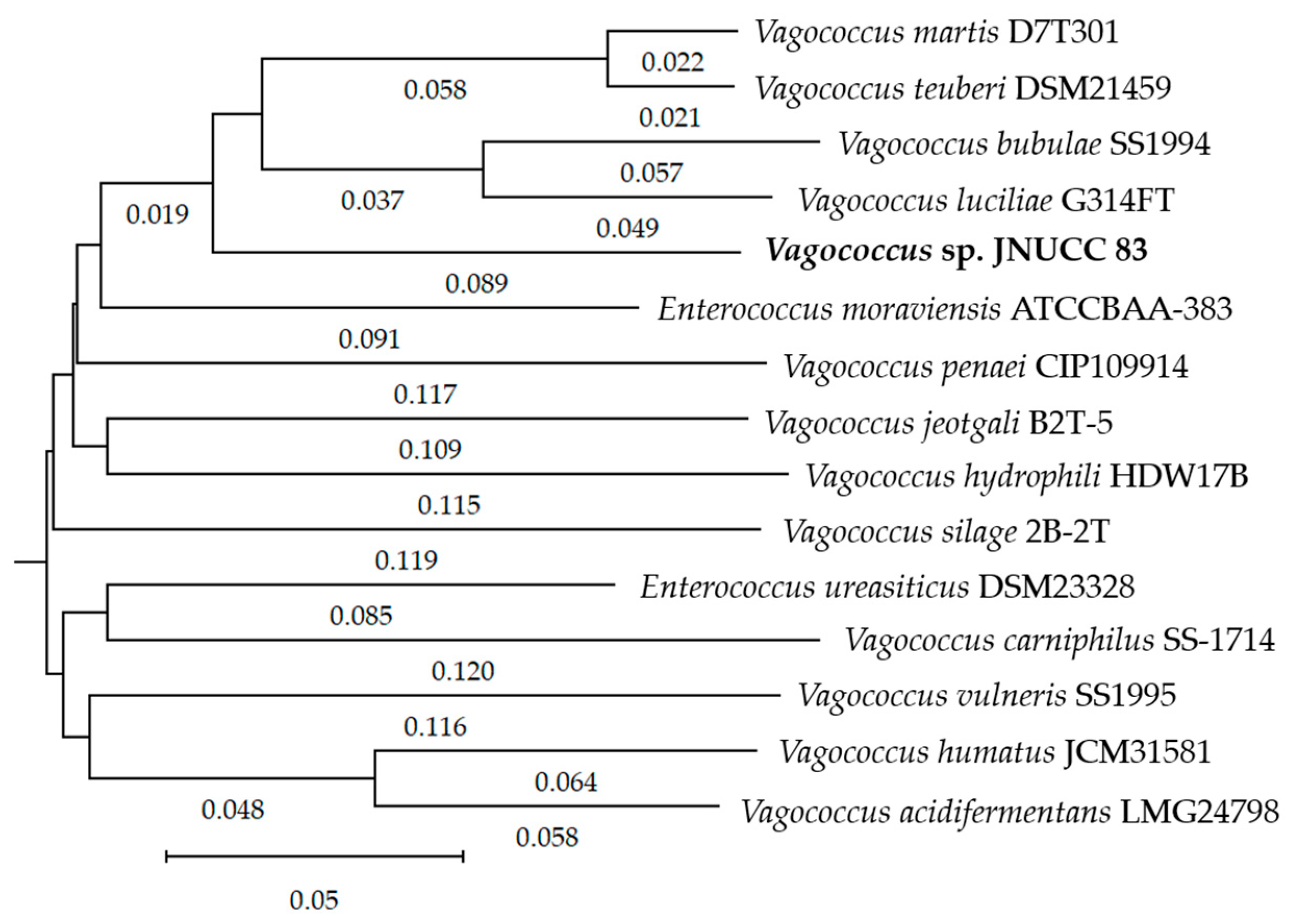
In the GBDP phylogenomic tree, JNUCC 83 was placed within the genus Vagococcus as a distinct and well-supported lineage, clearly separated from all currently described Vagococcus-type strains (Figure 2). The tree topology consistently indicated an independent phylogenetic position of JNUCC 83, while species-level relatedness was subsequently evaluated using dDDH and ANI analyses.
Consistent with the phylogenomic topology, pairwise dDDH estimates between JNUCC 83 and its closest relatives—including V. martis D7T301, V. teuberi DSM 21459, V. luciliae G314FT, and V. bubulae SS1994—were low. Using the recommended GBDP formula d4, dDDH values ranged from approximately 21.0% to 27.2%, with confidence intervals well below the 70% species delineation threshold (Table 3). Comparable trends were observed for the dDDH formulas d0 and d6, further reinforcing the genomic distinctness of JNUCC 83. The small G+C content differences between JNUCC 83 and these taxa (generally ≤0.5% for the closest Vagococcus species) indicate genus-level coherence while remaining fully compatible with species-level separation.
To further corroborate these results, average nucleotide identity was assessed using OrthoANIu. The comparison between JNUCC 83 and V. martis D7T301 yielded an OrthoANIu value of 83.32% with 53.32% genome coverage, which is far below the commonly accepted 95–96% ANI threshold for species circumscription (Table 4, OrthoANIu). This finding independently supports the dDDH-based conclusion that JNUCC 83 does not belong to any previously described Vagococcus species [46].
In parallel, phylogenetic reconstruction based on the 16S rRNA gene sequence placed JNUCC 83 within the genus Vagococcus but on an independent branch distinct from closely related type strains such as V. martis, V. teuberi, V. luciliae, and V. bubulae. Although the overall topology of the 16S rRNA gene tree was congruent with the genome-based phylogeny, branch support values were generally lower, reflecting the limited resolving power of single-gene markers for species-level discrimination within this genus.
Taken together, the concordant evidence from genome-scale phylogenomics (GBDP), low dDDH values, subthreshold OrthoANIu similarity, and consistent 16S rRNA gene placement demonstrates that Vagococcus sp. JNUCC 83 represents a phylogenetically coherent yet clearly distinct lineage within the genus Vagococcus. Based on these results, JNUCC 83 does not warrant the proposal of a novel genus and is considered a species-level distinct lineage within the genus Vagococcus; however, a formal proposal of a novel species is beyond the scope of the present study.

3.4. Functional Classification Based on EggNOG/COG Analysis

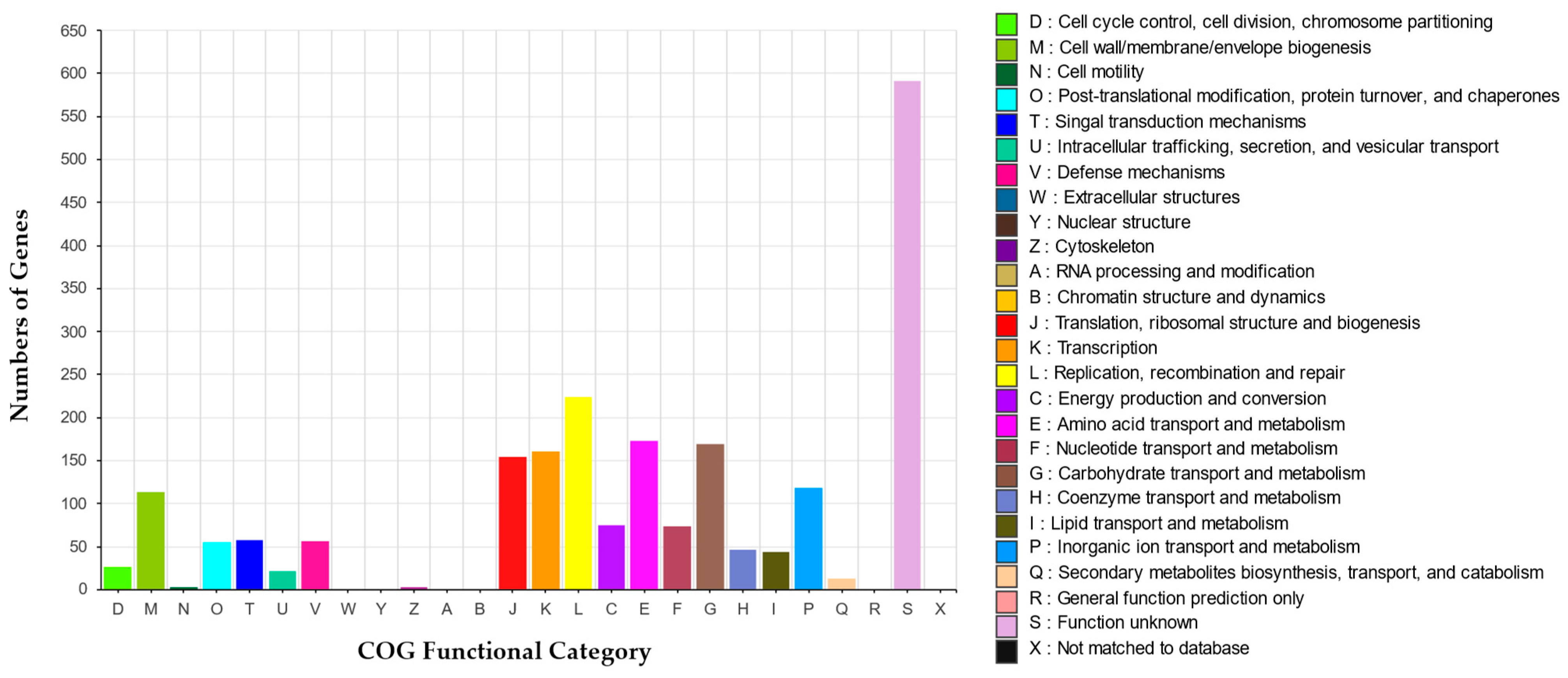
The functional annotation of the Vagococcus sp. JNUCC 83 genome was conducted using the EggNOG database with assignment to Clusters of Orthologous Groups (COGs) [62]. A total of 2170 protein-coding genes were successfully assigned to at least one COG functional category, providing an overview of the functional composition of the genome (Figure 3).
The largest proportion of genes was assigned to COG category S (function unknown), comprising 591 genes, which is a common feature observed in many bacterial genomes and reflects the presence of lineage-specific or poorly characterized proteins. This relatively high proportion suggests that Vagococcus sp. JNUCC 83 may harbor strain-specific genes potentially associated with adaptation to its plant-associated niche [49].
Among the well-characterized functional categories, genes involved in translation, ribosomal structure, and biogenesis (COG category J; 155 genes) and amino acid transport and metabolism (COG category E; 173 genes) were abundant, indicating a genome primarily enriched in protein synthesis capacity and amino acid metabolism. These functional categories are closely associated with the utilization and turnover of nitrogen-containing compounds, rather than specialized nitrogen assimilation pathways. The prominence of COG category K (transcription; 160 genes) further highlights the presence of diverse transcriptional regulators, supporting flexible gene regulation in response to environmental conditions [63].
A substantial number of genes were also assigned to COG category L (replication, recombination, and repair; 224 genes), consistent with the presence of multiple DNA repair and genome maintenance systems identified during genome annotation. This enrichment suggests robust mechanisms for maintaining genome integrity, which may be advantageous for survival under fluctuating environmental stresses encountered on plant surfaces [64].
Genes related to energy production and conversion (COG category C; 75 genes) and carbohydrate transport and metabolism (COG category G; 169 genes) were also well represented, reflecting metabolic versatility and the ability to utilize diverse carbon sources. This metabolic profile is consistent with adaptation to nutrient-variable environments such as floral ecosystems [65].
Additionally, COG category P (inorganic ion transport and metabolism; 118 genes) and COG category H (coenzyme transport and metabolism; 47 genes) indicate the presence of systems supporting redox balance and enzymatic cofactor homeostasis. In contrast, categories associated with secondary metabolite biosynthesis, transport, and catabolism (COG category Q; 13 genes) were relatively underrepresented, in agreement with antiSMASH results showing a limited repertoire of specialized secondary metabolite biosynthetic gene clusters [66].
Overall, the EggNOG/COG functional profile of Vagococcus sp. JNUCC 83 is characteristic of a bacterium primarily adapted to core metabolic functions, genome maintenance, and environmental responsiveness rather than extensive secondary metabolite production. The dominance of housekeeping-related categories, together with a modest number of genes involved in specialized metabolism, supports the interpretation that this strain relies mainly on fundamental physiological processes and metabolic flexibility to persist in its plant-associated habitat.

3.5. Genome Mining and Secondary Metabolite Biosynthetic Gene Cluster Analysis

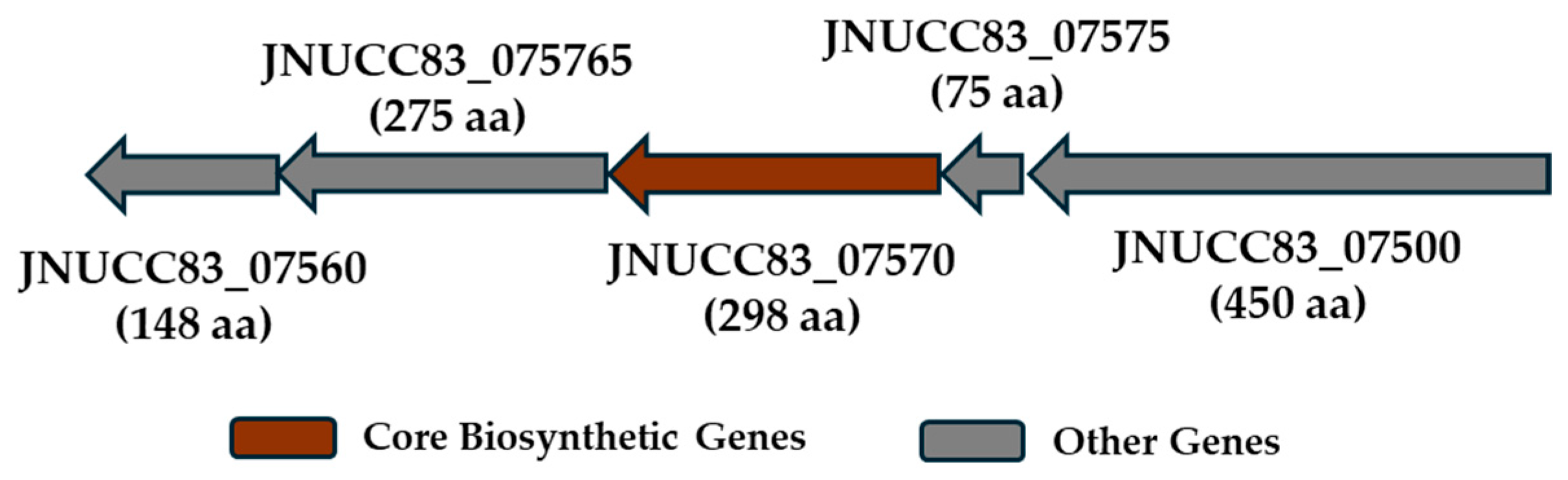
The secondary metabolite biosynthetic potential of Vagococcus sp. JNUCC 83 was examined using antiSMASH based on the complete chromosomal genome sequence (GenBank accession number CM132204.1). This analysis identified a locus annotated as a terpene-precursor-type biosynthetic gene cluster, whose core biosynthetic gene encodes a polyprenyl synthase family protein (JNUCC83_07570). Polyprenyl synthases catalyze the sequential condensation of the universal isoprenoid precursors isopentenyl diphosphate (IPP) and dimethylallyl diphosphate (DMAPP) to generate prenyl diphosphate intermediates, which serve as essential building blocks in isoprenoid metabolism (Figure 4) [67,68].
Structural comparison using the AlphaFold Protein Structure Database [69] revealed that JNUCC83_07570 shows high structural confidence (average pLDDT > 90) and significant sequence similarity to experimentally curated farnesyl diphosphate synthases (FPPSs) registered in Swiss-Prot. Specifically, it shares approximately 47–48% sequence identity with IspA from Bacillus subtilis (UniProt ID: P54383) and Fps from Micrococcus luteus (UniProt ID: O66126), showing a conserved alignment across the catalytic core. FPPS enzymes belong to the class I trans-prenyltransferase family and are characterized by an all-α-helical fold and two conserved Asp-rich (DDXXD) motifs that coordinate divalent metal ions and facilitate diphosphate-dependent chain elongation. These enzymes predominantly catalyze the formation of farnesyl diphosphate (FPP, C15) or closely related medium-chain prenyl diphosphate intermediates, rather than long-chain polyprenyl products [70,71].
Consistent with this structural assignment, the locus was classified as “terpene-precursor” rather than “terpene”, as it lacks hallmark enzymes required for the biosynthesis of dedicated terpene secondary metabolites, such as terpene cyclases, scaffold-specific prenyltransferases, and downstream tailoring enzymes (e.g., oxidoreductases) responsible for cyclization and structural diversification [72,73]. Accordingly, the predicted metabolic output of this cluster is best interpreted as the production of prenyl diphosphate intermediates, most likely farnesyl diphosphate or related C15 isoprenoid precursors, which are broadly utilized in primary cellular metabolism, including membrane-associated processes and cell envelope biosynthesis, rather than as end-product terpenoid natural compounds [74,75,76].
The genomic context further supports this interpretation, as the locus is embedded within a region enriched in housekeeping and genome maintenance-related genes, including DNA repair protein RecN, an arginine repressor, a TlyA family RNA methyltransferase, and the small and large exodeoxyribonuclease VII subunits. The predominance of genes involved in DNA repair, transcriptional regulation, and RNA/DNA processing indicates that this region is closely associated with core cellular physiology rather than specialized secondary metabolism [77,78,79]. Collectively, these features suggest that the identified locus primarily contributes to the supply of essential isoprenoid precursors supporting fundamental cellular functions, and it is therefore best described as a prenyl diphosphate precursor-producing gene cluster in Vagococcus sp. JNUCC 83.

4. Conclusions

In this study, we presented the complete genome sequence and comprehensive genomic characterization of Vagococcus sp. JNUCC 83, a flower-associated strain isolated from Camellia japonica on Jeju Island. Flowers represent transient and heterogeneous plant-associated microhabitats, and the isolation source provides an important ecological context for interpreting the genomic features of this strain. The high-quality genome assembly and annotation revealed a compact chromosomal architecture with characteristics typical of the genus Vagococcus, including low GC content, multiple rRNA operons, and a complete set of tRNA genes, which together suggest efficient core metabolism and adaptability to dynamic environmental conditions.
Genome-based phylogenomic analyses using GBDP and dDDH, supported by 16S rRNA gene phylogeny, demonstrated that JNUCC 83 represents a phylogenetically coherent yet clearly distinct lineage within the genus Vagococcus. The consistently low dDDH values relative to all currently described type strains indicate species-level genomic distinctness, highlighting the utility of whole-genome-based approaches for resolving taxonomic boundaries within Vagococcus and related lactic acid bacteria.
Functional annotation based on EggNOG/COG analysis showed that the genome of Vagococcus sp. JNUCC 83 is predominantly enriched in genes associated with core metabolic processes, transcriptional regulation, replication, and genome maintenance, rather than extensive secondary metabolite biosynthesis. In line with this functional profile, genome mining identified a terpene-precursor-type biosynthetic locus encoding a polyprenyl synthase, which is most plausibly interpreted as contributing to essential isoprenoid precursor supply required for primary cellular functions, rather than specialized secondary metabolism.
Overall, this study expands current knowledge of plant- and flower-associated Vagococcus lineages and provides a genome-based framework for interpreting their potential ecological adaptation in plant-associated niches beyond pathogenic contexts. While functional and ecological roles remain to be experimentally validated, the genomic features of Vagococcus sp. JNUCC 83 support its consideration as a biologically benign, environmentally adapted microbial resource, providing a scientific basis for future studies on flower-derived beneficial bacteria and their potential relevance to postbiotic and cosmetic research.

Author Contributions

Conceptualization, C.-G.H.; methodology, K.-A.H., M.N.K., and J.-H.K.; investigation, K.-A.H. and J.-H.K.; resources, K.-A.H.; data curation, J.-H.K.; formal analysis, C.-G.H.; writing—original draft preparation, C.-G.H.; writing—review and editing, C.-G.H.; supervision, C.-G.H.; project administration, C.-G.H.; funding acquisition, C.-G.H. All authors have read and agreed to the published version of the manuscript.

Funding

This work was supported by the 2025 education, research, and student guidance grant funded by Jeju National University.

Institutional Review Board Statement

Not applicable.

Informed Consent Statement

Not applicable.

Data Availability Statement

The complete genome sequence of Vagococcus sp. JNUCC 83 has been deposited in the NCBI database (https://www.ncbi.nlm.nih.gov/, accessed on 15 January 2026) under the GenBank accession number CM132204.1 (chromosome). Five extrachromosomal replicons have also been deposited under the accession numbers JBSJXX010000001.1–JBSJXX010000006.1. The corresponding BioProject and RefSeq assembly accession numbers are GCA_053667615.1 and GCF_053667615.1, respectively. All data generated or analyzed during this study are publicly available in these repositories.

Conflicts of Interest

The authors declare no conflicts of interest. The funders had no role in the design of the study; in the collection, analyses, or interpretation of data; in the writing of the manuscript; or in the decision to publish the results.

References

  1. Collins, M.D.; Ash, C.; Farrow, J.A.; Wallbanks, S.; Williams, A.M. 16S ribosomal ribonucleic acid sequence analyses of lactococci and related taxa. Description of Vagococcus fluvialis gen. nov., sp. nov. J. Appl. Bacteriol. 1989, 67, 453–460. [Google Scholar] [CrossRef]
  2. Hyun, D.W.; Tak, E.J.; Kim, P.S.; Bae, J.W. Description of Vagococcus coleopterorum sp. nov., isolated from the intestine of the diving beetle, Cybister lewisianus, and Vagococcus hydrophili sp. nov., isolated from the intestine of the dark diving beetle, Hydrophilus acuminatus, and emended description of the genus Vagococcus. J. Microbiol. 2021, 59, 132–141. [Google Scholar] [CrossRef] [PubMed]
  3. Pot, B.; Devriese, L.A.; Hommez, J.; Miry, C.; Vandemeulebroecke, K.; Kersters, K.; Haesebrouck, F. Characterization and identification of Vagococcus fluvialis strains isolated from domestic animals. J. Appl. Bacteriol. 1994, 77, 362–369. [Google Scholar] [CrossRef]
  4. Teixeira, L.M.; Carvalho, M.G.; Merquior, V.L.; Steigerwalt, A.G.; Brenner, D.J.; Facklam, R.R. Phenotypic and genotypic characterization of Vagococcus fluvialis, including strains isolated from human sources. J. Clin. Microbiol. 1997, 35, 2778–2781. [Google Scholar] [CrossRef]
  5. Brunswick, J.; Spiro, J.; Wisniewski, P. Vagococcus: An under-recognized and emerging cause of antibiotic-resistant infection. IDCases 2024, 36, e01995. [Google Scholar] [CrossRef]
  6. Lawson, P.A.; Falsen, E.; Cotta, M.A.; Whitehead, T.R. Vagococcus elongatus sp. nov., isolated from a swine-manure storage pit. Int. J. Syst. Evol. Microbiol. 2007, 57, 751–754. [Google Scholar] [CrossRef] [PubMed]
  7. Sundararaman, A.; Srinivasan, S.; Lee, S.S. Vagococcus humatus sp. nov., isolated from soil beneath a decomposing pig carcass. Int. J. Syst. Evol. Microbiol. 2017, 67, 330–335. [Google Scholar] [CrossRef] [PubMed]
  8. Wallbanks, S.; Martinez-Murcia, A.J.; Fryer, J.L.; Phillips, B.A.; Collins, M.D. 16S rRNA sequence determination for members of the genus Carnobacterium and related lactic acid bacteria and description of Vagococcus salmoninarum sp. nov. Int. J. Syst. Bacteriol. 1990, 40, 224–230. [Google Scholar] [CrossRef]
  9. Lawson, P.A.; Foster, G.; Falsen, E.; Ohlén, M.; Collins, M.D. Vagococcus lutrae sp. nov., isolated from the common otter (Lutra lutra). Int. J. Syst. Bacteriol. 1999, 49, 1251–1254. [Google Scholar] [CrossRef]
  10. Hoyles, L.; Lawson, P.A.; Foster, G.; Falsen, E.; Ohlén, M.; Grainger, J.M.; Collins, M.D. Vagococcus fessus sp. nov., isolated from a seal and a harbour porpoise. Int. J. Syst. Evol. Microbiol. 2000, 50, 1151–1154. [Google Scholar] [CrossRef]
  11. Shewmaker, P.L.; Steigerwalt, A.G.; Morey, R.E.; Carvalho, M.D.G.S.; Elliott, J.A.; Joyce, K.; Barrett, T.J.; Teixeira, L.M.; Facklam, R.R. Vagococcus carniphilus sp. nov., isolated from ground beef. Int. J. Syst. Evol. Microbiol. 2004, 54, 1505–1510. [Google Scholar] [CrossRef] [PubMed][Green Version]
  12. Jaffrès, E.; Prévost, H.; Rossero, A.; Joffraud, J.; Dousset, X. Vagococcus penaei sp. nov., isolated from spoilage microbiota of cooked shrimp (Penaeus vannamei). Int. J. Syst. Evol. Microbiol. 2010, 60, 2159–2164. [Google Scholar] [CrossRef] [PubMed]
  13. Wang, L.; Cui, Y.S.; Kwon, C.S.; Lee, S.T.; Lee, J.S.; Im, W.T. Vagococcus acidifermentans sp. nov., isolated from an acidogenic fermentation bioreactor. Int. J. Syst. Evol. Microbiol. 2011, 61, 1123–1126. [Google Scholar] [CrossRef]
  14. Shewmaker, P.L.; Whitney, A.M.; Gulvik, C.A.; Humrighouse, B.W.; Gartin, J.; Moura, H.; Barr, J.R.; Moore, E.R.B.; Karlsson, R.; Pinto, T.C.A.; et al. Vagococcus bubulae sp. nov., isolated from ground beef, and Vagococcus vulneris sp. nov., isolated from a human foot wound. Int. J. Syst. Evol. Microbiol. 2019, 69, 2268–2276. [Google Scholar] [CrossRef]
  15. Goh, S.H.; Facklam, R.R.; Chang, M.; Hill, J.E.; Tyrrell, G.J.; Burns, E.C.; Chan, D.; He, C.; Rahim, T.; Shaw, C.; et al. Identification of Enterococcus species and phenotypically similar Lactococcus and Vagococcus species by reverse checkerboard hybridization to chaperonin 60 gene sequences. J. Clin. Microbiol. 2000, 38, 3953–3959. [Google Scholar] [CrossRef] [PubMed]
  16. Al-Ahmad, A.; Pelz, K.; Schirrmeister, J.F.; Hellwig, E.; Pukall, R. Characterization of the first oral Vagococcus isolate from a root-filled tooth with periradicular lesions. Curr. Microbiol. 2008, 57, 235–238. [Google Scholar] [CrossRef]
  17. Racero, L.; Barberis, C.; Traglia, G.; Loza, M.S.; Vay, C.; Almuzara, M. Infections due to Vagococcus spp. Microbiological and clinical aspects and literature review. Enferm. Infecc. Microbiol. Clin. (Engl. Ed.) 2021, 39, 335–339. [Google Scholar] [CrossRef]
  18. Ruiz-Zarzuela, I.; de Bias, I.; Gironés, O.; Ghittino, C.; Múazquiz, J.L. Isolation of Vagococcus salmoninarum in rainbow trout, Oncorhynchus mykiss (Walbaum), broodstocks: Characterization of the pathogen. Vet. Res. Commun. 2005, 29, 553–562. [Google Scholar] [CrossRef]
  19. Yardimci, B.; Didinen, B.I.; Onuk, E.E.; Metin, S.; Ciftci, A.; Kubilay, A.; Pekmezci, G.Z.; Eralp, H. Immunohistochemical evaluation of experimental Vagococcus salmoninarum infection in rainbow trout (Oncorhynchus mykiss, Walbaum 1792). J. Fish Dis. 2016, 39, 547–553. [Google Scholar] [CrossRef]
  20. Michel, C.; Pelletier, C.; Boussaha, M.; Douet, D.G.; Lautraite, A.; Tailliez, P. Diversity of lactic acid bacteria associated with fish and the fish farm environment, established by amplified rRNA gene restriction analysis. Appl. Environ. Microbiol. 2007, 73, 2947–2955. [Google Scholar] [CrossRef]
  21. Matajira, C.E.C.; Poor, A.P.; Moreno, L.Z.; Monteiro, M.S.; Dalmutt, A.C.; Gomes, V.T.M.; Dutra, M.C.; Barbosa, M.R.F.; Sato, M.I.Z.; Moreno, A.M. Vagococcus sp. a porcine pathogen: Molecular and phenotypic characterization of strains isolated from diseased pigs in Brazil. J. Infect. Dev. Ctries. 2020, 14, 1314–1319. [Google Scholar] [CrossRef] [PubMed]
  22. Matsuo, T.; Mori, N.; Kawai, F.; Sakurai, A.; Toyoda, M.; Mikami, Y.; Uehara, Y.; Furukawa, K. Vagococcus fluvialis as a causative pathogen of bloodstream and decubitus ulcer infection: Case report and systematic review of the literature. J. Infect. Chemother. 2021, 27, 359–363. [Google Scholar] [CrossRef] [PubMed]
  23. Garcia, V.; Abat, C.; Rolain, J.M. Report of the first Vagococcus lutrae human infection, Marseille, France. New Microbes New Infect. 2015, 9, 56–57. [Google Scholar] [CrossRef]
  24. Yilmaz, M.; Arslan, T.; Atalay Oral, M.; Kubilay, A. Antibiotic susceptibility and resistance genes profiles of Vagococcus salmoninarum in a rainbow trout (Oncorhyncus mykiss, Walbaum) farm. PeerJ 2024, 12, e17194. [Google Scholar] [CrossRef] [PubMed]
  25. Román, L.; Real, F.; Sorroza, L.; Padilla, D.; Acosta, B.; Grasso, V.; Bravo, J.; Acosta, F. The in vitro effect of probiotic Vagococcus fluvialis on the innate immune parameters of Sparus aurata and Dicentrarchus labrax. Fish Shellfish Immunol. 2012, 33, 1071–1075. [Google Scholar] [CrossRef]
  26. Sorroza, L.; Padilla, D.; Acosta, F.; Román, L.; Grasso, V.; Vega, J.; Real, F. Characterization of the probiotic strain Vagococcus fluvialis in the protection of European sea bass (Dicentrarchus labrax) against vibriosis by Vibrio anguillarum. Vet. Microbiol. 2012, 155, 369–373. [Google Scholar] [CrossRef]
  27. Rosenbergová, Z.; Oftedal, T.F.; Ovchinnikov, K.V.; Thiyagarajah, T.; Rebroš, M.; Diep, D.B. Identification of a Novel Two-Peptide Lantibiotic from Vagococcus fluvialis. Microbiol. Spectr. 2022, 10, e0095422. [Google Scholar] [CrossRef]
  28. Han, J.H.; Kim, H.S. Skin Deep: The Potential of Microbiome Cosmetics. J. Microbiol. 2024, 62, 181–199. [Google Scholar] [CrossRef]
  29. Arora, R.; Kaur, R.; Babbar, R.; Dhingra, S.; Dhingra, A.K.; Grewal, A.S. Evolving Advances in the Cosmetic use of Probiotics and Postbiotics: Health, Regulatory and Marketing Aspects. Curr. Pharm. Biotechnol. 2024, 25, 1349–1361. [Google Scholar] [CrossRef]
  30. Corinaldesi, C.; Barone, G.; Marcellini, F.; Dell’Anno, A.; Danovaro, R. Marine Microbial-Derived Molecules and Their Potential Use in Cosmeceutical and Cosmetic Products. Mar. Drugs 2017, 15, 118. [Google Scholar] [CrossRef]
  31. Ko, M.S.; Moon, M.Y.; Hyun, C.G. Genomic and Metabolomic Characterization of Kitasatospora griseola JNUCC 62 from Mulyeongari Oreum and Its Cosmeceutical Potential. Fermentation 2025, 11, 671. [Google Scholar] [CrossRef]
  32. Kim, J.H.; Liang, X.; Kim, M.N.; Hyun, C.G. Taxonomic Characterization, Whole-Genome Sequencing, and Cosmetic Potential of Lysinibacillus sp. JNUCC 51 Isolated from Baengnokdam Crater Lake, Mt. Halla. Microorganisms 2025, 13, 2786. [Google Scholar] [CrossRef]
  33. An, S.H.; Ko, K.H.; Jang, W.G.; Hyun, C.G. Mystique and Pristine Microbiome of Jeju Lava (Yongam) Seawater: Comparative Insights with Mineral Water and Adjacent Seawater. Water 2025, 17, 3306. [Google Scholar] [CrossRef]
  34. Castronovo, L.M.; Vassallo, A.; Mengoni, A.; Miceli, E.; Bogani, P.; Firenzuoli, F.; Fani, R.; Maggini, V. Medicinal Plants and Their Bacterial Microbiota: A Review on Antimicrobial Compounds Production for Plant and Human Health. Pathogens 2021, 10, 106. [Google Scholar] [CrossRef]
  35. Burger, H.; Ayasse, M.; Dötterl, S.; Kreissl, S.; Galizia, C.G. Perception of floral volatiles involved in host-plant finding behaviour: Comparison of a bee specialist and generalist. J. Comp. Physiol. A Neuroethol. Sens. Neural Behav. Physiol. 2013, 199, 751–761. [Google Scholar] [CrossRef]
  36. Fanai, A.; Bohia, B.; Lalremruati, F.; Lalhriatpuii, N.; Lalrokimi; Lalmuanpuii, R.; Singh, P.K.; Zothanpuia. Plant growth promoting bacteria (PGPB)-induced plant adaptations to stresses: An updated review. PeerJ 2024, 12, e17882. [Google Scholar] [CrossRef] [PubMed]
  37. Singh, A.; Mazahar, S.; Chapadgaonkar, S.S.; Giri, P.; Shourie, A. Phyto-microbiome to mitigate abiotic stress in crop plants. Front. Microbiol. 2023, 14, 1210890. [Google Scholar] [CrossRef] [PubMed]
  38. Souak, D.; Barreau, M.; Courtois, A.; André, V.; Duclairoir Poc, C.; Feuilloley, M.G.J.; Gault, M. Challenging Cosmetic Innovation: The Skin Microbiota and Probiotics Protect the Skin from UV-Induced Damage. Microorganisms 2021, 9, 936. [Google Scholar] [CrossRef]
  39. Rodriguez Jimenez, A.; Guiglielmoni, N.; Goetghebuer, L.; Dechamps, E.; George, I.F.; Flot, J.F. Comparative genome analysis of Vagococcus fluvialis reveals abundance of mobile genetic elements in sponge-isolated strains. BMC Genom. 2022, 23, 618. [Google Scholar] [CrossRef]
  40. Bankevich, A.; Nurk, S.; Antipov, D.; Gurevich, A.A.; Dvorkin, M.; Kulikov, A.S.; Lesin, V.M.; Nikolenko, S.I.; Pham, S.; Prjibelski, A.D.; et al. SPAdes: A new genome assembly algorithm and its applications to single-cell sequencing. J. Comput. Biol. 2012, 19, 455–477. [Google Scholar] [CrossRef] [PubMed]
  41. Tatusova, T.; DiCuccio, M.; Badretdin, A.; Chetvernin, V.; Nawrocki, E.P.; Zaslavsky, L.; Lomsadze, A.; Pruitt, K.D.; Borodovsky, M.; Ostell, J. NCBI prokaryotic genome annotation pipeline. Nucleic Acids Res. 2016, 44, 6614–6624. [Google Scholar] [CrossRef]
  42. Lomsadze, A.; Gemayel, K.; Tang, S.; Borodovsky, M. Modeling leaderless transcription and atypical genes results in more accurate gene prediction in prokaryotes. Genome Res. 2018, 28, 1079–1089. [Google Scholar] [CrossRef]
  43. Parks, D.H.; Imelfort, M.; Skennerton, C.T.; Hugenholtz, P.; Tyson, G.W. CheckM: Assessing the quality of microbial genomes recovered from isolates, single cells, and metagenomes. Genome Res. 2015, 25, 1043–1055. [Google Scholar] [CrossRef]
  44. Meier-Kolthoff, J.P.; Göker, M. TYGS is an automated high-throughput platform for state-of-the-art genome-based taxonomy. Nat. Commun. 2019, 10, 2182. [Google Scholar] [CrossRef]
  45. Lefort, V.; Desper, R.; Gascuel, O. FastME 2.0: A Comprehensive, Accurate, and Fast Distance-Based Phylogeny Inference Program. Mol. Biol. Evol. 2015, 32, 2798–2800. [Google Scholar] [CrossRef]
  46. Yoon, S.H.; Ha, S.M.; Lim, J.; Kwon, S.; Chun, J. A large-scale evaluation of algorithms to calculate average nucleotide identity. Antonie Van Leeuwenhoek 2017, 110, 1281–1286. [Google Scholar] [CrossRef]
  47. Seemann, T. Prokka: Rapid prokaryotic genome annotation. Bioinformatics 2014, 30, 2068–2069. [Google Scholar] [CrossRef] [PubMed]
  48. Cantalapiedra, C.P.; Hernández-Plaza, A.; Letunic, I.; Bork, P.; Huerta-Cepas, J. eggNOG-mapper v2: Functional Annotation, Orthology Assignments, and Domain Prediction at the Metagenomic Scale. Mol. Biol. Evol. 2021, 38, 5825–5829. [Google Scholar] [CrossRef] [PubMed]
  49. Galperin, M.Y.; Wolf, Y.I.; Makarova, K.S.; Vera Alvarez, R.; Landsman, D.; Koonin, E.V. COG database update: Focus on microbial diversity, model organisms, and widespread pathogens. Nucleic Acids Res. 2021, 49, D274–D281. [Google Scholar] [CrossRef] [PubMed]
  50. del Solar, G.; Giraldo, R.; Ruiz-Echevarría, M.J.; Espinosa, M.; Díaz-Orejas, R. Replication and control of circular bacterial plasmids. Microbiol. Mol. Biol. Rev. 1998, 62, 434–464. [Google Scholar] [CrossRef] [PubMed]
  51. Garcillán-Barcia, M.P.; Francia, M.V.; de la Cruz, F. The diversity of conjugative relaxases and its application in plasmid classification. FEMS Microbiol. Rev. 2009, 33, 657–687. [Google Scholar] [CrossRef] [PubMed]
  52. Smillie, C.; Garcillán-Barcia, M.P.; Francia, M.V.; Rocha, E.P.; de la Cruz, F. Mobility of plasmids. Microbiol. Mol. Biol. Rev. 2010, 74, 434–452. [Google Scholar] [CrossRef]
  53. Mahillon, J.; Chandler, M. Insertion sequences. Microbiol. Mol. Biol. Rev. 1998, 62, 725–774. [Google Scholar] [CrossRef]
  54. Hayes, F. Toxins-antitoxins: Plasmid maintenance, programmed cell death, and cell cycle arrest. Science 2003, 301, 1496–1499. [Google Scholar] [CrossRef]
  55. Yamaguchi, Y.; Park, J.H.; Inouye, M. Toxin-antitoxin systems in bacteria and archaea. Annu. Rev. Genet. 2011, 45, 61–79. [Google Scholar] [CrossRef] [PubMed]
  56. Nies, D.H. Efflux-mediated heavy metal resistance in prokaryotes. FEMS Microbiol. Rev. 2003, 27, 313–339. [Google Scholar] [CrossRef] [PubMed]
  57. Silver, S.; Phung, L.T. A bacterial view of the periodic table: Genes and proteins for toxic inorganic ions. J. Ind. Microbiol. Biotechnol. 2005, 32, 587–605. [Google Scholar] [CrossRef]
  58. Ben Braïek, O.; Smaoui, S. Enterococci: Between Emerging Pathogens and Potential Probiotics. Biomed. Res. Int. 2019, 2019, 5938210. [Google Scholar] [CrossRef]
  59. Frost, L.S.; Leplae, R.; Summers, A.O.; Toussaint, A. Mobile genetic elements: The agents of open source evolution. Nat. Rev. Microbiol. 2005, 3, 722–732. [Google Scholar] [CrossRef]
  60. Nordström, K.; Austin, S.J. Mechanisms that contribute to the stable segregation of plasmids. Annu. Rev. Genet. 1989, 23, 37–69. [Google Scholar] [CrossRef]
  61. Novick, R.P. Plasmid incompatibility. Microbiol. Rev. 1987, 51, 381–395. [Google Scholar] [CrossRef][Green Version]
  62. Huerta-Cepas, J.; Szklarczyk, D.; Heller, D.; Hernández-Plaza, A.; Forslund, S.K.; Cook, H.; Mende, D.R.; Letunic, I.; Rattei, T.; Jensen, L.J.; et al. eggNOG 5.0: A hierarchical, functionally and phylogenetically annotated orthology resource based on 5090 organisms and 2502 viruses. Nucleic Acids Res. 2019, 47, D309–D314. [Google Scholar] [CrossRef]
  63. Galperin, M.Y.; Kristensen, D.M.; Makarova, K.S.; Wolf, Y.I.; Koonin, E.V. Microbial genome analysis: The COG approach. Brief. Bioinform. 2019, 20, 1063–1070. [Google Scholar] [CrossRef] [PubMed]
  64. Rocha, E.P.; Cornet, E.; Michel, B. Comparative and evolutionary analysis of the bacterial homologous recombination systems. PLoS Genet. 2005, 1, e15. [Google Scholar] [CrossRef]
  65. Konstantinidis, K.T.; Tiedje, J.M. Genomic insights that advance the species definition for prokaryotes. Proc. Natl. Acad. Sci. USA 2005, 102, 2567–2572. [Google Scholar] [CrossRef] [PubMed]
  66. Galperin, M.Y.; Makarova, K.S.; Wolf, Y.I.; Koonin, E.V. Expanded microbial genome coverage and improved protein family annotation in the COG database. Nucleic Acids Res. 2015, 43, D261–D269. [Google Scholar] [CrossRef]
  67. Blin, K.; Shaw, S.; Vader, L.; Szenei, J.; Reitz, Z.L.; Augustijn, H.E.; Cediel-Becerra, J.D.D.; de Crécy-Lagard, V.; Koetsier, R.A.; Williams, S.E.; et al. antiSMASH 8.0: Extended gene cluster detection capabilities and analyses of chemistry, enzymology, and regulation. Nucleic Acids Res. 2025, 53, W32–W38. [Google Scholar] [CrossRef] [PubMed]
  68. Wallrapp, F.H.; Pan, J.J.; Ramamoorthy, G.; Almonacid, D.E.; Hillerich, B.S.; Seidel, R.; Patskovsky, Y.; Babbitt, P.C.; Almo, S.C.; Jacobson, M.P.; et al. Prediction of function for the polyprenyl transferase subgroup in the isoprenoid synthase superfamily. Proc. Natl. Acad. Sci. USA 2013, 110, E1196–E1202. [Google Scholar] [CrossRef]
  69. Bertoni, D.; Tsenkov, M.; Magana, P.; Nair, S.; Pidruchna, I.; Querino Lima Afonso, M.; Midlik, A.; Paramval, U.; Lawal, D.; Tanweer, A.; et al. AlphaFold Protein Structure Database 2025: A redesigned interface and updated structural coverage. Nucleic Acids Res. 2026, 54, D358–D362. [Google Scholar] [CrossRef]
  70. Chang, H.Y.; Cheng, T.H.; Wang, A.H. Structure, catalysis, and inhibition mechanism of prenyltransferase. IUBMB Life 2021, 73, 40–63. [Google Scholar] [CrossRef]
  71. Rudolf, J.D.; Alsup, T.A.; Xu, B.; Li, Z. Bacterial terpenome. Nat. Prod. Rep. 2021, 38, 905–980. [Google Scholar] [CrossRef]
  72. Helfrich, E.J.N.; Lin, G.M.; Voigt, C.A.; Clardy, J. Bacterial terpene biosynthesis: Challenges and opportunities for pathway engineering. Beilstein J. Org. Chem. 2019, 15, 2889–2906. [Google Scholar] [CrossRef]
  73. Chen, C.C.; Zhang, L.; Yu, X.; Ma, L.; Ko, T.P.; Guo, R.T. Versatile cis-isoprenyl diphosphate synthase superfamily members in catalyzing carbon–carbon bond formation. ACS Catal. 2020, 10, 4717–4725. [Google Scholar] [CrossRef]
  74. Roney, I.J.; Rudner, D.Z. Two new enzymes that liberate undecaprenyl-phosphate to replenish the carrier lipid pool during envelope stress. mBio 2025, 16, e0371024. [Google Scholar] [CrossRef] [PubMed]
  75. Nowicka, B.; Kruk, J. Occurrence, biosynthesis and function of isoprenoid quinones. Biochim. Biophys. Acta 2010, 1797, 1587–1605. [Google Scholar] [CrossRef] [PubMed]
  76. McLean, E.K.; Lenhart, J.S.; Simmons, L.A. RecA Is Required for the Assembly of RecN into DNA Repair Complexes on the Nucleoid. J. Bacteriol. 2021, 203, e0024021. [Google Scholar] [CrossRef] [PubMed]
  77. Makarova, K.S.; Mironov, A.A.; Gelfand, M.S. Conservation of the binding site for the arginine repressor in all bacterial lineages. Genome Biol. 2001, 2, research0013.1. [Google Scholar] [CrossRef]
  78. Satta, A.; Esquirol, L.; Ebert, B.E.; Newman, J.; Peat, T.S.; Plan, M.; Schenk, G.; Vickers, C.E. Molecular characterization of cyanobacterial short-chain prenyltransferases and discovery of a novel GGPP phosphatase. FEBS J. 2022, 289, 6672–6693. [Google Scholar] [CrossRef]
  79. Briaud, P.; Gautier, T.; Rong, V.; Mereghetti, L.; Lanotte, P.; Hiron, A. The Streptococcus agalactiae Exonuclease ExoVII Is Required for Resistance to Exogenous DNA-Damaging Agents. J. Bacteriol. 2023, 205, e0002423. [Google Scholar] [CrossRef]
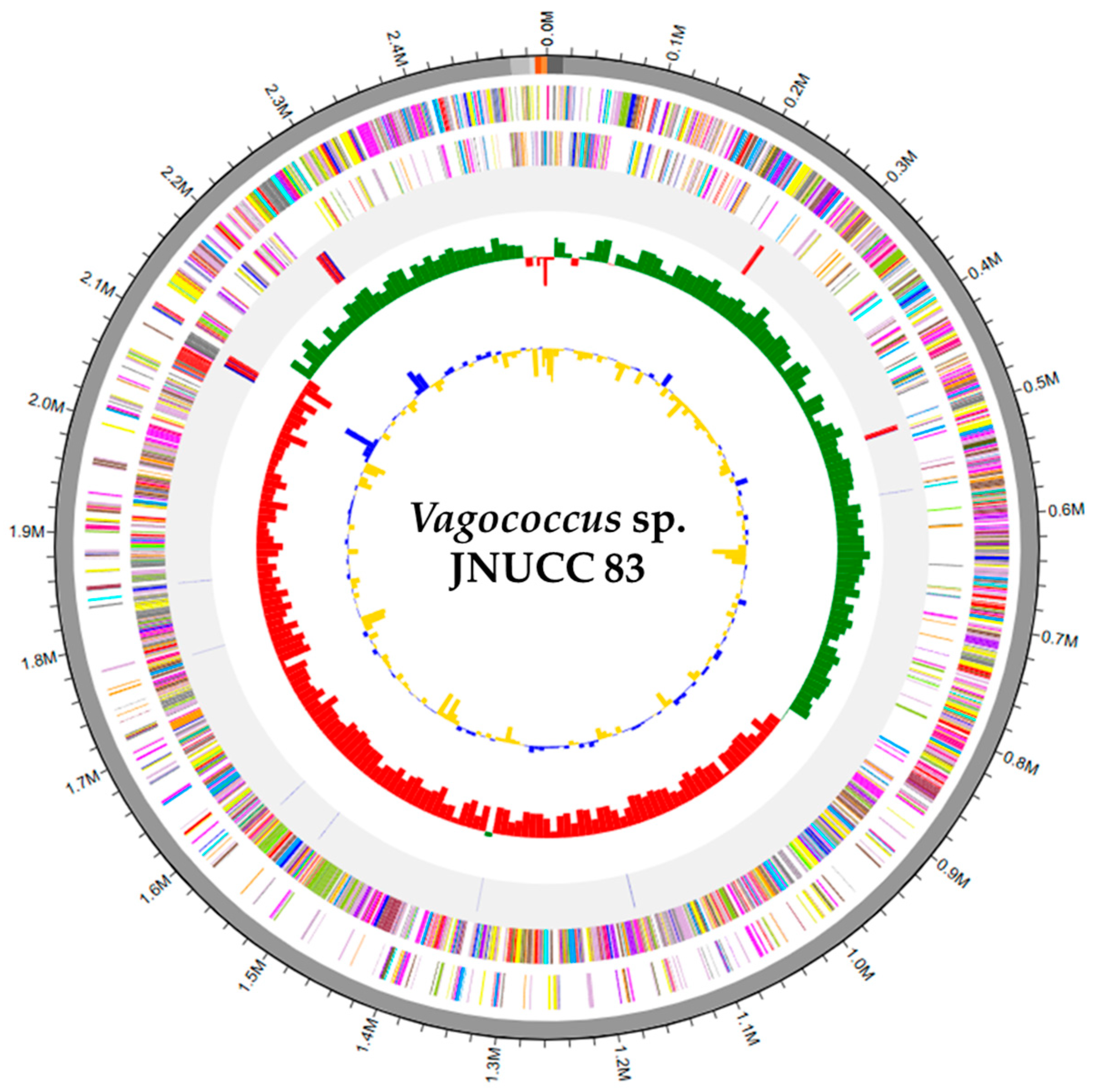
Figure 1. Circular genome map of Vagococcus sp. JNUCC 83. The circular genome map represents a pseudogenome constructed from six assembled contigs, consisting of one chromosome and five small contigs, including plasmid-like elements. From the outer to the inner rings: (1) contigs; (2) protein-coding sequences (CDSs) located on the forward strand; (3) CDSs located on the reverse strand, with genes colored according to COG functional categories; (4) rRNA and tRNA genes; (5) GC skew, with values higher than the genomic mean shown in green and lower values shown in red; and (6) GC ratio, with values higher than the genomic mean shown in blue and lower values shown in yellow. GC skew and GC ratio were calculated using 10 kb sliding windows for the chromosome, which were reduced to 1 kb for small contigs.
Microbiolres 17 00023 g001
Figure 2. Phylogenomic tree of Vagococcus sp. JNUCC 83 and related Vagococcus species based on TYGS analysis. The genome-based phylogenomic tree was reconstructed using the Genome BLAST Distance Phylogeny (GBDP) method implemented in the Type (Strain) Genome Server (TYGS), based on complete chromosomal genome sequences. The strain analyzed in this study (Vagococcus sp. JNUCC 83) is highlighted in bold to distinguish it from reference and type strains. Branch lengths represent GBDP distances, and the scale bar indicates 0.05 substitutions per site. Vagococcus sp. JNUCC 83 forms a distinct and well-supported lineage within the genus Vagococcus, clearly separated from all currently described type strains. Enterococcus species were included as outgroup taxa.
Microbiolres 17 00023 g002
Figure 3. Distribution of protein-coding genes of Vagococcus sp. JNUCC 83 across COG functional categories based on EggNOG annotation. The y-axis indicates the number of genes assigned to each category, and letters correspond to standard COG functional classifications.
Microbiolres 17 00023 g003
Figure 4. Genomic organization of a terpene-precursor-type gene cluster in Vagococcus sp. JNUCC 83. The cluster was identified by antiSMASH analysis of the complete chromosomal genome (CM132204.1). The core biosynthetic gene JNUCC83_07570, encoding a polyprenyl synthase family protein, is highlighted in brown, while surrounding genes are shown in gray. Arrows indicate transcriptional direction, and gene lengths (aa, amino acids) are shown in parentheses. The absence of terpene cyclases and downstream tailoring enzymes supports classification of this locus as a terpene-precursor rather than a dedicated terpene biosynthetic gene cluster.
Microbiolres 17 00023 g004
Table 1. General genomic features of Vagococcus sp. strain JNUCC 83.
Vagococcus sp. Strain JNUCC 83
Genome size (bp)2,513,280
Total number of contigs6
Contigs N50 (bp)2,472,896
G+C content (%)33.58
Total number of predicted genes2492
Total number of protein-coding genes2371
Total number of pseudogenes37
Total number of tRNA-coding genes62
Total number of rRNA-coding genes (5S, 16S, 23S)18 (6, 6, 6)
Genome size (bp), total number of contigs, contigs N50 (bp), and overall G+C content (%) were calculated from the complete assembly, including the chromosome (CM132204.1; 2,472,896 bp; circular) and five extrachromosomal replicons (unnamed1–unnamed5). All other genomic features (e.g., numbers of predicted genes, protein-coding genes, pseudogenes, tRNA genes, and rRNA operon components) refer to the complete circular chromosome sequence (CM132204.1) unless otherwise stated.
Table 2. Genomic features of small plasmid-like replicons in Vagococcus sp. JNUCC 83.
RepliconGenBank
Accession
Size
(bp)
GC
Content
(%)
Replication
Protein
Mobility/Recombination GenesStability/Resistance FeaturesPutative Classification
1JBSJXX010000001.113,91827RepMobV relaxase; IS3-family transposaseRelE/RelB toxin–antitoxin systemPlasmid-like replicon
2JBSJXX010000003.115,76230RepIntegrase (tyrosine recombinase)RelE/ParE- and Fst-family toxinsPlasmid-like replicon
3JBSJXX010000004.1412634.5RepDNA/RNA non-specific endonucleaseCryptic plasmid
4JBSJXX010000005.1503529.5RepPredicted lipase-family proteinSmall plasmid-like element
5JBSJXX010000006.1486331.5Heavy metal-translocating P-type ATPase; ArsR/SmtB regulatorResistance-associated plasmid-like element
Rep indicates a plasmid replication protein.
Table 3. Digital DNA–DNA hybridization (dDDH) and G+C content differences between Vagococcus sp. strain JNUCC 83 and closely related type strains.
Subject StraindDDH
(d0, in %)
C.I.
(d0, in %)
dDDH
(d4, in %)
C.I.
(d4, in %)
dDDH
(d6, in %)
C.I.
(d6, in %)
G+C Content
Difference
(in %)
Vagococcus martis D7T30159[55.4–62.5]27.2[24.9–29.7]48.9[45.9–52.0]0.18
Vagococcus teuberi DSM 2145952.5[49.0–55.9]26.5[24.2–29.0]44.2[41.2–47.2]0.52
Enterococcus moraviensis ATCC BAA-38313[10.3–16.3]25.8[23.5–28.3]13.4[11.0–16.2]2.39
Vagococcus luciliae G314FT46.5[43.1–49.9]25.5[23.2–28.0]39.7[36.7–42.7]0.2
Vagococcus bubulae SS199446[42.6–49.4]25.4[23.0–27.8]39.3[36.4–42.4]0.03
Vagococcus jeotgali B2T-516[13.0–19.5]23.3[21.1–25.8]16.1[13.6–19.0]0.23
Vagococcus acidifermentans LMG 2479813[10.3–16.3]22.6[20.3–25.1]13.4[11.0–16.2]10.75
Vagococcus vulneris SS199514[11.2–17.4]22.1[19.8–24.5]14.3[11.8–17.1]1.03
Vagococcus humatus JCM 3158113.6[10.8–16.9]21.4[19.1–23.8]13.9[11.5–16.7]1.21
Vagococcus penaei CIP 10991414.5[11.6–17.9]21.4[19.2–23.9]14.7[12.2–17.5]1.48
Vagococcus silagei 2B-2T14.2[11.4–17.6]21.3[19.0–23.7]14.4[12.0–17.3]1.11
Vagococcus hydrophili HDW17B15.2[12.3–18.6]21[18.8–23.5]15.3[12.8–18.2]0.77
Vagococcus carniphilus SS-171415.2[12.3–18.6]20.4[18.2–22.8]15.2[12.8–18.1]1.06
Vagococcus fluvialis DSM 573115.4[12.5–18.8]19.8[17.6–22.2]15.4[12.9–18.3]1.18
Digital DNA–DNA hybridization (dDDH) values were calculated using the Genome-to-Genome Distance Calculator (GGDC) implemented in the Type (Strain) Genome Server (TYGS) based on the Genome BLAST Distance Phylogeny (GBDP) approach. dDDH estimates are reported according to the formulas d0, d4, and d6, with the corresponding confidence intervals (C.I.) shown in brackets. G+C content differences (%) represent absolute differences between the complete chromosomal genome of Vagococcus sp. JNUCC 83 (CM132204.1) and each reference type strain. The commonly accepted species delineation threshold for dDDH is 70%.
Table 4. OrthoANIu results comparing Vagococcus sp. strain JNUCC 83 and V. martis D7T301.
MetricVagococcus sp. JNUCC 83V. martis D7T301
Genome length (bp)2,513,2802,560,200
Aligned length (bp)1,340,045-
Coverage (%)53.3252.34
OrthoANIu value (%)83.32-
Disclaimer/Publisher’s Note: The statements, opinions and data contained in all publications are solely those of the individual author(s) and contributor(s) and not of MDPI and/or the editor(s). MDPI and/or the editor(s) disclaim responsibility for any injury to people or property resulting from any ideas, methods, instructions or products referred to in the content.