Article

Low Afterglow Composite Scintillator for Real-Time X-Ray Imaging

1
School of Materials Science and Engineering, Shandong University, Jinan 250061, China
2
Institute for Advanced Interdisciplinary Research (IAIR), School of Chemistry and Chemical Engineering, University of Jinan, Jinan 250022, China
3
School of Materials Science and Engineering, University of Jinan, Jinan 250100, China
4
State Key Laboratory of Crystal Materials, Institute of Crystal Materials, Shandong University, No. 27 Shanda South Road, Jinan 250100, China
*
Author to whom correspondence should be addressed.
Materials 2026, 19(2), 437; https://doi.org/10.3390/ma19020437
Submission received: 14 December 2025 / Revised: 13 January 2026 / Accepted: 21 January 2026 / Published: 22 January 2026
(This article belongs to the Special Issue Halide Perovskite Crystal Materials and Optoelectronic Devices)

Abstract

Rare-earth fluoride nanocrystals have emerged as promising scintillator materials due to their excellent optical properties, environmental stability, and ease of fabrication into flexible screens. However, their practical application is often hindered by persistent afterglow, a phenomenon caused by deep trap states that capture and slowly release charge carriers after X-ray excitation, which leads to signal overlap and image artifacts in dynamic imaging scenarios. This study addresses this critical challenge by developing Ce3+/Tb3+ co-doped NaLuF4 nanoscintillators with suppressed afterglow. By introducing Ce3+ions as dopants into the Tb3+-activated NaLuF4 host, we successfully quenched the characteristic long afterglow without compromising the intrinsic radioluminescence efficiency of the Tb3+ centers. The optimized nanocrystals were subsequently incorporated into a poly (vinyl alcohol) matrix to fabricate transparent, high-loading composite scintillator films. The resulting films exhibit negligible afterglow, maintain high spatial resolution, and demonstrate excellent radiation stability. This work presents an effective strategy for suppressing afterglow in rare-earth fluoride scintillators through targeted ion doping, which paves the way for their application in real-time, high-quality X-ray imaging technologies such as medical diagnostics and industrial inspection.

Graphical Abstract

1. Introduction

Rare-earth fluoride scintillators have attracted considerable attention in recent years owing to their superior optical properties and environmental stability, rendering them applicable in fields such as bio-imaging, medical diagnostics, and industrial inspection [1,2,3,4,5,6,7]. In particular, rare-earth fluoride nanocrystals feature facile preparation, which enables the fabrication of flexible scintillator screens [8,9,10]. For example, Ou et al. reported the integration of rare-earth fluoride scintillators with polydimethylsiloxane to fabricate a transparent film, achieving high-resolution 3D imaging [11]. Thus, benefiting from the high X-ray attenuation and light yield, rare-earth fluoride scintillators are suitable candidate materials for scintillator applications [12,13,14,15,16].
Although rare-earth fluorides exhibit favorable properties for X-ray imaging, they face a significant challenge in the form of afterglow [17,18,19,20,21]. Afterglow originates from deep trap states, such as Frenkel defects (F-ion vacancies and interstitials), which form within the crystal lattice upon excitation by high-energy X-rays [22]. These defects trap electrons and cause their slow release, resulting in persistent luminescence that can last from hours to days [15,23,24,25,26]. Such prolonged afterglow overlaps with subsequent imaging signals, thereby reducing the timeliness and accuracy of dynamic imaging, especially in scenarios that require rapid responses, such as medical computed tomography scans, where it may lead to image blurring or artifacts [27,28].
So far, researchers have adopted various strategies to mitigate the long afterglow of scintillating materials [14,29,30,31,32]. For instance, Zhang et al. used Cu+ doping to suppress the X-ray-induced afterglow in Cs2LiYCl6: Ce crystals, thereby decreasing the concentration of deep trap states and shortening the afterglow duration [31]. Similarly, rare earth doping is used to suppress the afterglow of phosphors. Tian et al. successfully synthesized a series of Sb3+, Bi3+, Sm3+, and Yb3+ doped RbI: Tl scintillators, which exhibited enhanced light yield and weakened afterglow characteristics [32]. Consequently, the co-doping strategy serves as a highly viable and effective method for substantially suppressing the afterglow frequently encountered in scintillators, and it has emerged as a crucial technique for advancing scintillator development.
In this study, well-dispersed NaLuF4 nanocrystals were synthesized via a solvothermal method. By introducing Ce3+, we successfully suppressed the X-ray-induced long afterglow typically observed in Tb3+-doped NaLuF4 systems. Notably, Ce3+ doping not only effectively quenched the persistent luminescence but also maintained the intrinsic radioluminescence efficiency of the Tb3+ centers. Subsequently, the optimized nanocrystals were incorporated into a poly(vinyl alcohol) (PVA) matrix to fabricate transparent scintillator films with high nanoparticle-loading capacity. The resulting composite films exhibited negligible afterglow, high spatial resolution and excellent radiation stability, demonstrating significant potential for real-time X-ray imaging applications.

2. Materials and Methods

2.1. Materials

All reagents were used as received without any purification. Ammonium fluoride (NH4F, 90%), cerium Chloride (CeCl3·6H2O, 99.99%), yttrium chloride (YCl3·6H2O, 99.99%), terbium chloride (TbCl3·6H2O, 99.99%), lutetium chloride (LuCl3·6H2O, 99.99%) sodium oleate (C18H31O2Na, 98%), 1-octadecene (C18H36, ≥90%), glycerol (C3H8O3, 99%) were purchased from Shanghai Macklin Biochemical Co., Ltd., Shanghai, China. Polyvinyl alcohol (PVA-1788L) was purchased from Anhui Wanwei Group Co., Ltd., Chaohu, China. Cyclohexane (C6H12, AR) were purchased from Tianjin Fuyu Fine Chemical Co., Ltd., Tianjin, China. The oleic acid (OA, 90%) was purchased from Sigma-Aldrich Co., Ltd., St. Louis, MO, USA. Hydrochloric acid (HCl, 12 M in water) was purchased from Yantai Far East Fine Chemical Co., Ltd., Yantai, China.

2.2. Preparation of Ln3+-Doped Naluf4 Nanoparticles

The Ln3+-doped NaLuF4 nanoparticles were synthesized via a modified solvothermal method. First, 6.4 mmol of rare-earth chlorides (Lu0.85-xTb0.15CexCl3), 25.6 mmol sodium oleate, 20 mL of oleic acid, and 20 mL of 1-octadecene were mixed in a 100 mL flask. The mixture was heated to 150 °C for 1 h under vacuum with stirring. After cooling, a transparent lanthanide-oleate precursor was obtained. Then, 38.4 mmol of ammonium fluoride powders were added under N2, heated at 160 °C for 1 h, and degassed for 10 min. Next, the reaction mixture was heated to 320 °C for 30 min under N2 with stirring. After cooling, the nanocrystals were precipitated with ethanol, collected by centrifugation, washed with cyclohexane and ethanol three times, and finally dispersed in cyclohexane.
Subsequently, core–shell nanoparticles were synthesized using the obtained nanocrystals as seeds. Similarly, a transparent lanthanide-oleate precursor was prepared by reacting yttrium chloride with sodium oleate. The core nanocrystals were then introduced into this precursor solution. The mixture was heated to 80 °C for 10 min to evaporate the cyclohexane solvent. Next, 12.8 mmol of ammonium fluoride was added under a nitrogen atmosphere, and the reaction mixture was heated at 160 °C for 1 h, followed by degassing under vacuum for 10 min. Subsequently, under a continuous nitrogen flow, the temperature was raised to 290 °C and maintained for 120 min with vigorous stirring, after which the system was cooled to room temperature. Finally, the resulting nanocrystals were precipitated by adding ethanol, collected via centrifugation, washed three times with cyclohexane and ethanol to remove impurities, and finally re-dispersed in cyclohexane for storage or further use.

2.3. Preparation of Scintillator Screen

The scintillator screen was fabricated via a drop-casting method. In a typical procedure, ligand-free nanocrystals were uniformly dispersed in a PVA aqueous solution (100 mg/mL) at a mass ratio of 4:1 (NPs:PVA). The resulting homogeneous suspension was then transferred into a disposable polystyrene Petri dish. The composite film was then dried under ambient laboratory conditions for 3 h to obtain a transparent scintillator screen.

2.4. Characterization

Steady-state photoluminescence (PL) and radioluminescence (RL) spectra were recorded with a fluorescence spectrometer (FS5, Edinburgh Instruments, Livingston, UK) equipped with a UV light and portable X-ray tube (MAGPRO, Moxtek, Orem, UT, USA) as the excitation source, respectively. Transmission electron microscopy (TEM) images were acquired with a JEM-2100Plus microscope (JEOL, Tokyo, Japan) operated at 200 kV. Powder X-ray diffraction (XRD) patterns were collected at room temperature with an X-ray diffractometer (Rigaku Ultima IV, Tokyo, Japan) using Cu Kα radiation (λ = 1.5406 Å). The scanning rate was 10° min−1 with a step size of 0.02°. Optical photographs of the films were taken with a digital camera (Canon EOS 90D, Tokyo, Japan). X-ray imaging was carried out on digital camera (Canon EOS 90D) coupled with a portable X-ray tube (MAGPRO, Moxtek). The X-ray tube was operated at 50 kV and 100 μA.

3. Results and Discussion

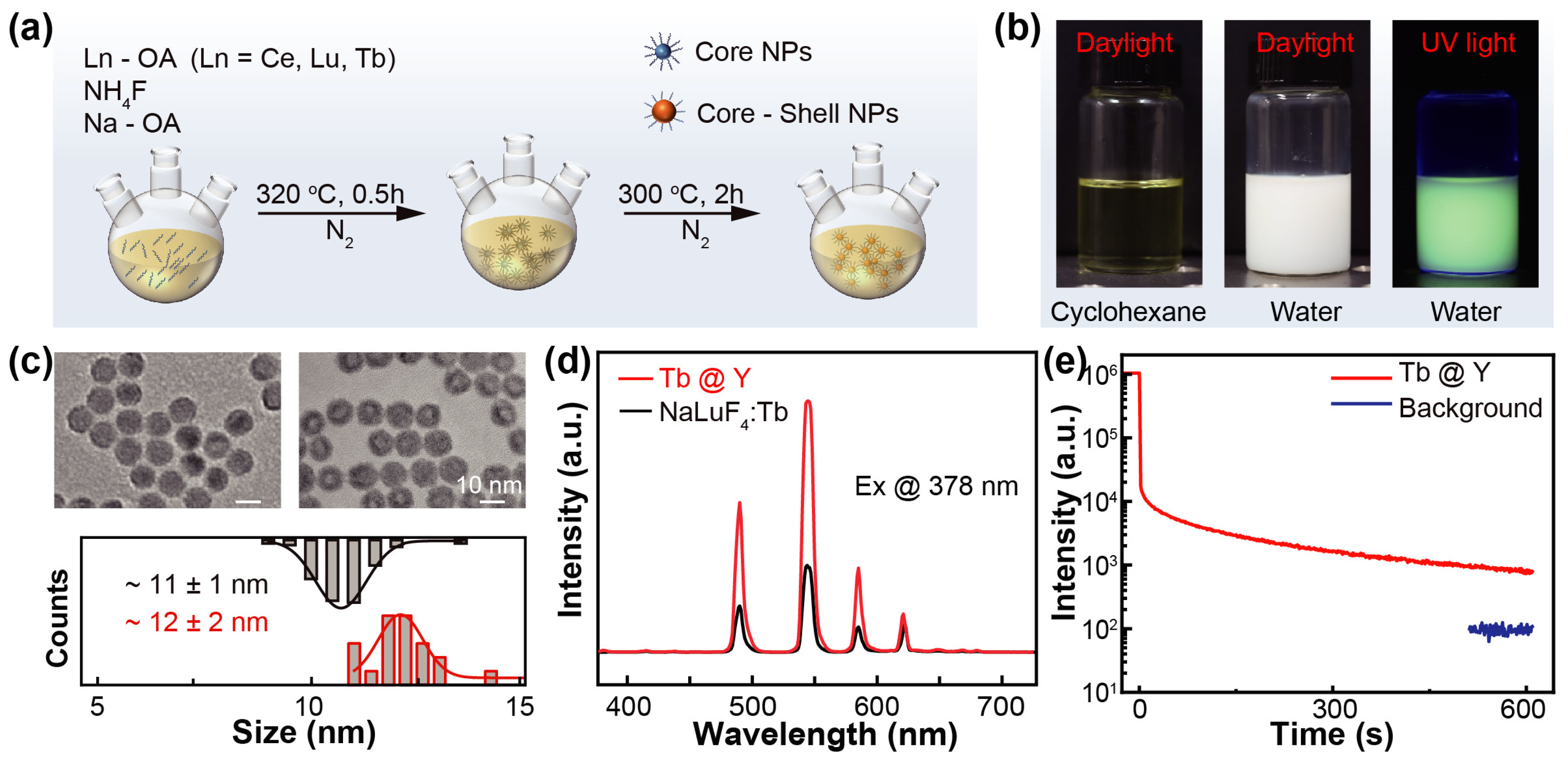
To prepare Tb3+-doped NaLuF4 nanoparticles on a large scale, an improved solvothermal method was employed, as shown in Figure 1a. The optimal doping ratio of Tb3+ in NaLuF4 nanoparticles was determined to be 15% [26]. Exceeding this ratio reduces the luminescence intensity owing to self-quenching from interactions between Tb3+ ions. The synthesized nanoparticles were dispersed in cyclohexane to form a transparent and yellowish liquid. The nanoparticles were then modified with hydrochloric acid (2M), yielding a well-dispersed aqueous system that exhibited bright green emission under UV light (Figure 1b).
To enhance the luminescent performance, a core–shell architecture was fabricated by coating the nanoparticles with an inert NaYF4 layer. The resulting core–shell NaLuF4:Tb3+@NaYF4 nanoparticles had an average size of 12 nm, compared with 11 nm for the uncoated NaLuF4:Tb3+ cores (Figure 1c). The shell significantly increased the emission intensity by suppressing nonradiative recombination at surface defects (Figure 1d) [33,34]. Under X-ray excitation, the core–shell nanoparticles exhibited a strong afterglow lasting up to 600 s, with an intensity more than tenfold above the background signal (Figure 1e). However, this prolonged afterglow can degrade temporal resolution in dynamic X-ray imaging, underscoring the need for effective afterglow-suppression strategies to preserve image quality.
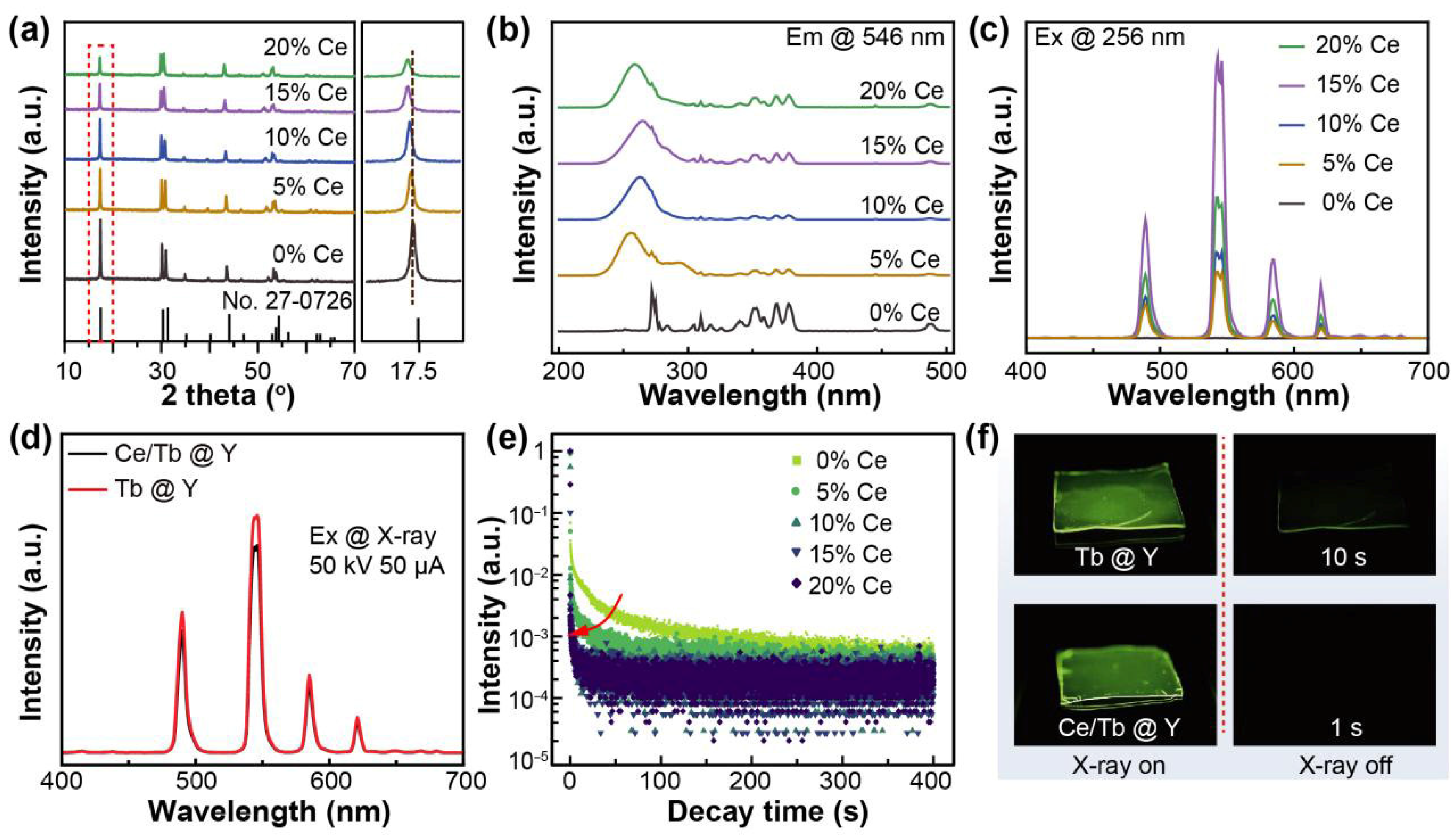
To mitigate the afterglow, a series of Ce3+-doped NaLuF4:Tb3+@NaYF4 samples were synthesized. X-ray diffraction (XRD) patterns confirmed the phase purity of all samples, which matched the standard hexagonal NaLuF4 structure (JCPDS No. 27-0726) without detectable impurities (Figure 2a). As the Ce3+ concentration increased, the diffraction peaks shifted slightly to lower angles. This shift is attributed to lattice expansion caused by the substitution of smaller Lu3+ (0.86 Å) or Tb3+ (0.92 Å) ions with larger Ce3+ ions (1.01 Å). Photoluminescence excitation (PLE) spectra monitored at 546 nm showed characteristic Tb3+ excitation peaks, along with an emerging broad band ascribed to Ce3+ (Figure 2b). The presence of this Ce3+ band suggests potential energy transfer from Ce3+ to Tb3+, which is consistent with previous studies [35,36]. Under 256 nm excitation, the Tb3+ emission intensity increased with Ce3+ concentration and peaked at 15% doping (Figure 2c). Upon X-ray excitation, both doped and undoped samples exhibited similar emission profiles (Figure 2d). Notably, even at 15% Ce3+ doping, the RL intensity decreased only marginally, indicating that Ce3+ doping had no obviously effect on the scintillation efficiency of the Tb3+ ions.
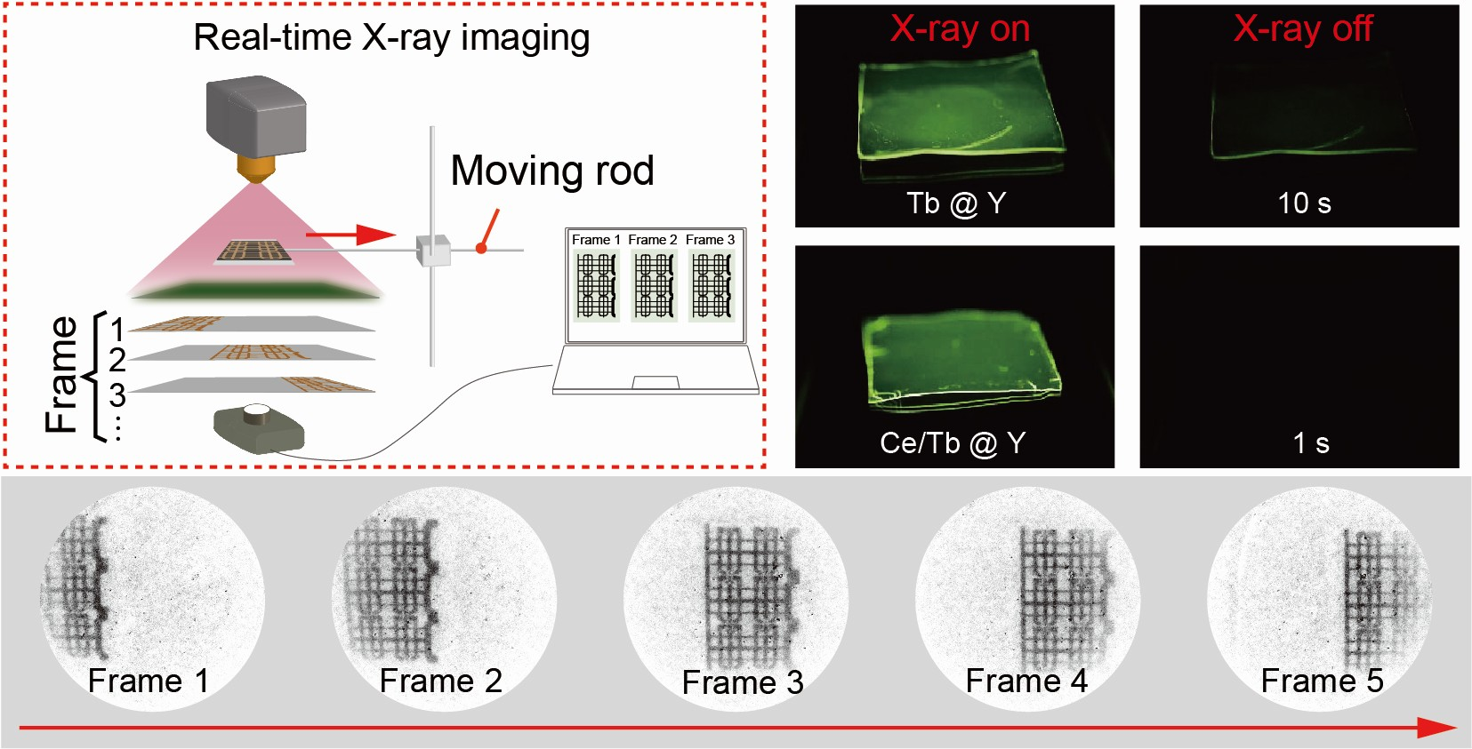
As shown in Figure 2e, the afterglow decay results suggested that the incorporation of Ce3+ ions efficiently suppressed the long-lasting afterglow of NaLuF4:Tb3+@NaYF4 nanoparticles. As the Ce3+ concentration increased from 0 to 15%, the decay time (to 1% of the RL intensity) was decreased from 6 s to 0.1 s, corresponding to a 60-fold reduction. As the Ce3+ ions concentration exceeded 15%, the time decreased slightly while the RL intensity obviously decreased. Consequently, 15% Ce3+-doped NaLuF4:Tb3+@NaYF4 was chosen for the subsequent fabrication of the scintillator screen. Additionally, the RL and afterglow images of both the undoped and 15% Ce3+-doped NaLuF4:Tb3+@NaYF4 nanoparticles films showed a similar afterglow suppression (Figure 2f).
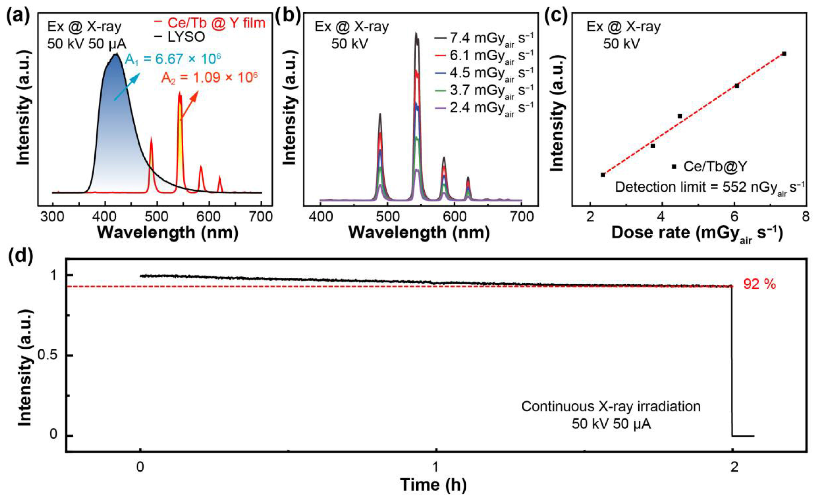
Owing to the suppression of afterglow, Ce3+-doped NaLuF4:Tb3+@NaYF4 nanoparticles are a promising candidate for X-ray imaging. Typically, transparent nanocomposite films were fabricated by combining Ce3+-doped NaLuF4:Tb3+@NaYF4 nanoparticles with PVA using a simple drop-casting method. Compared with a commercial scintillator (i.e., 500 μm LYSO single crystal), the luminescence intensity of the film (50 μm) was comparable despite their large difference in thickness (Figure 3a). The dose-dependent RL of the Ce3+-doped NaLuF4:Tb3+@NaYF4/PVA film showed a linear response to the X-ray dose rate, as illustrated in Figure 3b,c. The calculated detection limit was 552 nGyair s−1, approximately an order of magnitude lower than that required for medical diagnosis (~5.5 μGy/s) [37]. To assess its stability under X-ray irradiation, extended exposure tests were performed. After 2 h X-ray irradiation, the film maintains 92% of its initial luminescence intensity (Figure 3d).
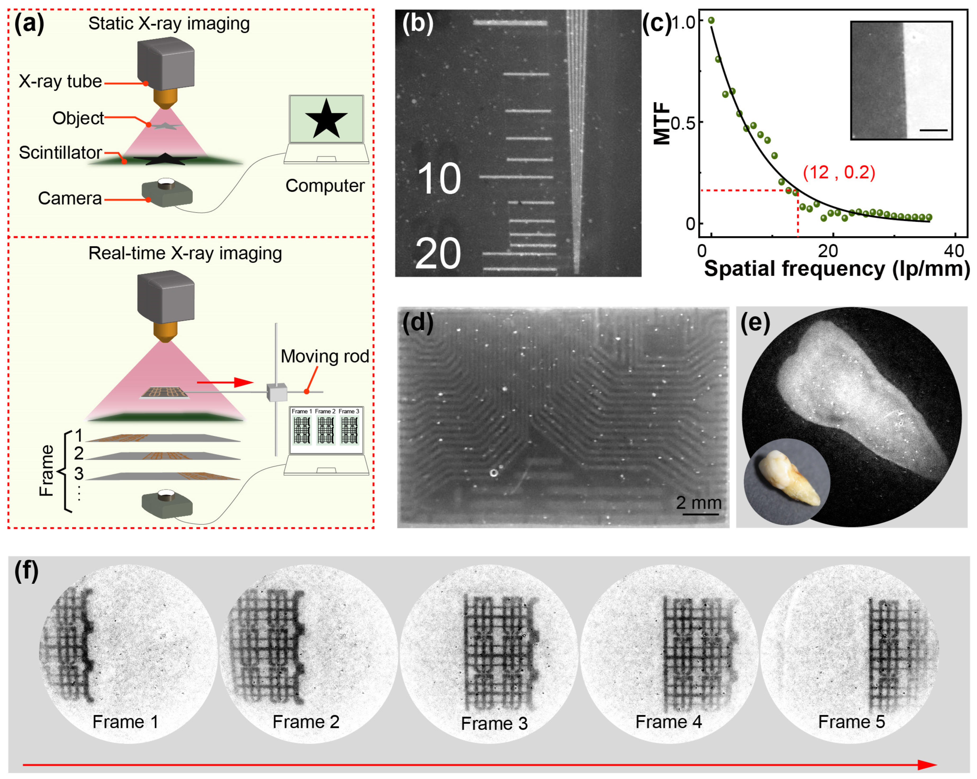
To further verify its X-ray imaging capabilities, a home-made X-ray imaging testbed was developed, consisting of an X-ray source, a Ce3+-doped NaLuF4:Tb3+@NaYF4/PVA film, and a digital camera (Figure 4a). In static X-ray imaging, the high transparency of the film enables the acquisition of high-quality images due to the reduced light scattering. To this end, resolution test targets and MTF were employed to evaluate the spatial resolution of the Ce3+-doped NaLuF4:Tb3+@NaYF4/PVA film, which exceeded 12 lp mm−1 (Figure 4b,c). Furthermore, an integrated circuit was employed in X-ray imaging, which demonstrated high resolution with distinct lines (Figure 4d). For biological-tissue imaging, an artificial-dentures sample was used, yielding similarly clear imaging results (Figure 4e).
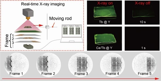
To demonstrate real-time X-ray imaging capability, a moving rod was incorporated into a custom testbed designed for the Ce3+-doped NaLuF4:Tb3+@NaYF4/PVA scintillator screen. In a proof-of-concept experiment, a copper pattern was used as the imaging target, moving at a speed of 1 cm/s (Figure 4a). In the continuous-shooting experiment, the captured images showed no blurring or artifacts (Figure 4f and Video S1), indicating excellent real-time imaging performance even for moving targets. This fast temporal response was crucial for real-time X-ray imaging, as it enables the rapid conversion of X-rays to visible light and captures dynamic changes in the target. Overall, these results demonstrated outstanding real-time imaging capabilities, laying a solid foundation for further development and application in medical and industrial non-destructive X-ray imaging.

4. Conclusions

In summary, this work demonstrates an effective strategy for suppressing X-ray-induced afterglow in Tb3+-doped NaLuF4 nanocrystals via Ce3+ co-doping. The Ce3+ ions enabled efficient energy transfer and suppressed afterglow while largely preserving the radioluminescence intensity. When composited with PVA, the fabricated scintillator film exhibited a low detection limit of 552 nGyairs−1 and outstanding radiation stability, retaining 92% of its initial luminescence after prolonged X-ray irradiation. Static and dynamic imaging tests confirmed the high imaging capability of the material. Notably, a spatial resolution exceeding 12 lp/mm was achieved with negligible afterglow interference in real-time imaging. These results provide a viable route to developing high-performance scintillators for fast, high-resolution X-ray imaging applications such as medical diagnostics and non-destructive testing.

Supplementary Materials

The following supporting information can be downloaded at https://www.mdpi.com/article/10.3390/ma19020437/s1, Video S1: Low afterglow composite scintillator for real-time X-ray imaging.

Author Contributions

X.C. supervised the project, conceived the original idea. X.Z. prepared the materials, fabricated devices, performed device performance tests and the manuscript. Y.L. and N.L. assisted in the material preparation and performance test. Z.C. and Y.Z. undertook the revision of the manuscript. All authors commented on the manuscript. All authors have read and agreed to the published version of the manuscript.

Funding

This work was sponsored financially by the Natural Science Foundation of Shandong Province (ZR2024ME181); Young Talent of Lifting engineering for Science and Technology in Shandong, China (SDAST2024QTA010); Shandong Province Science and Technology Smes Ability Improvement Project (2025TSGCCZZB0493).

Data Availability Statement

The original contributions presented in this study are included in the article/Supplementary Materials. Further inquiries can be directed to the corresponding author.

Conflicts of Interest

The authors declare no conflicts of interest.

References

  1. Lei, L.; Yi, M.; Wang, Y.; Hua, Y.; Zhang, J.; Prasad, P.N.; Xu, S. Dual Heterogeneous Interfaces Enhance X-Ray Excited Persistent Luminescence for Low-Dose 3D Imaging. Nat. Commun. 2024, 15, 1140. [Google Scholar] [CrossRef]
  2. Ling, Y.; Zhao, X.; Hao, P.; Song, Y.; Liu, J.; Zhao, L.; Qian, Y.; Guo, C. Gd3+-Sensitized Rare Earth Fluoride Scintillators for High-Resolution Flexible X-Ray Imaging. Chem. Eng. J. 2023, 476, 146790. [Google Scholar] [CrossRef]
  3. Lee, G.; Savage, N.; Wagner, B.; Zhang, Y.; Jacobs, B.; Menkara, H.; Summers, C.; Kang, Z. Synthesis and Luminescence Properties of Transparent Nanocrystalline GdF3:Tb Glass-Ceramic Scintillator. J. Lumin. 2014, 147, 363–366. [Google Scholar] [CrossRef]
  4. Lu, H.; Xu, X.; Feng, G.; Sun, B.; Wang, S.; Wu, S. Terbium Doped LiLuF4 Nanocrystal Scintillator-Based Flexible Composite Film for High Resolution X-Ray Imaging. RSC Adv. 2022, 12, 4615–4623. [Google Scholar] [CrossRef]
  5. Jiang, X.; Xia, S.; Zhang, J.; Ju, D.; Liu, Y.; Hu, X.; Wang, L.; Chen, Z.; Tao, X. Exploring Organic Metal Halides with Reversible Temperature-Responsive Dual-Emissive Photoluminescence. ChemSusChem 2019, 12, 5228–5232. [Google Scholar] [CrossRef] [PubMed]
  6. Feng, A.; Jiang, X.; Zhang, X.; Zheng, X.; Zheng, W.; Mohammed, O.F.; Chen, Z.; Bakr, O.M. Shape Control of Metal Halide Perovskite Single Crystals: From Bulk to Nanoscale. Chem. Mater. 2020, 32, 7602–7617. [Google Scholar] [CrossRef]
  7. Zhang, X.; Shi, Y.; Wang, X.; Liu, Y.; Zhang, Y. Flexible and Transparent Ceramic Nanocomposite for Laboratory X-Ray Imaging of Micrometer Resolution. ACS Nano 2022, 16, 21576–21582. [Google Scholar] [CrossRef]
  8. Yang, T.; Wu, P.; Chen, R.; Ni, J.; Xu, C.; Liu, X.; Wang, S.; Liu, S. Enhanced Luminescence of Tb Doping Nanoscintillators Based on NaLuF4 Host Matrix for X-Ray Imaging. J. Lumin. 2024, 269, 120498. [Google Scholar] [CrossRef]
  9. Xu, W.; Zhou, M.; Deng, R.; Liu, X.; Yao, L.; Zhou, J.; Qiu, J. Single Activator Doped Fluoride Nanoparticles for X-Ray Excited High-Resolution and Delayed Flexible Nonplanar Imaging. Chem. Eng. J. 2024, 499, 156406. [Google Scholar] [CrossRef]
  10. Wang, S.; Hua, C.; Wang, L.; Wang, C.; Liu, L.; Ren, J.; Zhang, J. Double Gain of Efficient UV-C Phosphor and Fast Scintillator Based on Pr3+-Doped KY3F10 Nano-Glass Composites. Adv. Opt. Mater. 2024, 12, 2302086. [Google Scholar] [CrossRef]
  11. Ou, X.; Qin, X.; Huang, B.; Zan, J.; Wu, Q.; Hong, Z.; Xie, L.; Bian, H.; Yi, Z.; Chen, X.; et al. High-Resolution X-Ray Luminescence Extension Imaging. Nature 2021, 590, 410–415. [Google Scholar] [CrossRef]
  12. Sun, B.; Winardi, I.; Miaule, S.; Pei, Q. Luminescent Lanthanides Doped NaGdF4 Nanoparticles–Plastic Nanocomposites for Gamma Ray Scintillation. Nucl. Instrum. Methods Phys. Res. Sect. A Accel. Spectrometers Detect. Assoc. Equip. 2026, 1082, 171087. [Google Scholar] [CrossRef]
  13. Yang, D.; Xu, J.; Li, L.; Zhang, X.; Xu, Z.; Yang, Y.; Dong, Z.; Xu, T.; Cheng, C.; Xu, Q.; et al. Reversible X-Ray Memory Imaging for Cadmium-Based Perovskites: Deep Trap-Driven Radioluminescence Enhancement. Adv. Mater. 2026, e20865. [Google Scholar] [CrossRef]
  14. Zou, H.; Zhu, W.; Zhao, J.; Zhou, S.; Xu, S.; Lei, L. Sub-10 nm Lanthanide-Doped Lu6O5F8 Nanoscintillators for Real-Time High-Resolution Dynamic 3D X-Ray Imaging. Adv. Funct. Mater. 2024, 34, 2409156. [Google Scholar] [CrossRef]
  15. Lu, H.; Xu, X.; Li, Y.; Feng, G.; Yang, J.; Huang, X.; Luo, Q.; Wang, S.; Wu, S. Heterovalent Co-Doped LiLuF4 Composite Flexible Films as Persistent Luminescence Scintillator for X-Ray High-Resolution Extension Imaging. Adv. Opt. Mater. 2024, 12, 2303134. [Google Scholar] [CrossRef]
  16. Qiao, P.; Yang, J.; Ma, H.; Lei, L. Tuning Scintillation Property of CsLu2F7: Pr Crystals for X-Ray Imaging. Opt. Mater. 2024, 148, 114812. [Google Scholar] [CrossRef]
  17. Li, Z.; Li, H.; Sun, H.-T. X-Ray-Activated UVA Long Persistent Luminescence from Defective Fluoride Elpasolites. J. Rare Earths 2020, 38, 124–129. [Google Scholar] [CrossRef]
  18. Zhou, C.; Li, D.; Lei, L.; Bai, G.; Wang, H.; Zhang, X.; Xu, S.; Qiu, J. Thermo-Enhanced Afterglow Emission in Tm3+ Doped Fluoride Nanoparticles for Anti-Counterfeiting Application after X-Ray Excitation. Ceram. Int. 2023, 49, S0272884223019612. [Google Scholar] [CrossRef]
  19. Lei, L.; Wang, Y.; Xu, W.; Ye, R.; Hua, Y.; Deng, D.; Chen, L.; Prasad, P.N.; Xu, S. Manipulation of Time-Dependent Multicolour Evolution of X-Ray Excited Afterglow in Lanthanide-Doped Fluoride Nanoparticles. Nat. Commun. 2022, 13, 5739. [Google Scholar] [CrossRef]
  20. Li, W.; Lin, J.; Huang, W.; Wang, Q.; Zhang, H.; Zhang, X.; Zhuang, J.; Liu, Y.; Qu, S.; Lei, B. Delivery of Luminescent Particles to Plants for Information Encoding and Storage. Light Sci. Appl. 2024, 13, 217. [Google Scholar] [CrossRef]
  21. Yang, H.; Zhang, A.; Xu, S.; Lei, L. Optimizing Crystallinity to Enhance X-Ray Excited Persistent Luminescence Intensity of NaLuF4: Gd/Tb Nanoparticles. J. Lumin. 2023, 263, 120040. [Google Scholar] [CrossRef]
  22. Li, L.; Li, T.; Hu, Y.; Cai, C.; Li, Y.; Zhang, X.; Liang, B.; Yang, Y.; Qiu, J. Mechanism of the Trivalent Lanthanides’ Persistent Luminescence in Wide Bandgap Materials. Light Sci. Appl. 2022, 11, 51. [Google Scholar] [CrossRef]
  23. Lu, H.; Xu, X.; Zhang, Z.; Zhang, X.; Huang, R.; Feng, G.; Xu, J.; Wu, S.; Zheng, Y.; Huang, X.; et al. Trap-Modulated Fluoride Scintillators for Thermal Mapping-Assisted X-Ray 3D Tomographic Imaging. Chem. Eng. J. 2025, 518, 164480. [Google Scholar] [CrossRef]
  24. Huang, K.; Dou, X.; Zhang, Y.; Gao, X.; Lin, J.; Qu, J.; Li, Y.; Huang, P.; Han, G. Enhancing Light and X-Ray Charging in Persistent Luminescence Nanocrystals for Orthogonal Afterglow Anti-Counterfeiting. Adv. Funct. Mater. 2021, 31, 2009920. [Google Scholar] [CrossRef]
  25. Hu, Y.; Yang, Y.; Zhang, X.; Wang, X.; Li, X.; Li, Y.; Li, T.; Zhang, H. X-Ray-Excited Super-Long Green Persistent Luminescence from Tb3+ Monodoped β-NaYF4. J. Phys. Chem. C 2020, 124, 24940–24948. [Google Scholar] [CrossRef]
  26. Sun, S.; Shao, S.; Fan, W.; Zhang, W.; Xu, F.; Wei, H.; Cao, B.; Wu, Y. Enhanced X-Ray-Induced Luminescence and Afterglow of NaLuF4:Gd3+/Tb3+ Nanoparticles with Ca2+ Doping. J. Lumin. 2023, 261, 119929. [Google Scholar] [CrossRef]
  27. Buzug, T.M. Computed Tomography. In Springer Handbook of Medical Technology; Springer: Berlin/Heidelberg, Germany, 2011; pp. 311–342. [Google Scholar] [CrossRef]
  28. Kalender, W.A. X-Ray Computed Tomography. Phys. Med. Biol. 2006, 51, R29. [Google Scholar] [CrossRef]
  29. Ma, J.; Zhu, W.; Lei, L.; Deng, D.; Hua, Y.; Yang, Y.M.; Xu, S.; Prasad, P.N. Highly Efficient NaGdF4:Ce/Tb Nanoscintillator with Reduced Afterglow and Light Scattering for High-Resolution X-Ray Imaging. ACS Appl. Mater. Interfaces 2021, 13, 44596–44603. [Google Scholar] [CrossRef]
  30. Zou, H.; Yi, M.; Xu, S.; Lei, L. A Hollow NaBiF4:Tb Nanoscintillator with Ultra-Weak Afterglow for High-Resolution X-Ray Imaging. Chem. Commun. 2023, 59, 11732–11735. [Google Scholar] [CrossRef]
  31. Zhang, Y.; Tong, Y.; Zong, L.; Ren, G.; Wu, Y. Effects of Cu+ and Sc3+ Codoping on Luminescence and Scintillation Properties of Cs2LiYCl6:Ce Single Crystals. J. Rare Earths 2025, 43, 2637–2645. [Google Scholar] [CrossRef]
  32. Miyazaki, K.; Nakauchi, D.; Kato, T.; Kawaguchi, N.; Yanagida, T. Suppression of Afterglow in RbI:Tl Scintillator by Co-Doping. J. Mater. Sci. Mater. Electron. 2023, 34, 1082. [Google Scholar] [CrossRef]
  33. Gao, W.; Cheng, X.; Xing, Y.; Han, S.; Chen, B.; Han, Q.; Yan, X.; Liu, J.; Liu, L.; Dong, J. Enhancement of Red Upconversion Emission Intensity of Ho3+ Ions in NaLuF4:Yb3+/Ho3+/Ce3+@NaLuF4 Core–Shell Nanoparticles. J. Rare Earths 2022, 40, 517–525. [Google Scholar] [CrossRef]
  34. Juarez, X.G.; Ko, H.; Jang, H.S. Strong Upconversion–Downshifting Green Emission from Tb3+ Ions in Core/Shell/Shell-Structured Nanophosphors. Res. Chem. Intermed. 2018, 44, 4641–4650. [Google Scholar] [CrossRef]
  35. Adusumalli, V.N.K.B.; Koppisetti, H.V.S.R.M.; Ganguli, S.; Sarkar, S.; Mahalingam, V. Tuning the Energy Transfer Efficiency between Ce3+ and Ln3+ Ions (Ln=Tm, Sm, Tb, Dy) by Controlling the Crystal Phase of NaYF4 Nanocrystals. Chem. Eur. J. 2017, 23, 994–1000. [Google Scholar] [CrossRef]
  36. Boyer, J.-C.; Gagnon, J.; Cuccia, L.A.; Capobianco, J.A. Synthesis, Characterization, and Spectroscopy of NaGdF4: Ce 3+,Tb3+/NaYF4 Core/Shell Nanoparticles. Chem. Mater. 2007, 19, 3358–3360. [Google Scholar] [CrossRef]
  37. Shearer, D.R.; Bopaiah, M. Dose Rate Limitations of Integrating Survey Meters for Diagnostic X-Ray Surveys. Health Phys. 2000, 79, 20–21. [Google Scholar] [CrossRef] [PubMed]
Figure 1. (a) Schematic diagram of the nanoparticle preparation process. (b) Luminescence images of the nanoparticles under sunlight and UV excitation. (c) TEM images and particle-size distributions of NaLuF4:Tb and NaLuF4:Tb@NaYF4 nanocrystals. (d) The luminescence spectra of NaLuF4:Tb and NaLuF4:Tb@NaYF4 nanocrystals. (e) The afterglow decay of NaLuF4:Tb@NaYF4 nanocrystals following 5 min of X-ray illumination.
Materials 19 00437 g001
Figure 2. (a) X-ray diffraction (XRD) patterns, (b) photoluminescence excitation (PLE) spectra, and (c) photoluminescence (PL) spectra of a series of Ce-doped samples with varying doping concentrations (0%, 5%, 10%, 15%, and 20%). (d) The radioluminescence (RL) spectra of undoped and Ce3+-doped NaLuF4:Tb3+ nanoparticles. (e) The normalized afterglow of the series of Ce3+-doped samples with different doping concentrations. (f) The RL and afterglow images of the undoped and Ce3+ doped NaLuF4:Tb3+@NaYF4 nanoparticles.
Materials 19 00437 g002
Figure 3. (a) Comparison of RL between a Ce3+-doped NaLuF4:Tb3+@NaYF4/PVA film (50 μm) and LYSO:Ce single crystal (500 μm). The integrated intensity (A1: 6.67 × 10⁶ and A2: 1.08 × 10⁶) was comparable despite a tenfold difference in thickness (50 μm vs. 500 μm). (b) RL of the Ce3+-doped NaLuF4:Tb3+@NaYF4/PVA film at various dose rates. (c) Detection limit of the Ce3+-doped NaLuF4:Tb3+@NaYF4/PVA film. (d) Radiation stability of the Ce3+-doped NaLuF4:Tb3+@NaYF4/PVA film under 50 keV and 50 μA.
Materials 19 00437 g003
Figure 4. (a) Schematic diagram of static X-ray imaging and real-time X-ray imaging process. (b) Image of a resolution test target and (c) Modulation transfer functions (MTF) of a slanted-edge image revealed a spatial resolution of 12 lp/mm. (d) Static imaging line pattern of a line-pair pattern and (e) a circuit board. (f) Real-time X-ray imaging of a copper pattern on a moving rod, with images acquired at 1 s interval. (ISO 25600, F/2.8, exposure 1/60 s).
Materials 19 00437 g004
Disclaimer/Publisher’s Note: The statements, opinions and data contained in all publications are solely those of the individual author(s) and contributor(s) and not of MDPI and/or the editor(s). MDPI and/or the editor(s) disclaim responsibility for any injury to people or property resulting from any ideas, methods, instructions or products referred to in the content.