
Exploring the Journey of Zinc Oxide Nanoparticles (ZnO-NPs) toward Biomedical Applications

 , , , , , , , and
Abstract
:1. Introduction
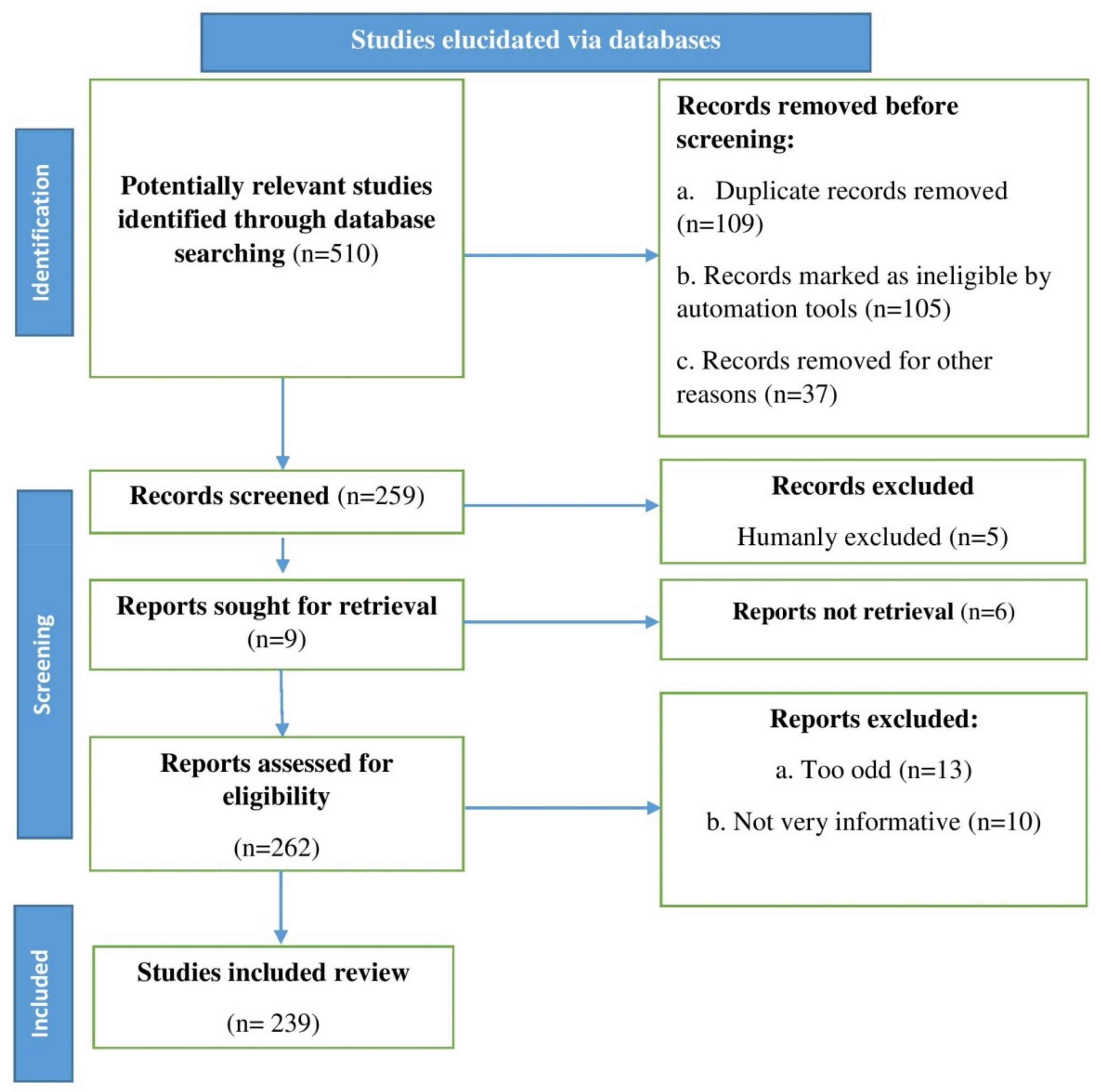
2. Methodology
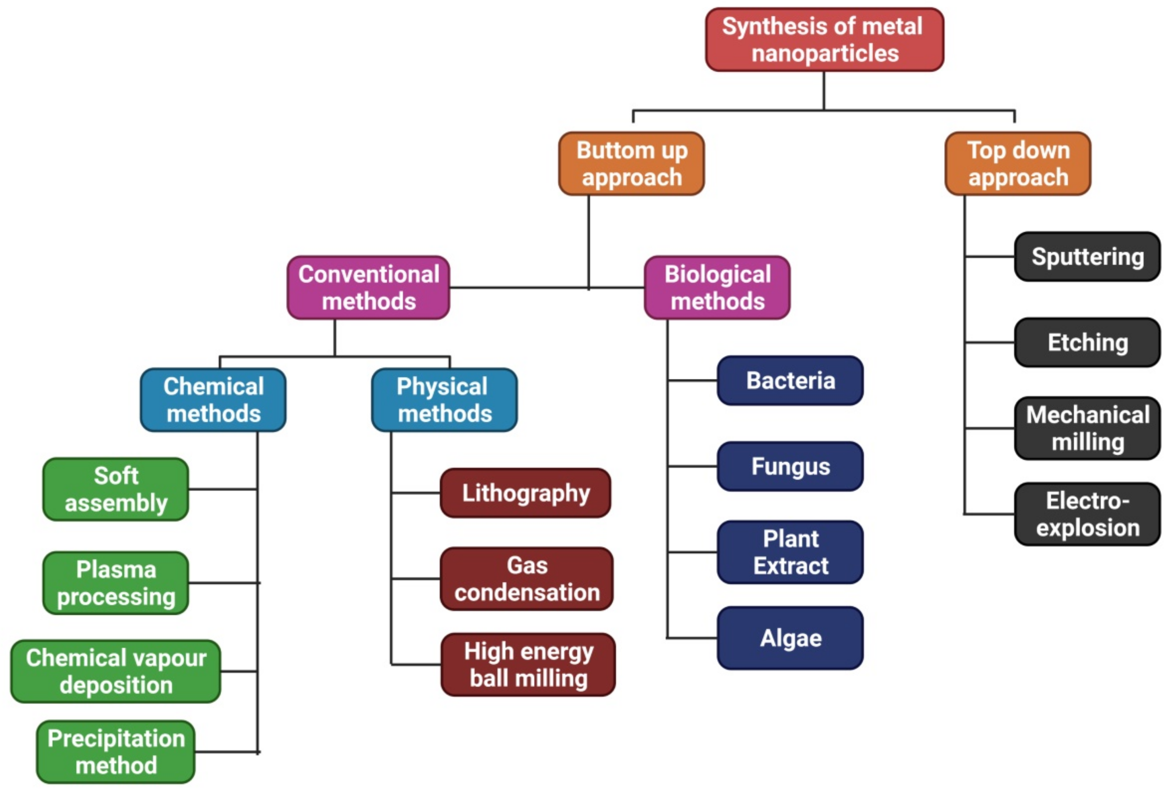
3. Traditional Synthesis of ZnO Nanomaterials
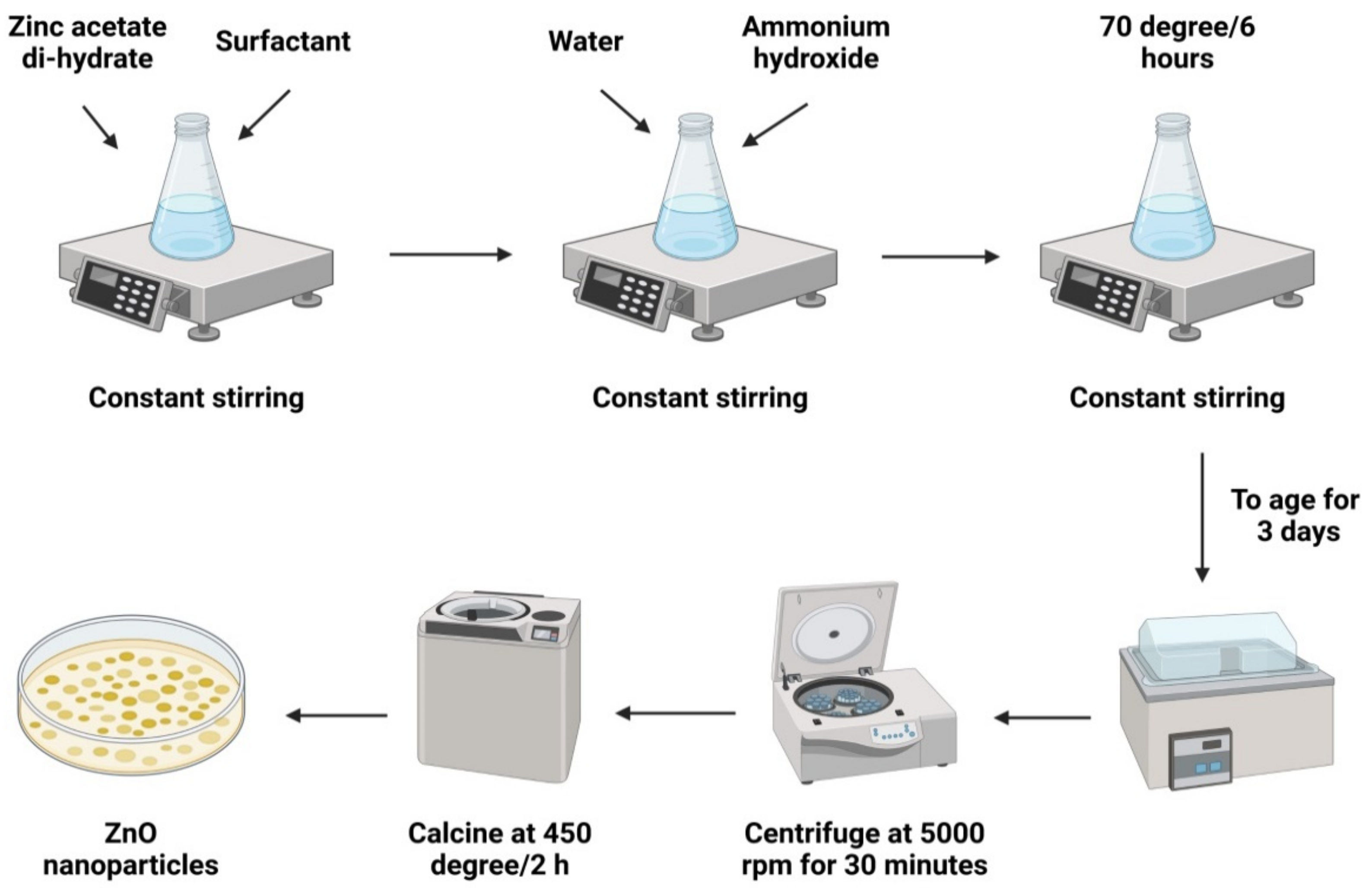
3.1. Sol-Gel Technique
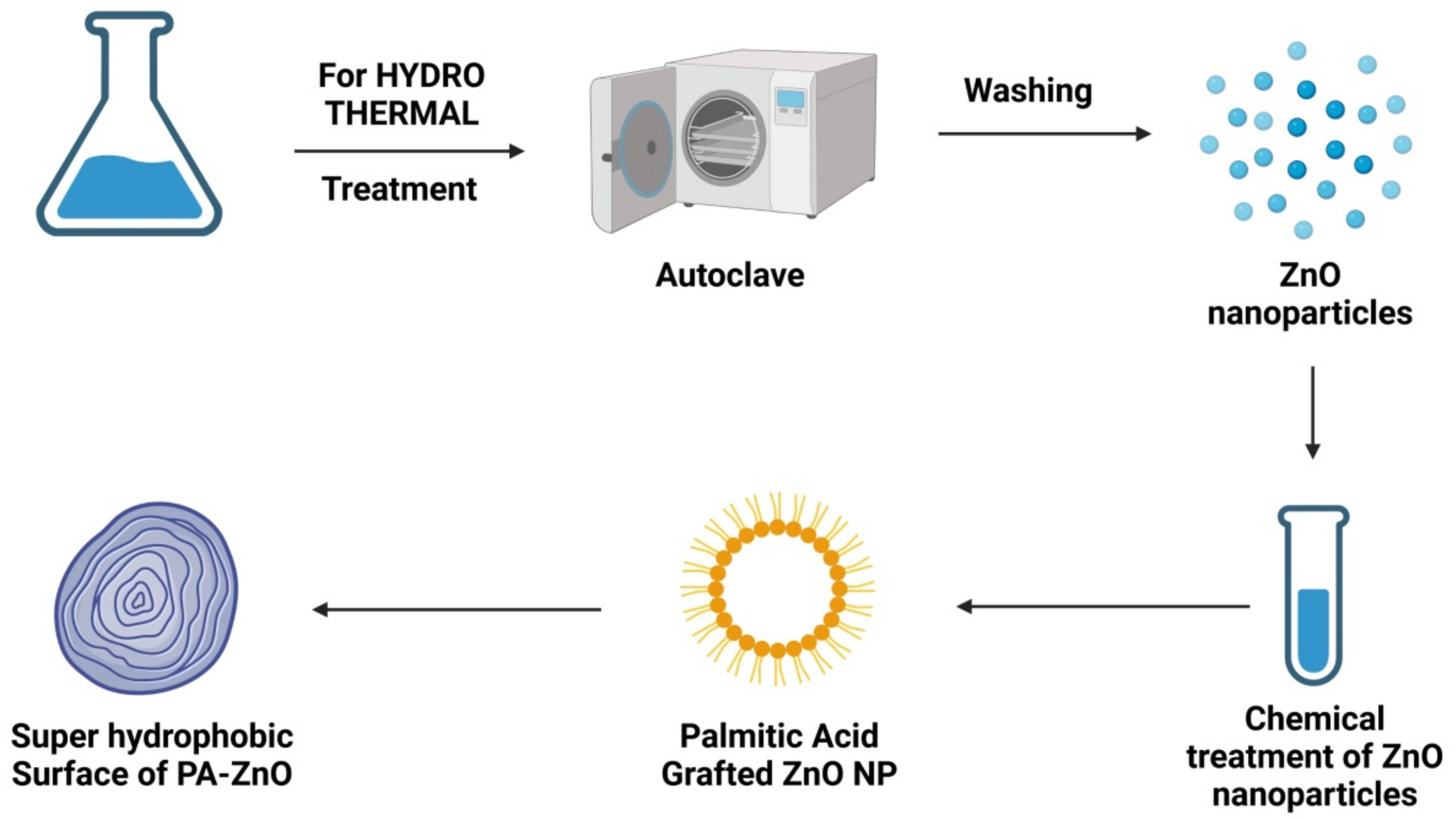
3.2. Hydrothermal Technique
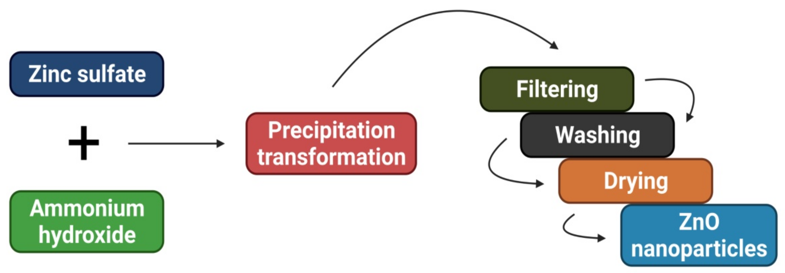
3.3. Co-Precipitation Technique
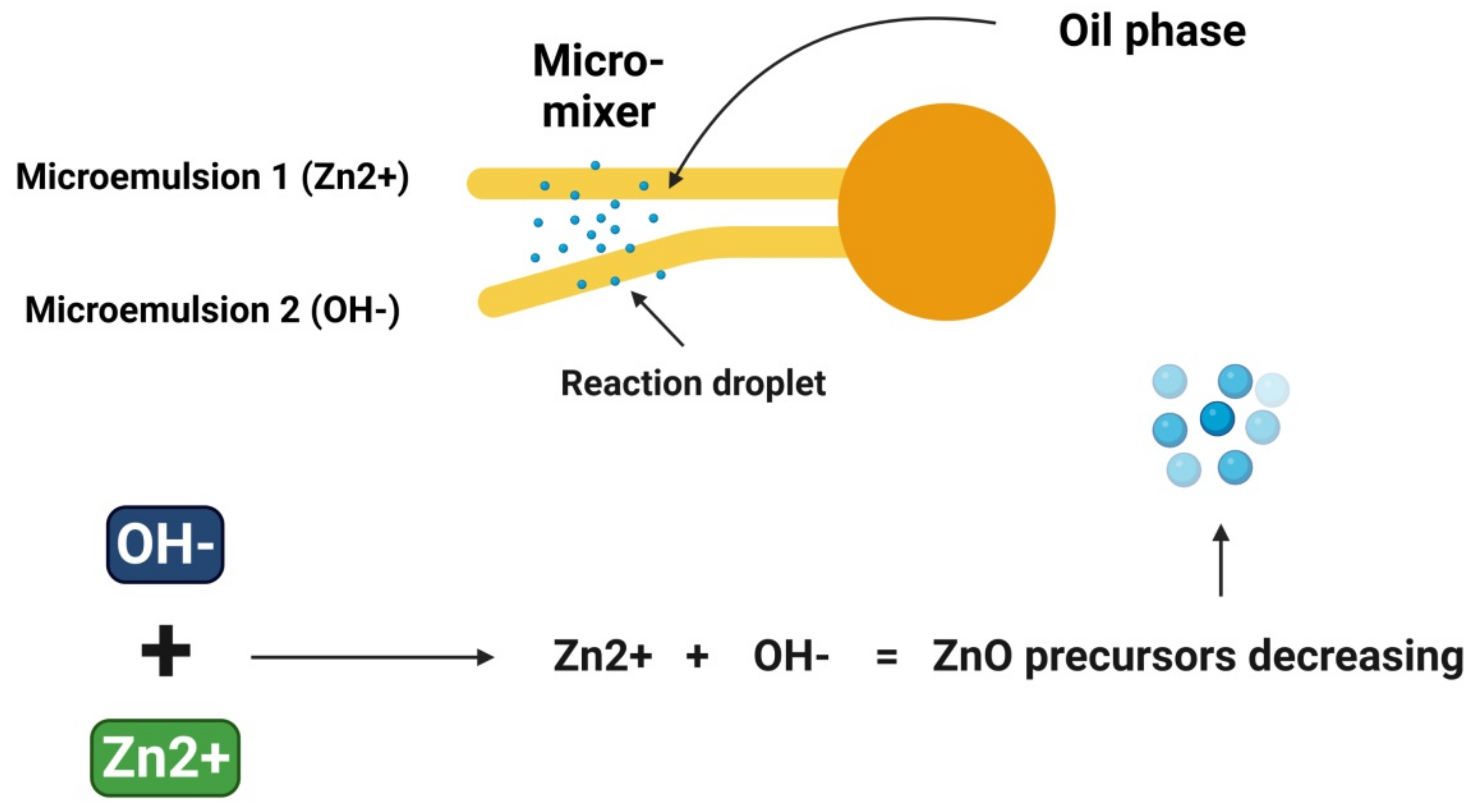
3.4. Microemulsion Technique
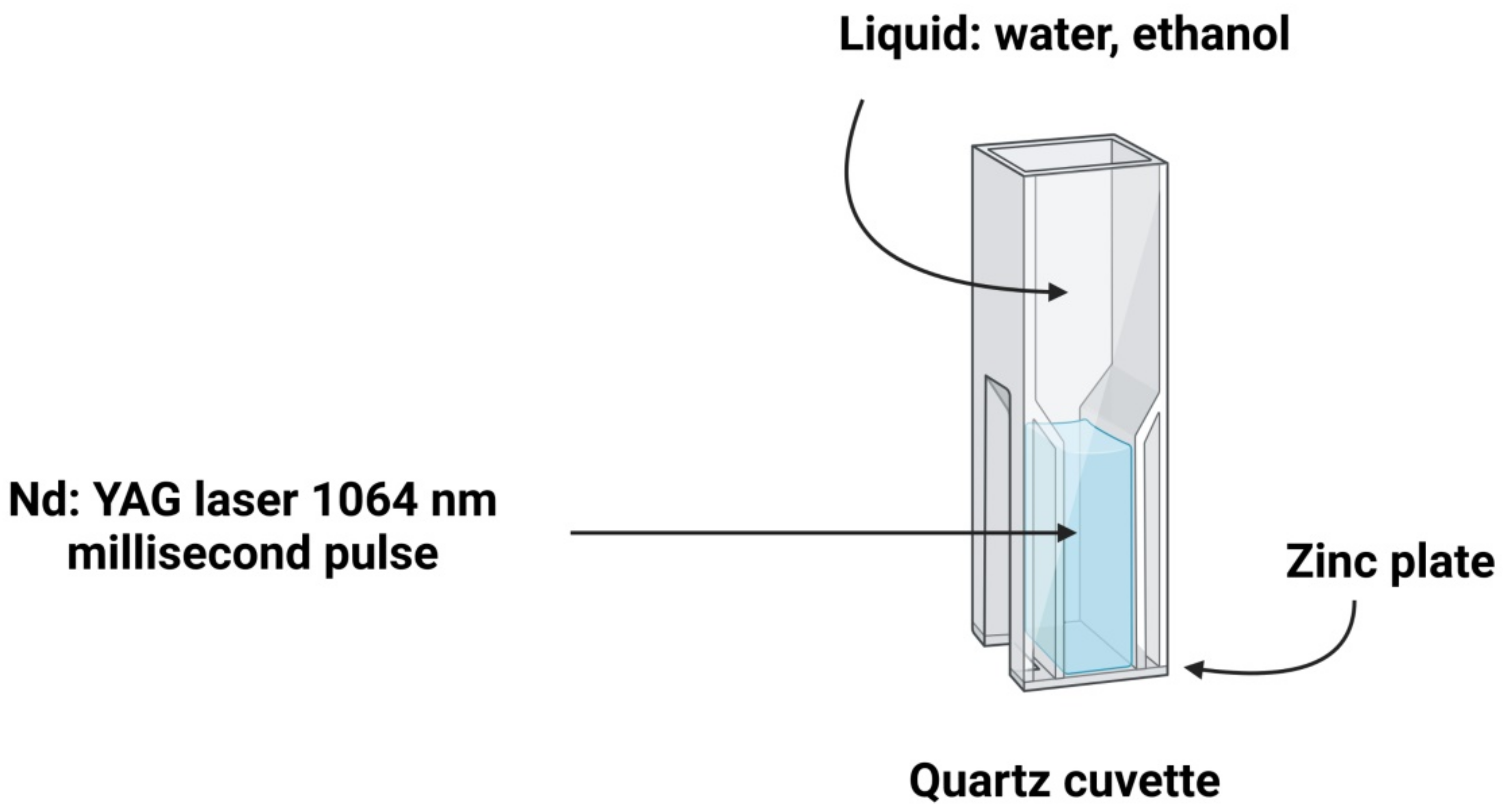
3.5. Laser Ablation Technique
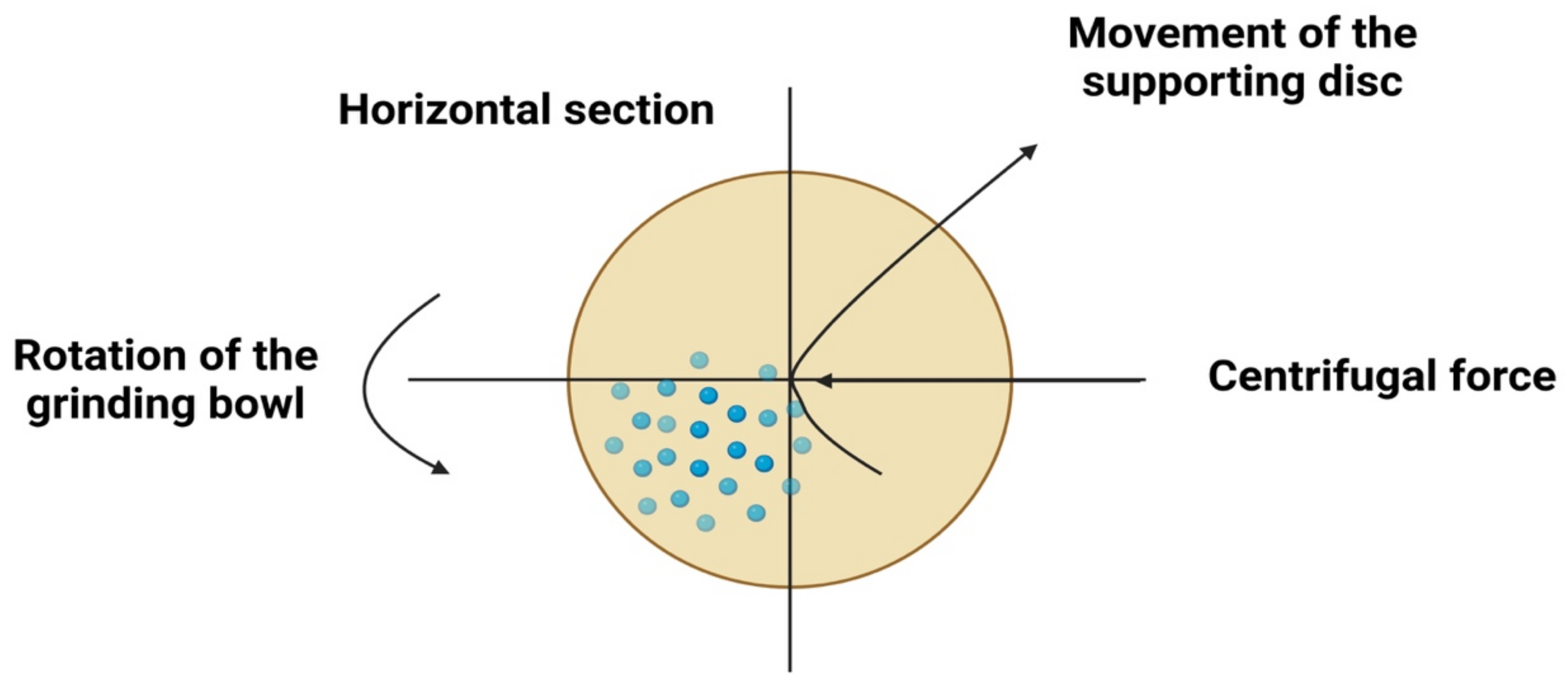
3.6. High-Energy Ball Milling Techniques
4. Green Synthesis of ZnO-NPs
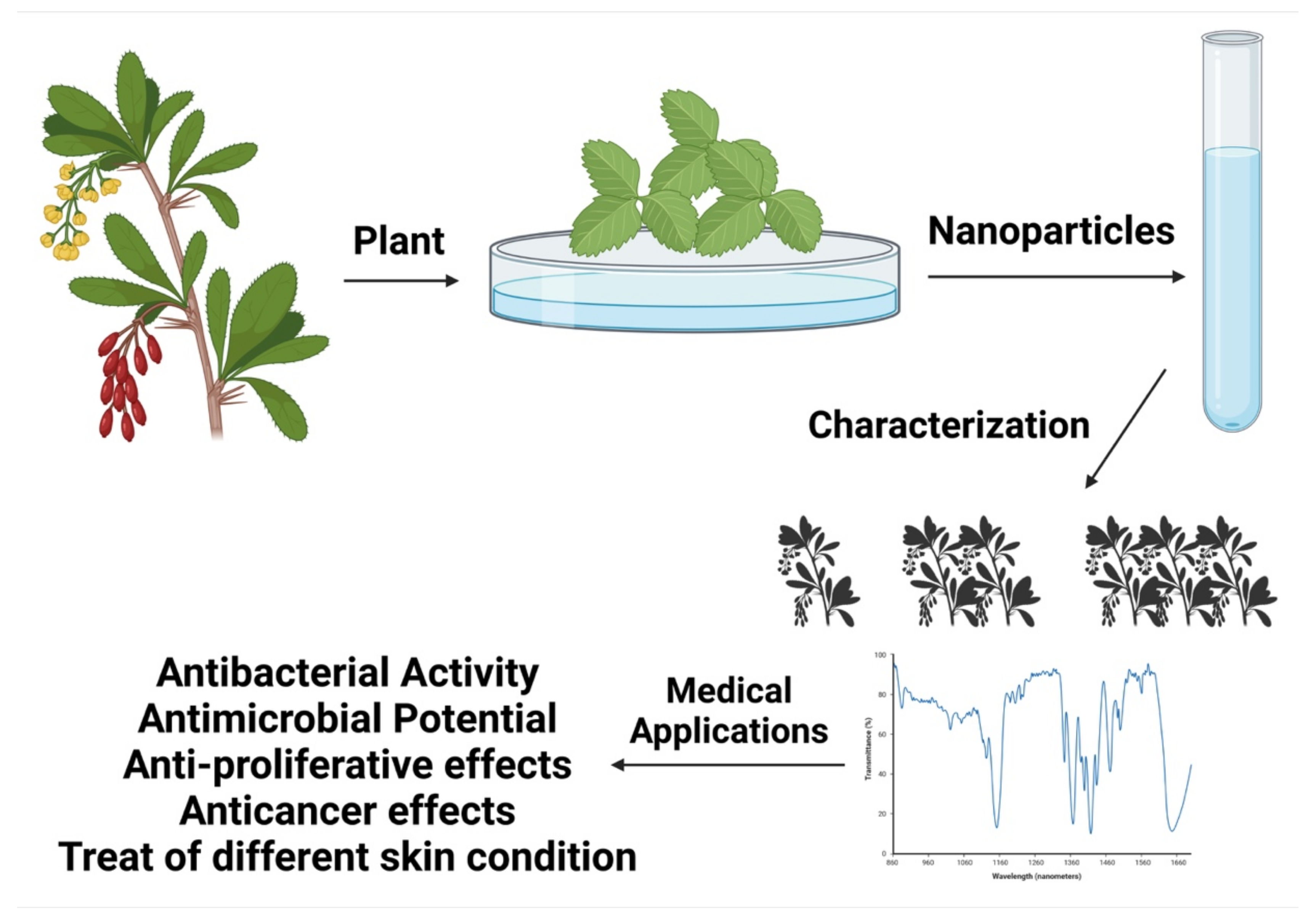
4.1. Green Synthesis of ZnO-NPs Using Plant Extract
4.2. ZnO-NPs Green Synthesis Using Bacteria
4.3. ZnO-NPs Green Synthesis Using Microalgae and Macroalgae
4.4. ZnO-NPs Green Synthesis Using Fungus Theorem
4.5. ZnO-NPs Green Synthesis Using Other Green Sources
5. Biomedical Applications of Green-Synthesized ZnO-NPs
5.1. ZnO-NPs Antibacterial Activity
5.2. ZnO-NPs Antimicrobial Potential
5.3. Proliferating Cells Selective Killers
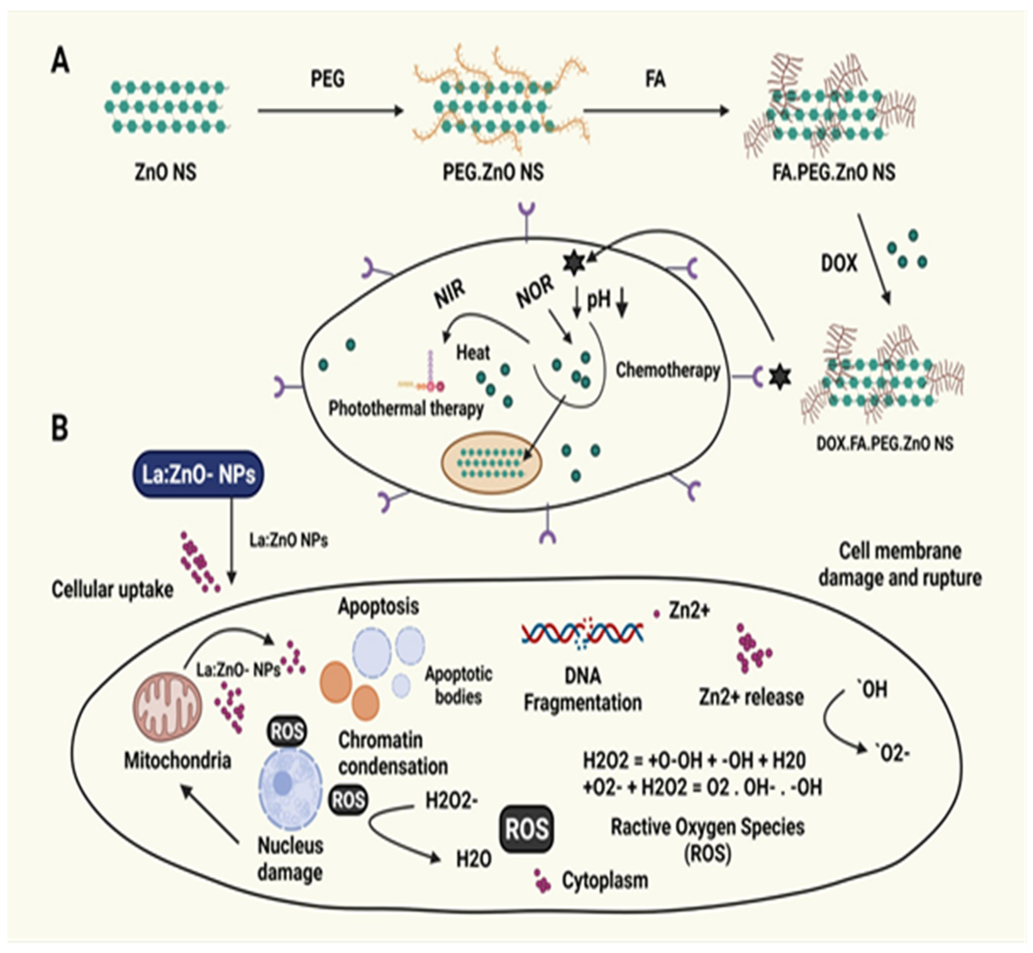
5.4. ZnO-NPs Anticancer Effects

| Platform | Raw Material | Size | System | Targeted Cell Line | Reference |
|---|---|---|---|---|---|
| Fungi-mediated | Pichia kudriavzevii yeast | 10–61 nm | ZnO-NPs | MCF-7, breast | [173] |
| Penicillium chrysogenum fungus | 29–37 nm | ZnO-NPs | MCF-7, breast HCT-116, colon | [174] | |
| Aspergillus niger fungus | 80–130 nm | ZnO-NPs | HepG2, liver | [175] | |
| Aspergillus niger fungus | 11.8–17.6 nm | ZnO-NPs | A549, lung | [176] | |
| Aspergillus terreus fungus | 28–63 nm | L-asparginase—ZnO-NPs | MCF-7, breast | [177] | |
| Algae and plant-mediated | Sargassum muticum algae extract | 30–57 nm | ZnO-NPs | HepG2, liver | [178] |
| Sargassum muticum algae extract | 50–100 nm | ZnO-NPs | WEHI-3, murine leukemia | [179] | |
| Sargassum muticum algae extract | 3–8 nm | ZnO-NPs | PANC-1, pancreas CaOV-3, ovarian COLO205, colon HL-60, leukemia | [180] | |
| Gracilaria edulis algae extract | 4.04 ± 1.81 nm; length 1.39 ± 0.6 nm; width | ZnO-NPs | SiHa, cervical | [181] | |
| Rehmanniae radix plant extract | 10–12 m | ZnONPs | MG-63 bone | [182] | |
| Myristica fragans plant extract | 100–200 nm | ZnONPs | HeoG2, liver | [183] | |
| Albizia lebbeck stem bark | 66.25 nm | ZnONPs | MCF-7, breast MDAMB231, breast | [184] | |
| Mangifera infica leaves | 45–60 nm | ZnO-NPs | A549, lung | [185] | |
| Pongamia pinnata seeds | 30.4–40.8 nm | ZnO-NPs | MCF-7, breast | [186] | |
| Eclipta prostrata leaves | 20–1.3 nm | ZnO-NPs | HepG2, liver | [187] | |
| Borassus flabellifer fruit extract | 110 nm | ZnO-NPs loaded with DOX | MDAMB231, breast | [188] | |
| Ziziphus nummalaria leaves | 17.33 m | ZnO-NPs | HeLa, cervical | [189] | |
| Laurus nobilis leaves | 47.27 nm | ZnO-NPs | A549, lung | [152] | |
| Nephelium lappaceum peel | - | ZnO-NPs | HepG2, liver | [190] | |
| Tecoma castanifolia flower | 70–75 nm | ZnO-NPs | A549, lung | [191] | |
| Gymnema sylvestre, plant extract | 38 nm 33/27/23 nm | ZnO-NPs La/Nd/Ce—ZnO-NPs | A498, kidney | [158] | |
| Costus pictus, leaves | 20–80 nm | ZnO-NPs | DLA, Daltons lymphoma ascites | [5] | |
| Protein mediated | Collagen protein | 20–50 nm | ZnO-NPs | HepG2, liver | [192] |
| Milk casein protein | 9.3–13.7 nm | ZnO-NPs loaded with curcumin | MCF-7, breast HeLa, cervical MDAMB231, breast MG-63, bone | [193] | |
| Tocopherol lipid | 100 nm | Chitosan coated ZnO-NPs | HeLa, cervical | [194] |
5.5. Treatment of Different Skin Conditions
5.6. Drug Delivery
5.7. Bioimaging
6. Toxicity Associated with ZnO-NPs
7. Conclusions and Future Perspectives
Author Contributions
Funding
Institutional Review Board Statement
Informed Consent Statement
Data Availability Statement
Conflicts of Interest
References
- Fouad, H.; Yang, G.; El-Sayed, A.A.; Mao, G.; Khalafallah, D.; Saad, M.; Ga’al, H.; Ibrahim, E.; Mo, J. Green synthesis of AgNP-ligand complexes and their toxicological effects on Nilaparvata lugens. J. Nanobiotechnol. 2021, 19, 318. [Google Scholar] [CrossRef] [PubMed]
- Amendola, V.; Amans, D.; Ishikawa, Y.; Koshizaki, N.; Scirè, S.; Compagnini, G.; Reichenberger, S.; Barcikowski, S. Room-Temperature Laser Synthesis in Liquid of Oxide, Metal-Oxide Core-Shells, and Doped Oxide Nanoparticles. Chemistry 2020, 26, 9206–9242. [Google Scholar] [CrossRef] [PubMed]
- Faisal, S.; Jan, H.; Shah, S.A.; Shah, S.; Khan, A.; Akbar, M.T.; Rizwan, M.; Jan, F.; Wajidullah; Akhtar, N.; et al. Green Synthesis of Zinc Oxide (ZnO) Nanoparticles Using Aqueous Fruit Extracts of Myristica fragrans: Their Characterizations and Biological and Environmental Applications. ACS Omega 2021, 6, 9709–9722. [Google Scholar] [CrossRef] [PubMed]
- Długosz, O.; Szostak, K.; Staroń, A.; Pulit-Prociak, J.; Banach, M. Methods for Reducing the Toxicity of Metal and Metal Oxide NPs as Biomedicine. Materials 2020, 13, 279. [Google Scholar] [CrossRef] [Green Version]
- Suresh, J.; Pradheesh, G.; Alexramani, V.; Sundrarajan, M.; Hong, S.I. Green synthesis and characterization of zinc oxide nanoparticle using insulin plant (Costus pictus D. Don) and investigation of its antimicrobial as well as anticancer activities. Adv. Nat. Sci. Nanosci. Nanotechnol. 2018, 9, 015008. [Google Scholar] [CrossRef]
- Fadeeva, I.V.; Goldberg, M.A.; Preobrazhensky, I.I.; Mamin, G.V.; Davidova, G.A.; Agafonova, N.V.; Fosca, M.; Russo, F.; Barinov, S.M.; Cavalu, S.; et al. Improved cytocompatibility and antibacterial properties of zinc-substituted brushite bone cement based on β-tricalcium phosphate. J. Mater. Sci. Mater. Med. 2021, 32, 99. [Google Scholar] [CrossRef] [PubMed]
- Jafarirad, S.; Mehrabi, M.; Divband, B.; Kosari-Nasab, M. Biofabrication of zinc oxide nanoparticles using fruit extract of Rosa canina and their toxic potential against bacteria: A mechanistic approach. Mater. Sci. Eng. C 2016, 59, 296–302. [Google Scholar] [CrossRef]
- Huang, Z.; Pan, C.; Huang, P.; Si, P.; Wu, W.; Xu, C.; Zhou, J.; Li, X. Effects of ZnO nanoparticles on the microstructure, mechanical properties and wettability of polypyrrole–polydopamine nanocomposites coated on W substrate. Mater. Today Commun. 2021, 28, 102620. [Google Scholar] [CrossRef]
- Waclawik, E.R.; Chang, J.; Ponzoni, A.; Concina, I.; Zappa, D.; Comini, E.; Motta, N.; Faglia, G.; Sberveglieri, G. Functionalised zinc oxide nanowire gas sensors: Enhanced NO(2) gas sensor response by chemical modification of nanowire surfaces. Beilstein J. Nanotechnol. 2012, 3, 368–377. [Google Scholar] [CrossRef] [Green Version]
- Xu, J.; Pan, Q.; Shun, Y.; Tian, Z. Grain size control and gas sensing properties of ZnO gas sensor. Sens. Actuators B Chem. 2000, 66, 277–279. [Google Scholar] [CrossRef]
- Fritea, L.; Banica, F.; Costea, T.O.; Moldovan, L.; Dobjanschi, L.; Muresan, M.; Cavalu, S. Metal Nanoparticles and Carbon-Based Nanomaterials for Improved Performances of Electrochemical (Bio)Sensors with Biomedical Applications. Materials 2021, 14, 6319. [Google Scholar] [CrossRef] [PubMed]
- Cross, S.E.; Innes, B.; Roberts, M.S.; Tsuzuki, T.; Robertson, T.A.; McCormick, P. Human skin penetration of sunscreen nanoparticles: In-vitro assessment of a novel micronized zinc oxide formulation. Skin Pharmacol. Physiol. 2007, 20, 148–154. [Google Scholar] [CrossRef] [PubMed]
- Grigorjeva, L.; Millers, D.; Grabis, J.; Monty, C.; Kalinko, A.; Smits, K.; Pankratov, V.; Łojkowski, W. Luminescence Properties of ZnO Nanocrystals and Ceramics. IEEE Trans. Nucl. Sci. 2008, 55, 1551–1555. [Google Scholar] [CrossRef]
- Ko, Y.H.; Lee, S.H.; Yu, J.S. Zinc Oxide Nanostructures for Optoelectronic and Energy Devices. Available online: https://spie.org/news/5270-zinc-oxide-nanostructures-for-optoelectronic-and-energy-devices (accessed on 23 December 2021).
- Gordillo, G. New materials used as optical window in thin film solar cells. Surf. Rev. Lett. 2012, 9, 1675–1680. [Google Scholar] [CrossRef]
- Thirumal, S.; Senthilkumar, S.R.; Sivakumar, T. Green tea (Camellia sinensis) mediated synthesis of zinc oxide (ZnO) nanoparticles and studies on their antimicrobial activities. Artic. Int. J. Pharm. Pharm. Sci. 2014, 6, 461–465. [Google Scholar]
- Cavalu, S.; Antoniac, I.V.; Mohan, A.; Bodog, F.; Doicin, C.; Mates, I.; Ulmeanu, M.; Murzac, R.; Semenescu, A. Nanoparticles and Nanostructured Surface Fabrication for Innovative Cranial and Maxillofacial Surgery. Materials 2020, 13, 5391. [Google Scholar] [CrossRef]
- Beek, W.J.E.; Wienk, M.M.; Janssen, R.A.J. Efficient hybrid solar cells from zinc oxide nanoparticles and a conjugated polymer. Adv. Mater. 2004, 16, 1009–1013. [Google Scholar] [CrossRef]
- Rajamanickam, D.; Shanthi, M. Photocatalytic degradation of an organic pollutant by zinc oxide—Solar process. Arab. J. Chem. 2016, 9, S1858–S1868. [Google Scholar] [CrossRef] [Green Version]
- Farouk, A.; Textor, T.; Schollmeyer, E.; Tarbuk, A.; Grancacic, A.M. Sol-gel-derived inorganic-organic hybrid polymers filled with ZnO nanoparticles as an ultraviolet protection finish for textiles. Autex Res. J. 2010, 10, 58–63. [Google Scholar]
- Przybyszewska, M.; Zaborski, M. Effect of ionic liquids and surfactants on zinc oxide nanoparticle activity in crosslinking of acrylonitrile butadiene elastomer. J. Appl. Polym. Sci. 2010, 116, 155–164. [Google Scholar] [CrossRef]
- Padmavathy, N.; Vijayaraghavan, R. Enhanced bioactivity of ZnO nanoparticles-an antimicrobial study. Sci. Technol. Adv. Mater. 2008, 9, 035004. [Google Scholar] [CrossRef] [PubMed]
- Hasnidawani, J.N.; Azlina, H.N.; Norita, H.; Bonnia, N.N.; Ratim, S.; Ali, E.S. Synthesis of ZnO Nanostructures Using Sol-Gel Method. Procedia Chem. 2016, 19, 211–216. [Google Scholar] [CrossRef] [Green Version]
- Prakash, V.; Niyogi, U.K.; Diwan, R.K. Characterization of synthesized copper oxide nanopowders and their use in nanofluids for enhancement of thermal conductivity. Indian J. Pure Appl. Phys. 2015, 53, 753–758. [Google Scholar]
- Suntako, R. Effect of zinc oxide nanoparticles synthesized by a precipitation method on mechanical and morphological properties of the CR foam. Bull. Mater. Sci. 2015, 38, 1033–1038. [Google Scholar] [CrossRef] [Green Version]
- Vijayakumar, S.; Mahadevan, S.; Arulmozhi, P.; Sriram, S.; Praseetha, P.K. Green synthesis of zinc oxide nanoparticles using Atalantia monophylla leaf extracts: Characterization and antimicrobial analysis. Mater. Sci. Semicond. Process. 2018, 82, 39–45. [Google Scholar] [CrossRef]
- Sawai, J.; Yoshikawa, T. Quantitative evaluation of antifungal activity of metallic oxide powders (MgO, CaO and ZnO) by an indirect conductimetric assay. J. Appl. Microbiol. 2004, 96, 803–809. [Google Scholar] [CrossRef] [PubMed]
- Tirla, A.; Vesa, C.M.; Cavalu, S. Severe Cardiac and Metabolic Pathology Induced by Steroid Abuse in a Young Individual. Diagnostics 2021, 11, 1313. [Google Scholar] [CrossRef]
- Parthiban, C.; Sundaramurthy, N. Biosynthesis, Characterization of ZnO Nanoparticles by Using Pyrus Pyrifolia Leaf Extract and Their Photocatalytic Activity. Int. J. Innov. Res. Sci. Eng. Technol. 2015, 4, 9710–9718. [Google Scholar]
- Sundrarajan, M.; Ambika, S.; Bharathi, K. Plant-extract mediated synthesis of ZnO nanoparticles using Pongamia pinnata and their activity against pathogenic bacteria. Adv. Powder Technol. 2015, 26, 1294–1299. [Google Scholar] [CrossRef]
- Lingaraju, K.; Raja Naika, H.; Manjunath, K.; Basavaraj, R.B.; Nagabhushana, H.; Nagaraju, G.; Suresh, D. Biogenic synthesis of zinc oxide nanoparticles using Ruta graveolens (L.) and their antibacterial and antioxidant activities. Appl. Nanosci. 2016, 6, 703–710. [Google Scholar] [CrossRef] [Green Version]
- Page, M.J.; McKenzie, J.E.; Bossuyt, P.M.; Boutron, I.; Hoffmann, T.C.; Mulrow, C.D.; Shamseer, L.; Tetzlaff, J.M.; Moher, D. Updating guidance for reporting systematic reviews: Development of the PRISMA 2020 statement. J. Clin. Epidemiol. 2021, 134, 103–112. [Google Scholar] [CrossRef] [PubMed]
- Page, M.J.; McKenzie, J.E.; Bossuyt, P.M.; Boutron, I.; Hoffmann, T.C.; Mulrow, C.D.; Shamseer, L.; Tetzlaff, J.M.; Akl, E.A.; Brennan, S.E.; et al. The PRISMA 2020 statement: An updated guideline for reporting systematic reviews. BMJ 2021, 372, n160. [Google Scholar] [CrossRef] [PubMed]
- Cavalu, S.; Kamel, E.; Laslo, V.; Fritea, L.; Costea, T.; Antoniac, I.V.; Vasile, E.; Antoniac, A.; Semenescu, A.; Mohan, A.; et al. Eco-Friendly, Facile and Rapid Way for Synthesis of Selenium Nanoparticles Production, Structural and Morphological Characterisation. Rev. Chim. 2018, 68, 2963–2966. [Google Scholar] [CrossRef]
- Rane, A.V.; Kanny, K.; Abitha, V.K.; Thomas, S. Methods for Synthesis of Nanoparticles and Fabrication of Nanocomposites. Synth. Inorg. Nanomater. 2018, 121–139. [Google Scholar] [CrossRef]
- Shah, M.; Fawcett, D.; Sharma, S.; Tripathy, S.K.; Poinern, G.E.J. Green Synthesis of Metallic Nanoparticles via Biological Entities. Materials 2015, 8, 7278–7308. [Google Scholar] [CrossRef] [PubMed] [Green Version]
- Kolodziejczak-Radzimska, A.; Jesionowski, T. Zinc Oxide-From Synthesis to Application: A Review. Materials 2014, 7, 2833–2881. [Google Scholar] [CrossRef] [Green Version]
- Nadaroğlu, H.; Alayli Güngör, A.; İnce, S. Synthesis of Nanoparticles by Green Synthesis Method. Int. J. Innov. Res. Rev. 2017, 1, 6–9. [Google Scholar]
- Somiya, S. Handbook of Advanced Ceramics: Materials, Applications, Processing, and Properties, 2nd ed.; Academic Press: Cambridge, MA, USA; Elsevier: Amsterdam, The Netherlands, 2013; pp. 1–1229. [Google Scholar] [CrossRef]
- Pooyan, S.S. Sol-gel process and its application in Nanotechnology. J. Polym. Eng. Technol. 2005, 13, 38–41. [Google Scholar]
- Simon, V.; Cavalu, S.; Simon, S.; Mocuta, H.; Vanea, E.; Prinz, M.; Neumann, M. Surface functionalisation of sol-gel derived aluminosilicates in simulated body fluids. Solid State Ionics 2009, 180, 764–769. [Google Scholar] [CrossRef]
- Carter, B.C.; Norton, M.G. Ceramic Materials: Science and Engineering; Springer Science & Business Media: Berlin/Heidelberg, Germany, 2007; 716p. [Google Scholar]
- Al Abdullah, K.; Awad, S.; Zaraket, J.; Salame, C. Synthesis of ZnO Nanopowders By Using Sol-Gel and Studying Their Structural and Electrical Properties at Different Temperature. Energy Procedia 2017, 119, 565–570. [Google Scholar] [CrossRef]
- Jurablu, S.; Farahmandjou, M.; Firoozabadi, T.P. Sol-Gel Synthesis of Zinc Oxide (ZnO) Nanoparticles: Study of Structural and Optical Properties. J. Sci. Islam. Repub. Iran 2015, 26, 281–285. [Google Scholar]
- Alwan, R.M.; Kadhim, Q.A.; Sahan, K.M.; Ali, R.A.; Mahdi, R.J.; Kassim, N.A.; Jassim, A.N. Synthesis of Zinc Oxide Nanoparticles via Sol—Gel Route and Their Characterization. Nanosci. Nanotechnol. 2015, 5, 1–6. [Google Scholar] [CrossRef]
- Yang, G.; Park, S.J. Conventional and Microwave Hydrothermal Synthesis and Application of Functional Materials: A Review. Materials 2019, 12, 1177. [Google Scholar] [CrossRef] [PubMed] [Green Version]
- Komarneni, S. Nanophase Materials by Hydrothermal, Microwave-Hydrothermal and Microwave-Solvothermal Methods. Available online: https://www.jstor.org/stable/24109979 (accessed on 24 December 2021).
- Cavalu, S.; Antoniac, I.V.; Fritea, L.; Mates, I.M.; Milea, C.; Laslo, V.; Vicas, S.; Mohan, A. Surface Modifications of the Titanium Mesh for Cranioplasty Using Selenium Nanoparticles Coating. J. Adhes. Sci. Technol. 2018, 32, 2509–2522. [Google Scholar] [CrossRef]
- Parhi, P.; Kramer, J.; Manivannan, V. Microwave initiated hydrothermal synthesis of nano-sized complex fluorides, KMF3 (K = Zn, Mn, Co, and Fe). J. Mater. Sci. 2008, 43, 5540–5545. [Google Scholar] [CrossRef]
- Sonawane, G.H.; Patil, S.P.; Sonawane, S.H. Nanocomposites and Its Applications. Appl. Nanomater. 2018, 2018, 1–22. [Google Scholar] [CrossRef]
- Bharti, D.B.; Bharati, A.V. Synthesis of ZnO nanoparticles using a hydrothermal method and a study its optical activity. Luminescence 2017, 32, 317–320. [Google Scholar] [CrossRef]
- Wirunmongkol, T.; O-Charoen, N.; Pavasupree, S. Simple Hydrothermal Preparation of Zinc Oxide Powders Using Thai Autoclave Unit. Energy Procedia 2013, 34, 801–807. [Google Scholar] [CrossRef] [Green Version]
- Pudovkin, M.S.; Zelenikhin, P.V.; Shtyreva, V.; Morozov, O.A.; Koryakovtseva, D.A.; Pavlov, V.V.; Osin, Y.N.; Evtugyn, V.G.; Akhmadeev, A.A.; Nizamutdinov, A.S.; et al. Coprecipitation Method of Synthesis, Characterization, and Cytotoxicity of Pr3+:LaF3 (CPr = 3, 7, 12, 20, 30%) Nanoparticles. J. Nanotechnol. 2018, 2018, 8516498. [Google Scholar] [CrossRef] [Green Version]
- Mostafavi, E.; Babaei, A.; Ataie, A. Synthesis of Nano-Structured La0.6Sr0.4Co0.2Fe0.8O3 Perovskite by Co-Precipitation Method. J. Ultrafine Grained Nanostructured Mater. 2015, 48, 45–52. [Google Scholar] [CrossRef]
- Wang, B.; Wei, Q.; Qu, S. Synthesis and Characterization of Uniform and Crystalline Magnetite Nanoparticles via Oxidation-precipitation and Modified co-precipitation Methods. Int. J. Electrochem. Sci. 2013, 8, 3786–3793. [Google Scholar]
- Costenaro, D.; Carniato, F.; Gatti, G.; Marchese, L.; Bisio, C. Preparation of luminescent ZnO nanoparticles modified with aminopropyltriethoxy silane for optoelectronic applications. New J. Chem. 2013, 37, 2103–2109. [Google Scholar] [CrossRef]
- Purwaningsih, S.Y.; Pratapa, S.; Triwikantoro; Darminto. Synthesis of nano-sized ZnO particles by co-precipitation method with variation of heating time. AIP Conf. Proc. 2016, 1710, 030040. [Google Scholar] [CrossRef]
- Adam, R.E.; Pozina, G.; Willander, M.; Nur, O. Synthesis of ZnO nanoparticles by co-precipitation method for solar driven photodegradation of Congo red dye at different pH. Photonics Nanostruct.-Fundam. Appl. 2018, 32, 11–18. [Google Scholar] [CrossRef]
- Wang, Y.; Zhang, X.; Wang, A.; Li, X.; Wang, G.; Zhao, L. Synthesis of ZnO nanoparticles from microemulsions in a flow type microreactor. Chem. Eng. J. 2014, 235, 191–197. [Google Scholar] [CrossRef]
- Li, X.; He, G.; Xiao, G.; Liu, H.; Wang, M. Synthesis and morphology control of ZnO nanostructures in microemulsions. J. Colloid Interface Sci. 2009, 333, 465–473. [Google Scholar] [CrossRef] [PubMed]
- Mintcheva, N.; Aljulaih, A.A.; Wunderlich, W.; Kulinich, S.A.; Iwamori, S. Laser-Ablated ZnO Nanoparticles and Their Photocatalytic Activity toward Organic Pollutants. Materials 2018, 11, 1127. [Google Scholar] [CrossRef] [Green Version]
- Amendola, V.; Meneghetti, M. Laser ablation synthesis in solution and size manipulation of noble metal nanoparticles. Phys. Chem. Chem. Phys. 2009, 11, 3805–3821. [Google Scholar] [CrossRef]
- Al-Dahash, G.; Mubder Khilkala, W.; Abd Alwahid, S.N. Preparation and Characterization of ZnO Nanoparticles by Laser Ablation in NaOH Aqueous Solution. Iran. J. Chem. Chem. Eng. 2018, 37, 11–16. [Google Scholar] [CrossRef]
- Farahani, S.V.; Mahmoodi, A.; Goranneviss, M. The effect of laser environment on the characteristics of ZnO nanoparticles by laser ablation. Int. Nano Lett. 2015, 6, 45–49. [Google Scholar] [CrossRef] [Green Version]
- Hodaei, A.; Ataie, A.; Mostafavi, E. Intermediate milling energy optimization to enhance the characteristics of barium hexaferrite magnetic nanoparticles. J. Alloys Compd. 2015, 640, 162–168. [Google Scholar] [CrossRef]
- Piras, C.C.; Fernández-Prieto, S.; De Borggraeve, W.M. Ball milling: A green technology for the preparation and functionalisation of nanocellulose derivatives. Nanoscale Adv. 2019, 1, 937–947. [Google Scholar] [CrossRef] [Green Version]
- Yang, L. Nanotechnology-enhanced metals and alloys for orthopedic implants. Nanotechnol.-Enhanced Orthop. Mater. 2015, 2015, 27–47. [Google Scholar] [CrossRef]
- Edelstein, A.S. Nanomaterials. In Encyclopedia of Materials: Science and Technology, 2nd ed.; Naval Research Laboratory: Washington, DC, USA, 2001; pp. 5916–5927. [Google Scholar] [CrossRef]
- Dargatz, B.; Gonzalez-Julian, J.; Guillon, O.; Rinaldi, A.; Prommalikit, C.; Mekprasart, W.; Pecharapa, W. Effect of Milling Speed and Time on Ultrafine ZnO Powder by High Energy Ball Milling Technique. J. Phys. Conf. Ser. 2019, 1259, 012023. [Google Scholar] [CrossRef] [Green Version]
- Mohammadi, N.; Mirhosseini, M.; Shirzad, M.; Dehghan Hamdan, A.; Yazdani, N. Synthesizing Zno Nanoparticles by High-Energy Milling and Investigating Their Antimicrobial Effect. SSU_Journals 2015, 23, 2070–2082. [Google Scholar]
- Heinlaan, M.; Ivask, A.; Blinova, I.; Dubourguier, H.C.; Kahru, A. Toxicity of nanosized and bulk ZnO, CuO and TiO2 to bacteria Vibrio fischeri and crustaceans Daphnia magna and Thamnocephalus platyurus. Chemosphere 2008, 71, 1308–1316. [Google Scholar] [CrossRef]
- Qu, J.; Yuan, X.; Wang, X.; Shao, P. Zinc accumulation and synthesis of ZnO nanoparticles using Physalis alkekengi L. Environ. Pollut. 2011, 159, 1783–1788. [Google Scholar] [CrossRef]
- Qu, J.; Luo, C.; Hou, J. Synthesis of ZnO nanoparticles from Zn-hyperaccumulator (Sedum alfredii Hance) plants. Micro Nano Lett. 2011, 6, 174–176. [Google Scholar] [CrossRef]
- Ochieng, P.E.; Iwuoha, E.; Michira, I.; Masikini, M.; Ondieki, J.; Githira, P.; Kamau, G.N. Green Route Synthesis and Characterization of ZnO Nanoparticles using spathodea campanulata. Int. J. Biochem. 2015, 23, 53–61. [Google Scholar]
- Rajeshkumar, S.; Malarkodi, C.; Vanaja, M.; Annadurai, G. Anticancer and enhanced antimicrobial activity of biosynthesizd silver nanoparticles against clinical pathogens. J. Mol. Struct. 2016, 1116, 165–173. [Google Scholar] [CrossRef]
- Yasmin, A.; Ramesh, K.; Rajeshkumar, S. Optimization and stabilization of gold nanoparticles by using herbal plant extract with microwave heating. Nano Converg. 2014, 1, 1–7. [Google Scholar] [CrossRef] [PubMed] [Green Version]
- Arfat, Y.A.; Benjakul, S.; Prodpran, T.; Sumpavapol, P.; Songtipya, P. Properties and antimicrobial activity of fish protein isolate/fish skin gelatin film containing basil leaf essential oil and zinc oxide nanoparticles. Food Hydrocoll. 2014, 41, 265–273. [Google Scholar] [CrossRef]
- Vickers, N.J. Animal Communication: When I’m Calling You, Will You Answer Too? Curr. Biol. 2017, 27, R713–R715. [Google Scholar] [CrossRef] [PubMed]
- Anbuvannan, M.; Ramesh, M.; Viruthagiri, G.; Shanmugam, N.; Kannadasan, N. Anisochilus carnosus leaf extract mediated synthesis of zinc oxide nanoparticles for antibacterial and photocatalytic activities. Mater. Sci. Semicond. Process. 2015, 39, 621–628. [Google Scholar] [CrossRef]
- Fu, L.; Fu, Z. Plectranthus amboinicus leaf extract-assisted biosynthesis of ZnO nanoparticles and their photocatalytic activity. Ceram. Int. 2015, 41, 2492–2496. [Google Scholar] [CrossRef]
- Rancan, F.; Wiehe, A.; Nöbel, M.; Senge, M.O.; Al Omari, S.; Böhm, F.; John, M.; Röder, B. Influence of substitutions on asymmetric dihydroxychlorins with regard to intracellular uptake, subcellular localization and photosensitization of Jurkat cells. J. Photochem. Photobiol. B Biol. 2005, 78, 17–28. [Google Scholar] [CrossRef]
- Bhuyan, T.; Mishra, K.; Khanuja, M.; Prasad, R.; Varma, A. Biosynthesis of zinc oxide nanoparticles from Azadirachta indica for antibacterial and photocatalytic applications. Mater. Sci. Semicond. Process. 2015, 32, 55–61. [Google Scholar] [CrossRef]
- Madan, H.R.; Sharma, S.C.; Udayabhanu; Suresh, D.; Vidya, Y.S.; Nagabhushana, H.; Rajanaik, H.; Anantharaju, K.S.; Prashantha, S.C.; Sadananda Maiya, P. Facile green fabrication of nanostructure ZnO plates, bullets, flower, prismatic tip, closed pine cone: Their antibacterial, antioxidant, photoluminescent and photocatalytic properties. Spectrochim. Acta Part A Mol. Biomol. Spectrosc. 2016, 152, 404–416. [Google Scholar] [CrossRef]
- Qian, Y.; Yao, J.; Russel, M.; Chen, K.; Wang, X. Characterization of green synthesized nano-formulation (ZnO–A. vera) and their antibacterial activity against pathogens. Environ. Toxicol. Pharmacol. 2015, 39, 736–746. [Google Scholar] [CrossRef]
- Ali, K.; Dwivedi, S.; Azam, A.; Saquib, Q.; Al-Said, M.S.; Alkhedhairy, A.A.; Musarrat, J. Aloe vera extract functionalized zinc oxide nanoparticles as nanoantibiotics against multi-drug resistant clinical bacterial isolates. J. Colloid Interface Sci. 2016, 472, 145–156. [Google Scholar] [CrossRef]
- Agarwal, H.; Venkat Kumar, S.; Rajeshkumar, S. A review on green synthesis of zinc oxide nanoparticles—An eco-friendly approach. Resour. Technol. 2017, 3, 406–413. [Google Scholar] [CrossRef]
- Nagajyothi, P.C.; Sreekanth, T.V.M.; Tettey, C.O.; Jun, Y.I.; Mook, S.H. Characterization, antibacterial, antioxidant, and cytotoxic activities of ZnO nanoparticles using Coptidis Rhizoma. Bioorg. Med. Chem. Lett. 2014, 24, 4298–4303. [Google Scholar] [CrossRef] [PubMed]
- Singh, A.; Neelam; Kaushik, M. Physicochemical investigations of zinc oxide nanoparticles synthesized from Azadirachta Indica (Neem) leaf extract and their interaction with Calf-Thymus DNA. Results Phys. 2019, 13, 102168. [Google Scholar] [CrossRef]
- Malaikozhundan, B.; Vinodhini, J. Nanopesticidal effects of Pongamia pinnata leaf extract coated zinc oxide nanoparticle against the Pulse beetle, Callosobruchus maculatus. Mater. Today Commun. 2018, 14, 106–115. [Google Scholar] [CrossRef]
- Abbes, N.; Bekri, I.; Cheng, M.; Sejri, N.; Cheikhrouhou, M.; Xu, J. Green Synthesis and Characterization of Zinc Oxide Nanoparticles Using Mulberry Fruit and Their Antioxidant Activity. Mater. Sci. 2021, 2021, 1–7. [Google Scholar] [CrossRef]
- Wary, R.R.; Baglari, S.; Brahma, D.; Gautam, U.K.; Kalita, P.; Baruah, M.B. Synthesis, characterization, and photocatalytic activity of ZnO nanoparticles using water extract of waste coconut husk. Environ. Sci. Pollut. Res. Int. 2022. [Google Scholar] [CrossRef]
- Thema, F.T.; Manikandan, E.; Dhlamini, M.S.; Maaza, M. Green synthesis of ZnO nanoparticles via Agathosma betulina natural extract. Mater. Lett. 2015, 161, 124–127. [Google Scholar] [CrossRef]
- Dobrucka, R.; Długaszewska, J. Biosynthesis and antibacterial activity of ZnO nanoparticles using Trifolium pratense flower extract. Saudi J. Biol. Sci. 2016, 23, 517–523. [Google Scholar] [CrossRef] [Green Version]
- Akintelu, S.A.; Folorunso, A.S. A Review on Green Synthesis of Zinc Oxide Nanoparticles Using Plant Extracts and Its Biomedical Applications. BioNanoScience 2020, 10, 848–863. [Google Scholar] [CrossRef]
- Ramesh, M.; Anbuvannan, M.; Viruthagiri, G. Green synthesis of ZnO nanoparticles using Solanum nigrum leaf extract and their antibacterial activity. Spectrochim. Acta Part A Mol. Biomol. Spectrosc. 2015, 136, 864–870. [Google Scholar] [CrossRef]
- Abel, S.; Tesfaye, J.L.; Nagaprasad, N.; Shanmugam, R.; Dwarampudi, L.P.; Krishnaraj, R. Synthesis and Characterization of Zinc Oxide Nanoparticles Using Moringa Leaf Extract. J. Nanomater. 2021, 2021, 4525770. [Google Scholar] [CrossRef]
- Albukhaty, S.; Al-Karagoly, H.; Dragh, M.A. Synthesis of zinc oxide nanoparticles and evaluated its activity against bacterial isolates. J. Biotech Res. 2020, 11, 47–53. [Google Scholar]
- Noorian, S.A.; Hemmatinejad, N.; Navarro, J.A.R. Ligand modified cellulose fabrics as support of zinc oxide nanoparticles for UV protection and antimicrobial activities. Int. J. Biol. Macromol. 2020, 154, 1215–1226. [Google Scholar] [CrossRef] [PubMed]
- Rajiv, P.; Rajeshwari, S.; Venckatesh, R. Bio-Fabrication of zinc oxide nanoparticles using leaf extract of Parthenium hysterophorus L. and its size-dependent antifungal activity against plant fungal pathogens. Spectrochim. Acta Part A Mol. Biomol. Spectrosc. 2013, 112, 384–387. [Google Scholar] [CrossRef]
- Zheng, Y.; Huang, Y.; Shi, H.; Fu, L. Green biosynthesis of ZnO nanoparticles by plectranthus amboinicus leaf extract and their application for electrochemical determination of norfloxacin. Inorg. Nano-Metal Chem. 2019, 49, 277–282. [Google Scholar] [CrossRef]
- Rajashekara, S.; Shrivastava, A.; Sumhitha, S.; Kumari, S. Biomedical Applications of Biogenic Zinc Oxide Nanoparticles Manufactured from Leaf Extracts of Calotropis gigantea (L.) Dryand. BioNanoScience 2020, 10, 654–671. [Google Scholar] [CrossRef]
- Kavithaa, K.; Paulpandi, M.; Ponraj, T.; Murugan, K.; Sumathi, S. Induction of intrinsic apoptotic pathway in human breast cancer (MCF-7) cells through facile biosynthesized zinc oxide nanorods. Karbala Int. J. Mod. Sci. 2016, 2, 46–55. [Google Scholar] [CrossRef] [Green Version]
- Vaishnavi Devi, B.; Rajasekar, A.; Rajeshkumar, S. Antiinflammatory Activity of Zinc Oxide Nanoparticles Synthesised Using Grape Seed Extract: An In Vitro Study. Available online: https://www.ikppress.org/index.php/PCBMB/article/view/5364 (accessed on 24 February 2022).
- Normah, N.; Juleanti, N.; Palapa, N.R.; Taher, T.; Siregar, P.M.S.B.N.; Wijaya, A.; Lesbani, A. Hydrothermal carbonization of rambutan peel (Nephelium lappaceum L.) as a Green and low-cost adsorbent for Fe(II) removal from aqueous solutions. Chem. Ecol. 2022, 2022, 1–17. [Google Scholar] [CrossRef]
- Antonescu, A.-I.; Miere, F.; Fritea, L.; Ganea, M.; Zdrinca, M.; Dobjanschi, L.; Antonescu, A.; Vicas, S.I.; Bodog, F.; Sindhu, R.K.; et al. Perspectives on the Combined Effects of Ocimum basilicum and Trifolium pratense Extracts in Terms of Phytochemical Profile and Pharmacological Effects. Plants 2021, 10, 1390. [Google Scholar] [CrossRef]
- Raliya, R.; Tarafdar, J.C. ZnO Nanoparticle Biosynthesis and Its Effect on Phosphorous-Mobilizing Enzyme Secretion and Gum Contents in Clusterbean (Cyamopsis tetragonoloba L.). Agric. Res. 2013, 2, 48–57. [Google Scholar] [CrossRef] [Green Version]
- Reddy, K.O.; Maheswari, C.U.; Shukla, M.; Rajulu, A.V. Chemical composition and structural characterization of Napier grass fibers. Mater. Lett. 2012, 67, 35–38. [Google Scholar] [CrossRef]
- Tripathi, R.M.; Bhadwal, A.S.; Gupta, R.K.; Singh, P.; Shrivastav, A.; Shrivastav, B.R. ZnO nanoflowers: Novel biogenic synthesis and enhanced photocatalytic activity. J. Photochem. Photobiol. B Biol. 2014, 141, 288–295. [Google Scholar] [CrossRef] [PubMed]
- Mehta, S.K.; Kumar, S.; Chaudhary, S.; Bhasin, K.K. Effect of cationic surfactant head groups on synthesis, growth and agglomeration behavior of ZnS nanoparticles. Nanoscale Res. Lett. 2009, 4, 1197–1208. [Google Scholar] [CrossRef] [PubMed] [Green Version]
- Kundu, D.; Hazra, C.; Chatterjee, A.; Chaudhari, A.; Mishra, S. Extracellular biosynthesis of zinc oxide nanoparticles using Rhodococcus pyridinivorans NT2: Multifunctional textile finishing, biosafety evaluation and in vitro drug delivery in colon carcinoma. J. Photochem. Photobiol. B Biol. 2014, 140, 194–204. [Google Scholar] [CrossRef] [PubMed]
- Xiao, X.; Wu, Z.C.; Chou, K.C. A Multi-Label Classifier for Predicting the Subcellular Localization of Gram-Negative Bacterial Proteins with Both Single and Multiple Sites. PLoS ONE 2011, 6, e20592. [Google Scholar] [CrossRef] [Green Version]
- Mittal, H.; Morajkar, P.P.; Al Alili, A.; Alhassan, S.M. In-Situ Synthesis of ZnO Nanoparticles using Gum Arabic Based Hydrogels as a Self-template for Effective Malachite Green Dye Adsorption. J. Polym. Environ. 2020, 28, 1637–1653. [Google Scholar] [CrossRef]
- Khan, F.; Kang, M.G.; Jo, D.M.; Chandika, P.; Jung, W.K.; Kang, H.W.; Kim, Y.M. Phloroglucinol-Gold and -Zinc Oxide Nanoparticles: Antibiofilm and Antivirulence Activities towards Pseudomonas aeruginosa PAO1. Mar. Drugs 2021, 19, 601. [Google Scholar] [CrossRef]
- Gudkov, S.V.; Burmistrov, D.E.; Serov, D.A.; Rebezov, M.B.; Semenova, A.A.; Lisitsyn, A.B. A Mini Review of Antibacterial Properties of ZnO Nanoparticles. Front. Phys. 2021, 9, 641481. [Google Scholar] [CrossRef]
- Verbič, A.; Brenčič, K.; Primc, G.; Gorjanc, M. Importance of Protocol Design for Suitable Green In Situ Synthesis of ZnO on Cotton Using Aqueous Extract of Japanese Knotweed Leaves as Reducing Agent. Forests 2022, 13, 143. [Google Scholar] [CrossRef]
- Bird, S.M.; El-Zubir, O.; Rawlings, A.E.; Leggett, G.J.; Staniland, S.S. A novel design strategy for nanoparticles on nanopatterns: Interferometric lithographic patterning of Mms6 biotemplated magnetic nanoparticles. J. Mater. Chem. C 2016, 4, 3948–3955. [Google Scholar] [CrossRef] [Green Version]
- Rao, M.D.; Gautam, P. Synthesis and characterization of ZnO nanoflowers using Chlamydomonas reinhardtii: A green approach. Environ. Prog. Sustain. Energy 2016, 35, 1020–1026. [Google Scholar] [CrossRef]
- Deekala, V.; Babu, B.K.; Rudraraju, R. Pharmacological studies of zinc oxide nanoparticles. Indian J. Biochem. Biophys. 2019, 56, 500–505. [Google Scholar]
- Sanaeimehr, Z.; Javadi, I.; Namvar, F. Antiangiogenic and antiapoptotic effects of green-synthesized zinc oxide nanoparticles using Sargassum muticum algae extraction. Cancer Nanotechnol. 2018, 9, 3. [Google Scholar] [CrossRef] [PubMed]
- Azizi, S.; Ahmad, M.B.; Namvar, F.; Mohamad, R. Green biosynthesis and characterization of zinc oxide nanoparticles using brown marine macroalga Sargassum muticum aqueous extract. Mater. Lett. 2014, 116, 275–277. [Google Scholar] [CrossRef]
- Pati, R.; Mehta, R.K.; Mohanty, S.; Padhi, A.; Sengupta, M.; Vaseeharan, B.; Goswami, C.; Sonawane, A. Topical application of zinc oxide nanoparticles reduces bacterial skin infection in mice and exhibits antibacterial activity by inducing oxidative stress response and cell membrane disintegration in macrophages. Nanomed. Nanotechnol. Biol. Med. 2014, 10, 1195–1208. [Google Scholar] [CrossRef]
- Jaidev, L.R.; Narasimha, G. Fungal mediated biosynthesis of silver nanoparticles, characterization and antimicrobial activity. Colloids Surfaces B Biointerfaces 2010, 81, 430–433. [Google Scholar] [CrossRef]
- Hoffmann, M.R.; Martin, S.T.; Choi, W.; Bahnemann, D.W. Environmental Applications of Semiconductor Photocatalysis. Chem. Rev. 2002, 95, 69–96. [Google Scholar] [CrossRef]
- Shamsuzzaman; Mashrai, A.; Khanam, H.; Aljawfi, R.N. Biological synthesis of ZnO nanoparticles using C. albicans and studying their catalytic performance in the synthesis of steroidal pyrazolines. Arab. J. Chem. 2017, 10, S1530–S1536. [Google Scholar] [CrossRef] [Green Version]
- Najibi Ilkhechi, N.; Mozammel, M.; Yari Khosroushahi, A. Antifungal effects of ZnO, TiO 2 and ZnO-TiO 2 nanostructures on Aspergillus flavus. Pestic. Biochem. Physiol. 2021, 176, 104869. [Google Scholar] [CrossRef]
- Chandrasekaran, R.; Gnanasekar, S.; Seetharaman, P.; Keppanan, R.; Arockiaswamy, W.; Sivaperumal, S. Formulation of Carica papaya latex-functionalized silver nanoparticles for its improved antibacterial and anticancer applications. J. Mol. Liq. 2016, 219, 232–238. [Google Scholar] [CrossRef]
- Nagarajan, S.; Arumugam Kuppusamy, K. Extracellular synthesis of zinc oxide nanoparticle using seaweeds of gulf of Mannar, India. J. Nanobiotechnol. 2013, 11, 39. [Google Scholar] [CrossRef] [PubMed] [Green Version]
- Asokan, A.; Ramachandran, T.; Ramaswamy, R.; Koushik, C.V.; Muthusamy, M. Preparation and Characterization of Zinc Oxide Nanoparticles and a Study of the Anti-microbial Property of Cotton Fabric Treated with the Particles. J. Text. Apparel. Technol. Manag. 2010, 6, 1–7. [Google Scholar]
- Ambika, S.; Sundrarajan, M. Green biosynthesis of ZnO nanoparticles using Vitex negundo L. extract: Spectroscopic investigation of interaction between ZnO nanoparticles and human serum albumin. J. Photochem. Photobiol. B Biol. 2015, 149, 143–148. [Google Scholar] [CrossRef] [PubMed]
- Mukhtar, S.S.; Hassan, A.S.; Morsy, N.M.; Hafez, T.S.; Hassaneen, H.M.; Saleh, F.M. Overview on Synthesis, Reactions, Applications, and Biological Activities of Schiff Bases. Egypt. J. Chem. 2021, 64, 6541–6554. [Google Scholar] [CrossRef]
- Shehabeldine, A.M.; Hashem, A.H.; Wassel, A.R.; Hasanin, M. Antimicrobial and Antiviral Activities of Durable Cotton Fabrics Treated with Nanocomposite Based on Zinc Oxide Nanoparticles, Acyclovir, Nanochitosan, and Clove Oil. Appl. Biochem. Biotechnol. 2021, 194, 783–800. [Google Scholar] [CrossRef]
- Burgess, R. Medical applications of nanoparticles and nanomaterials. Stud. Health Technol. Inform. 2009, 149, 257–283. [Google Scholar] [CrossRef]
- Salata, O.V. Applications of nanoparticles in biology and medicine. J. Nanobiotechnol. 2004, 2, 3. [Google Scholar] [CrossRef] [Green Version]
- Zhang, L.; Gu, F.X.; Chan, J.M.; Wang, A.Z.; Langer, R.S.; Farokhzad, O.C. Nanoparticles in medicine: Therapeutic applications and developments. Clin. Pharmacol. Ther. 2008, 83, 761–769. [Google Scholar] [CrossRef]
- Newman, M.D.; Stotland, M.; Ellis, J.I. The safety of nanosized particles in titanium dioxide- and zinc oxide-based sunscreens. J. Am. Acad. Dermatol. 2009, 61, 685–692. [Google Scholar] [CrossRef]
- Zhang, L.; Jiang, Y.; Ding, Y.; Daskalakis, N.; Jeuken, L.; Povey, M.; O’Neill, A.J.; York, D.W. Mechanistic investigation into antibacterial behaviour of suspensions of ZnO nanoparticles against E. coli. J. Nanoparticle Res. 2010, 12, 1625–1636. [Google Scholar] [CrossRef]
- Huang, Z.; Zheng, X.; Yan, D.; Yin, G.; Liao, X.; Kang, Y.; Yao, Y.; Huang, D.; Hao, B. Toxicological effect of ZnO nanoparticles based on bacteria. Langmuir 2008, 24, 4140–4144. [Google Scholar] [CrossRef] [PubMed]
- Limbach, L.K.; Wick, P.; Manser, P.; Grass, R.N.; Bruinink, A.; Stark, W.J. Exposure of engineered nanoparticles to human lung epithelial cells: Influence of chemical composition and catalytic activity on oxidative stress. Environ. Sci. Technol. 2007, 41, 4158–4163. [Google Scholar] [CrossRef] [PubMed]
- Husseiny, M.I.; El-Aziz, M.A.; Badr, Y.; Mahmoud, M.A. Biosynthesis of gold nanoparticles using Pseudomonas aeruginosa. Spectrochim. Acta-Part A Mol. Biomol. Spectrosc. 2007, 67, 1003–1006. [Google Scholar] [CrossRef] [PubMed]
- Cleetus, C.M.; Primo, F.A.; Fregoso, G.; Raveendran, N.L.; Noveron, J.C.; Spencer, C.T.; Ramana, C.V.; Joddar, B. Alginate Hydrogels with Embedded ZnO Nanoparticles for Wound Healing Therapy. Int. J. Nanomed. 2020, 15, 5097–5111. [Google Scholar] [CrossRef] [PubMed]
- Kumar, R.; Singh, R.; Singh, M.; Kumar, P. ZnO nanoparticle-grafted PLA thermoplastic composites for 3D printing applications: Tuning of thermal, mechanical, morphological and shape memory effect. J. Thermoplast. Compos. Mater. 2020. [Google Scholar] [CrossRef]
- Premanathan, M.; Karthikeyan, K.; Jeyasubramanian, K.; Manivannan, G. Selective toxicity of ZnO nanoparticles toward Gram-positive bacteria and cancer cells by apoptosis through lipid peroxidation. Nanomed. Nanotechnol. Biol. Med. 2011, 7, 184–192. [Google Scholar] [CrossRef]
- Sangeetha, G.; Rajeshwari, S.; Venckatesh, R. Green synthesis of zinc oxide nanoparticles by aloe barbadensis miller leaf extract: Structure and optical properties. Mater. Res. Bull. 2011, 46, 2560–2566. [Google Scholar] [CrossRef]
- Saravanan, M.; Gopinath, V.; Chaurasia, M.K.; Syed, A.; Ameen, F.; Purushothaman, N. Green synthesis of anisotropic zinc oxide nanoparticles with antibacterial and cytofriendly properties. Microb. Pathog. 2018, 115, 57–63. [Google Scholar] [CrossRef]
- Abinaya, M.; Vaseeharan, B.; Divya, M.; Sharmili, A.; Govindarajan, M.; Alharbi, N.S.; Kadaikunnan, S.; Khaled, J.M.; Benelli, G. Bacterial exopolysaccharide (EPS)-coated ZnO nanoparticles showed high antibiofilm activity and larvicidal toxicity against malaria and Zika virus vectors. J. Trace Elem. Med. Biol. 2018, 45, 93–103. [Google Scholar] [CrossRef]
- Suresh, D.; Nethravathi, P.C.; Udayabhanu; Rajanaika, H.; Nagabhushana, H.; Sharma, S.C. Green synthesis of multifunctional zinc oxide (ZnO) nanoparticles using Cassia fistula plant extract and their photodegradative, antioxidant and antibacterial activities. Mater. Sci. Semicond. Process. 2015, 31, 446–454. [Google Scholar] [CrossRef]
- Kalappurakkal, R. Molecular Taxonomic Identification, Biosynthesis and in vitro Antibacterial Activity of ZNO Nanoparticles Using Boerhavia diffusa Against MRSA In vitro screening and characterization of mycoendophytes residing in Withania somnifera for potent anticancer agents View project Bioprospecting of hydrolytic bacteria from mangroves View project. Artic. Int. J. Toxicol. Pharmacol. Res. 2016, 8, 40–44. [Google Scholar]
- Anitha, R.; Ramesh, K.V.; Ravishankar, T.N.; Sudheer Kumar, K.H.; Ramakrishnappa, T. Cytotoxicity, antibacterial and antifungal activities of ZnO nanoparticles prepared by the Artocarpus gomezianus fruit mediated facile green combustion method. J. Sci. Adv. Mater. Devices 2018, 3, 440–451. [Google Scholar] [CrossRef]
- Elavarasan, N.; Kokila, K.; Inbasekar, G.; Sujatha, V. Evaluation of photocatalytic activity, antibacterial and cytotoxic effects of green synthesized ZnO nanoparticles by Sechium edule leaf extract. Res. Chem. Intermed. 2016, 43, 3361–3376. [Google Scholar] [CrossRef]
- Karthik, S.; Siva, P.; Balu, K.S.; Suriyaprabha, R.; Rajendran, V.; Maaza, M. Acalypha indica–mediated green synthesis of ZnO nanostructures under differential thermal treatment: Effect on textile coating, hydrophobicity, UV resistance, and antibacterial activity. Adv. Powder Technol. 2017, 28, 3184–3194. [Google Scholar] [CrossRef]
- Raja, A.; Ashokkumar, S.; Pavithra Marthandam, R.; Jayachandiran, J.; Khatiwada, C.P.; Kaviyarasu, K.; Ganapathi Raman, R.; Swaminathan, M. Eco-friendly preparation of zinc oxide nanoparticles using Tabernaemontana divaricata and its photocatalytic and antimicrobial activity. J. Photochem. Photobiol. B Biol. 2018, 181, 53–58. [Google Scholar] [CrossRef] [PubMed]
- Vijayakumar, S.; Vaseeharan, B.; Malaikozhundan, B.; Shobiya, M. Laurus nobilis leaf extract mediated green synthesis of ZnO nanoparticles: Characterization and biomedical applications. Biomed. Pharmacother. 2016, 84, 1213–1222. [Google Scholar] [CrossRef]
- Steffy, K.; Shanthi, G.; Maroky, A.S.; Selvakumar, S. Enhanced antibacterial effects of green synthesized ZnO NPs using Aristolochia indica against Multi-drug resistant bacterial pathogens from Diabetic Foot Ulcer. J. Infect. Public Health 2018, 11, 463–471. [Google Scholar] [CrossRef]
- Sharmila, G.; Muthukumaran, C.; Sandiya, K.; Santhiya, S.; Sakthi Pradeep, R.; Manoj Kumar, N.; Suriyanarayanan, N.; Thirumarimurugan, M. Biosynthesis, characterization, and antibacterial activity of zinc oxide nanoparticles derived from Bauhinia tomentosa leaf extract. J. Nanostructure Chem. 2018, 8, 293–299. [Google Scholar] [CrossRef] [Green Version]
- Ishwarya, R.; Vaseeharan, B.; Kalyani, S.; Banumathi, B.; Govindarajan, M.; Alharbi, N.S.; Kadaikunnan, S.; Al-anbr, M.N.; Khaled, J.M.; Benelli, G. Facile green synthesis of zinc oxide nanoparticles using Ulva lactuca seaweed extract and evaluation of their photocatalytic, antibiofilm and insecticidal activity. J. Photochem. Photobiol. B Biol. 2018, 178, 249–258. [Google Scholar] [CrossRef]
- Aiswarya Devi, S.; Harshiny, M.; Udaykumar, S.; Gopinath, P.; Matheswaran, M. Strategy of metal iron doping and green-mediated ZnO nanoparticles: Dissolubility, antibacterial and cytotoxic traits. Toxicol. Res. 2017, 6, 854–865. [Google Scholar] [CrossRef] [Green Version]
- Chai, H.Y.; Lam, S.M.; Sin, J.C. Green synthesis of magnetic Fe-doped ZnO nanoparticles via Hibiscus rosa-sinensis leaf extracts for boosted photocatalytic, antibacterial and antifungal activities. Mater. Lett. 2019, 242, 103–106. [Google Scholar] [CrossRef]
- Chopra, H.; Bibi, S.; Islam, F.; Ahmad, S.U.; Olawale, O.A.; Alhumaydhi, F.A.; Marzouki, R.; Baig, A.A.; Emran, T.B. Emerging Trends in the Delivery of Resveratrol by Nanostructures: Applications of Nanotechnology in Life Sciences. J. Nanomater. 2022, 2022, 3083728. [Google Scholar] [CrossRef]
- Wang, X.; Chen, H.; Zheng, Y.; Ma, M.; Chen, Y.; Zhang, K.; Zeng, D.; Shi, J. Au-nanoparticle coated mesoporous silica nanocapsule-based multifunctional platform for ultrasound mediated imaging, cytoclasis and tumor ablation. Biomaterials 2013, 34, 2057–2068. [Google Scholar] [CrossRef] [PubMed]
- Ramamurthy, C.H.; Sampath, K.S.; Arunkumar, P.; Kumar, M.S.; Sujatha, V.; Premkumar, K.; Thirunavukkarasu, C. Green synthesis and characterization of selenium nanoparticles and its augmented cytotoxicity with doxorubicin on cancer cells. Bioprocess Biosyst. Eng. 2013, 36, 1131–1139. [Google Scholar] [CrossRef]
- Zhang, H.; Shan, Y.; Dong, L. A comparison of TiO2and ZnO nanoparticles as photosensitizers in photodynamic therapy for cancer. J. Biomed. Nanotechnol. 2014, 10, 1450–1457. [Google Scholar] [CrossRef]
- Taccola, L.; Raffa, V.; Riggio, C.; Vittorio, O.; Iorio, M.C.; Vanacore, R.; Pietrabissa, A.; Cuschieri, A. Zinc oxide nanoparticles as selective killers of proliferating cells. Int. J. Nanomed. 2011, 6, 1129–1140. [Google Scholar] [CrossRef] [Green Version]
- Akhtar, M.J.; Ahamed, M.; Kumar, S.; Majeed Khan, M.A.; Ahmad, J.; Alrokayan, S.A. Zinc oxide nanoparticles selectively induce apoptosis in human cancer cells through reactive oxygen species. Int. J. Nanomed. 2012, 7, 845–857. [Google Scholar] [CrossRef] [Green Version]
- Reddy, K.M.; Feris, K.; Bell, J.; Wingett, D.G.; Hanley, C.; Punnoose, A. Selective toxicity of zinc oxide nanoparticles to prokaryotic and eukaryotic systems. Appl. Phys. Lett. 2007, 90, 213902. [Google Scholar] [CrossRef] [Green Version]
- Ivask, A.; Titma, T.; Visnapuu, M.; Vija, H.; Kakinen, A.; Sihtmae, M.; Pokhrel, S.; Madler, L.; Heinlaan, M.; Kisand, V.; et al. Toxicity of 11 Metal Oxide Nanoparticles to Three Mammalian Cell Types in vitro. Curr. Top. Med. Chem. 2015, 15, 1914–1929. [Google Scholar] [CrossRef]
- Gojova, A.; Guo, B.; Kota, R.S.; Rutledge, J.C.; Kennedy, I.M.; Barakat, A.I. Induction of inflammation in vascular endothelial cells by metal oxide nanoparticles: Effect of particle composition. Environ. Health Perspect. 2007, 115, 403–409. [Google Scholar] [CrossRef]
- Hanley, C.; Thurber, A.; Hanna, C.; Punnoose, A.; Zhang, J.; Wingett, D.G. The influences of cell Type and ZnO nanoparticle size on immune cell cytotoxicity and cytokine induction. Nanoscale Res. Lett. 2009, 4, 1409–1420. [Google Scholar] [CrossRef] [PubMed] [Green Version]
- Deng, X.; Luan, Q.; Chen, W.; Wang, Y.; Wu, M.; Zhang, H.; Jiao, Z. Nanosized zinc oxide particles induce neural stem cell apoptosis. Nanotechnology 2009, 20, 115101. [Google Scholar] [CrossRef] [PubMed]
- Nie, S.; Xing, Y.; Kim, G.J.; Simons, J.W. Nanotechnology applications in cancer. Annu. Rev. Biomed. Eng. 2007, 9, 257–288. [Google Scholar] [CrossRef] [PubMed] [Green Version]
- Hellman, S. Improving the Therapeutic Index in Breast Cancer Treatment: The Richard and Hinda Rosenthal Foundation Award Lecture. Available online: https://pubmed.ncbi.nlm.nih.gov/7438067/ (accessed on 27 December 2021).
- Huang, P.S.; Oliff, A. Drug-targeting strategies in cancer therapy. Curr. Opin. Genet. Dev. 2001, 11, 104–110. [Google Scholar] [CrossRef]
- Bosanquet, A.G.; Bell, P.B. Ex vivo therapeutic index by drug sensitivity assay using fresh human normal and tumor cells. J. Exp. Ther. Oncol. 2004, 4, 145–154. [Google Scholar] [PubMed]
- Moghaddam, A.B.; Moniri, M.; Azizi, S.; Rahim, R.A.; Ariff, A.B.; Navaderi, M.; Mohamad, R. Eco-Friendly Formulated Zinc Oxide Nanoparticles: Induction of Cell Cycle Arrest and Apoptosis in the MCF-7 Cancer Cell Line. Genes 2017, 8, 281. [Google Scholar] [CrossRef] [Green Version]
- Hussain, A.; Oves, M.; Alajmi, M.F.; Hussain, I.; Amir, S.; Ahmed, J.; Rehman, M.T.; El-Seedi, H.R.; Ali, I. Biogenesis of ZnO nanoparticles using Pandanus odorifer leaf extract: Anticancer and antimicrobial activities. RSC Adv. 2019, 9, 15357–15369. [Google Scholar] [CrossRef] [Green Version]
- Gao, Y.; Anand, M.A.V.; Ramachandran, V.; Karthikkumar, V.; Shalini, V.; Vijayalakshmi, S.; Ernest, D. Biofabrication of Zinc Oxide Nanoparticles from Aspergillus niger, Their Antioxidant, Antimicrobial and Anticancer Activity. J. Clust. Sci. 2019, 30, 937–946. [Google Scholar] [CrossRef]
- Majeed, S.; Danish, M.; Norazmi, F.S.B. Fungal Derived Zinc Oxide Nanoparticles and Their Antibacterial and Anticancer Activities Against Human Alveoli Lung Cancer A-549 Cell Line. Adv. Sci. Eng. Med. 2018, 10, 551–556. [Google Scholar] [CrossRef]
- Baskar, G.; Chandhuru, J.; Sheraz Fahad, K.; Praveen, A.S.; Chamundeeswari, M.; Muthukumar, T. Anticancer activity of fungal l-asparaginase conjugated with zinc oxide nanoparticles. J. Mater. Sci. Mater. Med. 2015, 26, 43. [Google Scholar] [CrossRef]
- Chopra, H.; Bibi, S.; Mishra, A.K.; Tirth, V.; Yerramsetty, S.V.; Murali, S.V.; Ahmad, S.U.; Mohanta, Y.K.; Attia, M.S.; Algahtani, A.; et al. Nanomaterials: A Promising Therapeutic Approach for Cardiovascular Diseases. J. Nanomater. 2022, 2022, 4155729. [Google Scholar] [CrossRef]
- Safavi, E.; Homayouni-Tabrizi, M.; Karimi, E.; Mohammad, G.R.K.S. Biosynthesis of zinc oxide nanoparticles using anjbar (root of Persicaria bistorta) extract and their cytotoxic effects on human breast cancer cell line (MCF-7). IET Nanobiotechnol. 2019, 13, 736–741. [Google Scholar] [CrossRef] [PubMed]
- Namvar, F.; Azizi, S.; Rahman, H.S.; Mohamad, R.; Rasedee, A.; Soltani, M.; Rahim, R.A. Green synthesis, characterization, and anticancer activity of hyaluronan/zinc oxide nanocomposites. Onco. Targets. Ther. 2016, 9, 4549–4559. [Google Scholar] [CrossRef] [PubMed] [Green Version]
- Asik, R.M.; Gowdhami, B.; Jaabir, M.S.M.; Archunan, G.; Suganthy, N. Anticancer potential of zinc oxide nanoparticles against cervical carcinoma cells synthesized via biogenic route using aqueous extract of Gracilaria edulis. Mater. Sci. Eng. C 2019, 103, 109840. [Google Scholar] [CrossRef]
- Cheng, J.; Wang, X.; Qiu, L.; Li, Y.; Marraiki, N.; Elgorban, A.M.; Xue, L. Green synthesized zinc oxide nanoparticles regulates the apoptotic expression in bone cancer cells MG-63 cells. J. Photochem. Photobiol. B Biol. 2020, 202, 111644. [Google Scholar] [CrossRef] [PubMed]
- Ashokan, A.P.; Paulpandi, M.; Dinesh, D.; Murugan, K.; Vadivalagan, C.; Benelli, G. Toxicity on Dengue Mosquito Vectors Through Myristica fragrans-Synthesized Zinc Oxide Nanorods, and Their Cytotoxic Effects on Liver Cancer Cells (HepG2). J. Clust. Sci. 2017, 28, 205–226. [Google Scholar] [CrossRef]
- Umar, H.; Kavaz, D.; Rizaner, N. Biosynthesis of zinc oxide nanoparticles using Albizia lebbeck stem bark, and evaluation of its antimicrobial, antioxidant, and cytotoxic activities on human breast cancer cell lines. Int. J. Nanomed. 2018, 14, 87–100. [Google Scholar] [CrossRef] [Green Version]
- Rajeshkumar, S.; Kumar, S.V.; Ramaiah, A.; Agarwal, H.; Lakshmi, T.; Roopan, S.M. Biosynthesis of zinc oxide nanoparticles usingMangifera indica leaves and evaluation of their antioxidant and cytotoxic properties in lung cancer (A549) cells. Enzyme Microb. Technol. 2018, 117, 91–95. [Google Scholar] [CrossRef]
- Malaikozhundan, B.; Vaseeharan, B.; Vijayakumar, S.; Pandiselvi, K.; Kalanjiam, M.A.R.; Murugan, K.; Benelli, G. Biological therapeutics of Pongamia pinnata coated zinc oxide nanoparticles against clinically important pathogenic bacteria, fungi and MCF-7 breast cancer cells. Microb. Pathog. 2017, 104, 268–277. [Google Scholar] [CrossRef]
- Chung, I.M.; Rahuman, A.A.; Marimuthu, S.; Kirthi, A.V.; Anbarasan, K.; Rajakumar, G. An Investigation of the Cytotoxicity and Caspase-Mediated Apoptotic Effect of Green Synthesized Zinc Oxide Nanoparticles Using Eclipta prostrata on Human Liver Carcinoma Cells. Nanomaterials 2015, 5, 1317–1330. [Google Scholar] [CrossRef]
- Vimala, K.; Shanthi, K.; Sundarraj, S.; Kannan, S. Synergistic effect of chemo-photothermal for breast cancer therapy using folic acid (FA) modified zinc oxide nanosheet. J. Colloid Interface Sci. 2017, 488, 92–108. [Google Scholar] [CrossRef] [PubMed]
- Padalia, H.; Chanda, S. Characterization, antifungal and cytotoxic evaluation of green synthesized zinc oxide nanoparticles using Ziziphus nummularia leaf extract. Artif. Cells Nanomed. Biotechnol. 2017, 45, 1751–1761. [Google Scholar] [CrossRef] [PubMed] [Green Version]
- Yuvakkumar, R.; Suresh, J.; Saravanakumar, B.; Joseph Nathanael, A.; Hong, S.I.; Rajendran, V. Rambutan peels promoted biomimetic synthesis of bioinspired zinc oxide nanochains for biomedical applications. Spectrochim. Acta Part A Mol. Biomol. Spectrosc. 2015, 137, 250–258. [Google Scholar] [CrossRef] [PubMed]
- Sharmila, G.; Thirumarimurugan, M.; Muthukumaran, C. Green synthesis of ZnO nanoparticles using Tecoma castanifolia leaf extract: Characterization and evaluation of its antioxidant, bactericidal and anticancer activities. Microchem. J. 2019, 145, 578–587. [Google Scholar] [CrossRef]
- Vijayakumar, S.; Vaseeharan, B. Antibiofilm, anti cancer and ecotoxicity properties of collagen based ZnO nanoparticles. Adv. Powder Technol. 2018, 29, 2331–2345. [Google Scholar] [CrossRef]
- Somu, P.; Paul, S. A biomolecule-assisted one-pot synthesis of zinc oxide nanoparticles and its bioconjugate with curcumin for potential multifaceted therapeutic applications. New J. Chem. 2019, 43, 11934–11948. [Google Scholar] [CrossRef]
- Wu, H.; Zhang, J. Chitosan-based zinc oxide nanoparticle for enhanced anticancer effect in cervical cancer: A physicochemical and biological perspective. Saudi Pharm. J. 2018, 26, 205–210. [Google Scholar] [CrossRef]
- Karthikeyan, M.; Jafar Ahamed, A.; Karthikeyan, C.; Vijaya Kumar, P. Enhancement of antibacterial and anticancer properties of pure and REM doped ZnO nanoparticles synthesized using Gymnema sylvestre leaves extract. SN Appl. Sci. 2019, 1, 355. [Google Scholar] [CrossRef] [Green Version]
- Hughes, G.; McLean, N.R. Zinc oxide tape: A useful dressing for the recalcitrant finger-tip and soft-tissue injury. Arch. Emerg. Med. 1988, 5, 223–227. [Google Scholar] [CrossRef] [Green Version]
- Mitchnick, M.A.; Fairhurst, D.; Pinnell, S.R. Microfine zinc oxide (Z-cote) as a photostable UVA/UVB sunblock agent. J. Am. Acad. Dermatol. 1999, 40, 85–90. [Google Scholar] [CrossRef]
- Agren, M.S. Percutaneous Absorption of Zinc from Zinc Oxide Applied Topically to Intact Skin in Man. Dermatology 1990, 180, 36–39. [Google Scholar] [CrossRef]
- Asharani, P.V.; Lian Wu, Y.; Gong, Z.; Valiyaveettil, S. Toxicity of silver nanoparticles in zebrafish models. Nanotechnology 2008, 19, 255102. [Google Scholar] [CrossRef] [PubMed]
- Taylor, E.; Webster, T.J. Reducing infections through nanotechnology and nanoparticles. Int. J. Nanomed. 2011, 6, 1463. [Google Scholar] [CrossRef] [Green Version]
- Yuan, Q.; Hein, S.; Misra, R.D.K. New generation of chitosan-encapsulated ZnO quantum dots loaded with drug: Synthesis, characterization and in vitro drug delivery response. Acta Biomater. 2010, 6, 2732–2739. [Google Scholar] [CrossRef] [PubMed]
- Nie, L.; Gao, L.; Feng, P.; Zhang, J.; Fu, X.; Liu, Y.; Yan, X.; Wang, T. Three-dimensional functionalized tetrapod-like ZnO nanostructures for plasmid DNA delivery. Small 2006, 2, 621–625. [Google Scholar] [CrossRef] [PubMed]
- Zhang, P.; Liu, W. ZnO QD@PMAA-co-PDMAEMA nonviral vector for plasmid DNA delivery and bioimaging. Biomaterials 2010, 31, 3087–3094. [Google Scholar] [CrossRef]
- Antoine, T.E.; Mishra, Y.K.; Trigilio, J.; Tiwari, V.; Adelung, R.; Shukla, D. Prophylactic, therapeutic and neutralizing effects of zinc oxide tetrapod structures against herpes simplex virus type-2 infection. Antivir. Res. 2012, 96, 363–375. [Google Scholar] [CrossRef] [Green Version]
- Kalpana, V.N.; Devi Rajeswari, V. A Review on Green Synthesis, Biomedical Applications, and Toxicity Studies of ZnO NPs. Bioinorg. Chem. Appl. 2018, 2018, 3569758. [Google Scholar] [CrossRef]
- Irfan, M.; Munir, H.; Ismail, H. Moringa oleifera gum based silver and zinc oxide nanoparticles: Green synthesis, characterization and their antibacterial potential against MRSA. Biomater. Res. 2021, 25, 17. [Google Scholar] [CrossRef]
- Upadhyaya, L.; Singh, J.; Agarwal, V.; Pandey, A.C.; Verma, S.P.; Das, P.; Tewari, R.P. Efficient water soluble nanostructured ZnO grafted O-carboxymethyl chitosan/curcumin-nanocomposite for cancer therapy. Process Biochem. 2015, 50, 678–688. [Google Scholar] [CrossRef]
- Barick, K.C.; Nigam, S.; Bahadur, D. Nanoscale assembly of mesoporous ZnO: A potential drug carrier. J. Mater. Chem. 2010, 20, 6446–6452. [Google Scholar] [CrossRef]
- Tan, L.; Liu, J.; Zhou, W.; Wei, J.; Peng, Z. A novel thermal and pH responsive drug delivery system based on ZnO@PNIPAM hybrid nanoparticles. Mater. Sci. Eng. C. Mater. Biol. Appl. 2014, 45, 524–529. [Google Scholar] [CrossRef] [PubMed]
- Kishwar, S.; Asif, M.H.; Nur, O.; Willander, M.; Larsson, P.O. Intracellular ZnO Nanorods Conjugated with Protoporphyrin for Local Mediated Photochemistry and Efficient Treatment of Single Cancer Cell. Nanoscale Res. Lett. 2010, 5, 1669–1674. [Google Scholar] [CrossRef] [PubMed] [Green Version]
- Muhammad, F.; Guo, M.; Guo, Y.; Qi, W.; Qu, F.; Sun, F.; Zhao, H.; Zhu, G. Acid degradable ZnO quantum dots as a platform for targeted delivery of an anticancer drug. J. Mater. Chem. 2011, 21, 13406–13412. [Google Scholar] [CrossRef]
- Hong, E.J.; Sivakumar, P.; Ravichandran, V.; Choi, D.G.; Kim, Y.S.; Shim, M.S. Pro-Oxidant Drug-Loaded Au/ZnO Hybrid Nanoparticles for Cancer-Specific Chemo-Photodynamic Combination Therapy. ACS Biomater. Sci. Eng. 2019, 5, 5209–5217. [Google Scholar] [CrossRef]
- Chen, T.; Zhao, T.; Wei, D.; Wei, Y.; Li, Y.; Zhang, H. Core-shell nanocarriers with ZnO quantum dots-conjugated Au nanoparticle for tumor-targeted drug delivery. Carbohydr. Polym. 2013, 92, 1124–1132. [Google Scholar] [CrossRef]
- Ahmad, J.; Wahab, R.; Siddiqui, M.A.; Musarrat, J.; Al-Khedhairy, A.A. Zinc oxide quantum dots: A potential candidate to detain liver cancer cells. Bioprocess Biosyst. Eng. 2015, 38, 155–163. [Google Scholar] [CrossRef]
- Stepankova, H.; Swiatkowski, M.; Kruszynski, R.; Svec, P.; Michalkova, H.; Smolikova, V.; Ridoskova, A.; Splichal, Z.; Michalek, P.; Richtera, L.; et al. The Anti-Proliferative Activity of Coordination Compound-Based ZnO Nanoparticles as a Promising Agent Against Triple Negative Breast Cancer Cells. Int. J. Nanomed. 2021, 16, 4431–4449. [Google Scholar] [CrossRef]
- Liu, M.; Sun, X.; Liao, Z.; Li, Y.; Qi, X.; Qian, Y.; Fenniri, H.; Zhao, P.; Shen, J. Zinc oxide end-capped Fe 3 O 4@mSiO 2 core-shell nanocarriers as targeted and responsive drug delivery system for chemo-/ions synergistic therapeutics. Drug Deliv. 2019, 26, 732–743. [Google Scholar] [CrossRef] [Green Version]
- Zhang, Z.Y.; Xiong, H.M. Photoluminescent ZnO nanoparticles and their biological applications. Materials 2015, 8, 3101–3127. [Google Scholar] [CrossRef]
- Mohammed, Y.H.; Barkauskas, D.S.; Holmes, A.; Grice, J.; Roberts, M.S. Noninvasive in vivo human multiphoton microscopy: A key method in proving nanoparticulate zinc oxide sunscreen safety. J. Biomed. Opt. 2020, 25, 1–19. [Google Scholar] [CrossRef] [PubMed] [Green Version]
- Jaatinen, E.A.; Fernando, J.F.S.; Shortell, M.P.; Walden, S.L. Accurate determination of nonlinear refraction in ZnO and Au composite nanostructures. Opt. Mater. Express 2020, 10, 653–661. [Google Scholar] [CrossRef]
- Prasanna, A.P.S.; Venkataprasanna, K.S.; Pannerselvam, B.; Asokan, V.; Jeniffer, R.S.; Venkatasubbu, G.D. Multifunctional ZnO/SiO2 Core/Shell Nanoparticles for Bioimaging and Drug Delivery Application. J. Fluoresc. 2020, 30, 1075–1083. [Google Scholar] [CrossRef] [PubMed]
- Couto, N.; Newton, J.R.A.; Russo, C.; Karunakaran, E.; Achour, B.; Al-Majdoub, Z.M.; Sidaway, J.; Rostami-Hodjegan, A.; Clench, M.R.; Barber, J. Label-Free Quantitative Proteomics and Substrate-Based Mass Spectrometry Imaging of Xenobiotic Metabolizing Enzymes in Ex Vivo Human Skin and a Human Living Skin Equivalent Model. Drug Metab. Dispos. 2021, 49, 39–52. [Google Scholar] [CrossRef] [PubMed]
- Kilin, V.; Campargue, G.; Fureraj, I.; Sakong, S.; Sabri, T.; Riporto, F.; Vieren, A.; Mugnier, Y.; Mas, C.; Staedler, D.; et al. Wavelength-Selective Nonlinear Imaging and Photo-Induced Cell Damage by Dielectric Harmonic Nanoparticles. ACS Nano 2020, 14, 4087–4095. [Google Scholar] [CrossRef] [PubMed]
- Chattopadhyay, S.; Kumawat, A.; Misra, K.P.; Halder, N.; Bandyopadhyay, A.; Antony, A.; Rao, A.; Poornesh, P.; Jedryka, J.; Ozga, K.; et al. Micro-strain administered SHG intensity enhancement by heavy Ce doping in co-precipitated ZnO nanoparticles. Mater. Sci. Eng. B Solid-State Mater. Adv. Technol. 2021, 266, 115041. [Google Scholar] [CrossRef]
- Zhang, H.J.; Xiong, H.M.; Ren, Q.G.; Xia, Y.Y.; Kong, J.L. ZnO@silica core-shell nanoparticles with remarkable luminescence and stability in cell imaging. J. Mater. Chem. 2012, 22, 13159–13165. [Google Scholar] [CrossRef]
- Prabhu, S.; Binu Parvathy, A.; Ashok Kumar, R.; Sandhya, V. Potential Effect of Zinc Oxide Nanoparticle as a Natural Antibiotic Drug against Bacterial Species. Available online: https://www.ikprress.org/index.php/PCBMB/article/view/5442 (accessed on 25 February 2022).
- Jiang, J.; Pi, J.; Cai, J. The Advancing of Zinc Oxide Nanoparticles for Biomedical Applications. Bioinorg. Chem. Appl. 2018, 2018, 1062562. [Google Scholar] [CrossRef]
- Santhoshkumar, J.; Kumar, S.V.; Rajeshkumar, S. Synthesis of zinc oxide nanoparticles using plant leaf extract against urinary tract infection pathogen. Resour. Technol. 2017, 3, 459–465. [Google Scholar] [CrossRef]
- Arfat, Y.A.; Ahmed, J.; Al Hazza, A.; Jacob, H.; Joseph, A. Comparative effects of untreated and 3-methacryloxypropyltrimethoxysilane treated ZnO nanoparticle reinforcement on properties of polylactide-based nanocomposite films. Int. J. Biol. Macromol. 2017, 101, 1041–1050. [Google Scholar] [CrossRef]
- Mirzaei, H.; Darroudi, M. Zinc oxide nanoparticles: Biological synthesis and biomedical applications. Ceram. Int. 2017, 43, 907–914. [Google Scholar] [CrossRef]
- Osmond, M.J.; McCall, M.J. Zinc oxide nanoparticles in modern sunscreens: An analysis of potential exposure and hazard. Nanotoxicology 2010, 4, 15–41. [Google Scholar] [CrossRef] [PubMed]
- Markowska-Szczupak, A.; Endo-Kimura, M.; Paszkiewicz, O.; Kowalska, E. Are Titania Photocatalysts and Titanium Implants Safe? Review on the Toxicity of Titanium Compounds. Nanomaterials 2020, 10, 2065. [Google Scholar] [CrossRef] [PubMed]
- Siddiqi, K.S.; ur Rahman, A.; Tajuddin; Husen, A. Properties of Zinc Oxide Nanoparticles and Their Activity against Microbes. Nanoscale Res. Lett. 2018, 13, 141. [Google Scholar] [CrossRef] [PubMed]
- Singh, T.; Shukla, S.; Kumar, P.; Wahla, V.; Bajpai, V.K. Application of nanotechnology in food science: Perception and overview. Front. Microbiol. 2017, 8, 1501. [Google Scholar] [CrossRef] [PubMed] [Green Version]
- Freire, K.; Ordóñez Ramos, F.; Soria, D.B.; Pabón Gelves, E.; Di Virgilio, A.L. Cytotoxicity and DNA damage evaluation of TiO 2 and ZnO nanoparticles. Uptake in lung cells in culture. Toxicol. Res. 2021, 10, 192–202. [Google Scholar] [CrossRef] [PubMed]
- Cavalu, S.; Fritea, L.; Brocks, M.; Barbaro, K.; Murvai, G.; Costea, T.O.; Antoniac, I.; Verona, C.; Romani, M.; Latini, A.; et al. Novel Hybrid Composites Based on PVA/SeTiO2 Nanoparticles and Natural Hydroxyapatite for Orthopedic Applications: Correlations between Structural, Morphological and Biocompatibility Properties. Materials 2020, 13, 2077. [Google Scholar] [CrossRef]
- Alghsham, R.S.; Satpathy, S.R.; Bodduluri, S.R.; Hegde, B.; Jala, V.R.; Twal, W.; Burlison, J.A.; Sunkara, M.; Haribabu, B. Zinc Oxide Nanowires Exposure Induces a Distinct Inflammatory Response via CCL11-Mediated Eosinophil Recruitment. Front. Immunol. 2019, 10, 2604. [Google Scholar] [CrossRef] [Green Version]
- Khan, M.; Naqvi, A.H.; Ahmad, M. Comparative study of the cytotoxic and genotoxic potentials of zinc oxide and titanium dioxide nanoparticles. Toxicol. Rep. 2015, 2, 765–774. [Google Scholar] [CrossRef] [Green Version]
- Liu, J.; Kang, Y.; Yin, S.; Song, B.; Wei, L.; Chen, L.; Shao, L. Zinc oxide nanoparticles induce toxic responses in human neuroblastoma SHSY5Y cells in a size-dependent manner. Int. J. Nanomed. 2017, 12, 8085. [Google Scholar] [CrossRef] [Green Version]
- Keerthana, S.; Kumar, A. Potential risks and benefits of zinc oxide nanoparticles: A systematic review. Crit. Rev. Toxicol. 2020, 50, 47–71. [Google Scholar] [CrossRef] [PubMed]


| Common Name | Plant (Family) | Extraction Part | Functional Group | Shape | Size (nm) | References |
|---|---|---|---|---|---|---|
| Coptis Rhizome | Coptidis rhizoma (Ranunculaceae) | Dried Rhizome | Primary and secondary amine, aromatic, aliphatic amine, alcohol, carboxylic acid, alkyl halide, and alkynes. | Spherical, rod-shaped | 2.9–25.2 (TEM) | [87] |
| Neem | Azadirachta indica (Meliaceae) | Fresh leaves | Amine, alcohol, ketone, carboxylic acid | Spherical | 18 (XRD) | [88] |
| Indian beech | Pongamia pinnata (Legumes) | Fresh leaves | O-H stretching, C=O spreading carboxylic acid or their ester, C-O-H bending mode. | Spherical, hexagonal, nanorod | 26 (XRD), agglomeration of 100 (DLS, SEM, TEM) | [89] |
| Red Rubin basil | Ocimum basilicum (Lamiaceae) | Leaf extract | - | Hexagonal (wurtzite) | 50 (TEM, EDS), 14.28 (XRD) | [90] |
| Bhuiamla, stone breaker | Phyllanthus niruri (Phyllanthaceae) | Leaf extract | O-H, C-H, C-O stretching, aromatic aldehyde. | Hexagonal wurtzite, quasi-spherical | 25.61 (FE-SEM & XRD) | [91] |
| Buchu | Agathosma betulina (Rutaceae) | Dry leaves | O-H of hydroxyl group, Zn-O stretching band | Quasi-spherical agglomerates | 15.8 (TEM), 12–26 (HRTEM) | [92] |
| Red clover | Trifolium pratense (Legumes) | Flower | Hydroxyl, -C-O, -C-O-C, C=C stretching mode. | Spherical | 60–70 (XRD) | [93] |
| Kapurli | Anisochilus carnosus (Lamiaceae) | Leaf extract | O-H of water, alcohol, phenol C-H of alkane, O-H of carboxylic acid, C=O of the nitro group. | Hexagonal wurtzite, quasi-spherical | 56.14 (30 mL of extract), 49.55 (40 mL), 38.59 (50 mL) [XRD], 20–40 (FE-SEM), 30–40 (TEM) | [79] |
| Water hyacinth | E. crassipes (Pontederiaceae) | Leaf extract | - | Spherical without aggregation | 32–36 (SEM & TEM), 32 (XRD) | [94] |
| Dog rose | Rosa canina (Rosaceae) | Fruit extract | C-O and C=O of esters, hydroxyl, C-H stretching. | Spherical | [13.3 (CH), 11.3 (MI)] (XRD), [25–204 (CH), 21–243 (MI)] (DLS), | [7] |
| Black nightshade | Solanum nigrum (Solanaceae) | Leaf extract | O-H, aldehydic C-H, amide III bands of protein, carboxyl side group, C-N of amine, the carbonyl group | Wurtzite hexagonal, quasi-spherical | 20–30(XRD and FE-SEM),29.79(TEM) | [95] |
| Aloe vera | Aloe vera (Liliaceae) | Freeze-dried leaf peel | - | Spherical, hexagonal | 25–65 (SEM & TEM) | [84] |
| Neem | Azadirachta indica (Meliaceae) | Leaf | Amide II was stretching band, C-N stretching band of aliphatic, aromatic amide, an aliphatic amine, alcohol, phenol, secondary amine, C-H of alkane and aromatics, C=C-H of alkynes, C=O, C-C of an alkane. | Spherical | 9.6–25.5 (TEM) | [82] |
| Drumstick tree | Moringa oleifera (Moringaceae) | Leaf | O-H, C-H of alkane, C=O of alcohol, carboxylic acid | Spherical and granular nano-sized shape with a group of aggregates | 24 (XRD), 16–20 (FE-SEM) | [96] |
| Coconut | Cocus nucifera (Arecaceae) | Coconut water | O-H of alcohol and a carboxylic acid, C=O of ketones, C-N of aromatic and aliphatic amines, | Spherical and predominantly hexagonal without any agglomeration | 20–80 (TEM), 21.2 (XRD) | [97] |
| Cotton | Gossypium (Malvaceae) | Cellulosic fiber | O-H, [C=O, C-O, C-O-C] (due to Zn precursor) | Wurtzite, spherical, nanorod | 13 (XRD) | [98] |
| Santa maria feverfew, carrot grass, congress weed | Parthenium hysterophorus (Asteraceae) | Leaf extract | N-H bending & N-H stretching mode, a phosphorus compound, secondary sulfonamide, monosubstituted alkyne, amine salt, vinyl cis-tri substituted | Spherical, hexagonal | 22–35 (50% plant extract), 75–90 (25% plant extract) (XRD, TEM) | [99] |
| Neem | Azadirachta indica (Meliaceae) | Fresh leaves | O-H between H2O and CO2, carbonate moieties | Hexagonal disk, nanobuds | 10–30 (TEM), 9–40 (XRD) | [83] |
| Mexican mint | Plectranthusamboinicus (Lamiaceae) | Leaf extract | Zn-O, C-O of C-O-SO3, phosphorus compound | Rod-shaped nanoparticles with agglomerates | 50–180 (SEM) | [100] |
| Crown flower | Calatropis gigantea (Apocynaceae) | Fresh leaves | - | Spherical-shaped forming agglomerates | 30–35 (SEM) | [101] |
| Nochi | Vitex negundo (Lamiaceae) | Flowers | - | Hexagonal | 38.17 (XRD), 10–130 (DLS) | [30] |
| Sandalwood | S. album (Santalaceae) | Leaves | N-H stretching of amide II, carboxylate group, carbonyl stretching, O-H of alcohol | Nano rods | 100 (DLS & SEM), 70–140 (TEM) | [102] |
| Nochi | Vitex negundo (Lamiaceae) | Leaf | OH, C-H, C=C stretching band. | Spherical | 75–80 (SEM & EDX), 38.17 (XRD) | [103] |
| Rambutan | Nephelium lappaceum (Sapindaceae) | Fruit peels | O-H stretching, H-O-H bending | Needle-shaped forming agglomerate | 50.95 (XRD) | [104] |
| Aloe Vera | Aloe Vera (Liliaceae) | Leaf extract | O-H of phenol, amines, O-H of alcohol, and C-H of alkanes, the amide of protein and enzymes. | Spherical, oval, hexagonal | 8–20 (XRD) | [85] |
| African tulip tree | Sphathodea campanulata (Bignoniaceae) | Leaf extract | O-H stretching of polyphenols, nitrile group, C-H, C=O group | Spherical | 30–50 (TEM) | [105] |
| Family | Bacterial Strain | Functional Group | Shape | Size (nm) | References |
|---|---|---|---|---|---|
| Bacillaceae | Lactobacillus sporogens | - | Hexagonal unit cell | 5–15 (TEM), 11 (XRD) | [112] |
| Pseudomonadaceae | Pseudomonas aeruginosa | O-H stretching vibration, -CH of aliphatic stretching vibration, ester carbonyl group. | Spherical | 35–80 (TEM), 27 (XRD), 81 (DLS) | [113] |
| Pseudomonadaceae | Aeromonas hydrophila | Phosphorus compound, vinyl cis-trisubstituted, monosubstituted alkyne | Spherical, oval | 57.72 (AFM), 42–64 (XRD) | [114] |
| Bacillaceae | B.licheniformis | 0-H, N-H,-C-O (carbonyl stretching in the amide I and amide II linkage of protein), C-N stretching bond. | Nanoflowers | 200 with nanopetals 40 in width and 400 in length (TEM) | [108] |
| Nocardiaceae | Rhodococcus pyridinivorans | Phosphorus compound, secondary sulfornamide, monosubstituted alkyne, β-lactone, amine salt, amide II stretching band, enol of 1-3-di ketone, a hydroxy aryl ketone, amide I bending band, alkane, mononuclear benzene band. | Hexagonal phase, roughly spherical | 100–120 (FE-SEM), 120–130 (XRD) | [110] |
| Enterobacteriacea | Serratia ureilytica (HM475278) | - | Spherical- to nanoflower-shaped | 170–250 (30 min), 300–600 (60 min), 185–365 (90 min) [SEM] | [115] |
| Algal Strain | Family | Size (nm) | Shape | Functional Group | Reference |
|---|---|---|---|---|---|
| Chlamydomonas reinhardtii | Chlamydomonaceae | 55–80 (HR-SEM), 21 (XRD) | Nanorod, nanoflower, porous nanosheet | C=O stretching, N-H bending band of amide I and amide II, C=O stretch of zinc acetate, C-O-C of polysaccharide | [117] |
| S. myriocystum | Sargassaceae | 46.6 (DLS), 20–36 (AFM) | Spherical, radial, triangle, hexagonal, rod | O-H and C=O stretching band, carboxylic acid | [118] |
| Sargassum muticum | Sargassaceae | 30–57 (FE-SEM), 42 (XRD) | Hexagonal wurtzite | Asymmetric stretching band of the sulfate group, an asymmetric C-O band associated with C-O-SO3 & -OH group, sulfated polysaccharides | [119] |
| Family | Fungal Strain | Functional Group | Shape | Size (nm) | Reference |
|---|---|---|---|---|---|
| Trichocomaceae | Aspergillus strain | - | Spherical forming aggregates | 50–120 (SEM) | [125] |
| Trichocomaceae | Aspergillus terreus | C-N bond of primary amine, C-O of a primary alcohol, primary and secondary alcohol, N=O aromatic nitro compound, alkyl C=C, amide, open-chain amino group | Spherical | 54.8–82.6 (SEM), 29 (XRD) | [126] |
| Candida albicans | - | Quasi-spherical, hexagonal phase (wurtzite structure) | 25 (XRD), 15–25 (SEM), 20 (TEM) | [124] | |
| Trichocomaceae | Aspergillus fumigatus TFR-8 | - | Oblate spherical and hexagonal forming aggregates | 1.2–6.8 (DLS), 100 (agglomerate) | [106] |
| Others | Size (nm) | Shape | Functional Group | References |
|---|---|---|---|---|
| Egg albumin | 16 (XRD), 10–20 (TEM), 8–22 (AFM) | Spherical, Hexagonal wurtzite | Hydroxyl group | [129] |
| L-alanine | 50–110 (TEM, SEM) | - | Hydroxyl group, C-O vibration of Schiff- base. | [130] |
| Soluble starch | 50 (SEM) | - | - | [131] |
| Platform | Raw Material | Size | System | Targeted Bacteria | Reference |
|---|---|---|---|---|---|
| Bacteria-mediated | Bacillus megaterium | 45–95 nm | ZnO-NPs |
| [144] |
| Bacillus licheniformis | 10–100 nm | ZnO-NPs |
| [145] | |
| Plant-mediated | Cassia fistula | 5–15 nm | ZnO-NPs |
| [146] |
| Trifolium pretense | 60–70 nm | ZnO-NPs |
| [93] | |
| Boerhavia diffusa | 140 nm | ZnO-NPs |
| [147] | |
| Artocarpus gomezianus | 39, 35, 31 nm prepared with 5, 10 and 15 mL of 10% extract | ZnO-NPs |
| [148] | |
| Sechium edule | 30–70 nm | ZnO-NPs |
| [149] | |
| Azadirachta indica | 9.6–25.5 nm | ZnO-NPs |
| [82] | |
| Azadirachta indica | 9–40 nm | ZnO-NPs |
| [83] | |
| Acalypha indica | 20 nm | ZnO-NPs |
| [150] | |
| Tabernaemontana divaricata | 20–50 nm | ZnO-NPs |
| [151] | |
| Laurus nobilis | 47.27 nm | ZnO-NPs |
| [152] | |
| Ruta graveolens | 28 nm | ZnO-NPs |
| [31] | |
| Aristolochia indica | 22.5 nm | ZnONPs |
| [153] | |
| Allium sativum | 14 and 27 nm | ZnO-NPs |
| [153] | |
| Bauhinia tomentosa | 22–94 nm | ZnO-NPs |
| [154] | |
| Ulva lactuca | 10–50 nm | ZnO-NPs |
| [155] | |
| Amaranthus spinosus | 243 nm undoped/197 nm 1%-Fe-ZnO-NPs | Undoped and Fe-doped ZnO-NPs |
| [156] | |
| Hibiscus rosa-sinensis | 15–170 nm | Fe-doped ZnO-NPs |
| [157] | |
| G. sylvestre | 138 nm, 52 nm, 59 nm, and 63 nm for undoped, La-, Ce-, and Nd-doped | Lanthanum-, cerium-, and neodymium-doped ZnO-NPs |
| [158] |
| Materials | Cell Line | Drug | References |
|---|---|---|---|
| ZnO (Tetrapod) | CHO-K1, HeLa, Vero, VK2/E6 | - | [204] |
| ZnO@PMAA-co-PDMAEMA-NPs | COS-7 | DNA | [205] |
| ZnO/Carboxymethyl Cellulose (CMC) | L929, MA104 | Curcumin | [206] |
| Curcumin/O-CMCS/n-ZnO nanocomposites | MA 104 | Curcumin | [207] |
| Mesoporous ZnO | - | DOX | [208] |
| ZnO@PNIPAM-NPs | - | DOX | [209] |
| ZnO-NPs | T47D | PPDME | [210] |
| ZnO-NPs | HeLa | DOX | [211] |
| ZnO/PEG-NPs | Gram-positive microorganisms | DOX | [212] |
| ZnO/Au-NPs | Hela | Camptothecin | [213] |
| ZnO-QDs | HepG2 | - | [214] |
| Chitosan/ZnO-NPs | - | DOX | [201] |
| ZnO | cancerous T, activated human T | - | [215] |
| ZnO@Polymer-NPs | U251 | DOX | [216] |
| Model | Type of Material | Size (nm) | Reference |
|---|---|---|---|
| Skin tissue/cellular architecture | ZnO-NPs | 15–30 | [218] |
| KB cells | ZnO Nanocrystals | <100 | [219] |
| S. oneidensis | CdSe(S)/ZnO-QDs | 2–4 | [220] |
| Human skin and rat liver cells | ZnO-NPs | 26–30 | [221] |
| Plants tissues cell implosion | ZnO-NPs | 2–200 | [222] |
| Blood cells of zebrafish; roots and shoots of Arabidopsis plants | ZnO-NPs | 10–300 | [223] |
| Hela cells | ZnO@silica-NPs | 2–5 | [224] |
| Skin | ZnO-NPs | 21 | [225] |
| B16F10 cells | ZnO/Au@PEG-NPs | 45–98 | [226] |
Publisher’s Note: MDPI stays neutral with regard to jurisdictional claims in published maps and institutional affiliations. |
© 2022 by the authors. Licensee MDPI, Basel, Switzerland. This article is an open access article distributed under the terms and conditions of the Creative Commons Attribution (CC BY) license (https://creativecommons.org/licenses/by/4.0/).
Share and Cite
Islam, F.; Shohag, S.; Uddin, M.J.; Islam, M.R.; Nafady, M.H.; Akter, A.; Mitra, S.; Roy, A.; Emran, T.B.; Cavalu, S. Exploring the Journey of Zinc Oxide Nanoparticles (ZnO-NPs) toward Biomedical Applications. Materials 2022, 15, 2160. https://doi.org/10.3390/ma15062160
Islam F, Shohag S, Uddin MJ, Islam MR, Nafady MH, Akter A, Mitra S, Roy A, Emran TB, Cavalu S. Exploring the Journey of Zinc Oxide Nanoparticles (ZnO-NPs) toward Biomedical Applications. Materials. 2022; 15(6):2160. https://doi.org/10.3390/ma15062160
Chicago/Turabian StyleIslam, Fahadul, Sheikh Shohag, Md. Jalal Uddin, Md. Rezaul Islam, Mohamed H. Nafady, Aklima Akter, Saikat Mitra, Arpita Roy, Talha Bin Emran, and Simona Cavalu. 2022. "Exploring the Journey of Zinc Oxide Nanoparticles (ZnO-NPs) toward Biomedical Applications" Materials 15, no. 6: 2160. https://doi.org/10.3390/ma15062160
APA StyleIslam, F., Shohag, S., Uddin, M. J., Islam, M. R., Nafady, M. H., Akter, A., Mitra, S., Roy, A., Emran, T. B., & Cavalu, S. (2022). Exploring the Journey of Zinc Oxide Nanoparticles (ZnO-NPs) toward Biomedical Applications. Materials, 15(6), 2160. https://doi.org/10.3390/ma15062160