Article

Evaluation of Drug-Loading Ability of Poly(Lactic Acid)/Hydroxyapatite Core–Shell Particles

1
National Institute of Advanced Industrial Science and Technology (AIST), 2266-98 Anagahora, Shimoshidami, Moriyama-ku, Nagoya 463-8560, Japan
2
Department of Applied Chemistry, College of Engineering, Chubu University, Matsumoto-cho, Kasugai 487-8501, Japan
3
Department of Energy Engineering, Graduate School of Engineering, Nagoya University, Furo-cho, Chikusa-ku, Nagoya 464-8603, Japan
*
Authors to whom correspondence should be addressed.
Materials 2021, 14(8), 1959; https://doi.org/10.3390/ma14081959
Submission received: 26 February 2021 / Revised: 1 April 2021 / Accepted: 12 April 2021 / Published: 14 April 2021
(This article belongs to the Special Issue Bioceramics and Related Hybrid Materials for Tissue Reconstruction)

Abstract

:
Poly(lactic acid)/hydroxyapatite (PLA/HAp) core–shell particles are prepared using the emulsification method. These particles are safe for living organisms because they are composed of biodegradable polymers and biocompatible ceramics. These particles are approximately 50–100 nm in size, and their hydrophobic substance loading can be controlled. Hence, PLA/HAp core–shell particles are expected to be used as drug delivery carriers for hydrophobic drugs. In this work, PLA/HAp core–shell particles with a loading of vitamin K1 were prepared, and their drug-loading ability was evaluated. The particles were 40–80 nm in diameter with a PLA core and a HAp shell. The particle size increased with an increase in the vitamin K1 loading. The drug-loading capacity (LC) value of the particles, an indicator of their drug-loading ability, was approximately 250%, which is higher than the previously reported values. The amount of vitamin K1 released from the particles increased as the pH of the soaking solution decreased because the HAp shell easily dissolved under the acidic conditions. The PLA/HAp particles prepared in this work were found to be promising candidates for drug delivery carriers because of their excellent drug-loading ability and pH sensitivity.

1. Introduction

Carriers that form part of drug delivery systems (DDS carriers) have attracted considerable attention [1]. DDS carriers are expected to deliver drugs to the appropriate sites in living organisms effectively and safely. In addition, the components of the DDS carriers should not remain in the body. Several materials, such as phospholipids, polyethylene glycol (PEG), and biodegradable polymers, have been used as DDS carriers. Phospholipids are liposomes composed of bilayer membranes [2]. The liposome is hydrophilic inside and has a hydrophobic membrane on the outside. PEG is a polymeric micelle composed of biocompatible molecules [3]. Polymeric micelles are formed from hydrophilic and hydrophobic polymers by the self-association of block copolymers. The degradation rate of microspheres with biodegradable polymers, such as poly(lactic acid) (PLA) and poly(lactic glycolic acid) (PLGA), can reportedly be controlled in vivo by adjusting their composition [4,5,6,7,8,9]. PLGA microspheres loaded with superparamagnetic iron oxide nanoparticles and dexamethasone acetate have been reported for the treatment of local inflammatory diseases [7]. PLGA microspheres/poly(vinyl alcohol) (PVA) hydrogel composites with dexamethasone and vascular endothelial growth factor have been reported to stimulate angiogenesis [8]. Inflamed areas exhibit greater vascular permeability and leakage of larger molecules than normal areas [10]; this is known as the enhanced permeability and retention effect (EPR effect) [11,12]. Nano-sized DDS carriers (20–200 nm) were designed to passively target inflammatory sites, which have interendothelial pores in the blood vessels [13]. Doxorubicin (DOX)-loaded liposomal preparations have been approved for the treatment of cancers such as Kaposi’s sarcoma using the EPR effect [14].
Hydroxyapatite (Ca10(PO4)6(OH)2 or HAp), a well-known bioceramic, is an inorganic component of bone. HAp exhibits excellent biocompatibility and bioresorbability [15]. Thus, various DDS carriers using HAp have been reported [16,17,18,19,20]. Tetracycline hydrochloride, an antibiotic, loaded onto porous HAp was composited with polycaprolactone, and the drug-release behavior of the composite was controlled by varying the mixing ratio [16]. Nanocomposites of DOX-loaded HAp and folic acid have shown enhanced drug effects on tumors [19].
Seventy percent of novel drugs are hydrophobic and the number of these drugs continues to increase [21]. Thus, the ability to load hydrophobic substances is a great advantage for DDS carriers. DDS carriers may reduce the burden on patients by decreasing the frequency of drug administration. However, carriers must have a large drug-loading capacity with a controlled release. Hence, improvements in the drug-loading capacity of DDS carriers have been studied [22,23]. Polymeric micelles have been developed to improve the drug-loading capacity via π–π stacking interactions [22] and by inducing additional π–π interactions [23]. The drug-loading capacity values of polymeric micelles with π–π stacking interactions and additional π–π interactions are 18% and 16%, respectively, which are larger than those of other poly(ε-caprolactone) and poly(l-lactide) polymer micelles (3–5%) [24]. Carbon dot particles can absorb drugs on their surfaces with a drug-loading capacity range of 310–439% [25]. Core–shell particles are normally composed of a core encapsulated within a shell, and the shell is expected to inhibit the reactivity and solubility of the materials loaded in the core. DOX-loaded poly(ethylene oxide)–poly(propylene oxide)–poly(ethylene oxide) block copolymers have been reported to be useful for multidrug-resistant cancer cells [26]. DOX-loaded Fe3O4/mesoporous nano-silica core–shell particles with a drug-loading capacity of 20% exhibit a suppression effect on the degradation and elution of DOX as compared to the administration of free DOX [27].
PLA particles for application as DDS carriers can be prepared using the emulsion method. However, it is necessary to use surfactants to stabilize the interface of these particles [28]. Moreover, a large amount of surfactants is required to prepare small particles [29]. Most surfactants are nonbiodegradable and tend to remain in the resulting particles [29]. In our previous work, we prepared PLA/HAp core–shell particles using the emulsification method without involving any surfactant [30,31,32]. The surface of PLA was stabilized with calcium ions, which bonded with the carboxyl groups in PLA [33]. Subsequently, HAp precipitated on the carboxyl groups, which bonded with the calcium ions; thus, the HAp shell was formed after aging for a certain duration [30,31]. Hydrophobic substances could be easily encapsulated within the PLA micelles of PLA/HAp core–shell particles. The particles were found to be safe for living organisms because they were composed of PLA and HAp. The particles were 40–100 nm in size and were expected to show the EPR effect on tumors and inflammatory sites. The present work represents a fundamental study of the drug-loading ability of PLA/HAp core–shell particles for application as DDS carriers. Vitamin K1 was chosen as the model drug for the evaluation of drug-loading capacity as it is a liquid fat-soluble vitamin. Vitamin K1-loaded PLA/HAp core–shell particles were prepared and their drug-loading capacity and drug-release behavior in phosphate buffer solutions were investigated.

2. Materials and Methods

2.1. Materials

Acetone (99.5%, Wako Pure Chemical Industries, Osaka, Japan), vitamin K1 (97%, Wako Pure Chemical Industries), Ca(CH3COO)2·H2O (99%, Wako Pure Chemical Industries), (NH4)2HPO4 (99%, Wako Pure Chemical Industries), PLA ((C6H8O4)n, Mw 10–18 kDa, Sigma-Aldrich, St. Louis, Missouri, United States), and ultrapure water (milli-Q, resistivity > 18.2 MΩ·cm) were used to prepare the PLA/HAp core–shell particles. A phosphate buffer solution was prepared using Na2HPO4 (99%, Wako Pure Chemical Industries) and KH2PO4 (99.5%, Wako Pure Chemical Industries) for drug-release testing.

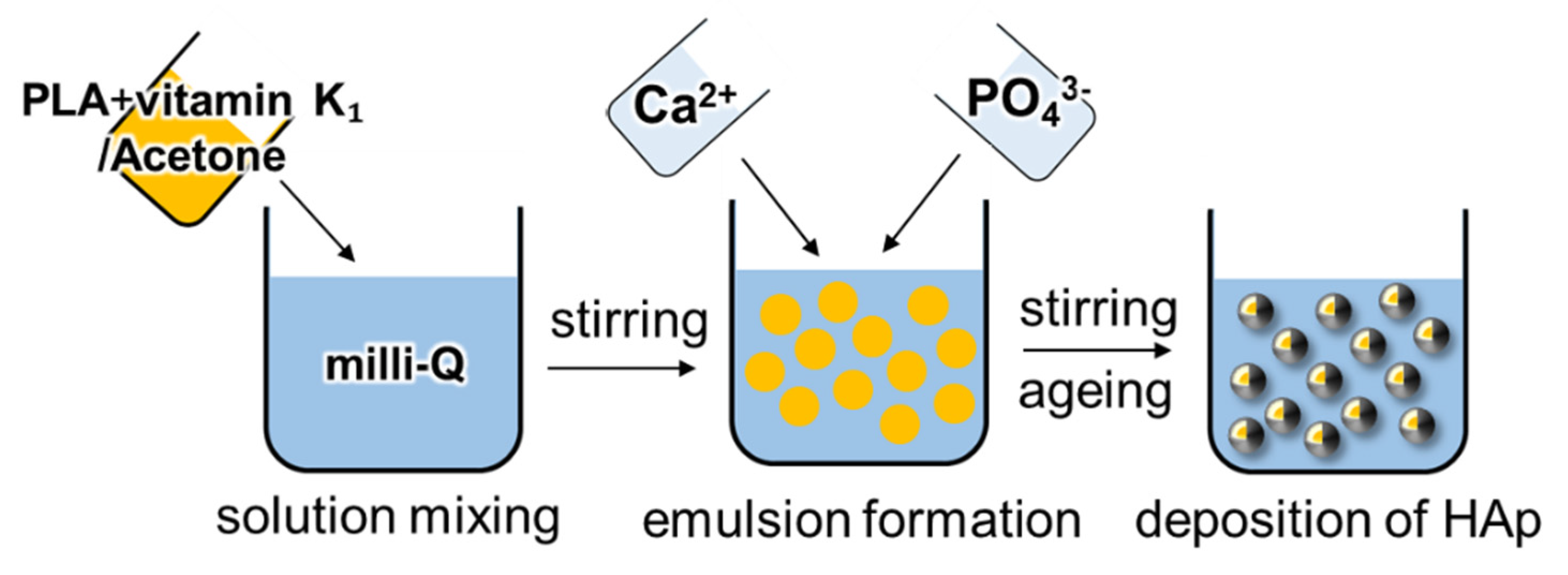
2.2. Preparation of Vitamin K1-Loaded PLA/HAp Core–Shell Particles

Vitamin K1-loaded PLA/HAp core–shell particles were prepared using an emulsification method (Scheme 1). The details of the preparation of the PLA/HAp core–shell particles are described in our previous paper [30,31,32]. In brief, PLA (20 mg) and various amounts of vitamin K1 (0, 50, 100, and 200 mg) were dissolved in 4 mL of acetone (denoted as PLHA-Vx, where x is the amount of vitamin K1, (x = 0, 50, 100, 200)). This solution was then added to 160 mL of ultrapure water. To the resulting mixture, 20 mL of a 20 mM calcium acetate solution and 20 mL of a 12 mM potassium phosphate solution were added under stirring at 25 °C. The resulting solutions were aged for three days. The pH of the solutions was measured before and 1 h after the addition of the phosphate ions and after aging for 72 h (Table 1). The aged solutions were centrifuged (6000 rpm, 10 min). The supernatant was removed, and the particles were washed with ultrapure water. The particles were resuspended in 20 mL of ultrapure water. The residual vitamin K1 in the container was collected by dissolving it in ethanol, and its concentration was measured using ultraviolet–visible absorption spectroscopy (UV–Vis; JASCO Corporation, V-750) over the wavelength range of 200–900 nm (bandwidth: 2.0 nm; scanning speed: 400 nm/min). The absorption spectrum of vitamin K1 was set to 273 nm.

2.3. Characterization of the Vitamin K1-Loaded PLA/HAp Core–Shell Particles

The crystalline phase of PLHA-Vx was evaluated using X-ray diffraction (XRD; Rigaku, Tokyo, Japan). The XRD conditions were as follows: CuKα radiation (40 kV, 30 mA); 1.0 °/min, and a 2θ range of 3–60°. PLHA-Vx was evaluated by the attenuated total reflection (ATR) method using a Fourier transform infrared spectrometer (FT-IR; JASCO Corporation, Tokyo, Japan) in the range of 400–4000 cm−1. The morphology of PLHA-Vx was observed using field emission scanning electron microscopy (FE-SEM; Hitachi, Tokyo, Japan). The PLHA-Vx-containing solutions were dropped onto an aluminum sample stage and dried gently at 37 °C. Subsequently, PLHA-Vx was coated with a layer of amorphous osmium (Meiwafosis, Tokyo, Japan). The diameters of the PLHA-Vx particles were measured using a laser diffraction particle size analyzer (SALD-7500nano, Shimadzu, Kyoto, Japan). Particles weighing 5 mg were dispersed in 100 mL of ultrapure water and sonicated during the measurement. The PLHA-V0 solution was directly dropped onto a carbon grid and dried. The samples were observed using transmission electron microscopy (TEM; JEOL, Tokyo, Japan). The organic/inorganic ratio of PLHA-Vx was evaluated using thermogravimetry–differential thermal analysis (TG–DTA; Rigaku, Tokyo, Japan). PLHA-Vx (5 mg) was used for the analysis, and the same amount of Al2O3 was used as the reference. TG–DTA was performed at room temperature (approximately 25 °C) to 1000 °C (2 °C/min) under air flow (200 mL/min). For the XRD, FT-IR, and TG–DTA measurements, freeze-dried particles were used.

2.4. Drug-Release Test of the Vitamin K1-Loaded PLA/HAp Core–Shell Particles
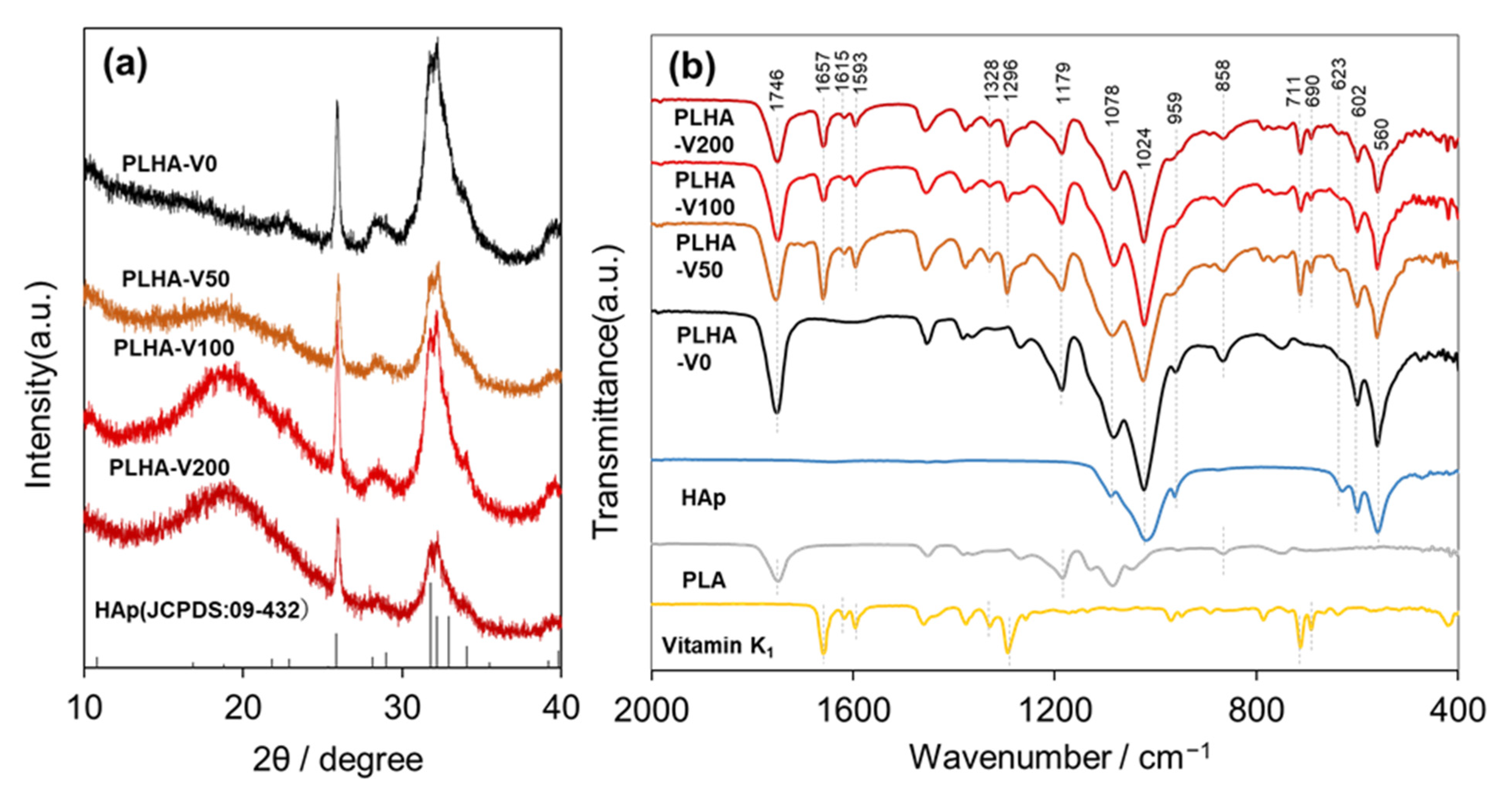
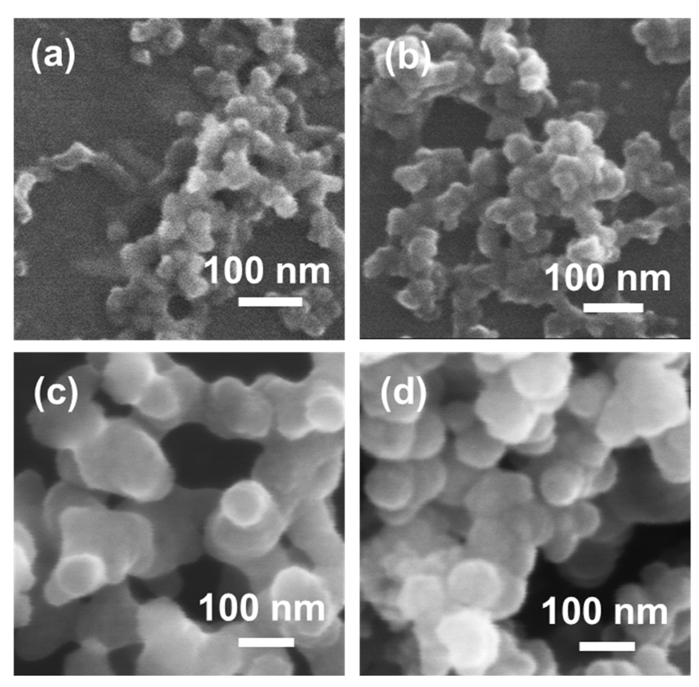
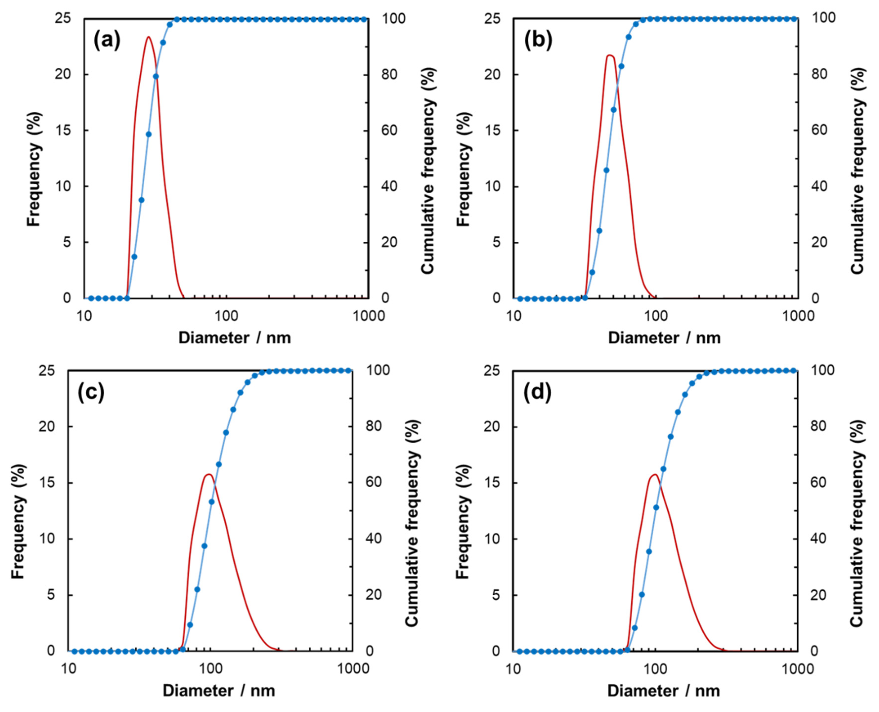
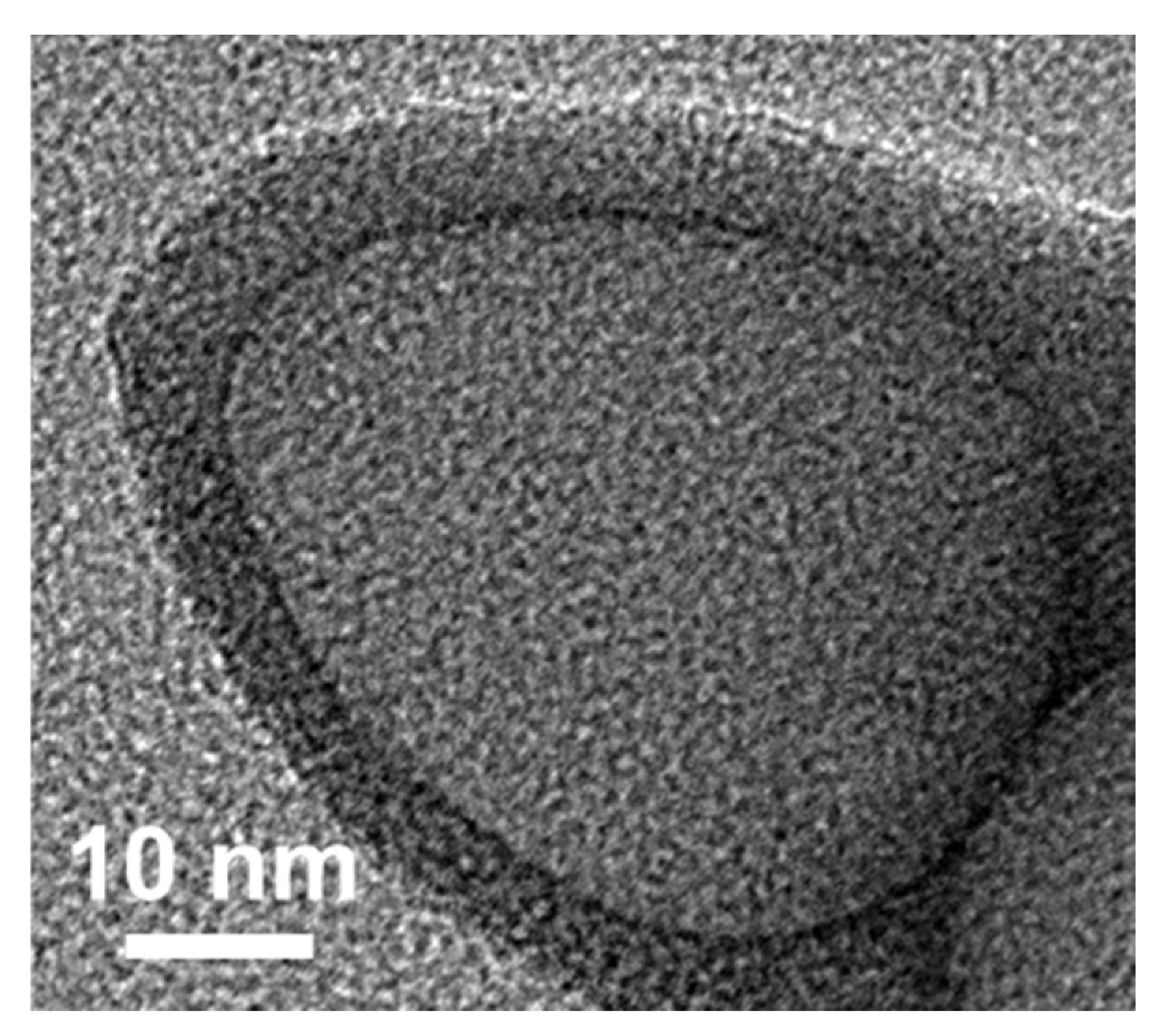
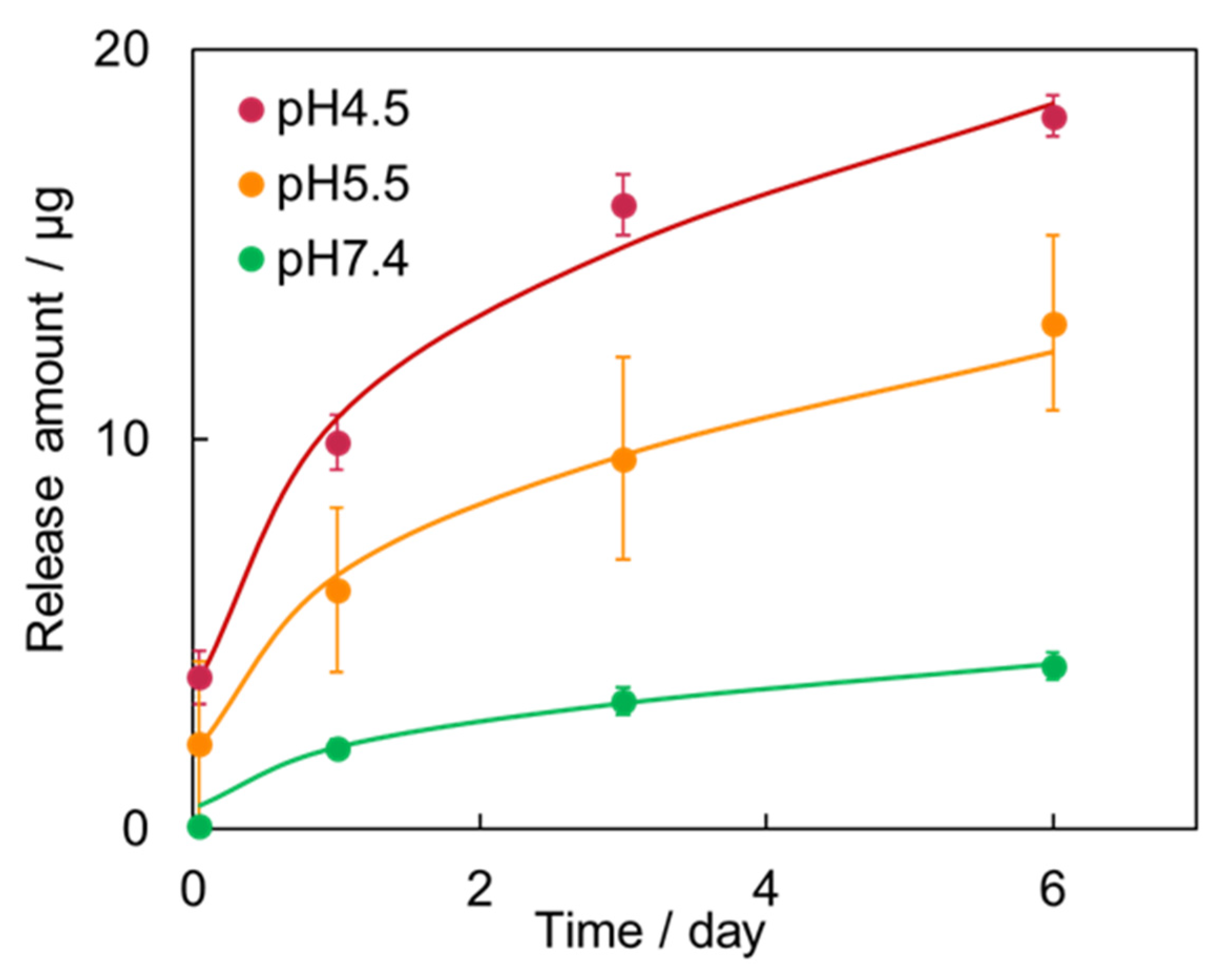
PLHA-V200 was chosen for the drug-release test because it contained the highest amount of drug among all the PLHA-Vx samples investigated. PLHA-V200 was added to phosphate buffer solutions with the pH values of 4.5, 5.5, and 7.4, and 1.88 mg of PLHA-V200 was soaked in 1 mL of the buffer solutions. The solutions were maintained at 37 °C for 1–6 days and stirred during the release test to prevent particle agglomeration. The phosphate buffer solutions (pH 4.5, 5.5, and 7.4) were prepared by mixing 66 mM Na2HPO4 and KH2PO4 in the Na2HPO4:KH2PO4 ratios of 0:20, 1:19, and 16:4 at pH = 4.5, 5.5, and 7.4, respectively. In this work, a protein-free phosphate buffer solution was selected for the drug-release test to investigate the fundamental properties of the PLA/HAp particles. The supernatant of the solution was collected by centrifugation (14,000 rpm) and subjected to Ultraviolet Visible Absorption Spectroscopy analysis (UV–Vis, JASCO Corporation, Tokyo, Japan).

3. Results

During the particle preparation, the pH of the PLHA-Vx solutions decreased after the addition of the phosphate ions (Table 1), indicating the precipitation of calcium phosphate. It is known that the pH decreases with precipitation of HAp [34]. The XRD patterns of PLHA-Vx are shown in Figure 1a. PLHA-Vx exhibited intense peaks at 2θ = 26° and 32°, corresponding to the 002 and 211 planes of HAp (JCPDS: 09-432), respectively. In addition, a halo peak at around 2θ = 18° was observed for PLHA-V50, -V100, and -V200. The FT-IR spectra of the particles are shown in Figure 1b. The bands corresponding to the phosphate group of HAp were the ν2 bending vibration at 469 cm−1; the ν4 bending vibration at 560, 602, and 623 cm−1; the ν1 symmetric stretching vibration at 959 cm−1; and the ν3 asymmetric stretching vibration at 1024 cm−1 [35,36,37]. The carboxyl group of PLA was assigned to the band at 858 cm−1 for the –C–COO stretching vibration, 1078 cm−1 for the COC symmetric stretching vibration, 1179 cm−1 for the COC asymmetric stretching vibration and CH3 asymmetric bending vibration, and 1746 cm−1 for the C=O stretching vibration [38]. In addition, the vitamin K1 bands of PLHA-V50, -V100, and -V200 were located at 1296 and 1328 cm−1 for the –C=C and C–C groups, 1374 cm−1 for the CH3 group, 1457 cm−1 for the CH2 group, 1593 and 1615 cm−1 for the C=C group, and 1657 cm−1 for the C=O group [39].
The PLHA-Vx particles were spherical, as shown in the SEM images in Figure 2. The particle size distribution is shown in Figure 3. The average particle sizes of PLHA-V0, -V50, -V100, and -V200 were 27, 47, 104, and 105 nm, respectively. The particle size was increased with an increasing drug-loading amount. The TEM image of PLHA-V0 is shown in Figure 4. The inner gray sphere and black shell were approximately 40 nm in diameter and 6 nm in thickness, respectively. The percentages of the organic/inorganic components in PLHA-Vx were estimated using TG–DTA (Table 1), where the inorganic component of PLHA-Vx was HAp, and the organic component was the sum of PLA and vitamin K1. The amount of residual vitamin K1 in the container for the preparation of PLHA-V200 was measured to be 109 mg using UV–Vis spectroscopy. The loading amount of vitamin K1 in PLHA-V200 was calculated to be 91 mg.
The vitamin K1-releasing behavior of PLHA-V200 in the phosphate buffer at various pH levels is shown in Figure 5. PLHA-V200 was chosen for the release test because it contained the largest amount of vitamin K1 among all the PLHA-Vx samples investigated. Vitamin K1 was not found to undergo the initial burst release behavior in PLHA-V200 at all pH levels. The amount of vitamin K1 released in the phosphate buffer at pH 4.5, 5.6, and 7.4, was 18, 13, and 5 µg, respectively, and this amount increased as the pH of the phosphate buffer solution decreased.

4. Discussion

The XRD patterns of the PLHA-Vx particles showed peaks corresponding to HAp. PLHA-V50, -V100, and -V200 showed a halo peak centered at 2θ = 18°, which may have originated from vitamin K1. The intensity of this peak increased with an increase in the vitamin K1 content. This peak was confirmed for a mixture of PLHA-V0 and vitamin K1 (Figure S1 in Supplementary Materials). In addition, the XRD peaks of PLHA-Vx were not sharp, which were similar to those of biological HAp [40,41]. The FT-IR spectra of PLHA-Vx showed bands corresponding to the phosphate and carboxyl groups of HAp and PLA, respectively. The bands corresponding to vitamin K1 were observed in the spectra of PLHA-V50, -V100, and -V200, whereas these bands were not observed for PLHA-V0. The hierarchical architecture of PLHA-V0 was composed of a PLA core and a HAp shell, as shown in the TEM image. Additionally, the PLHA-Vx particles were spherical in shape, as confirmed by the SEM images, and no other precipitation was visible. Thus, PLHA-Vx was successfully prepared as a core–shell structure by loading vitamin K1.
The diameters of the PLHA-V0, -V50, -V100, and -V200 particles were 27, 47, 104, and 105 nm, respectively, and the size of particles increased with an increase in the amount of vitamin K1. Additionally, the organic component of PLHA-Vx increased with the increasing amounts of vitamin K1. Hence, the particles expanded with an increase in the amount of drug that was encapsulated.
The loading capacity (LC) of DDS carriers is an indicator of their drug-loading ability and can be calculated using the following equation [25,42,43]:
LC   ( % ) = W d W c × 100
where Wd is the mass of the drug (mg) and Wc is the mass of the DDS carriers (mg). In this work, the LC can be altered as follows.
LC   ( % ) = R V R H + R P × 100
where RV, RH, and RP are the weight ratios of vitamin K1, HAp, and PLA in the particles, respectively. The RH and (RV + RP) ratios were obtained using TG–DTA (Table 2). In the case of PLHA-V200, the RV can be altered as follows:
R V = 84.5 R p
where RH is 15.5, as determined from the TG–DTA results. The weight ratio of RV and RP can be obtained as follows:
R V : R p = W V : W p
where WV and Wp are the weights of vitamin K1 and PLA (mg) in PLHA-V200, respectively. WP is the amount of PLA used for particle preparation, which was 20 mg. WV is the amount of vitamin K1 loaded into PLHA-V200, as obtained from the UV–Vis spectrum, which was 109 mg. RV:RP can be calculated as 71.4:13.1, from the values of WV and WP above. The weight ratio of HAp, PLA, and vitamin K1 in PLHA-V200 is 15.5, 13.1, and 71.4%, respectively. Consequently, the LC value of PLHA-V200 was calculated to be 250%. Zhang et al. reported that polymeric micelles with anticancer agents, which were prepared by exploiting the π–π interactions between biodegradable polymers and disulfide bonds, exhibited an LC value of 18% [22]. Li et al. reported that polymeric micelles with π–π conjugated moieties as lipophilic segments for delivering anticancer agents exhibited an LC value of 15% [23]. Al-Amin et al. reported liposome particles with an LC value of 12% [44]. Therefore, PLHA-Vx exhibited an excellent drug-loading capacity, with an LC value of 250%.
The amount of vitamin K1 released from the particles increased as the pH of the phosphate buffer solution decreased. HAp is poorly soluble under neutral and basic conditions [45] and readily dissolves under acidic conditions. Thus, the HAp shell can easily dissolve at pH 4.5, and therefore, a larger amount of vitamin K1 was released compared with that at pH 7.4. Furthermore, PLHA-Vx showed no initial burst release behavior, and vitamin K1 was released gradually during immersion. The release behavior of PLHA-V200 showed good correlation with the Korsmeyer–Peppas equation in pH 4.5, 5.5, and 7.4 (R2 > 0.95) [46]. These results suggest that PLA/HAp core–shell particles are excellent candidates for DDS carriers with a larger drug-loading capacity and pH sensitivity.

5. Conclusions

PLHA-Vx was prepared, and its drug-loading ability was evaluated. PLHA-Vx was composed of PLA and HAp and was spherical in shape with a diameter of 40–80 nm. The diameter of PLHA-Vx increased with an increase in the amount of vitamin K1. Thus, vitamin K1 was successfully encapsulated in the PLA/HAp particles. The LC value of PLHA-V200 was 250%, which is larger than those reported previously. PLHA-V200 showed pH sensitivity and no initial burst release behavior. Thus, PLHA-Vx is a potential candidate for DDS carriers because of its excellent drug-loading ability and pH sensitivity.

Supplementary Materials

The following are available online at https://www.mdpi.com/article/10.3390/ma14081959/s1, Figure S1: XRD patterns of PLHA-V0 and PLHA-V0 + vitamin K1 mixture.

Author Contributions

Conceptualization, S.L., T.M., K.K., A.S.-N., M.S. and F.N.: methodology; S.S., S.L., K.K. and F.N.: validation; S.S., S.L., T.M., K.K., A.S.-N. and F.N.: formal analysis; S.S., S.L. and F.N.: investigation; S.S., S.L. and F.N.: resources; S.S., S.L. and F.N.: data curation; S.S., S.L. and F.N.: writing—original draft preparation; S.S., S.L. and F.N.: writing—review and editing; S.L. T.M., K.K., A.S.-N., M.S. and F.N.: visualization; S.S., S.L. and F.N.: supervision; S.L. and F.N.: project administration; and F.N.: funding acquisition. All authors have read and agreed to the published version of the manuscript.

Funding

This work was supported by JST A-STEP Grant Number JPMJTS1624 and AMED under Grant Number JP20he0622038.

Institutional Review Board Statement

Not applicable.

Informed Consent Statement

Not applicable.

Data Availability Statement

Data sharing is not applicable to this article.

Acknowledgments

The authors acknowledge T. Matsubara (Nagoya Institute of Technology) for providing TEM analysis of the nanoparticles.

Conflicts of Interest

The authors declare no conflict of interest.

References

  1. Ju, Y.; Guo, H.; Edman, M.; Hamm-Alvarez, S.F. Application of advances in endocytosis and membrane trafficking to drug delivery. Adv. Drug Del. Rev. 2020, 157, 118–141. [Google Scholar] [CrossRef]
  2. Blume, G.; Cevc, G. Liposomes for the sustained drug release in vivo. Biochim. Biophys. Acta 1990, 1029, 91–97. [Google Scholar] [CrossRef]
  3. Nagasaki, Y.; Okada, T.; Scholz, C.; Iijima, M.; Kato, M.; Kataoka, K. The Reactive Polymeric Micelle Based on An Aldehyde-Ended Poly(ethylene glycol)/Poly(lactide) Block Copolymer. Macromolecules 1998, 31, 1473–1479. [Google Scholar] [CrossRef]
  4. Peltonen, L.; Aitta, J.; Hyvönen, S.; Karjalainen, M.; Hirvonen, J. Improved entrapment efficiency of hydrophilic drug substance during nanoprecipitation of poly (I) lactide nanoparticles. AAPS PharmSciTech 2009, 5, 115. [Google Scholar] [CrossRef] [Green Version]
  5. Ogawa, Y.; Okada, H.; Yamamoto, Y.; Shimamoto, T. In Vivo Release Profiles of Leuprolide Acetate from Microcapsules Prepared with Polylactic Acids or Copoly (Lactic/Glycolic) Acids and In Vivo Degradation of These Polymers. Chem. Pharm. Bull. 1988, 36, 2576–2581. [Google Scholar] [CrossRef] [PubMed] [Green Version]
  6. Nagai, Y.; Lee, J.J.; Yamane, H. Hydrolytic degradation of low molecular weigh poly(lactic acid)s and their drug eluting behavior. J. Soc. Mater. Sci. Jpn. 2011, 60, 2–7. [Google Scholar] [CrossRef] [Green Version]
  7. Fredenberg, S.; Wahlgren, M.; Reslow, M.; Axelsson, A. The mechanisms of drug release in poly(lactic-co-glycolic acid)-based drug delivery systems—A review. Int. J. Pharm. 2011, 415, 34–52. [Google Scholar] [CrossRef]
  8. Butoescu, N.; Seemayer, C.A.; Foti, M.; Jordan, O.; Doelker, E. Dexamethasone-containing PLGA superparamagnetic microparticles as carriers for the local treatment of arthritis. Biomaterials 2009, 30, 1772–1780. [Google Scholar] [CrossRef]
  9. Patil, S.D.; Papadmitrakopoulos, F.; Burgess, D.J. Concurrent delivery of dexamethasone and VEGF for localized inflammation control and angiogenesis. J. Control. Release 2007, 117, 68–79. [Google Scholar] [CrossRef]
  10. Torchilin, V. Tumor delivery of macromolecular drugs based on the EPR effect. Adv. Drug Del. Rev. 2011, 63, 131–135. [Google Scholar] [CrossRef]
  11. Matsumura, Y.; Maeda, H. A new concept for macromolecular therapeutics in cancer chemotherapy: Mechanism of tumoritropic accumulation of proteins and the antitumor agent smancs. Cancer Res. 1986, 46, 6387. [Google Scholar] [PubMed]
  12. Maeda, H. SMANCS and polymer-conjugated macromolecular drugs: Advantages in cancer chemotherapy. Adv. Drug Del. Rev. 1991, 6, 181–202. [Google Scholar] [CrossRef]
  13. Hollis, C.P.; Weiss, H.L.; Leggas, M.; Evers, B.M.; Gemeinhart, R.A.; Li, T. Biodistribution and bioimaging studies of hybrid paclitaxel nanocrystals: Lessons learned of the EPR effect and image-guided drug delivery. J. Control. Release 2013, 172, 12–21. [Google Scholar] [CrossRef] [PubMed] [Green Version]
  14. Fang, J.; Nakamura, H.; Maeda, H. The EPR effect: Unique features of tumor blood vessels for drug delivery, factors involved, and limitations and augmentation of the effect. Adv. Drug Del. Rev. 2011, 63, 136–151. [Google Scholar] [CrossRef] [PubMed]
  15. Oyane, A.; Tsurushima, H.; Sogo, Y.; Ito, A.; Mutsuzaki, H. Development of apatite based biomaterials utilizing, biologically functional molecules. New Glass 2009, 24, 35–42. [Google Scholar]
  16. Kim, H.-W.; Knowles, J.C.; Kim, H.-E. Hydroxyapatite/poly(ε-caprolactone) composite coatings on hydroxyapatite porous bone scaffold for drug delivery. Biomaterials 2004, 25, 1279–1287. [Google Scholar] [CrossRef]
  17. dos Apostolos, R.C.R.; Andrade, G.F.; da Silva, W.M.; de Assis Gomes, D.; de Miranda, M.C.; de Sousa, E.M.B. Hybrid polymeric systems of mesoporous silica/hydroxyapatite nanoparticles applied as antitumor drug delivery platform. Int. J. Appl. Ceram. Technol. 2019, 16, 1836–1849. [Google Scholar] [CrossRef]
  18. Kundu, B.; Ghosh, D.; Sinha, M.K.; Sen, P.S.; Balla, V.K.; Das, N.; Basu, D. Doxorubicin-intercalated nano-hydroxyapatite drug-delivery system for liver cancer: An animal model. Ceram. Int. 2013, 39, 9557–9566. [Google Scholar] [CrossRef]
  19. Sun, W.; Fan, J.L.; Wang, S.Z.; Kang, Y.; Du, J.J.; Peng, X.J. Biodegradable Drug-Loaded Hydroxyapatite Nanotherapeutic Agent for Targeted Drug Release in Tumors. ACS Appl. Mater. Interfaces 2018, 10, 7832–7840. [Google Scholar] [CrossRef]
  20. Verma, G.; Shetake, N.G.; Pandrekar, S.; Pandey, B.N.; Hassan, P.A.; Priyadarsini, K.I. Development of surface functionalized hydroxyapatite nanoparticles for enhanced specificity towards tumor cells. Eur. J. Pharm. Sci. 2020, 144, 105206. [Google Scholar] [CrossRef] [PubMed]
  21. Kawabata, Y.; Wada, K.; Nakatani, M.; Yamada, S.; Onoue, S. Formulation design for poorly water-soluble drugs based on biopharmaceutics classification system: Basic approaches and practical applications. Int. J. Pharm. 2011, 420, 1–10. [Google Scholar] [CrossRef] [PubMed]
  22. Zhang, H.; Yan, J.; Mei, H.; Cai, S.; Li, S.; Cheng, F.; Cao, J.; He, B. High-drug-loading capacity of redox-activated biodegradable nanoplatform for active targeted delivery of chemotherapeutic drugs. Regen. Biomater. 2020, 7, 359–369. [Google Scholar] [CrossRef] [PubMed]
  23. Li, Y.; Su, T.; Li, S.; Lai, Y.; He, B.; Gu, Z. Polymeric micelles with π–π conjugated moiety on glycerol dendrimer as lipophilic segments for anticancer drug delivery. Biomater. Sci. 2014, 2, 775–783. [Google Scholar] [CrossRef]
  24. Cheng, F.; Guan, X.; Cao, H.; Su, T.; Cao, J.; Chen, Y.; Cai, M.; He, B.; Gu, Z.; Luo, X. Characteristic of core materials in polymeric micelles effect on their micellar properties studied by experimental and dpd simulation methods. Int. J. Pharm. 2015, 492, 152–160. [Google Scholar] [CrossRef]
  25. Peng, Z.; Li, S.; Han, X.; Al-Youbi, A.O.; Bashammakh, A.S.; El-Shahawi, M.S.; Leblanc, R.M. Determination of the composition, encapsulation efficiency and loading capacity in protein drug delivery systems using circular dichroism spectroscopy. Anal. Chim. Acta 2016, 937, 113–118. [Google Scholar] [CrossRef] [PubMed]
  26. Batrakova, E.V.; Dorodnych, T.Y.; Klinskii, E.Y.; Kliushnenkova, E.N.; Shemchukova, O.B.; Goncharova, O.N.; Arjakov, S.A.; Alakhov, V.Y.; Kabanov, A.V. Anthracycline antibiotics non-covalently incorporated into the block copolymer micelles: In vivo evaluation of anti-cancer activity. Br. J. Cancer 1996, 74, 1545–1552. [Google Scholar] [CrossRef] [PubMed] [Green Version]
  27. Chen, Y.; Chen, H.; Zeng, D.; Tian, Y.; Chen, F.; Feng, J.; Shi, J. Core/Shell Structured Hollow Mesoporous Nanocapsules: A Potential Platform for Simultaneous Cell Imaging and Anticancer Drug Delivery. ACS Nano 2010, 4, 6001–6013. [Google Scholar] [CrossRef]
  28. Danhier, F.; Ansorena, E.; Silva, J.M.; Coco, R.; Le Breton, A.; Préat, V. PLGA-based nanoparticles: An overview of biomedical applications. J. Control. Release 2012, 161, 505–522. [Google Scholar] [CrossRef]
  29. Rao, J.P.; Geckeler, K.E. Polymer nanoparticles: Preparation techniques and size-control parameters. Prog. Polym. Sci. 2011, 36, 887–913. [Google Scholar] [CrossRef]
  30. Nagata, F.; Miyajima, T.; Kato, K. Preparation of phylloquinone-loaded poly(lactic acid)/hydroxyapatite core–shell particles and their drug release behavior. Adv. Powder Technol. 2016, 27, 903–907. [Google Scholar] [CrossRef]
  31. Hanasaki, M.; Nagata, F.; Miyajima, T.; Kato, K. Controlling particle size of poly(lactic acid)/hydroxyapatite nanoparticles. Trans. Mat. Res. Soc. Jpn. 2018, 43, 135–138. [Google Scholar] [CrossRef]
  32. Lee, S.; Miyajima, T.; Sugawara-Narutaki, A.; Kato, K.; Nagata, F. Development of paclitaxel-loaded poly(lactic acid)/hydroxyapatite core–shell nanoparticles as a stimuli-responsive drug delivery system. R. Soc. Open Sci. 2021, 8. [Google Scholar] [CrossRef]
  33. Sato, K.; Kogure, T.; Kumagai, Y.; Tanaka, J. Crystal Orientation of Hydroxyapatite Induced by Ordered Carboxyl Groups. J. Colloid Interface Sci. 2001, 240, 133–138. [Google Scholar] [CrossRef] [PubMed]
  34. Tanizawa, Y.; Sawamura, K.; Suzuki, T. Inhibition of hydroxyapatite formation and growth by condensed phosphate. Chem. Soc. Jpn. 1989, 1989, 1706–1711. [Google Scholar] [CrossRef] [Green Version]
  35. Rehman, I.; Bonfield, W. Characterization of hydroxyapatite and carbonated apatite by photo acoustic FTIR spectroscopy. J. Mater. Sci. Mater. Med. 1997, 8, 1–4. [Google Scholar] [CrossRef] [PubMed]
  36. Maçon, A.L.B.; Lee, S.; Poologasundarampillai, G.; Kasuga, T.; Jones, J.R. Synthesis and dissolution behaviour of CaO/SrO-containing sol–gel-derived 58S glasses. J. Mater. Sci. 2017, 52, 8858–8870. [Google Scholar] [CrossRef]
  37. Lee, S.; Nakano, T.; Kasuga, T. Structure, dissolution behavior, cytocompatibility, and antibacterial activity of silver-containing calcium phosphate invert glasses. J. Biomed. Mater. Res. A 2017, 105, 3127–3135. [Google Scholar] [CrossRef] [PubMed] [Green Version]
  38. Kister, G.; Cassanas, G.; Vert, M. Effects of morphology, conformation and configuration on the IR and Raman spectra of various poly(lactic acid)s. Polymer 1998, 39, 267–273. [Google Scholar] [CrossRef]
  39. Breton, J.; Burie, J.R.; Berthomieu, C.; Berger, G.; Nabedryk, E. The binding sites of quinones in photosynthetic bacterial reaction centers investigated by light-induced FTIR difference spectroscopy: Assignment of the QA vibrations in Rhodobacter sphaeroides using 18O- or 13C-labeled ubiquinone and vitamin K1. Biochemistry 1994, 33, 4953–4965. [Google Scholar] [CrossRef] [PubMed]
  40. Kokubo, T.; Kushitani, H.; Sakka, S.; Kitsugi, T.; Yamamuro, T. Solutions able to reproduce in vivo surface-structure changes in bioactive glass-ceramic A-W. J. Biomed. Mater. Res. 1990, 24, 721–734. [Google Scholar] [CrossRef] [PubMed]
  41. Kuśnieruk, S.; Wojnarowicz, J.; Chodara, A.; Chudoba, T.; Gierlotka, S.; Lojkowski, W. Influence of hydrothermal synthesis parameters on the properties of hydroxyapatite nanoparticles. Beilstein J. Nanotechnol. 2016, 7, 1586–1601. [Google Scholar] [CrossRef] [PubMed] [Green Version]
  42. Wang, S.-B.; Chen, A.-Z.; Weng, L.-J.; Chen, M.-Y.; Xie, X.-L. Effect of Drug-loading Methods on Drug Load, Encapsulation Efficiency and Release Properties of Alginate/Poly-L-Arginine/Chitosan Ternary Complex Microcapsules. Macromol. Biosci. 2004, 4, 27–30. [Google Scholar] [CrossRef]
  43. El-Say, K.M. Maximizing the encapsulation efficiency and the bioavailability of controlled-release cetirizine microspheres using Draper-Lin small composite design. Drug Des. Dev. Ther. 2016, 10. [Google Scholar] [CrossRef] [PubMed] [Green Version]
  44. Al-Amin, M.D.; Bellato, F.; Mastrotto, F.; Garofalo, M.; Malfanti, A.; Salmaso, S.; Caliceti, P. Dexamethasone Loaded Liposomes by Thin-Film Hydration and Microfluidic Procedures: Formulation Challenges. Int. J. Mol. Sci. 2020, 21, 1611. [Google Scholar] [CrossRef] [PubMed] [Green Version]
  45. Brown, W.E.; Patel, P.R.; Chow, L.C. Formation of CaHPO4 2H2O from Enamel Mineral and Its Relationship to Caries Mechanism. J. Dent. Res. 1975, 54, 475–481. [Google Scholar] [CrossRef] [PubMed]
  46. Juhász, Á.; Ungor, D.; Berta, K.; Seres, L.; Csapó, E. Spreadsheet-based nonlinear analysis of in vitro release properties of a model drug from colloidal carriers. J. Mol. Liq. 2021, 328, 115405. [Google Scholar] [CrossRef]
Scheme 1. Preparation of the vitamin K1-loaded PLA/HAp core–shell particle solutions.
Materials 14 01959 sch001
Figure 1. (a) X-ray diffraction (XRD) patterns and (b) Fourier transform infrared (FT-IR) spectra for the PLHA-Vx particles.
Materials 14 01959 g001
Figure 2. Scanning electron microscopy (SEM) images of (a) PLHA-V0, (b) PLHA-V50, (c) PLHA-V100, and (d) PLHA-V200.
Materials 14 01959 g002
Figure 3. Particle diameter distribution of (a) PLHA-V0, (b) PLHA-V50, (c) PLHA-V100, and (d) PLHA-V200. Red solid lines represent the appearance frequency, and blue solid lines represent the cumulative frequency.
Materials 14 01959 g003
Figure 4. TEM image of PLHA-V0.
Materials 14 01959 g004
Figure 5. Vitamin K1-releasing behavior of PLHA-V200 in phosphate buffer at various pH levels. Error bars represent the standard deviation (n = 3). Solid lines represent Korsmeyer–Peppas fitted release profiles.
Materials 14 01959 g005
Table 1. pH values of the PLHA-Vx solutions under different conditions.
Sample CodeAddition of Phosphate IonAfter Aging (72 h)
Before1 h after
PLHA-V07.616.816.19
PLHA-V507.676.866.23
PLHA-V1007.616.806.13
PLHA-V2007.706.886.35
Table 2. Organic/inorganic percentage of PLHA-Vx.
SampleHAp (%)Organic Component (%)
PLHA-V050.349.7
PLHA-V5027.872.2
PLHA-V10024.975.1
PLHA-V20015.584.5
Publisher’s Note: MDPI stays neutral with regard to jurisdictional claims in published maps and institutional affiliations.

