Synthesis of Fe- and Co-Doped TiO2 with Improved Photocatalytic Activity Under Visible Irradiation Toward Carbamazepine Degradation


 ,
, 
Abstract
:1. Introduction
2. Materials and Methods
2.1. Reagents
2.2. Photocatalyst Preparation
2.3. Characterization
2.4. Photocatalytic Activity Experiments
3. Results and Discussion
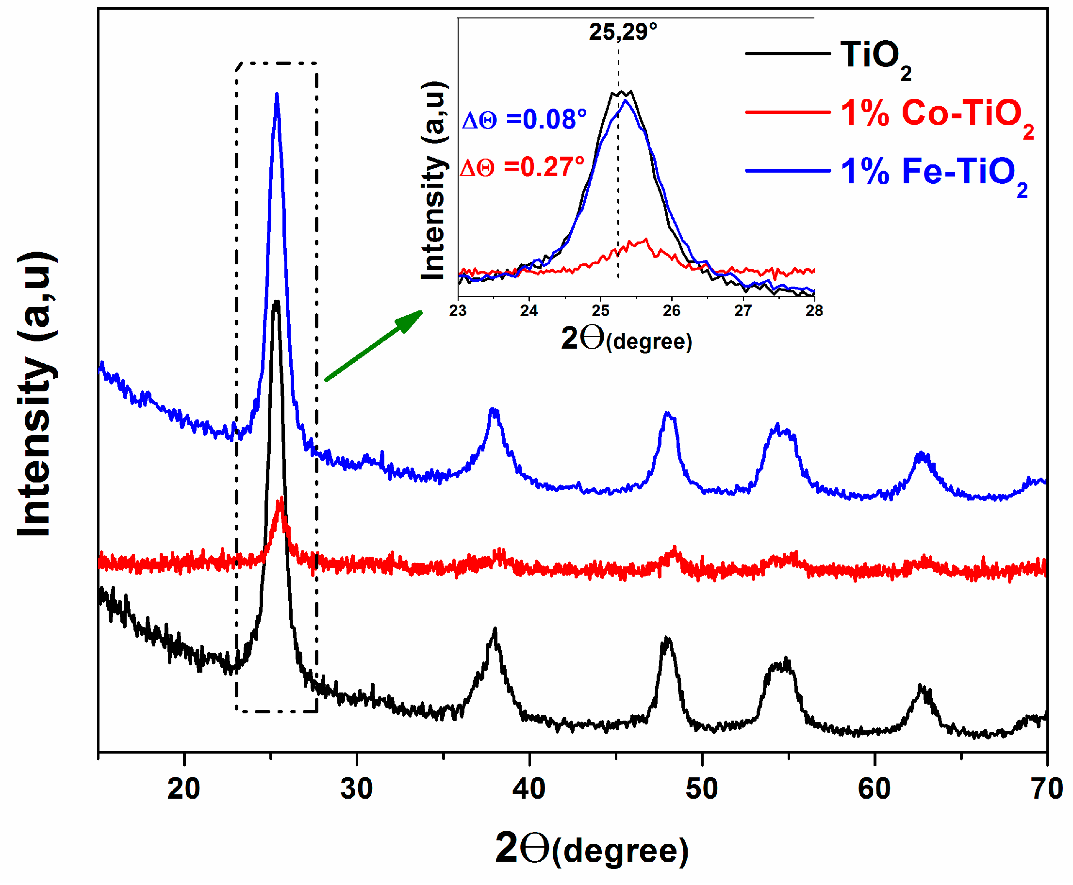
3.1. XRD Analysis
3.2. FTIR Studies
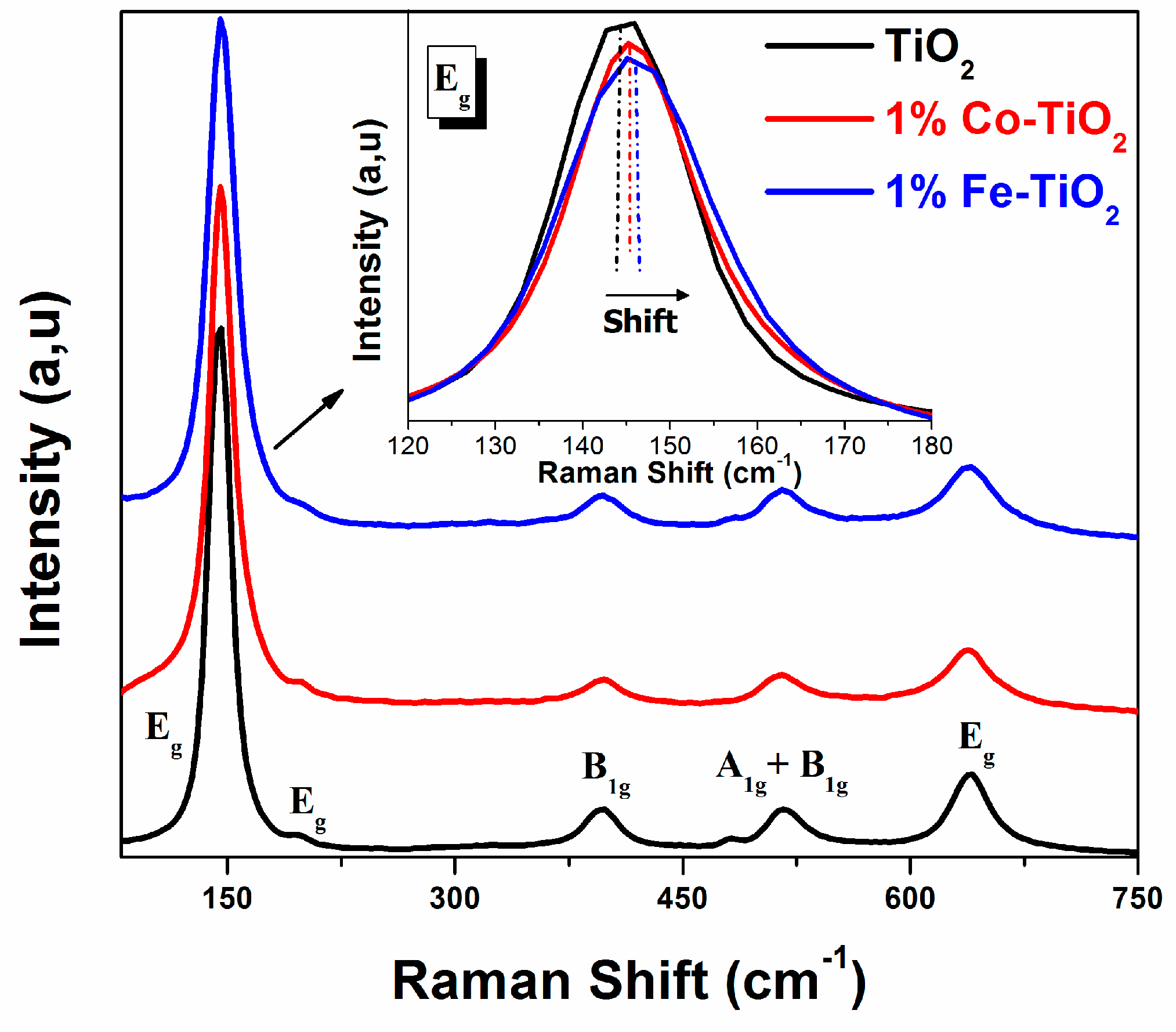
3.3. Raman Studies
3.4. Optical Absorption Studies
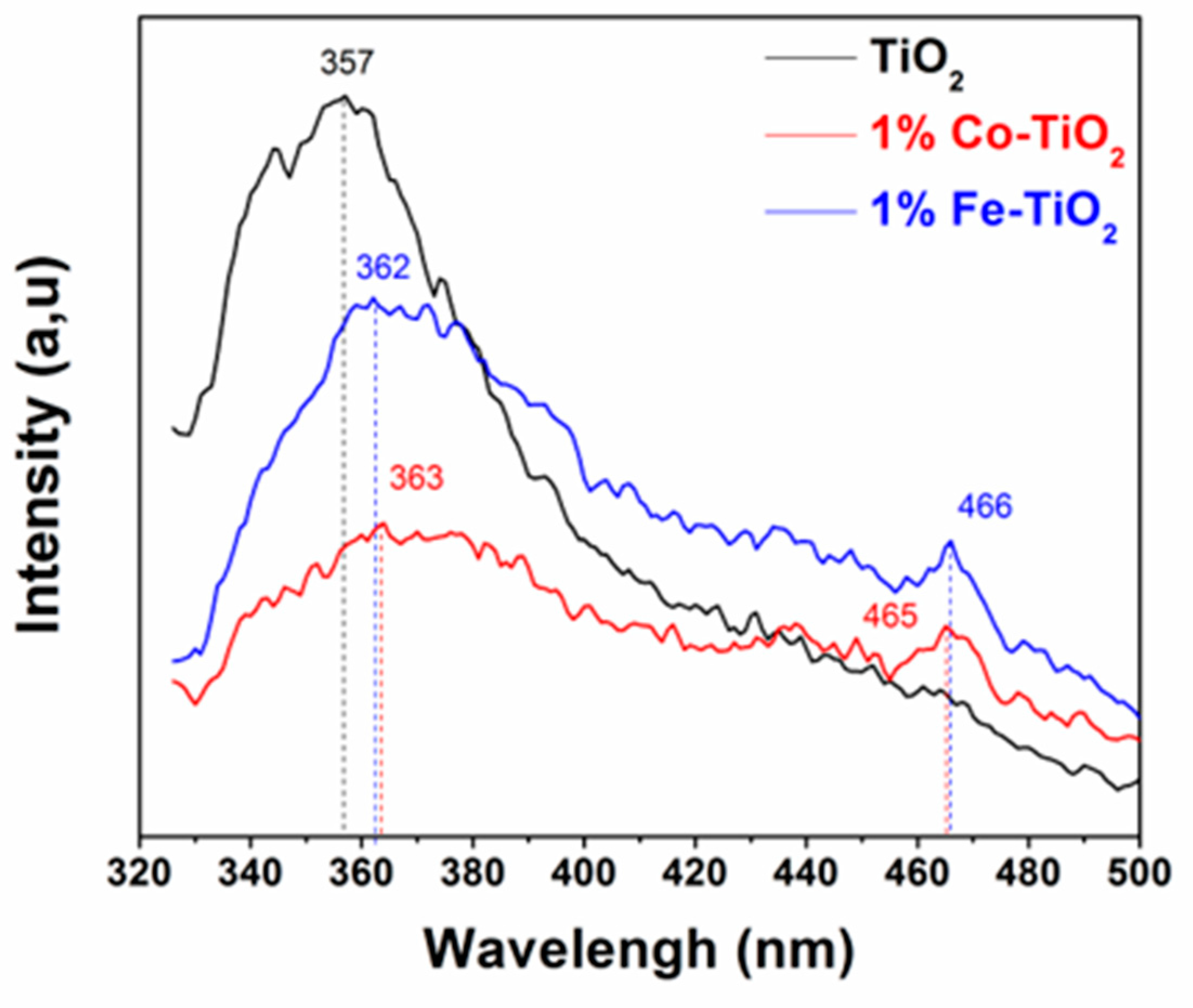
3.5. Photoluminescence Studies
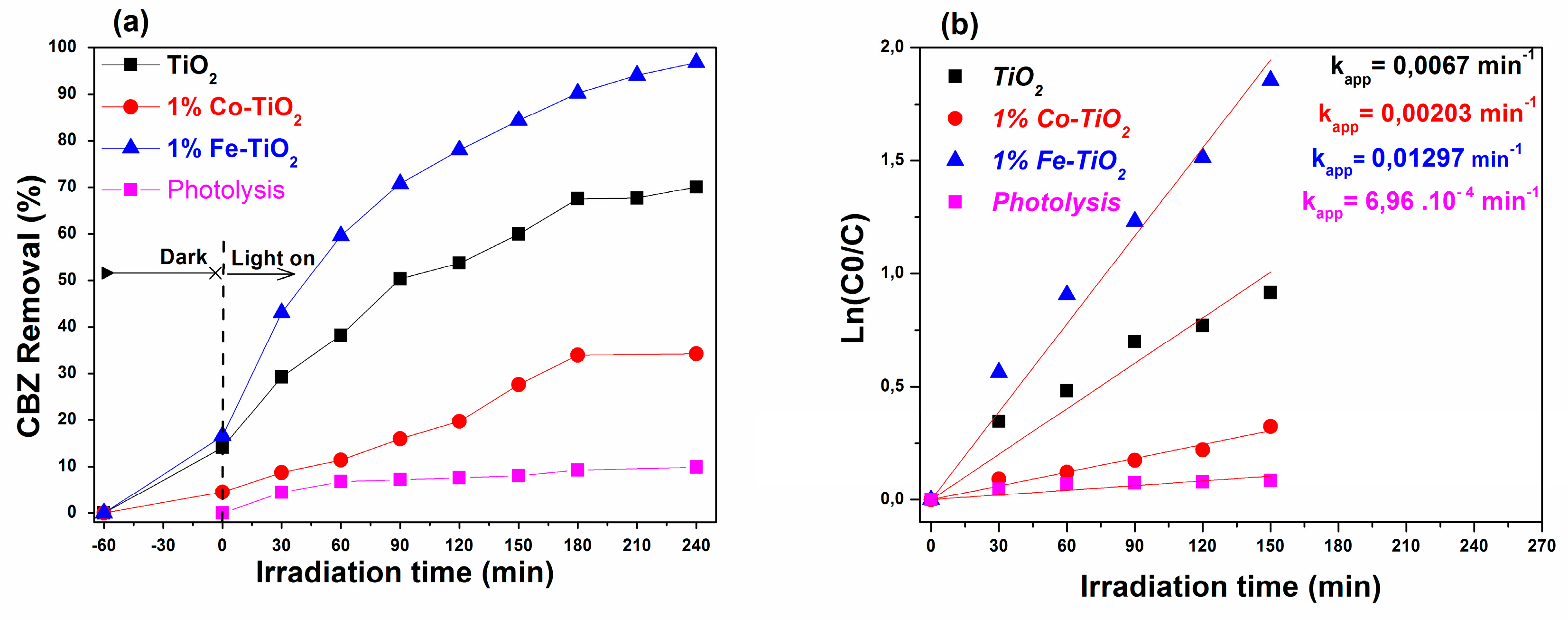
3.6. Photocatalytic Performance
3.6.1. UV-A Light Irradiation
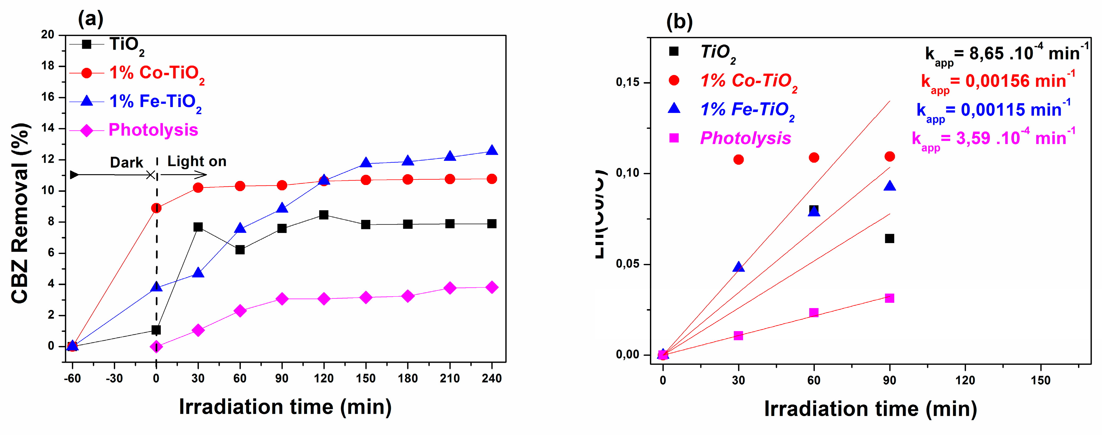
3.6.2. Visible-Light Irradiation
4. Conclusion
Author Contributions
Funding
Acknowledgments
Conflicts of Interest
References
- Mohapatra, D.P.; Brar, S.K.; Daghrir, R.; Tyagi, R.D.; Picard, P.; Surampalli, R.Y.; Drogui, P. Photocatalytic degradation of carbamazepine in wastewater by using a new class of whey-stabilized nanocrystalline TiO2 and ZnO. Sci. Total Environ. 2014, 485–486, 263–269. [Google Scholar] [CrossRef] [PubMed]
- Liu, Z.; Zhou, X.; Chen, X.; Dai, C.; Zhang, J.; Zhang, Y. Biosorption of clofibric acid and carbamazepine in aqueous solution by agricultural waste rice straw. J. Environ. Sci. (China) 2013, 25, 2384–2395. [Google Scholar] [CrossRef]
- He, Y.; Sutton, N.B.; Rijnaarts, H.H.H.; Langenhoff, A.A.M. Degradation of pharmaceuticals in wastewater using immobilized TiO2 photocatalysis under simulated solar irradiation. Appl. Catal. B Environ. 2016, 182, 132–141. [Google Scholar] [CrossRef]
- Miao, X.S.; Metcalfe, C.D. Determination of carbamazepine and its metabolites in aqueous samples using liquid chromatography—Electrospray tandem mass spectrometry. Anal. Chem. 2003, 75, 3731–3738. [Google Scholar] [CrossRef] [PubMed]
- Teo, H.L.; Wong, L.; Liu, Q.; Teo, T.L.; Lee, T.K.; Lee, H.K. Simple and accurate measurement of carbamazepine in surface water by use of porous membrane-protected micro-solid-phase extraction coupled with isotope dilution mass spectrometry. Anal. Chim. Acta 2016, 912, 49–57. [Google Scholar] [CrossRef]
- Leucht, S.; Helfer, B.; Dold, M.; Kissling, W.; Mcgrath, J. Carbamazepine for schizophrenia. Cochrane Database Syst. Rev. 2014, 2014. [Google Scholar] [CrossRef]
- Kinney, C.A.; Furlong, E.T.; Zaugg, S.D.; Burkhardt, M.R.; Werner, S.L.; Cahill, J.D.; Jorgensen, G.R. Survey of Organic Wastewater Contaminants in Biosolids Destined for Land Application. Environ. Sci. Technol. 2006, 40, 7207–7215. [Google Scholar] [CrossRef]
- Ferrari, B.; Paxéus, N.; Giudice, R.L.; Pollio, A.; Garric, J. Ecotoxicological impact of pharmaceuticals found in treated wastewaters: Study of carbamazepine, clofibric acid, and diclofenac. Ecotoxicol. Environ. Saf. 2003, 55, 359–370. [Google Scholar] [CrossRef]
- Donner, E.; Kosjek, T.; Qualmann, S.; Kusk, K.O.; Heath, E.; Revitt, D.M.; Ledin, A.; Andersen, H.R. Ecotoxicity of carbamazepine and its UV photolysis transformation products. Sci. Total Environ. 2013, 443, 870–876. [Google Scholar] [CrossRef]
- Zhou, H.; Liu, X.; Chen, X.; Ying, T.; Ying, Z. Characteristics of removal of waste-water marking pharmaceuticals with typical hydrophytes in the urban rivers. Sci. Total Environ. 2018, 636, 1291–1302. [Google Scholar] [CrossRef]
- Jaria, G.; Silva, C.P.; Oliveira, J.A.B.P.; Santos, S.M.; Gil, M.V.; Otero, M.; Calisto, V.; Esteves, V.I. Production of highly efficient activated carbons from industrial wastes for the removal of pharmaceuticals from water—A full factorial design. J. Hazard. Mater. 2019, 212–218. [Google Scholar] [CrossRef] [PubMed]
- Carabin, A.; Drogui, P.; Robert, D. Photo-degradation of carbamazepine using TiO2 suspended photocatalysts. J. Taiwan Inst. Chem. Eng. 2015, 54, 109–117. [Google Scholar] [CrossRef]
- Wu, J.; Cagnetta, G.; Wang, B.; Cui, Y.; Deng, S.; Wang, Y.; Huang, J.; Yu, G. Efficient degradation of carbamazepine by organo-montmorillonite supported nCoFe2 O4 -activated peroxymonosulfate process. Chem. Eng. J. 2019, 824–836. [Google Scholar] [CrossRef]
- Kashale, A.A.; Rasal, A.S.; Kamble, G.P.; Ingole, V.H.; Dwivedi, P.K.; Rajoba, S.J.; Jadhav, L.D.; Ling, Y.-C.; Chang, J.-Y.; Ghule, A.V. Biosynthesized Co-doped TiO2 nanoparticles based anode for lithium-ion battery application and investigating the influence of dopant concentrations on its performance. Compos. Part B Eng. 2019, 167, 44–50. [Google Scholar] [CrossRef]
- Wan, H.; Yao, W.; Zhu, W.; Tang, Y.; Ge, H.; Shi, X.; Duan, T. Fe-N co-doped SiO2@TiO2 yolk-shell hollow nanospheres with enhanced visible light photocatalytic degradation. Appl. Surf. Sci. 2018, 444, 355–363. [Google Scholar] [CrossRef]
- D’Amato, C.; Giovannetti, R.; Zannotti, M.; Rommozzi, E.; Minicucci, M.; Gunnella, R.; Di Cicco, A. Band Gap Implications on Nano-TiO2 Surface Modification with Ascorbic Acid for Visible Light-Active Polypropylene Coated Photocatalyst. Nanomaterials 2018, 8, 599. [Google Scholar] [CrossRef] [PubMed]
- Liu, J.; Li, Y.; Ke, J.; Wang, S.; Wang, L.; Xiao, H. Black NiO-TiO2 nanorods for solar photocatalysis: Recognition of electronic structure and reaction mechanism. Appl. Catal. B Environ. 2018, 224, 705–714. [Google Scholar] [CrossRef]
- El Mragui, A.; Daou, I.; Zegaoui, O. Influence of the preparation method and ZnO/(ZnO + TiO2) weight ratio on the physicochemical and photocatalytic properties of ZnO-TiO2nanomaterials. Catal. Today 2018, 29–34. [Google Scholar]
- Niu, X.; Yan, W.; Shao, C.; Zhao, H.; Yang, J. Hydrothermal synthesis of Mo-C co-doped TiO2 and coupled with fluorine-doped tin oxide (FTO) for high-efficiency photodegradation of methylene blue and tetracycline: Effect of donor-acceptor passivated co-doping. Appl. Surf. Sci. 2019, 466, 882–892. [Google Scholar] [CrossRef]
- Raza, W.; Haque, M.M.; Muneer, M.; Fleisch, M.; Hakki, A.; Bahnemann, D. Photocatalytic degradation of different chromophoric dyes in aqueous phase using la and Mo doped TiO2 hybrid carbon spheres. J. Alloy. Compd. 2015, 632, 837–844. [Google Scholar] [CrossRef]
- Wang, E.; Yang, W.; Cao, Y. Unique surface chemical species on indium doped TiO2 and their effect on the visible light photocatalytic activity. J. Phys. Chem. C 2009, 113, 20912–20917. [Google Scholar] [CrossRef]
- Chen, J.; Qiu, F.; Xu, W.; Cao, S.; Zhu, H. Recent progress in enhancing photocatalytic efficiency of TiO2-based materials. Appl. Catal. A Gen. 2015, 495, 131–140. [Google Scholar] [CrossRef]
- Lv, T.; Zhao, J.; Chen, M.; Shen, K.; Zhang, D.; Zhang, J.; Zhang, G.; Liu, Q. Boosted Visible-Light Photodegradation of Methylene Blue by V and Co Co-Doped TiO2. Materials 2018, 11, 1946. [Google Scholar] [CrossRef] [PubMed]
- Ma, X.; Zhou, W.; Chen, Y. Structure and Photocatalytic Properties of Mn-Doped TiO2 Loaded on Wood-Based Activated Carbon Fiber Composites. Materials 2017, 10, 631. [Google Scholar]
- El Mragui, A.; Zegaoui, O.; Daou, I.; Esteves da Silva, J.C.G. Preparation, characterization, and photocatalytic activity under UV and visible light of Co, Mn, and Ni mono-doped and (P,Mo) and (P,W) co-doped TiO2 nanoparticles: A comparative study. Environ. Sci. Pollut. Res. 2019. [Google Scholar] [CrossRef]
- El Mragui, A.; Zegaoui, O.; Daou, I. Synthesis, characterization and photocatalytic properties under visible light of doped and co-doped TiO2-based nanoparticles. Mater. Today Proc. 2019, 13, 857–865. [Google Scholar] [CrossRef]
- Ilie, A.G.; Scarisoareanu, M.; Morjan, I.; Dutu, E.; Badiceanu, M.; Mihailescu, I. Principal component analysis of Raman spectra for TiO2 nanoparticle characterization. Appl. Surf. Sci. 2017, 417, 93–103. [Google Scholar] [CrossRef]
- Adyani, S.M.; Ghorbani, M. A comparative study of physicochemical and photocatalytic properties of visible light responsive Fe, Gd and P single and tri-doped TiO2 nanomaterials. J. Rare Earths 2018, 36, 72–85. [Google Scholar] [CrossRef]
- Luu, C.L.; Nguyen, Q.T.; Ho, S.T. Synthesis and characterization of Fe-doped TiO2 photocatalyst by the sol-gel method. Adv. Nat. Sci. Nanosci. Nanotechnol. 2010, 1, 0–5. [Google Scholar] [CrossRef]
- Komaraiah, D.; Radha, E.; Kalarikkal, N.; Sivakumar, J.; Ramana Reddy, M.V.; Sayanna, R. Structural, optical and photoluminescence studies of sol-gel synthesized pure and iron doped TiO2 photocatalysts. Ceram. Int. 2019, 45, 25060–25068. [Google Scholar] [CrossRef]
- Asiltürk, M.; Sayılkan, F.; Arpaç, E. Effect of Fe3+ ion doping to TiO2 on the photocatalytic degradation of Malachite Green dye under UV and vis-irradiation. J. Photochem. Photobiol. A Chem. 2009, 203, 64–71. [Google Scholar] [CrossRef]
- Gaur, L.K.; Kumar, P.; Kushavah, D.; Khiangte, K.R.; Mathpal, M.C.; Agrahari, V.; Gairola, S.P.; Soler, M.A.G.; Swart, H.C.; Agarwal, A. Laser induced phase transformation influenced by Co doping in TiO2 nanoparticles. J. Alloys Compd. 2019, 780, 25–34. [Google Scholar] [CrossRef]
- Zhu, J.; Chen, F.; Zhang, J.; Chen, H.; Anpo, M. Fe3+-TiO2 photocatalysts prepared by combining sol–gel method with hydrothermal treatment and their characterization. J. Photochem. Photobiol. A Chem. 2006, 180, 196–204. [Google Scholar] [CrossRef]
- Khan, M.; Cao, W. Cationic (V, Y)-codoped TiO2 with enhanced visible light induced photocatalytic activity: A combined experimental and theoretical study. J. Appl. Phys. 2013, 114, 183514. [Google Scholar] [CrossRef]
- Burton, A.W.; Ong, K.; Rea, T.; Chan, I.Y. On the estimation of average crystallite size of zeolites from the Scherrer equation: A critical evaluation of its application to zeolites with one-dimensional pore systems. Microporous Mesoporous Mater. 2009, 117, 75–90. [Google Scholar] [CrossRef]
- Siddhapara, K.; Shah, D. Characterization of nanocrystalline cobalt doped TiO2 sol–gel material. J. Cryst. Growth 2012, 352, 224–228. [Google Scholar] [CrossRef]
- Dahlan, D.; Md Saad, S.K.; Berli, A.U.; Bajili, A.; Umar, A.A. Synthesis of two-dimensional nanowall of Cu-Doped TiO2 and its application as photoanode in DSSCs. Phys. E Low-Dimens. Syst. Nanostruct. 2017, 91, 185–189. [Google Scholar] [CrossRef]
- Choudhury, B.; Choudhury, A. Luminescence characteristics of cobalt doped TiO2 nanoparticles. J. Lumin. 2012, 132, 178–184. [Google Scholar] [CrossRef]
- Majeed Khan, M.A.; Siwach, R.; Kumar, S.; Alhazaa, A.N. Role of Fe doping in tuning photocatalytic and photoelectrochemical properties of TiO2 for photodegradation of methylene blue. Opt. Laser Technol. 2019, 118, 170–178. [Google Scholar] [CrossRef]
- Alamgir; Khan, W.; Ahmad, S.; Mehedi Hassan, M.; Naqvi, A.H. Structural phase analysis, band gap tuning and fluorescence properties of Co doped TiO2 nanoparticles. Opt. Mater. (Amst.) 2014, 38, 278–285. [Google Scholar] [CrossRef]
- Wang, J.A.; Limas-Ballesteros, R.; López, T.; Moreno, A.; Gómez, R.; Novaro, O.; Bokhimi, X. Quantitative Determination of Titanium Lattice Defects and Solid-State Reaction Mechanism in Iron-Doped TiO2 Photocatalysts. J. Phys. Chem. B 2001, 105, 9692–9698. [Google Scholar] [CrossRef]
- Pirzada, B.M.; Mehraj, O.; Bhat, S.A.; Sabir, S. Efficient visible-light-driven Photocatalytic activity and enhanced charge transfer properties over Mo-doped WO3/TiO2 nanocomposites. J. Environ. Chem. Eng. 2018, 6, 3204–3212. [Google Scholar] [CrossRef]
- Das, K.; Sharma, S.N.; Kumar, M.; De, S.K. Morphology dependent luminescence properties of Co doped TiO2 nanostructures. J. Phys. Chem. C 2009, 113, 14783–14792. [Google Scholar] [CrossRef]
- Kanmani, S.S.; Ramachandran, K. Synthesis and characterization of TiO2/ZnO core/shell nanomaterials for solar cell applications. Renew Energy 2012, 43, 149–156. [Google Scholar] [CrossRef]
- Fagan, R.; McCormack, D.; Hinder, S.; Pillai, S. Photocatalytic Properties of g-C3N4–TiO2 Heterojunctions under UV and Visible Light Conditions. Materials 2016, 9, 286. [Google Scholar] [CrossRef]
- Zhang, W.F.; He, Y.L.; Zhang, M.S.; Yin, Z.; Chen, Q. Raman scattering study on anatase TiO2 nanocrystals. J. Phys. D Appl. Phys 2000, 33, 912–916. [Google Scholar] [CrossRef]
- Choi, H.C.; Jung, Y.M.; Kim, S. Bin Size effects in the Raman spectra of TiO2 nanoparticles. Vib. Spectrosc. 2005, 37, 33–38. [Google Scholar] [CrossRef]
- Garza-Arévalo, J.I.; García-Montes, I.; Reyes, M.H.; Guzmán-Mar, J.L.; Rodríguez-González, V.; Reyes, L.H. Fe doped TiO2 photocatalyst for the removal of As (III) under visible radiation and its potential application on the treatment of As-contaminated groundwater. Mater. Res. Bull. 2016, 73, 145–152. [Google Scholar] [CrossRef]
- Lu, J.G.; Fujita, S.; Kawaharamura, T.; Nishinaka, H.; Kamada, Y.; Ohshima, T.; Ye, Z.Z.; Zeng, Y.J.; Zhang, Y.Z.; Zhu, L.P.; et al. Carrier concentration dependence of band gap shift in n-type ZnO:Al films. J. Appl. Phys. 2007, 101. [Google Scholar] [CrossRef]
- Fabbiyola, S.; Sailaja, V.; Kennedy, L.J.; Bououdina, M.; Judith Vijaya, J. Optical and magnetic properties of Cu-doped ZnO nanoparticles. J. Alloys Compd. 2017, 694, 522–531. [Google Scholar] [CrossRef]
- Bakhshayesh, A.M.; Bakhshayesh, N. Enhanced short circuit current density of dye-sensitized solar cells aided by Sr, V co-doped TiO2 particles. Mater. Sci. Semicond. Process. 2016, 41, 92–101. [Google Scholar] [CrossRef]
- Dholam, R.; Patel, N.; Miotello, A. Efficient H2 production by water-splitting using indium–tin-oxide/V-doped TiO2 multilayer thin film photocatalyst. Int. J. Hydrogen Energy 2011, 36, 6519–6528. [Google Scholar] [CrossRef]
- Shahzad, A.; Rasool, K.; Nawaz, M.; Miran, W.; Jang, J.; Moztahida, M.; Mahmoud, K.A.; Lee, D.S. Heterostructural TiO2/Ti3C2Tx (MXene) for photocatalytic degradation of antiepileptic drug carbamazepine. Chem. Eng. J. 2018, 349, 748–755. [Google Scholar] [CrossRef]
- Murugadoss, G.; Jayavel, R.; Rajesh Kumar, M. Systematic investigation of structural and morphological studies on doped TiO2 nanoparticles for solar cell applications. Superlattices Microstruct. 2014, 76, 349–361. [Google Scholar] [CrossRef]
- Joshi, K.; Rawat, M.; Gautam, S.K.; Singh, R.G.; Ramola, R.C.; Singh, F. Band gap widening and narrowing in Cu-doped ZnO thin films. J. Alloys Compd. 2016, 680, 252–258. [Google Scholar] [CrossRef]
- Kaleji, B.K.; Sarraf-Mamoory, R.; Fujishima, A. Influence of Nb dopant on the structural and optical properties of nanocrystalline TiO2 thin films. Mater. Chem. Phys. 2012, 132, 210–215. [Google Scholar] [CrossRef]
- Rana, M.P.S.; Singh, F.; Negi, S.; Gautam, S.K.; Singh, R.G.; Ramola, R.C. Band gap engineering and low temperature transport phenomenon in highly conducting antimony doped tin oxide thin films. Ceram. Int. 2016, 42, 5932–5941. [Google Scholar] [CrossRef]
- Burstein, E. Anomalous optical absorption limit in InSb. Phys. Rev. 1954, 93, 632–633. [Google Scholar] [CrossRef]
- Li, G.H.; Yang, L.; Jin, Y.X.; Zhang, L.D. Structural and optical properties of TiO2 thin film and TiO2+2 wt.% ZnFe2O4 composite film prepared by r.f. sputtering. Thin Solid Film. 2000, 368, 163–167. [Google Scholar] [CrossRef]
- DeLoach, J.D.; Scarel, G.; Aita, C.R. Correlation between Titania film structure and near ultraviolet optical absorption. J. Appl. Phys. 1999, 85, 2377–2384. [Google Scholar] [CrossRef]
- Vorontsov, A.V.; Valdés, H. Quantum size effect and visible light activity of anatase nanosheet quantum dots. J. Photochem. Photobiol. A Chem. 2019, 379, 39–46. [Google Scholar] [CrossRef]
- Iwamoto, M.; Abe, T.; Tachibana, Y. Control of bandgap of iron oxide through its encapsulation into SiO2-based mesoporous materials. J. Mol. Catal. A Chem. 2000, 155, 143–153. [Google Scholar] [CrossRef]
- Udayabhaskar, R.; Mangalaraja, R.V.; Sahlevani, S.F.; Perarasu, V.T.; Karthikeyan, B.; Contreras, D.; Gracia-Pinilla, M.A. Graphene induced band gap widening and luminescence quenching in ceria:graphene nanocomposites. J. Alloys Compd. 2019, 770, 1221–1228. [Google Scholar] [CrossRef]
- Wang, Y.; Herron, N. Quantum size eSects on the exciton energy ofCdS clusters Ying. Phys. Rev. B 1990, 42, 7253–7255. [Google Scholar] [CrossRef]
- Cao, J.; Nie, W.; Huang, L.; Ding, Y.; Lv, K.; Tang, H. Photocatalytic activation of sulfite by nitrogen vacancy modified graphitic carbon nitride for efficient degradation of carbamazepine. Appl. Catal. B Environ. 2019, 241, 18–27. [Google Scholar] [CrossRef]
- Yu, J.; Yu, H.; Ao, C.H.; Lee, S.C.; Yu, J.C.; Ho, W. Preparation, characterization and photocatalytic activity of in situ Fe-doped TiO2 thin films. Thin Solid Film. 2006, 496, 273–280. [Google Scholar] [CrossRef]
- D’Amato, C.A.; Giovannetti, R.; Zannotti, M.; Rommozzi, E.; Ferraro, S.; Seghetti, C.; Minicucci, M.; Gunnella, R.; Di Cicco, A. Enhancement of visible-light photoactivity by polypropylene coated plasmonic Au/TiO2 for dye degradation in water solution. Appl. Surf. Sci. 2018, 441, 575–587. [Google Scholar] [CrossRef]
- Choudhury, B.; Choudhury, A. Oxygen vacancy and dopant concentration dependent magnetic properties of Mn doped TiO2 nanoparticle. Curr. Appl. Phys. 2013, 13, 1025–1031. [Google Scholar] [CrossRef]
- Diak, M.; Klein, M.; Klimczuk, T.; Lisowski, W.; Remita, H.; Zaleska-Medynska, A.; Grabowska, E. Photoactivity of decahedral TiO2 loaded with bimetallic nanoparticles: Degradation pathway of phenol-1- 13 C and hydroxyl radical formation. Appl. Catal. B Environ. 2017, 200, 56–71. [Google Scholar] [CrossRef]
- Srinivasulu, T.; Saritha, K.; Reddy, K.T.R. Synthesis and characterization of Fe-doped ZnO thin films deposited by chemical spray pyrolysis. Mod. Electron. Mater. 2017, 3, 76–85. [Google Scholar] [CrossRef]
- Jia, T.; Fu, F.; Yu, D.; Cao, J.; Sun, G. Facile synthesis and characterization of N-doped TiO2 /C nanocomposites with enhanced visible-light photocatalytic performance. Appl. Surf. Sci. 2018, 430, 438–447. [Google Scholar] [CrossRef]
- Lin, L.; Wang, H.; Jiang, W.; Mkaouar, A.R.; Xu, P. Comparison study on photocatalytic oxidation of pharmaceuticals by TiO2-Fe and TiO2-reduced graphene oxide nanocomposites immobilized on optical fibers. J. Hazard. Mater. 2017, 333, 162–168. [Google Scholar] [CrossRef] [PubMed]
- Liqiang, J.; Yichun, Q.; Baiqi, W.; Shudan, L.; Baojiang, J.; Libin, Y.; Wei, F.; Honggang, F.; Jiazhong, S. Review of photoluminescence performance of nano-sized semiconductor materials and its relationships with photocatalytic activity. Sol. Energy Mater. Sol. Cells 2006, 90, 1773–1787. [Google Scholar] [CrossRef]
- Gao, T.; Chen, Z.; Zhu, Y.; Niu, F.; Huang, Q.; Qin, L.; Sun, X.; Huang, Y. Synthesis of BiFeO3 nanoparticles for the visible-light induced photocatalytic property. Mater. Res. Bull. 2014, 59, 6–12. [Google Scholar] [CrossRef]
- Akurati, K.K.; Vital, A.; Fortunato, G.; Hany, R.; Nueesch, F.; Graule, T. Flame synthesis of TiO2 nanoparticles with high photocatalytic activity. Solid State Sci. 2007, 9, 247–257. [Google Scholar] [CrossRef]


| Samples | Peak (101) Position 2θ (°) | dhkl | Average Crystallite Size (nm) |
|---|---|---|---|
| TiO2 | 25.29 | 0.98 | 7.45 |
| 1 wt.% Fe-TiO2 | 25.37 | 0.74 | 7.40 |
| 1 wt.% Co-TiO2 | 25.56 | 0.33 | 7.78 |
| Samples | Egap | UV-A Light | Visible Light | ||||
|---|---|---|---|---|---|---|---|
| MO Removal (%) | Rate Constant, k (min−1) | R2 | MO Removal (%) | Rate Constant, k (min−1) | R2 | ||
| TiO2 | 3.12 | 70.06 | 0.0067 | 0.97 | 7.88 | 8.64 × 10⁻⁴ | 0.84 |
| 1 wt.% Fe-TiO2 | 3.32 | 96.9 | 0.01297 | 0.99 | 12.54 | 0.00115 | 0.97 |
| 1 wt.% Co-TiO2 | 3.05 | 34.21 | 0.00203 | 0.99 | 10.77 | 0.00156 | 0.81 |
© 2019 by the authors. Licensee MDPI, Basel, Switzerland. This article is an open access article distributed under the terms and conditions of the Creative Commons Attribution (CC BY) license (http://creativecommons.org/licenses/by/4.0/).
Share and Cite
El Mragui, A.; Logvina, Y.; Pinto da Silva, L.; Zegaoui, O.; Esteves da Silva, J.C.G. Synthesis of Fe- and Co-Doped TiO2 with Improved Photocatalytic Activity Under Visible Irradiation Toward Carbamazepine Degradation. Materials 2019, 12, 3874. https://doi.org/10.3390/ma12233874
El Mragui A, Logvina Y, Pinto da Silva L, Zegaoui O, Esteves da Silva JCG. Synthesis of Fe- and Co-Doped TiO2 with Improved Photocatalytic Activity Under Visible Irradiation Toward Carbamazepine Degradation. Materials. 2019; 12(23):3874. https://doi.org/10.3390/ma12233874
Chicago/Turabian StyleEl Mragui, Abderrahim, Yuliya Logvina, Luís Pinto da Silva, Omar Zegaoui, and Joaquim C.G. Esteves da Silva. 2019. "Synthesis of Fe- and Co-Doped TiO2 with Improved Photocatalytic Activity Under Visible Irradiation Toward Carbamazepine Degradation" Materials 12, no. 23: 3874. https://doi.org/10.3390/ma12233874
APA StyleEl Mragui, A., Logvina, Y., Pinto da Silva, L., Zegaoui, O., & Esteves da Silva, J. C. G. (2019). Synthesis of Fe- and Co-Doped TiO2 with Improved Photocatalytic Activity Under Visible Irradiation Toward Carbamazepine Degradation. Materials, 12(23), 3874. https://doi.org/10.3390/ma12233874