Beyond Stenosis: Mechanism-Based Multimodality Imaging and Invasive Coronary Function Testing for Endotype Definition in ANOCA/INOCA

 ,
,  , ,
, , 
Abstract
1. Introduction
2. Materials and Methods
3. Definitions and Clinically Actionable Endotypes
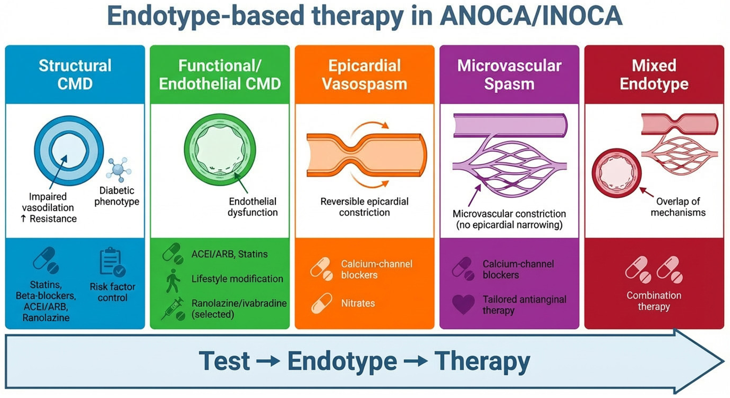
3.1. Structural Coronary Microvascular Dysfunction (CMD)
3.2. Functional/Endothelial CMD
3.3. Epicardial Vasospasm (Vasospastic Angina)
3.4. Microvascular Spasm
4. Pathophysiological Rationale
4.1. Coronary Flow as the Final Common Pathway
4.2. Pressure-Based Versus Flow-Based Physiology
4.3. Mechanisms of Coronary Microvascular Dysfunction
4.4. Vasomotor Disorders and Dynamic Flow Limitation
4.5. Prognostic Implications of Flow Impairment
4.6. From Pathophysiology to Mechanism-Based Care
5. Non-Invasive Testing: From Ischaemia Detection to Functional Endotyping
5.1. CCTA: An Anatomic Gatekeeper and Plaque Phenotype Tool
5.2. PET: Absolute Myocardial Blood Flow and Flow Reserve
- •
- epicardial flow-limiting disease,
- •
- diffuse atherosclerosis,
- •
- structural or functional coronary microvascular dysfunction, or combinations thereof
5.2.1. Diagnostic Patterns and Endotype Inference
5.2.2. Thresholds and Standardisation Challenges
5.2.3. Prognostic Implications
5.2.4. Limitations
5.3. Stress CMR: Quantitative Perfusion and Myocardial Phenotyping
5.3.1. Quantitative Perfusion Mapping
5.3.2. Integrated Myocardial Characterisation
5.3.3. Diagnostic Performance and Incremental Value
5.3.4. Limitations and Technical Considerations
5.4. PET Versus CMR: Complementary Rather than Competitive
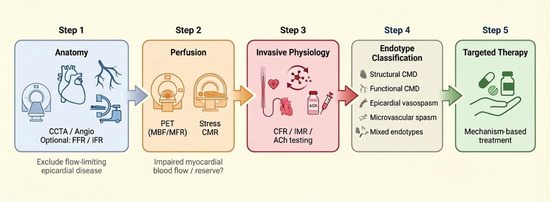
5.5. Bridge: From Anatomy to Physiology to Mechanism
6. Invasive Coronary Function Testing: ESC-Aligned “Complete the Endotype” Strategy
6.1. Why ICFT Is Increasingly Central: Concept and Rationale
6.2. Core Invasive Measurements: Epicardial Physiology, CFR, and Microvascular Resistance
6.3. Acetylcholine Provocation Testing: Epicardial vs. Microvascular Spasm
6.4. Integrated ICFT: Completing the Endotype
6.5. Evidence of Clinical Utility: CorMicA and Stratified Therapy
7. Linking Endotype to Therapy
7.1. From Mechanism to Management: A Paradigm Shift
7.2. Structural Coronary Microvascular Dysfunction: A Systemic Cardiometabolic Target
7.3. Functional CMD: Targeting Endothelial Dysfunction
7.4. Vasospastic Angina and Microvascular Spasm: Suppressing Abnormal Vasoconstriction
7.5. Mixed Endotypes: The Rule Rather than the Exception
7.6. Evidence Supporting Stratified Therapy
7.7. Toward Precision Medicine in ANOCA/INOCA
8. Conclusions
8.1. Practical Implementation and Barriers
8.2. Future Perspectives
Author Contributions
Funding
Institutional Review Board Statement
Informed Consent Statement
Data Availability Statement
Acknowledgments
Conflicts of Interest
Abbreviations
| ACh | acetylcholine |
| ACEi | angiotensin-converting enzyme inhibitors |
| ANOCA | angina with non-obstructive coronary arteries |
| ARB | angiotensin receptor blockers |
| CAD | coronary artery disease |
| CCB | calcium-channel blockers |
| CCS | chronic coronary syndromes |
| CCTA | coronary computed tomography angiography |
| CFR | coronary flow reserve |
| CMD | coronary microvascular dysfunction |
| CMR | cardiovascular magnetic resonance |
| COVADIS | Coronary Vasomotion Disorders International Study Group |
| ECG | electrocardiogram |
| ECV | extracellular volume |
| ESC | European Society of Cardiology |
| FFR | fractional flow reserve |
| FFR-CT | computed tomography-derived fractional flow reserve |
| ICA | invasive coronary angiography |
| ICFT | invasive coronary function testing |
| iFR | instantaneous wave-free ratio |
| IMR | index of microcirculatory resistance |
| INOCA | ischaemia with non-obstructive coronary arteries |
| LGE | late gadolinium enhancement |
| MBF | myocardial blood flow |
| MFR | myocardial flow reserve |
| MPR | myocardial perfusion reserve |
| NO | nitric oxide |
| PET | positron emission tomography |
| VSA | vasospastic angina |
References
- Vrints, C.; Andreotti, F.; Koskinas, K.C.; Rossello, X.; Adamo, M.; Ainslie, J.; Banning, A.P.; Budaj, A.; Buechel, R.R.; Chiariello, G.A.; et al. 2024 ESC Guidelines for the management of chronic coronary syndromes. Eur. Heart J. 2024, 45, 3415–3537. [Google Scholar] [CrossRef]
- Samuels, B.A.; Shah, S.M.; Widmer, R.J.; Kobayashi, Y.; Miner, S.E.S.; Taqueti, V.R.; Jeremias, A.; Albadri, A.; Blair, J.A.; Kearney, K.E.; et al. Comprehensive Management of ANOCA, Part 1-Definition, Patient Population, and Diagnosis: JACC State-of-the-Art Review. J. Am. Coll. Cardiol. 2023, 82, 1245–1263. [Google Scholar] [CrossRef]
- Mátyás, B.B.; Benedek, I.; Rat, N.; Gerculy, R.; Benedek, T. Coronary Plaque Vulnerability and Pericoronary Adipose Tissue Inflammation: Emerging Insights from Advanced CT Imaging. Medicina 2026, 62, 630. [Google Scholar] [CrossRef]
- Kunadian, V.; Chieffo, A.; Camici, P.G.; Berry, C.; Escaned, J.; Maas, A.H.E.M.; Prescott, E.; Karam, N.; Appelman, Y.; Fraccaro, C.; et al. An EAPCI Expert Consensus Document on Ischaemia with Non-Obstructive Coronary Arteries in Collaboration with European Society of Cardiology Working Group on Coronary Pathophysiology & Microcirculation Endorsed by Coronary Vasomotor Disorders International Study Group. Eur. Heart J. 2020, 41, 3504–3520. [Google Scholar] [CrossRef]
- Beltrame, J.F.; Crea, F.; Kaski, J.C.; Ogawa, H.; Ong, P.; Sechtem, U.; Shimokawa, H.; Bairey Merz, C.N.; Coronary Vasomotion Disorders International Study Group (COVADIS). International standardization of diagnostic criteria for vasospastic angina. Eur. Heart J. 2017, 38, 2565–2568. [Google Scholar] [CrossRef] [PubMed]
- Ong, P.; Camici, P.G.; Beltrame, J.F.; Crea, F.; Shimokawa, H.; Sechtem, U.; Kaski, J.C.; Bairey Merz, C.N.; Coronary Vasomotion Disorders International Study Group (COVADIS). International standardization of diagnostic criteria for microvascular angina. Int. J. Cardiol. 2018, 250, 16–20. [Google Scholar] [CrossRef] [PubMed]
- Sinha, A.; Rahman, H.; Perera, D. Reply: Rethinking False Positive Exercise Electrocardiographic Stress Tests by Assessing Coronary Microvascular Function. J. Am. Coll. Cardiol. 2024, 83, e195–e196. [Google Scholar] [CrossRef] [PubMed]
- Leone, A.M.; Galante, D.; Viceré, A.; Marrone, A.; Verardi, F.M.; Giuliana, C.; Pollio Benvenuto, C.; Viccaro, V.; Todisco, S.; Erriquez, A.; et al. Functional coronary assessment in angina with intermediate coronary stenosis: The #FullPhysiology approach. Eur. Heart J. 2025, 46, 978–980. [Google Scholar] [CrossRef]
- van de Hoef, T.P.; Lee, J.M.; Boerhout, C.K.M.; de Waard, G.A.; Jung, J.H.; Lee, S.H.; Mejía-Rentería, H.; Hoshino, M.; Echavarria-Pinto, M.; Meuwissen, M.; et al. Combined Assessment of FFR and CFR for Decision Making in Coronary Revascularization: From the Multicenter International ILIAS Registry. JACC Cardiovasc. Interv. 2022, 15, 1047–1056. [Google Scholar] [CrossRef]
- Seitz, A.; Martínez Pereyra, V.; Sechtem, U.; Ong, P. Update on coronary artery spasm 2022—A narrative review. Int. J. Cardiol. 2022, 359, 1–6. [Google Scholar] [CrossRef]
- Suda, A.; Takahashi, J.; Hao, K.; Kikuchi, Y.; Shindo, T.; Ikeda, S.; Sato, K.; Sugisawa, J.; Matsumoto, Y.; Miyata, S.; et al. Coronary Functional Abnormalities in Patients with Angina and Nonobstructive Coronary Artery Disease. J. Am. Coll. Cardiol. 2019, 74, 2350–2360. [Google Scholar] [CrossRef] [PubMed]
- Woudstra, J.; Dimitriu-Leen, A.C.; Crooijmans, C.; Yosofi, B.; Meeder, J.G.; Meuwissen, M.; De Vos, A.M.J.; Paradies, V.; Bijvank, E.G.M.O.; Winkler, P.; et al. Microvascular vasomotor dysfunction is more prevalent in female versus epicardial vasomotor dysfunction in male ANOCA patients. Int. J. Cardiol. 2026, 450, 134226. [Google Scholar] [CrossRef] [PubMed]
- Del Buono, M.G.; Montone, R.A.; Camilli, M.; Carbone, S.; Narula, J.; Lavie, C.J.; Niccoli, G.; Crea, F. Coronary Microvascular Dysfunction Across the Spectrum of Cardiovascular Diseases: JACC State-of-the-Art Review. J. Am. Coll. Cardiol. 2021, 78, 1352–1371. [Google Scholar] [CrossRef]
- Pijls, N.H.; de Bruyne, B.; Peels, K.; van der Voort, P.H.; Bonnier, H.J.; Bartunek, J.; Koolen, J.J. Measurement of fractional flow reserve to assess the functional severity of coronary-artery stenoses. N. Engl. J. Med. 1996, 334, 1703–1708. [Google Scholar] [CrossRef]
- Parlati, A.L.M.; Nardi, E.; Sucato, V.; Madaudo, C.; Leo, G.; Rajah, T.; Marzano, F.; Prastaro, M.; Gargiulo, P.; Paolillo, S.; et al. ANOCA, INOCA, MINOCA: The New Frontier of Coronary Syndromes. J. Cardiovasc. Dev. Dis. 2025, 12, 64. [Google Scholar] [CrossRef]
- Duncker, D.J.; Koller, A.; Merkus, D.; Canty, J.M. Regulation of coronary blood flow in health and ischemic heart disease. Prog. Cardiovasc. Dis. 2015, 57, 409–422. [Google Scholar] [CrossRef]
- Camici, P.G.; Crea, F. Coronary microvascular dysfunction. N. Engl. J. Med. 2007, 356, 830–840. [Google Scholar] [CrossRef] [PubMed]
- Gould, K.L. Does coronary flow trump coronary anatomy? JACC Cardiovasc. Imaging 2009, 2, 1009–1023. [Google Scholar] [CrossRef]
- Tonino, P.A.; De Bruyne, B.; Pijls, N.H.; Siebert, U.; Ikeno, F.; van’ t Veer, M.; Klauss, V.; Manoharan, G.; Engstrøm, T.; Oldroyd, K.G.; et al. Fractional flow reserve versus angiography for guiding percutaneous coronary intervention. N. Engl. J. Med. 2009, 360, 213–224. [Google Scholar] [CrossRef] [PubMed]
- Gould, K.L.; Kirkeeide, R.L.; Buchi, M. Coronary flow reserve as a physiologic measure of stenosis severity. J. Am. Coll. Cardiol. 1990, 15, 459–474. [Google Scholar] [CrossRef]
- Berry, C.; Corcoran, D.; Hennigan, B.; Watkins, S.; Layland, J.; Oldroyd, K.G. Fractional flow reserve-guided management in stable coronary disease and acute myocardial infarction: Recent developments. Eur. Heart J. 2015, 36, 3155–3164. [Google Scholar] [CrossRef] [PubMed]
- Stegehuis, V.E.; Wijntjens, G.W.; Piek, J.J.; van de Hoef, T.P. Fractional Flow Reserve or Coronary Flow Reserve for the Assessment of Myocardial Perfusion: Implications of FFR as an Imperfect Reference Standard for Myocardial Ischemia. Curr. Cardiol. Rep. 2018, 20, 77. [Google Scholar] [CrossRef] [PubMed]
- Johnson, N.P.; Kirkeeide, R.L.; Gould, K.L. Is discordance of coronary flow reserve and fractional flow reserve due to methodology or clinically relevant coronary pathophysiology? JACC Cardiovasc. Imaging 2012, 5, 193–202. [Google Scholar] [CrossRef]
- Ahn, S.G.; Suh, J.; Hung, O.Y.; Lee, H.S.; Bouchi, Y.H.; Zeng, W.; Gandhi, R.; Eshtehardi, P.; Gogas, B.D.; Samady, H. Discordance Between Fractional Flow Reserve and Coronary Flow Reserve: Insights from Intracoronary Imaging and Physiological Assessment. JACC Cardiovasc. Interv. 2017, 10, 999–1007. [Google Scholar] [CrossRef]
- Kelshiker, M.A.; Seligman, H.; Howard, J.P.; Rahman, H.; Foley, M.; Nowbar, A.N.; A Rajkumar, C.; Shun-Shin, M.J.; Ahmad, Y.; Sen, S.; et al. Coronary flow reserve and cardiovascular outcomes: A systematic review and meta-analysis. Eur. Heart J. 2022, 43, 1582–1593. [Google Scholar] [CrossRef]
- Lee, J.M.; Jung, J.-H.; Hwang, D.; Park, J.; Fan, Y.; Na, S.-H.; Doh, J.-H.; Nam, C.-W.; Shin, E.-S.; Koo, B.-K. Coronary Flow Reserve and Microcirculatory Resistance in Patients with Intermediate Coronary Stenosis. J. Am. Coll. Cardiol. 2016, 67, 1158–1169. [Google Scholar] [CrossRef]
- Rahman, H.; Demir, O.M.; Khan, F.; Ryan, M.; Ellis, H.; Mills, M.T.; Chiribiri, A.; Webb, A.; Perera, D. Physiological Stratification of Patients with Angina Due to Coronary Microvascular Dysfunction. J. Am. Coll. Cardiol. 2020, 75, 2538–2549. [Google Scholar] [CrossRef]
- Crea, F.; Camici, P.G.; Bairey Merz, C.N. Coronary microvascular dysfunction: An update. Eur. Heart J. 2014, 35, 1101–1111. [Google Scholar] [CrossRef]
- Lanza, G.A.; Careri, G.; Crea, F. Mechanisms of coronary artery spasm. Circulation 2011, 124, 1774–1782. [Google Scholar] [CrossRef]
- Hubert, A.; Seitz, A.; Pereyra, V.M.; Bekeredjian, R.; Sechtem, U.; Ong, P. Coronary Artery Spasm: The Interplay Between Endothelial Dysfunction and Vascular Smooth Muscle Cell Hyperreactivity. Eur. Cardiol. 2020, 15, e12. [Google Scholar] [CrossRef] [PubMed]
- Picard, F.; Sayah, N.; Spagnoli, V.; Adjedj, J.; Varenne, O. Vasospastic angina: A literature review of current evidence. Arch. Cardiovasc. Dis. 2019, 112, 44–55. [Google Scholar] [CrossRef]
- Takahashi, J.; Suda, A.; Nishimiya, K.; Godo, S.; Yasuda, S.; Shimokawa, H. Pathophysiology and Diagnosis of Coronary Functional Abnormalities. Eur. Cardiol. 2021, 16, e30. [Google Scholar] [CrossRef]
- Mileva, N.; Nagumo, S.; Mizukami, T.; Sonck, J.; Berry, C.; Gallinoro, E.; Monizzi, G.; Candreva, A.; Munhoz, D.; Vassilev, D.; et al. Prevalence of Coronary Microvascular Disease and Coronary Vasospasm in Patients with Nonobstructive Coronary Artery Disease: Systematic Review and Meta-Analysis. J. Am. Heart Assoc. 2022, 11, e023207. [Google Scholar] [CrossRef]
- Ziadi, M.C.; Dekemp, R.A.; Williams, K.A.; Guo, A.; Chow, B.J.; Renaud, J.M.; Ruddy, T.D.; Sarveswaran, N.; Tee, R.E.; Beanlands, R.S. Impaired myocardial flow reserve on rubidium-82 positron emission tomography imaging predicts adverse outcomes in patients assessed for myocardial ischemia. J. Am. Coll. Cardiol. 2011, 58, 740–748. [Google Scholar] [CrossRef]
- Højstrup, S.; Hansen, K.W.; Talleruphuus, U.; Marner, L.; Bjerking, L.; Jakobsen, L.; Christiansen, E.H.; Bouchelouche, K.; Wiinberg, N.; Guldbrandsen, K.; et al. Myocardial Flow Reserve, an Independent Prognostic Marker of All-Cause Mortality Assessed by 82Rb PET Myocardial Perfusion Imaging: A Danish Multicenter Study. Circ. Cardiovasc. Imaging 2023, 16, e015184. [Google Scholar] [CrossRef]
- Boerhout, C.K.M.; de Waard, G.A.; Lee, J.M.; Mejia-Renteria, H.; Lee, S.H.; Jung, J.-H.; Hoshino, M.; Echavarria-Pinto, M.; Meuwissen, M.; Matsuo, H.; et al. Prognostic value of structural and functional coronary microvascular dysfunction in patients with non-obstructive coronary artery disease; from the multicentre international ILIAS registry. EuroIntervention 2022, 18, 719–728. [Google Scholar] [CrossRef] [PubMed]
- Hwang, D.; Park, S.; Koo, B.-K. Reply: Ischemia with Nonobstructive Coronary Artery Disease. JACC Asia 2023, 3, 687. [Google Scholar] [CrossRef] [PubMed]
- Miner, S.; Mejia-Renteria, H.; Leone, A.M.; Velollari, O.; Sykes, R.; Biscaglia, S.; Esposito, G.; Galante, D.; Oreglia, J.; Ang, D.; et al. Endotypes of angina with non-obstructive coronary arteries: A prospective multicentre study. Eur. Heart J. 2026, 47, 594–604. [Google Scholar] [CrossRef] [PubMed]
- Hokimoto, S.; Kaikita, K.; Yasuda, S.; Tsujita, K.; Ishihara, M.; Matoba, T.; Matsuzawa, Y.; Mitsutake, Y.; Mitani, Y.; Murohara, T.; et al. JCS/CVIT/JCC 2023 guideline focused update on diagnosis and treatment of vasospastic angina (coronary spastic angina) and coronary microvascular dysfunction. J. Cardiol. 2023, 82, 293–341. [Google Scholar] [CrossRef] [PubMed]
- Jenkins, K.; Pompei, G.; Ganzorig, N.; Brown, S.; Beltrame, J.; Kunadian, V. Vasospastic angina: A review on diagnostic approach and management. Ther. Adv. Cardiovasc. Dis. 2024, 18, 17539447241230400. [Google Scholar] [CrossRef]
- Welch, I.; Case, B. Treatment options for patients with coronary microvascular dysfunction. Future Cardiol. 2025, 21, 735–738. [Google Scholar] [CrossRef] [PubMed]
- Ford, T.J.; Stanley, B.; Good, R.; Rocchiccioli, P.; McEntegart, M.; Watkins, S.; Eteiba, H.; Shaukat, A.; Lindsay, M.; Robertson, K.; et al. Stratified Medical Therapy Using Invasive Coronary Function Testing in Angina: The CorMicA Trial. J. Am. Coll. Cardiol. 2018, 72, 2841–2855. [Google Scholar] [CrossRef]
- Ford, T.J.; Stanley, B.; Sidik, N.; Good, R.; Rocchiccioli, P.; McEntegart, M.; Watkins, S.; Eteiba, H.; Shaukat, A.; Lindsay, M.; et al. 1-Year Outcomes of Angina Management Guided by Invasive Coronary Function Testing (CorMicA). JACC Cardiovasc. Interv. 2020, 13, 33–45. [Google Scholar] [CrossRef]
- Hansen, B.; Holtzman, J.N.; Juszczynski, C.; Khan, N.; Kaur, G.; Varma, B.; Gulati, M. Ischemia with No Obstructive Arteries (INOCA): A Review of the Prevalence, Diagnosis and Management. Curr. Probl. Cardiol. 2023, 48, 101420. [Google Scholar] [CrossRef]
- Kolossváry, M.; Szilveszter, B.; Merkely, B.; Maurovich-Horvat, P. Plaque imaging with CT—A comprehensive review on coronary CT angiography based risk assessment. Cardiovasc. Diagn. Ther. 2017, 7, 489–506. [Google Scholar] [CrossRef] [PubMed]
- Nieman, K.; García-García, H.M.; Hideo-Kajita, A.; Collet, C.; Dey, D.; Pugliese, F.; Weissman, G.; Tijssen, J.G.; Leipsic, J.; Opolski, M.P.; et al. Standards for quantitative assessments by coronary computed tomography angiography (CCTA): An expert consensus document of the society of cardiovascular computed tomography (SCCT). J. Cardiovasc. Comput. Tomogr. 2024, 18, 429–443. [Google Scholar] [CrossRef] [PubMed]
- Motoyama, S.; Sarai, M.; Harigaya, H.; Anno, H.; Inoue, K.; Hara, T.; Naruse, H.; Ishii, J.; Hishida, H.; Wong, N.D.; et al. Computed tomographic angiography characteristics of atherosclerotic plaques subsequently resulting in acute coronary syndrome. J. Am. Coll. Cardiol. 2009, 54, 49–57. [Google Scholar] [CrossRef]
- Murthy, V.L.; Lee, B.C.; Sitek, A.; Naya, M.; Moody, J.; Polavarapu, V.; Ficaro, E.P.; Di Carli, M.F. Comparison and prognostic validation of multiple methods of quantification of myocardial blood flow with 82Rb PET. J. Nucl. Med. 2014, 55, 1952–1958. [Google Scholar] [CrossRef]
- Schindler, T.H.; Fearon, W.F.; Pelletier-Galarneau, M.; Ambrosio, G.; Sechtem, U.; Ruddy, T.D.; Patel, K.K.; Bhatt, D.L.; Bateman, T.M.; Gewirtz, H.; et al. Myocardial Perfusion PET for the Detection and Reporting of Coronary Microvascular Dysfunction: A JACC: Cardiovascular Imaging Expert Panel Statement. JACC Cardiovasc. Imaging 2023, 16, 536–548. [Google Scholar] [CrossRef] [PubMed]
- Vohra, S.; Sakul, S.; Agarwal, V.; Patel, K.K. Quantification of myocardial blood flow using PET: Current status, clinical applications, and future directions. Prog. Cardiovasc. Dis. 2025, 93, 10–20. [Google Scholar] [CrossRef]
- Kato, Y.; Chehab, O.; Ambale-Venkatesh, B.; Lima, J.A.C. Quantitative myocardial perfusion imaging across PET, SPECT, CMR, and CT. Front. Radiol. 2026, 6, 1760241. [Google Scholar] [CrossRef] [PubMed]
- Sciagrà, R.; Lubberink, M.; Hyafil, F.; Saraste, A.; Slart, R.H.J.A.; Agostini, D.; Nappi, C.; Georgoulias, P.; Bucerius, J.; Rischpler, C.; et al. EANM procedural guidelines for PET/CT quantitative myocardial perfusion imaging. Eur. J. Nucl. Med. Mol. Imaging 2021, 48, 1040–1069. [Google Scholar] [CrossRef]
- Taqueti, V.R.; Di Carli, M.F. Coronary Microvascular Disease Pathogenic Mechanisms and Therapeutic Options: JACC State-of-the-Art Review. J. Am. Coll. Cardiol. 2018, 72, 2625–2641. [Google Scholar] [CrossRef] [PubMed]
- Ramirez, G.; Builoff, V.; Miller, R.J.; Lemley, M.; Carvajal-Juarez, I.; Alexanderson, E.; Rosamond, T.L.; Song, N.; Travin, M.I.; Slipczuk, L.; et al. Multicenter Evaluation of Myocardial Flow Reserve as a Prognostic Marker for Mortality in 13N-Ammonia PET Myocardial Perfusion Imaging. Circ. Cardiovasc. Imaging 2026, 19, e018729. [Google Scholar] [CrossRef]
- Valenta, I.; Quercioli, A.; Schindler, T.H. Diagnostic value of PET-measured longitudinal flow gradient for the identification of coronary artery disease. JACC Cardiovasc. Imaging 2014, 7, 387–396. [Google Scholar] [CrossRef] [PubMed]
- Fukushima, K.; Javadi, M.S.; Higuchi, T.; Bravo, P.E.; Chien, D.; Lautamäki, R.; Merrill, J.; Nekolla, S.G.; Bengel, F.M. Impaired global myocardial flow dynamics despite normal left ventricular function and regional perfusion in chronic kidney disease: A quantitative analysis of clinical 82Rb PET/CT studies. J. Nucl. Med. 2012, 53, 887–893. [Google Scholar] [CrossRef]
- Dekemp, R.A. Toward improved standardization of PET myocardial blood flow. J. Nucl. Cardiol. 2023, 30, 1297–1299. [Google Scholar] [CrossRef]
- Czernin, J.; Müller, P.; Chan, S.; Brunken, R.C.; Porenta, G.; Krivokapich, J.; Chen, K.; Chan, A.; Phelps, M.E.; Schelbert, H.R. Influence of age and hemodynamics on myocardial blood flow and flow reserve. Circulation 1993, 88, 62–69. [Google Scholar] [CrossRef]
- Huck, D.M.; Weber, B.N.; Brown, J.M.; Lopez, D.; Hainer, J.; Blankstein, R.; Dorbala, S.; Divakaran, S.; Di Carli, M.F. Prognostic value of myocardial flow reserve vs corrected myocardial flow reserve in patients without obstructive coronary artery disease. J. Nucl. Cardiol. 2024, 37, 101854. [Google Scholar] [CrossRef]
- Sperry, B.W.; Metzinger, M.P.; Ibrahim, A.O.; Thompson, R.C.; Cho, Y.J.; Jones, P.G.; McGhie, A.I.; Bateman, T.M. Age- and Sex-Specific Myocardial Blood Flow Values in Patients Without Coronary Atherosclerosis on Rb-82 PET Myocardial Perfusion Imaging. Circ. Cardiovasc. Imaging 2024, 17, e016577. [Google Scholar] [CrossRef] [PubMed]
- Packard, R.R.S.; Maddahi, J.; Pelletier-Galarneau, M.; Al-Mallah, M.H.; Coelho, M.; Dorbala, S.; Galt, J.; Hyun, M.; Menon, N.; Miller, E.J.; et al. SNMMI/EANM/ASNC/ACNM Procedure Standard/Practice Guideline for 18F-Flurpiridaz PET Myocardial Perfusion Imaging and Blood Flow Quantitation. J. Nucl. Med. 2025, 66, 1538–1554. [Google Scholar] [CrossRef]
- Bijloo, I.; Woudstra, J.; Aribas, E.; Elias-Smale, S.E.; Vink, C.; Piek, J.J.; Duncker, D.J.; van Lennep, J.E.R.; Kavousi, M.; Appelman, Y. Non-invasive and invasive diagnostic modalities for the assessment of coronary vasomotor dysfunction in ANOCA patients. EuroIntervention 2026, 22, e283–e291. [Google Scholar] [CrossRef]
- Kotecha, T.; Martinez-Naharro, A.; Boldrini, M.; Knight, D.; Hawkins, P.; Kalra, S.; Patel, D.; Coghlan, G.; Moon, J.; Plein, S.; et al. Automated Pixel-Wise Quantitative Myocardial Perfusion Mapping by CMR to Detect Obstructive Coronary Artery Disease and Coronary Microvascular Dysfunction: Validation Against Invasive Coronary Physiology. JACC Cardiovasc. Imaging 2019, 12, 1958–1969. [Google Scholar] [CrossRef]
- Hsu, L.-Y.; Jacobs, M.; Benovoy, M.; Ta, A.D.; Conn, H.M.; Winkler, S.; Greve, A.M.; Chen, M.Y.; Shanbhag, S.M.; Bandettini, W.P.; et al. Diagnostic Performance of Fully Automated Pixel-Wise Quantitative Myocardial Perfusion Imaging by Cardiovascular Magnetic Resonance. JACC Cardiovasc. Imaging 2018, 11, 697–707. [Google Scholar] [CrossRef]
- Crawley, R.; Kunze, K.P.; Milidonis, X.; Highton, J.; McElroy, S.; Frey, S.M.; Hoefler, D.; Karamanli, C.; Wong, N.C.K.; Backhaus, S.J.; et al. High-resolution free-breathing automated quantitative myocardial perfusion by cardiovascular magnetic resonance for the detection of functionally significant coronary artery disease. Eur. Heart J. Cardiovasc. Imaging 2024, 25, 914–925. [Google Scholar] [CrossRef] [PubMed]
- Messroghli, D.R.; Moon, J.C.; Ferreira, V.M.; Grosse-Wortmann, L.; He, T.; Kellman, P.; Mascherbauer, J.; Nezafat, R.; Salerno, M.; Schelbert, E.B.; et al. Clinical recommendations for cardiovascular magnetic resonance mapping of T1, T2, T2* and extracellular volume: A consensus statement by the Society for Cardiovascular Magnetic Resonance (SCMR) endorsed by the European Association for Cardiovascular Imaging (EACVI). J. Cardiovasc. Magn. Reson. 2017, 19, 75, Erratum in J. Cardiovasc. Magn. Reson. 2018, 20, 9. https://doi.org/10.1186/s12968-017-0408-9. [Google Scholar] [CrossRef]
- Dorfman, A.L.; Geva, T.; Samyn, M.M.; Greil, G.; Krishnamurthy, R.; Messroghli, D.; Festa, P.; Secinaro, A.; Soriano, B.; Taylor, A.; et al. SCMR expert consensus statement for cardiovascular magnetic resonance of acquired and non-structural pediatric heart disease. J. Cardiovasc. Magn. Reson. 2022, 24, 44. [Google Scholar] [CrossRef] [PubMed]
- Plein, S.; Kozerke, S.; Suerder, D.; Luescher, T.F.; Greenwood, J.P.; Boesiger, P.; Schwitter, J. High spatial resolution myocardial perfusion cardiac magnetic resonance for the detection of coronary artery disease. Eur. Heart J. 2008, 29, 2148–2155. [Google Scholar] [CrossRef] [PubMed]
- Nagel, E.; Greenwood, J.P.; McCann, G.P.; Bettencourt, N.; Shah, A.M.; Hussain, S.T.; Perera, D.; Plein, S.; Bucciarelli-Ducci, C.; Paul, M.; et al. Magnetic Resonance Perfusion or Fractional Flow Reserve in Coronary Disease. N. Engl. J. Med. 2019, 380, 2418–2428. [Google Scholar] [CrossRef]
- Ge, Y.; Antiochos, P.; Steel, K.; Bingham, S.; Abdullah, S.; Chen, Y.-Y.; Mikolich, J.R.; Arai, A.E.; Bandettini, W.P.; Shanbhag, S.M.; et al. Prognostic Value of Stress CMR Perfusion Imaging in Patients with Reduced Left Ventricular Function. JACC Cardiovasc. Imaging 2020, 13, 2132–2145. [Google Scholar] [CrossRef]
- Wang, S.; Kim, P.; Wang, H.; Ng, M.-Y.; Arai, A.E.; Singh, A.; Mushtaq, S.; Sin, T.H.; Tada, Y.; Hillier, E.; et al. Myocardial Blood Flow Quantification Using Stress Cardiac Magnetic Resonance Improves Detection of Coronary Artery Disease. JACC Cardiovasc. Imaging 2024, 17, 1428–1441. [Google Scholar] [CrossRef]
- Chang, A.; Kang, N.; Chung, J.; Gupta, A.R.; Parwani, P. Evaluation of Ischemia with No Obstructive Coronary Arteries (INOCA) and Contemporary Applications of Cardiac Magnetic Resonance (CMR). Medicina 2023, 59, 1570. [Google Scholar] [CrossRef]
- Chiribiri, A.; Arai, A.E.; DiBella, E.; Hsu, L.-Y.; Ishida, M.; Jerosch-Herold, M.; Kozerke, S.; Milidonis, X.; Nezafat, R.; Plein, S.; et al. Society for cardiovascular magnetic resonance expert consensus statement on quantitative myocardial perfusion cardiovascular magnetic resonance imaging. J. Cardiovasc. Magn. Reson. 2025, 27, 101940. [Google Scholar] [CrossRef]
- Ford, T.J.; Yii, E.; Sidik, N.; Good, R.; Rocchiccioli, P.; McEntegart, M.; Watkins, S.; Eteiba, H.; Shaukat, A.; Lindsay, M.; et al. Ischemia and No Obstructive Coronary Artery Disease: Prevalence and Correlates of Coronary Vasomotion Disorders. Circ. Cardiovasc. Interv. 2019, 12, e008126. [Google Scholar] [CrossRef] [PubMed]
- Namba, H.F.; Boerhout, C.K.M.; Damman, P.; Kunadian, V.; Escaned, J.; Ong, P.; Perera, D.; Berry, C.; van de Hoef, T.P.; Piek, J.J. Invasive coronary function testing in clinical practice: Implementing the 2024 ESC guidelines on chronic coronary syndromes. Int. J. Cardiol. 2025, 430, 133176. [Google Scholar] [CrossRef]
- Collet, C.; Yong, A.; Munhoz, D.; Akasaka, T.; Berry, C.; Blair, J.E.A.; Collison, D.; Engstrøm, T.; Escaned, J.; Fearon, W.F.; et al. A Systematic Approach to the Evaluation of the Coronary Microcirculation Using Bolus Thermodilution: CATH CMD. J. Soc. Cardiovasc. Angiogr. Interv. 2024, 3, 101934. [Google Scholar] [CrossRef] [PubMed]
- Fearon, W.F.; Kobayashi, Y. Invasive Assessment of the Coronary Microvasculature: The Index of Microcirculatory Resistance. Circ. Cardiovasc. Interv. 2017, 10, e005361. [Google Scholar] [CrossRef] [PubMed]
- Gallinoro, E.; Bertolone, D.T.; Fernandez-Peregrina, E.; Paolisso, P.; Bermpeis, K.; Esposito, G.; Gomez-Lopez, A.; Candreva, A.; Mileva, N.; Belmonte, M.; et al. Reproducibility of bolus versus continuous thermodilution for assessment of coronary microvascular function in patients with ANOCA. EuroIntervention 2023, 19, e155–e166. [Google Scholar] [CrossRef]
- Rahman, H.; Ryan, M.; Lumley, M.; Modi, B.; McConkey, H.; Ellis, H.; Scannell, C.; Clapp, B.; Marber, M.; Webb, A.; et al. Coronary Microvascular Dysfunction Is Associated with Myocardial Ischemia and Abnormal Coronary Perfusion During Exercise. Circulation 2019, 140, 1805–1816. [Google Scholar] [CrossRef]
- Wang, D.; Li, X.; Feng, W.; Zhou, H.; Peng, W.; Wang, X. Diagnostic and prognostic value of angiography-derived index of microvascular resistance: A systematic review and meta-analysis. Front. Cardiovasc. Med. 2024, 11, 1360648. [Google Scholar] [CrossRef]
- Montone, R.A.; Rinaldi, R.; Del Buono, M.G.; Gurgoglione, F.; La Vecchia, G.; Russo, M.; Caffè, A.; Burzotta, F.; Leone, A.M.; Romagnoli, E.; et al. Safety and prognostic relevance of acetylcholine testing in patients with stable myocardial ischaemia or myocardial infarction and non-obstructive coronary arteries. EuroIntervention 2022, 18, e666–e676. [Google Scholar] [CrossRef] [PubMed]
- Ashokprabhu, N.D.; Quesada, O.; Alvarez, Y.R.; Henry, T.D. INOCA/ANOCA: Mechanisms and novel treatments. Am. Heart J. Plus 2023, 30, 100302. [Google Scholar] [CrossRef]
- Montone, R.A.; Rinaldi, R.; Niccoli, G.; Andò, G.; Gragnano, F.; Piccolo, R.; Pelliccia, F.; Moscarella, E.; Zimarino, M.; Fabris, E.; et al. Optimizing Management of Stable Angina: A Patient-Centered Approach Integrating Revascularization, Medical Therapy, and Lifestyle Interventions. J. Am. Coll. Cardiol. 2024, 84, 744–760. [Google Scholar] [CrossRef] [PubMed]
- Masiero, G.; Aurigemma, C.; Biscaglia, S.; Bruno, F.; Burzotta, F.; Campo, G.; Cangemi, S.; Chieffo, A.; Di Mario, C.; Esposito, G.; et al. Implementazione delle nuove raccomandazioni sull’impiego dei test funzionali coronarici e dell’imaging intravascolare nell’angioplastica coronarica per le sindromi coronariche croniche: Documento di consenso GISE/ANMCO/SIC [GISE/ANMCO/SIC Consensus document on the implementation of the new recommendations for the use of coronary functional testing and intravascular imaging in coronary angioplasty for chronic coronary syndromes]. G. Ital. Cardiol. 2025, 26, 941–953. (In Italian) [Google Scholar] [CrossRef]



| Modality/Test | Key Outputs | Endotype Signal | Clinical Role | Limitations | Therapeutic Implications |
|---|---|---|---|---|---|
| CCTA | Stenosis severity; plaque burden and phenotype | Non-obstructive plaque → atherosclerotic substrate; normal CCTA does not exclude CMD or vasospasm | Anatomical gatekeeper; selection for functional testing | Heavy calcification; motion artefacts | Intensify preventive therapy; proceed to functional testing if symptoms persist |
| Stress PET (MBF/MFR) | Rest and stress MBF; global and regional MFR | Global reduction in MFR/MBF → structural CMD or diffuse coronary dysfunction | First-/second-line functional assessment | Haemodynamic variability; tracer and software differences; limited detection of spasm | Supports CMD diagnosis; risk stratification; guides cardiometabolic optimisation |
| Stress CMR (quantitative) | Stress MBF/MPR; regional perfusion; LGE; T1/ECV | Extensive perfusion impairment → CMD; tissue abnormalities may suggest alternative myocardial pathology | Alternative to PET; integrated “one-stop” evaluation | Arrhythmias; motion artefacts; variability in analysis pipelines | Guides CMD management; avoids misclassification; identifies alternative myocardial pathology |
| ICA + FFR/iFR | Epicardial physiology | Flow-limiting epicardial disease | Assessment of obstructive CAD prior to ICFT; excludes focal flow-limiting epicardial disease | Does not directly assess coronary blood flow or microvascular dysfunction; non-focal atherosclerotic disease may affect interpretation | Guides revascularisation decisions; a normal FFR/iFR does not exclude coronary vascular dysfunction |
| CFR (wire-based) | Coronary flow reserve | Reduced CFR suggests CMD but lacks specificity | Component of ICFT | Influenced by haemodynamics and epicardial disease | Requires integration with resistance indices and vasoreactivity testing |
| Microvascular resistance (IMR) | Microvascular resistance index | Elevated resistance → structural CMD | Core component of microvascular assessment | Technique-dependent | Supports CMD-targeted therapy; avoids inappropriate vasodilator-only strategies |
| Acetylcholine provocation | Vasomotor response; symptoms; ECG changes | Epicardial vs. microvascular spasm; mixed phenotypes | Diagnosis of vasospastic disorders | Requires expertise; protocol variability | Calcium-channel blockers first-line; nitrates and trigger modification |
| Integrated ICFT | Comprehensive physiological assessment | CMD, vasospasm, or mixed endotypes | ESC-recommended in persistent symptoms | Requires dedicated infrastructure | Enables stratified therapy; improves symptoms and quality of life |
| Endotype | Pathophysiological Target | First-Line Therapy | Mechanism-Based Pharmacological Strategies | Clinical Considerations |
|---|---|---|---|---|
| Structural CMD | Increased microvascular resistance, impaired vasodilatory capacity | Risk factor modification + β-blockers | ACEi/ARBs and statins to improve endothelial function and microvascular remodelling; β-blockers to reduce myocardial oxygen demand; consider ranolazine or ivabradine in selected patients | Disease-modifying therapy is central; focus on cardiometabolic optimisation rather than pure vasodilation |
| Functional (endothelium-dependent) CMD | Endothelial dysfunction, impaired NO-mediated vasodilation | ACEi/ARBs + statins | Therapies targeting endothelial function (ACEi, statins); consider CCBs if vasomotor component present; ranolazine may improve perfusion in selected patients | Overlap with vasospasm is common; treatment response may be heterogeneous |
| Endothelium-independent CMD | Impaired microvascular vasodilatory response (adenosine pathway) | β-blockers (particularly when increased myocardial oxygen demand predominates) | β-blockers to reduce demand; consider adjunctive agents (ranolazine, ivabradine, nicorandil) in persistent symptoms | Requires differentiation from endothelial dysfunction; often identified by reduced CFR/IMR abnormalities |
| Epicardial VSA | Hyperreactivity of vascular smooth muscle, transient epicardial spasm | CCBs | Dihydropyridine or non-dihydropyridine CCBs; long-acting nitrates as second-line; nicorandil in refractory cases | Avoid non-selective β-blockers; trigger control (e.g., smoking) is essential |
| Microvascular spasm | Microvascular vasoconstriction (ACh-induced) | CCBs | Similar to vasospastic angina; consider nitrates and nicorandil; potential role for ranolazine or Rho-kinase inhibitors (investigational) | Diagnosis requires invasive testing; response often variable |
| Mixed endotypes | Combined CMD + vasospasm | CCBs + risk factor control | Combination therapy targeting both vasodilation and microvascular dysfunction (CCBs + ACEi/ARB + statins ± antianginals) | Present in up to 40–60% of patients; requires individualised therapy |
Disclaimer/Publisher’s Note: The statements, opinions and data contained in all publications are solely those of the individual author(s) and contributor(s) and not of MDPI and/or the editor(s). MDPI and/or the editor(s) disclaim responsibility for any injury to people or property resulting from any ideas, methods, instructions or products referred to in the content. |
© 2026 by the authors. Published by MDPI on behalf of the Lithuanian University of Health Sciences. Licensee MDPI, Basel, Switzerland. This article is an open access article distributed under the terms and conditions of the Creative Commons Attribution (CC BY) license.
Share and Cite
Granata, L.G.; Marchetta, M.; Giubilato, S.; Sangiorgi, G.M.; Francese, G.M.; Andò, G. Beyond Stenosis: Mechanism-Based Multimodality Imaging and Invasive Coronary Function Testing for Endotype Definition in ANOCA/INOCA. Medicina 2026, 62, 910. https://doi.org/10.3390/medicina62050910
Granata LG, Marchetta M, Giubilato S, Sangiorgi GM, Francese GM, Andò G. Beyond Stenosis: Mechanism-Based Multimodality Imaging and Invasive Coronary Function Testing for Endotype Definition in ANOCA/INOCA. Medicina. 2026; 62(5):910. https://doi.org/10.3390/medicina62050910
Chicago/Turabian StyleGranata, Lucio Giuseppe, Marcello Marchetta, Simona Giubilato, Giuseppe Massimo Sangiorgi, Giuseppina Maura Francese, and Giuseppe Andò. 2026. "Beyond Stenosis: Mechanism-Based Multimodality Imaging and Invasive Coronary Function Testing for Endotype Definition in ANOCA/INOCA" Medicina 62, no. 5: 910. https://doi.org/10.3390/medicina62050910
APA StyleGranata, L. G., Marchetta, M., Giubilato, S., Sangiorgi, G. M., Francese, G. M., & Andò, G. (2026). Beyond Stenosis: Mechanism-Based Multimodality Imaging and Invasive Coronary Function Testing for Endotype Definition in ANOCA/INOCA. Medicina, 62(5), 910. https://doi.org/10.3390/medicina62050910