

Subcutaneous Myoepithelioma in the Extremity: A Potential Pitfall in the Differential Diagnosis of Subcutaneous Tumors


Abstract
1. Introduction
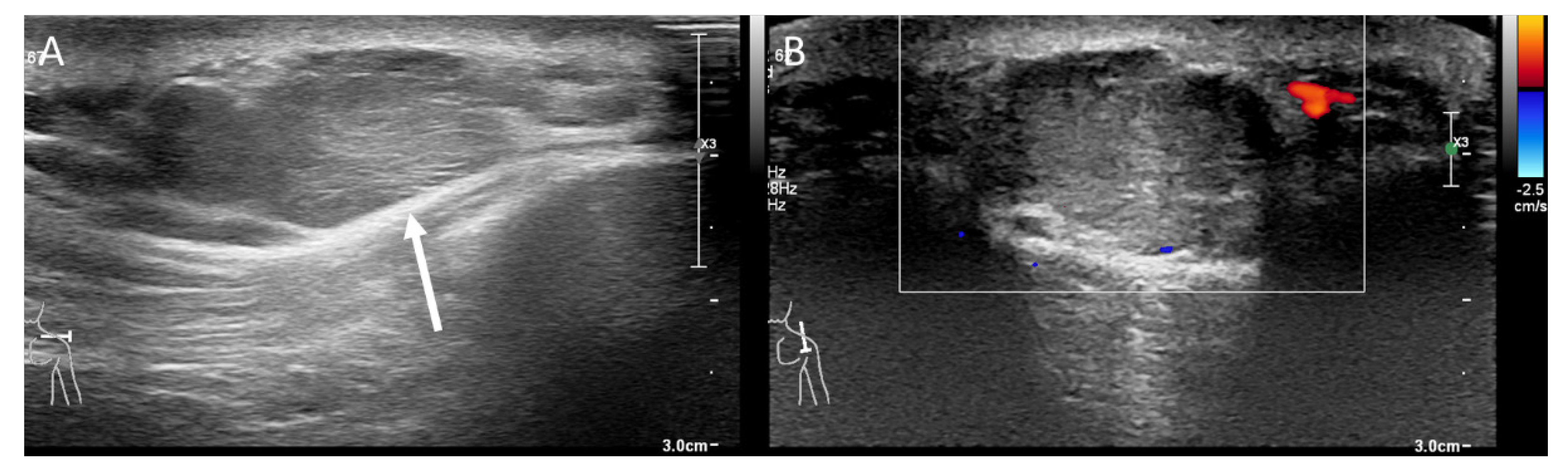
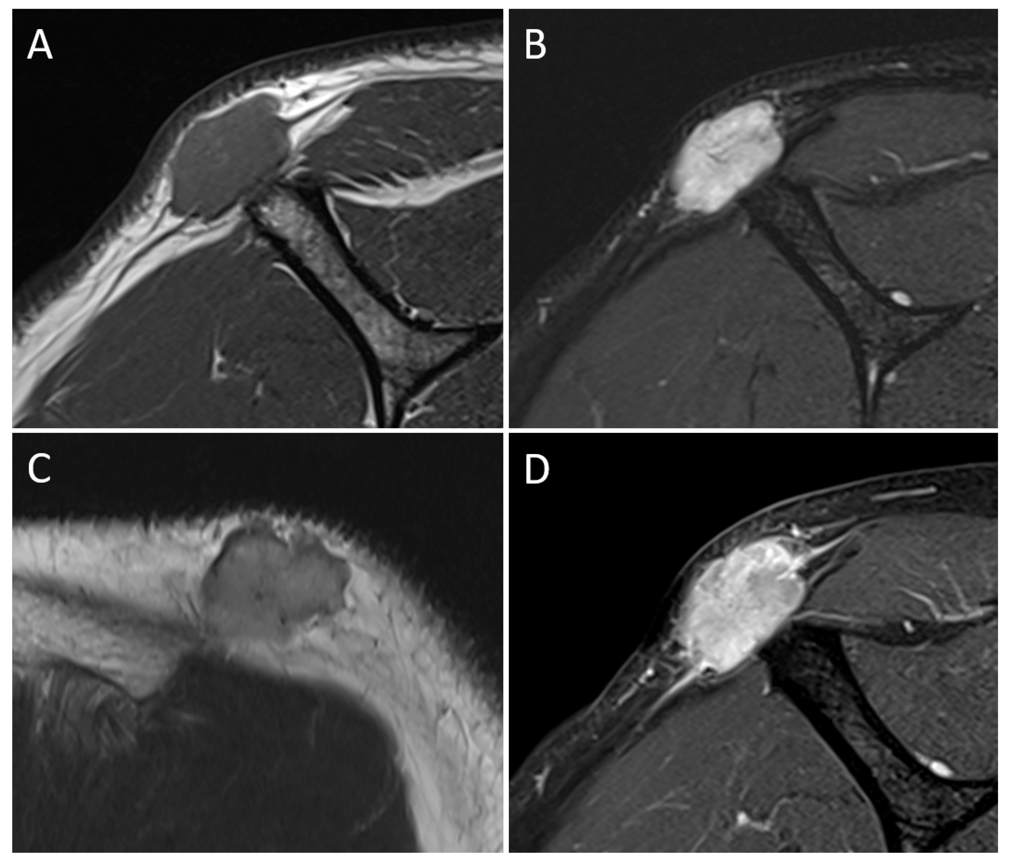
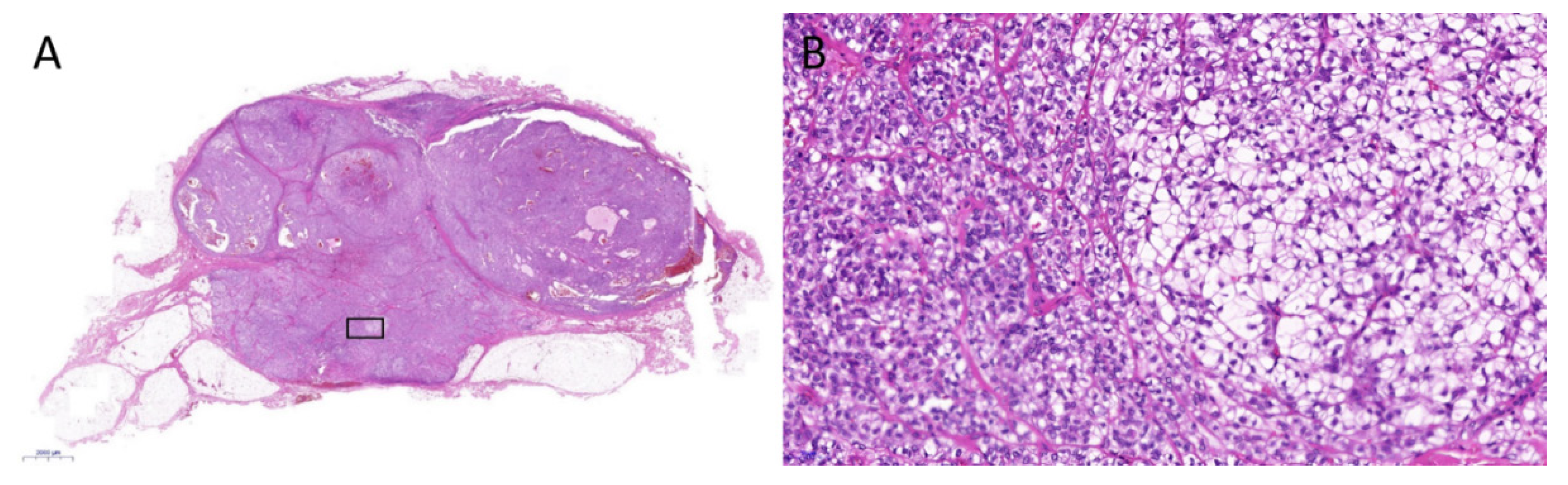
2. Case Report
3. Discussion
4. Conclusions
Author Contributions
Funding
Institutional Review Board Statement
Informed Consent Statement
Data Availability Statement
Conflicts of Interest
References
- Rastrelli, M.; Del Fiore, P.; Damiani, G.B.; Mocellin, S.; Tropea, S.; Spina, R.; Costa, A.; Cavallin, F.; Rossi, C.R. Myoepithelioma of the soft tissue: A systematic review of clinical reports. Eur. J. Surg. Oncol. 2019, 45, 1520–1526. [Google Scholar] [CrossRef] [PubMed]
- Balachander, N.; Masthan, K.M.; Babu, N.A.; Anbazhagan, V. Myoepithelial cells in pathology. J. Pharm. Bioallied. Sci. 2015, 7, S190–S193. [Google Scholar] [CrossRef]
- Kilpatrick, S.E.; Hitchcock, M.G.; Kraus, M.D.; Calonje, E.; Fletcher, C.D. Mixed tumors and myoepitheliomas of soft tissue: A clinicopathologic study of 19 cases with a unifying concept. Am. J. Surg. Pathol. 1997, 21, 13–22. [Google Scholar] [CrossRef] [PubMed]
- Elsensohn, A.; Mo, J.H.; Maly, T.J.; Lee, P.K.; de Feraudy, S. Myoepithelioma of Soft Tissue With Both Squamous and Adipocytic Metaplasia. Am. J. Dermatopathol. 2018, 40, 142–144. [Google Scholar] [CrossRef] [PubMed]
- Kadlub, N.; Galiani, E.; Fraitag, S.; Boudjema, S.; Vazquez, M.P.; Coulomb, A.; Picard, A. Soft tissue myoepithelioma of the scalp in a 11-year-old girl: A challenging diagnosis. Pediatr. Dermatol. 2012, 29, 345–348. [Google Scholar] [CrossRef] [PubMed]
- Plaza, J.A.; Brenn, T.; Chung, C.; Salim, S.; Linos, K.D.; Jour, G.; Duran Rincon, J.; Wick, M.; Sangueza, M.; Gru, A.A. Histomorphological and immunophenotypical spectrum of cutaneous myoepitheliomas: A series of 35 cases. J. Cutan. Pathol. 2021, 48, 847–855. [Google Scholar] [CrossRef]
- Fletcher, C.D.M.; World Health Organization. International Agency for Research on Cancer. In WHO Classification of Tumours of Soft Tissue and Bone, 4th ed.; IARC Press: Lyon, France, 2013; p. 468. [Google Scholar]
- Hornick, J.L.; Fletcher, C.D. Cutaneous myoepithelioma: A clinicopathologic and immunohistochemical study of 14 cases. Hum. Pathol. 2004, 35, 14–24. [Google Scholar] [CrossRef]
- Mentzel, T.; Requena, L.; Kaddu, S.; Soares de Aleida, L.M.; Sangueza, O.P.; Kutzner, H. Cutaneous myoepithelial neoplasms: Clinicopathologic and immunohistochemical study of 20 cases suggesting a continuous spectrum ranging from benign mixed tumor of the skin to cutaneous myoepithelioma and myoepithelial carcinoma. J. Cutan. Pathol. 2003, 30, 294–302. [Google Scholar] [CrossRef]
- Flucke, U.; Palmedo, G.; Blankenhorn, N.; Slootweg, P.J.; Kutzner, H.; Mentzel, T. EWSR1 gene rearrangement occurs in a subset of cutaneous myoepithelial tumors: A study of 18 cases. Mod. Pathol. 2011, 24, 1444–1450. [Google Scholar] [CrossRef]
- Frost, M.W.; Steiniche, T.; Damsgaard, T.E.; Stolle, L.B. Primary cutaneous myoepithelial carcinoma: A case report and review of the literature. Apmis 2014, 122, 369–379. [Google Scholar] [CrossRef]
- Hornick, J.L.; Fletcher, C.D. Myoepithelial tumors of soft tissue: A clinicopathologic and immunohistochemical study of 101 cases with evaluation of prognostic parameters. Am. J. Surg. Pathol. 2003, 27, 1183–1196. [Google Scholar] [CrossRef] [PubMed]
- Blacksin, M.F.; Ha, D.H.; Hameed, M.; Aisner, S. Superficial soft-tissue masses of the extremities. Radiographics 2006, 26, 1289–1304. [Google Scholar] [CrossRef] [PubMed]
- Sharon, C.E.; Straker, R.J., 3rd; Karakousis, G.C. The Role of Imaging in Soft Tissue Sarcoma Diagnosis and Management. Surg. Clin. N. Am. 2022, 102, 539–550. [Google Scholar] [CrossRef]
- Jacobson, J.A.; Middleton, W.D.; Allison, S.J.; Dahiya, N.; Lee, K.S.; Levine, B.D.; Lucas, D.R.; Murphey, M.D.; Nazarian, L.N.; Siegel, G.W.; et al. Ultrasonography of Superficial Soft-Tissue Masses: Society of Radiologists in Ultrasound Consensus Conference Statement. Radiology 2022, 304, 18–30. [Google Scholar] [CrossRef]
- Inampudi, P.; Jacobson, J.A.; Fessell, D.P.; Carlos, R.C.; Patel, S.V.; Delaney-Sathy, L.O.; van Holsbeeck, M.T. Soft-tissue lipomas: Accuracy of sonography in diagnosis with pathologic correlation. Radiology 2004, 233, 763–767. [Google Scholar] [CrossRef]
- Behan, M.; Kazam, E. The echographic characteristics of fatty tissues and tumors. Radiology 1978, 129, 143–151. [Google Scholar] [CrossRef] [PubMed]
- Carra, B.J.; Bui-Mansfield, L.T.; O’Brien, S.D.; Chen, D.C. Sonography of musculoskeletal soft-tissue masses: Techniques, pearls, and pitfalls. AJR. Am. J. Roentgenol. 2014, 202, 1281–1290. [Google Scholar] [CrossRef]
- Catalano, O.; Roldán, F.A.; Varelli, C.; Bard, R.; Corvino, A.; Wortsman, X. Skin cancer: Findings and role of high-resolution ultrasound. J. Ultrasound 2019, 22, 423–431. [Google Scholar] [CrossRef] [PubMed]
- Paixao, C.; Lustig, J.P.; Causeret, S.; Chaigneau, L.; Danner, A.; Aubry, S. Tumors and pseudotumors of the soft tissues: Imaging semiology and strategy. J. Clin. Imaging Sci. 2021, 11, 13. [Google Scholar] [CrossRef] [PubMed]
- Kransdorf, M.J.; Bancroft, L.W.; Peterson, J.J.; Murphey, M.D.; Foster, W.C.; Temple, H.T. Imaging of fatty tumors: Distinction of lipoma and well-differentiated liposarcoma. Radiology 2002, 224, 99–104. [Google Scholar] [CrossRef]
- Calleja, M.; Dimigen, M.; Saifuddin, A. MRI of superficial soft tissue masses: Analysis of features useful in distinguishing between benign and malignant lesions. Skelet. Radiol. 2012, 41, 1517–1524. [Google Scholar] [CrossRef] [PubMed]
- Shu, H.; Ma, Q.; Li, A.; Wang, P.; Gao, Y.; Yao, Q.; Hu, Y.; Ye, X. Diagnostic Performance of US and MRI in Predicting Malignancy of Soft Tissue Masses: Using a Scoring System. Front. Oncol. 2022, 12, 853232. [Google Scholar] [CrossRef] [PubMed]
- Rastrelli, M.; Passuello, N.; Cecchin, D.; Basso, U.; Tosi, A.L.; Rossi, C.R. Metastatic malignant soft tissue myoepithelioma: A case report showing complete response after locoregional and systemic therapy. J. Surg. Case Rep. 2013, 2013, rjt109. [Google Scholar] [CrossRef] [PubMed]
- Hashimoto, K.; Nishimura, S.; Chikugo, T.; Kakinoki, R.; Akagi, M. Soft Tissue Myoepithelioma of the Shoulder. Acta Med. Okayama 2020, 74, 531–535. [Google Scholar] [CrossRef]
- Trevino, M.; Moorthy, C.; Kafchinski, L.; Bustamante, D. Foot plantar soft tissue malignant myoepithelioma tumor: Case report and review of the literature. Clin. Imaging 2020, 61, 90–94. [Google Scholar] [CrossRef]
- Lim, H.-C.; Yu, I.-K.; Park, M.-J.; Jang, D.-S. Computed Tomography and Magnetic Resonance Imaging of Myoepitheliloma in the Soft Palate: A Case Report. J. Korean Soc. Radiol. 2011, 65, 23. [Google Scholar] [CrossRef]
- Kim, H.S.; Lee, W.M.; Choi, S.M. Myoepitheliomas of the soft palate: Helical CT findings in two patients. Korean J. Radiol. 2007, 8, 552–555. [Google Scholar] [CrossRef]
- Chamberlain, F.; Cojocaru, E.; Scaranti, M.; Noujaim, J.; Constantinou, A.; Thway, K.; Fisher, C.; Messiou, C.; Strauss, D.C.; Miah, A.; et al. Adult soft tissue myoepithelial carcinoma: Treatment outcomes and efficacy of chemotherapy. Med. Oncol. 2019, 37, 13. [Google Scholar] [CrossRef]


| Author, Year | Age (Years) /Sex | Location | Size (cm) | Pathologic Diagnosis | US Findings | MRI Findings |
|---|---|---|---|---|---|---|
| Rastrelli et al., 2013 [24] | 61/M | Leg, subcutaneous layer | Myoepithelial carcinoma | Lobulated heterogeneous hyperechoic mass | ||
| Hashimoto et al., 2020 [25] | 72/W | Shoulder, intramuscular | 8.3 × 6.5 | Myoepithelioma | Mass with low signal intensity on T1WI, high signal intensity on T2WI, and heterogeneous enhancement | |
| Trevino et al., 2020 [26] | 12/M | Plantar foot, intramuscular | 5 × 2 | Myoepithelial carcinoma | Mass with intermediate signal intensity on T1WI, heterogeneous high signal intensity on T2WI/STIR, and intense enhancement | |
| Current case | 69/M | Shoulder, subcutaneous layer | 2.8 × 1.7 | Myoepithelioma | Lobulated hyperechoic mass with internal striations and no internal vascularity | Mass with low signal intensity on T1WI, high signal intensity on fat-suppressed T2WI, intermediate signal intensity on T2WI, and intense enhancement with adjacent fascial thickening |
Disclaimer/Publisher’s Note: The statements, opinions and data contained in all publications are solely those of the individual author(s) and contributor(s) and not of MDPI and/or the editor(s). MDPI and/or the editor(s) disclaim responsibility for any injury to people or property resulting from any ideas, methods, instructions or products referred to in the content. |
© 2023 by the authors. Licensee MDPI, Basel, Switzerland. This article is an open access article distributed under the terms and conditions of the Creative Commons Attribution (CC BY) license (https://creativecommons.org/licenses/by/4.0/).
Share and Cite
Koo, M.; Wi, Y.C.; Kim, J.; Lee, S.-W. Subcutaneous Myoepithelioma in the Extremity: A Potential Pitfall in the Differential Diagnosis of Subcutaneous Tumors. Medicina 2023, 59, 667. https://doi.org/10.3390/medicina59040667
Koo M, Wi YC, Kim J, Lee S-W. Subcutaneous Myoepithelioma in the Extremity: A Potential Pitfall in the Differential Diagnosis of Subcutaneous Tumors. Medicina. 2023; 59(4):667. https://doi.org/10.3390/medicina59040667
Chicago/Turabian StyleKoo, Minsun, Young Chan Wi, Jimin Kim, and Sheen-Woo Lee. 2023. "Subcutaneous Myoepithelioma in the Extremity: A Potential Pitfall in the Differential Diagnosis of Subcutaneous Tumors" Medicina 59, no. 4: 667. https://doi.org/10.3390/medicina59040667
APA StyleKoo, M., Wi, Y. C., Kim, J., & Lee, S.-W. (2023). Subcutaneous Myoepithelioma in the Extremity: A Potential Pitfall in the Differential Diagnosis of Subcutaneous Tumors. Medicina, 59(4), 667. https://doi.org/10.3390/medicina59040667