Review

Zebrafish Models of Diamond-Blackfan Anemia: A Tool for Understanding the Disease Pathogenesis and Drug Discovery

Frontier Science Research Center, University of Miyazaki, 5200 Kihara, Kiyotake, Miyazaki 889-1692, Japan
*
Author to whom correspondence should be addressed.
Pharmaceuticals 2019, 12(4), 151; https://doi.org/10.3390/ph12040151
Submission received: 3 September 2019 / Revised: 8 October 2019 / Accepted: 8 October 2019 / Published: 9 October 2019
(This article belongs to the Special Issue Zebrafish as a Powerful Tool for Drug Discovery)

Abstract

:
Diamond-Blackfan anemia (DBA) is a rare bone marrow failure syndrome characterized by red blood cell aplasia. Currently, mutations in 19 ribosomal protein genes have been identified in patients. However, the pathogenic mechanism of DBA remains unknown. Recently, several DBA models were generated in zebrafish (Danio rerio) to elucidate the molecular pathogenesis of disease and to explore novel treatments. Zebrafish have strong advantages in drug discovery due to their rapid development and transparency during embryogenesis and their applicability to chemical screens. Together with mice, zebrafish have now become a powerful tool for studying disease mechanisms and drug discovery. In this review, we introduce recent advances in DBA drug development and discuss the usefulness of zebrafish as a disease model.

1. Introduction

Mutations in the genes encoding ribosome components and ribosome biogenesis factors lead to disease conditions called ‘ribosomopathies’ [1,2]. The patients commonly display hematopoietic failures, skin and skeletal abnormalities, and cancer predisposition. Currently, various ribosomal protein (RP) genes and ribosome biogenesis or assembly factor genes have been identified as the disease-causing genes (Table 1). Diamond-Blackfan anemia (DBA; MIM# 105650) is the first discovered and most widely studied ribosomopathy, in which mutations in as many as 19 RP genes have been identified [2,3,4]. Although extensive efforts have been made during the last two decades, the pathogenic mechanism of DBA remains poorly understood, and there are no clinically effective treatments available. This is because of the limited availability of patient samples and the lack of appropriate animal models, which is a common issue for ribosomopathies.
To overcome these problems, attempts have been made to develop various disease models that exhibit DBA phenotypes, including patient-derived cultured cells or iPS cells, knockdown or knockout mouse models of causative genes, and others [5,6,7,8]. However, patient cells are scarce and difficult to obtain. Additionally, it is rather difficult to recapitulate the complex patient phenotypes with cultured cells. Knockout mice harboring a homozygous mutation of the causative RP genes died in a very early stage of development, and the heterozygous mutants displayed no abnormal symptoms [7].
The zebrafish (Danio rerio) is a small freshwater fish originating from India and is a useful model animal for studying human diseases [9,10,11]. There are many attractive advantages of using zebrafish as a DBA model, including not only the fast development and transparency of embryos but also the conserved hematopoietic regulation in humans. In addition, various technologies are available to create disease models, such as the knockdown of a target gene using morpholino antisense oligonucleotides (MOs) or the knockout using a CRISPR/Cas9 system [12,13]. Valuable resources are also available. There is a large resource center at the University of Oregon called ZIRC (Zebrafish International Resource Center), where thousands of mutant lines are maintained and detailed information about phenotypes and relevant literature is provided.
For these reasons, zebrafish have been recognized as a promising animal model of DBA, and several groups, including us, successfully developed valuable zebrafish models of this disease [10,14]. These models enabled us to gain an in-depth understanding of the disease mechanism and to provide platforms for empowering future studies. In this review, we introduce recent progress in developing zebrafish DBA models and exploring drug candidates for DBA.

2. Clinical Features and Associated Genes

DBA is an inherited pure red cell aplasia and displays hypoplastic anemia with reduced erythroid progenitors in the bone marrow [15]. Approximately half of the patients also display congenital anomalies, including thumb deformity, webbed neck, cleft palate, and cardiac malformation. In addition, these patients have an increased risk of cancer development, such as acute myeloid leukemia, myelodysplastic syndrome, colon cancer, and osteogenic sarcoma [2]. Patients are generally diagnosed during infancy or early childhood with an incidence rate of approximately 7 per million live births.
Treatment with corticosteroids, such as dexamethasone, can improve the anemia phenotype in 80% of cases, which is the first-line treatment for DBA [15]. However, patients often lose their response over long-term corticosteroid therapy and become dependent on regular red blood cell transfusions, resulting in adverse iron overload that has to be managed through the life [16]. Hematopoietic stem cell transplantation, the only curative treatment of DBA, is also available, but this procedure is restricted to those with a matched related donor and still exhibits a considerable risk of mortality associated with the treatment. Thus, there is a strong demand for new therapeutics for DBA.
To date, heterozygous mutations in 19 RP genes (RPS7, RPS10, RPS15A, RPS17, RPS19, RPS24, RPS26, RPS27, RPS28, RPS29, RPL5, RPL11, RPL15, RPL18, RPL26, RPL27, RPL31, RPL35, RPL35A) have been identified in DBA patients [17,18]. These mutations account for ~70% of the mutations found in patients. In addition, mutations in GATA1, a hematopoietic master transcription factor gene necessary for proper erythroid production and mutations in TSR2, an RPS26 chaperone protein gene, were also identified in a small number of patients [19,20].
Various studies have shown that defects in ribosome biogenesis causes nucleolar stress that triggers a p53 signaling pathway, which highlights a possible role of p53 in DBA pathogenesis [21,22]. Several ribosomal proteins, including RPL5 and RPL11, have been identified as key players that mediate p53 responses through interactions with murine double minute 2 (MDM2), the negative regulator of p53. Analyses of mouse and zebrafish DBA models also implicated p53 as the major contributor to the pathophysiology of DBA (see later sections) [23,24]. However, it remains controversial whether the p53-dependent pathway directly links to an anemia phenotype in DBA [25,26]. In addition, the observation that RPL5 and RPL11 are frequently mutated in the patients challenges the role of p53 in DBA pathophysiology. Recently, it was proposed that ribosome defects caused by RP haploinsufficiency or its faulty assembly resulted in reduced translation of a subset of transcripts such as GATA1 mRNA, thereby impeding erythroid lineage commitment in DBA [27,28]. Although the mechanism underlying such selective alterations of mRNA translation is not fully understood, it may explain the pathogenic mechanisms of ribosomopathies including DBA.

3. Mouse and Zebrafish DBA Models

3.1. Mouse Models

To understand the disease mechanisms and to evaluate novel therapies, the generation of animal models is crucial. The first attempt to create a mouse model for DBA was carried out by Matsson et al. in 2004 [7], where they disrupted the mouse ortholog of human RPS19, which is the most frequently mutated gene in DBA patients. However, this trial ended up with embryonic lethality for mice harboring homozygous loss of Rps19 and a normal hematopoietic phenotype for mice harboring heterozygous loss of Rps19. Four years later, another mouse model of DBA was developed that carries a heterozygous missense mutation in Rps19 (Dsk3 mutant). However, unlike DBA patients, the Dsk3 mutants showed only mild anemia and a more prominent pigmented phenotype without any physical deformities [24]. Furthermore, a transgenic mouse model expressing an RPS19 missense mutation at codon 62 (RPS19R62W) demonstrated growth retardation and a mild anemia with a reduced number of erythroid progenitors [29]. However, this mutation was thought to act by a dominant negative mechanism, which is different from the generally believed haploinsufficiency model for DBA.
In 2011, Jaako et al. finally generated a mouse model of DBA with decreased erythrocyte production using transgenic RNA interference, where the synthesis of Rps19 was downregulated by a doxycycline-regulated Rps19-targeting shRNA [8]. The Rps19-deficient mice displayed macrocytic anemia, leukocytopenia, and variable platelet counts. Subsequently, Morgado-Palacin et al. generated mice with an inducible Rpl11-null allele and demonstrated that heterozygous loss of Rpl11 in adult mice impaired erythrocyte maturation [30]. Interestingly, these mice also showed reduced p53 responses and increased cMYC levels. Together, these defects resulted in anemia and cancer susceptibility, thereby recapitulating DBA. Rps19 knockdown and Rpl11 haploinsufficient mice proved to be useful, but they have not become widely used due to their limited availability. Moreover, attempting high-throughput exploration of drug candidates using such mouse models is generally difficult.

3.2. Zebrafish Models

To compensate for the difficulties and problems encountered by the mouse models, we and other groups focused on zebrafish for more convenient and high-throughput analyses. Knockdown of zebrafish rps19 nicely recapitulated DBA phenotypes [14,23,25], which displayed a drastic reduction in red blood cells (Figure 1). Interestingly, as observed in DBA patients, only erythroid cell differentiation was affected, but other myeloid and endothelial cells were unaffected. The knockdown embryos also showed deformities in the head and tail regions, which may be relevant to the skeletal abnormalities observed in the patients. These phenotypes were rescued by injection of wild-type rps19 mRNAs but not by mRNAs with patient-type mutations [14]. Subsequently, other RP gene knockdown models were also generated, and together with the rps19-knockdown embryos, these models have been used for exploring DBA pathogenesis [10].
As described above, a role of p53 signaling pathway is controversial. Danilova et al. showed that both developmental abnormalities and erythroid reduction were caused by activation of p53 [23], whereas Torihara et al. demonstrated that only developmental abnormalities are dependent on the p53 pathway but erythroid defect was independent, which was shown by a simultaneous knockdown of rps19 and tp53 in zebrafish [25]. Similarly, p53-inependent erythropoiesis failure was demonstrated in rpl35a and rps24 knockdown embryos using tp53 mutant zebrafish [31]. In contrast, a knockdown of rps7 was somewhat complicated, which exhibited both p53 dependent and independent features [32].
Valuable zebrafish mutant lines have also been developed. Amsterdam et al. generated several hundred lines of zebrafish, each heterozygous for a recessive embryonic lethal mutation, by insertional mutagenesis. These include RP gene mutants and surprisingly many of them developed malignant peripheral nerve sheath tumors, suggesting that many RP genes are cancer genes in zebrafish [11]. Subsequently, Taylor et al. found that one of these mutants, rps29-/-, displayed decreased red blood cell production and increased apoptosis in the head region during early development [33]. Both the hematopoietic and apoptotic phenotypes were rescued by p53 inactivation, which is in good agreement with a speculated role of p53 in the pathogenic mechanism of DBA. Similarly, rpl11 mutant identified from the same mutagenesis screen showed anemia, decreased hematopoietic stem cells, and activation of the p53 pathway with altered expression in genes involved in cell cycle arrest and apoptosis [34].
Recently, the progress of genome editing technologies, such as the TALEN and CRISPR/Cas9 systems, enabled us to develop zebrafish mutants more rapidly and precisely. Zhang et al. successfully generated an rps19 null mutant through TALEN-mediated gene targeting, which reproduced the erythroid defects of DBA, including a lack of mature red blood cells, decreased globin synthesis, and p53 activation [35]. Our group also generated several rps19 mutants that display decreased erythroid production by using the CRISPR/Cas9 system (Figure 1, unpublished data). All these mutants are useful resources for DBA research, especially for in vivo drug screening and evaluations of the efficacy.

4. Drug Development

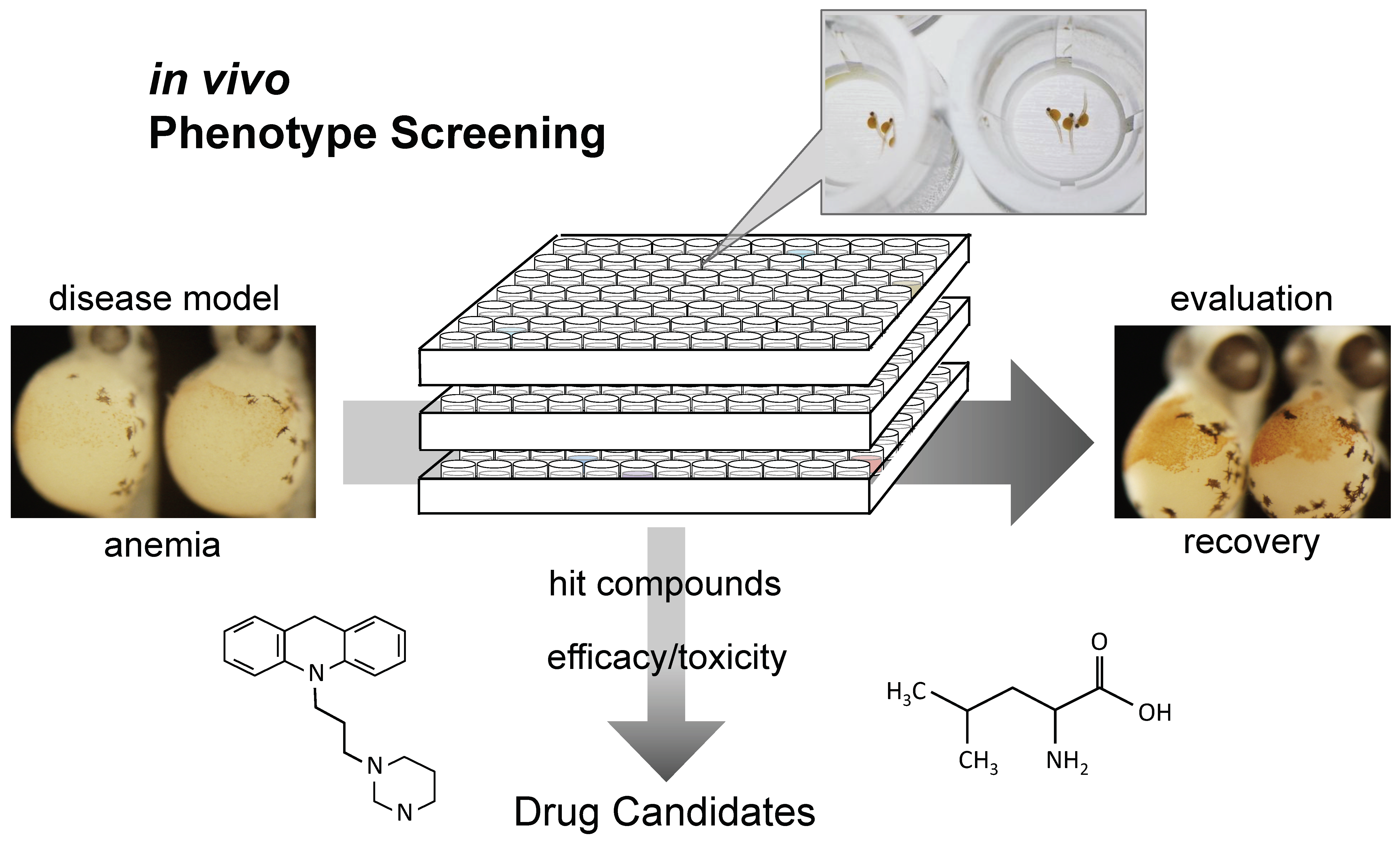
Corticosteroid treatments and chronic transfusion are the main therapeutic regimens in DBA. Although these therapies can alleviate anemia, both cause severe long-term toxicities. To develop new therapeutics for DBA, zebrafish provide a powerful tool for evaluating drug efficacy and screening compound libraries (Figure 2, Table 2).

4.1. l-leucine

The amino acid l-leucine was the first molecule found to improve anemia in a zebrafish model of DBA. Payne et al. treated rps19-knockdown zebrafish with l-leucine and found that it improved the anemia and developmental defects associated with DBA [36]. These findings were confirmed in primary human CD34+ cells after shRNA knockdown of RPS19. l-Leucine was also effective in a mouse model of DBA. Administration of l-leucine significantly improved anemia in Rps19-deficient mice, showing increased erythrocyte numbers and hemoglobin concentration, and associating with decreased p53 activity [37]. Because erythroid progenitor cells have a very high rate of proliferation and require a high level of ribosome biogenesis, the erythroid lineage may be particularly sensitive to reduced expression levels of ribosomal proteins. On the other hand, l-leucine is thought to boost translation by upregulating ribosome biosynthesis through the mTOR pathway [38]. This may explain why l-leucine is effective in zebrafish and mouse models of DBA. A pilot phase I/II study of l-leucine in the treatment of patients with transfusion-dependent DBA is in progress under ClinicalTrials.gov with the number NCT01362595.

4.2. Sotatercept

Sotatercept (ACE-011) is an activin receptor type IIA ligand trap that inhibits transforming growth factor beta (TGF-β) superfamily members, including growth differentiation factor 11 (GDF-11) and activin B [39]. It was generated by recombinant fusion of the extracellular domain of the human activin receptor IIA and the human immunoglobulin G1 Fc domain. GDF-11 is overexpressed in immature erythroblasts in β-thalassemia, and an activin receptor IIA ligand trap was able to correct ineffective erythropoiesis in patients [40]. Ear et al. investigated the ability of the murine ortholog of sotatercept (RAP-011) to restore erythroid levels in zebrafish models of DBA, including rps19 and rpl11 knockdown embryos and to elucidate the role of TGF-signaling molecules during ribosome stress [41]. They demonstrated that RAP-011 was able to elevate erythroid counts under normal physiological conditions and during ribosome deficiency in zebrafish. In addition, RAP-011 was shown to block the function of lefty1 (Lft1), an activin/nodal signaling antagonist, and its overexpression delayed the maturation of erythroid cells. In this context, increased expression of Lft1 was observed in rps19 and rpl11-mutant zebrafish. A safety and efficacy study of sotatercept in adults with transfusion-dependent DBA is in progress under ClinicalTrials.gov with the number NCT01464164.

4.3. Trifluoperazine

A chemical screen using zebrafish rps29-/- embryos identified several calmodulin (CaM) inhibitors that can rescue hemoglobin levels in mutant embryos [42]. Subsequently, the effects of the CaM inhibitor trifluoperazine (TFP) were examined in human and murine models. Treating RPS19-deficient cord blood-derived CD34+ cells with TFP relieved the erythroid differentiation block, and injection of TFP into an Rps19-deficient mouse model ameliorated erythroid production and hemoglobin level. In addition, TFP was suggested to inhibit p53 transcriptional activity through its c-terminal domain (CTD), thereby rescues the phenotypes caused by RP deficiency [43]. CaM inhibitors may be effective therapies for DBA patients. A phase I/II study to determine the safety of TFP in transfusion-dependent DBA has just started and can be found at ClinicalTrials.gov with the number NCT03966053.

4.4. SMER28

Screening of a chemical library using iPS cells from DBA patients identified SMER28, an inducer of autophagy, as a hit compound that enhances erythroid differentiation of iPS cells [44]. These cells were established from the fibroblasts of patients with RPS19 and RPL5 inactivating mutations. To assess the therapeutic potential of SMER28 in vivo, a zebrafish model of DBA was tested. Treatment with SMER28 improved erythroid production in rps29-/- zebrafish in a dose-dependent manner. More than 50% of embryos treated with 1 μM SMER28 showed high expression of hemoglobin compared to 20% for the controls. SMER28 was also examined its effects on mice with irradiation-induced anemia, which resulted in elevated RBC counts and hematocrit. Furthermore, the effects of SMER28 were tested in Rpl11+/fl/Mx1-Cre mice that displayed decreased erythropoiesis, in which deletion of Rpl11 was under the control of poly(I:C)-inducible Cre. The poly(I:C)-induced anemic mice receiving SMER28 (20 mg/kg) showed significant improvement of RBC counts [44]. Because the effects of SMER28 on human cells have been confirmed as well, SMER28 or its derivatives may be viable alternatives to current treatments for DBA. Clinical studies on the safety and efficacy of SMER28 are needed.

5. Conclusions

Zebrafish DBA models were generated and have now become powerful tools for studying DBA pathogenesis. More importantly, zebrafish have made in vivo chemical screening possible, which will lead to the identification of compounds that are effective for DBA therapies (Figure 2). L-Leucine, sotatercept, and trifluoperazine, identified using zebrafish DBA models, are now under evaluation in clinical trials. The divergent pathways illuminated by these chemicals also shed light on the pathophysiology of DBA. Zebrafish in vivo phenotype screening followed by validation of efficacy in mouse models will be a promising strategy to develop new therapeutics for rare diseases such as DBA.

Author Contributions

N.K. conceptualized and wrote the paper and T.U. wrote the paper.

Funding

This research received no external funding.

Conflicts of Interest

The authors declare no conflict of interest.

References

  1. Narla, A.; Ebert, B.L. Ribosomopathies: Human disorders of ribosome dysfunction. Blood 2010, 115, 3196–3205. [Google Scholar] [CrossRef] [PubMed]
  2. Aspesi, A.; Ellis, S.R. Rare ribosomopathies: Insights into mechanisms of cancer. Nat. Rev. Cancer 2019, 19, 228–238. [Google Scholar] [CrossRef] [PubMed]
  3. Diamond, L.K.; Blackfan, K.D. Hypoplastic anemia. Am. J. Dis. Child. 1938, 56, 464–467. [Google Scholar]
  4. Boria, I.; Garelli, E.; Gazda, H.T.; Aspesi, A.; Quarello, P.; Pavesi, E.; Ferrante, D.; Meerpohl, J.J.; Kartal, M.; Da Costa, L.; et al. The ribosomal basis of Diamond-Blackfan anemia: Mutation and database update. Hum. Mutat. 2010, 31, 1269–1279. [Google Scholar] [CrossRef] [PubMed]
  5. Lipton, J.M.; Kudisch, M.; Gross, R.; Nathan, D.G. Defective erythroid progenitor differentiation system in congenital hypoplastic (Diamond-Blackfan) anemia. Blood 1986, 67, 962–968. [Google Scholar] [PubMed]
  6. Garçon, L.; Ge, J.; Manjunath, S.H.; Mills, J.A.; Apicella, M.; Parikh, S.; Sullivan, L.M.; Podsakoff, G.M.; Gadue, P.; French, D.L.; et al. Ribosomal and hematopoietic defects in induced pluripotent stem cells derived from Diamond Blackfan anemia patients. Blood 2013, 122, 912–921. [Google Scholar] [CrossRef] [PubMed]
  7. Matsson, H.; Davey, E.J.; Draptchinskaia, N.; Hamaguchi, I.; Ooka, A.; Levéen, P.; Forsberg, E.; Karlsson, S.; Dahl, N. Targeted disruption of the ribosomal protein S19 gene is lethal prior to implantation. Mol. Cell. Biol. 2004, 24, 4032–4037. [Google Scholar] [CrossRef]
  8. Jaako, P.; Flygare, J.; Olsson, K.; Quere, R.; Ehinger, M.; Henson, A.; Ellis, S.; Schambach, A.; Baum, C.; Richter, J.; et al. Mice with ribosomal protein S19 deficiency develop bone marrow failure and symptoms like patients with Diamond-Blackfan anemia. Blood 2011, 118, 6087–6096. [Google Scholar] [CrossRef] [PubMed] [Green Version]
  9. Ablain, J.; Zon, L.I. Of fish and men: Using zebrafish to fight human diseases. Trends Cell Biol. 2013, 23, 584–586. [Google Scholar] [CrossRef]
  10. Oyarbide, U.; Topczewski, J.; Corey, S.J. Peering through zebrafish to understand inherited bone marrow failure syndromes. Haematologica 2019, 104, 13–24. [Google Scholar] [CrossRef]
  11. Amsterdam, A.; Sadler, K.C.; Lai, K.; Farrington, S.; Bronson, R.T.; Lees, J.A.; Hopkins, N. Many ribosomal protein genes are cancer genes in zebrafish. PLoS Biol. 2004, 2, E139. [Google Scholar] [CrossRef] [PubMed]
  12. Uechi, T.; Nakajima, Y.; Nakao, A.; Torihara, H.; Chakraborty, A.; Inoue, K.; Kenmochi, N. Ribosomal protein gene knockdown causes developmental defects in zebrafish. PLoS ONE 2006, 1, e37. [Google Scholar] [CrossRef] [PubMed]
  13. Hwang, W.Y.; Fu, Y.; Reyon, D.; Maeder, M.L.; Tsai, S.Q.; Sander, J.D.; Peterson, R.T.; Yeh, J.R.; Joung, J.K. Efficient genome editing in zebrafish using a CRISPR-Cas system. Nat. Biotechnol. 2013, 31, 227–229. [Google Scholar] [CrossRef] [PubMed]
  14. Uechi, T.; Nakajima, Y.; Chakraborty, A.; Torihara, H.; Higa, S.; Kenmochi, N. Deficiency of ribosomal protein S19 during early embryogenesis leads to reduction of erythrocytes in a zebrafish model of Diamond-Blackfan anemia. Hum. Mol. Genet. 2008, 17, 3204–3211. [Google Scholar] [CrossRef] [PubMed] [Green Version]
  15. Vlachos, A.; Ball, S.; Dahl, N.; Alter, B.P.; Sheth, S.; Ramenghi, U.; Meerpohl, J.; Karlsson, S.; Liu, J.M.; Leblanc, T.; et al. Diagnosing and treating Diamond Blackfan anaemia: Results of an international clinical consensus conference. Br. J. Haematol. 2008, 142, 859–876. [Google Scholar] [CrossRef] [PubMed]
  16. Lipton, J.M.; Atsidaftos, E.; Zyskind, I.; Vlachos, A. Improving clinical care and elucidating the pathophysiology of Diamond Blackfan anemia: An update from the Diamond Blackfan Anemia Registry. Pediatr. Blood Cancer 2006, 46, 558–564. [Google Scholar] [CrossRef] [PubMed]
  17. Draptchinskaia, N.; Gustavsson, P.; Andersson, B.; Pettersson, M.; Willig, T.N.; Dianzani, I.; Ball, S.; Tchernia, G.; Klar, J.; Matsson, H.; et al. The gene encoding ribosomal protein S19 is mutated in Diamond-Blackfan anaemia. Nat. Genet. 1999, 21, 169–175. [Google Scholar] [CrossRef] [PubMed]
  18. Ulirsch, J.C.; Verboon, J.M.; Kazerounian, S.; Guo, M.H.; Yuan, D.; Ludwig, L.S.; Handsaker, R.E.; Abdulhay, N.J.; Fiorini, C.; Genovese, G.; et al. The genetic landscape of Diamond-Blackfan anemia. Am. J. Hum. Genet. 2018, 103, 930–947. [Google Scholar] [CrossRef] [PubMed]
  19. Sankaran, V.G.; Ghazvinian, R.; Do, R.; Thiru, P.; Vergilio, J.A.; Beggs, A.H.; Sieff, C.A.; Orkin, S.H.; Nathan, D.G.; Lander, E.S.; et al. Exome sequencing identifies GATA1 mutations resulting in Diamond-Blackfan anemia. J. Clin. Investig. 2012, 122, 2439–2443. [Google Scholar] [CrossRef] [Green Version]
  20. Gripp, K.W.; Curry, C.; Olney, A.H.; Sandoval, C.; Fisher, J.; Chong, J.X.; Pilchman, L.; Sahraoui, R.; Stabley, D.L.; Sol-Church, K. Diamond-Blackfan anemia with mandibulofacial dystostosis is heterogeneous, including the novel DBA genes TSR2 and RPS28. Am. J. Med. Genet. A. 2014, 164, 2240–2249. [Google Scholar] [CrossRef]
  21. Ebert, B.; Lipton, J.M. Diamond Blackfan anemia and ribosome biogenesis: Introduction. Semin. Hematol. 2011, 48, 73–74. [Google Scholar] [CrossRef] [PubMed]
  22. Chakraborty, A.; Uechi, T.; Kenmochi, N. Guarding the ‘translation apparatus’: Defective ribosome biogenesis and the p53 signaling pathway. Wiley Interdiscip. Rev. RNA 2011, 2, 507–522. [Google Scholar] [CrossRef] [PubMed]
  23. Danilova, N.; Sakamoto, K.M.; Lin, S. Ribosomal protein S19 deficiency in zebrafish leads to developmental abnormalities and defective erythropoiesis through activation of p53 protein family. Blood 2008, 112, 5228–5237. [Google Scholar] [CrossRef] [PubMed] [Green Version]
  24. McGowan, K.A.; Li, J.Z.; Park, C.Y.; Beaudry, V.; Tabor, H.K.; Sabnis, A.J.; Zhang, W.; Fuchs, H.; de Angelis, M.H.; Myers, R.M.; et al. Ribosomal mutations cause p53-mediated dark skin and pleiotropic effects. Nat. Genet. 2008, 40, 963–970. [Google Scholar] [CrossRef] [PubMed] [Green Version]
  25. Torihara, H.; Uechi, T.; Chakraborty, A.; Shinya, M.; Sakai, N.; Kenmochi, N. Erythropoiesis failure due to RPS19 deficiency is independent of an activated Tp53 response in a zebrafish model of Diamond-Blackfan anaemia. Br. J. Haematol. 2011, 152, 648–654. [Google Scholar] [CrossRef] [PubMed]
  26. Chakraborty, A.; Uechi, T.; Nakajima, Y.; Gazda, H.T.; O’Donohue, M.F.; Gleizes, P.E.; Kenmochi, N. Cross talk between TP53 and c-Myc in the pathophysiology of Diamond-Blackfan anemia: Evidence from RPL11-deficient in vivo and in vitro models. Biochem. Biophys. Res. Commun. 2018, 495, 1839–1845. [Google Scholar] [CrossRef] [PubMed]
  27. Ludwig, L.S.; Gazda, H.T.; Eng, J.C.; Eichhorn, S.W.; Thiru, P.; Ghazvinian, R.; George, T.I.; Gotlib, J.R.; Beggs, A.H.; Sieff, C.A.; et al. Altered translation of GATA1 in Diamond-Blackfan anemia. Nat. Med. 2014, 20, 748–753. [Google Scholar] [CrossRef] [PubMed] [Green Version]
  28. Khajuria, R.K.; Munschauer, M.; Ulirsch, J.C.; Fiorini, C.; Ludwig, L.S.; McFarland, S.K.; Abdulhay, N.J.; Specht, H.; Keshishian, H.; Mani, D.R.; et al. Ribosome levels selectively regulate translation and lineage commitment in human hematopoiesis. Cell 2018, 173, 90–103. [Google Scholar] [CrossRef] [PubMed]
  29. Devlin, E.E.; Dacosta, L.; Mohandas, N.; Elliott, G.; Bodine, D.M. A transgenic mouse model demonstrates a dominant negative effect of a point mutation in the RPS19 gene associated with Diamond-Blackfan anemia. Blood 2010, 116, 2826–2835. [Google Scholar] [CrossRef] [Green Version]
  30. Morgado-Palacin, L.; Varetti, G.; Llanos, S.; Gómez-López, G.; Martinez, D.; Serrano, M. Partial loss of Rpl11 in adult mice recapitulates Diamond-Blackfan anemia and promotes lymphomagenesis. Cell Rep. 2015, 13, 712–722. [Google Scholar] [CrossRef]
  31. Yadav, G.V.; Chakraborty, A.; Uechi, T.; Kenmochi, N. Ribosomal protein deficiency causes Tp53-independent erythropoiesis failure in zebrafish. Int. J. Biochem. Cell Biol. 2014, 49, 1–7. [Google Scholar] [CrossRef] [PubMed]
  32. Duan, J.; Ba, Q.; Wang, Z.; Hao, M.; Li, X.; Hu, P.; Zhang, D.; Zhang, R.; Wang, H. Knockdown of ribosomal protein S7 causes developmental abnormalities via p53 dependent and independent pathways in zebrafish. Int. J. Biochem. Cell Biol. 2011, 43, 1218–1227. [Google Scholar] [CrossRef] [PubMed]
  33. Taylor, A.M.; Humphries, J.M.; White, R.M.; Murphey, R.D.; Burns, C.E.; Zon, L.I. Hematopoietic defects in rps29 mutant zebrafish depend upon p53 activation. Exp. Hematol. 2012, 40, 228–237. [Google Scholar] [CrossRef] [PubMed]
  34. Danilova, N.; Sakamoto, K.M.; Lin, S. Ribosomal protein L11 mutation in zebrafish leads to haematopoietic and metabolic defects. Br. J. Haematol. 2011, 152, 217–228. [Google Scholar] [CrossRef] [PubMed]
  35. Zhang, Y.; Ear, J.; Yang, Z.; Morimoto, K.; Zhang, B.; Lin, S. Defects of protein production in erythroid cells revealed in a zebrafish Diamond-Blackfan anemia model for mutation in RPS19. Cell Death Dis. 2014, 5, e1352. [Google Scholar] [CrossRef] [PubMed]
  36. Payne, E.M.; Virgilio, M.; Narla, A.; Sun, H.; Levine, M.; Paw, B.H.; Berliner, N.; Look, A.T.; Ebert, B.L.; Khanna-Gupta, A. l-leucine improves the anemia and developmental defects associated with Diamond-Blackfan anemia and del(5q) MDS by activating the mTOR pathway. Blood 2012, 120, 2214–2224. [Google Scholar] [CrossRef] [PubMed]
  37. Jaako, P.; Debnath, S.; Olsson, K.; Bryder, D.; Flygare, J.; Karlsson, S. Dietary l-leucine improves the anemia in a mouse model for Diamond-Blackfan anemia. Blood 2012, 120, 2225–2228. [Google Scholar] [CrossRef] [PubMed]
  38. Stipanuk, M.H. Leucine and protein synthesis: mTOR and beyond. Nutr. Rev. 2007, 65, 122–129. [Google Scholar] [CrossRef] [PubMed]
  39. Carrancio, S.; Markovics, J.; Wong, P.; Leisten, J.; Castiglioni, P.; Groza, M.C.; Raymon, H.K.; Heise, C.; Daniel, T.; Chopra, R.; et al. An activin receptor IIA ligand trap promotes erythropoiesis resulting in a rapid induction of red blood cells and haemoglobin. Br. J. Haematol. 2014, 165, 870–882. [Google Scholar] [CrossRef] [PubMed]
  40. Dussiot, M.; Maciel, T.T.; Fricot, A.; Chartier, C.; Negre, O.; Veiga, J.; Grapton, D.; Paubelle, E.; Payen, E.; Beuzard, Y.; et al. An activin receptor IIA ligand trap corrects ineffective erythropoiesis in beta-thalassemia. Nat. Med. 2014, 20, 398–407. [Google Scholar] [CrossRef]
  41. Ear, J.; Huang, H.; Wilson, T.; Tehrani, Z.; Lindgren, A.; Sung, V.; Laadem, A.; Daniel, T.O.; Chopra, R.; Lin, S. RAP-011 improves erythropoiesis in zebrafish model of Diamond-Blackfan anemia through antagonizing lefty1. Blood 2015, 126, 880–890. [Google Scholar] [CrossRef] [PubMed] [Green Version]
  42. Taylor, A.M.; Raiser, D.M.; Humphries, J.M.; Ebert, B.L.; Zon, L.I. Calmodulin inhibition rescues the effects of ribosomal protein deficiency by modulating p53 activity in models of Diamond Blackfan anemia. Blood 2012, 120, 512. [Google Scholar]
  43. Macari, E.R.; Taylor, A.M.; Raiser, D.M.; Siva, K.; McGrath, K.; Humphries, J.M.; Flygare, J.; Ebert, B.L.; Zon, L.I. Calmodulin inhibition rescues DBA models with ribosomal protein deficiency through reduction of RSK signaling. Blood 2016, 128, 332. [Google Scholar]
  44. Doulatov, S.; Vo, L.T.; Macari, E.R.; Wahlster, L.; Kinney, M.A.; Taylor, A.M.; Barragan, J.; Gupta, M.; McGrath, K.; Lee, H.Y.; et al. Drug discovery for Diamond-Blackfan anemia using reprogrammed hematopoietic progenitors. Sci. Transl. Med. 2017, 9, eaah5645. [Google Scholar] [CrossRef] [PubMed]
  45. Chen, J.; Siva, K.; Rzymski, T.; Johansson, L.; Lundbäck, T.; Villacis, L.N.; Ek, F.; Wang, B.; George, A.J.; Wan, Y.; et al. Small molecule screens identify CDK8-inhibitors as candidate Diamond-Blackfan anemia drugs. Blood 2018, 132, 753. [Google Scholar]
  46. Flygare, J.; Estrada, V.R.; Shin, C.; Gupta, S.; Lodish, H.F. HIF1α synergizes with glucocorticoids to promote BFU-E progenitor self-renewal. Blood 2011, 117, 3435–3444. [Google Scholar] [CrossRef]
  47. Lee, H.Y.; Gao, X.; Barrasa, M.I.; Li, H.; Elmes, R.R.; Peters, L.L.; Lodish, H.F. PPAR-α and glucocorticoid receptor synergize to promote erythroid progenitor self-renewal. Nature 2015, 522, 474–477. [Google Scholar] [CrossRef] [PubMed]
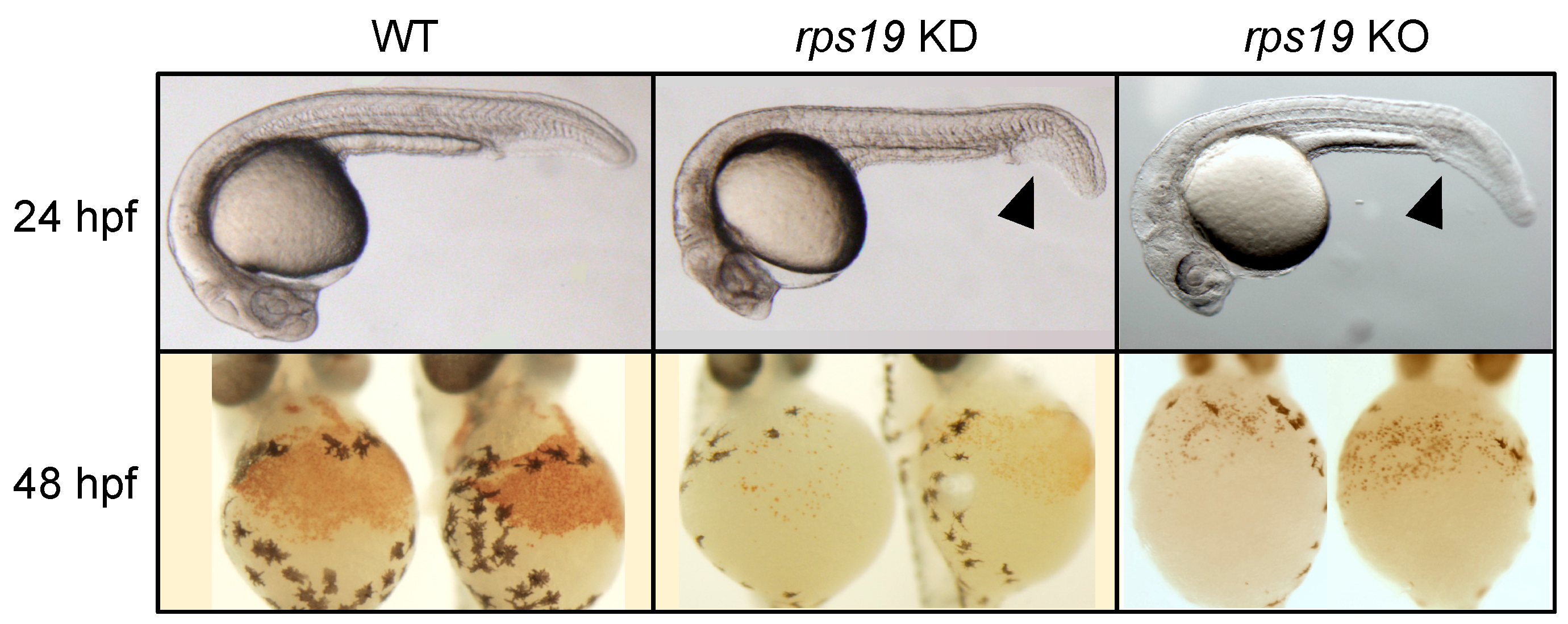
Figure 1. Zebrafish DBA models. Wild-type embryos show a high density of red blood cells (WT), whereas the blood production is drastically reduced in the rps19 knockdown (KD) and knockout (KO) embryos, as indicated by the absence of hemoglobin stained cells (lower panels). These embryos also display developmental deformities in head and tail regions (arrowheads). The abnormal phenotypes were rescued by injection of rps19 mRNA but not rescued by mRNA with patient type mutations in the knockdown embryos [14].
Pharmaceuticals 12 00151 g001
Figure 2. Zebrafish in vivo phenotype screening. Zebrafish provides a novel platform for screening compound libraries and evaluating drug efficacy in vivo, which will lead to find new therapeutics for rare diseases including DBA.
Pharmaceuticals 12 00151 g002
Table 1. Ribosomopathies and causative genes [1,2].
DiseaseGeneFunctionAbnormal Phenotypes
BloodTumorSkinSkeleton
Diamond-Blackfan anemia (DBA)RPS19 and other
18 RP genes
ribosomal protein+++
X-linked dyskeratosis
congenita (DC)
DKC1rRNA modification
enzyme
+++++
Cartilage-hair hypoplasia (CHH)RMRPcleavage of 5.8S
rRNA
+++++++
Shwachman-Diamond
syndrome
SBDSpromoting of 60S
subunit maturation
+++ ++
T-cell acute lymphoblastic
leukemia (T-ALL)
RPL5, RPL10, RPL22ribosomal protein++++ ++
5q- syndromeRPS14ribosomal protein+++
Treacher-Collins syndromeTCOF1, POLR1D,
POLR1C
rDNA transcription ++
Isolated congenital aspleniaRPSAribosomal protein
Note: Ribosomopathies are diseases caused by mutations in the genes encoding ribosomal proteins or RNAs or other ribosome biogenesis factors. They are characterized by hematologic and developmental disorders and often comprise bone marrow failure, skeletal and other developmental abnormalities and cancer predisposition. The mechanism by which a mutation in any ribosomal component results in hematologic disorder is still unclear, although the ribosome widely exists throughout the body.
Table 2. Drug candidates for DBA.
CompoundFunctionDisease ModelClinical Trial, Patent
L-Leucine [36,37,38]activator of protein synthesishCD34+, zebrafish, mouseNCT01362595
Sotatercept [39,40,41]activin receptor type II ligand trapzebrafishNCT01464164
Trifluoperazine [42,43]calmodulin inhibitorhCD34+, zebrafish, mouseNCT03966053
SMER28 [44]inducer of autophagyiPSC, zebrafish, mouseN/A
CDK8 inhibitor [45]inhibitor of cyclin-dependent kinase 8mouse fetal liver cellWO2017076968Al
Dimethyloxalylglycine * [46]prolyl hydroxylase inhibitormBFU-EN/A
GW7647 *, fenofibrate * [47]PPAR-a agonistmBFU-E, hCD34+, mouseN/A
Note: * combination with glucocorticoid. PPAR-a, peroxisome proliferator-activated receptor a; hCD34+, human CD34+ cells; mBFU-E, mouse burst-forming unit erythroid.