Short Note

Dicyclohexylammonium O,O’-Diphenyl Phosphate, [(C6H11)2NH2][(C6H5O)2P(O)(O)]: Spectroscopic Study, Crystal Structure, and Hirshfeld Surface Analysis

by
Samira Nazari Moghaddam
1,
Akbar Raissi Shabari
1,*,
Fahimeh Sabbaghi
2,
Mehrdad Pourayoubi
3,* and
Abdul Ajees Abdul Salam
4,*
1
Department of Chemistry, North Tehran Branch, Islamic Azad University, Tehran 1651153311, Iran
2
Department of Chemistry, Zanjan Branch, Islamic Azad University, Zanjan 45156-58145, Iran
3
Department of Chemistry, Faculty of Science, Ferdowsi University of Mashhad, Mashhad 9177948974, Iran
4
Department of Atomic and Molecular Physics, Centre for Applied Nanosciences, Manipal Institute of Technology Campus, Manipal Academy of Higher Education, Manipal, Karnataka 576 104, India
*
Authors to whom correspondence should be addressed.
Molbank 2019, 2019(1), M1051; https://doi.org/10.3390/M1051
Submission received: 27 January 2019 / Revised: 22 February 2019 / Accepted: 26 February 2019 / Published: 2 March 2019
(This article belongs to the Section Structure Determination)

Abstract

:
The title salt, [(C6H11)2NH2][(C6H5O)2P(O)(O)], crystallizes in the chiral space group P212121, composed of achiral cation and anion components. The strong charge-assisted N–H…O hydrogen bonds build a linear assembly along the a axis, including a non-centrosymmetric C 2 2 (6) chain graph-set motif. The intra and intermolecular C–H…O interactions as well as the C–H…π-electron ring interactions also exist in the crystal structure. Fingerprint plots are used for a detailed investigation of intermolecular interactions participating in the crystal packing. The spectroscopic features (IR, 1H NMR, 13C{1H} NMR, 31P{1H} NMR, and mass) are studied.

1. Introduction

The charge-assisted hydrogen bonds were observed in some cation–anion compounds including phosphorus–oxygen-based anions. Typical examples are the [(Cl)2P(O)(O)] anion and also the anions with the (S)(O)P(O)(O), (C)2P(O)(O), (C)(N)P(O)(O), and (O)P(O)(S)(S) skeletons [1,2,3,4]. The Cambridge Structural Database (CSD) survey was investigated to compare the hydrogen bond strengths in some phosphate-based classes of salts and neutral closely related structures [4].
The chiral crystal structures from chiral or achiral molecules/components were also studied, and the diversity of hydrogen bond motifs was investigated [5,6,7].
In this article, the chiral crystal structure [(C6H11)2NH2][(C6H5O)2P(O)(O)], including achiral components, is investigated, and the synergistic cooperation of N–H…O, C–H…O, and C–H…π interactions in the crystal packing is discussed. The synthesis procedure and spectroscopic characterization of the title compound are detailed. A comparison of the cation and anion components of the title salt with the structures including similar components from the CSD is presented.

2. Results and Discussion

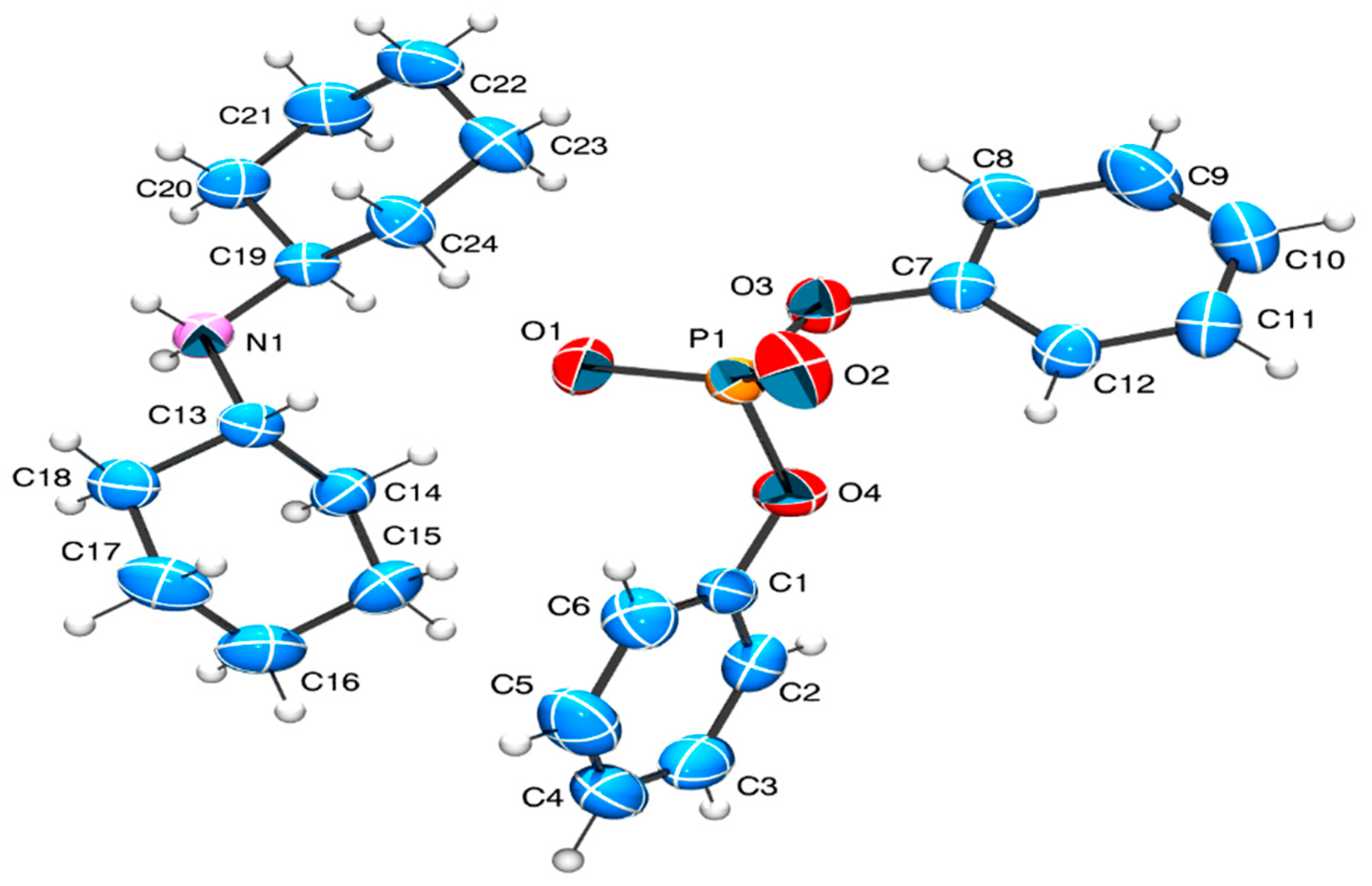
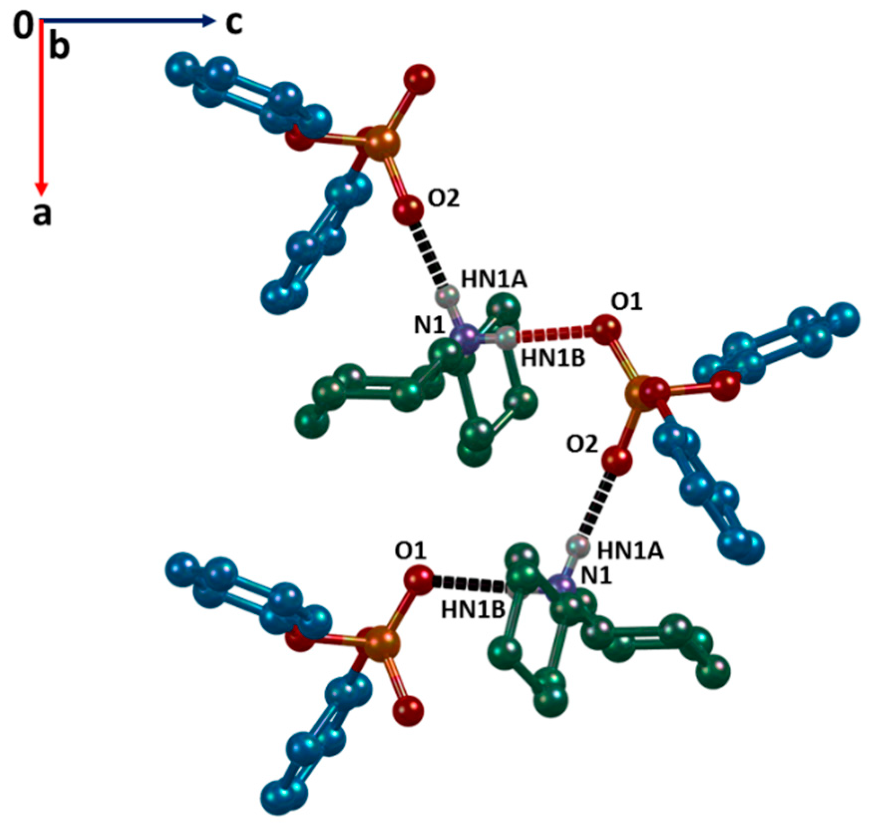
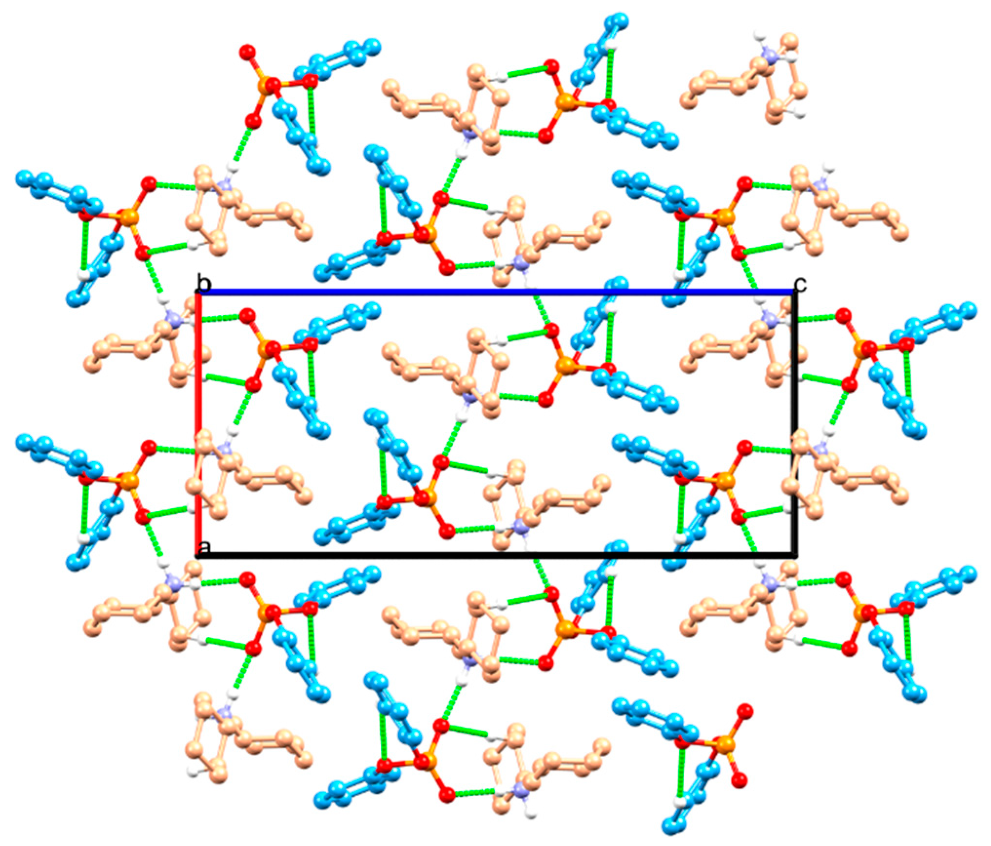
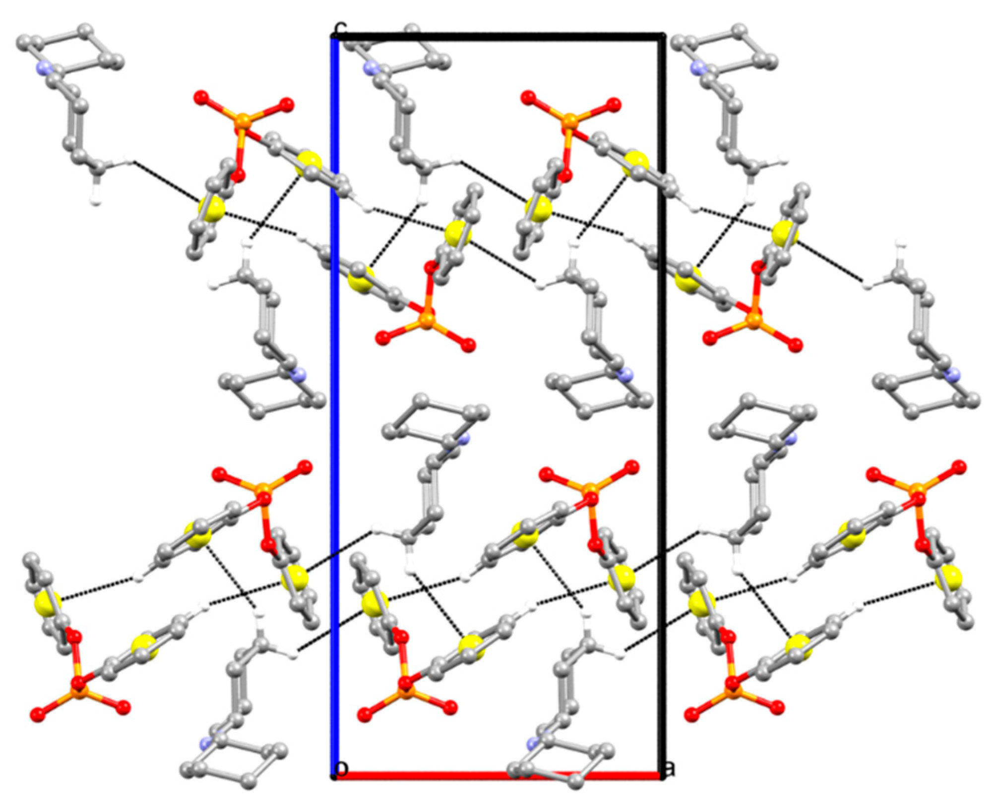
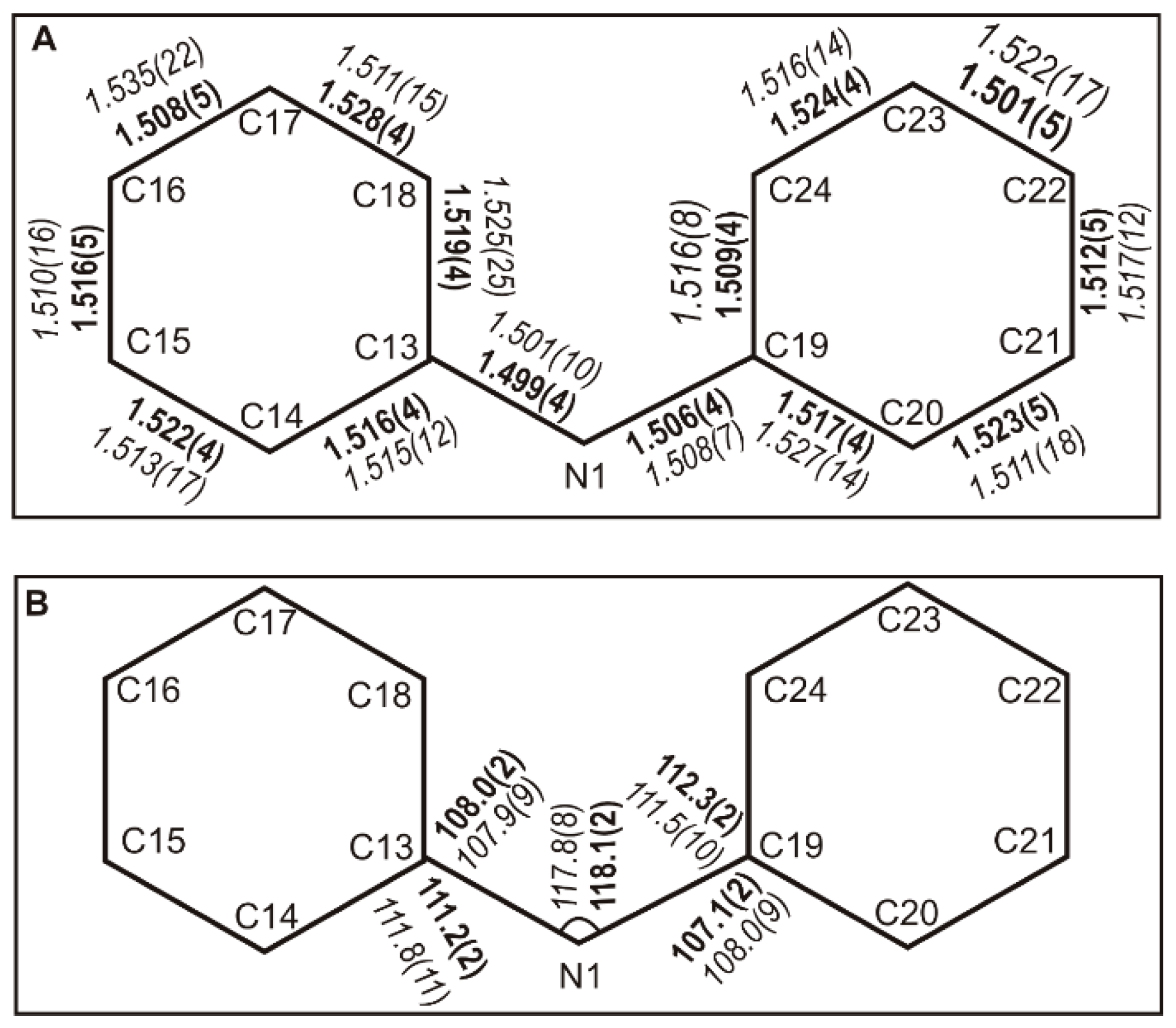
2.1. Structure Description

The title compound crystallizes in the orthorhombic system with chiral space group P212121, and the asymmetric unit is composed of one cation and one anion components (Figure 1). Selected bond distances and angles are given in Table 1. In general, all bond distances are within the characteristic values according to the atoms involved and are in the ranges of similar bonds observed in analogous structures. For example, the phosphorus–oxygen bond lengths of the [P(O)(O)] segment are 1.469(2) and 1.459(2) Å, which are slightly more than the typical P=O bond length (1.45 Å) [8].
In the cation, the cyclohexyl rings adopt a chair conformation, and the NH2 unit is situated in the equatorial position with respect to the rings. The large C–N–C angle, of 118.1(2)°, is a result of the steric bulk of the cyclohexyl rings, as has been also observed in the structures with the same cation, typically in [(C6H11)2NH2][C6H5PO2(OH)] (with CSD refcode ZARGOQ and the C–N–C angle of 118.80(10)°) [9].
In the anion, the phosphorus atom has a distorted tetrahedral (O)2P(O)(O) environment, with the bond angles within 96.31(11)° and 119.96(12)°. These extreme values correspond to the O3–P1–O4 and O2–P1–O1 angles, respectively (O1 and O2 are the atoms in the [P(O)(O)] segment; O3 and O4 are the ester oxygen atoms). Similar trends of O–P–O angles were observed in structures with [(C6H5O)2P(O)(O)] anion, like for example the structures with the CSD refcodes PABMUA and PABNAH [10]. The P–O–C angles of 124.5(2)° and 126.80(18)° show a few more “s” characters with respect to the sp2 hybridization. These values in the anion with a (C-O)2(O)P(O) core are in accordance with the previously reported P–O–C angles, typically in the (C-O)2P(X)(N)-based structures (X = O, S) [3] deposited in the CSD. The similarities and differences of cations and anions in the title structure with the selected related structures in the CSD are compared and discussed in Section 2.2.
In the crystal structure of the title compound, the cations and anions are hydrogen-bonded to each other, through N–H…O hydrogen bonds (N1…O1 = 2.806(3) Å and N1…O2 = 2.730(3) Å), building a linear arrangement along the a axis (Figure 2, Table 2). This assembly includes the non-centrosymmetric C 2 2 (6) graph-set motif. The strength of N–H…O hydrogen bonds is a result of the assistance of positive and negative charges in hydrogen bonding interactions.
In addition, the crystal packing shows one weak intermolecular C–H…O hydrogen bond (C24…O2 = 3.548(4) Å), and one intramolecular C–H…O hydrogen bond (C12…O4 = 3.139(4) Å) (Table 2). The C–H…O interactions do not extend the dimensionality of hydrogen bond pattern made by N–H…O hydrogen bonds. Figure 3 indicates a view of crystal packing with the relevant N–H…O and C–H…O hydrogen bonds. This assembly includes an R 2 2 (8) hydrogen-bonded ring motif, made by the cooperation of N1–HN1B…O1 and C24–H24B…O2 hydrogen bonds. The two phenyl rings of the anion also involved in the C–H…π-electron ring interactions, as the acceptors; the geometries are given in Table 2. The C–H…π interactions network is also formed along the a axis (Figure 4). In all contacts noted, the acceptor sites (O, π) belong to the anion.

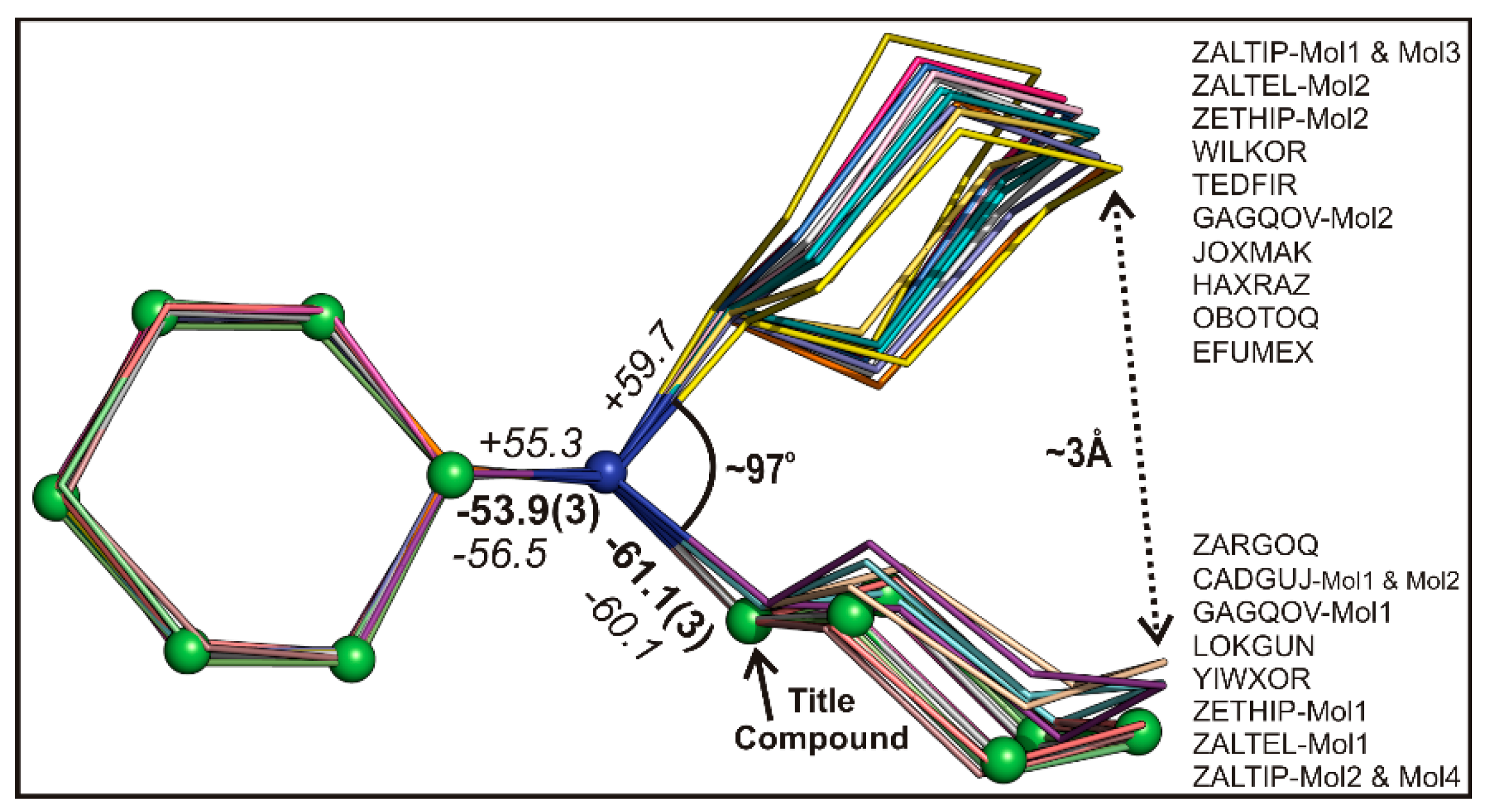
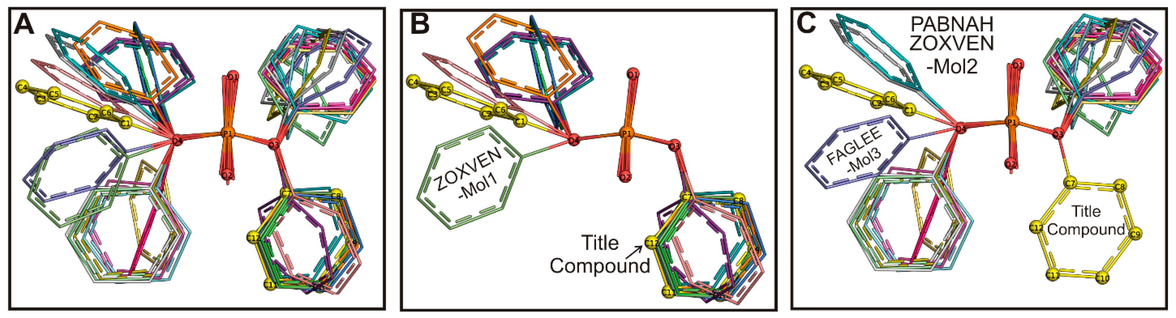
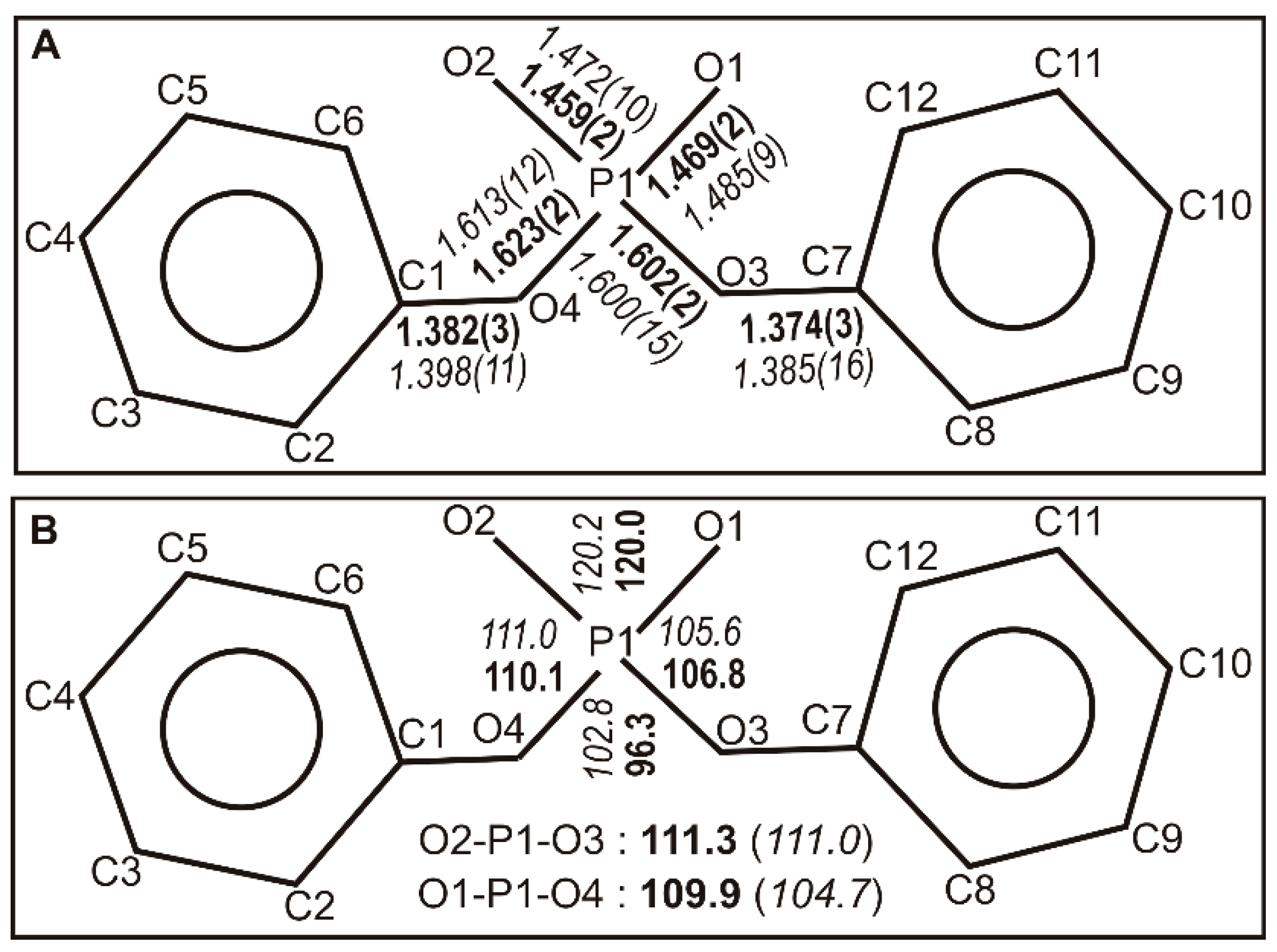
2.2. Structural Comparison

CSD study has been carried out to understand the similarities and conformational changes of the title compound. Fourteen structures (CSD REFCODES: CADGUJ [11], EFUMEX [12], GAGQOV [13], HAXRAZ [14], JOXMAK [15], LOKGUN [16], OBOTOQ [17], TEDFIR [18], WILKOR [19], YIWXOR [20], ZALTEL [21], ZALTIP [21], ZARGOQ [9], and ZETHIP [2]) containing the cation “dicyclohexylammonium” moiety were retrieved from the CSD. The bond lengths (Supplementary Table S1, Figure 5A), selected bond angles (Supplementary Table S2, Figure 5B), and selected torsion angles C14–C13–N1–C19 and C24–C19–N1–C13 (Supplementary Table S3, Figure 6) of the cation component are compared with the literature. From the results, it is evident that the bond lengths of dicyclohexylammonium moiety of the title compound are well-matched with the reported literature values (Figure 5A). The bond angles C13–N1–C19 (118.1°), C14–C13–N1 (111.2°), C18–C13–N1 (108.0°), C20–C19–N1 (107.1°), and C24–C19–N1 (112.3°) match the average bond angles calculated from the literature data as well (Figure 5B).
The conformational changes of the two cyclohexyl rings are compared with the structures retrieved from the CSD and superimposed with the cation moiety of the title compound. The structures were superimposed with one of the cyclohexyl rings including the nitrogen atom (N1–C19–C20–C21–C22–C23–C24) as shown in Figure 6. Two significant conformational flexibilities were observed among these structures. The cation moiety of the title compound adopts a −sc (synclinal) conformation with the torsion angles C14–C13–N1–C19 and C24–C19–N1–C13 of −61.1(3)° and −53.9(3)°, respectively, which bear a resemblance to the average literature values (Supplementary Table S3, and Figure 6). Ten molecules arising from the eight structures CADGUJ (Molecules 1 and 2), GAGQOV (Molecule 1), LOKGUN, YIWXOR, ZALTEL (Molecule 1), ZARGOQ, ZALTIP (Molecules 2 and 4), and ZETHIP (Molecule 1) adopt similar conformation as the title compound while remaining structures adopt +synclinal conformation. The distance of rings between the two groups is found to be ~3.0 Å with an approximate angle of 97° (Figure 6).
The bond lengths excluding the two phenyl rings, selected bond angles, and torsion angles representing Ph-O parts of the anion moiety of the title compound were compared with the reported structures extracted from the CSD. The 12 model structures (CSD REFCODES: CUVMUB [23], DUWWEX [24], ETADIM [25], FAGLEE [26], FAWYOR [27], HUHTEJ [28], OFESID [29], PABMUA [10], PABNAH [10], RUXWOY [30], VOVCOY [31], and ZOXVEN [32]) containing anion “diphenyl phosphate” were retrieved from the CSD. Figure 7 shows the distances of the C–O (C1–O4, C7–O3), P–OPh (P1–O3, P1–O4), P–O (non-ester; P1–O1, P1–O2) (Supplementary Table S4), and O–P–O bond angles with the reported literature values. The bond lengths and bond angles that correspond to phosphate geometry are favorably comparable with the reported literature.
The torsion angles C7–O3–P1–O4 (−72.5°), and C1–O4–P1–O3 (−163.3°) of the diphenyl phosphate moiety of the title compound represent –synclinal and –antiperiplanar conformations, respectively. Superposition of the structures extracted from CSD with the anion moiety of the title compound based on O3–P1–O4 atoms shows two significant conformational flexibilities (Figure 8A). While the torsion angles representing C7–O3–P1–O4 of the entire structures show either −sc or +sc conformation, the torsion angles for C1–O4–P1–O3 show ±ap, ±sc, and ±ac conformations (Supplementary Table S6). The torsional angles of C7–O3–P1–O4 in the six CSD structures FAGLEE (Molecules 1 and 4), CUVMUB, FAWYOR (Molecule 2), PABMUA, RUXWOY, and ZOXVEN (Molecule 1) including the title compound adopt –synclinal conformation (Figure 8B). Superposition of these molecules with the title compound shows that FAGLEE (Molecule 1) is very close to the title compound, and the remaining molecules in this group are slightly deviated and clustered together, except ZOXVEN (Molecule 1). While one of the phenyl rings of the ZOXVEN adopts skew (–sc) conformation, the other one adopts trans (+ap) conformation. Thus, one of the phenyl rings of the ZOXVEN remarkably deviates from the other structures in this group (Figure 8B). The remaining molecules FAGLEE (Molecules 2 and 3), DUWWEX, ETADIM, HUHTEJ, OFESID, FAWYOR (Molecule 1), VOVCOY, PABNAH, and ZOXVEN (Molecule 2) are clustered into other groups based on the C7–O3–P1–O4 torsion angle (+sc) conformation (Figure 8C, Supplementary Table S6). In this group, ETADIM, FAGLEE (Molecule 2), HUHTEJ, OFESID, FAWYOR (Molecule 1), and VOVCOY molecules adopt similar conformation (+sc), Figure 8B, and the DUWWEX molecule adopts +ac conformation. The FAGLEE (Molecule 3) adopts +ap conformation, PABNAH, and ZOXVEN (Molecule 2) molecules both adopt –ac conformation, and these three structures are slightly deviating from other structures in this cluster (Figure 8C).
Thus, comparison of the cation and anion moieties of the title compound with the similar structures available in the CSD indicates that the title compound is well-matched with the reported literature.

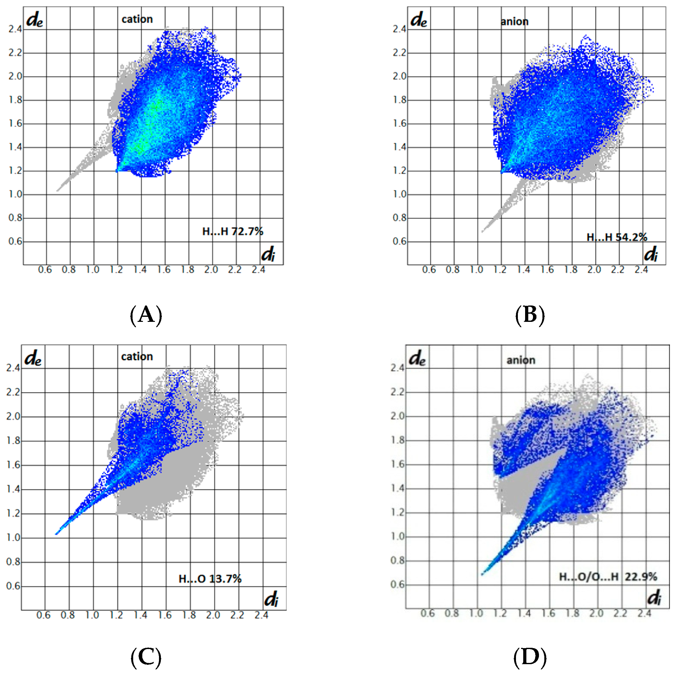
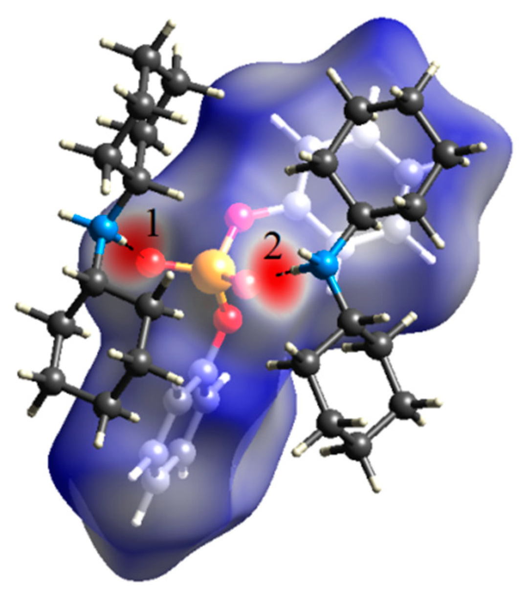
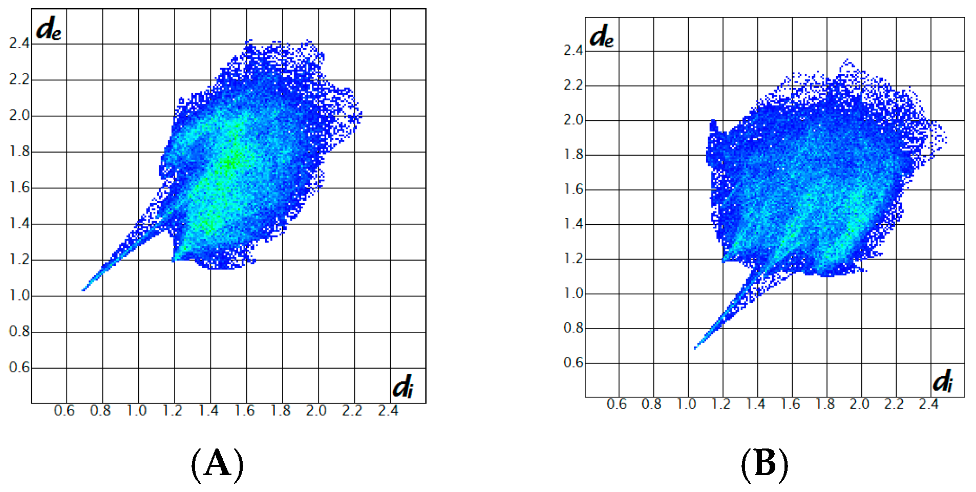
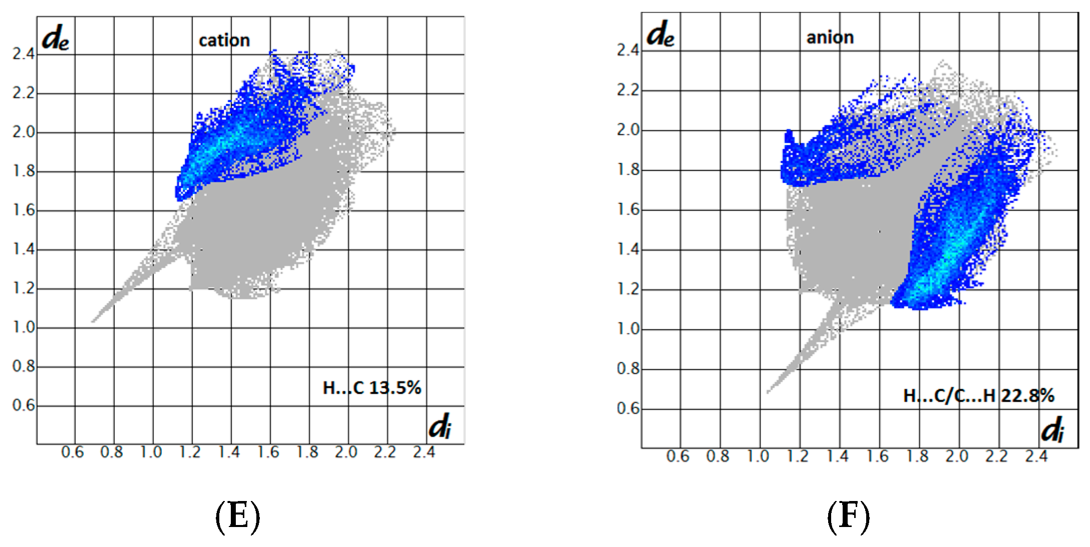
2.3. Hirshfeld Surface Analysis and Fingerprint Plots

The Hirshfeld surface analysis, which uses three-dimensional (3D) surfaces functions, as well as two-dimensional (2D) fingerprint plots [33,34], is a very useful graphical tool for identification and understanding of intermolecular interactions within a crystal structure. The Hirshfeld surfaces mapped with dnorm and corresponding shape index associated 2D fingerprint plots of the cation and anion of the title structure were generated using the Crystal Explorer software version 3.1 [35], with the structure file in the CIF format as the input.
In the Hirshfeld surfaces of the cation and anion, two large red areas can be seen, which correspond to two intermolecular N–H···O hydrogen bonds, as noted in the crystal structure section. The Hirshfeld surface map is typically represented for the anion and labels 1 and 2 denote to the hydrogen bonds noted (Figure 9); the Hirshfeld surface of the cation represents similar red areas with the hydrogen-bond donor sites, i.e., NH units, within the surface.
Two-dimensional fingerprint plots of the cation and anion are derived from the Hirshfeld surfaces, by plotting the fraction of points on the surface as a function of (de, di), where de and di introduce the distances from a point on the Hirshfeld surface to the nearest atoms outside and inside the surface, respectively. The full fingerprint plots of the cation and anion are given in Figure 10. The full plots were also divided into the figures illustrating different contacts observed in the crystal (Figure 11).
According to divided fingerprints, the contribution portions of contacts received by the cation are H···H 72.7%, H···O 13.7%, and H···C 13.5%. Similar contacts in the anion have the contribution portions of 54.2%, 22.9%, and 22.8%, respectively. Furthermore, a minor P···H contact is also seen for the anion with the contribution portion of 0.1%. In the fingerprint plots, the H···O contacts develop as one sharp spike, indicating the closest interaction in the crystal.

2.4. Spectroscopic Study

In the IR spectrum of the title salt, the intense doublet band with the maxima at 1239 and 1224 cm−1 is assigned to phosphorus–oxygen stretches of the [P=O(O)] moiety. The broad overlapped bands within 2471 to 3016 cm−1 are assigned to NH stretching frequencies, aliphatic and aromatic CH stretching frequencies, and the overtone of stretching frequencies of the phosphorus–oxygen bonds in the [P=O(O)] moiety. The broadening of these overlapped bands is related to strong N–H…O hydrogen bonds as were discussed in the X-ray description.
The 31P signal appears at −12.70 ppm, in comparison with the values of −11.7 ppm in the cation–anion compound [3-Cl-C6H4NH3][(C6H5O)2P(O)(O)] [30] and −0.81 ppm in the neutral compound (C6H5O)2((C6H5)(CH3)CHNH)P(O) [36]. The relatively high negative value is attributed to shielding caused by the negative charge on the oxygen atom. In the 1H NMR, the signal at 8.80 ppm corresponds to the NH2 protons, and the downfield chemical shift is due to the hydrogen bonding effect, similar to the one that was reported for a compound exhibiting hydrogen bonding in the X-ray structure [37]. The aliphatic and aromatic protons appear within 1.01 to 2.97 ppm and 7.02 to 7.28 ppm, as expected. In the 13C NMR spectrum, the doublet signals at 153.87 (2JPC = 7.0 Hz) and 120.34 ppm (3JPC = 5.1 Hz) are related to the ipso- and ortho- carbon atoms of C6H5 ring. The other six signals are singlet.
The mass spectrum indicates the peaks at m/z = 248, 247, 182, and 180, respectively attributed to the cations with formulas of [(C6H5O)2P(O)(O) − H]+, [(C6H5O)2P(O)(O) − 2H]+, [(C6H11)2NH2]+, and [(C6H11)2N]+.

3. Materials and Methods

3.1. Synthesis of [(C6H11)2NH2][(C6H5O)2P(O)(O)]

General procedure for the preparation of some dicyclohexylammonium di(para-substituted phenyl) phosphates was reported, together with the melting points and elemental analyses of different derivatives such as the title salt, i.e., with the para-substituent H [38]. The synthesis method was based on the reaction of N-methylpyridinium di(para-substituted phenyl phosphates) with HCl and dicyclohexylamine. The title salt reported in this article is the product obtained from hydrolysis in the synthesis process in a different reaction condition, as follows. A solution of dicyclohexylamine (1 mmol) and triethylamine (1 mmol) in acetonitrile was added to a solution of diphenylphosphoryl chloride (1 mmol) in the same solvent (at 273 K) under stirring. After 4 h, the mixture was transferred to a beaker in order to evaporate the solvent at room temperature over the course of a few days. The solid which formed was washed with distilled water and dried. Single crystals (yellow needles) were obtained from a solution of the product in a methanol–acetonitrile (1:1 v/v) mixture at room temperature. Yield: 80%. Mp: 188 °C (decomp.) (in literature: 188 to 189.5 °C [38]). Anal. Calc. for C24H34NO4P: C, 66.80; H, 7.94; N, 3.25. Found: C, 66.57; H, 7.90; N, 3.27. 1H NMR (DMSO-d6, 300.8 MHz): δ 8.80 (s, 2H, NH), 7.28 (apparent-t, J = 7.7 Hz, 4H), 7.16 (d, J = 7.8 Hz, 4H), 7.02 (t, J = 7.2 Hz, 2H), 2.97 (m, 2H), 1.01–1.97 (m, 20H). 13C{1H} NMR (DMSO-d6, 75.6 MHz): δ 153.87 (d, J = 7.0 Hz), 129.46, 122.82, 120.34 (d, J = 5.1 Hz), 52.24, 28.84, 25.25, 24.48. 31P{1H} NMR (DMSO-d6, 121.8 MHz): δ −12.70. IR (cm−1): 3016, 2934, 2859, 2760, 2669, 2564, 2471, 1595, 1495, 1458, 1389, 1239, 1224, 1166, 1096, 1032, 894, 754, 687. MS (70 eV, EI): 248 (4) [(C6H5O)2P(O)(O) − H]+, 247 (5) [(C6H5O)2P(O)(O)− 2H]+, 182 (13) [(C6H11)2NH2]+, 180 (76) [(C6H11)2N]+, 137 (100) [C9H15N]+, 99 (47) [C6H13N]+, 82 (76) [C6H10]+.

3.2. X-ray Data of Crystal Structure

The X-ray data were collected at 293 K with graphite monochromated Mo radiation (0.71073 Å) on a Bruker SMART APEXII area-detector diffractometer. The structure was solved with SHELXS97 [39] by the direct methods algorithm and refined using full-matrix least-squares on F2 with the SHELXL-2016/6 [40]. All carbon-bound H atoms were placed at calculated positions and were refined on their parent atoms with their Uiso set to 1.2Ueq. Nitrogen-bound H atoms were located in a difference Fourier map and refined isotropically with their Uiso set to 1.2Ueq of the carrier N atom.
The crystallographic data for C12H10O4P.C12H24N (M = 431.49 g/mol): Orthorhombic, space group P212121, a = 9.6610(4) Å, b = 10.9649(5) Å, c = 21.8524(11) Å, V = 2314.87(18) Å3, Z = 4, T = 293(2) K, F(000) = 928, μ(MoKα) = 0.148 mm−1, S = 1.041, Dcalc = 1.238 g/cm3, 5598 independent reflections, −12 ≤ h ≤ 11, −14 ≤ k ≤ 9, −28 ≤ l ≤ 28, 4466 reflections with I > 2σ(I), Rint = 0.0289. The final R1 was 0.0453 (F2 > 2σ(F2)) and wR2 (F2) was 0.1122, Δρmax (eÅ−3) = 0.543, Δρmin (eÅ−3) = −0.226. The molecular graphics were generated by Mercury [41] and OLEX2 [42]. Crystallographic data have been deposited with Cambridge Crystallographic Data Centre (CCDC-1887337). These data can be obtained free of charge [43] from the CCDC, 12 Union Road, Cambridge CB2 1EZ, UK; fax: +44-1223-336033; e-mail: [email protected].

4. Conclusions

The [(C6H11)2NH2][(C6H5O)2P(O)(O)] salt crystallizes in the chiral space group P212121, and the cation and anion components are assembled through relatively strong charge-assisted N–H…O hydrogen bonds, together with C–H…O and C–H…π weak interactions. The divided fingerprints show the most contribution portions of H…H contacts and then H…O and H…C interactions received by both cation and anion. The negative 31P signal in solution NMR is attributed to shielding caused by the negative charge on the oxygen atom. The cation and anion components of the title salt are well-matched with the structures including similar components from the CSD.

Supplementary Materials

The tables for structural comparisons with CSD, and 31P{1H} NMR, 1H NMR, 13C{1H} NMR, IR, Mass, and crystallographic data of synthesized compound are available online at https://www.mdpi.com/1422-8599/2019/1/M1051/s1.

Author Contributions

A.R.S. and F.S. are supervisors of S.N.M. in her PhD thesis. Designing of the experiment was done by A.R.S. and M.P., the experiment and Hirshfeld surface analysis were performed by F.S., M.P., and S.N.M., the spectroscopic studies were done by M.P., the paper was written by M.P. and A.A.A.S., and the X-ray crystallography experiment and structural comparisons were done by A.A.A.S.; the figures were prepared by M.P., A.A.A.S., and S.N.M.; all authors read and approved the manuscript.

Funding

This research received no external funding.

Acknowledgments

S.N.M. and A.R.S. thank the Islamic Azad University, North Tehran Branch for financial support. M.P. thanks the Ferdowsi University of Mashhad for spectroscopic studies. We thank Devadasan Velmurugan, UGC BSR Faculty, CAS in Crystallography and Biophysics, University of Madras, Guindy Campus, Chennai—600 025, India. Abdul Ajees Abdul Salam acknowledges research grant provided by the Manipal Academy of Higher Education (MAHE) (MAHE/REG/TD/DAMP-(T)) under the fellowship program offered by Manipal FAIMER Institute (M-FIILIPE).

Conflicts of Interest

The authors declare no conflict of interest.

References

  1. Xu, H.-J.; Tan, Q.-Y.; Pang, Y.-J. (2,4-Dihydroxybenzylidene)dimethyl-ammonium dichlorophosphinate. Acta Cryst. E 2009, 65, o353. [Google Scholar] [CrossRef] [PubMed]
  2. Krzyżanowska, B.; Stec, W.J.; Wieczorek, M.W.; Błaszczyk, J. Synthesis and assignment of absolute configuration of enantiomeric dicyclohexylammonium 2-oxo-2-thioxo-1,3,2-oxathiaphospholanes. Heteroat. Chem. 1994, 5, 533–539. [Google Scholar] [CrossRef]
  3. Sabbaghi, F.; Pourayoubi, M.; Dušek, M.; Eigner, V.; Bayat, S.; Damodaran, K.; Nečas, M.; Kučeráková, M. Analysis of P–O–C, P–S–C and P–O–P angles: A database survey completed with four new X-ray crystal structures. Struct. Chem. 2016, 27, 1831–1844. [Google Scholar] [CrossRef]
  4. Hamzehee, F.; Pourayoubi, M.; Nečas, M.; Choquesillo-Lazarte, D. Extensive analysis of N–H···O hydrogen bonding in four classes of phosphorus compounds: A combined experimental and database study. Acta Cryst. C 2017, 73, 287–297. [Google Scholar] [CrossRef] [PubMed]
  5. Eghbali Toularoud, M.; Pourayoubi, M.; Dušek, M.; Eigner, V.; Damodaran, K. Chiral one-dimensional hydrogen-bonded architectures constructed from single-enantiomer phosphoric triamides. Acta Cryst. C 2018, 74, 608–617. [Google Scholar] [CrossRef] [PubMed]
  6. Torabi Farkhani, E.; Pourayoubi, M.; Izadyar, M.; Andreev, P.V.; Shchegravina, E.S. Evaluation of N–H···S and N–H···π interactions in O,O’-diethyl N-(2,4,6-trimethylphenyl)thiophosphate: A combination of X-ray crystallographic and theoretical studies. Acta Cryst. C 2018, 74, 847–855. [Google Scholar] [CrossRef] [PubMed]
  7. Dey, A.; Pidcock, E. The relevance of chirality in space group analysis: A database study of common hydrogen-bonding motifs and their symmetry preferences. CrystEngComm 2008, 10, 1258–1264. [Google Scholar] [CrossRef]
  8. Corbridge, D.E.C. Phosphorus, Chemistry, Biochemistry & Technology; Elsevier: Amsterdam, The Netherlands, 2000; pp. 69–74. [Google Scholar]
  9. Diop, T.; Diop, L.; Maris, T.; Stoeckli-Evans, H. Dicyclohexylammonium hydrogen phenylphosphonate. Acta Cryst. E 2012, 68, o1432. [Google Scholar] [CrossRef] [PubMed]
  10. Holmes, R.R.; Day, R.O.; Yoshida, Y.; Holmes, J.M. Hydrogen-bonded phosphate esters. Synthesis and structure of imidazole-containing salts of diphenyl phosphate and (trichloromethyl)phosphonic acid. J. Am. Chem. Soc. 1992, 114, 1771–1778. [Google Scholar] [CrossRef]
  11. Bartczak, T.J. Structure of a racemic compound: The dicyclohexylammonium salt of (2R,4R,2S,4S)-cis-4-methyl-2-oxido-1,3,2-dioxaphosphorinane 2-sulphide, C12H24N+.C4H8O3PS−−. Acta Cryst. C 1983, 39, 1059–1062. [Google Scholar] [CrossRef]
  12. Kamenecka, T.M.; Overman, L.E.; Ly Sakata, S.K. Construction of substituted cyclohexanones by reductive cyclization of 7-oxo-2,8-alkadienyl esters. Org. Lett. 2002, 4, 79–82. [Google Scholar] [CrossRef] [PubMed]
  13. Krawczyk, H.; Śliwiński, M.; Wolf, W.M.; Bodalski, R. Diastereo- and enantioselective synthesis of bicyclic α-methylene-δ-valero-lactones by asymmetric michael reaction. Synlett 2004, 2004, 1995–1999. [Google Scholar] [CrossRef]
  14. Harger, M.J.P.; Sreedharan-Menon, R. Stereochemistry of the methoxide induced rearrangement of an α-bromophosphonamidate: Cleavage of the P–N and P–C bonds in the azaphosphiridine oxide intermediate 1. J. Chem. Soc. Perkin Trans. 1 1997, 4, 527–532. [Google Scholar] [CrossRef]
  15. Yamamoto, J.H.; Udachin, K.A.; Enright, G.D.; Carty, A.J. Phosphorus monoxide as a quadruply bridging ligand: Syntheses and X-ray crystal structures of Ru5(CO)154-PF) and [H2NCy2][Ru5(CO)154-PO)]. Chem. Commun. 1998, 20, 2259–2260. [Google Scholar] [CrossRef]
  16. Mazurek, J.; Lis, T. Crystal and molecular structure of potassium, ammonium and dicyclohexylammonium salts of (2-oxopropyl) phosphonic acid in monoionized state. J. Mol. Struct. 1999, 474, 143–155. [Google Scholar] [CrossRef]
  17. Diop, T.; Diop, L.; Diop, C.A.K.; Molloy, K.C.; Kociok-Köhn, G. Dicyclohexylammonium trimethylbis(hydrogen phenylphosphonato)stannate(IV). Acta Cryst. E 2011, 67, m1872–m1873. [Google Scholar] [CrossRef] [PubMed]
  18. Nifantev, E.E.; Kukhareva, T.S.; Popkova, T.N.; Dyachenko, V.I.; Kolomiets, A.F.; Magomedova, N.S.; Belskii, V.K. Synthesis and transformations of benzo-1,3,2-dioxaphosphor (V) inanes based on fluorinated derivatives of orthooxybenzyl alcohol. Zhurnal Obs. Khimii 1996, 66, 61–67. [Google Scholar]
  19. Bodalski, R.; Jankowski, S.; Glówka, M.L.; Filipiak, T.; Quin, L.D. Anchimeric participation of a methoxy group in a reaction of a metathiophosphate. J. Org. Chem. 1994, 59, 5173–5178. [Google Scholar] [CrossRef]
  20. Mikolajczyk, M.; Luczak, J.; Wieczorek, M.W.; Blaszczyk, J. Stereochemistry of organophosphorus cyclic compounds. Part 17. Synthesis of N-methyl-2-methoxy-1,3,2-oxazaphosphorinane-2-thione (III) and dicyclohexylammonium salt of N-methyl-2-oxo-2-thio-1,3,2-oxazaphosphorinane (V) and their crystal and molecular structures. Pol. J. Chem. 1993, 67, 1087–1097. [Google Scholar]
  21. Kashemirov, B.A.; Ju, J.-Y.; Bau, R.; McKenna, C.E. “Troika Acids”: Synthesis, structure, and fragmentation pathways of novel α-(hydroxyimino)phosphonoacetic acids. J. Am. Chem. Soc. 1995, 117, 7285–7286. [Google Scholar] [CrossRef]
  22. The PyMOL Molecular Graphic System, Open-Source Version 1.5, Schrödinger, LLC. Available online: www.pymol.org (accessed on 27 February 2019).
  23. Głowiak, T.; Wnek, I. Crystal and molecular structure of glycinium diphenylphosphate. J. Crystallogr. Spectrosc. Res. 1985, 15, 157–171. [Google Scholar] [CrossRef]
  24. Głowiak, T.; Podgórska, I. X-ray, spectroscopic and magnetic studies of hexaaquacopper(II) di(diphenylphosphate) diglycine. Inorg. Chim. Acta 1986, 125, 83–88. [Google Scholar] [CrossRef]
  25. Stockland, R.A., Jr.; Levine, A.M.; Giovine, M.T.; Guzei, I.A.; Cannistra, J.C. Reductive elimination from metal phosphonate complexes: Circumvention of competing protonolysis reactions. Organometallics 2004, 23, 647–656. [Google Scholar] [CrossRef]
  26. Toney, J.H.; Brock, C.P.; Marks, T.J. Aqueous coordination chemistry of vanadocene dichloride with nucleotides and phosphoesters. Mechanistic implications for a new class of antitumor agents. J. Am. Chem. Soc. 1986, 108, 7263–7274. [Google Scholar] [CrossRef]
  27. Głowiak, T.; Podgórska, I. Structure of tetraaqua-bis[μ-(glycylglycinato)-NOO’]-dicopper(II) bis(diphenyl phosphate) dihydrate. Acta Cryst. C 1987, 43, 53–57. [Google Scholar] [CrossRef]
  28. Małecka, M.; Rybarczyk-Pirek, A.; Grabowski, S.J.; Malinowska, K.; Ochocki, J. 3,5-Dimethyl-2H-pyrazol-1-ium diphenylphosphate. Acta Cryst. E 2002, 58, o1113–o1115. [Google Scholar] [CrossRef]
  29. Safin, D.A.; Babashkina, M.G.; Robeyns, K.; Mitoraj, M.P.; Kubisiak, P.; Brela, M.; Garcia, Y. Experimental and theoretical investigations of the NiII complex with N-phosphorylated thiourea iPrNHC(S)NHP(O)(OPh)2. CrystEngComm 2013, 15, 7845–7851. [Google Scholar] [CrossRef]
  30. Babashkina, M.G.; Robeyns, K.; Filinchuk, Y.; Safin, D.A. Detailed studies of the interaction of 3-chloroaniline with O,O’-diphenylphosphorylisothiocyanate. New J. Chem. 2016, 40, 1230–1236. [Google Scholar] [CrossRef]
  31. Dixon, R.P.; Geib, S.J.; Hamilton, A.D. Molecular recognition: Bis-acylguanidiniums provide a simple family of receptors for phosphodiesters. J. Am. Chem. Soc. 1992, 114, 365–366. [Google Scholar] [CrossRef]
  32. Král, V.; Furuta, H.; Shreder, K.; Lynch, V.; Sessler, J.L. Protonated sapphyrins. Highly effective phosphate receptors. J. Am. Chem. Soc. 1996, 118, 1595–1607. [Google Scholar] [CrossRef]
  33. McKinnon, J.J.; Jayatilaka, D.; Spackman, M.A. Towards quantitative analysis of intermolecular interactions with Hirshfeld surfaces. Chem. Commun. 2007, 37, 3814–3816. [Google Scholar] [CrossRef]
  34. Spackman, M.A.; McKinnon, J.J. Fingerprinting intermolecular interactions in molecular crystals. CrystEngComm 2002, 4, 378–392. [Google Scholar] [CrossRef]
  35. Wolff, S.K.; Grimwood, D.J.; McKinnon, J.J.; Turner, M.J.; Jayatilaka, D.; Spackman, M.A. Crystal Explorer; University of Western Australia: Perth, Australia, 2007. [Google Scholar]
  36. Sabbaghi, F.; Pourayoubi, M.; Nečas, M.; Damodaran, K. Two single-enantiomer amidophosphoesters: A database study on the chirality of (O)2P(O)N-based structures. Acta Cryst. C 2019, 75, 77–84. [Google Scholar] [CrossRef] [PubMed]
  37. Aranburu Leiva, A.I.; Kaur, M.; Benjamin, S.L.; Jones, A.M.; Langley, S.K.; Mewis, R.E. 1,8-bis(2-hydroxy-3,5-di-tert-butylbenzyl)-4,11-dibenzyl-1,4,8,11-tetraazacyclotetradecane. Molbank 2017, 2017, M963. [Google Scholar] [CrossRef]
  38. Osborne, D.W. The pyridinolysis of diaryl methyl phosphates. J. Org. Chem. 1964, 29, 3570–3574. [Google Scholar] [CrossRef]
  39. Sheldrick, G.M. A short history of SHELX. Acta Cryst. A 2008, 64, 112–122. [Google Scholar] [CrossRef] [PubMed]
  40. Sheldrick, G.M. Crystal structure refinement with SHELXL. Acta Cryst. C 2015, 71, 3–8. [Google Scholar] [CrossRef] [PubMed]
  41. Macrae, C.F.; Bruno, I.J.; Chisholm, J.A.; Edgington, P.R.; McCabe, P.; Pidcock, E.; Rodriguez-Monge, L.; Taylor, R.; van de Streek, J.; Wood, P.A. Mercury CSD 2.0—new features for the visualization and investigation of crystal structures. J. Appl. Cryst. 2008, 41, 466–470. [Google Scholar] [CrossRef]
  42. Dolomanov, O.V.; Bourhis, L.J.; Gildea, R.J.; Howard, J.A.K.; Puschmann, H. OLEX2: A complete structure solution, refinement and analysis program. J. Appl. Cryst. 2009, 42, 339–341. [Google Scholar] [CrossRef]
  43. CCDC. Available online: http://www.ccdc.cam.ac.uk/conts/retrieving.html (accessed on 27 January 2019).
Figure 1. The structure of [(C6H11)2NH2][(C6H5O)2P(O)(O)], showing the atom-numbering scheme for non-hydrogen atoms and displacement ellipsoids at the 50% probability level. Hydrogen atoms are drawn as spheres of arbitrary radii.
Molbank 2019 m1051 g001
Figure 2. A view of the one-dimensional array of the title cation–anion compound built from N–H…O hydrogen bonds. Color keys are red O, orange P, dark blue N; the C atoms of cation and anion components are shown as green and blue colors, respectively. The carbon-bonded hydrogen atoms were omitted for the sake of clarity, and the hydrogen bonds are shown as dashed lines.
Molbank 2019 m1051 g002
Figure 3. The molecular interactions of [(C6H11)2NH2][(C6H5O)2P(O)(O)] viewed along the b direction. Intra and intermolecular C–H…O interactions are shown along with the strong N–H…O intermolecular interactions. Hydrogen atoms not involved in the interactions are omitted for the sake of clarity, and the hydrogen bonds are shown as dashed lines. Color keys are red O, orange P, blue N; the C atoms of cation and anion components are shown as gold-orange and light blue colors, respectively.
Molbank 2019 m1051 g003
Figure 4. The C–H…π interactions of [(C6H11)2NH2][(C6H5O)2P(O)(O)] viewed along the b direction. Only the hydrogen atoms participating in C–H…π interactions are shown, and the C–H…π interactions are represented by dashed lines. Color keys are red O, orange P, blue N, gray C, and yellow for centroid of the phenyl ring.
Molbank 2019 m1051 g004
Figure 5. Structural comparison of the cation moiety of the title compound. (A) Bond lengths of the dicyclohexylammonium moiety are shown with the average literature values extracted from the Cambridge Structural Database (CSD). (B) Selected bond angles of the cation moiety of the title compound are marked with the literature values. The bond lengths and angles of the title compound are shown in boldface and the average bond lengths, and bond angles calculated from the 14 model structures extracted from the CSD are shown in normal italic.
Molbank 2019 m1051 g005
Figure 6. Conformational flexibility of cyclohexyl rings. The cation moiety of the title compound is shown in a ball-and-stick model, and carbon atoms are colored green. The remaining structures are shown in a simple line model. Molecules are marked using CSD REFCODES and molecules with more than one model in the asymmetric unit are marked as Mol1, Mol2, etc. The figure is rendered using PyMOL [22].
Molbank 2019 m1051 g006
Figure 7. Structural comparison of the anion moiety, diphenyl phosphate, of the title compound. (A) Bond lengths of the diphenyl phosphate moiety, except for two phenyl rings, are shown and compared with the literature values extracted from the CSD. (B) Selected bond angles of the anion moiety of the title compound are marked with the literature values. The bond lengths and angles of the title compound are shown in boldface and the average bond lengths, and bond angles calculated from the 12 model structures extracted from the CSD, are shown in normal italic.
Molbank 2019 m1051 g007
Figure 8. Structural comparison of diphenyl phosphate moiety of the title compound with the literature. (A) Superposition of diphenyl phosphate moieties extracted from the CSD with the title compound. The title compound is shown in yellow (carbon atoms), ball-and-stick model. Based on the torsional angle C7–O3–P1–O4, two major conformational flexibilities −sc and +sc were observed. The models representing −sc group are shown in (B), and the models that adopt +sc are shown in (C). The deviating models in both groups are marked.
Molbank 2019 m1051 g008
Figure 9. A view of the Hirshfeld surface map of the [(C6H5O)2P(O)(O)] anion. The interactions according to the labels are as follows, with the labels in parentheses: N1–HN1B…O1 (1) and N1–HN1A…O2 (2). The ball-and-stick models of the anion and two hydrogen-bonded cations are given inside and outside the surface, respectively.
Molbank 2019 m1051 g009
Figure 10. Full fingerprint plot of the cation (A) and anion (B).
Molbank 2019 m1051 g010
Figure 11. Divided fingerprint plots for the cation and anion components of the title salt: H…H of cation (A), H…H of anion (B), H…O of cation (C), H…O of anion (D), H…C of cation (E) and H…C of anion (F).
Molbank 2019 m1051 g011aMolbank 2019 m1051 g011b
Table 1. Selected bond distances (Å) and angles (°).
Bond Distances
O1–P11.469(2)C1–O41.382(3)
P1–O21.459(2)C7–O31.374(3)
O3–P11.602(2)C13–N11.499(4)
O4–P11.623(2)C19–N11.506(4)
Angles
O2–P1–O1119.96(12)O3–P1–O496.31(11)
O1–P1–O3106.78(12)C7–O3–P1126.80(18)
O2–P1–O3111.27(13)C1–O4–P1124.5(2)
O1–P1–O4109.86(12)C13–N1–C19118.1(2)
O2–P1–O4110.05(15)C2–C1–O4115.1(3)
Table 2. Hydrogen bond geometries (e.s.d.s are given in parentheses).
D–H…AD–H (Å)H…A (Å)DA (Å)D–H…A (°)
N1–HN1B…O1#10.83(3)1.97(3)2.806(3)177(3)
N1–HN1A…O2#20.90(3)1.84(3)2.730(3)176(3)
C12–H12…O40.932.623.139(4)115.8
C24–H24B…O2#10.972.653.548(4)154.9
C11–H11…Cg1#30.932.803.629(3)149
C15–H15B…Cg1#40.972.973.664(4)130
C15–H15A…Cg2#40.972.953.765(4)142
Symmetry codes: #1 x − 1/2, −y + 1/2, −z + 2; #2 x − 1, y, z; #3 −x + 2, y + 1/2, −z + 3/2; #4 −x + 1, y − 1/2, −z + 3/2.

