Heat Shock Proteins and Exercise in Skeletal Muscle Insulin Resistance: Protective Mechanisms and Therapeutic Potential


Abstract
1. Introduction
2. Heat Shock Proteins (HSPs)
3. HSP70 and Its Role in Muscle and Metabolic Health
3.1. Anti-Inflammatory Properties
3.2. HSP70 Regulation of Mitochondrial Function
4. Exercise-Induced Expression of Heat Shock Proteins (HSPs)
4.1. Exercise Dose–Response Effects and Heat Shock Proteins
4.1.1. Exercise, HSP70, and GLUT4 Regulation: Relevance to Insulin Resistance

| Experimental Model | Exercise Paradigm | Exercise Characteristics | Primary Tissue | HSP70 Response | Primary Mechanistic Effect | Physiological/Metabolic Outcome | Representative References |
|---|---|---|---|---|---|---|---|
| Human | Aerobic treadmill running | Acute, 45 min, ~75% VO2max | Skeletal muscle | ↑ HSP70 mRNA and protein | Activation of the heat shock response, enhanced protein folding, maintenance of proteostasis, and improved cellular stress tolerance | Protection against acute exercise-induced metabolic and oxidative stress | [72] |
| Human | Endurance exercise | Moderate intensity, acute bout | Plasma/ circulation | ↑ Extracellular HSP70 | Activation of systemic heat shock signaling and intercellular stress communication | Enhanced systemic adaptation to physiological stress and improved metabolic resilience | [76,77,78] |
| Human and Rodent | Aerobic and resistance training | Moderate-to-vigorous intensity, repeated training | Skeletal muscle | Sustained elevation of basal HSP70 | Improved protein quality control, reduced oxidative damage, attenuation of inflammatory signaling | Improved metabolic adaptation, enhanced exercise tolerance and greater insulin sensitivity | [66,67,68,69,70,83,84,85] |
| Rodent | Treadmill running | Acute endurance exercise | Skeletal muscle, heart, liver | ↑ HSP70 expression | Heat- and exercise-mediated activation of HSF1 and molecular chaperone pathways | Protection against exercise-induced cellular stress and maintenance of tissue homeostasis | [71] |
| Rodent | Heat treatment ± exercise | Chronic intervention | Skeletal muscle | ↑ HSP70 | Suppression of JNK and IKKβ signaling, reduced inflammatory activation, preservation of insulin signaling | Prevention of diet-induced insulin resistance and improvement of glucose homeostasis | [1,12,53,54] |
| Rodent | Mechanical loading/ overload | Chronic Physiological loading | Skeletal muscle | ↑ HSP70 | Activation of Akt–mTOR signaling and inhibition of FOXO and NF-κB pathways | Preservation of muscle mass, reduced disuse atrophy, and enhanced muscle regeneration | [38,39,40] |
| Human and Rodent | Aerobic and resistance exercise | Acute and chronic training | Skeletal muscle | ↑ HSP70 | Preservation of mitochondrial integrity, enhanced oxidative enzyme activity, improved mitochondrial quality control and biogenesis | Increased oxidative capacity, improved metabolic flexibility, and enhanced mitochondrial function | [13,48,57,58,59,60,61] |
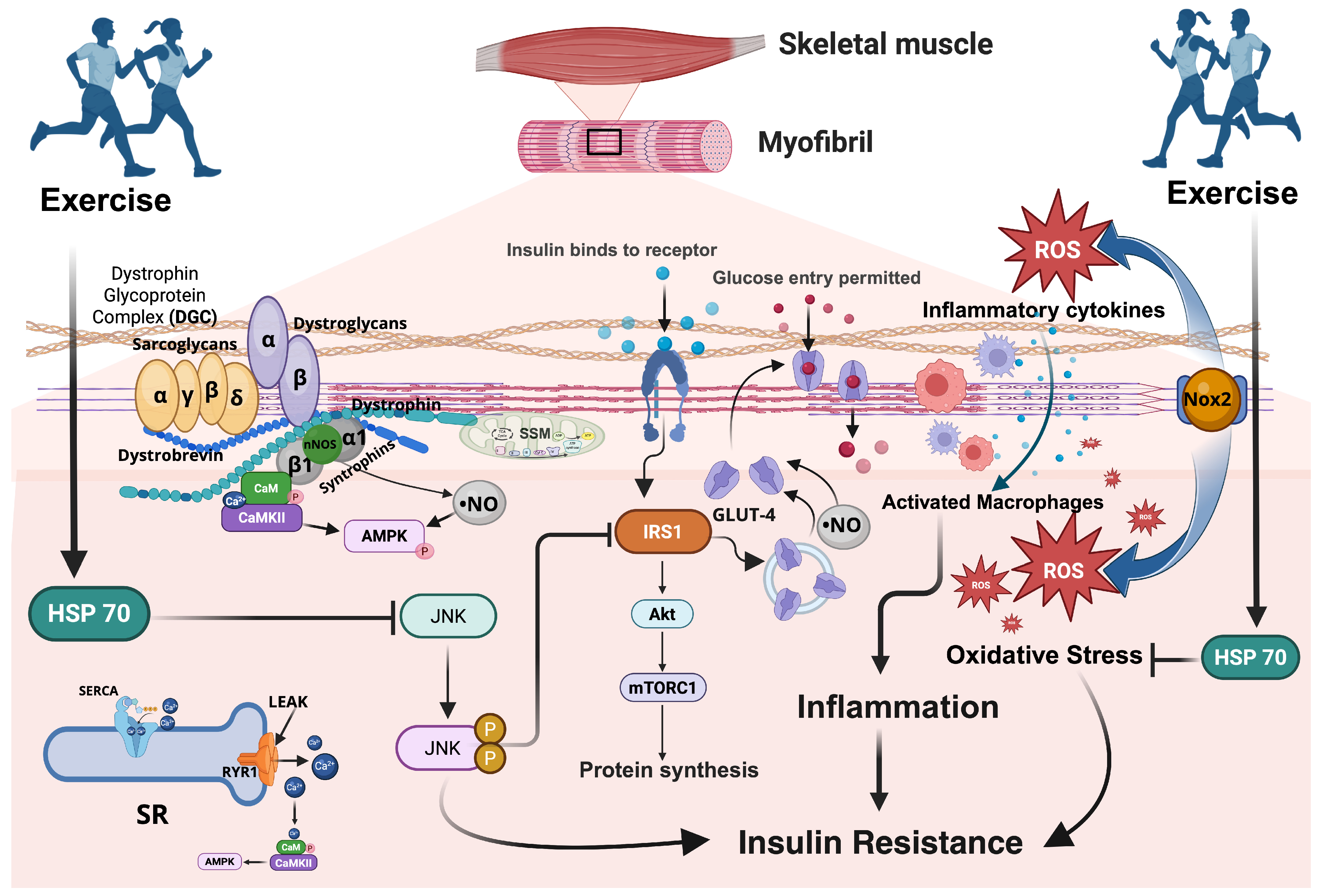
| Human and Rodent | Aerobic exercise | Acute and chronic exercise | Skeletal muscle | ↑ HSP70 | Stabilization of IRS-1/PI3K/Akt signaling, preservation of AMPK-mediated GLUT4 trafficking, reduction in ROS-induced impairment of insulin signaling | Increased GLUT4 translocation, enhanced glucose uptake, and improved insulin sensitivity | [48,63,92,93,94,95,96,97,98,99,100,101] |
4.1.2. Insulin-Dependent Pathway During Exercise
4.1.3. Insulin-Independent Pathway During Exercise
4.2. The Impact of Different Exercise Factors
5. Conclusions
Author Contributions
Funding
Institutional Review Board Statement
Informed Consent Statement
Data Availability Statement
Conflicts of Interest
Abbreviations
| Akt | Protein kinase B; serine/threonine kinase central to insulin signaling and glucose uptake |
| AMPK | AMP-activated protein kinase; key energy sensor promoting glucose uptake and fatty-acid oxidation |
| AS160 | Akt substrate of 160 kDa (also known as TBC1D4); regulates GLUT4 vesicle trafficking |
| ATP | Adenosine triphosphate; cellular energy currency |
| BGP-15 | Hydroximic acid derivative and pharmacologic HSP co-inducer that improves insulin sensitivity |
| CaMKII | Calcium/calmodulin-dependent protein kinase II; mediates calcium signaling during muscle contraction |
| CK | Creatine kinase; marker of muscle damage |
| DAMPs | Damage-associated molecular patterns; endogenous molecules triggering immune activation |
| eHSP70 | Extracellular heat shock protein 70; circulating form with immunomodulatory function |
| ER | Endoplasmic reticulum; site of protein folding and the unfolded-protein response (UPR) |
| FFA | Free fatty acid; lipid species contributing to insulin resistance when elevated |
| FOXO | Forkhead box O transcription factors; mediate muscle atrophy and oxidative stress responses |
| GLUT4 | Glucose transporter type 4; insulin-responsive transporter in skeletal muscle and adipose tissue |
| HFD | High-fat diet; experimental model for inducing metabolic dysfunction |
| HSP | Heat shock protein; conserved molecular chaperone family aiding in protein folding and stress tolerance |
| HSP70 (Hsp70) | Inducible 70-kDa heat shock protein; central focus of this review |
| Hsc70 | Heat shock cognate 70; constitutively expressed member of the HSP70 family |
| HSP72 | Common designation for inducible HSP70 isoform in human studies |
| HSP90 | 90-kDa heat shock protein; stabilizes signaling proteins including kinases and steroid receptors |
| HSF1 | Heat shock factor 1; transcriptional regulator of heat shock proteins |
| iHSP70 | Inducible/intracellular heat shock protein 70; stress-responsive isoform |
| IGF-1 | Insulin-like growth factor 1; anabolic growth factor activating PI3K/Akt/mTOR signaling |
| IL-6 | Interleukin-6; cytokine modulating inflammation and metabolic signaling |
| iNOS | Inducible nitric oxide synthase; produces nitric oxide during inflammatory stress |
| IRS-1 | Insulin receptor substrate-1; adaptor protein mediating insulin receptor signaling |
| JNK | c-Jun N-terminal kinase; stress-activated kinase impairing insulin signaling when overactive |
| LKB1 | Liver kinase B1; upstream activator of AMPK |
| MAPK | Mitogen-activated protein kinase; family of kinases including ERK, JNK, and p38 |
| mTOR | Mechanistic target of rapamycin; key regulator of growth and metabolism |
| mTORC1 | mTOR complex 1; regulates protein synthesis and cell growth |
| mTORC2 | mTOR complex 2; activates Akt by Ser473 phosphorylation |
| NF-κB | Nuclear factor-κB; transcription factor controlling inflammatory gene expression |
| nNOS | Neuronal nitric oxide synthase; enzyme generating NO in skeletal muscle |
| NO | Nitric oxide; vasodilator regulating blood flow and glucose delivery |
| Nox2 | NADPH oxidase 2; enzyme complex generating reactive oxygen species (ROS) |
| PGC-1α | Peroxisome proliferator-activated receptor gamma coactivator-1 alpha; master regulator of mitochondrial biogenesis |
| PI3K | Phosphoinositide 3-kinase; mediates insulin signaling downstream of IRS-1 |
| PKB | Protein kinase B (synonymous with Akt) |
| PPARα | Peroxisome proliferator-activated receptor alpha; nuclear receptor promoting fatty-acid oxidation |
| RNS | Reactive nitrogen species; nitrogen-derived oxidants affecting signaling and stress responses |
| ROS | Reactive oxygen species; oxygen-derived oxidants contributing to oxidative stress |
| SERCA | Sarco/endoplasmic reticulum Ca2+-ATPase; pumps calcium into SR for muscle relaxation |
| SIRT1 | Sirtuin 1; NAD+-dependent deacetylase regulating mitochondrial function and metabolism |
| SR | Sarcoplasmic reticulum; intracellular Ca2+ storage organelle in muscle |
| T2D | Type 2 diabetes mellitus |
| TBC1D1 | Tre-2/Bub2/Cdc16-domain family member 1; Rab GTPase-activating protein modulating GLUT4 vesicle traffic |
| TBC1D4 | Tre-2/Bub2/Cdc16-domain family member 4 (also AS160); regulates GLUT4 translocation |
| TNF-α | Tumor necrosis factor-alpha; pro-inflammatory cytokine implicated in insulin resistance |
| UPR | Unfolded protein response; ER-stress pathway maintaining proteostasis |
| VO2max | Maximal oxygen consumption; measure of aerobic capacity |
References
- Archer, A.E.; Von Schulze, A.T.; Geiger, P.C. Exercise, heat shock proteins and insulin resistance. Philos. Trans. R. Soc. Lond. Ser. B Biol. Sci. 2018, 373, 20160529. [Google Scholar] [CrossRef] [Scilit] [PubMed]
- Freeman, A.M.; Pennings, N. Insulin Resistance. In StatPearls; StatPearls Publishing LLC: Treasure Island, FL, USA, 2022. [Google Scholar]
- Buscemi, C.; Randazzo, C.; Barile, A.M.; Bo, S.; Ponzo, V.; Caldarella, R.; Malavazos, A.E.; Caruso, R.; Colombrita, P.; Lombardo, M.; et al. Factors associated with body weight gain and insulin-resistance: A longitudinal study. Nutr. Diabetes 2024, 14, 21. [Google Scholar] [CrossRef] [Scilit] [PubMed]
- Abdul-Ghani, M.A.; DeFronzo, R.A. Pathogenesis of insulin resistance in skeletal muscle. J. Biomed. Biotechnol. 2010, 2010, 476279. [Google Scholar] [CrossRef] [Scilit] [PubMed]
- Petersen, M.C.; Shulman, G.I. Mechanisms of Insulin Action and Insulin Resistance. Physiol. Rev. 2018, 98, 2133–2223. [Google Scholar] [CrossRef] [Scilit] [PubMed]
- Fujita, S.; Rasmussen, B.B.; Cadenas, J.G.; Grady, J.J.; Volpi, E. Effect of insulin on human skeletal muscle protein synthesis is modulated by insulin-induced changes in muscle blood flow and amino acid availability. Am. J. Physiol. Endocrinol. Metab. 2006, 291, E745–E754. [Google Scholar] [CrossRef] [Scilit] [PubMed]
- Richter, E.A. Is GLUT4 translocation the answer to exercise-stimulated muscle glucose uptake? Am. J. Physiol. Endocrinol. Metab. 2021, 320, E240–E243. [Google Scholar] [CrossRef] [Scilit] [PubMed]
- Ormazabal, V.; Nair, S.; Elfeky, O.; Aguayo, C.; Salomon, C.; Zuñiga, F.A. Association between insulin resistance and the development of cardiovascular disease. Cardiovasc. Diabetol. 2018, 17, 122. [Google Scholar] [CrossRef] [Scilit] [PubMed]
- Manrique, C.; Sowers, J.R. Insulin resistance and skeletal muscle vasculature: Significance, assessment and therapeutic modulators. Cardiorenal Med. 2014, 4, 244–256. [Google Scholar] [CrossRef] [Scilit] [PubMed]
- Liu, J.; Liu, Z. Muscle Insulin Resistance and the Inflamed Microvasculature: Fire from Within. Int. J. Mol. Sci. 2019, 20, 562. [Google Scholar] [CrossRef] [Scilit] [PubMed]
- Hooper, P.L. Hot-tub therapy for type 2 diabetes mellitus. N. Engl. J. Med. 1999, 341, 924–925. [Google Scholar] [CrossRef] [Scilit] [PubMed]
- Gupte, A.A.; Bomhoff, G.L.; Swerdlow, R.H.; Geiger, P.C. Heat treatment improves glucose tolerance and prevents skeletal muscle insulin resistance in rats fed a high-fat diet. Diabetes 2009, 58, 567–578. [Google Scholar] [CrossRef] [Scilit] [PubMed]
- Kuppuswami, J.; Senthilkumar, G.P. Nutri-stress, mitochondrial dysfunction, and insulin resistance—Role of heat shock proteins. Cell Stress Chaperones 2023, 28, 35–48. [Google Scholar] [CrossRef] [Scilit] [PubMed]
- Drozdova, P.; Bedulina, D.; Madyarova, E.; Rivarola-Duarte, L.; Schreiber, S.; Stadler, P.F.; Luckenbach, T.; Timofeyev, M. Description of strongly heat-inducible heat shock protein 70 transcripts from Baikal endemic amphipods. Sci. Rep. 2019, 9, 8907. [Google Scholar] [CrossRef] [Scilit] [PubMed]
- Lawler, J.M.; Kwak, H.B.; Song, W.; Parker, J.L. Exercise training reverses downregulation of HSP70 and antioxidant enzymes in porcine skeletal muscle after chronic coronary artery occlusion. Am. J. Physiol. Regul. Integr. Comp. Physiol. 2006, 291, R1756–R1763. [Google Scholar] [CrossRef] [Scilit] [PubMed]
- Krause, M.; Heck, T.G.; Bittencourt, A.; Scomazzon, S.P.; Newsholme, P.; Curi, R.; de Bittencourt, P.I.H., Jr. The Chaperone Balance Hypothesis: The Importance of the Extracellular to Intracellular HSP70 Ratio to Inflammation-Driven Type 2 Diabetes, the Effect of Exercise, and the Implications for Clinical Management. Mediat. Inflamm. 2015, 2015, 249205. [Google Scholar] [CrossRef] [Scilit] [PubMed]
- Bittencourt, A.; Porto, R.R. eHSP70/iHSP70 and divergent functions on the challenge: Effect of exercise and tissue specificity in response to stress. Clin. Physiol. Funct. Imaging 2017, 37, 99–105. [Google Scholar] [CrossRef] [Scilit] [PubMed]
- Morino, S.; Kondo, T.; Sasaki, K.; Adachi, H.; Suico, M.A.; Sekimoto, E.; Matsuda, T.; Shuto, T.; Araki, E.; Kai, H. Mild electrical stimulation with heat shock ameliorates insulin resistance via enhanced insulin signaling. PLoS ONE 2008, 3, e4068. [Google Scholar] [CrossRef] [Scilit] [PubMed]
- Song, W.; Kwak, H.B.; Kim, J.H.; Lawler, J.M. Exercise training modulates the nitric oxide synthase profile in skeletal muscle from old rats. J. Gerontol. A Biol. Sci. Med. Sci. 2009, 64, 540–549. [Google Scholar] [CrossRef] [Scilit] [PubMed]
- Lee, W.J.; Kim, M.; Park, H.-S.; Kim, H.S.; Jeon, M.J.; Oh, K.S.; Koh, E.H.; Won, J.C.; Kim, M.-S.; Oh, G.T. AMPK activation increases fatty acid oxidation in skeletal muscle by activating PPARα and PGC-1. Biochem. Biophys. Res. Commun. 2006, 340, 291–295. [Google Scholar] [CrossRef] [Scilit] [PubMed]
- Ritossa, F. A new puffing pattern induced by temperature shock and DNP in Drosophila. Experientia 1962, 18, 571–573. [Google Scholar] [CrossRef] [Scilit]
- Xu, Q.; Metzler, B.; Jahangiri, M.; Mandal, K. Molecular chaperones and heat shock proteins in atherosclerosis. Am. J. Physiol. Heart Circ. Physiol. 2012, 302, H506–H514. [Google Scholar] [CrossRef] [Scilit] [PubMed]
- Szyller, J.; Bil-Lula, I. Heat Shock Proteins in Oxidative Stress and Ischemia/Reperfusion Injury and Benefits from Physical Exercises: A Review to the Current Knowledge. Oxidative Med. Cell Longev. 2021, 2021, 6678457. [Google Scholar] [CrossRef] [Scilit] [PubMed]
- Wang, X.; Chen, M.; Zhou, J.; Zhang, X. HSP27, 70 and 90, anti-apoptotic proteins, in clinical cancer therapy. Int. J. Oncol. 2014, 45, 18–30. [Google Scholar] [CrossRef] [Scilit] [PubMed]
- Rohde, M.; Daugaard, M.; Jensen, M.H.; Helin, K.; Nylandsted, J.; Jäättelä, M. Members of the heat-shock protein 70 family promote cancer cell growth by distinct mechanisms. Genes Dev. 2005, 19, 570–582. [Google Scholar] [CrossRef] [Scilit] [PubMed]
- Smith, R.S.; Meyers, D.A.; Peters, S.P.; Moore, W.C.; Wenzel, S.A.; Bleecker, E.R.; Hawkins, G.A. Sequence analysis of HSPA1A and HSPA1B in a multi-ethnic study population. DNA Seq. 2007, 18, 47–53. [Google Scholar] [CrossRef] [Scilit] [PubMed]
- Ramirez, V.P.; Stamatis, M.; Shmukler, A.; Aneskievich, B.J. Basal and stress-inducible expression of HSPA6 in human keratinocytes is regulated by negative and positive promoter regions. Cell Stress Chaperones 2015, 20, 95–107. [Google Scholar] [CrossRef] [Scilit] [PubMed]
- Gbotsyo, Y.A.; Rowarth, N.M.; Weir, L.K.; MacRae, T.H. Short-term cold stress and heat shock proteins in the crustacean Artemia franciscana. Cell Stress Chaperones 2020, 25, 1083–1097. [Google Scholar] [CrossRef] [Scilit] [PubMed]
- Nie, H.; Liu, L.; Wang, H.; Huo, Z.; Yan, X. Stress levels over time in Ruditapes philippinarum: The effects of hypoxia and cold stress on hsp70 gene expression. Aquac. Rep. 2018, 12, 1–4. [Google Scholar] [CrossRef] [Scilit]
- Roh, J.S.; Sohn, D.H. Damage-associated molecular patterns in inflammatory diseases. Immune Netw. 2018, 18, e27. [Google Scholar] [CrossRef] [Scilit] [PubMed]
- Jeyachandran, S.; Chellapandian, H.; Park, K.; Kwak, I.S. A Review on the Involvement of Heat Shock Proteins (Extrinsic Chaperones) in Response to Stress Conditions in Aquatic Organisms. Antioxidants 2023, 12, 1444. [Google Scholar] [CrossRef] [Scilit] [PubMed]
- Lanneau, D.; Brunet, M.; Frisan, E.; Solary, E.; Fontenay, M.; Garrido, C. Heat shock proteins: Essential proteins for apoptosis regulation. J. Cell. Mol. Med. 2008, 12, 743–761. [Google Scholar] [CrossRef] [Scilit] [PubMed]
- Li, D.Y.; Liang, S.; Wen, J.H.; Tang, J.X.; Deng, S.L.; Liu, Y.X. Extracellular HSPs: The Potential Target for Human Disease Therapy. Molecules 2022, 27, 2361. [Google Scholar] [CrossRef] [Scilit] [PubMed]
- Jagodzinski, M.; Hankemeier, S.; van Griensven, M.; Bosch, U.; Krettek, C.; Zeichen, J. Influence of cyclic mechanical strain and heat of human tendon fibroblasts on HSP-72. Eur. J. Appl. Physiol. 2006, 96, 249–256. [Google Scholar] [CrossRef] [Scilit] [PubMed]
- Morimoto, R.I. Heat shock: The role of transient inducible responses in cell damage, transformation, and differentiation. Cancer Cells 1991, 3, 295–301. [Google Scholar] [PubMed]
- Nollen, E.A.; Morimoto, R.I. Chaperoning signaling pathways: Molecular chaperones as stress-sensingheat shock’proteins. J. Cell Sci. 2002, 115, 2809–2816. [Google Scholar] [CrossRef] [Scilit] [PubMed]
- Hu, C.; Yang, J.; Qi, Z.; Wu, H.; Wang, B.; Zou, F.; Mei, H.; Liu, J.; Wang, W.; Liu, Q. Heat shock proteins: Biological functions, pathological roles, and therapeutic opportunities. MedComm 2022, 3, e161. [Google Scholar] [CrossRef] [Scilit] [PubMed]
- Pomella, S.; Cassandri, M.; Antoniani, F.; Crotti, S.; Mediani, L.; Silvestri, B.; Medici, M.; Rota, R.; Rosa, A.; Carra, S. Heat Shock Proteins: Important Helpers for the Development, Maintenance and Regeneration of Skeletal Muscles. Muscles 2023, 2, 187–203. [Google Scholar] [CrossRef] [Scilit] [PubMed]
- Dodd, S.; Hain, B.; Judge, A. Hsp70 prevents disuse muscle atrophy in senescent rats. Biogerontology 2009, 10, 605–611. [Google Scholar] [PubMed]
- Senf, S.M.; Dodd, S.L.; Judge, A.R. FOXO signaling is required for disuse muscle atrophy and is directly regulated by Hsp70. Am. J. Physiol.-Cell Physiol. 2010, 298, C38–C45. [Google Scholar] [CrossRef] [Scilit] [PubMed]
- Mansilla, M.J.; Montalban, X.; Espejo, C. Heat Shock Protein 70: Roles in Multiple Sclerosis. Mol. Med. 2012, 18, 1018–1028. [Google Scholar] [CrossRef] [Scilit] [PubMed]
- Daugaard, M.; Rohde, M.; Jäättelä, M. The heat shock protein 70 family: Highly homologous proteins with overlapping and distinct functions. FEBS Lett. 2007, 581, 3702–3710. [Google Scholar] [CrossRef] [Scilit] [PubMed]
- Schroeder, H.T.; De Lemos Muller, C.H.; Heck, T.G.; Krause, M.; Homem de Bittencourt, P.I. Heat shock response during the resolution of inflammation and its progressive suppression in chronic-degenerative inflammatory diseases. Cell Stress Chaperones 2024, 29, 116–142. [Google Scholar] [CrossRef] [Scilit] [PubMed]
- Chen, D.; Wang, Y.; Chin, E.R. Activation of the endoplasmic reticulum stress response in skeletal muscle of G93A*SOD1 amyotrophic lateral sclerosis mice. Front. Cell Neurosci. 2015, 9, 170. [Google Scholar] [CrossRef] [Scilit] [PubMed]
- Henstridge, D.C.; Whitham, M.; Febbraio, M.A. Chaperoning to the metabolic party: The emerging therapeutic role of heat-shock proteins in obesity and type 2 diabetes. Mol. Metab. 2014, 3, 781–793. [Google Scholar] [CrossRef] [Scilit] [PubMed]
- Chung, J.; Nguyen, A.-K.; Henstridge, D.C.; Holmes, A.G.; Chan, M.H.S.; Mesa, J.L.; Lancaster, G.I.; Southgate, R.J.; Bruce, C.R.; Duffy, S.J.; et al. HSP72 protects against obesity-induced insulin resistance. Proc. Natl. Acad. Sci. USA 2008, 105, 1739–1744. [Google Scholar] [CrossRef] [Scilit] [PubMed]
- Rowles, J.E.; Keane, K.N.; Gomes Heck, T.; Cruzat, V.; Verdile, G.; Newsholme, P. Are Heat Shock Proteins an Important Link between Type 2 Diabetes and Alzheimer Disease? Int. J. Mol. Sci. 2020, 21, 8204. [Google Scholar] [CrossRef] [Scilit] [PubMed]
- Drew, B.G.; Ribas, V.; Le, J.A.; Henstridge, D.C.; Phun, J.; Zhou, Z.; Soleymani, T.; Daraei, P.; Sitz, D.; Vergnes, L.; et al. HSP72 is a mitochondrial stress sensor critical for Parkin action, oxidative metabolism, and insulin sensitivity in skeletal muscle. Diabetes 2014, 63, 1488–1505. [Google Scholar] [CrossRef] [Scilit] [PubMed]
- Borges, T.J.; Wieten, L.; van Herwijnen, M.J.; Broere, F.; van der Zee, R.; Bonorino, C.; van Eden, W. The anti-inflammatory mechanisms of Hsp70. Front. Immunol. 2012, 3, 95. [Google Scholar] [CrossRef] [Scilit] [PubMed]
- Senf, S.M.; Howard, T.M.; Ahn, B.; Ferreira, L.F.; Judge, A.R. Loss of the inducible Hsp70 delays the inflammatory response to skeletal muscle injury and severely impairs muscle regeneration. PLoS ONE 2013, 8, e62687. [Google Scholar] [CrossRef] [Scilit] [PubMed]
- Fan, W.; Gao, X.K.; Rao, X.S.; Shi, Y.P.; Liu, X.C.; Wang, F.Y.; Liu, Y.F.; Cong, X.X.; He, M.Y.; Xu, S.B.; et al. Hsp70 Interacts with Mitogen-Activated Protein Kinase (MAPK)-Activated Protein Kinase 2 To Regulate p38MAPK Stability and Myoblast Differentiation during Skeletal Muscle Regeneration. Mol. Cell. Biol. 2018, 38, e00211–e00218. [Google Scholar] [CrossRef] [Scilit] [PubMed]
- Mulyani, W.R.W.; Sanjiwani, M.I.D.; Sandra, S.; Prabawa, I.P.Y.; Lestari, A.A.W.; Wihandani, D.M.; Suastika, K.; Saraswati, M.R.; Bhargah, A.; Manuaba, I. Chaperone-Based Therapeutic Target Innovation: Heat Shock Protein 70 (HSP70) for Type 2 Diabetes Mellitus. Diabetes Metab. Syndr. Obes. 2020, 13, 559–568. [Google Scholar] [CrossRef] [Scilit] [PubMed]
- Geiger, P.C.; Gupte, A.A. Heat shock proteins are important mediators of skeletal muscle insulin sensitivity. Exerc. Sport Sci. Rev. 2011, 39, 34–42. [Google Scholar] [CrossRef] [Scilit] [PubMed]
- Adachi, H.; Kondo, T.; Ogawa, R.; Sasaki, K.; Morino-Koga, S.; Sakakida, M.; Kawashima, J.; Motoshima, H.; Furukawa, N.; Tsuruzoe, K.; et al. An acylic polyisoprenoid derivative, geranylgeranylacetone protects against visceral adiposity and insulin resistance in high-fat-fed mice. Am. J. Physiol. Endocrinol. Metab. 2010, 299, E764–E771. [Google Scholar] [CrossRef] [Scilit] [PubMed]
- Takano, C.; Ogawa, E.; Hayakawa, S. Insulin Resistance in Mitochondrial Diabetes. Biomolecules 2023, 13, 126. [Google Scholar] [CrossRef] [Scilit] [PubMed]
- Young, J.C.; Hoogenraad, N.J.; Hartl, F.U. Molecular chaperones Hsp90 and Hsp70 deliver preproteins to the mitochondrial import receptor Tom70. Cell 2003, 112, 41–50. [Google Scholar] [CrossRef] [Scilit] [PubMed]
- Henstridge, D.C.; Bruce, C.R.; Drew, B.G.; Tory, K.; Kolonics, A.; Estevez, E.; Chung, J.; Watson, N.; Gardner, T.; Lee-Young, R.S.; et al. Activating HSP72 in rodent skeletal muscle increases mitochondrial number and oxidative capacity and decreases insulin resistance. Diabetes 2014, 63, 1881–1894. [Google Scholar] [CrossRef] [Scilit] [PubMed]
- Song, T.; Yin, F.; Wang, Z.; Zhang, H.; Liu, P.; Guo, Y.; Tang, Y.; Zhang, Z. Hsp70-Bim interaction facilitates mitophagy by recruiting parkin and TOMM20 into a complex. Cell. Mol. Biol. Lett. 2023, 28, 46. [Google Scholar] [CrossRef] [Scilit] [PubMed]
- Boutant, M.; Cantó, C. SIRT1 metabolic actions: Integrating recent advances from mouse models. Mol. Metab. 2014, 3, 5–18. [Google Scholar] [CrossRef] [Scilit] [PubMed]
- Halling, J.F.; Jessen, H.; Nøhr-Meldgaard, J.; Buch, B.T.; Christensen, N.M.; Gudiksen, A.; Ringholm, S.; Neufer, P.D.; Prats, C.; Pilegaard, H. PGC-1α regulates mitochondrial properties beyond biogenesis with aging and exercise training. Am. J. Physiol.-Endocrinol. Metab. 2019, 317, E513–E525. [Google Scholar] [CrossRef] [Scilit] [PubMed]
- Egawa, T.; Ohno, Y.; Goto, A.; Ikuta, A.; Suzuki, M.; Ohira, T.; Yokoyama, S.; Sugiura, T.; Ohira, Y.; Yoshioka, T. AICAR-induced activation of AMPK negatively regulates myotube hypertrophy through the HSP72-mediated pathway in C2C12 skeletal muscle cells. Am. J. Physiol.-Endocrinol. Metab. 2014, 306, E344–E354. [Google Scholar] [CrossRef] [Scilit] [PubMed]
- Henstridge, D.C.; Febbraio, M.A.; Hargreaves, M. Heat shock proteins and exercise adaptations. Our knowledge thus far and the road still ahead. J. Appl. Physiol. 2016, 120, 683–691. [Google Scholar] [CrossRef] [Scilit] [PubMed]
- Alagar Boopathy, L.R.; Jacob-Tomas, S.; Alecki, C.; Vera, M. Mechanisms tailoring the expression of heat shock proteins to proteostasis challenges. J. Biol. Chem. 2022, 298, 101796. [Google Scholar] [CrossRef] [Scilit] [PubMed]
- Gurley, J.M.; Griesel, B.A.; Olson, A.L. Increased skeletal muscle GLUT4 expression in obese mice after voluntary wheel running exercise is posttranscriptional. Diabetes 2016, 65, 2911–2919, Erratum in Diabetes 2016, 65, 3812. [Google Scholar] [CrossRef] [Scilit] [PubMed]
- Webb, R.; Hughes, M.G.; Thomas, A.W.; Morris, K. The Ability of Exercise-Associated Oxidative Stress to Trigger Redox-Sensitive Signalling Responses. Antioxidants 2017, 6, 63. [Google Scholar] [CrossRef] [Scilit] [PubMed]
- Houmard, J.A.; Tanner, C.J.; Slentz, C.A.; Duscha, B.D.; McCartney, J.S.; Kraus, W.E. Effect of the volume and intensity of exercise training on insulin sensitivity. J. Appl. Physiol. 2004, 96, 101–106. [Google Scholar] [CrossRef] [Scilit] [PubMed]
- Liu, Y.; Lormes, W.; Wang, L.; Reissnecker, S.; Steinacker, J.M. Different skeletal muscle HSP70 responses to high-intensity strength training and low-intensity endurance training. Eur. J. Appl. Physiol. 2004, 91, 330–335. [Google Scholar] [CrossRef] [Scilit] [PubMed]
- Paulsen, G.; Vissing, K.; Kalhovde, J.M.; Ugelstad, I.; Bayer, M.L.; Kadi, F.; Schjerling, P.; Hallén, J.; Raastad, T. Maximal eccentric exercise induces a rapid accumulation of small heat shock proteins on myofibrils and a delayed HSP70 response in humans. Am. J. Physiol. Regul. Integr. Comp. Physiol. 2007, 293, R844–R853. [Google Scholar] [CrossRef] [Scilit] [PubMed]
- Thompson, H.S.; Maynard, E.B.; Morales, E.R.; Scordilis, S.P. Exercise-induced HSP27, HSP70 and MAPK responses in human skeletal muscle. Acta Physiol. Scand. 2003, 178, 61–72. [Google Scholar] [CrossRef] [Scilit] [PubMed]
- Morton, J.P.; Maclaren, D.P.; Cable, N.T.; Campbell, I.T.; Evans, L.; Kayani, A.C.; McArdle, A.; Drust, B. Trained men display increased basal heat shock protein content of skeletal muscle. Med. Sci. Sports Exerc. 2008, 40, 1255–1262. [Google Scholar] [CrossRef] [Scilit] [PubMed]
- Salo, D.C.; Donovan, C.M.; Davies, K.J. HSP70 and other possible heat shock or oxidative stress proteins are induced in skeletal muscle, heart, and liver during exercise. Free Radic. Biol. Med. 1991, 11, 239–246. [Google Scholar] [CrossRef] [Scilit] [PubMed]
- Dozic, S.; Howden, E.J.; Bell, J.R.; Mellor, K.M.; Delbridge, L.M.D.; Weeks, K.L. Cellular Mechanisms Mediating Exercise-Induced Protection against Cardiotoxic Anthracycline Cancer Therapy. Cells 2023, 12, 1312. [Google Scholar] [CrossRef] [Scilit] [PubMed]
- Furrer, R.; Hawley, J.A.; Handschin, C. The molecular athlete: Exercise physiology from mechanisms to medals. Physiol. Rev. 2023, 103, 1693–1787. [Google Scholar] [CrossRef] [Scilit] [PubMed]
- Bonello, J.P.; Locke, M. HSP72 expression is specific to skeletal muscle contraction type. Cell Stress Chaperones 2019, 24, 709–718. [Google Scholar] [CrossRef] [Scilit] [PubMed]
- Ogawa, K.; Seta, R.; Shimizu, T.; Shinkai, S.; Calderwood, S.K.; Nakazato, K.; Takahashi, K. Plasma adenosine triphosphate and heat shock protein 72 concentrations after aerobic and eccentric exercise. Exerc. Immunol. Rev. 2011, 17, 136–149. [Google Scholar] [PubMed]
- Peake, J.; Nosaka, K.; Suzuki, K. Characterization of inflammatory responses to eccentric exercise in humans. Exerc. Immunol. Rev. 2005, 11, 64–85. [Google Scholar] [PubMed]
- Walsh, R.C.; Koukoulas, I.; Garnham, A.; Moseley, P.L.; Hargreaves, M.; Febbraio, M.A. Exercise increases serum Hsp72 in humans. Cell Stress Chaperones 2001, 6, 386–393. [Google Scholar] [CrossRef] [Scilit]
- Edwards, J.P.; Walsh, N.P.; Diment, P.C.; Roberts, R. Anxiety and perceived psychological stress play an important role in the immune response after exercise. Exerc. Immunol. Rev. 2018, 24, 26–34. [Google Scholar] [PubMed]
- Krüger, K.; Reichel, T.; Zeilinger, C. Role of heat shock proteins 70/90 in exercise physiology and exercise immunology and their diagnostic potential in sports. J. Appl. Physiol. 2019, 126, 916–927. [Google Scholar] [CrossRef] [Scilit] [PubMed]
- Kurashova, N.A.; Madaeva, I.M.; Kolesnikova, L.I. Expression of HSP70 Heat-Shock Proteins under Oxidative Stress. Adv. Gerontol. 2020, 10, 20–25. [Google Scholar] [CrossRef] [Scilit]
- Henstridge, D.C.; Forbes, J.M.; Penfold, S.A.; Formosa, M.F.; Dougherty, S.; Gasser, A.; de Courten, M.P.; Cooper, M.E.; Kingwell, B.A.; de Courten, B. The relationship between heat shock protein 72 expression in skeletal muscle and insulin sensitivity is dependent on adiposity. Metabolism 2010, 59, 1556–1561. [Google Scholar] [CrossRef] [Scilit] [PubMed]
- Liu, Y.; Steinacker, J.M. Changes in skeletal muscle heat shock proteins: Pathological significance. Front. Biosci. 2001, 6, D12–D25. [Google Scholar] [CrossRef] [Scilit] [PubMed]
- Murlasits, Z.; Cutlip, R.G.; Geronilla, K.B.; Rao, K.M.; Wonderlin, W.F.; Alway, S.E. Resistance training increases heat shock protein levels in skeletal muscle of young and old rats. Exp. Gerontol. 2006, 41, 398–406. [Google Scholar] [CrossRef] [Scilit] [PubMed]
- Dimauro, I.; Mercatelli, N.; Caporossi, D. Exercise-induced ROS in heat shock proteins response. Free Radic. Biol. Med. 2016, 98, 46–55. [Google Scholar] [CrossRef] [Scilit] [PubMed]
- Yamada, P.; Amorim, F.; Moseley, P.; Schneider, S. Heat Shock Protein 72 Response to Exercise in Humans. Sports Med. 2008, 38, 715–733. [Google Scholar] [CrossRef] [Scilit] [PubMed]
- Milne, K.J.; Noble, E.G. Exercise-induced elevation of HSP70 is intensity dependent. J. Appl. Physiol. 2002, 93, 561–568. [Google Scholar] [CrossRef] [Scilit] [PubMed]
- Smolka, M.B.; Zoppi, C.C.; Alves, A.A.; Silveira, L.R.; Marangoni, S.; Pereira-Da-Silva, L.; Novello, J.C.; Macedo, D.V. HSP72 as a complementary protection against oxidative stress induced by exercise in the soleus muscle of rats. Am. J. Physiol. Regul. Integr. Comp. Physiol. 2000, 279, R1539–R1545. [Google Scholar] [CrossRef] [Scilit] [PubMed]
- Milne, K.J.; Wolff, S.; Noble, E.G. Myocardial accumulation and localization of the inducible 70-kDa heat shock protein, Hsp70, following exercise. J. Appl. Physiol. 2012, 113, 853–860. [Google Scholar] [CrossRef] [Scilit] [PubMed]
- Costa-Beber, L.C.; Heck, T.G.; Fiorin, P.B.G.; Ludwig, M.S. HSP70 as a biomarker of the thin threshold between benefit and injury due to physical exercise when exposed to air pollution. Cell Stress Chaperones 2021, 26, 889–915. [Google Scholar] [CrossRef] [Scilit] [PubMed]
- van Gerwen, J.; Shun-Shion, A.S.; Fazakerley, D.J. Insulin signalling and GLUT4 trafficking in insulin resistance. Biochem. Soc. Trans. 2023, 51, 1057–1069. [Google Scholar] [CrossRef] [Scilit] [PubMed]
- Kondo, M.; Kamiya, H.; Himeno, T.; Naruse, K.; Nakashima, E.; Watarai, A.; Shibata, T.; Tosaki, T.; Kato, J.; Okawa, T.; et al. Therapeutic efficacy of bone marrow-derived mononuclear cells in diabetic polyneuropathy is impaired with aging or diabetes. J. Diabetes Investig. 2015, 6, 140–149. [Google Scholar] [CrossRef] [Scilit] [PubMed]
- Richter, E.A.; Hargreaves, M. Exercise, GLUT4, and skeletal muscle glucose uptake. Physiol. Rev. 2013, 93, 993–1017. [Google Scholar] [CrossRef] [Scilit] [PubMed]
- Angelini, G.; Castagneto-Gissey, L.; Salinari, S.; Bertuzzi, A.; Anello, D.; Pradhan, M.; Zschätzsch, M.; Ritter, P.; Le Roux, C.W.; Rubino, F.; et al. Upper gut heat shock proteins HSP70 and GRP78 promote insulin resistance, hyperglycemia, and non-alcoholic steatohepatitis. Nat. Commun. 2022, 13, 7715. [Google Scholar] [CrossRef] [Scilit] [PubMed]
- Flores-Opazo, M.; McGee, S.L.; Hargreaves, M. Exercise and GLUT4. Exerc. Sport Sci. Rev. 2020, 48, 110–118. [Google Scholar] [CrossRef] [Scilit] [PubMed]
- Małkowska, P. Positive Effects of Physical Activity on Insulin Signaling. Curr. Issues Mol. Biol. 2024, 46, 5467–5487. [Google Scholar] [CrossRef] [Scilit] [PubMed]
- Morgan, J.R.; Prasad, K.; Jin, S.; Augustine, G.J.; Lafer, E.M. Uncoating of clathrin-coated vesicles in presynaptic terminals: Roles for Hsc70 and auxilin. Neuron 2001, 32, 289–300. [Google Scholar] [CrossRef] [Scilit] [PubMed]
- Mukund, K.; Subramaniam, S. Skeletal muscle: A review of molecular structure and function, in health and disease. Wiley Interdiscip. Rev. Syst. Biol. Med. 2020, 12, e1462. [Google Scholar] [CrossRef] [Scilit] [PubMed]
- Porter, C.; Reidy, P.T.; Bhattarai, N.; Sidossis, L.S.; Rasmussen, B.B. Resistance Exercise Training Alters Mitochondrial Function in Human Skeletal Muscle. Med. Sci. Sports Exerc. 2015, 47, 1922–1931. [Google Scholar] [CrossRef] [Scilit] [PubMed]
- Powers, S.K.; Jackson, M.J. Exercise-induced oxidative stress: Cellular mechanisms and impact on muscle force production. Physiol. Rev. 2008, 88, 1243–1276. [Google Scholar] [CrossRef] [Scilit] [PubMed]
- Sylow, L.; Kleinert, M.; Richter, E.A.; Jensen, T.E. Exercise-stimulated glucose uptake-regulation and implications for glycaemic control. Nat. Rev. Endocrinol. 2017, 13, 133–148. [Google Scholar] [CrossRef] [Scilit] [PubMed]
- Wang, L.; Schumann, U.; Liu, Y.; Prokopchuk, O.; Steinacker, J.M. Heat shock protein 70 (Hsp70) inhibits oxidative phosphorylation and compensates ATP balance through enhanced glycolytic activity. J. Appl. Physiol. 2012, 113, 1669–1676. [Google Scholar] [CrossRef] [Scilit] [PubMed]
- Lawler, J.M.; Rodriguez, D.A.; Hord, J.M. Mitochondria in the middle: Exercise preconditioning protection of striated muscle. J. Physiol. 2016, 594, 5161–5183. [Google Scholar] [CrossRef] [Scilit] [PubMed]
- Sticka, K.D.; Schnurr, T.M.; Jerome, S.P.; Dajles, A.; Reynolds, A.J.; Duffy, L.K.; Knall, C.M.; Dunlap, K.L. Exercise Increases Glucose Transporter-4 Levels on Peripheral Blood Mononuclear Cells. Med. Sci. Sports Exerc. 2018, 50, 938–944. [Google Scholar] [CrossRef] [Scilit] [PubMed]
- Zheng, M.; Wang, P. Role of insulin receptor substance-1 modulating PI3K/Akt insulin signaling pathway in Alzheimer’s disease. 3 Biotech 2021, 11, 179. [Google Scholar] [CrossRef] [Scilit] [PubMed]
- Merz, K.E.; Thurmond, D.C. Role of Skeletal Muscle in Insulin Resistance and Glucose Uptake. Compr. Physiol. 2020, 10, 785–809. [Google Scholar] [CrossRef] [Scilit] [PubMed]
- Hers, I.; Tavaré, J.M. Mechanism of feedback regulation of insulin receptor substrate-1 phosphorylation in primary adipocytes. Biochem. J. 2005, 388, 713–720. [Google Scholar] [CrossRef] [Scilit] [PubMed]
- Chua, B.T.; Gallego-Ortega, D.; Ramirez de Molina, A.; Ullrich, A.; Lacal, J.C.; Downward, J. Regulation of Akt(ser473) phosphorylation by choline kinase in breast carcinoma cells. Mol. Cancer 2009, 8, 131. [Google Scholar] [CrossRef] [Scilit] [PubMed]
- Albert, V.; Svensson, K.; Shimobayashi, M.; Colombi, M.; Muñoz, S.; Jimenez, V.; Handschin, C.; Bosch, F.; Hall, M.N. mTORC2 sustains thermogenesis via Akt-induced glucose uptake and glycolysis in brown adipose tissue. EMBO Mol. Med. 2016, 8, 232–246. [Google Scholar] [CrossRef] [Scilit] [PubMed]
- Sano, H.; Kane, S.; Sano, E.; Mîinea, C.P.; Asara, J.M.; Lane, W.S.; Garner, C.W.; Lienhard, G.E. Insulin-stimulated Phosphorylation of a Rab GTPase-activating Protein Regulates GLUT4 Translocation. J. Biol. Chem. 2003, 278, 14599–14602. [Google Scholar] [CrossRef] [Scilit] [PubMed]
- Eickelschulte, S.; Hartwig, S.; Leiser, B.; Lehr, S.; Joschko, V.; Chokkalingam, M.; Chadt, A.; Al-Hasani, H. AKT/AMPK-mediated phosphorylation of TBC1D4 disrupts the interaction with insulin-regulated aminopeptidase. J. Biol. Chem. 2021, 296, 100637. [Google Scholar] [CrossRef] [Scilit] [PubMed]
- Chiu, T.T.; Sun, Y.; Koshkina, A.; Klip, A. Rac-1 Superactivation Triggers Insulin-independent Glucose Transporter 4 (GLUT4) Translocation That Bypasses Signaling Defects Exerted by c-Jun N-terminal kinase (JNK)- and Ceramide-induced Insulin Resistance. J. Biol. Chem. 2013, 288, 17520–17531. [Google Scholar] [CrossRef] [Scilit] [PubMed]
- Demir, S.; Nawroth, P.P.; Herzig, S.; Ekim Üstünel, B. Emerging Targets in Type 2 Diabetes and Diabetic Complications. Adv. Sci. 2021, 8, e2100275. [Google Scholar] [CrossRef] [Scilit] [PubMed]
- Yoneyama, K.; Mori, N.; Sato, T.; Yoda, A.; Xie, X.; Okamoto, M.; Iwanaga, M.; Ohnishi, T.; Nishiwaki, H.; Asami, T.; et al. Conversion of carlactone to carlactonoic acid is a conserved function of MAX1 homologs in strigolactone biosynthesis. New Phytol. 2018, 218, 1522–1533. [Google Scholar] [CrossRef] [Scilit] [PubMed]
- Edick, A.M.; Auclair, O.; Burgos, S.A. Role of Grb10 in mTORC1-dependent regulation of insulin signaling and action in human skeletal muscle cells. Am. J. Physiol. Endocrinol. Metab. 2020, 318, E173–E183. [Google Scholar] [CrossRef] [Scilit] [PubMed]
- Szwed, A.; Kim, E.; Jacinto, E. Regulation and metabolic functions of mTORC1 and mTORC2. Physiol. Rev. 2021, 101, 1371–1426. [Google Scholar] [CrossRef] [Scilit] [PubMed]
- Hardie, D.G. AMP-activated protein kinase: A cellular energy sensor with a key role in metabolic disorders and in cancer. Biochem. Soc. Trans. 2011, 39, 1–13. [Google Scholar] [CrossRef] [Scilit] [PubMed]
- Habegger, K.M.; Hoffman, N.J.; Ridenour, C.M.; Brozinick, J.T.; Elmendorf, J.S. AMPK enhances insulin-stimulated GLUT4 regulation via lowering membrane cholesterol. Endocrinology 2012, 153, 2130–2141. [Google Scholar] [CrossRef] [Scilit] [PubMed]
- Cartee, G.D. Mechanisms for greater insulin-stimulated glucose uptake in normal and insulin-resistant skeletal muscle after acute exercise. Am. J. Physiol. Endocrinol. Metab. 2015, 309, E949–E959. [Google Scholar] [CrossRef] [Scilit] [PubMed]
- Araki, S.; Osuka, K.; Takata, T.; Tsuchiya, Y.; Watanabe, Y. Coordination between Calcium/Calmodulin-Dependent Protein Kinase II and Neuronal Nitric Oxide Synthase in Neurons. Int. J. Mol. Sci. 2020, 21, 7997. [Google Scholar] [CrossRef] [Scilit] [PubMed]
- Treebak, J.T.; Glund, S.; Deshmukh, A.; Klein, D.K.; Long, Y.C.; Jensen, T.E.; Jørgensen, S.B.; Viollet, B.; Andersson, L.; Neumann, D.; et al. AMPK-mediated AS160 phosphorylation in skeletal muscle is dependent on AMPK catalytic and regulatory subunits. Diabetes 2006, 55, 2051–2058. [Google Scholar] [CrossRef] [Scilit] [PubMed]
- Rose, A.J.; Richter, E.A. Skeletal muscle glucose uptake during exercise: How is it regulated? Physiology 2005, 20, 260–270. [Google Scholar] [CrossRef] [Scilit] [PubMed]
- Olver, T.D.; Laughlin, M.H.; Padilla, J. Exercise and Vascular Insulin Sensitivity in the Skeletal Muscle and Brain. Exerc. Sport Sci. Rev. 2019, 47, 66–74. [Google Scholar] [CrossRef] [Scilit] [PubMed]
- Qian, L.; Zhu, Y.; Deng, C.; Liang, Z.; Chen, J.; Chen, Y.; Wang, X.; Liu, Y.; Tian, Y.; Yang, Y. Peroxisome proliferator-activated receptor gamma coactivator-1 (PGC-1) family in physiological and pathophysiological process and diseases. Signal Transduct. Target. Ther. 2024, 9, 50. [Google Scholar] [CrossRef] [Scilit] [PubMed]
- Abu Shelbayeh, O.; Arroum, T.; Morris, S.; Busch, K.B. PGC-1α Is a Master Regulator of Mitochondrial Lifecycle and ROS Stress Response. Antioxidants 2023, 12, 1075. [Google Scholar] [CrossRef] [Scilit] [PubMed]
- Rogers, R.S.; Morris, E.M.; Wheatley, J.L.; Archer, A.E.; McCoin, C.S.; White, K.S.; Wilson, D.R.; Meers, G.M.; Koch, L.G.; Britton, S.L.; et al. Deficiency in the Heat Stress Response Could Underlie Susceptibility to Metabolic Disease. Diabetes 2016, 65, 3341–3351. [Google Scholar] [CrossRef] [Scilit] [PubMed]
- Bird, S.R.; Hawley, J.A. Update on the effects of physical activity on insulin sensitivity in humans. BMJ Open Sport Exerc. Med. 2016, 2, e000143. [Google Scholar] [CrossRef] [Scilit] [PubMed]
- Ren, J.M.; Semenkovich, C.F.; Gulve, E.A.; Gao, J.; Holloszy, J.O. Exercise induces rapid increases in GLUT4 expression, glucose transport capacity, and insulin-stimulated glycogen storage in muscle. J. Biol. Chem. 1994, 269, 14396–14401. [Google Scholar] [CrossRef] [Scilit] [PubMed]
- Lollo, P.C.; Moura, C.S.; Morato, P.N.; Amaya-Farfan, J. Differential response of heat shock proteins to uphill and downhill exercise in heart, skeletal muscle, lung and kidney tissues. J. Sports Sci. Med. 2013, 12, 461–466. [Google Scholar] [PubMed]
- Milne, S.; Orbell, S.; Sheeran, P. Combining motivational and volitional interventions to promote exercise participation: Protection motivation theory and implementation intentions. BR. J. Health Psychol. 2002, 7, 163–184. [Google Scholar] [CrossRef] [Scilit] [PubMed]
- Heck, T.G. What artificial intelligence knows about 70 kDa heat shock proteins, and how we will face this ChatGPT era. Cell Stress Chaperones 2023, 28, 225–229. [Google Scholar] [CrossRef] [Scilit] [PubMed]
- Heck, T.G.; Scomazzon, S.P.; Nunes, P.R.; Schöler, C.M.; da Silva, G.S.; Bittencourt, A.; Faccioni-Heuser, M.C.; Krause, M.; Bazotte, R.B.; Curi, R.; et al. Acute exercise boosts cell proliferation and the heat shock response in lymphocytes: Correlation with cytokine production and extracellular-to-intracellular HSP70 ratio. Cell Stress Chaperones 2017, 22, 271–291. [Google Scholar] [CrossRef] [Scilit] [PubMed]
- Ogata, T.; Oishi, Y.; Higashida, K.; Higuchi, M.; Muraoka, I. Prolonged exercise training induces long-term enhancement of HSP70 expression in rat plantaris muscle. Am. J. Physiol. Regul. Integr. Comp. Physiol. 2009, 296, R1557–R1563. [Google Scholar] [CrossRef] [Scilit] [PubMed]
- Konopka, A.R.; Douglass, M.D.; Kaminsky, L.A.; Jemiolo, B.; Trappe, T.A.; Trappe, S.; Harber, M.P. Molecular adaptations to aerobic exercise training in skeletal muscle of older women. J. Gerontol. A Biol. Sci. Med. Sci. 2010, 65, 1201–1207. [Google Scholar] [CrossRef] [Scilit] [PubMed]
- Gibala, M.J.; Little, J.P.; Macdonald, M.J.; Hawley, J.A. Physiological adaptations to low-volume, high-intensity interval training in health and disease. J. Physiol. 2012, 590, 1077–1084. [Google Scholar] [CrossRef] [Scilit] [PubMed]

Disclaimer/Publisher’s Note: The statements, opinions and data contained in all publications are solely those of the individual author(s) and contributor(s) and not of MDPI and/or the editor(s). MDPI and/or the editor(s) disclaim responsibility for any injury to people or property resulting from any ideas, methods, instructions or products referred to in the content. |
© 2026 by the authors. Licensee MDPI, Basel, Switzerland. This article is an open access article distributed under the terms and conditions of the Creative Commons Attribution (CC BY) license.
Share and Cite
Othman, M.A.; Kim, J.H.; Kamal, K.Y.; Lawler, J.M. Heat Shock Proteins and Exercise in Skeletal Muscle Insulin Resistance: Protective Mechanisms and Therapeutic Potential. Int. J. Mol. Sci. 2026, 27, 6587. https://doi.org/10.3390/ijms27156587
Othman MA, Kim JH, Kamal KY, Lawler JM. Heat Shock Proteins and Exercise in Skeletal Muscle Insulin Resistance: Protective Mechanisms and Therapeutic Potential. International Journal of Molecular Sciences. 2026; 27(15):6587. https://doi.org/10.3390/ijms27156587
Chicago/Turabian StyleOthman, Mariam A., Joo Hyun Kim, Khaled Y. Kamal, and John M. Lawler. 2026. "Heat Shock Proteins and Exercise in Skeletal Muscle Insulin Resistance: Protective Mechanisms and Therapeutic Potential" International Journal of Molecular Sciences 27, no. 15: 6587. https://doi.org/10.3390/ijms27156587
APA StyleOthman, M. A., Kim, J. H., Kamal, K. Y., & Lawler, J. M. (2026). Heat Shock Proteins and Exercise in Skeletal Muscle Insulin Resistance: Protective Mechanisms and Therapeutic Potential. International Journal of Molecular Sciences, 27(15), 6587. https://doi.org/10.3390/ijms27156587