Impaired Myogenic Differentiation Is a Shared Feature Across Genetic Myopathies

 , , , , , and
Abstract
1. Introduction
1.1. Methods
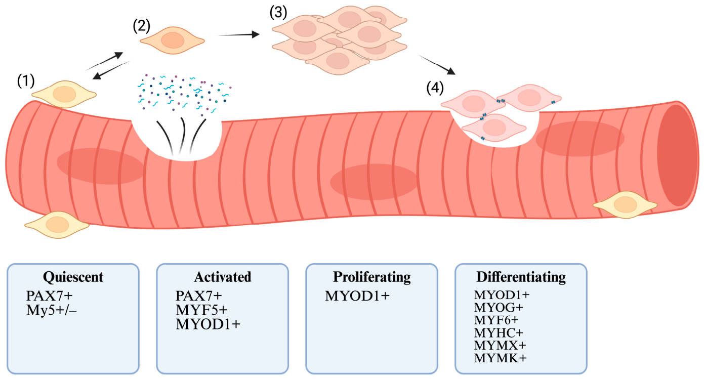
1.2. Overview of Myogenic Differentiation
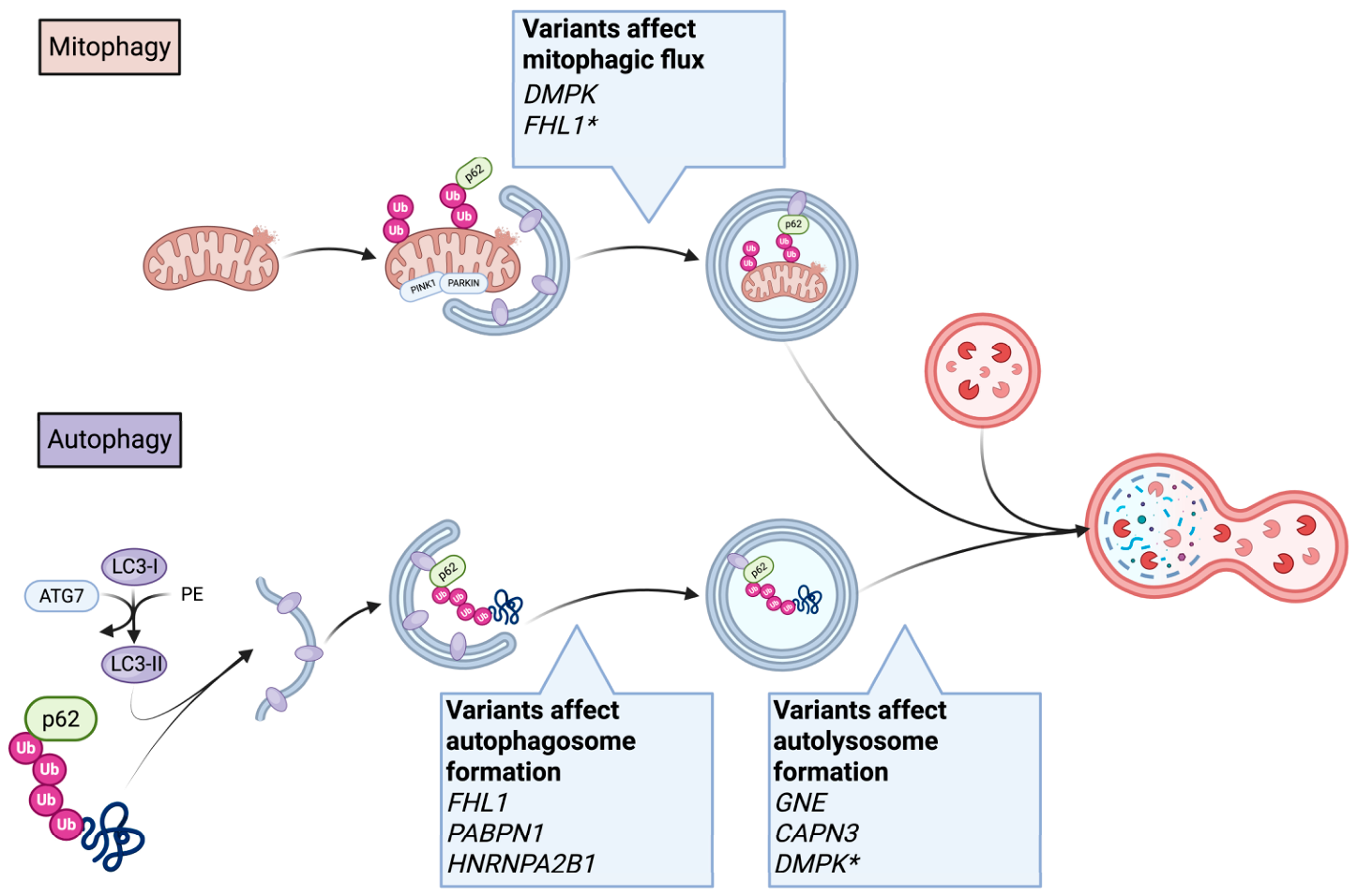
1.3. Autophagy as a Regulator of Differentiation
1.4. Mitochondrial Remodeling and Differentiation
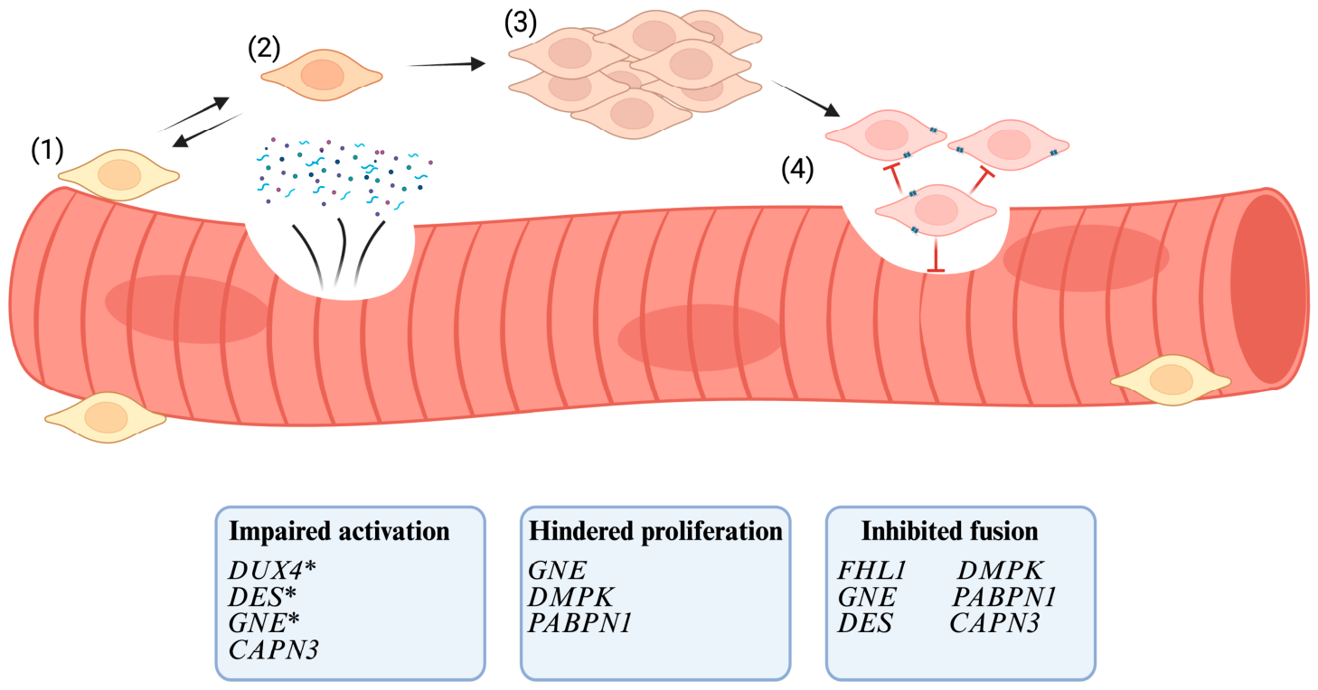
1.5. Satellite Cell Dysfunction as a Disease Mechanism in Myopathies
2. FHL1opathy
2.1. Etiology
2.2. FHL1 Affects Myogenic Differentiation
2.3. Autophagy Dysregulation
2.4. Critical Analysis
2.5. Possible Therapeutic Strategies
3. Oculopharyngeal Muscular Dystrophy (OPMD)
3.1. Etiology
3.2. PABPN1 Affects Myogenic Differentiation
3.3. Autophagy Dysregulation
3.4. Critical Analysis
3.5. Possible Therapeutic Strategies
4. Myotonic Dystrophy
4.1. Etiology
4.2. Toxic RNA Impairs Myogenic Differentiation
4.3. Autophagy and Mitophagy Dysregulation
4.4. Critical Analysis
4.5. Possible Therapeutic Strategies
5. Facioscapulohumeral Dystrophy (FSHD)
5.1. Etiology
5.2. DUX4 Affects Myogenic Differentiation
5.3. Critical Analysis
5.4. Possible Therapeutic Strategies
6. GNE Myopathy
6.1. Etiology
6.2. GNE Affects Myogenic Differentiation
6.3. Autophagy Dysregulation
6.4. Critical Analysis
6.5. Possible Therapeutic Strategies
7. Desminopathy
7.1. Etiology
7.2. Desmin Affects Myogenic Differentiation
7.3. Nuclear Stability Promotes Differentiation
7.4. Mitochondrial Dynamics
7.5. Critical Analysis
8. Limb Girdle Muscular Dystrophy Type 2A/R1 (LGMD2A/R1)
8.1. Etiology
8.2. Calpain-3 Affects Myogenic Differentiation
8.3. Autophagy Dysregulation
8.4. Mitochondrial Dynamics
8.5. Critical Analysis
8.6. Possible Therapeutic Strategies
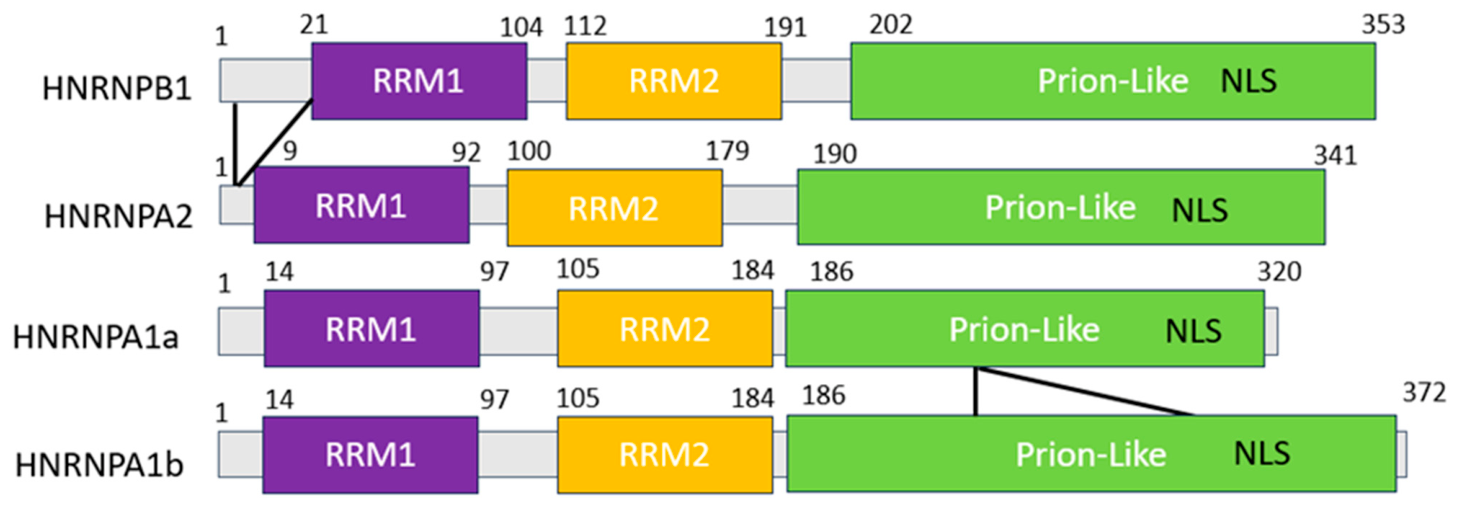
9. HNRNPA2/B1 and HNRNPA1
9.1. Etiology
9.2. HNRNPA2/B1 Affects Myogenic Differentiation
9.3. Cytotoxic Stress Granules or m6A Nuclear RNA Processing Events as a Putative Disease Mechanism
9.4. Autophagy Dysregulation
9.5. Critical Analysis
10. Future Work and Implications for Myopathies
10.1. Contribution of Myopathic Variants to Disease Phenotype
10.2. Expanding the List of Satellite Cell-Opathies
Author Contributions
Funding
Data Availability Statement
Acknowledgments
Conflicts of Interest
References
- Bentzinger, C.F.; Wang, Y.X.; Rudnicki, M.A. Building Muscle: Molecular Regulation of Myogenesis. Cold Spring Harb. Perspect. Biol. 2012, 4, a008342. [Google Scholar] [CrossRef] [Scilit] [PubMed]
- Yin, H.; Price, F.; Rudnicki, M.A. Satellite Cells and the Muscle Stem Cell Niche. Physiol. Rev. 2013, 93, 23–67. [Google Scholar] [CrossRef] [Scilit] [PubMed]
- Sousa-Victor, P.; García-Prat, L.; Muñoz-Cánoves, P. Control of Satellite Cell Function in Muscle Regeneration and Its Disruption in Ageing. Nat. Rev. Mol. Cell Biol. 2022, 23, 204–226. [Google Scholar] [CrossRef] [Scilit] [PubMed]
- Kaczmarek, A.; Kaczmarek, M.; Ciałowicz, M.; Clemente, F.M.; Wolański, P.; Badicu, G.; Murawska-Ciałowicz, E. The Role of Satellite Cells in Skeletal Muscle Regeneration-The Effect of Exercise and Age. Biology 2021, 10, 1056. [Google Scholar] [CrossRef] [Scilit] [PubMed]
- Chen, B.; Shan, T. The Role of Satellite and Other Functional Cell Types in Muscle Repair and Regeneration. J. Muscle Res. Cell Motil. 2019, 40, 1–8. [Google Scholar] [CrossRef] [Scilit] [PubMed]
- Kuang, S.; Kuroda, K.; Le Grand, F.; Rudnicki, M.A. Asymmetric Self-Renewal and Commitment of Satellite Stem Cells in Muscle. Cell 2007, 129, 999–1010. [Google Scholar] [CrossRef] [Scilit] [PubMed]
- Ancel, S.; Michaud, J.; Sizzano, F.; Tauzin, L.; Oliveira, M.; Migliavacca, E.; Schüler, S.C.; Raja, S.; Dammone, G.; Karaz, S.; et al. A Dual-Color PAX7 and MYF5 in Vivo Reporter to Investigate Muscle Stem Cell Heterogeneity in Regeneration and Aging. Stem Cell Rep. 2024, 19, 1024–1040. [Google Scholar] [CrossRef] [Scilit] [PubMed]
- Dall’Agnese, A.; Caputo, L.; Nicoletti, C.; di Iulio, J.; Schmitt, A.; Gatto, S.; Diao, Y.; Ye, Z.; Forcato, M.; Perera, R.; et al. Transcription Factor-Directed Re-Wiring of Chromatin Architecture for Somatic Cell Nuclear Reprogramming toward Trans-Differentiation. Mol. Cell 2019, 76, 453–472.e8. [Google Scholar] [CrossRef] [Scilit] [PubMed]
- Mal, A.; Harter, M.L. MyoD Is Functionally Linked to the Silencing of a Muscle-Specific Regulatory Gene Prior to Skeletal Myogenesis. Proc. Natl. Acad. Sci. USA 2003, 100, 1735–1739. [Google Scholar] [CrossRef] [Scilit] [PubMed]
- Battistelli, C.; Garbo, S.; Maione, R. MyoD-Induced Trans-Differentiation: A Paradigm for Dissecting the Molecular Mechanisms of Cell Commitment, Differentiation and Reprogramming. Cells 2022, 11, 3435. [Google Scholar] [CrossRef] [Scilit] [PubMed]
- Adhikari, A.; Kim, W.; Davie, J. Myogenin Is Required for Assembly of the Transcription Machinery on Muscle Genes during Skeletal Muscle Differentiation. PLoS ONE 2021, 16, e0245618. [Google Scholar] [CrossRef] [Scilit] [PubMed]
- Ganassi, M.; Badodi, S.; Ortuste Quiroga, H.P.; Zammit, P.S.; Hinits, Y.; Hughes, S.M. Myogenin Promotes Myocyte Fusion to Balance Fibre Number and Size. Nat. Commun. 2018, 9, 4232. [Google Scholar] [CrossRef] [Scilit] [PubMed]
- Lehka, L.; Rędowicz, M.J. Mechanisms Regulating Myoblast Fusion: A Multilevel Interplay. Semin. Cell Dev. Biol. 2020, 104, 81–92. [Google Scholar] [CrossRef] [Scilit] [PubMed]
- Pizza, F.X.; Buckley, K.H. Regenerating Myofibers after an Acute Muscle Injury: What Do We Really Know about Them? Int. J. Mol. Sci. 2023, 24, 12545. [Google Scholar] [CrossRef] [Scilit] [PubMed]
- Xia, Q.; Huang, X.; Huang, J.; Zheng, Y.; March, M.E.; Li, J.; Wei, Y. The Role of Autophagy in Skeletal Muscle Diseases. Front. Physiol. 2021, 12, 638983. [Google Scholar] [CrossRef] [Scilit] [PubMed]
- Masiero, E.; Sandri, M. Autophagy Inhibition Induces Atrophy and Myopathy in Adult Skeletal Muscles. Autophagy 2010, 6, 307–309. [Google Scholar] [CrossRef] [Scilit] [PubMed]
- Nemazanyy, I.; Blaauw, B.; Paolini, C.; Caillaud, C.; Protasi, F.; Mueller, A.; Proikas-Cezanne, T.; Russell, R.C.; Guan, K.-L.; Nishino, I.; et al. Defects of Vps15 in Skeletal Muscles Lead to Autophagic Vacuolar Myopathy and Lysosomal Disease. EMBO Mol. Med. 2013, 5, 870–890. [Google Scholar] [CrossRef] [Scilit] [PubMed]
- Paré, M.F.; Baechler, B.L.; Fajardo, V.A.; Earl, E.; Wong, E.; Campbell, T.L.; Tupling, A.R.; Quadrilatero, J. Effect of Acute and Chronic Autophagy Deficiency on Skeletal Muscle Apoptotic Signaling, Morphology, and Function. Biochim. Biophys. Acta Mol. Cell Res. 2017, 1864, 708–718. [Google Scholar] [CrossRef] [Scilit] [PubMed]
- Fortini, P.; Ferretti, C.; Iorio, E.; Cagnin, M.; Garribba, L.; Pietraforte, D.; Falchi, M.; Pascucci, B.; Baccarini, S.; Morani, F.; et al. The Fine Tuning of Metabolism, Autophagy and Differentiation during in Vitro Myogenesis. Cell Death Dis. 2016, 7, e2168. [Google Scholar] [CrossRef] [Scilit] [PubMed]
- McMillan, E.M.; Quadrilatero, J. Autophagy Is Required and Protects against Apoptosis during Myoblast Differentiation. Biochem. J. 2014, 462, 267–277. [Google Scholar] [CrossRef] [Scilit] [PubMed]
- Baechler, B.L.; Bloemberg, D.; Quadrilatero, J. Mitophagy Regulates Mitochondrial Network Signaling, Oxidative Stress, and Apoptosis during Myoblast Differentiation. Autophagy 2019, 15, 1606–1619. [Google Scholar] [CrossRef] [Scilit] [PubMed]
- Tang, A.H.; Rando, T.A. Induction of Autophagy Supports the Bioenergetic Demands of Quiescent Muscle Stem Cell Activation. EMBO J. 2014, 33, 2782–2797. [Google Scholar] [CrossRef] [Scilit] [PubMed]
- García-Prat, L.; Martínez-Vicente, M.; Perdiguero, E.; Ortet, L.; Rodríguez-Ubreva, J.; Rebollo, E.; Ruiz-Bonilla, V.; Gutarra, S.; Ballestar, E.; Serrano, A.L.; et al. Autophagy Maintains Stemness by Preventing Senescence. Nature 2016, 529, 37–42. [Google Scholar] [CrossRef] [Scilit] [PubMed]
- Goh, K.Y.; Lee, W.X.; Choy, S.M.; Priyadarshini, G.K.; Chua, K.; Tan, Q.H.; Low, S.Y.; Chin, H.S.; Wong, C.S.; Huang, S.-Y.; et al. FOXO-Regulated DEAF1 Controls Muscle Regeneration through Autophagy. Autophagy 2024, 20, 2632–2654. [Google Scholar] [CrossRef] [Scilit] [PubMed]
- Slavin, M.B.; Memme, J.M.; Oliveira, A.N.; Moradi, N.; Hood, D.A. Regulatory Networks Coordinating Mitochondrial Quality Control in Skeletal Muscle. Am. J. Physiol. Cell Physiol. 2022, 322, C913–C926. [Google Scholar] [CrossRef] [Scilit] [PubMed]
- Iqbal, S.; Ostojic, O.; Singh, K.; Joseph, A.-M.; Hood, D.A. Expression of Mitochondrial Fission and Fusion Regulatory Proteins in Skeletal Muscle during Chronic Use and Disuse. Muscle Nerve 2013, 48, 963–970. [Google Scholar] [CrossRef] [Scilit] [PubMed]
- Axelrod, C.L.; Fealy, C.E.; Mulya, A.; Kirwan, J.P. Exercise Training Remodels Human Skeletal Muscle Mitochondrial Fission and Fusion Machinery towards a Pro-Elongation Phenotype. Acta Physiol. 2019, 225, e13216. [Google Scholar] [CrossRef] [Scilit] [PubMed]
- Iqbal, S.; Hood, D.A. The Role of Mitochondrial Fusion and Fission in Skeletal Muscle Function and Dysfunction. Front. Biosci. 2015, 20, 157–172. [Google Scholar] [CrossRef] [Scilit] [PubMed]
- Zaman, M.; Shutt, T.E. The Role of Impaired Mitochondrial Dynamics in MFN2-Mediated Pathology. Front. Cell Dev. Biol. 2022, 10, 858286. [Google Scholar] [CrossRef] [Scilit] [PubMed]
- Bloemberg, D.; Quadrilatero, J. Effect of Mitochondrial Fission Inhibition on C2C12 Differentiation. Data Brief 2016, 7, 634–640. [Google Scholar] [CrossRef] [Scilit] [PubMed]
- Gambarotto, L.; Metti, S.; Chrisam, M.; Cerqua, C.; Sabatelli, P.; Armani, A.; Zanon, C.; Spizzotin, M.; Castagnaro, S.; Strappazzon, F.; et al. Ambra1 Deficiency Impairs Mitophagy in Skeletal Muscle. J. Cachexia Sarcopenia Muscle 2022, 13, 2211–2224. [Google Scholar] [CrossRef] [Scilit] [PubMed]
- Narendra, D.P.; Youle, R.J. The Role of PINK1-Parkin in Mitochondrial Quality Control. Nat. Cell Biol. 2024, 26, 1639–1651. [Google Scholar] [CrossRef] [Scilit] [PubMed]
- Peker, N.; Donipadi, V.; Sharma, M.; McFarlane, C.; Kambadur, R. Loss of Parkin Impairs Mitochondrial Function and Leads to Muscle Atrophy. Am. J. Physiol. Cell Physiol. 2018, 315, C164–C185. [Google Scholar] [CrossRef] [Scilit] [PubMed]
- Gouspillou, G.; Godin, R.; Piquereau, J.; Picard, M.; Mofarrahi, M.; Mathew, J.; Purves-Smith, F.M.; Sgarioto, N.; Hepple, R.T.; Burelle, Y.; et al. Protective Role of Parkin in Skeletal Muscle Contractile and Mitochondrial Function. J. Physiol. 2018, 596, 2565–2579. [Google Scholar] [CrossRef] [Scilit] [PubMed]
- Cairns, G.; Thumiah-Mootoo, M.; Abbasi, M.R.; Gourlay, M.; Racine, J.; Larionov, N.; Prola, A.; Khacho, M.; Burelle, Y. PINK1 Deficiency Alters Muscle Stem Cell Fate Decision and Muscle Regenerative Capacity. Stem Cell Rep. 2024, 19, 673–688. [Google Scholar] [CrossRef] [Scilit] [PubMed]
- Campello, S.; Scorrano, L. Mitochondrial Shape Changes: Orchestrating Cell Pathophysiology. EMBO Rep. 2010, 11, 678–684. [Google Scholar] [CrossRef] [Scilit] [PubMed]
- Baker, N.; Wade, S.; Triolo, M.; Girgis, J.; Chwastek, D.; Larrigan, S.; Feige, P.; Fujita, R.; Crist, C.; Rudnicki, M.A.; et al. The Mitochondrial Protein OPA1 Regulates the Quiescent State of Adult Muscle Stem Cells. Cell Stem Cell 2022, 29, 1315–1332.e9. [Google Scholar] [CrossRef] [Scilit] [PubMed]
- Ryall, J.G.; Dell’Orso, S.; Derfoul, A.; Juan, A.; Zare, H.; Feng, X.; Clermont, D.; Koulnis, M.; Gutierrez-Cruz, G.; Fulco, M.; et al. The NAD(+)-Dependent SIRT1 Deacetylase Translates a Metabolic Switch into Regulatory Epigenetics in Skeletal Muscle Stem Cells. Cell Stem Cell 2015, 16, 171–183. [Google Scholar] [CrossRef] [Scilit] [PubMed]
- Hong, X.; Isern, J.; Campanario, S.; Perdiguero, E.; Ramírez-Pardo, I.; Segalés, J.; Hernansanz-Agustín, P.; Curtabbi, A.; Deryagin, O.; Pollán, A.; et al. Mitochondrial Dynamics Maintain Muscle Stem Cell Regenerative Competence throughout Adult Life by Regulating Metabolism and Mitophagy. Cell Stem Cell 2022, 29, 1298–1314.e10, Erratum in Cell Stem Cell 2022, 29, 1506–1508. [Google Scholar] [CrossRef] [Scilit] [PubMed]
- Folmes, C.D.L.; Nelson, T.J.; Martinez-Fernandez, A.; Arrell, D.K.; Lindor, J.Z.; Dzeja, P.P.; Ikeda, Y.; Perez-Terzic, C.; Terzic, A. Somatic Oxidative Bioenergetics Transitions into Pluripotency-Dependent Glycolysis to Facilitate Nuclear Reprogramming. Cell Metab. 2011, 14, 264–271. [Google Scholar] [CrossRef] [Scilit] [PubMed]
- Remels, A.H.V.; Langen, R.C.J.; Schrauwen, P.; Schaart, G.; Schols, A.M.W.J.; Gosker, H.R. Regulation of Mitochondrial Biogenesis during Myogenesis. Mol. Cell. Endocrinol. 2010, 315, 113–120. [Google Scholar] [CrossRef] [Scilit] [PubMed]
- Gaitanos, G.C.; Williams, C.; Boobis, L.H.; Brooks, S. Human Muscle Metabolism during Intermittent Maximal Exercise. J. Appl. Physiol. 1993, 75, 712–719. [Google Scholar] [CrossRef] [Scilit] [PubMed]
- Gonzalez-Chapa, J.A.; Macêdo, M.B.; Lood, C. The Emerging Role of Mitochondrial Dysfunction in the Pathogenesis of Idiopathic Inflammatory Myopathies. Rambam Maimonides Med. J. 2023, 14, e0006. [Google Scholar] [CrossRef] [Scilit] [PubMed]
- Irazoki, A.; Gordaliza-Alaguero, I.; Frank, E.; Giakoumakis, N.N.; Seco, J.; Palacín, M.; Gumà, A.; Sylow, L.; Sebastián, D.; Zorzano, A. Disruption of Mitochondrial Dynamics Triggers Muscle Inflammation through Interorganellar Contacts and Mitochondrial DNA Mislocation. Nat. Commun. 2023, 14, 108. [Google Scholar] [CrossRef] [Scilit] [PubMed]
- Ganassi, M.; Zammit, P.S. Involvement of Muscle Satellite Cell Dysfunction in Neuromuscular Disorders: Expanding the Portfolio of Satellite Cell-Opathies. Eur. J. Transl. Myol. 2022, 32, 10064. [Google Scholar] [CrossRef] [Scilit] [PubMed]
- Ganassi, M.; Muntoni, F.; Zammit, P.S. Defining and Identifying Satellite Cell-Opathies within Muscular Dystrophies and Myopathies. Exp. Cell Res. 2022, 411, 112906. [Google Scholar] [CrossRef] [Scilit] [PubMed]
- Dumont, N.A.; Wang, Y.X.; von Maltzahn, J.; Pasut, A.; Bentzinger, C.F.; Brun, C.E.; Rudnicki, M.A. Dystrophin Expression in Muscle Stem Cells Regulates Their Polarity and Asymmetric Division. Nat. Med. 2015, 21, 1455–1463. [Google Scholar] [CrossRef] [Scilit] [PubMed]
- Sarde, L.; Letort, G.; Varet, H.; Laville, V.; Fernandes, J.; Tajbakhsh, S.; Evano, B. Impaired Stem Cell Migration and Divisions in Duchenne Muscular Dystrophy Revealed by Live Imaging. Nat. Commun. 2026, 17, 1769. [Google Scholar] [CrossRef] [Scilit] [PubMed]
- Storey, E.C.; Holt, I.; Morris, G.E.; Fuller, H.R. Muscle Cell Differentiation and Development Pathway Defects in Emery-Dreifuss Muscular Dystrophy. Neuromuscul. Disord. 2020, 30, 443–456. [Google Scholar] [CrossRef] [Scilit] [PubMed]
- Brooke, M.H.; Neville, H.E. Reducing Body Myopathy. Neurology 1972, 22, 829–840. [Google Scholar] [CrossRef] [Scilit] [PubMed]
- Schessl, J.; Zou, Y.; McGrath, M.J.; Cowling, B.S.; Maiti, B.; Chin, S.S.; Sewry, C.; Battini, R.; Hu, Y.; Cottle, D.L.; et al. Proteomic Identification of FHL1 as the Protein Mutated in Human Reducing Body Myopathy. J. Clin. Investig. 2008, 118, 904–912. [Google Scholar] [CrossRef] [Scilit] [PubMed]
- Windpassinger, C.; Schoser, B.; Straub, V.; Hochmeister, S.; Noor, A.; Lohberger, B.; Farra, N.; Petek, E.; Schwarzbraun, T.; Ofner, L.; et al. An X-Linked Myopathy with Postural Muscle Atrophy and Generalized Hypertrophy, Termed XMPMA, Is Caused by Mutations in FHL1. Am. J. Hum. Genet. 2008, 82, 88–99. [Google Scholar] [CrossRef] [Scilit] [PubMed]
- Cowling, B.S.; Cottle, D.L.; Wilding, B.R.; D’Arcy, C.E.; Mitchell, C.A.; McGrath, M.J. Four and a Half LIM Protein 1 Gene Mutations Cause Four Distinct Human Myopathies: A Comprehensive Review of the Clinical, Histological and Pathological Features. Neuromuscul. Disord. 2011, 21, 237–251. [Google Scholar] [CrossRef] [Scilit] [PubMed]
- Malfatti, E.; Olivé, M.; Taratuto, A.L.; Richard, P.; Brochier, G.; Bitoun, M.; Gueneau, L.; Laforêt, P.; Stojkovic, T.; Maisonobe, T.; et al. Skeletal Muscle Biopsy Analysis in Reducing Body Myopathy and Other FHL1-Related Disorders. J. Neuropathol. Exp. Neurol. 2013, 72, 833–845. [Google Scholar] [CrossRef] [Scilit] [PubMed]
- Gueneau, L.; Bertrand, A.T.; Jais, J.-P.; Salih, M.A.; Stojkovic, T.; Wehnert, M.; Hoeltzenbein, M.; Spuler, S.; Saitoh, S.; Verschueren, A.; et al. Mutations of the FHL1 Gene Cause Emery-Dreifuss Muscular Dystrophy. Am. J. Hum. Genet. 2009, 85, 338–353. [Google Scholar] [CrossRef] [Scilit] [PubMed]
- Schessl, J.; Taratuto, A.L.; Sewry, C.; Battini, R.; Chin, S.S.; Maiti, B.; Dubrovsky, A.L.; Erro, M.G.; Espada, G.; Robertella, M.; et al. Clinical, Histological and Genetic Characterization of Reducing Body Myopathy Caused by Mutations in FHL1. Brain 2009, 132, 452–464. [Google Scholar] [CrossRef] [Scilit] [PubMed]
- Jokela, M.; Huovinen, S.; Palmio, J.; Saukkonen, A.-M.; Penttilä, S.; Udd, B. Gluteus Maximus Hypertrophy: A Diagnostic Clue in Four and a Half LIM Domain 1-Mutated Reducing Body Myopathy. Neuromuscul. Disord. 2017, 27, 962–963. [Google Scholar] [CrossRef] [Scilit] [PubMed]
- Astrea, G.; Schessl, J.; Clement, E.; Tosetti, M.; Mercuri, E.; Rutherford, M.; Cioni, G.; Bönnemann, C.G.; Muntoni, F.; Battini, R. Muscle MRI in FHL1-Linked Reducing Body Myopathy. Neuromuscul. Disord. 2009, 19, 689–691. [Google Scholar] [CrossRef] [Scilit] [PubMed]
- Schreckenbach, T.; Henn, W.; Kress, W.; Roos, A.; Maschke, M.; Feiden, W.; Dillmann, U.; Schulz, J.B.; Weis, J.; Claeys, K.G. Novel FHL1 Mutation in a Family with Reducing Body Myopathy. Muscle Nerve 2013, 47, 127–134. [Google Scholar] [CrossRef] [Scilit] [PubMed]
- Cejas, C.P.; Serra, M.M.; Galvez, D.F.G.; Cavassa, E.A.; Taratuto, A.L.; Vazquez, G.A.; Massaro, M.E.L.; Schteinschneider, A. V Muscle MRI in Pediatrics: Clinical, Pathological and Genetic Correlation. Pediatr. Radiol. 2017, 47, 724–735. [Google Scholar] [CrossRef] [Scilit] [PubMed]
- Greene, W.K.; Baker, E.; Rabbitts, T.H.; Kees, U.R. Genomic Structure, Tissue Expression and Chromosomal Location of the LIM-Only Gene, SLIM1. Gene 1999, 232, 203–207. [Google Scholar] [CrossRef] [Scilit] [PubMed]
- Cowling, B.S.; McGrath, M.J.; Nguyen, M.-A.; Cottle, D.L.; Kee, A.J.; Brown, S.; Schessl, J.; Zou, Y.; Joya, J.; Bönnemann, C.G.; et al. Identification of FHL1 as a Regulator of Skeletal Muscle Mass: Implications for Human Myopathy. J. Cell Biol. 2008, 183, 1033–1048. [Google Scholar] [CrossRef] [Scilit] [PubMed]
- Han, S.; Cui, C.; He, H.; Shen, X.; Chen, Y.; Wang, Y.; Li, D.; Zhu, Q.; Yin, H. FHL1 Regulates Myoblast Differentiation and Autophagy through Its Interaction with LC3. J. Cell. Physiol. 2020, 235, 4667–4678. [Google Scholar] [CrossRef] [Scilit] [PubMed]
- Lin, Y.; Ban, R.; Qiao, L.; Chen, J.; Liu, M.; Liu, J.; Shi, Q. Identification of Novel FHL1 Mutations Associated with X-Linked Scapuloperoneal Myopathy in Unrelated Chinese Patients. J. Hum. Genet. 2023, 68, 477–484. [Google Scholar] [CrossRef] [Scilit] [PubMed]
- Park, Y.-E.; Kim, D.-S.; Shin, J.-H. Myofibrillar Myopathy Caused by a Novel FHL1 Mutation Presenting a Mild Myopathy with Ankle Contracture. Clin. Neurol. Neurosurg. 2019, 180, 48–51. [Google Scholar] [CrossRef] [Scilit] [PubMed]
- Borch, J.D.S.; Krag, T.; Holm-Yildiz, S.D.; Cetin, H.; Solheim, T.A.; Fornander, F.; Straub, V.; Duno, M.; Vissing, J. Three Novel FHL1 Variants Cause a Mild Phenotype of Emery-Dreifuss Muscular Dystrophy. Hum. Mutat. 2022, 43, 1234–1238. [Google Scholar] [CrossRef] [Scilit] [PubMed]
- Shalaby, S.; Hayashi, Y.K.; Goto, K.; Ogawa, M.; Nonaka, I.; Noguchi, S.; Nishino, I. Rigid Spine Syndrome Caused by a Novel Mutation in Four-and-a-Half LIM Domain 1 Gene (FHL1). Neuromuscul. Disord. 2008, 18, 959–961. [Google Scholar] [CrossRef] [Scilit] [PubMed]
- Domenighetti, A.A.; Chu, P.-H.; Wu, T.; Sheikh, F.; Gokhin, D.S.; Guo, L.T.; Cui, Z.; Peter, A.K.; Christodoulou, D.C.; Parfenov, M.G.; et al. Loss of FHL1 Induces an Age-Dependent Skeletal Muscle Myopathy Associated with Myofibrillar and Intermyofibrillar Disorganization in Mice. Hum. Mol. Genet. 2014, 23, 209–225. [Google Scholar] [CrossRef] [Scilit] [PubMed]
- Nguyen, M.T.; Min, K.-H.; Lee, W. MiR-183-5p Induced by Saturated Fatty Acids Regulates the Myogenic Differentiation by Directly Targeting FHL1 in C2C12 Myoblasts. BMB Rep. 2020, 53, 605–610. [Google Scholar] [CrossRef] [Scilit] [PubMed]
- Wilding, B.R.; McGrath, M.J.; Bonne, G.; Mitchell, C.A. FHL1 Mutants That Cause Clinically Distinct Human Myopathies Form Protein Aggregates and Impair Myoblast Differentiation. J. Cell Sci. 2014, 127, 2269–2281. [Google Scholar] [CrossRef] [Scilit] [PubMed]
- Horsley, V.; Jansen, K.M.; Mills, S.T.; Pavlath, G.K. IL-4 Acts as a Myoblast Recruitment Factor during Mammalian Muscle Growth. Cell 2003, 113, 483–494. [Google Scholar] [CrossRef] [Scilit] [PubMed]
- Horsley, V.; Friday, B.B.; Matteson, S.; Kegley, K.M.; Gephart, J.; Pavlath, G.K. Regulation of the Growth of Multinucleated Muscle Cells by an NFATC2-Dependent Pathway. J. Cell Biol. 2001, 153, 329–338. [Google Scholar] [CrossRef] [Scilit] [PubMed]
- Ehlers, M.L.; Celona, B.; Black, B.L. NFATc1 Controls Skeletal Muscle Fiber Type and Is a Negative Regulator of MyoD Activity. Cell Rep. 2014, 8, 1639–1648. [Google Scholar] [CrossRef] [Scilit] [PubMed]
- McCullagh, K.J.A.; Calabria, E.; Pallafacchina, G.; Ciciliot, S.; Serrano, A.L.; Argentini, C.; Kalhovde, J.M.; Lømo, T.; Schiaffino, S. NFAT Is a Nerve Activity Sensor in Skeletal Muscle and Controls Activity-Dependent Myosin Switching. Proc. Natl. Acad. Sci. USA 2004, 101, 10590–10595, Correction in Proc. Natl. Acad. Sci. USA 2024, 121, e2424252121. [Google Scholar] [CrossRef] [Scilit] [PubMed]
- O’Connor, R.S.; Mills, S.T.; Jones, K.A.; Ho, S.N.; Pavlath, G.K. A Combinatorial Role for NFAT5 in Both Myoblast Migration and Differentiation during Skeletal Muscle Myogenesis. J. Cell Sci. 2007, 120, 149–159. [Google Scholar] [CrossRef] [Scilit] [PubMed]
- Semsarian, C.; Wu, M.J.; Ju, Y.K.; Marciniec, T.; Yeoh, T.; Allen, D.G.; Harvey, R.P.; Graham, R.M. Skeletal Muscle Hypertrophy Is Mediated by a Ca2+-Dependent Calcineurin Signalling Pathway. Nature 1999, 400, 576–581. [Google Scholar] [CrossRef] [Scilit] [PubMed]
- Ding, J.; Cong, Y.F.; Liu, B.; Miao, J.; Wang, L. Aberrant Protein Turn-Over Associated With Myofibrillar Disorganization in FHL1 Knockout Mice. Front. Genet. 2018, 9, 273. [Google Scholar] [CrossRef] [Scilit] [PubMed]
- Komatsu, M.; Waguri, S.; Ueno, T.; Iwata, J.; Murata, S.; Tanida, I.; Ezaki, J.; Mizushima, N.; Ohsumi, Y.; Uchiyama, Y.; et al. Impairment of Starvation-Induced and Constitutive Autophagy in Atg7-Deficient Mice. J. Cell Biol. 2005, 169, 425–434. [Google Scholar] [CrossRef] [Scilit] [PubMed]
- Pan, H.-Y.; Ladd, A.V.; Biswal, M.R.; Valapala, M. Role of Nuclear Factor of Activated T Cells (NFAT) Pathway in Regulating Autophagy and Inflammation in Retinal Pigment Epithelial Cells. Int. J. Mol. Sci. 2021, 22, 8684. [Google Scholar] [CrossRef] [Scilit] [PubMed]
- Shathasivam, T.; Kislinger, T.; Gramolini, A.O. Genes, Proteins and Complexes: The Multifaceted Nature of FHL Family Proteins in Diverse Tissues. J. Cell. Mol. Med. 2010, 14, 2702–2720. [Google Scholar] [CrossRef] [Scilit] [PubMed]
- Martin, B.; Schneider, R.; Janetzky, S.; Waibler, Z.; Pandur, P.; Kühl, M.; Behrens, J.; von der Mark, K.; Starzinski-Powitz, A.; Wixler, V. The LIM-Only Protein FHL2 Interacts with Beta-Catenin and Promotes Differentiation of Mouse Myoblasts. J. Cell Biol. 2002, 159, 113–122. [Google Scholar] [CrossRef] [Scilit] [PubMed]
- Hojayev, B.; Rothermel, B.A.; Gillette, T.G.; Hill, J.A. FHL2 Binds Calcineurin and Represses Pathological Cardiac Growth. Mol. Cell. Biol. 2012, 32, 4025–4034. [Google Scholar] [CrossRef] [Scilit] [PubMed]
- Liu, Z.; Han, S.; Wang, Y.; Cui, C.; Zhu, Q.; Jiang, X.; Yang, C.; Du, H.; Yu, C.; Li, Q.; et al. The LIM-Only Protein FHL2 Is Involved in Autophagy to Regulate the Development of Skeletal Muscle Cell. Int. J. Biol. Sci. 2019, 15, 838–846. [Google Scholar] [CrossRef] [Scilit] [PubMed]
- Zhu, Y.; Li, P.; Dan, X.; Kang, X.; Ma, Y.; Shi, Y. MiR-377 Inhibits Proliferation and Differentiation of Bovine Skeletal Muscle Satellite Cells by Targeting FHL2. Genes 2022, 13, 947. [Google Scholar] [CrossRef] [Scilit] [PubMed]
- Dennhag, N.; Kahsay, A.; Nissen, I.; Nord, H.; Chermenina, M.; Liu, J.; Arner, A.; Liu, J.-X.; Backman, L.J.; Remeseiro, S.; et al. Fhl2b Mediates Extraocular Muscle Protection in Zebrafish Models of Muscular Dystrophies and Its Ectopic Expression Ameliorates Affected Body Muscles. Nat. Commun. 2024, 15, 1950. [Google Scholar] [CrossRef] [Scilit] [PubMed]
- Bai, W.; Zhang, Y.; Ma, J.; Du, M.; Xu, H.; Wang, J.; Zhang, L.; Li, W.; Hou, Y.; Liu, X.; et al. FHL3 Promotes the Formation of Fast Glycolytic Muscle Fibers by Interacting with YY1 and Muscle Glycolytic Metabolism. Cell. Mol. Life Sci. 2023, 80, 27. [Google Scholar] [CrossRef] [Scilit] [PubMed]
- Zhang, Y.; Li, W.; Zhu, M.; Li, Y.; Xu, Z.; Zuo, B. FHL3 Differentially Regulates the Expression of MyHC Isoforms through Interactions with MyoD and PCREB. Cell. Signal. 2016, 28, 60–73. [Google Scholar] [CrossRef] [Scilit] [PubMed]
- Cottle, D.L.; McGrath, M.J.; Cowling, B.S.; Coghill, I.D.; Brown, S.; Mitchell, C.A. FHL3 Binds MyoD and Negatively Regulates Myotube Formation. J. Cell Sci. 2007, 120, 1423–1435. [Google Scholar] [CrossRef] [Scilit] [PubMed]
- Han, S.; Cui, C.; Wang, Y.; He, H.; Shen, X.; Chen, Y.; Liu, Z.; Zhu, Q.; Li, D.; Yin, H. FHL3 Negatively Regulates the Differentiation of Skeletal Muscle Satellite Cells in Chicken. 3 Biotech 2019, 9, 206. [Google Scholar] [CrossRef] [Scilit] [PubMed]
- Morgan, M.J.; Madgwick, A.J. The LIM Proteins FHL1 and FHL3 Are Expressed Differently in Skeletal Muscle. Biochem. Biophys. Res. Commun. 1999, 255, 245–250. [Google Scholar] [CrossRef] [Scilit] [PubMed]
- Chu, P.H.; Ruiz-Lozano, P.; Zhou, Q.; Cai, C.; Chen, J. Expression Patterns of FHL/SLIM Family Members Suggest Important Functional Roles in Skeletal Muscle and Cardiovascular System. Mech. Dev. 2000, 95, 259–265. [Google Scholar] [CrossRef] [Scilit] [PubMed]
- Tabor, L.C.; Plowman, E.K.; Romero-Clark, C.; Youssof, S. Oropharyngeal Dysphagia Profiles in Individuals with Oculopharyngeal Muscular Dystrophy. Neurogastroenterol. Motil. 2018, 30, e13251. [Google Scholar] [CrossRef] [Scilit] [PubMed]
- Kroon, R.H.M.J.M.; Kalf, J.G.; de Swart, B.J.M.; van der Sluijs, B.M.; Glennon, J.C.; Raz, V.; van Engelen, B.G.; Horlings, C.G.C. Longitudinal Assessment of Strength, Functional Capacity, Oropharyngeal Function, and Quality of Life in Oculopharyngeal Muscular Dystrophy. Neurology 2021, 97, e1475–e1483. [Google Scholar] [CrossRef] [Scilit] [PubMed]
- Brais, B.; Bouchard, J.P.; Xie, Y.G.; Rochefort, D.L.; Chrétien, N.; Tomé, F.M.; Lafrenière, R.G.; Rommens, J.M.; Uyama, E.; Nohira, O.; et al. Short GCG Expansions in the PABP2 Gene Cause Oculopharyngeal Muscular Dystrophy. Nat. Genet. 1998, 18, 164–167, Correction in Nat. Genet. 1998, 19, 404. [Google Scholar] [CrossRef] [Scilit] [PubMed]
- Kerwitz, Y.; Kühn, U.; Lilie, H.; Knoth, A.; Scheuermann, T.; Friedrich, H.; Schwarz, E.; Wahle, E. Stimulation of Poly(A) Polymerase through a Direct Interaction with the Nuclear Poly(A) Binding Protein Allosterically Regulated by RNA. EMBO J. 2003, 22, 3705–3714. [Google Scholar] [CrossRef] [Scilit] [PubMed]
- Banerjee, A.; Apponi, L.H.; Pavlath, G.K.; Corbett, A.H. PABPN1: Molecular Function and Muscle Disease. FEBS J. 2013, 280, 4230–4250. [Google Scholar] [CrossRef] [Scilit] [PubMed]
- Tavanez, J.P.; Calado, P.; Braga, J.; Lafarga, M.; Carmo-Fonseca, M. In Vivo Aggregation Properties of the Nuclear Poly(A)-Binding Protein PABPN1. RNA 2005, 11, 752–762. [Google Scholar] [CrossRef] [Scilit] [PubMed]
- Shademan, M.; Mei, H.; van Engelen, B.; Ariyurek, Y.; Kloet, S.; Raz, V. PABPN1 Loss-of-Function Causes APA-Shift in Oculopharyngeal Muscular Dystrophy. HGG Adv. 2024, 5, 100269. [Google Scholar] [CrossRef] [Scilit] [PubMed]
- Riaz, M.; Raz, Y.; van der Slujis, B.; Dickson, G.; van Engelen, B.; Vissing, J.; Raz, V. Cytokine Genes as Potential Biomarkers for Muscle Weakness in OPMD. Hum. Mol. Genet. 2016, 25, 4282–4287. [Google Scholar] [CrossRef] [Scilit] [PubMed]
- Anvar, S.Y.; Raz, Y.; Verway, N.; van der Sluijs, B.; Venema, A.; Goeman, J.J.; Vissing, J.; van der Maarel, S.M.; ’t Hoen, P.A.C.; van Engelen, B.G.M.; et al. A Decline in PABPN1 Induces Progressive Muscle Weakness in Oculopharyngeal Muscle Dystrophy and in Muscle Aging. Aging 2013, 5, 412–426. [Google Scholar] [CrossRef] [Scilit] [PubMed]
- Apponi, L.H.; Corbett, A.H.; Pavlath, G.K. Control of MRNA Stability Contributes to Low Levels of Nuclear Poly(A) Binding Protein 1 (PABPN1) in Skeletal Muscle. Skelet. Muscle 2013, 3, 23. [Google Scholar] [CrossRef] [Scilit] [PubMed]
- Kim, Y.J.; Noguchi, S.; Hayashi, Y.K.; Tsukahara, T.; Shimizu, T.; Arahata, K. The Product of an Oculopharyngeal Muscular Dystrophy Gene, Poly(A)-Binding Protein 2, Interacts with SKIP and Stimulates Muscle-Specific Gene Expression. Hum. Mol. Genet. 2001, 10, 1129–1139. [Google Scholar] [CrossRef] [Scilit] [PubMed]
- Wang, Q.; Bag, J. Ectopic Expression of a Polyalanine Expansion Mutant of Poly(A)-Binding Protein N1 in Muscle Cells in Culture Inhibits Myogenesis. Biochem. Biophys. Res. Commun. 2006, 340, 815–822. [Google Scholar] [CrossRef] [Scilit] [PubMed]
- Apponi, L.H.; Leung, S.W.; Williams, K.R.; Valentini, S.R.; Corbett, A.H.; Pavlath, G.K. Loss of Nuclear Poly(A)-Binding Protein 1 Causes Defects in Myogenesis and MRNA Biogenesis. Hum. Mol. Genet. 2010, 19, 1058–1065. [Google Scholar] [CrossRef] [Scilit] [PubMed]
- Mallon, S.D.; Bos, E.; Sheikhhassani, V.; Shademan, M.; Voortman, L.M.; Mashaghi, A.; Sharp, T.H.; Raz, V. Polyalanine Expansion in PABPN1 Alters the Structure and Dynamics of Its Nuclear Aggregates in Differentiated Muscle Cells. FASEB J. 2025, 39, e70748. [Google Scholar] [CrossRef] [Scilit] [PubMed]
- Périé, S.; Mamchaoui, K.; Mouly, V.; Blot, S.; Bouazza, B.; Thornell, L.-E.; St Guily, J.L.; Butler-Browne, G. Premature Proliferative Arrest of Cricopharyngeal Myoblasts in Oculo-Pharyngeal Muscular Dystrophy: Therapeutic Perspectives of Autologous Myoblast Transplantation. Neuromuscul. Disord. 2006, 16, 770–781. [Google Scholar] [CrossRef] [Scilit] [PubMed]
- García-Castañeda, M.; Vega, A.V.; Rodríguez, R.; Montiel-Jaen, M.G.; Cisneros, B.; Zarain-Herzberg, A.; Avila, G. Functional Impact of an Oculopharyngeal Muscular Dystrophy Mutation in PABPN1. J. Physiol. 2017, 595, 4167–4187. [Google Scholar] [CrossRef] [Scilit] [PubMed]
- Olie, C.S.; van der Wal, E.; Cikes, D.; Maton, L.; de Greef, J.C.; Lin, I.-H.; Chen, Y.-F.; Kareem, E.; Penninger, J.M.; Kessler, B.M.; et al. Cytoskeletal Disorganization Underlies PABPN1-Mediated Myogenic Disability. Sci. Rep. 2020, 10, 17621, Correction in Sci. Rep. 2021, 11, 6429. [Google Scholar] [CrossRef] [Scilit] [PubMed]
- Zhang, Y.; Zeuthen, C.; Zhu, C.; Wu, F.; Mezzell, A.T.; Whitlow, T.J.; Choo, H.J.; Vest, K.E. Pharyngeal Pathology in a Mouse Model of Oculopharyngeal Muscular Dystrophy Is Associated with Impaired Basal Autophagy in Myoblasts. Front. Cell Dev. Biol. 2022, 10, 986930. [Google Scholar] [CrossRef] [Scilit] [PubMed]
- Raz, V.; Dickson, G.; ’t Hoen, P.A.C. Dysfunctional Transcripts Are Formed by Alternative Polyadenylation in OPMD. Oncotarget 2017, 8, 73516–73528. [Google Scholar] [CrossRef] [Scilit] [PubMed]
- Ishtayeh, H.; Galves, M.; Barnatan, T.T.; Berdichevsky, Y.; Amer-Sarsour, F.; Pasmanik-Chor, M.; Braverman, I.; Blumen, S.C.; Ashkenazi, A. Oculopharyngeal Muscular Dystrophy Mutations Link the RNA-Binding Protein HNRNPQ to Autophagosome Biogenesis. Aging Cell 2023, 22, e13949. [Google Scholar] [CrossRef] [Scilit] [PubMed]
- Périé, S.; Trollet, C.; Mouly, V.; Vanneaux, V.; Mamchaoui, K.; Bouazza, B.; Marolleau, J.P.; Laforêt, P.; Chapon, F.; Eymard, B.; et al. Autologous Myoblast Transplantation for Oculopharyngeal Muscular Dystrophy: A Phase I/IIa Clinical Study. Mol. Ther. 2014, 22, 219–225. [Google Scholar] [CrossRef] [Scilit] [PubMed]
- Malerba, A.; Klein, P.; Bachtarzi, H.; Jarmin, S.A.; Cordova, G.; Ferry, A.; Strings, V.; Espinoza, M.P.; Mamchaoui, K.; Blumen, S.C.; et al. PABPN1 Gene Therapy for Oculopharyngeal Muscular Dystrophy. Nat. Commun. 2017, 8, 14848. [Google Scholar] [CrossRef] [Scilit] [PubMed]
- Gagnon, C.; Petitclerc, É.; Kierkegaard, M.; Mathieu, J.; Duchesne, É.; Hébert, L.J. A 9-Year Follow-up Study of Quantitative Muscle Strength Changes in Myotonic Dystrophy Type 1. J. Neurol. 2018, 265, 1698–1705. [Google Scholar] [CrossRef] [Scilit] [PubMed]
- Liao, Q.; Zhang, Y.; He, J.; Huang, K. Global Prevalence of Myotonic Dystrophy: An Updated Systematic Review and Meta-Analysis. Neuroepidemiology 2022, 56, 163–173. [Google Scholar] [CrossRef] [Scilit] [PubMed]
- Mathieu, J.; Prévost, C. Epidemiological Surveillance of Myotonic Dystrophy Type 1: A 25-Year Population-Based Study. Neuromuscul. Disord. 2012, 22, 974–979. [Google Scholar] [CrossRef] [Scilit] [PubMed]
- Johnson, N.E.; Butterfield, R.J.; Mayne, K.; Newcomb, T.; Imburgia, C.; Dunn, D.; Duval, B.; Feldkamp, M.L.; Weiss, R.B. Population-Based Prevalence of Myotonic Dystrophy Type 1 Using Genetic Analysis of Statewide Blood Screening Program. Neurology 2021, 96, e1045–e1053. [Google Scholar] [CrossRef] [Scilit] [PubMed]
- Dogan, C.; De Antonio, M.; Hamroun, D.; Varet, H.; Fabbro, M.; Rougier, F.; Amarof, K.; Arne Bes, M.-C.; Bedat-Millet, A.-L.; Behin, A.; et al. Gender as a Modifying Factor Influencing Myotonic Dystrophy Type 1 Phenotype Severity and Mortality: A Nationwide Multiple Databases Cross-Sectional Observational Study. PLoS ONE 2016, 11, e0148264. [Google Scholar] [CrossRef] [Scilit] [PubMed]
- Provenzano, M.; Ikegami, K.; Bates, K.; Gaynor, A.; Hartman, J.M.; Jones, A.; Butler, A.; Berggren, K.N.; Dekdebrun, J.; Hung, M.; et al. The Splice Index as a Prognostic Biomarker of Strength and Function in Myotonic Dystrophy Type 1. J. Clin. Investig. 2025, 135, e185426. [Google Scholar] [CrossRef] [Scilit] [PubMed]
- Meola, G. Clinical Aspects, Molecular Pathomechanisms and Management of Myotonic Dystrophies. Acta Myol. 2013, 32, 154–165. [Google Scholar] [PubMed]
- Conte, T.C.; Duran-Bishop, G.; Orfi, Z.; Mokhtari, I.; Deprez, A.; Côté, I.; Molina, T.; Kim, T.-Y.; Tellier, L.; Roussel, M.-P.; et al. Clearance of Defective Muscle Stem Cells by Senolytics Restores Myogenesis in Myotonic Dystrophy Type 1. Nat. Commun. 2023, 14, 4033. [Google Scholar] [CrossRef] [Scilit] [PubMed]
- Furling, D.; Lemieux, D.; Taneja, K.; Puymirat, J. Decreased Levels of Myotonic Dystrophy Protein Kinase (DMPK) and Delayed Differentiation in Human Myotonic Dystrophy Myoblasts. Neuromuscul. Disord. 2001, 11, 728–735. [Google Scholar] [CrossRef] [Scilit] [PubMed]
- Timchenko, N.A.; Iakova, P.; Cai, Z.J.; Smith, J.R.; Timchenko, L.T. Molecular Basis for Impaired Muscle Differentiation in Myotonic Dystrophy. Mol. Cell. Biol. 2001, 21, 6927–6938. [Google Scholar] [CrossRef] [Scilit] [PubMed]
- Lee, J.E.; Lee, J.Y.; Wilusz, J.; Tian, B.; Wilusz, C.J. Systematic Analysis of Cis-Elements in Unstable MRNAs Demonstrates That CUGBP1 Is a Key Regulator of MRNA Decay in Muscle Cells. PLoS ONE 2010, 5, e11201. [Google Scholar] [CrossRef] [Scilit] [PubMed]
- Peng, X.; Shen, X.; Chen, X.; Liang, R.; Azares, A.R.; Liu, Y. Celf1 Regulates Cell Cycle and Is Partially Responsible for Defective Myoblast Differentiation in Myotonic Dystrophy RNA Toxicity. Biochim. Biophys. Acta 2015, 1852, 1490–1497. [Google Scholar] [CrossRef] [Scilit] [PubMed]
- García-Puga, M.; Saenz-Antoñanzas, A.; Gerenu, G.; Arrieta-Legorburu, A.; Fernández-Torrón, R.; Zulaica, M.; Saenz, A.; Elizazu, J.; Nogales-Gadea, G.; Gadalla, S.M.; et al. Senescence Plays a Role in Myotonic Dystrophy Type 1. JCI Insight 2022, 7, e159357. [Google Scholar] [CrossRef] [Scilit] [PubMed]
- Hasuike, Y.; Mochizuki, H.; Nakamori, M. Expanded CUG Repeat RNA Induces Premature Senescence in Myotonic Dystrophy Model Cells. Front. Genet. 2022, 13, 865811. [Google Scholar] [CrossRef] [Scilit] [PubMed]
- Nakamori, M.; Hamanaka, K.; Thomas, J.D.; Wang, E.T.; Hayashi, Y.K.; Takahashi, M.P.; Swanson, M.S.; Nishino, I.; Mochizuki, H. Aberrant Myokine Signaling in Congenital Myotonic Dystrophy. Cell Rep. 2017, 21, 1240–1252. [Google Scholar] [CrossRef] [Scilit] [PubMed]
- Yadava, R.S.; Mandal, M.; Giese, J.M.; Rigo, F.; Bennett, C.F.; Mahadevan, M.S. Modeling Muscle Regeneration in RNA Toxicity Mice. Hum. Mol. Genet. 2021, 30, 1111–1130. [Google Scholar] [CrossRef] [Scilit] [PubMed]
- Malatesta, M.; Giagnacovo, M.; Renna, L.V.; Cardani, R.; Meola, G.; Pellicciari, C. Cultured Myoblasts from Patients Affected by Myotonic Dystrophy Type 2 Exhibit Senescence-Related Features: Ultrastructural Evidence. Eur. J. Histochem. 2011, 55, e26. [Google Scholar] [CrossRef] [Scilit] [PubMed]
- Renna, L.V.; Cardani, R.; Botta, A.; Rossi, G.; Fossati, B.; Costa, E.; Meola, G. Premature Senescence in Primary Muscle Cultures of Myotonic Dystrophy Type 2 Is Not Associated with P16 Induction. Eur. J. Histochem. 2014, 58, 2444. [Google Scholar] [CrossRef] [Scilit] [PubMed]
- Pelletier, R.; Hamel, F.; Beaulieu, D.; Patry, L.; Haineault, C.; Tarnopolsky, M.; Schoser, B.; Puymirat, J. Absence of a Differentiation Defect in Muscle Satellite Cells from DM2 Patients. Neurobiol. Dis. 2009, 36, 181–190. [Google Scholar] [CrossRef] [Scilit] [PubMed]
- Cardani, R.; Baldassa, S.; Botta, A.; Rinaldi, F.; Novelli, G.; Mancinelli, E.; Meola, G. Ribonuclear Inclusions and MBNL1 Nuclear Sequestration Do Not Affect Myoblast Differentiation but Alter Gene Splicing in Myotonic Dystrophy Type 2. Neuromuscul. Disord. 2009, 19, 335–343. [Google Scholar] [CrossRef] [Scilit] [PubMed]
- Arandel, L.; Polay Espinoza, M.; Matloka, M.; Bazinet, A.; De Dea Diniz, D.; Naouar, N.; Rau, F.; Jollet, A.; Edom-Vovard, F.; Mamchaoui, K.; et al. Immortalized Human Myotonic Dystrophy Muscle Cell Lines to Assess Therapeutic Compounds. Dis. Model. Mech. 2017, 10, 487–497. [Google Scholar] [CrossRef] [Scilit] [PubMed]
- André, L.M.; Ausems, C.R.M.; Wansink, D.G.; Wieringa, B. Abnormalities in Skeletal Muscle Myogenesis, Growth, and Regeneration in Myotonic Dystrophy. Front. Neurol. 2018, 9, 368. [Google Scholar] [CrossRef] [Scilit] [PubMed]
- Bargiela, A.; Cerro-Herreros, E.; Fernandez-Costa, J.M.; Vilchez, J.J.; Llamusi, B.; Artero, R. Increased Autophagy and Apoptosis Contribute to Muscle Atrophy in a Myotonic Dystrophy Type 1 Drosophila Model. Dis. Model. Mech. 2015, 8, 679–690. [Google Scholar] [CrossRef] [Scilit] [PubMed]
- Brockhoff, M.; Rion, N.; Chojnowska, K.; Wiktorowicz, T.; Eickhorst, C.; Erne, B.; Frank, S.; Angelini, C.; Furling, D.; Rüegg, M.A.; et al. Targeting Deregulated AMPK/MTORC1 Pathways Improves Muscle Function in Myotonic Dystrophy Type I. J. Clin. Investig. 2017, 127, 549–563. [Google Scholar] [CrossRef] [Scilit] [PubMed]
- Leduc-Gaudet, J.-P.; Franco-Romero, A.; Cefis, M.; Moamer, A.; Broering, F.E.; Milan, G.; Sartori, R.; Chaffer, T.J.; Dulac, M.; Marcangeli, V.; et al. MYTHO Is a Novel Regulator of Skeletal Muscle Autophagy and Integrity. Nat. Commun. 2023, 14, 1199. [Google Scholar] [CrossRef] [Scilit] [PubMed]
- Beffy, P.; Del Carratore, R.; Masini, M.; Furling, D.; Puymirat, J.; Masiello, P.; Simili, M. Altered Signal Transduction Pathways and Induction of Autophagy in Human Myotonic Dystrophy Type 1 Myoblasts. Int. J. Biochem. Cell Biol. 2010, 42, 1973–1983. [Google Scholar] [CrossRef] [Scilit] [PubMed]
- Song, K.-Y.; Guo, X.-M.; Wang, H.-Q.; Zhang, L.; Huang, S.-Y.; Huo, Y.-C.; Zhang, G.; Feng, J.-Z.; Zhang, R.-R.; Ma, Y.; et al. MBNL1 Reverses the Proliferation Defect of Skeletal Muscle Satellite Cells in Myotonic Dystrophy Type 1 by Inhibiting Autophagy via the MTOR Pathway. Cell Death Dis. 2020, 11, 545. [Google Scholar] [CrossRef] [Scilit] [PubMed]
- Rizzo, M.; Beffy, P.; Del Carratore, R.; Falleni, A.; Pretini, V.; D’Aurizio, R.; Botta, A.; Evangelista, M.; Stoccoro, A.; Coppedè, F.; et al. Activation of the Interferon Type I Response Rather than Autophagy Contributes to Myogenesis Inhibition in Congenital DM1 Myoblasts. Cell Death Dis. 2018, 9, 1071. [Google Scholar] [CrossRef] [Scilit] [PubMed]
- Kleefeld, F.; Horvath, R.; Pinal-Fernandez, I.; Mammen, A.L.; Casal-Dominguez, M.; Hathazi, D.; Melchert, S.; Hahn, K.; Sickmann, A.; Muselmann-Genschow, C.; et al. Multi-Level Profiling Unravels Mitochondrial Dysfunction in Myotonic Dystrophy Type 2. Acta Neuropathol. 2024, 147, 19. [Google Scholar] [CrossRef] [Scilit] [PubMed]
- García-Puga, M.; Saenz-Antoñanzas, A.; Fernández-Torrón, R.; de Munain, A.L.; Matheu, A. Myotonic Dystrophy Type 1 Cells Display Impaired Metabolism and Mitochondrial Dysfunction That Are Reversed by Metformin. Aging 2020, 12, 6260–6275. [Google Scholar] [CrossRef] [Scilit] [PubMed]
- Rahman, A.F.; Quadrilatero, J. Emerging Role of Mitophagy in Myoblast Differentiation and Skeletal Muscle Remodeling. Semin. Cell Dev. Biol. 2023, 143, 54–65. [Google Scholar] [CrossRef] [Scilit] [PubMed]
- Ravel-Chapuis, A.; Atieh, S.A.; Fahmi, C.; Jasmin, B.J. Disruptions of Cell Signaling Pathways in Myotonic Dystrophy Type 1 Skeletal Muscle, Their Pathogenic Impact, and Potential for Combinatorial Therapeutics. J. Biol. Chem. 2026, 302, 111219. [Google Scholar] [CrossRef] [Scilit] [PubMed]
- Costamagna, D.; Costelli, P.; Sampaolesi, M.; Penna, F. Role of Inflammation in Muscle Homeostasis and Myogenesis. Mediat. Inflamm. 2015, 2015, 805172. [Google Scholar] [CrossRef] [Scilit] [PubMed]
- Morriss, G.R.; Rajapakshe, K.; Huang, S.; Coarfa, C.; Cooper, T.A. Mechanisms of Skeletal Muscle Wasting in a Mouse Model for Myotonic Dystrophy Type 1. Hum. Mol. Genet. 2018, 27, 2789–2804. [Google Scholar] [CrossRef] [Scilit] [PubMed]
- Pascual-Gilabert, M.; Artero, R.; López-Castel, A. The Myotonic Dystrophy Type 1 Drug Development Pipeline: 2022 Edition. Drug Discov. Today 2023, 28, 103489. [Google Scholar] [CrossRef] [Scilit] [PubMed]
- Klein, A.F.; Varela, M.A.; Arandel, L.; Holland, A.; Naouar, N.; Arzumanov, A.; Seoane, D.; Revillod, L.; Bassez, G.; Ferry, A.; et al. Peptide-Conjugated Oligonucleotides Evoke Long-Lasting Myotonic Dystrophy Correction in Patient-Derived Cells and Mice. J. Clin. Investig. 2019, 129, 4739–4744. [Google Scholar] [CrossRef] [Scilit] [PubMed]
- Furling, D.; Doucet, G.; Langlois, M.-A.; Timchenko, L.; Belanger, E.; Cossette, L.; Puymirat, J. Viral Vector Producing Antisense RNA Restores Myotonic Dystrophy Myoblast Functions. Gene Ther. 2003, 10, 795–802. [Google Scholar] [CrossRef] [Scilit] [PubMed]
- Bassez, G.; Audureau, E.; Hogrel, J.-Y.; Arrouasse, R.; Baghdoyan, S.; Bhugaloo, H.; Gourlay-Chu, M.-L.; Le Corvoisier, P.; Peschanski, M. Improved Mobility with Metformin in Patients with Myotonic Dystrophy Type 1: A Randomized Controlled Trial. Brain 2018, 141, 2855–2865. [Google Scholar] [CrossRef] [Scilit] [PubMed]
- Preston, M.K.; Tawil, R.; Wang, L.H. Facioscapulohumeral Muscular Dystrophy. In GeneReviews®; Adam, M.P., Ed.; University of Washington: Seattle, WA, USA, 1993. [Google Scholar]
- Hamel, J.; Johnson, N.; Tawil, R.; Martens, W.B.; Dilek, N.; McDermott, M.P.; Heatwole, C. Patient-Reported Symptoms in Facioscapulohumeral Muscular Dystrophy (PRISM-FSHD). Neurology 2019, 93, e1180–e1192. [Google Scholar] [CrossRef] [Scilit] [PubMed]
- Sposìto, R.; Pasquali, L.; Galluzzi, F.; Rocchi, A.; Solìto, B.; Soragna, D.; Tupler, R.; Siciliano, G. Facioscapulohumeral Muscular Dystrophy Type 1A in Northwestern Tuscany: A Molecular Genetics-Based Epidemiological and Genotype-Phenotype Study. Genet. Test. 2005, 9, 30–36. [Google Scholar] [CrossRef] [Scilit] [PubMed]
- Tonini, M.M.O.; Passos-Bueno, M.R.; Cerqueira, A.; Matioli, S.R.; Pavanello, R.; Zatz, M. Asymptomatic Carriers and Gender Differences in Facioscapulohumeral Muscular Dystrophy (FSHD). Neuromuscul. Disord. 2004, 14, 33–38. [Google Scholar] [CrossRef] [Scilit] [PubMed]
- Tassin, A.; Laoudj-Chenivesse, D.; Vanderplanck, C.; Barro, M.; Charron, S.; Ansseau, E.; Chen, Y.W.; Mercier, J.; Coppée, F.; Belayew, A. DUX4 Expression in FSHD Muscle Cells: How Could Such a Rare Protein Cause a Myopathy? J. Cell. Mol. Med. 2013, 17, 76–89. [Google Scholar] [CrossRef] [Scilit] [PubMed]
- Snider, L.; Geng, L.N.; Lemmers, R.J.L.F.; Kyba, M.; Ware, C.B.; Nelson, A.M.; Tawil, R.; Filippova, G.N.; van der Maarel, S.M.; Tapscott, S.J.; et al. Facioscapulohumeral Dystrophy: Incomplete Suppression of a Retrotransposed Gene. PLoS Genet. 2010, 6, e1001181. [Google Scholar] [CrossRef] [Scilit] [PubMed]
- Runfola, V.; Giambruno, R.; Caronni, C.; Pannese, M.; Andolfo, A.; Gabellini, D. MATR3 Is an Endogenous Inhibitor of DUX4 in FSHD Muscular Dystrophy. Cell Rep. 2023, 42, 113120. [Google Scholar] [CrossRef] [Scilit] [PubMed]
- Wallace, L.M.; Garwick, S.E.; Mei, W.; Belayew, A.; Coppee, F.; Ladner, K.J.; Guttridge, D.; Yang, J.; Harper, S.Q. DUX4, a Candidate Gene for Facioscapulohumeral Muscular Dystrophy, Causes P53-Dependent Myopathy In Vivo. Ann. Neurol. 2011, 69, 540–552. [Google Scholar] [CrossRef] [Scilit] [PubMed]
- Block, G.J.; Narayanan, D.; Amell, A.M.; Petek, L.M.; Davidson, K.C.; Bird, T.D.; Tawil, R.; Moon, R.T.; Miller, D.G. Wnt/β-Catenin Signaling Suppresses DUX4 Expression and Prevents Apoptosis of FSHD Muscle Cells. Hum. Mol. Genet. 2013, 22, 4661–4672. [Google Scholar] [CrossRef] [Scilit] [PubMed]
- Bosnakovski, D.; Xu, Z.; Gang, E.J.; Galindo, C.L.; Liu, M.; Simsek, T.; Garner, H.R.; Agha-Mohammadi, S.; Tassin, A.; Coppée, F.; et al. An Isogenetic Myoblast Expression Screen Identifies DUX4-Mediated FSHD-Associated Molecular Pathologies. EMBO J. 2008, 27, 2766–2779. [Google Scholar] [CrossRef] [Scilit] [PubMed]
- Lemmers, R.J.L.F.; van der Vliet, P.J.; Klooster, R.; Sacconi, S.; Camaño, P.; Dauwerse, J.G.; Snider, L.; Straasheijm, K.R.; van Ommen, G.J.; Padberg, G.W.; et al. A Unifying Genetic Model for Facioscapulohumeral Muscular Dystrophy. Science 2010, 329, 1650–1653. [Google Scholar] [CrossRef] [Scilit] [PubMed]
- Wijmenga, C.; Hewitt, J.E.; Sandkuijl, L.A.; Clark, L.N.; Wright, T.J.; Dauwerse, H.G.; Gruter, A.M.; Hofker, M.H.; Moerer, P.; Williamson, R. Chromosome 4q DNA Rearrangements Associated with Facioscapulohumeral Muscular Dystrophy. Nat. Genet. 1992, 2, 26–30. [Google Scholar] [CrossRef] [Scilit] [PubMed]
- Lemmers, R.J.L.F.; de Kievit, P.; Sandkuijl, L.; Padberg, G.W.; van Ommen, G.-J.B.; Frants, R.R.; van der Maarel, S.M. Facioscapulohumeral Muscular Dystrophy Is Uniquely Associated with One of the Two Variants of the 4q Subtelomere. Nat. Genet. 2002, 32, 235–236. [Google Scholar] [CrossRef] [Scilit] [PubMed]
- van Deutekom, J.C.; Wijmenga, C.; van Tienhoven, E.A.; Gruter, A.M.; Hewitt, J.E.; Padberg, G.W.; van Ommen, G.J.; Hofker, M.H.; Frants, R.R. FSHD Associated DNA Rearrangements Are Due to Deletions of Integral Copies of a 3.2 Kb Tandemly Repeated Unit. Hum. Mol. Genet. 1993, 2, 2037–2042. [Google Scholar] [CrossRef] [Scilit] [PubMed]
- Sacconi, S.; Lemmers, R.J.L.F.; Balog, J.; van der Vliet, P.J.; Lahaut, P.; van Nieuwenhuizen, M.P.; Straasheijm, K.R.; Debipersad, R.D.; Vos-Versteeg, M.; Salviati, L.; et al. The FSHD2 Gene SMCHD1 Is a Modifier of Disease Severity in Families Affected by FSHD1. Am. J. Hum. Genet. 2013, 93, 744–751. [Google Scholar] [CrossRef] [Scilit] [PubMed]
- Lemmers, R.J.L.F.; Tawil, R.; Petek, L.M.; Balog, J.; Block, G.J.; Santen, G.W.E.; Amell, A.M.; van der Vliet, P.J.; Almomani, R.; Straasheijm, K.R.; et al. Digenic Inheritance of an SMCHD1 Mutation and an FSHD-Permissive D4Z4 Allele Causes Facioscapulohumeral Muscular Dystrophy Type 2. Nat. Genet. 2012, 44, 1370–1374. [Google Scholar] [CrossRef] [Scilit] [PubMed]
- Jagannathan, S.; Shadle, S.C.; Resnick, R.; Snider, L.; Tawil, R.N.; van der Maarel, S.M.; Bradley, R.K.; Tapscott, S.J. Model Systems of DUX4 Expression Recapitulate the Transcriptional Profile of FSHD Cells. Hum. Mol. Genet. 2016, 25, 4419–4431. [Google Scholar] [CrossRef] [Scilit] [PubMed]
- Schätzl, T.; Todorow, V.; Kaiser, L.; Weinschrott, H.; Schoser, B.; Deigner, H.-P.; Meinke, P.; Kohl, M. Meta-Analysis towards FSHD Reveals Misregulation of Neuromuscular Junction, Nuclear Envelope, and Spliceosome. Commun. Biol. 2024, 7, 640. [Google Scholar] [CrossRef] [Scilit] [PubMed]
- Knopp, P.; Krom, Y.D.; Banerji, C.R.S.; Panamarova, M.; Moyle, L.A.; den Hamer, B.; van der Maarel, S.M.; Zammit, P.S. DUX4 Induces a Transcriptome More Characteristic of a Less-Differentiated Cell State and Inhibits Myogenesis. J. Cell Sci. 2016, 129, 3816–3831. [Google Scholar] [CrossRef] [Scilit] [PubMed]
- Banerji, C.R.S.; Zammit, P.S. PAX7 Target Gene Repression Is a Superior FSHD Biomarker than DUX4 Target Gene Activation, Associating with Pathological Severity and Identifying FSHD at the Single-Cell Level. Hum. Mol. Genet. 2019, 28, 2224–2236. [Google Scholar] [CrossRef] [Scilit] [PubMed]
- Banerji, C.R.S.; Panamarova, M.; Hebaishi, H.; White, R.B.; Relaix, F.; Severini, S.; Zammit, P.S. PAX7 Target Genes Are Globally Repressed in Facioscapulohumeral Muscular Dystrophy Skeletal Muscle. Nat. Commun. 2017, 8, 2152. [Google Scholar] [CrossRef] [Scilit] [PubMed]
- Balog, J.; Thijssen, P.E.; Shadle, S.; Straasheijm, K.R.; van der Vliet, P.J.; Krom, Y.D.; van den Boogaard, M.L.; de Jong, A.; Lemmers, R.J.L.F.; Tawil, R.; et al. Increased DUX4 Expression during Muscle Differentiation Correlates with Decreased SMCHD1 Protein Levels at D4Z4. Epigenetics 2015, 10, 1133–1142, Correction in Epigenetics 2016, 11, 175. [Google Scholar] [CrossRef] [Scilit] [PubMed]
- Arahata, K.; Ishihara, T.; Fukunaga, H.; Orimo, S.; Lee, J.H.; Goto, K.; Nonaka, I. Inflammatory Response in Facioscapulohumeral Muscular Dystrophy (FSHD): Immunocytochemical and Genetic Analyses. Muscle Nerve Suppl. 1995, 2, S56–S66. [Google Scholar] [CrossRef] [Scilit] [PubMed]
- Greco, A.; Mul, K.; Jaeger, M.H.; Dos Santos, J.C.; Koenen, H.; de Jong, L.; Mann, R.; Fütterer, J.; Netea, M.G.; Pruijn, G.J.M.; et al. IL-6 and TNF Are Potential Inflammatory Biomarkers in Facioscapulohumeral Muscular Dystrophy. J. Neuromuscul. Dis. 2024, 11, 327–347. [Google Scholar] [CrossRef] [Scilit] [PubMed]
- Bosnakovski, D.; Shams, A.S.; Yuan, C.; da Silva, M.T.; Ener, E.T.; Baumann, C.W.; Lindsay, A.J.; Verma, M.; Asakura, A.; Lowe, D.A.; et al. Transcriptional and Cytopathological Hallmarks of FSHD in Chronic DUX4-Expressing Mice. J. Clin. Investig. 2020, 130, 2465–2477. [Google Scholar] [CrossRef] [Scilit] [PubMed]
- Savarese, M.; Sarparanta, J.; Vihola, A.; Jonson, P.H.; Johari, M.; Rusanen, S.; Hackman, P.; Udd, B. Panorama of the Distal Myopathies. Acta Myol. 2020, 39, 245–265. [Google Scholar] [CrossRef] [Scilit] [PubMed]
- Johnson, J.O.; Pioro, E.P.; Boehringer, A.; Chia, R.; Feit, H.; Renton, A.E.; Pliner, H.A.; Abramzon, Y.; Marangi, G.; Winborn, B.J.; et al. Mutations in the Matrin 3 Gene Cause Familial Amyotrophic Lateral Sclerosis. Nat. Neurosci. 2014, 17, 664–666. [Google Scholar] [CrossRef] [Scilit] [PubMed]
- Khan, M.; Chen, X.X.L.; Dias, M.; Santos, J.R.; Kour, S.; You, J.; van Bruggen, R.; Youssef, M.M.M.; Wan, Y.-W.; Liu, Z.; et al. MATR3 Pathogenic Variants Differentially Impair Its Cryptic Splicing Repression Function. FEBS Lett. 2024, 598, 415–436. [Google Scholar] [CrossRef] [Scilit] [PubMed]
- Mensch, A.; Meinhardt, B.; Bley, N.; Hüttelmaier, S.; Schneider, I.; Stoltenburg-Didinger, G.; Kraya, T.; Müller, T.; Zierz, S. The p.S85C-Mutation in MATR3 Impairs Stress Granule Formation in Matrin-3 Myopathy. Exp. Neurol. 2018, 306, 222–231. [Google Scholar] [CrossRef] [Scilit] [PubMed]
- Kao, C.S.; van Bruggen, R.; Kim, J.R.; Chen, X.X.L.; Chan, C.; Lee, J.; Cho, W.I.; Zhao, M.; Arndt, C.; Maksimovic, K.; et al. Selective Neuronal Degeneration in MATR3 S85C Knock-in Mouse Model of Early-Stage ALS. Nat. Commun. 2020, 11, 5304. [Google Scholar] [CrossRef] [Scilit] [PubMed]
- Malik, A.M.; Wu, J.J.; Gillies, C.A.; Doctrove, Q.A.; Li, X.; Huang, H.; Tank, E.H.M.; Shakkottai, V.G.; Barmada, S. Neuronal Activity Regulates Matrin 3 Abundance and Function in a Calcium-Dependent Manner through Calpain-Mediated Cleavage and Calmodulin Binding. Proc. Natl. Acad. Sci. USA 2023, 120, e2206217120. [Google Scholar] [CrossRef] [Scilit] [PubMed]
- Ramesh, N.; Kour, S.; Anderson, E.N.; Rajasundaram, D.; Pandey, U.B. RNA-Recognition Motif in Matrin-3 Mediates Neurodegeneration through Interaction with HnRNPM. Acta Neuropathol. Commun. 2020, 8, 138. [Google Scholar] [CrossRef] [Scilit] [PubMed]
- Zhao, M.; Kao, C.S.; Arndt, C.; Tran, D.D.; Cho, W.I.; Maksimovic, K.; Chen, X.X.L.; Khan, M.; Zhu, H.; Qiao, J.; et al. Knockdown of Genes Involved in Axonal Transport Enhances the Toxicity of Human Neuromuscular Disease-Linked MATR3 Mutations in Drosophila. FEBS Lett. 2020, 594, 2800–2818. [Google Scholar] [CrossRef] [Scilit] [PubMed]
- Pollini, D.; Loffredo, R.; Maniscalco, F.; Cardano, M.; Micaelli, M.; Bonomo, I.; Licata, N.V.; Peroni, D.; Tomaszewska, W.; Rossi, A.; et al. Multilayer and MATR3-Dependent Regulation of MRNAs Maintains Pluripotency in Human Induced Pluripotent Stem Cells. iScience 2021, 24, 102197. [Google Scholar] [CrossRef] [Scilit] [PubMed]
- Liu, T.; Zhu, Q.; Kai, Y.; Bingham, T.; Wang, S.; Cha, H.J.; Mehta, S.; Schlaeger, T.M.; Yuan, G.-C.; Orkin, S.H. Matrin3 Mediates Differentiation through Stabilizing Chromatin Loop-Domain Interactions and YY1 Mediated Enhancer-Promoter Interactions. Nat. Commun. 2024, 15, 1274. [Google Scholar] [CrossRef] [Scilit] [PubMed]
- Cha, H.J.; Uyan, Ö.; Kai, Y.; Liu, T.; Zhu, Q.; Tothova, Z.; Botten, G.A.; Xu, J.; Yuan, G.-C.; Dekker, J.; et al. Inner Nuclear Protein Matrin-3 Coordinates Cell Differentiation by Stabilizing Chromatin Architecture. Nat. Commun. 2021, 12, 6241. [Google Scholar] [CrossRef] [Scilit] [PubMed]
- Hindi, S.M.; Petrany, M.J.; Greenfeld, E.; Focke, L.C.; Cramer, A.A.W.; Whitt, M.A.; Khairallah, R.J.; Ward, C.W.; Chamberlain, J.S.; Podbilewicz, B.; et al. Enveloped Viruses Pseudotyped with Mammalian Myogenic Cell Fusogens Target Skeletal Muscle for Gene Delivery. Cell 2023, 186, 2062–2077.e17, Erratum in Cell 2023, 186, 3520. [Google Scholar] [CrossRef] [Scilit] [PubMed]
- Winokur, S.T.; Barrett, K.; Martin, J.H.; Forrester, J.R.; Simon, M.; Tawil, R.; Chung, S.-A.; Masny, P.S.; Figlewicz, D.A. Facioscapulohumeral Muscular Dystrophy (FSHD) Myoblasts Demonstrate Increased Susceptibility to Oxidative Stress. Neuromuscul. Disord. 2003, 13, 322–333. [Google Scholar] [CrossRef] [Scilit] [PubMed]
- Argov, Z.; Eisenberg, I.; Grabov-Nardini, G.; Sadeh, M.; Wirguin, I.; Soffer, D.; Mitrani-Rosenbaum, S. Hereditary Inclusion Body Myopathy: The Middle Eastern Genetic Cluster. Neurology 2003, 60, 1519–1523. [Google Scholar] [CrossRef] [Scilit] [PubMed]
- Kayashima, T.; Matsuo, H.; Satoh, A.; Ohta, T.; Yoshiura, K.; Matsumoto, N.; Nakane, Y.; Niikawa, N.; Kishino, T. Nonaka Myopathy Is Caused by Mutations in the UDP-N-Acetylglucosamine-2-Epimerase/N-Acetylmannosamine Kinase Gene (GNE). J. Hum. Genet. 2002, 47, 77–79. [Google Scholar] [CrossRef] [Scilit] [PubMed]
- Nishino, I.; Carrillo-Carrasco, N.; Argov, Z. GNE Myopathy: Current Update and Future Therapy. J. Neurol. Neurosurg. Psychiatry 2015, 86, 385–392. [Google Scholar] [CrossRef] [Scilit] [PubMed]
- Carrillo, N.; Malicdan, M.C.; Huizing, M. GNE Myopathy: Etiology, Diagnosis, and Therapeutic Challenges. Neurotherapeutics 2018, 15, 900–914. [Google Scholar] [CrossRef] [Scilit] [PubMed]
- Bottega, R.; Marzollo, A.; Marinoni, M.; Athanasakis, E.; Persico, I.; Bianco, A.M.; Faleschini, M.; Valencic, E.; Simoncini, D.; Rossini, L.; et al. GNE-Related Thrombocytopenia: Evidence for a Mutational Hotspot in the ADP/Substrate Domain of the GNE Bifunctional Enzyme. Haematologica 2022, 107, 750–754. [Google Scholar] [CrossRef] [Scilit] [PubMed]
- Futterer, J.; Dalby, A.; Lowe, G.C.; Johnson, B.; Simpson, M.A.; Motwani, J.; Williams, M.; Watson, S.P.; Morgan, N.V.; UK GAPP Study Group. Mutation in GNE Is Associated with Severe Congenital Thrombocytopenia. Blood 2018, 132, 1855–1858. [Google Scholar] [CrossRef] [Scilit] [PubMed]
- Schauer, R. Sialic Acids: Fascinating Sugars in Higher Animals and Man. Zoology 2004, 107, 49–64. [Google Scholar] [CrossRef] [Scilit] [PubMed]
- Milman Krentsis, I.; Sela, I.; Eiges, R.; Blanchard, V.; Berger, M.; Becker Cohen, M.; Mitrani-Rosenbaum, S. GNE Is Involved in the Early Development of Skeletal and Cardiac Muscle. PLoS ONE 2011, 6, e21389. [Google Scholar] [CrossRef] [Scilit] [PubMed]
- Schwarzkopf, M.; Knobeloch, K.-P.; Rohde, E.; Hinderlich, S.; Wiechens, N.; Lucka, L.; Horak, I.; Reutter, W.; Horstkorte, R. Sialylation Is Essential for Early Development in Mice. Proc. Natl. Acad. Sci. USA 2002, 99, 5267–5270. [Google Scholar] [CrossRef] [Scilit] [PubMed]
- Noguchi, S.; Keira, Y.; Murayama, K.; Ogawa, M.; Fujita, M.; Kawahara, G.; Oya, Y.; Imazawa, M.; Goto, Y.-I.; Hayashi, Y.K.; et al. Reduction of UDP-N-Acetylglucosamine 2-Epimerase/N-Acetylmannosamine Kinase Activity and Sialylation in Distal Myopathy with Rimmed Vacuoles. J. Biol. Chem. 2004, 279, 11402–11407. [Google Scholar] [CrossRef] [Scilit] [PubMed]
- Singh, R.; Arya, R. GNE Myopathy and Cell Apoptosis: A Comparative Mutation Analysis. Mol. Neurobiol. 2016, 53, 3088–3101. [Google Scholar] [CrossRef] [Scilit] [PubMed]
- Harazi, A.; Becker-Cohen, M.; Zer, H.; Moshel, O.; Hinderlich, S.; Mitrani-Rosenbaum, S. The Interaction of UDP-N-Acetylglucosamine 2-Epimerase/N-Acetylmannosamine Kinase (GNE) and Alpha-Actinin 2 Is Altered in GNE Myopathy M743T Mutant. Mol. Neurobiol. 2017, 54, 2928–2938. [Google Scholar] [CrossRef] [Scilit] [PubMed]
- Weidemann, W.; Stelzl, U.; Lisewski, U.; Bork, K.; Wanker, E.E.; Hinderlich, S.; Horstkorte, R. The Collapsin Response Mediator Protein 1 (CRMP-1) and the Promyelocytic Leukemia Zinc Finger Protein (PLZF) Bind to UDP-N-Acetylglucosamine 2-Epimerase/N-Acetylmannosamine Kinase (GNE), the Key Enzyme of Sialic Acid Biosynthesis. FEBS Lett. 2006, 580, 6649–6654. [Google Scholar] [CrossRef] [Scilit] [PubMed]
- Pogoryelova, O.; González Coraspe, J.A.; Nikolenko, N.; Lochmüller, H.; Roos, A. GNE Myopathy: From Clinics and Genetics to Pathology and Research Strategies. Orphanet J. Rare Dis. 2018, 13, 70. [Google Scholar] [CrossRef] [Scilit] [PubMed]
- Schmitt, R.E.; Smith, D.Y.; Cho, D.S.; Kirkeby, L.A.; Resch, Z.T.; Liewluck, T.; Niu, Z.; Milone, M.; Doles, J.D. Myogenesis Defects in a Patient-Derived IPSC Model of Hereditary GNE Myopathy. NPJ Regen. Med. 2022, 7, 48. [Google Scholar] [CrossRef] [Scilit] [PubMed]
- Neu, C.T.; Weilepp, L.; Bork, K.; Gesper, A.; Horstkorte, R. GNE Deficiency Impairs Myogenesis in C2C12 Cells and Cannot Be Rescued by ManNAc Supplementation. Glycobiology 2024, 34, cwae004. [Google Scholar] [CrossRef] [Scilit] [PubMed]
- Ilouz, N.; Harazi, A.; Guttman, M.; Daya, A.; Ruppo, S.; Yakovlev, L.; Mitrani-Rosenbaum, S. In Vivo and in Vitro Genome Editing to Explore GNE Functions. Front. Genome Ed. 2022, 4, 930110. [Google Scholar] [CrossRef] [Scilit] [PubMed]
- Chen, W.; Chen, Y.; Liu, Y.; Wang, X. Autophagy in Muscle Regeneration: Potential Therapies for Myopathies. J. Cachexia Sarcopenia Muscle 2022, 13, 1673–1685. [Google Scholar] [CrossRef] [Scilit] [PubMed]
- Zhang, T.; Yin, X.; Yu, X.; Shang, R.; Lu, L.; Miao, J. Metformin Protects Fibroblasts from Patients with GNE Myopathy by Restoring Autophagic Flux via an AMPK/MTOR-Independent Pathway. Biomed. Pharmacother. 2023, 164, 114958. [Google Scholar] [CrossRef] [Scilit] [PubMed]
- Chan, Y.M.; Lee, P.; Jungles, S.; Morris, G.; Cadaoas, J.; Skrinar, A.; Vellard, M.; Kakkis, E. Substantial Deficiency of Free Sialic Acid in Muscles of Patients with GNE Myopathy and in a Mouse Model. PLoS ONE 2017, 12, e0173261. [Google Scholar] [CrossRef] [Scilit] [PubMed]
- Yonekawa, T.; Malicdan, M.C.V.; Cho, A.; Hayashi, Y.K.; Nonaka, I.; Mine, T.; Yamamoto, T.; Nishino, I.; Noguchi, S. Sialyllactose Ameliorates Myopathic Phenotypes in Symptomatic GNE Myopathy Model Mice. Brain 2014, 137, 2670–2679. [Google Scholar] [CrossRef] [Scilit] [PubMed]
- Ito, M.; Sugihara, K.; Asaka, T.; Toyama, T.; Yoshihara, T.; Furuichi, K.; Wada, T.; Asano, M. Glycoprotein Hyposialylation Gives Rise to a Nephrotic-like Syndrome That Is Prevented by Sialic Acid Administration in GNE V572L Point-Mutant Mice. PLoS ONE 2012, 7, e29873. [Google Scholar] [CrossRef] [Scilit] [PubMed]
- Huizing, M.; Yardeni, T.; Fuentes, F.; Malicdan, M.C.V.; Leoyklang, P.; Volkov, A.; Dekel, B.; Brede, E.; Blake, J.; Powell, A.; et al. Rationale and Design for a Phase 1 Study of N-Acetylmannosamine for Primary Glomerular Diseases. Kidney Int. Rep. 2019, 4, 1454–1462. [Google Scholar] [CrossRef] [Scilit] [PubMed]
- Niethamer, T.K.; Yardeni, T.; Leoyklang, P.; Ciccone, C.; Astiz-Martinez, A.; Jacobs, K.; Dorward, H.M.; Zerfas, P.M.; Gahl, W.A.; Huizing, M. Oral Monosaccharide Therapies to Reverse Renal and Muscle Hyposialylation in a Mouse Model of GNE Myopathy. Mol. Genet. Metab. 2012, 107, 748–755. [Google Scholar] [CrossRef] [Scilit] [PubMed]
- Lochmüller, H.; Behin, A.; Caraco, Y.; Lau, H.; Mirabella, M.; Tournev, I.; Tarnopolsky, M.; Pogoryelova, O.; Woods, C.; Lai, A.; et al. A Phase 3 Randomized Study Evaluating Sialic Acid Extended-Release for GNE Myopathy. Neurology 2019, 92, e2109–e2117. [Google Scholar] [CrossRef] [Scilit] [PubMed]
- Suzuki, N.; Mori-Yoshimura, M.; Katsuno, M.; Takahashi, M.P.; Yamashita, S.; Oya, Y.; Hashizume, A.; Yamada, S.; Nakamori, M.; Izumi, R.; et al. Safety and Efficacy of Aceneuramic Acid in GNE Myopathy: Open-Label Extension Study. J. Neurol. Neurosurg. Psychiatry 2024, 95, 1093–1094. [Google Scholar] [CrossRef] [Scilit] [PubMed]
- Mori-Yoshimura, M.; Suzuki, N.; Katsuno, M.; Takahashi, M.P.; Yamashita, S.; Oya, Y.; Hashizume, A.; Yamada, S.; Nakamori, M.; Izumi, R.; et al. Efficacy Confirmation Study of Aceneuramic Acid Administration for GNE Myopathy in Japan. Orphanet J. Rare Dis. 2023, 18, 241. [Google Scholar] [CrossRef] [Scilit] [PubMed]
- Suzuki, N.; Mori-Yoshimura, M.; Katsuno, M.; Takahashi, M.P.; Yamashita, S.; Oya, Y.; Hashizume, A.; Yamada, S.; Nakamori, M.; Izumi, R.; et al. Phase II/III Study of Aceneuramic Acid Administration for GNE Myopathy in Japan. J. Neuromuscul. Dis. 2023, 10, 555–566. [Google Scholar] [CrossRef] [Scilit] [PubMed]
- Carrillo, N.; Malicdan, M.C.; Leoyklang, P.; Shrader, J.A.; Joe, G.; Slota, C.; Perreault, J.; Heiss, J.D.; Class, B.; Liu, C.-Y.; et al. Safety and Efficacy of N-Acetylmannosamine (ManNAc) in Patients with GNE Myopathy: An Open-Label Phase 2 Study. Genet. Med. 2021, 23, 2067–2075. [Google Scholar] [CrossRef] [Scilit] [PubMed]
- Multi-Center Study of ManNAc for GNE Myopathy (MAGiNE). Identifier NCT04231266. Available online: https://clinicaltrials.gov/study/NCT04231266 (accessed on 2 March 2026).
- Dalakas, M.C.; Park, K.Y.; Semino-Mora, C.; Lee, H.S.; Sivakumar, K.; Goldfarb, L.G. Desmin Myopathy, a Skeletal Myopathy with Cardiomyopathy Caused by Mutations in the Desmin Gene. N. Engl. J. Med. 2000, 342, 770–780. [Google Scholar] [CrossRef] [Scilit] [PubMed]
- Lazarides, E. Intermediate Filaments as Mechanical Integrators of Cellular Space. Nature 1980, 283, 249–256. [Google Scholar] [CrossRef] [Scilit] [PubMed]
- Herrmann, H.; Bär, H.; Kreplak, L.; Strelkov, S.V.; Aebi, U. Intermediate Filaments: From Cell Architecture to Nanomechanics. Nat. Rev. Mol. Cell Biol. 2007, 8, 562–573. [Google Scholar] [CrossRef] [Scilit] [PubMed]
- Winter, L.; Wittig, I.; Peeva, V.; Eggers, B.; Heidler, J.; Chevessier, F.; Kley, R.A.; Barkovits, K.; Strecker, V.; Berwanger, C.; et al. Mutant Desmin Substantially Perturbs Mitochondrial Morphology, Function and Maintenance in Skeletal Muscle Tissue. Acta Neuropathol. 2016, 132, 453–473. [Google Scholar] [CrossRef] [Scilit] [PubMed]
- Clemen, C.S.; Herrmann, H.; Strelkov, S.V.; Schröder, R. Desminopathies: Pathology and Mechanisms. Acta Neuropathol. 2013, 125, 47–75. [Google Scholar] [CrossRef] [Scilit] [PubMed]
- Van Spaendonck-Zwarts, K.Y.; Van Hessem, L.; Jongbloed, J.D.H.; De Walle, H.E.K.; Capetanaki, Y.; Van der Kooi, A.J.; Van Langen, I.M.; Van den Berg, M.P.; Van Tintelen, J.P. Desmin-Related Myopathy. Clin. Genet. 2011, 80, 354–366. [Google Scholar] [CrossRef] [Scilit] [PubMed]
- Mado, K.; Chekulayev, V.; Shevchuk, I.; Puurand, M.; Tepp, K.; Kaambre, T. On the Role of Tubulin, Plectin, Desmin, and Vimentin in the Regulation of Mitochondrial Energy Fluxes in Muscle Cells. Am. J. Physiol. Cell Physiol. 2019, 316, C657–C667. [Google Scholar] [CrossRef] [Scilit] [PubMed]
- Georgatos, S.D.; Maison, C. Integration of Intermediate Filaments into Cellular Organelles. Int. Rev. Cytol. 1996, 164, 91–138. [Google Scholar] [CrossRef] [Scilit] [PubMed]
- Selcen, D.; Engel, A.G. Myofibrillar Myopathy Caused by Novel Dominant Negative Alpha B-Crystallin Mutations. Ann. Neurol. 2003, 54, 804–810. [Google Scholar] [CrossRef] [Scilit] [PubMed]
- Goldfarb, L.G.; Vicart, P.; Goebel, H.H.; Dalakas, M.C. Desmin Myopathy. Brain 2004, 127, 723–734. [Google Scholar] [CrossRef] [Scilit] [PubMed]
- Claeyssen, C.; Bulangalire, N.; Bastide, B.; Agbulut, O.; Cieniewski-Bernard, C. Desmin and Its Molecular Chaperone, the AB-Crystallin: How Post-Translational Modifications Modulate Their Functions in Heart and Skeletal Muscles? Biochimie 2024, 216, 137–159. [Google Scholar] [CrossRef] [Scilit] [PubMed]
- Dimauro, I.; Caporossi, D. Alpha B-Crystallin in Muscle Disease Prevention: The Role of Physical Activity. Molecules 2022, 27, 1147. [Google Scholar] [CrossRef] [Scilit] [PubMed]
- Sarparanta, J.; Jonson, P.H.; Kawan, S.; Udd, B. Neuromuscular Diseases Due to Chaperone Mutations: A Review and Some New Results. Int. J. Mol. Sci. 2020, 21, 1409. [Google Scholar] [CrossRef] [Scilit] [PubMed]
- Capetanaki, Y.; Milner, D.J.; Weitzer, G. Desmin in Muscle Formation and Maintenance: Knockouts and Consequences. Cell Struct. Funct. 1997, 22, 103–116. [Google Scholar] [CrossRef] [Scilit] [PubMed]
- Weitzer, G.; Milner, D.J.; Kim, J.U.; Bradley, A.; Capetanaki, Y. Cytoskeletal Control of Myogenesis: A Desmin Null Mutation Blocks the Myogenic Pathway during Embryonic Stem Cell Differentiation. Dev. Biol. 1995, 172, 422–439. [Google Scholar] [CrossRef] [Scilit] [PubMed]
- Li, Z.; Mericskay, M.; Agbulut, O.; Butler-Browne, G.; Carlsson, L.; Thornell, L.E.; Babinet, C.; Paulin, D. Desmin Is Essential for the Tensile Strength and Integrity of Myofibrils but Not for Myogenic Commitment, Differentiation, and Fusion of Skeletal Muscle. J. Cell Biol. 1997, 139, 129–144. [Google Scholar] [CrossRef] [Scilit] [PubMed]
- Eiber, N.; Fröb, F.; Schowalter, M.; Thiel, C.; Clemen, C.S.; Schröder, R.; Hashemolhosseini, S. Lack of Desmin in Mice Causes Structural and Functional Disorders of Neuromuscular Junctions. Front. Mol. Neurosci. 2020, 13, 567084. [Google Scholar] [CrossRef] [Scilit] [PubMed]
- Allen, R.E.; Rankin, L.L.; Greene, E.A.; Boxhorn, L.K.; Johnson, S.E.; Taylor, R.G.; Pierce, P.R. Desmin Is Present in Proliferating Rat Muscle Satellite Cells but Not in Bovine Muscle Satellite Cells. J. Cell. Physiol. 1991, 149, 525–535. [Google Scholar] [CrossRef] [Scilit] [PubMed]
- Kuisk, I.R.; Li, H.; Tran, D.; Capetanaki, Y. A Single MEF2 Site Governs Desmin Transcription in Both Heart and Skeletal Muscle during Mouse Embryogenesis. Dev. Biol. 1996, 174, 1–13. [Google Scholar] [CrossRef] [Scilit] [PubMed]
- Carlsson, L.; Fisher, C.; Sjöberg, G.; Robson, R.M.; Sejersen, T.; Thornell, L.E. Cytoskeletal Derangements in Hereditary Myopathy with a Desmin L345P Mutation. Acta Neuropathol. 2002, 104, 493–504. [Google Scholar] [CrossRef] [Scilit] [PubMed]
- Nédellec, P.; Edling, Y.; Perret, E.; Fardeau, M.; Vicart, P. Glucocorticoid Treatment Induces Expression of Small Heat Shock Proteins in Human Satellite Cell Populations: Consequences for a Desmin-Related Myopathy Involving the R120G Alpha B-Crystallin Mutation. Neuromuscul. Disord. 2002, 12, 457–465. [Google Scholar] [CrossRef] [Scilit] [PubMed]
- Joanne, P.; Hovhannisyan, Y.; Bencze, M.; Daher, M.-T.; Parlakian, A.; Toutirais, G.; Gao-Li, J.; Lilienbaum, A.; Li, Z.; Kordeli, E.; et al. Absence of Desmin Results in Impaired Adaptive Response to Mechanical Overloading of Skeletal Muscle. Front. Cell Dev. Biol. 2021, 9, 662133. [Google Scholar] [CrossRef] [Scilit] [PubMed]
- Frock, R.L.; Kudlow, B.A.; Evans, A.M.; Jameson, S.A.; Hauschka, S.D.; Kennedy, B.K. Lamin A/C and Emerin Are Critical for Skeletal Muscle Satellite Cell Differentiation. Genes Dev. 2006, 20, 486–500. [Google Scholar] [CrossRef] [Scilit] [PubMed]
- Hakibilen, C.; Delort, F.; Daher, M.-T.; Joanne, P.; Cabet, E.; Cardoso, O.; Bourgois-Rocha, F.; Tian, C.; Rivas, E.; Madruga, M.; et al. Desmin Modulates Muscle Cell Adhesion and Migration. Front. Cell Dev. Biol. 2022, 10, 783724. [Google Scholar] [CrossRef] [Scilit] [PubMed]
- Capetanaki, Y.; Papathanasiou, S.; Diokmetzidou, A.; Vatsellas, G.; Tsikitis, M. Desmin Related Disease: A Matter of Cell Survival Failure. Curr. Opin. Cell Biol. 2015, 32, 113–120. [Google Scholar] [CrossRef] [Scilit] [PubMed]
- Lockard, V.G.; Bloom, S. Trans-Cellular Desmin-Lamin B Intermediate Filament Network in Cardiac Myocytes. J. Mol. Cell. Cardiol. 1993, 25, 303–309. [Google Scholar] [CrossRef] [Scilit] [PubMed]
- Chapman, M.A.; Zhang, J.; Banerjee, I.; Guo, L.T.; Zhang, Z.; Shelton, G.D.; Ouyang, K.; Lieber, R.L.; Chen, J. Disruption of Both Nesprin 1 and Desmin Results in Nuclear Anchorage Defects and Fibrosis in Skeletal Muscle. Hum. Mol. Genet. 2014, 23, 5879–5892. [Google Scholar] [CrossRef] [Scilit] [PubMed]
- Kural-Mangıt, E.; Dinçer, P.R. Physical Evidence on Desmin-Lamin B Interaction. Cytoskeleton 2021, 78, 14–17. [Google Scholar] [CrossRef] [Scilit] [PubMed]
- Cetin, N.; Balci-Hayta, B.; Gundesli, H.; Korkusuz, P.; Purali, N.; Talim, B.; Tan, E.; Selcen, D.; Erdem-Ozdamar, S.; Dincer, P. A Novel Desmin Mutation Leading to Autosomal Recessive Limb-Girdle Muscular Dystrophy: Distinct Histopathological Outcomes Compared with Desminopathies. J. Med. Genet. 2013, 50, 437–443. [Google Scholar] [CrossRef] [Scilit] [PubMed]
- Kayman Kürekçi, G.; Acar, A.C.; Dinçer, P.R. Loss of the Nuclear Envelope Protein LAP1B Disrupts the Myogenic Differentiation of Patient-Derived Fibroblasts. Int. J. Mol. Sci. 2022, 23, 13615. [Google Scholar] [CrossRef] [Scilit] [PubMed]
- Zi-Yi, Z.; Qin, Q.; Fei, Z.; Cun-Yu, C.; Lin, T. Nesprin Proteins: Bridging Nuclear Envelope Dynamics to Muscular Dysfunction. Cell Commun. Signal. 2024, 22, 208. [Google Scholar] [CrossRef] [Scilit] [PubMed]
- Lattanzi, G.; Cenni, V.; Marmiroli, S.; Capanni, C.; Mattioli, E.; Merlini, L.; Squarzoni, S.; Maraldi, N.M. Association of Emerin with Nuclear and Cytoplasmic Actin Is Regulated in Differentiating Myoblasts. Biochem. Biophys. Res. Commun. 2003, 303, 764–770. [Google Scholar] [CrossRef] [Scilit] [PubMed]
- Bonne, G.; Di Barletta, M.R.; Varnous, S.; Bécane, H.M.; Hammouda, E.H.; Merlini, L.; Muntoni, F.; Greenberg, C.R.; Gary, F.; Urtizberea, J.A.; et al. Mutations in the Gene Encoding Lamin A/C Cause Autosomal Dominant Emery-Dreifuss Muscular Dystrophy. Nat. Genet. 1999, 21, 285–288. [Google Scholar] [CrossRef] [Scilit] [PubMed]
- Wang, N.; Tytell, J.D.; Ingber, D.E. Mechanotransduction at a Distance: Mechanically Coupling the Extracellular Matrix with the Nucleus. Nat. Rev. Mol. Cell Biol. 2009, 10, 75–82. [Google Scholar] [CrossRef] [Scilit] [PubMed]
- Bao, M.; Liu, S.; Yu, X.-Y.; Wu, C.; Chen, Q.; Ding, H.; Shen, C.; Wang, B.; Wang, S.; Song, Y.-H.; et al. Runx1 Promotes Satellite Cell Proliferation during Ischemia—Induced Muscle Regeneration. Biochem. Biophys. Res. Commun. 2018, 503, 2993–2997. [Google Scholar] [CrossRef] [Scilit] [PubMed]
- Milner, D.J.; Mavroidis, M.; Weisleder, N.; Capetanaki, Y. Desmin Cytoskeleton Linked to Muscle Mitochondrial Distribution and Respiratory Function. J. Cell Biol. 2000, 150, 1283–1297. [Google Scholar] [CrossRef] [Scilit] [PubMed]
- Reipert, S.; Steinböck, F.; Fischer, I.; Bittner, R.E.; Zeöld, A.; Wiche, G. Association of Mitochondria with Plectin and Desmin Intermediate Filaments in Striated Muscle. Exp. Cell Res. 1999, 252, 479–491. [Google Scholar] [CrossRef] [Scilit] [PubMed]
- Clemen, C.S.; Stöckigt, F.; Strucksberg, K.-H.; Chevessier, F.; Winter, L.; Schütz, J.; Bauer, R.; Thorweihe, J.-M.; Wenzel, D.; Schlötzer-Schrehardt, U.; et al. The Toxic Effect of R350P Mutant Desmin in Striated Muscle of Man and Mouse. Acta Neuropathol. 2015, 129, 297–315. [Google Scholar] [CrossRef] [Scilit] [PubMed]
- Smolina, N.; Khudiakov, A.; Knyazeva, A.; Zlotina, A.; Sukhareva, K.; Kondratov, K.; Gogvadze, V.; Zhivotovsky, B.; Sejersen, T.; Kostareva, A. Desmin Mutations Result in Mitochondrial Dysfunction Regardless of Their Aggregation Properties. Biochim. Biophys. Acta Mol. Basis Dis. 2020, 1866, 165745. [Google Scholar] [CrossRef] [Scilit] [PubMed]
- Alam, S.; Abdullah, C.S.; Aishwarya, R.; Miriyala, S.; Panchatcharam, M.; Peretik, J.M.; Wayne Orr, A.; James, J.; Robbins, J.; Bhuiyan, M.S. Aberrant Mitochondrial Fission Is Maladaptive in Desmin Mutation–Induced Cardiac Proteotoxicity. J. Am. Heart Assoc. 2018, 7, e009289. [Google Scholar] [CrossRef] [Scilit] [PubMed]
- Winter, L.; Unger, A.; Berwanger, C.; Spörrer, M.; Türk, M.; Chevessier, F.; Strucksberg, K.H.; Schlötzer-Schrehardt, U.; Wittig, I.; Goldmann, W.H.; et al. Imbalances in Protein Homeostasis Caused by Mutant Desmin. Neuropathol. Appl. Neurobiol. 2019, 45, 476–494. [Google Scholar] [CrossRef] [Scilit] [PubMed]
- Capetanaki, Y. Desmin Cytoskeleton: A Potential Regulator of Muscle Mitochondrial Behavior and Function. Trends Cardiovasc. Med. 2002, 12, 339–348. [Google Scholar] [CrossRef] [Scilit] [PubMed]
- Zemirli, N.; Morel, E.; Molino, D. Mitochondrial Dynamics in Basal and Stressful Conditions. Int. J. Mol. Sci. 2018, 19, 564. [Google Scholar] [CrossRef] [Scilit] [PubMed]
- Cereghetti, G.M.; Stangherlin, A.; Martins De Brito, O.; Chang, C.R.; Blackstone, C.; Bernardi, P.; Scorrano, L. Dephosphorylation by Calcineurin Regulates Translocation of Drp1 to Mitochondria. Proc. Natl. Acad. Sci. USA 2008, 105, 15803–15808. [Google Scholar] [CrossRef] [Scilit] [PubMed]
- Giovarelli, M.; Zecchini, S.; Martini, E.; Garrè, M.; Barozzi, S.; Ripolone, M.; Napoli, L.; Coazzoli, M.; Vantaggiato, C.; Roux-Biejat, P.; et al. Drp1 Overexpression Induces Desmin Disassembling and Drives Kinesin-1 Activation Promoting Mitochondrial Trafficking in Skeletal Muscle. Cell Death Differ. 2020, 27, 2383–2401. [Google Scholar] [CrossRef] [Scilit] [PubMed]
- Rainer, P.P.; Dong, P.; Sorge, M.; Fert-Bober, J.; Holewinski, R.J.; Wang, Y.; Foss, C.A.; An, S.S.; Baracca, A.; Solaini, G.; et al. Desmin Phosphorylation Triggers Preamyloid Oligomers Formation and Myocyte Dysfunction in Acquired Heart Failure. Circ. Res. 2018, 122, e75–e83. [Google Scholar] [CrossRef] [Scilit] [PubMed]
- Iqbal, S.; Hood, D.A. Oxidative Stress-Induced Mitochondrial Fragmentation and Movement in Skeletal Muscle Myoblasts. Am. J. Physiol. Cell Physiol. 2014, 306, C1176–C1183. [Google Scholar] [CrossRef] [Scilit] [PubMed]
- Chiang, W.-Y.; Yu, H.W.; Wu, M.-C.; Huang, Y.-M.; Chen, Y.-Q.; Lin, J.-W.; Liu, Y.-W.; You, L.-R.; Chiou, A.; Kuo, J.-C. Matrix Mechanics Regulates Muscle Regeneration by Modulating Kinesin-1 Activity. Biomaterials 2024, 308, 122551. [Google Scholar] [CrossRef] [Scilit] [PubMed]
- Lindén, M.; Li, Z.; Paulin, D.; Gotow, T.; Leterrier, J.F. Effects of Desmin Gene Knockout on Mice Heart Mitochondria. J. Bioenerg. Biomembr. 2001, 33, 333–341. [Google Scholar] [CrossRef] [Scilit] [PubMed]
- Perdiz, D.; Lorin, S.; Leroy-Gori, I.; Poüs, C. Stress-Induced Hyperacetylation of Microtubule Enhances Mitochondrial Fission and Modulates the Phosphorylation of Drp1 at 616Ser. Cell. Signal. 2017, 39, 32–43. [Google Scholar] [CrossRef] [Scilit] [PubMed]
- Quintana, M.T.; Parry, T.L.; He, J.; Yates, C.C.; Sidorova, T.N.; Murray, K.T.; Bain, J.R.; Newgard, C.B.; Muehlbauer, M.J.; Eaton, S.C.; et al. Cardiomyocyte-Specific Human Bcl2-Associated Anthanogene 3 P209L Expression Induces Mitochondrial Fragmentation, Bcl2-Associated Anthanogene 3 Haploinsufficiency, and Activates P38 Signaling. Am. J. Pathol. 2016, 186, 1989–2007. [Google Scholar] [CrossRef] [Scilit] [PubMed]
- Du, Z.; Zhu, T.; Lin, M.; Bao, Y.; Qiao, J.; Lv, G.; Xie, Y.; Li, Q.; Quan, J.; Xu, C.; et al. A Novel Mutation in Human EMD Gene and Mitochondrial Dysfunction in Emerin Knockdown Cardiomyocytes. J. Cell. Mol. Med. 2022, 26, 5054–5066. [Google Scholar] [CrossRef] [Scilit] [PubMed]
- Bardakov, S.N.; Sorochanu, I.; Mkrtchyan, L.A.; Slesarenko, Y.S.; Tsargush, V.A.; Limaev, I.S.; Isaev, A.A.; Yakovlev, I.A.; Deev, R.V. Calpainopathy (Limb-Girdle Muscular Dystrophy Type R1): Clinical Features, Diagnostic Approaches, and Biotechnological Treatment Methods. J. Neuromuscul. Dis. 2025, 12, 594–618. [Google Scholar] [CrossRef] [Scilit] [PubMed]
- Lasa-Elgarresta, J.; Mosqueira-Martín, L.; Naldaiz-Gastesi, N.; Sáenz, A.; López de Munain, A.; Vallejo-Illarramendi, A. Calcium Mechanisms in Limb-Girdle Muscular Dystrophy with CAPN3 Mutations. Int. J. Mol. Sci. 2019, 20, 4548. [Google Scholar] [CrossRef] [Scilit] [PubMed]
- Beckmann, J.S.; Spencer, M. Calpain 3, the “Gatekeeper” of Proper Sarcomere Assembly, Turnover and Maintenance. Neuromuscul. Disord. 2008, 18, 913–921. [Google Scholar] [CrossRef] [Scilit] [PubMed]
- Duguez, S.; Bartoli, M.; Richard, I. Calpain 3: A Key Regulator of the Sarcomere? FEBS J. 2006, 273, 3427–3436. [Google Scholar] [CrossRef] [Scilit] [PubMed]
- Kramerova, I.; Kudryashova, E.; Venkatraman, G.; Spencer, M.J. Calpain 3 Participates in Sarcomere Remodeling by Acting Upstream of the Ubiquitin-Proteasome Pathway. Hum. Mol. Genet. 2005, 14, 2125–2134, Correction in Hum. Mol. Genet. 2007, 16, 1006. [Google Scholar] [CrossRef] [Scilit] [PubMed]
- de Morrée, A.; Lutje Hulsik, D.; Impagliazzo, A.; van Haagen, H.H.H.B.M.; de Galan, P.; van Remoortere, A.; ’t Hoen, P.A.C.; van Ommen, G.B.; Frants, R.R.; van der Maarel, S.M. Calpain 3 Is a Rapid-Action, Unidirectional Proteolytic Switch Central to Muscle Remodeling. PLoS ONE 2010, 5, e11940. [Google Scholar] [CrossRef] [Scilit] [PubMed]
- Hauerslev, S.; Sveen, M.-L.; Duno, M.; Angelini, C.; Vissing, J.; Krag, T.O. Calpain 3 Is Important for Muscle Regeneration: Evidence from Patients with Limb Girdle Muscular Dystrophies. BMC Musculoskelet. Disord. 2012, 13, 43. [Google Scholar] [CrossRef] [Scilit] [PubMed]
- Yajima, Y.; Kawashima, S. Calpain Function in the Differentiation of Mesenchymal Stem Cells. Biol. Chem. 2002, 383, 757–764. [Google Scholar] [CrossRef] [Scilit] [PubMed]
- Sáenz, A.; Leturcq, F.; Cobo, A.M.; Poza, J.J.; Ferrer, X.; Otaegui, D.; Camaño, P.; Urtasun, M.; Vílchez, J.; Gutiérrez-Rivas, E.; et al. LGMD2A: Genotype-Phenotype Correlations Based on a Large Mutational Survey on the Calpain 3 Gene. Brain 2005, 128, 732–742. [Google Scholar] [CrossRef] [Scilit] [PubMed]
- Kramerova, I.; Kudryashova, E.; Wu, B.; Germain, S.; Vandenborne, K.; Romain, N.; Haller, R.G.; Verity, M.A.; Spencer, M.J. Mitochondrial Abnormalities, Energy Deficit and Oxidative Stress Are Features of Calpain 3 Deficiency in Skeletal Muscle. Hum. Mol. Genet. 2009, 18, 3194–3205. [Google Scholar] [CrossRef] [Scilit] [PubMed]
- Kramerova, I.; Torres, J.A.; Eskin, A.; Nelson, S.F.; Spencer, M.J. Calpain 3 and CaMKIIβ Signaling Are Required to Induce HSP70 Necessary for Adaptive Muscle Growth after Atrophy. Hum. Mol. Genet. 2018, 27, 1642–1653. [Google Scholar] [CrossRef] [Scilit] [PubMed]
- Yalvac, M.E.; Amornvit, J.; Braganza, C.; Chen, L.; Hussain, S.-R.A.; Shontz, K.M.; Montgomery, C.L.; Flanigan, K.M.; Lewis, S.; Sahenk, Z. Impaired Regeneration in Calpain-3 Null Muscle Is Associated with Perturbations in MTORC1 Signaling and Defective Mitochondrial Biogenesis. Skelet. Muscle 2017, 7, 27. [Google Scholar] [CrossRef] [Scilit] [PubMed]
- Jahnke, V.E.; Peterson, J.M.; Van Der Meulen, J.H.; Boehler, J.; Uaesoontrachoon, K.; Johnston, H.K.; Defour, A.; Phadke, A.; Yu, Q.; Jaiswal, J.K.; et al. Mitochondrial Dysfunction and Consequences in Calpain-3-Deficient Muscle. Skelet. Muscle 2020, 10, 37. [Google Scholar] [CrossRef] [Scilit] [PubMed]
- Villani, K.R.; Zhong, R.; Henley-Beasley, C.S.; Rastelli, G.; Boncompagni, S.; Barton, E.R.; Wei-LaPierre, L. Loss of Calpain 3 Dysregulates Store-Operated Calcium Entry and Its Exercise Response in Mice. bioRxiv 2024. [Google Scholar] [CrossRef] [Scilit] [PubMed]
- Terešak, P.; Lapao, A.; Subic, N.; Boya, P.; Elazar, Z.; Simonsen, A. Regulation of PRKN-Independent Mitophagy. Autophagy 2022, 18, 24–39. [Google Scholar] [CrossRef] [Scilit] [PubMed]
- Guo, C.; Sun, L.; Chen, X.; Zhang, D. Oxidative Stress, Mitochondrial Damage and Neurodegenerative Diseases. Neural Regen. Res. 2013, 8, 2003–2014. [Google Scholar] [CrossRef] [PubMed]
- Zhong, S.; Chen, W.; Wang, B.; Gao, C.; Liu, X.; Song, Y.; Qi, H.; Liu, H.; Wu, T.; Wang, R.; et al. Energy Stress Modulation of AMPK/FoxO3 Signaling Inhibits Mitochondria-Associated Ferroptosis. Redox Biol. 2023, 63, 102760. [Google Scholar] [CrossRef] [Scilit] [PubMed]
- Su, C.-J.; Shen, Z.; Cui, R.-X.; Huang, Y.; Xu, D.-L.; Zhao, F.-L.; Pan, J.; Shi, A.-M.; Liu, T.; Yu, Y.-L. Thioredoxin-Interacting Protein (TXNIP) Regulates Parkin/PINK1-Mediated Mitophagy in Dopaminergic Neurons Under High-Glucose Conditions: Implications for Molecular Links Between Parkinson’s Disease and Diabetes. Neurosci. Bull. 2020, 36, 346–358. [Google Scholar] [CrossRef] [Scilit] [PubMed]
- El-Khoury, R.; Traboulsi, S.; Hamad, T.; Lamaa, M.; Sawaya, R.; Ahdab-Barmada, M. Divergent Features of Mitochondrial Deficiencies in LGMD2A Associated With Novel Calpain-3 Mutations. J. Neuropathol. Exp. Neurol. 2019, 78, 88–98. [Google Scholar] [CrossRef] [Scilit] [PubMed]
- Sahenk, Z.; Ozes, B.; Murrey, D.; Myers, M.; Moss, K.; Yalvac, M.E.; Ridgley, A.; Chen, L.; Mendell, J.R. Systemic Delivery of AAVrh74.TMCK.HCAPN3 Rescues the Phenotype in a Mouse Model for LGMD2A/R1. Mol. Ther. Methods Clin. Dev. 2021, 22, 401–414. [Google Scholar] [CrossRef] [Scilit] [PubMed]
- Selvaraj, S.; Dhoke, N.R.; Kiley, J.; Mateos-Aierdi, A.J.; Tungtur, S.; Mondragon-Gonzalez, R.; Killeen, G.; Oliveira, V.K.P.; López de Munain, A.; Perlingeiro, R.C.R. Gene Correction of LGMD2A Patient-Specific IPSCs for the Development of Targeted Autologous Cell Therapy. Mol. Ther. 2019, 27, 2147–2157. [Google Scholar] [CrossRef] [Scilit] [PubMed]
- Bibee, K.P.; Cheng, Y.-J.; Ching, J.K.; Marsh, J.N.; Li, A.J.; Keeling, R.M.; Connolly, A.M.; Golumbek, P.T.; Myerson, J.W.; Hu, G.; et al. Rapamycin Nanoparticles Target Defective Autophagy in Muscular Dystrophy to Enhance Both Strength and Cardiac Function. FASEB J. 2014, 28, 2047–2061. [Google Scholar] [CrossRef] [Scilit] [PubMed]
- Lasa-Elgarresta, J.; Mosqueira-Martín, L.; González-Imaz, K.; Marco-Moreno, P.; Gerenu, G.; Mamchaoui, K.; Mouly, V.; López de Munain, A.; Vallejo-Illarramendi, A. Targeting the Ubiquitin-Proteasome System in Limb-Girdle Muscular Dystrophy With CAPN3 Mutations. Front. Cell Dev. Biol. 2022, 10, 822563. [Google Scholar] [CrossRef] [Scilit] [PubMed]
- Vainshtein, A.; Tryon, L.D.; Pauly, M.; Hood, D.A. Role of PGC-1α during Acute Exercise-Induced Autophagy and Mitophagy in Skeletal Muscle. Am. J. Physiol. Cell Physiol. 2015, 308, C710–C719. [Google Scholar] [CrossRef] [Scilit] [PubMed]
- Kimonis, V. Inclusion Body Myopathy with Paget Disease of Bone and/or Frontotemporal Dementia. In GeneReviews®; Adam, M.P., Ed.; University of Washington: Seattle, WA, USA, 2007. [Google Scholar]
- Liu, Y.; Shi, S.L. The Roles of HnRNP A2/B1 in RNA Biology and Disease. Wiley Interdiscip. Rev. RNA 2021, 12, e1612. [Google Scholar] [CrossRef] [Scilit] [PubMed]
- McGlincy, N.J.; Tan, L.-Y.; Paul, N.; Zavolan, M.; Lilley, K.S.; Smith, C.W.J. Expression Proteomics of UPF1 Knockdown in HeLa Cells Reveals Autoregulation of HnRNP A2/B1 Mediated by Alternative Splicing Resulting in Nonsense-Mediated MRNA Decay. BMC Genom. 2010, 11, 565. [Google Scholar] [CrossRef] [Scilit] [PubMed]
- Alarcón, C.R.; Goodarzi, H.; Lee, H.; Liu, X.; Tavazoie, S.; Tavazoie, S.F. HNRNPA2B1 Is a Mediator of m(6)A-Dependent Nuclear RNA Processing Events. Cell 2015, 162, 1299–1308. [Google Scholar] [CrossRef] [Scilit] [PubMed]
- Jiang, X.; Liu, B.; Nie, Z.; Duan, L.; Xiong, Q.; Jin, Z.; Yang, C.; Chen, Y. The Role of M6A Modification in the Biological Functions and Diseases. Signal Transduct. Target. Ther. 2021, 6, 74. [Google Scholar] [CrossRef] [Scilit] [PubMed]
- Wheeler, J.R.; Whitney, O.N.; Vogler, T.O.; Nguyen, E.D.; Pawlikowski, B.; Lester, E.; Cutler, A.; Elston, T.; Dalla Betta, N.; Parker, K.R.; et al. RNA-Binding Proteins Direct Myogenic Cell Fate Decisions. Elife 2022, 11, e75844. [Google Scholar] [CrossRef] [Scilit] [PubMed]
- Zhang, J.; Sheng, H.; Zhang, L.; Li, X.; Guo, Y.; Wang, Y.; Guo, H.; Ding, X. Bta-MiR-206 and a Novel LncRNA-LncA2B1 Promote Myogenesis of Skeletal Muscle Satellite Cells via Common Binding Protein HNRNPA2B1. Cells 2023, 12, 1028. [Google Scholar] [CrossRef] [Scilit] [PubMed]
- Kim, H.J.; Mohassel, P.; Donkervoort, S.; Guo, L.; O’Donovan, K.; Coughlin, M.; Lornage, X.; Foulds, N.; Hammans, S.R.; Foley, A.R.; et al. Heterozygous Frameshift Variants in HNRNPA2B1 Cause Early-Onset Oculopharyngeal Muscular Dystrophy. Nat. Commun. 2022, 13, 2306. [Google Scholar] [CrossRef] [Scilit] [PubMed]
- Re, A.; Joshi, T.; Kulberkyte, E.; Morris, Q.; Workman, C.T. RNA-Protein Interactions: An Overview. Methods Mol. Biol. 2014, 1097, 491–521. [Google Scholar] [CrossRef] [Scilit] [PubMed]
- Xue, Y.C.; Ng, C.S.; Xiang, P.; Liu, H.; Zhang, K.; Mohamud, Y.; Luo, H. Dysregulation of RNA-Binding Proteins in Amyotrophic Lateral Sclerosis. Front. Mol. Neurosci. 2020, 13, 78. [Google Scholar] [CrossRef] [Scilit] [PubMed]
- Birsa, N.; Bentham, M.P.; Fratta, P. Cytoplasmic Functions of TDP-43 and FUS and Their Role in ALS. Semin. Cell Dev. Biol. 2020, 99, 193–201. [Google Scholar] [CrossRef] [Scilit] [PubMed]
- Kataoka, T.; Simos, G.; Ravanidis, S.; Doxakis, E. RNA-Binding Proteins Implicated in Mitochondrial Damage and Mitophagy. Front. Cell Dev. Biol. 2020, 8, 372. [Google Scholar] [CrossRef] [Scilit] [PubMed]
- Hackman, P.; Sarparanta, J.; Lehtinen, S.; Vihola, A.; Evilä, A.; Jonson, P.H.; Luque, H.; Kere, J.; Screen, M.; Chinnery, P.F.; et al. Welander Distal Myopathy Is Caused by a Mutation in the RNA-Binding Protein TIA1. Ann. Neurol. 2013, 73, 500–509. [Google Scholar] [CrossRef] [Scilit] [PubMed]
- Vanderweyde, T.; Apicco, D.J.; Youmans-Kidder, K.; Ash, P.E.A.; Cook, C.; Lummertz da Rocha, E.; Jansen-West, K.; Frame, A.A.; Citro, A.; Leszyk, J.D.; et al. Interaction of Tau with the RNA-Binding Protein TIA1 Regulates Tau Pathophysiology and Toxicity. Cell Rep. 2016, 15, 1455–1466. [Google Scholar] [CrossRef] [Scilit] [PubMed]
- Maharjan, N.; Künzli, C.; Buthey, K.; Saxena, S. C9ORF72 Regulates Stress Granule Formation and Its Deficiency Impairs Stress Granule Assembly, Hypersensitizing Cells to Stress. Mol. Neurobiol. 2017, 54, 3062–3077. [Google Scholar] [CrossRef] [Scilit] [PubMed]
- Protter, D.S.W.; Parker, R. Principles and Properties of Stress Granules. Trends Cell Biol. 2016, 26, 668–679. [Google Scholar] [CrossRef] [Scilit] [PubMed]
- Buchan, J.R.; Kolaitis, R.-M.; Taylor, J.P.; Parker, R. Eukaryotic Stress Granules Are Cleared by Autophagy and Cdc48/VCP Function. Cell 2013, 153, 1461–1474. [Google Scholar] [CrossRef] [Scilit] [PubMed]
- Kim, H.J.; Kim, N.C.; Wang, Y.-D.; Scarborough, E.A.; Moore, J.; Diaz, Z.; MacLea, K.S.; Freibaum, B.; Li, S.; Molliex, A.; et al. Mutations in Prion-like Domains in HnRNPA2B1 and HnRNPA1 Cause Multisystem Proteinopathy and ALS. Nature 2013, 495, 467–473. [Google Scholar] [CrossRef] [Scilit] [PubMed]
- Bartolomé-Nafría, A.; García-Pardo, J.; Ventura, S. Mutations in Human Prion-like Domains: Pathogenic but Not Always Amyloidogenic. Prion 2024, 18, 28–39. [Google Scholar] [CrossRef] [Scilit] [PubMed]
- Guil, S.; Long, J.C.; Cáceres, J.F. HnRNP A1 Relocalization to the Stress Granules Reflects a Role in the Stress Response. Mol. Cell. Biol. 2006, 26, 5744–5758. [Google Scholar] [CrossRef] [Scilit] [PubMed]
- Paul, K.R.; Molliex, A.; Cascarina, S.; Boncella, A.E.; Taylor, J.P.; Ross, E.D. Effects of Mutations on the Aggregation Propensity of the Human Prion-Like Protein HnRNPA2B1. Mol. Cell. Biol. 2017, 37, e00652-16. [Google Scholar] [CrossRef] [Scilit] [PubMed]
- Sun, Y.; Zhao, K.; Xia, W.; Feng, G.; Gu, J.; Ma, Y.; Gui, X.; Zhang, X.; Fang, Y.; Sun, B.; et al. The Nuclear Localization Sequence Mediates HnRNPA1 Amyloid Fibril Formation Revealed by CryoEM Structure. Nat. Commun. 2020, 11, 6349. [Google Scholar] [CrossRef] [Scilit] [PubMed]
- Han, X.; Zhan, F.; Yao, Y.; Cao, L.; Liu, J.; Yao, S. Clinical Heterogeneity in a Family with Flail Arm Syndrome and Review of HnRNPA1-Related Spectrum. Ann. Clin. Transl. Neurol. 2022, 9, 1910–1917. [Google Scholar] [CrossRef] [Scilit] [PubMed]
- Ren, M.; Zhao, H.; Gao, Y.; Chen, Q.; Zhao, X.; Yue, W. NUF2 Promotes Tumorigenesis by Interacting with HNRNPA2B1 via PI3K/AKT/MTOR Pathway in Ovarian Cancer. J. Ovarian Res. 2023, 16, 17. [Google Scholar] [CrossRef] [Scilit] [PubMed]
- Zheng, R.; Yu, Y.; Lv, L.; Zhang, Y.; Deng, H.; Li, J.; Zhang, B. M6A Reader HNRNPA2B1 Destabilization of ATG4B Regulates Autophagic Activity, Proliferation and Olaparib Sensitivity in Breast Cancer. Exp. Cell Res. 2023, 424, 113487. [Google Scholar] [CrossRef] [Scilit] [PubMed]
- Pinkus, J.L.; Amato, A.A.; Taylor, J.P.; Greenberg, S.A. Abnormal Distribution of Heterogeneous Nuclear Ribonucleoproteins in Sporadic Inclusion Body Myositis. Neuromuscul. Disord. 2014, 24, 611–616. [Google Scholar] [CrossRef] [Scilit] [PubMed]
- Grumati, P.; Coletto, L.; Sabatelli, P.; Cescon, M.; Angelin, A.; Bertaggia, E.; Blaauw, B.; Urciuolo, A.; Tiepolo, T.; Merlini, L.; et al. Autophagy Is Defective in Collagen VI Muscular Dystrophies, and Its Reactivation Rescues Myofiber Degeneration. Nat. Med. 2010, 16, 1313–1320. [Google Scholar] [CrossRef] [Scilit] [PubMed]
- Chrisam, M.; Pirozzi, M.; Castagnaro, S.; Blaauw, B.; Polishchuck, R.; Cecconi, F.; Grumati, P.; Bonaldo, P. Reactivation of Autophagy by Spermidine Ameliorates the Myopathic Defects of Collagen VI-Null Mice. Autophagy 2015, 11, 2142–2152. [Google Scholar] [CrossRef] [Scilit] [PubMed]
- Fiacco, E.; Castagnetti, F.; Bianconi, V.; Madaro, L.; De Bardi, M.; Nazio, F.; D’Amico, A.; Bertini, E.; Cecconi, F.; Puri, P.L.; et al. Autophagy Regulates Satellite Cell Ability to Regenerate Normal and Dystrophic Muscles. Cell Death Differ. 2016, 23, 1839–1849. [Google Scholar] [CrossRef] [Scilit] [PubMed]
- Arnold, L.; Henry, A.; Poron, F.; Baba-Amer, Y.; van Rooijen, N.; Plonquet, A.; Gherardi, R.K.; Chazaud, B. Inflammatory Monocytes Recruited after Skeletal Muscle Injury Switch into Antiinflammatory Macrophages to Support Myogenesis. J. Exp. Med. 2007, 204, 1057–1069. [Google Scholar] [CrossRef] [Scilit] [PubMed]
- Londhe, P.; Davie, J.K. Interferon-γ Resets Muscle Cell Fate by Stimulating the Sequential Recruitment of JARID2 and PRC2 to Promoters to Repress Myogenesis. Sci. Signal. 2013, 6, ra107. [Google Scholar] [CrossRef] [Scilit] [PubMed]
- Southerland, K.W.; Xu, Y.; Peters, D.T.; Lin, X.; Wei, X.; Xiang, Y.; Fei, K.; Olivere, L.A.; Morowitz, J.M.; Otto, J.; et al. Skeletal Muscle Regeneration Failure in Ischemic-Damaged Limbs Is Associated with pro-Inflammatory Macrophages and Premature Differentiation of Satellite Cells. Genome Med. 2023, 15, 95. [Google Scholar] [CrossRef] [Scilit] [PubMed]
- Deprez, A.; Orfi, Z.; Rieger, L.; Dumont, N.A. Impaired Muscle Stem Cell Function and Abnormal Myogenesis in Acquired Myopathies. Biosci. Rep. 2023, 43, BSR20220284. [Google Scholar] [CrossRef] [Scilit] [PubMed]
- Chang, N.C.; Chevalier, F.P.; Rudnicki, M.A. Satellite Cells in Muscular Dystrophy—Lost in Polarity. Trends Mol. Med. 2016, 22, 479–496. [Google Scholar] [CrossRef] [Scilit] [PubMed]
- Malik, A.M.; Barmada, S.J. Matrin 3 in Neuromuscular Disease: Physiology and Pathophysiology. JCI Insight 2021, 6, e143948. [Google Scholar] [CrossRef] [Scilit] [PubMed]
- Senderek, J.; Garvey, S.M.; Krieger, M.; Guergueltcheva, V.; Urtizberea, A.; Roos, A.; Elbracht, M.; Stendel, C.; Tournev, I.; Mihailova, V.; et al. Autosomal-Dominant Distal Myopathy Associated with a Recurrent Missense Mutation in the Gene Encoding the Nuclear Matrix Protein, Matrin 3. Am. J. Hum. Genet. 2009, 84, 511–518. [Google Scholar] [CrossRef] [Scilit] [PubMed]
- Banerjee, A.; Vest, K.E.; Pavlath, G.K.; Corbett, A.H. Nuclear Poly(A) Binding Protein 1 (PABPN1) and Matrin3 Interact in Muscle Cells and Regulate RNA Processing. Nucleic Acids Res. 2017, 45, 10706–10725. [Google Scholar] [CrossRef] [Scilit] [PubMed]
- Santos, J.R.; Park, J. MATR3’s Role beyond the Nuclear Matrix: From Gene Regulation to Its Implications in Amyotrophic Lateral Sclerosis and Other Diseases. Cells 2024, 13, 980. [Google Scholar] [CrossRef] [Scilit] [PubMed]
- Förch, P.; Puig, O.; Kedersha, N.; Martínez, C.; Granneman, S.; Séraphin, B.; Anderson, P.; Valcárcel, J. The Apoptosis-Promoting Factor TIA-1 Is a Regulator of Alternative Pre-MRNA Splicing. Mol. Cell 2000, 6, 1089–1098. [Google Scholar] [CrossRef] [Scilit] [PubMed]
- Welander, L. Myopathia Distalis Tarda Hereditaria; 249 Examined Cases in 72 Pedigrees. Acta Med. Scand. Suppl. 1951, 265, 1–124. [Google Scholar] [PubMed]
- Núñez, M.; Sánchez-Jiménez, C.; Alcalde, J.; Izquierdo, J.M. Long-Term Reduction of T-Cell Intracellular Antigens Reveals a Transcriptome Associated with Extracellular Matrix and Cell Adhesion Components. PLoS ONE 2014, 9, e113141. [Google Scholar] [CrossRef] [Scilit] [PubMed]
- Sánchez-Jiménez, C.; Izquierdo, J.M. T-Cell Intracellular Antigen (TIA)-Proteins Deficiency in Murine Embryonic Fibroblasts Alters Cell Cycle Progression and Induces Autophagy. PLoS ONE 2013, 8, e75127. [Google Scholar] [CrossRef] [Scilit] [PubMed]
- Reyes, R.; Alcalde, J.; Izquierdo, J.M. Depletion of T-Cell Intracellular Antigen Proteins Promotes Cell Proliferation. Genome Biol. 2009, 10, R87. [Google Scholar] [CrossRef] [Scilit] [PubMed]
- Fernández-Gómez, A.; Izquierdo, J.M. The Multifunctional Faces of T-Cell Intracellular Antigen 1 in Health and Disease. Int. J. Mol. Sci. 2022, 23, 1400. [Google Scholar] [CrossRef] [Scilit] [PubMed]
- Carrascoso, I.; Sánchez-Jiménez, C.; Silion, E.; Alcalde, J.; Izquierdo, J.M. A Heterologous Cell Model for Studying the Role of T-Cell Intracellular Antigen 1 in Welander Distal Myopathy. Mol. Cell. Biol. 2019, 39, e00299-18. [Google Scholar] [CrossRef] [Scilit] [PubMed]
- Sánchez-Jiménez, C.; Ludeña, M.D.; Izquierdo, J.M. T-Cell Intracellular Antigens Function as Tumor Suppressor Genes. Cell Death Dis. 2015, 6, e1669. [Google Scholar] [CrossRef] [Scilit] [PubMed]



| Disease | Gene | Onset Age | Inheritance | Phenotypic Variability | Differentiation Aspect Affected | Autophagy/Mitophagy |
|---|---|---|---|---|---|---|
| FHL1opathy | FHL1 | Early onset to adult | XMPMA, EDMD—X-linked recessive SPM—X-linked dominant RBM—Autosomal dominant | High—ranges from reducing body myopathy to Emery–Dreifuss phenotype | Fusion impaired by reduced expression or MRFs | Autophagosome assembly defects, possible increased mitophagy |
| OPMD | PABPN1 | Adult onset | Autosomal dominant | Low. Ptosis and dysphagia with variable late limb involvement | Fusion impaired by MRF sequestration in the nucleus | Possible autophagosome assembly defects |
| DM1, DM2 | DMPK, CNBP | Congenital to adult | Autosomal dominant | Very high—multisystem disorder with variable severity | Reduced expression of MRFs, possibly due to mRNA destabilization | Increased autophagic flux, decreased mitophagy |
| FSHD | DUX4 | Adolescent to adult | Autosomal dominant | High—ranges from asymptomatic carriers to severe disease | Suppression of MRF expression, as well as broad transcriptional dysregulation | |
| GNEM | GNE | Early adulthood onset | Autosomal recessive | Moderate—distal onset and consistent sparing of the quadriceps | Reduced expression of MRFs | Reduced autophagic activity |
| Desminopathy | DES | Adult onset | Primarily autosomal dominant | High—cardiac and respiratory involvement are variable | Desmin may regulate MRFs and variants may impair satellite cell migration | |
| Calpainopathy | CAPN3 | Adolescence to adult | Primarily autosomal recessive | Moderate—proximal onset with scapular winging | Impaired fusion, MRF expression, and myonuclear positioning | Impaired autophagosome clearance |
| IBMPFD, LGMD1E | HNRNPA2B1, HNRNPA1 | Adult onset | Autosomal dominant? | High—multisystem with inclusion body myopathy, Paget’s disease, dementia | RNA processing defects | Directly regulates autophagy activation through LC3 |
| Gene | Regulated by PAX7 | Dysregulated Autophagy | Differentially Expressed During Satellite Cell Activation | Myogenic Transcription Factors Affected |
|---|---|---|---|---|
| FHL1 | + | + | + | MYOG, NFAT |
| DUX4 | - | - | + | MYOD |
| GNE | + | + | + | PAX7, MYOD1, MYOG |
| DES | - | + | + | MEF2C, MYOD1, MYOG |
| HNRNPA2/B1 | - | + | + | |
| DMPK | - | + | + | PAX7, MYOD1, MYOG |
| PABPN1 | - | + | - | PAX7, MYOD1, MYOG, MYF5 |
| CAPN3 | - | + | - | PAX7, MYOD1, MYOG, NFAT |
Disclaimer/Publisher’s Note: The statements, opinions and data contained in all publications are solely those of the individual author(s) and contributor(s) and not of MDPI and/or the editor(s). MDPI and/or the editor(s) disclaim responsibility for any injury to people or property resulting from any ideas, methods, instructions or products referred to in the content. |
© 2026 by the authors. Licensee MDPI, Basel, Switzerland. This article is an open access article distributed under the terms and conditions of the Creative Commons Attribution (CC BY) license.
Share and Cite
Soule, T.G.B.; Slavin, M.B.; Pontifex, C.S.; Dabaja, M.Z.; Melnyk, A.; Dufour, A.; Dumont, N.A.; Shutt, T.E.; Pfeffer, G. Impaired Myogenic Differentiation Is a Shared Feature Across Genetic Myopathies. Int. J. Mol. Sci. 2026, 27, 6338. https://doi.org/10.3390/ijms27146338
Soule TGB, Slavin MB, Pontifex CS, Dabaja MZ, Melnyk A, Dufour A, Dumont NA, Shutt TE, Pfeffer G. Impaired Myogenic Differentiation Is a Shared Feature Across Genetic Myopathies. International Journal of Molecular Sciences. 2026; 27(14):6338. https://doi.org/10.3390/ijms27146338
Chicago/Turabian StyleSoule, Tyler G. B., Mikhaela B. Slavin, Carly S. Pontifex, Mohamed Z. Dabaja, Arthur Melnyk, Antoine Dufour, Nicolas A. Dumont, Timothy E. Shutt, and Gerald Pfeffer. 2026. "Impaired Myogenic Differentiation Is a Shared Feature Across Genetic Myopathies" International Journal of Molecular Sciences 27, no. 14: 6338. https://doi.org/10.3390/ijms27146338
APA StyleSoule, T. G. B., Slavin, M. B., Pontifex, C. S., Dabaja, M. Z., Melnyk, A., Dufour, A., Dumont, N. A., Shutt, T. E., & Pfeffer, G. (2026). Impaired Myogenic Differentiation Is a Shared Feature Across Genetic Myopathies. International Journal of Molecular Sciences, 27(14), 6338. https://doi.org/10.3390/ijms27146338