Failure to Fuse Shut Eyelids, a Novel Unique Sign in Affected Fetus with Homozygous PPP1R13L Pathogenic Variant—A Case Report and Review of the Literature

 ,
,
Abstract
1. Introduction
2. Detailed Case Report
3. Discussion
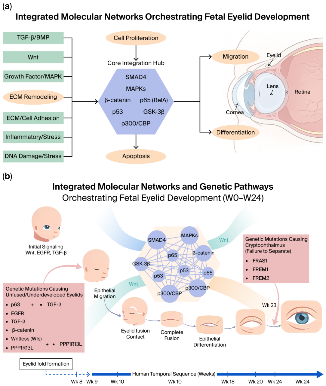
- SMADs (SMAD2/3, SMAD1/5/8, SMAD4) are central to eyelid development, playing crucial roles in both eyelid closing and reopening (Table 3, Figure 2). Evidence for this includes studies showing that TGF-βRII mutations cause non-fusion or delayed fusion of eyelids [45,46,47]. Additionally, the deletion of SMAD4 (a common Smad for both TGF-β and BMP pathways) in mice leads to eyelid non-fusion and corneal abnormalities [46,48].
- The Wnt pathway (involving β-catenin, GSK-3β, and p300/CBP) is essential for epithelial–mesenchymal interactions, placode formation, hair follicle (eyelash) development, and epidermal differentiation. β-catenin serves as the key effector, with its stability regulated by GSK-3β, and p300/CBP act as co-activators for β-catenin [21,29,45]. While its role is broad for skin, β-catenin has a central role in hair follicle (and thus eyelash) development and overall epidermal appendage formation, which is crucial for eyelid structure. Specifically, studies directly link β-catenin to eyelid development, showing that its conditional deletion in the epidermis causes severe eyelid malformations and non-fusion [31].
- The MAPK pathway, encompassing ERK, JNK, and p38, is activated downstream of growth factor receptors like EGFR and regulates crucial cellular processes such as proliferation, survival, and differentiation. This pathway is essential for eyelid fusion [49]. For instance, EGFR is critical for proper eyelid development and fusion; its signaling primarily utilizes the MAPK cascade (ERK, JNK, p38) to drive the epithelial proliferation and migration required for eyelid formation and subsequent separation [50]. The observation that EGFR loss results in an “eyes open at birth” phenotype—indicating a failure of eyelid fusion—directly implicates EGFR and its downstream MAPK signaling in this vital developmental process.
- NF-κB: While prominently associated with inflammation and immunity, the NF-κB pathway, including p65/RelA and IKKs, also plays crucial developmental roles in cell survival, proliferation, and differentiation, particularly within epithelia [51]. p65 (RelA) serves as a key transcriptional component, critical for NF-κB’s broad developmental functions, which encompass cell proliferation and survival essential for tissue growth, including that of the eyelid. The activation of p65 by upstream IKKα/β kinases links the core NF-κB pathway to proper eyelid development and fusion, with dysregulation known to lead to specific eyelid fusion defects [52].
- The p53 pathway, renowned as the “guardian of the genome,” primarily induces cell cycle arrest or apoptosis in response to cellular stress and damage. In a broader developmental context, p53 plays a crucial role in tissue sculpting through the regulation of programmed cell death. While programmed cell death is essential for processes such as eyelid separation and opening, p53’s direct involvement in this specific eyelid apoptosis appears more indirect or context-dependent. Instead of being a primary, constitutive inducer for all eyelid sculpting events, its role is likely amplified under specific stress conditions or serves to fine-tune developmental apoptotic pathways, a pattern observed in various other tissues [53].
- The integrin/ECM–receptor pathway involves integrins, transmembrane receptors critical for mediating cell-extracellular matrix (ECM) adhesion and crucial bidirectional signaling. These interactions profoundly influence cell migration, proliferation, differentiation, and overall tissue architecture. In the context of eyelid development and fusion, integrins are essential for structural integrity and dynamic tissue remodeling. While established for their role in wound healing [54]—a process that shares fundamental mechanisms with developmental fusion—integrins link the ECM to intracellular signaling pathways, including MAPK cascades. This regulation of cell migration and proliferation is vital for the precise formation and subsequent fusion of the eyelids. Specifically, the integrin subunit β1 plays a crucial role in epidermal differentiation, which is indispensable for forming the complex stratified epithelium of the eyelids and facilitating proper cell-matrix interactions during fusion [55].
- Co-activators p300 and CBP are histone acetyltransferases (HATs) that serve as crucial co-activators for a wide array of transcription factors, including β-catenin, p65, p53, and Smad proteins. By bridging these transcription factors to the basal transcriptional machinery, CBP/p300 effectively enhances gene expression [56]. While not specific to eyelid development, their indispensable role in modulating nearly all critical transcription factors and pathways relevant to development (e.g., Wnt, p53, Smads, NF-κB, MAPK targets) makes them indirect but essential players in every developmental process, including eyelid formation and fusion. Their precise contribution to eyelid development is thus context-dependent, determined by the specific transcription factors they co-activate.
4. Conclusions
Supplementary Materials
Author Contributions
Funding
Institutional Review Board Statement
Informed Consent Statement
Data Availability Statement
Acknowledgments
Conflicts of Interest
Abbreviations
| CEAs | Congenital eye anomalies |
| GA | Gestational age |
| woe2 | Waved with open eyelids 2 |
| KEGG | Kyoto Encyclopedia of Genes and Genomes |
References
- Dubucs, C.; Caillet, A.; Frémont, F.; Delteil, L.; Amanda, V.N.G.; Neville, J.; Damase, C. Prevalence of Congenital Ocular Anomalies in 15 Countries of Europe: Results From the Medikeye Study. Birth Defects Res. 2024, 116, e2414. [Google Scholar] [CrossRef] [Scilit] [PubMed]
- Stallings, E.B.; Isenburg, J.L.; Mai, C.T.; Liberman, R.F.; Moore, C.A.; Canfield, M.A.; Salemi, J.L.; Kirby, R.S.; Short, T.D.; Nembhard, W.N.; et al. Population-Based Birth Defects Data in the United States, 2011–2015: A Focus on Eye and Ear Defects. Birth Defects Res. 2018, 110, 1478–1486. [Google Scholar] [CrossRef] [Scilit] [PubMed]
- Saleh, G.M.; Hussain, B.; Verity, D.H.; Collin, J.R.O. A Surgical Strategy for the Correction of Fraser Syndrome Cryptophthalmos. Ophthalmology 2009, 116, 1707–1712.e1. [Google Scholar] [CrossRef] [Scilit] [PubMed]
- Tao, H.; Shimizu, M.; Kusumoto, R.; Ono, K.; Noji, S.; Ohuchi, H. A Dual Role of FGF10 in Proliferation and Coordinated Migration of Epithelial Leading Edge Cells during Mouse Eyelid Development. Development 2005, 132, 3217–3230. [Google Scholar] [CrossRef] [Scilit] [PubMed]
- Smyth, I.; Scambler, P. The Genetics of Fraser Syndrome and the Blebs Mouse Mutants. Hum. Mol. Genet. 2005, 14, R269–R274. [Google Scholar] [CrossRef] [Scilit] [PubMed]
- Petrou, P.; Pavlakis, E.; Dalezios, Y.; Chalepakis, G. Basement Membrane Localization of Frem3 Is Independent of the Fras1/Frem1/Frem2 Protein Complex within the Sublamina Densa. Matrix Biol. 2007, 26, 652–658. [Google Scholar] [CrossRef] [Scilit] [PubMed]
- Petrou, P.; Chiotaki, R.; Dalezios, Y.; Chalepakis, G. Overlapping and Divergent Localization of Frem1 and Fras1 and Its Functional Implications during Mouse Embryonic Development. Exp. Cell Res. 2007, 313, 910–920. [Google Scholar] [CrossRef] [Scilit] [PubMed]
- Kao, W.W.-Y.; Xia, Y.; Liu, C.-Y.; Saika, S. Signaling Pathways in Morphogenesis of Cornea and Eyelid. Ocul. Surf. 2008, 6, 9–23. [Google Scholar] [CrossRef] [Scilit]
- Ohuchi, H. Wakayama Symposium: Epithelial-Mesenchymal Interactions in Eyelid Development. Ocul. Surf. 2012, 10, 212–216. [Google Scholar] [CrossRef] [Scilit] [PubMed]
- Hayashi, Y.; Liu, C.-Y.; Jester, J.J.; Hayashi, M.; Wang, I.-J.; Funderburgh, J.L.; Saika, S.; Roughley, P.J.; Kao, C.W.-C.; Kao, W.W.-Y. Excess Biglycan Causes Eyelid Malformation by Perturbing Muscle Development and TGF-Alpha Signaling. Dev. Biol. 2005, 277, 222–234. [Google Scholar] [CrossRef] [Scilit] [PubMed]
- Pearson, A.A. The Development of the Eyelids. Part I. External Features. J. Anat. 1980, 130, 33–42. [Google Scholar] [PubMed]
- Sevel, D. A Reappraisal of the Development of the Eyelids. Eye 1988, 2, 123–129. [Google Scholar] [CrossRef] [Scilit] [PubMed]
- Tawfik, H.A.; Abdulhafez, M.H.; Fouad, Y.A.; Dutton, J.J. Embryologic and Fetal Development of the Human Eyelid. Ophthal. Plast. Reconstr. Surg. 2016, 32, 407–414. [Google Scholar] [CrossRef] [Scilit] [PubMed]
- Byun, T.H.; Kim, J.T.; Park, H.W.; Kim, W.K. Timetable for Upper Eyelid Development in Staged Human Embryos and Fetuses. Anat. Rec. 2011, 294, 789–796. [Google Scholar] [CrossRef] [Scilit] [PubMed]
- Willett, S.M.; Maenner, S.K.; Mayo, J.P. The Perceptual Consequences and Neurophysiology of Eye Blinks. Front. Syst. Neurosci. 2023, 17, 1242654. [Google Scholar] [CrossRef] [Scilit] [PubMed]
- Petrikovsky, B.M.; Kaplan, G.; Holsten, N. Eyelid Movements in Normal Human Fetuses. J. Clin. Ultrasound 2003, 31, 299–301. [Google Scholar] [CrossRef] [Scilit] [PubMed]
- Lavezzo, M.M.; Schellini, S.A.; Padovani, C.R.; Hirai, F.E. Eye Blink in Newborn and Preschool-Age Children. Acta Ophthalmol. 2008, 86, 275–278. [Google Scholar] [CrossRef] [PubMed]
- Falik-Zaccai, T.C.; Barsheshet, Y.; Mandel, H.; Segev, M.; Lorber, A.; Gelberg, S.; Kalfon, L.; Ben Haroush, S.; Shalata, A.; Gelernter-Yaniv, L.; et al. Sequence Variation in PPP1R13L Results in a Novel Form of Cardio-Cutaneous Syndrome. EMBO Mol. Med. 2017, 9, 319–336, Erratum in EMBO Mol. Med. 2017, 9, 1326. https://doi.org/10.15252/emmm.201708209. [Google Scholar] [CrossRef] [Scilit] [PubMed]
- Wonkam, A.; Toko, R.; Chelo, D.; Tekendo-Ngongang, C.; Kingue, S.; Dahoun, S. The 22q11.2 Deletion Syndrome in Congenital Heart Defects: Prevalence of Microdeletion Syndrome in Cameroon. Glob. Heart 2017, 12, 115–120. [Google Scholar] [CrossRef] [Scilit] [PubMed]
- Roelandt, M.A.; Devriendt, K.; de Llano-Pérula, M.C.; Raes, M.; Willems, G.; Verdonck, A. Dental and Craniofacial Characteristics in Patients With 14Q22.1–Q22.2 Deletion: A Case Series. Cleft Palate Craniofacial J. 2021, 58, 505–513. [Google Scholar] [CrossRef] [Scilit] [PubMed]
- Mandrile, G.; Dubois, A.; Hoffman, J.D.; Uliana, V.; Di Maria, E.; Malacarne, M.; Coviello, D.; Faravelli, F.; Zwolinski, S.; Hellens, S.; et al. 3q26.33–3q27.2 Microdeletion: A New Microdeletion Syndrome? Eur. J. Med. Genet. 2013, 56, 216–221. [Google Scholar] [CrossRef] [Scilit] [PubMed]
- Tassano, E.; Uccella, S.; Severino, M.; Giacomini, T.; Nardi, F.; Gimelli, G.; Tavella, E.; Ronchetto, P.; Malacarne, M.; Coviello, D. Expanding the Phenotype Associated with Interstitial 6p25.1p24.3 Microdeletion: A New Case and Review of the Literature. J. Genet. 2021, 100, 9. [Google Scholar] [CrossRef] [Scilit]
- Happ, H.; Schilter, K.F.; Weh, E.; Reis, L.M.; Semina, E. V 8q21.11 Microdeletion in Two Patients with Syndromic Peters Anomaly. Am. J. Med. Genet. A 2016, 170, 2471–2475. [Google Scholar] [CrossRef] [Scilit] [PubMed]
- Moein, H.-R.; Saeed, H.N.; Jacobs, D.S.; Rapoport, Y.; Yoon, M.K.; Shah, A.S.; Khan, H.; Raoof, D.; Jurkunas, U. V Exposure, Entropion, and Bilateral Corneal Ulceration in a Newborn as a Manifestation of Chromosome 22 Q11.2 Duplication Syndrome. Am. J. Ophthalmol. Case Rep. 2019, 13, 16–19. [Google Scholar] [CrossRef] [Scilit] [PubMed]
- Hanafusa, H.; Morisada, N.; Ishida, Y.; Sakata, R.; Morita, K.; Miura, S.; Ye, M.J.; Yamamoto, T.; Okamoto, N.; Nozu, K.; et al. The Smallest de Novo 20q11.2 Microdeletion Causing Intellectual Disability and Dysmorphic Features. Hum. Genome Var. 2017, 4, 17050. [Google Scholar] [CrossRef] [Scilit] [PubMed]
- van Haelst, M.M.; Maiburg, M.; Baujat, G.; Jadeja, S.; Monti, E.; Bland, E.; Pearce, K.; Hennekam, R.C.; Scambler, P.J. Molecular Study of 33 Families with Fraser Syndrome New Data and Mutation Review. Am. J. Med. Genet. A 2008, 146A, 2252–2257. [Google Scholar] [CrossRef] [Scilit] [PubMed]
- Pavlakis, E.; Chiotaki, R.; Chalepakis, G. The Role of Fras1/Frem Proteins in the Structure and Function of Basement Membrane. Int. J. Biochem. Cell Biol. 2011, 43, 487–495. [Google Scholar] [CrossRef] [Scilit] [PubMed]
- Kalayinia, S.; Mahdavi, M.; Houshmand, G.; Hesami, M.; Pourirahim, M.; Maleki, M. Novel Homozygous Stop-Gain Pathogenic Variant of PPP1R13L Gene Leads to Arrhythmogenic Cardiomyopathy. BMC Cardiovasc. Disord. 2022, 22, 359. [Google Scholar] [CrossRef] [Scilit] [PubMed]
- Toonen, J.; Liang, L.; Sidjanin, D.J. Waved with Open Eyelids 2 (Woe2) Is a Novel Spontaneous Mouse Mutation in the Protein Phosphatase 1, Regulatory (Inhibitor) Subunit 13 like (Ppp1r13l) Gene. BMC Genet. 2012, 13, 76. [Google Scholar] [CrossRef] [Scilit] [PubMed]
- Herron, B.J.; Rao, C.; Liu, S.; Laprade, L.; Richardson, J.A.; Olivieri, E.; Semsarian, C.; Millar, S.E.; Stubbs, L.; Beier, D.R. A Mutation in NFkB Interacting Protein 1 Results in Cardiomyopathy and Abnormal Skin Development in Wa3 Mice. Hum. Mol. Genet. 2005, 14, 667–677. [Google Scholar] [CrossRef] [Scilit] [PubMed]
- Zhu, X.; Senoo, M.; Millar, S.E.; Ma, G. Wnt/β-Catenin Signaling Controls Mouse Eyelid Growth by Mediating Epithelial-Mesenchymal Interactions. Ocul. Surf. 2023, 29, 486–494. [Google Scholar] [CrossRef] [Scilit] [PubMed]
- Luetteke, N.C.; Qiu, T.H.; Peiffer, R.L.; Oliver, P.; Smithies, O.; Lee, D.C. TGF Alpha Deficiency Results in Hair Follicle and Eye Abnormalities in Targeted and Waved-1 Mice. Cell 1993, 73, 263–278. [Google Scholar] [CrossRef] [Scilit] [PubMed]
- Mann, G.B.; Fowler, K.J.; Gabriel, A.; Nice, E.C.; Williams, R.L.; Dunn, A.R. Mice with a Null Mutation of the TGF Alpha Gene Have Abnormal Skin Architecture, Wavy Hair, and Curly Whiskers and Often Develop Corneal Inflammation. Cell 1993, 73, 249–261. [Google Scholar] [CrossRef] [Scilit] [PubMed]
- Zenz, R.; Scheuch, H.; Martin, P.; Frank, C.; Eferl, R.; Kenner, L.; Sibilia, M.; Wagner, E.F. C-Jun Regulates Eyelid Closure and Skin Tumor Development through EGFR Signaling. Dev. Cell 2003, 4, 879–889. [Google Scholar] [CrossRef] [Scilit] [PubMed]
- Robinson, R.A.; Lu, X.; Jones, E.Y.; Siebold, C. Biochemical and Structural Studies of ASPP Proteins Reveal Differential Binding to P53, P63, and P73. Structure 2008, 16, 259–268. [Google Scholar] [CrossRef] [Scilit] [PubMed]
- Carroll, J.M.; Luetteke, N.C.; Lee, D.C.; Watt, F.M. Role of Integrins in Mouse Eyelid Development: Studies in Normal Embryos and Embryos in Which There Is a Failure of Eyelid Fusion. Mech. Dev. 1998, 78, 37–45. [Google Scholar] [CrossRef] [Scilit] [PubMed]
- Dong, F.; Call, M.; Xia, Y.; Kao, W.W.-Y. Role of EGF Receptor Signaling on Morphogenesis of Eyelid and Meibomian Glands. Exp. Eye Res. 2017, 163, 58–63. [Google Scholar] [CrossRef] [Scilit] [PubMed]
- Vassalli, A.; Matzuk, M.M.; Gardner, H.A.; Lee, K.F.; Jaenisch, R. Activin/Inhibin Beta B Subunit Gene Disruption Leads to Defects in Eyelid Development and Female Reproduction. Genes Dev. 1994, 8, 414–427. [Google Scholar] [CrossRef] [Scilit] [PubMed]
- Mizoguchi, S.; Suzuki, K.; Zhang, J.; Yamanaka, O.; Liu, C.-Y.; Okada, Y.; Miyajima, M.; Kokado, M.; Kao, W.W.Y.; Yamada, G.; et al. Disruption of Eyelid and Cornea Morphogenesis by Epithelial β-Catenin Gain-of-Function. Mol. Vis. 2015, 21, 793–803. [Google Scholar] [PubMed]
- Liu, Y.; Kawai, K.; Khashabi, S.; Deng, C.; Liu, Y.-H.; Yiu, S. Inactivation of Smad4 Leads to Impaired Ocular Development and Cataract Formation. Biochem. Biophys. Res. Commun. 2010, 400, 476–482, Erratum in Biochem. Biophys. Res. Commun. 2011, 405, 706. [Google Scholar] [CrossRef] [Scilit] [PubMed]
- Rubinstein, T.J.; Weber, A.C.; Traboulsi, E.I. Molecular Biology and Genetics of Embryonic Eyelid Development. Ophthalmic Genet. 2016, 37, 252–259. [Google Scholar] [CrossRef] [Scilit] [PubMed]
- Bergamaschi, D.; Samuels, Y.; O’Neil, N.J.; Trigiante, G.; Crook, T.; Hsieh, J.-K.; O’Connor, D.J.; Zhong, S.; Campargue, I.; Tomlinson, M.L.; et al. IASPP Oncoprotein Is a Key Inhibitor of P53 Conserved from Worm to Human. Nat. Genet. 2003, 33, 162–167. [Google Scholar] [CrossRef] [Scilit] [PubMed]
- Cai, Y.; Qiu, S.; Gao, X.; Gu, S.-Z.; Liu, Z.-J. IASPP Inhibits P53-Independent Apoptosis by Inhibiting Transcriptional Activity of P63/P73 on Promoters of Proapoptotic Genes. Apoptosis 2012, 17, 777–783. [Google Scholar] [CrossRef] [Scilit] [PubMed]
- Allgayer, H.; Mahapatra, S.; Mishra, B.; Swain, B.; Saha, S.; Khanra, S.; Kumari, K.; Panda, V.K.; Malhotra, D.; Patil, N.S.; et al. Epithelial-to-Mesenchymal Transition (EMT) and Cancer Metastasis: The Status Quo of Methods and Experimental Models 2025. Mol. Cancer 2025, 24, 167. [Google Scholar] [CrossRef] [Scilit] [PubMed]
- Yoshida, Y.; Tanaka, S.; Umemori, H.; Minowa, O.; Usui, M.; Ikematsu, N.; Hosoda, E.; Imamura, T.; Kuno, J.; Yamashita, T.; et al. Negative Regulation of BMP/Smad Signaling by Tob in Osteoblasts. Cell 2000, 103, 1085–1097. [Google Scholar] [CrossRef] [Scilit] [PubMed]
- Luo, K. Signaling Cross Talk between TGF-β/Smad and Other Signaling Pathways. Cold Spring Harb. Perspect. Biol. 2017, 9, a022137. [Google Scholar] [CrossRef] [Scilit] [PubMed]
- Hachana, S.; Larrivée, B. TGF-β Superfamily Signaling in the Eye: Implications for Ocular Pathologies. Cells 2022, 11, 2336. [Google Scholar] [CrossRef] [Scilit] [PubMed]
- Huang, J.; Dattilo, L.K.; Rajagopal, R.; Liu, Y.; Kaartinen, V.; Mishina, Y.; Deng, C.-X.; Umans, L.; Zwijsen, A.; Roberts, A.B.; et al. FGF-Regulated BMP Signaling Is Required for Eyelid Closure and to Specify Conjunctival Epithelial Cell Fate. Development 2009, 136, 1741–1750. [Google Scholar] [CrossRef] [Scilit] [PubMed]
- Werner, S.; Grose, R. Regulation of Wound Healing by Growth Factors and Cytokines. Physiol. Rev. 2003, 83, 835–870. [Google Scholar] [CrossRef] [Scilit] [PubMed]
- Threadgill, D.W.; Dlugosz, A.A.; Hansen, L.A.; Tennenbaum, T.; Lichti, U.; Yee, D.; LaMantia, C.; Mourton, T.; Herrup, K.; Harris, R.C. Targeted Disruption of Mouse EGF Receptor: Effect of Genetic Background on Mutant Phenotype. Science 1995, 269, 230–234. [Google Scholar] [CrossRef] [Scilit] [PubMed]
- Li, Q.; Verma, I.M. NF-KappaB Regulation in the Immune System. Nat. Rev. Immunol. 2002, 2, 725–734, Erratum in Nat. Rev. Immunol. 2002, 2, 975. [Google Scholar] [CrossRef] [Scilit] [PubMed]
- Valovka, T.; Hottiger, M.O. P65 Controls NF-ΚB Activity by Regulating Cellular Localization of IκBβ. Biochem. J. 2011, 434, 253–263. [Google Scholar] [CrossRef] [Scilit] [PubMed]
- Armstrong, J.F.; Kaufman, M.H.; Harrison, D.J.; Clarke, A.R. High-Frequency Developmental Abnormalities in P53-Deficient Mice. Curr. Biol. 1995, 5, 931–936. [Google Scholar] [CrossRef] [Scilit] [PubMed]
- Longhurst, C.M.; Jennings, L.K. Integrin-Mediated Signal Transduction. Cell. Mol. Life Sci. 1998, 54, 514–526. [Google Scholar] [CrossRef] [Scilit] [PubMed]
- Piwko-Czuchra, A.; Koegel, H.; Meyer, H.; Bauer, M.; Werner, S.; Brakebusch, C.; Fässler, R. Beta1 Integrin-Mediated Adhesion Signalling Is Essential for Epidermal Progenitor Cell Expansion. PLoS ONE 2009, 4, e5488. [Google Scholar] [CrossRef] [Scilit] [PubMed]
- Goodman, R.H.; Smolik, S. CBP/P300 in Cell Growth, Transformation, and Development. Genes Dev. 2000, 14, 1553–1577. [Google Scholar] [CrossRef] [Scilit]



| Group | Dominant Phenotype When Applicable | HPO | HPO-Human Phenotype Ontology (jax.org) | Notable Representing Syndromes and Some Affiliated Genes or Locations | Reference | |
|---|---|---|---|---|---|---|
| Structural abnormalities mostly confined to the orbit and eyelid | Blepharophimosis | HP:0000581 | 129 | Autosomal-dominant blepharophimosis-ptosis-epicanthus inversus syndrome (BPES) | FOXL2 | PMID: 28280295 |
| Cryptophthalmos | HP:0001126 | 6 | Cryptophthalmos, unilateral or bilateral, isolated | FREM2 | OMIM-#123570 | |
| Microphthalmia/coloboma and skeletal dysplasia syndrome | MAB21L2 | Human Phenotype Ontology (jax.org) | ||||
| Eyelid aplasia or hypoplasis aplasia/hypoplasia of the eyelid | HP:0011226 | 44 | Neu-Laxova syndrome | PHGDH, PSAT1 | PMID: 35885441 | |
| Ablepharon-macrostomia syndrome | TWIST2 | PMID: 21595001 | ||||
| Congenital ptosis | Ptosis | HP:0000508 | 736 | Ptosis, congenital 1 | ZFHX4 | PMID: 28280295 |
| Ptosis, hereditary congenital 2 | XQ24–Q27.1 | |||||
| Duane syndrome | MAFB | Ptosis, Hereditary Congenital 2-MalaCards | ||||
| 3MC syndrome | COLEC10 | |||||
| Structural abnormalities involving the craniofacial region | Craniosynostosis | HP:0001363 | 180 | Non-syndromic craniosynostosis | Ras/ERK | PMID: 28808027 |
| Encephalocele | HP:0002084 | 120 | Frontorhiny | ALX3 | PMID: 25126130 | |
| PMID: 19409524 | ||||||
| Eyelid involvement as part of systemic disease | Treacher Collins syndrome | TCOF1, POLR1D, PLR1C and POLR1B | PMID: 34573374 | |||
| Goldenhar syndrome | unknown etiology | PMID: 30134872 | ||||
| Fraser syndrome | FRAS1 FREM1 FREM2 | PMID: 35791156 | ||||
| OMIM # 219000 | ||||||
| Gordon syndrome | PIEZO2 | OMIM # 114300 | ||||
| Crouzon syndrome | FGFR2 | OMIM # 123500 | ||||
| Blepharophimosis-ptosis-intellectual disability syndrome | UBE3B | PMID: 28280295 | ||||
| Lymphedema-distichiasis-syndrome | FOX2 | PMID: 28280295 | ||||
| Noonan syndrome | KRAS and multiple affiliated genes) | PMID: 29948256 | ||||
| Waardenburg syndrome subtypes | PAX3, MITF, SOX10 | PMID: 36620710 | ||||
| 46, XX sex reversal 5 | NR2F2 | Blepharophimosis, Ptosis, and Epicanthus Inversus Syndrome-GeneReviews®-NCBI Bookshelf (nih.gov) | ||||
| Say–Barber–Biesecker variant of Ohdo syndrome | KAT6B | |||||
| Disease affecting the neuromuscular junction (NMJ) | Möbius syndrome type 2 | 3q21–q22 | OMIM # 601471 | |||
| PMID: 25633065 | ||||||
| Congenital myasthenic syndrome 9 | MUSK | OMIM # 616325 | ||||
| Other congenital myasthenic syndromes | CHRNE, ALG14 | PMID: 28280295 | ||||
| Congenital myopathies | Many congenital muscular diseases may be accompanied by congenital ptosis with or without additional ocular anomalies, including congenital myopathies, mitochondrial myopathies, myotonic syndromes, muscular dystrophies, and other myopathies | Multiple affiliated genes | PMID: 35049410 | |||
| In congenital fibrosis of the extraocular muscles (CFEOM) syndromes (1 + 2 + 3) | KIF21A, PHOX2A/ARIX | PMID: 28280295 | ||||
| Congenital orbital masses/lesions that involve the eyelid or affect its movement Group structural abnormalities mostly confined to the orbit and eyelid Congenital ptosis structural abnormalities involving the craniofacial region | Teratoma | HP:0009792 | 11 | Sotos syndrome | NSD1 | PMID: 34027878 |
| PMID: 28695872 | ||||||
| Periorbital dermoid cyst | HP:0030668 | 1 | Otofaciocervical syndrome 2 | PAX1 | PMID: 34750318 | |
| Peripheral primitive neuroectodermal neoplasm | HP:0030067 | 62 | Mismatch repair cancer syndrome 1 | MLH1 | PMID: 34621833 | |
| Aggressive infantile fibromatosis (desmoid tumor) | HP:6001034 | 7 | Gardner syndrome | APC | ||
| Embryonal rhabdomyosarcoma | HP:0006743 | 4 | Rhabdomyosarcoma, embryonal, 2 | DICER1 | ||
| Rhabdomyosarcoma 1 | SLC22A18 | |||||
| Optic nerve sheath meningioma | HP:0500089 | 1 | Neurofibromatosis, type II | NF2 | PMID: 34621833 | |
| DOI: 10.2147/IMCRJ.S82795 | ||||||
| (A congenital case report was not located in the literature) | ||||||
| Periocular capillary hemangioma | HP:0500090 | 0 | PMID: 28540013 | |||
| Granular cell tumor of the eyelid or orbit Dominant phenotype when applicable | An HP was not located for the phenotype | Noonan syndrome | Multiple genes | PMID: 34621833 | ||
| PMID: 19953625 | ||||||
| (A congenital case report was not located in the literature) | ||||||
| Microdeletion Syndromes | Associated Eyelid Abnormalities | Microduplication Syndromes | Associated Eyelid Abnormalities |
|---|---|---|---|
| 16p11.2 PMID: 21465664 PMID: 38050025 PMID: 32373379 | Down-slanting palpebral fissures; ptosis; sagging lateral upper eyelids; eyelid eversion; long eyelashes and prominent eyes | 22q11.2 PMID: 23972321 PMID: 30505980 PMID: 27108843 | Highly variable, but commonly reported features include down-slanting palpebral fissures, ptosis (drooping eyelids), superior displacement of the eyebrows, hypertelorism (wide-set eyes), hooded eyelids, and, rarely, severe cases involving congenital entropion (inward-turning eyelids), ectropion (outward-turning eyelids), and corneal ulceration |
| 22q11.2 PMID: 17704945 PMID: 27182748 PMID: 26056486 | Deep-set eyes; eyelid hooding, short (narrow) palpebral fissures | 20q11.2 PMID: 23704076 PMID: 30893560 | Distinctive facial features can include epicanthus (skin folds at the inner corner of the eye), hypoplastic supraorbital ridges, and horizontal or down-slanting palpebral fissures |
| 14q22q23 PMID: 30268123 PMID: 22486322 | Ptosis and hypertelorism | 3q29 PMID: 33039685 PMID: 19298871 PMID: 19287140 | Ocular abnormalities can occur, with some cases involving a range of issues, including nystagmus, epicanthus, lagophthalmos (incomplete eyelid closure), entropion, and infantile glaucoma |
| 3q26.33–3q27.2 PMID: 23357683 PMID: 27525095 PMID: 24462885 | Including narrow horizontal palpebral fissures, epicanthal folds, and ptosis (blepharophimosis sequence) | 16p11.2 PMID: 38605127 PMID: 24891046 | Subtle and non-specific findings include down-slanting palpebral fissures, deep-set eyes, ptosis, hypertelorism, epicanthic folds, and subtle synophrys |
| 6p25 PMID: 15654696 PMID: 36941760 PMID: 15150541 | Proptosis (bulging eyes) and down-slanting palpebral fissures | Xq25 (STAG2) OMIM. #300387 | Facial dysmorphism can include lower palpebral eversion; this microduplication has also been reported in a patient with eyelid myoclonia and absences (Jeavons syndrome) |
| 8q21.11 PMID: 21802062 PMID: 37039706 PMID: 36341706 | Ptosis as part of facial dysmorphism |
| Molecule/Complex = | Common Function | Primary Pathway | Shared Pathways |
|---|---|---|---|
| SMAD4 | Transcription/Co-Smad | TGF-β/BMP | TGF-β, BMP4, SMAD |
| MAPKs | Kinase cascade | MAPK | MAPK, TGF-β, BMP, Int/ECM |
| β-catenin | Transcription factor | Wnt | Wnt, TGF-β/BMP (fibrosis) |
| p65 (RelA) | Transcription factor | NF-κB | NF-κB, p53, TGF-β |
| p53 | Tumor suppressor | p53 | p53, NF-κB, TGF-β/SMAD |
| GSK-3β | Kinase (destruction) | Wnt/MAPK | Wnt, MAPK, TGF-β |
| p300/CBP | Co-activator | General | Wnt, TGF-β, p53 |
| Integrins | Receptor/adhesion | ECM-Rec | Integrin, MAPK, TGF-β |
Disclaimer/Publisher’s Note: The statements, opinions and data contained in all publications are solely those of the individual author(s) and contributor(s) and not of MDPI and/or the editor(s). MDPI and/or the editor(s) disclaim responsibility for any injury to people or property resulting from any ideas, methods, instructions or products referred to in the content. |
© 2026 by the authors. Licensee MDPI, Basel, Switzerland. This article is an open access article distributed under the terms and conditions of the Creative Commons Attribution (CC BY) license.
Share and Cite
Shalata, Z.; Mintz, H.; Haddad, S.; Osman, K.; Mahroum, M.; Hadid, Y.; Shalata, A. Failure to Fuse Shut Eyelids, a Novel Unique Sign in Affected Fetus with Homozygous PPP1R13L Pathogenic Variant—A Case Report and Review of the Literature. Int. J. Mol. Sci. 2026, 27, 6991. https://doi.org/10.3390/ijms27156991
Shalata Z, Mintz H, Haddad S, Osman K, Mahroum M, Hadid Y, Shalata A. Failure to Fuse Shut Eyelids, a Novel Unique Sign in Affected Fetus with Homozygous PPP1R13L Pathogenic Variant—A Case Report and Review of the Literature. International Journal of Molecular Sciences. 2026; 27(15):6991. https://doi.org/10.3390/ijms27156991
Chicago/Turabian StyleShalata, Zaher, Hila Mintz, Sami Haddad, Khaled Osman, Mohammad Mahroum, Yarin Hadid, and Adel Shalata. 2026. "Failure to Fuse Shut Eyelids, a Novel Unique Sign in Affected Fetus with Homozygous PPP1R13L Pathogenic Variant—A Case Report and Review of the Literature" International Journal of Molecular Sciences 27, no. 15: 6991. https://doi.org/10.3390/ijms27156991
APA StyleShalata, Z., Mintz, H., Haddad, S., Osman, K., Mahroum, M., Hadid, Y., & Shalata, A. (2026). Failure to Fuse Shut Eyelids, a Novel Unique Sign in Affected Fetus with Homozygous PPP1R13L Pathogenic Variant—A Case Report and Review of the Literature. International Journal of Molecular Sciences, 27(15), 6991. https://doi.org/10.3390/ijms27156991