Immune Cells in Preeclampsia
 , , and
Abstract
1. Introduction
2. Th1/Th2 Cells
2.1. Th1/Th2 Cells in Normal Pregnancy
2.2. Th1/Th2 in Preeclampsia
3. Regulatory T Cells
3.1. Regulatory T Cells in Normal Pregnancy
3.2. Regulatory T Cells in Preeclampsia
4. Th17 Cells
4.1. Th17 Cells in Normal Pregnancy
4.2. Th17 Cells in Preeclampsia
5. B Cells
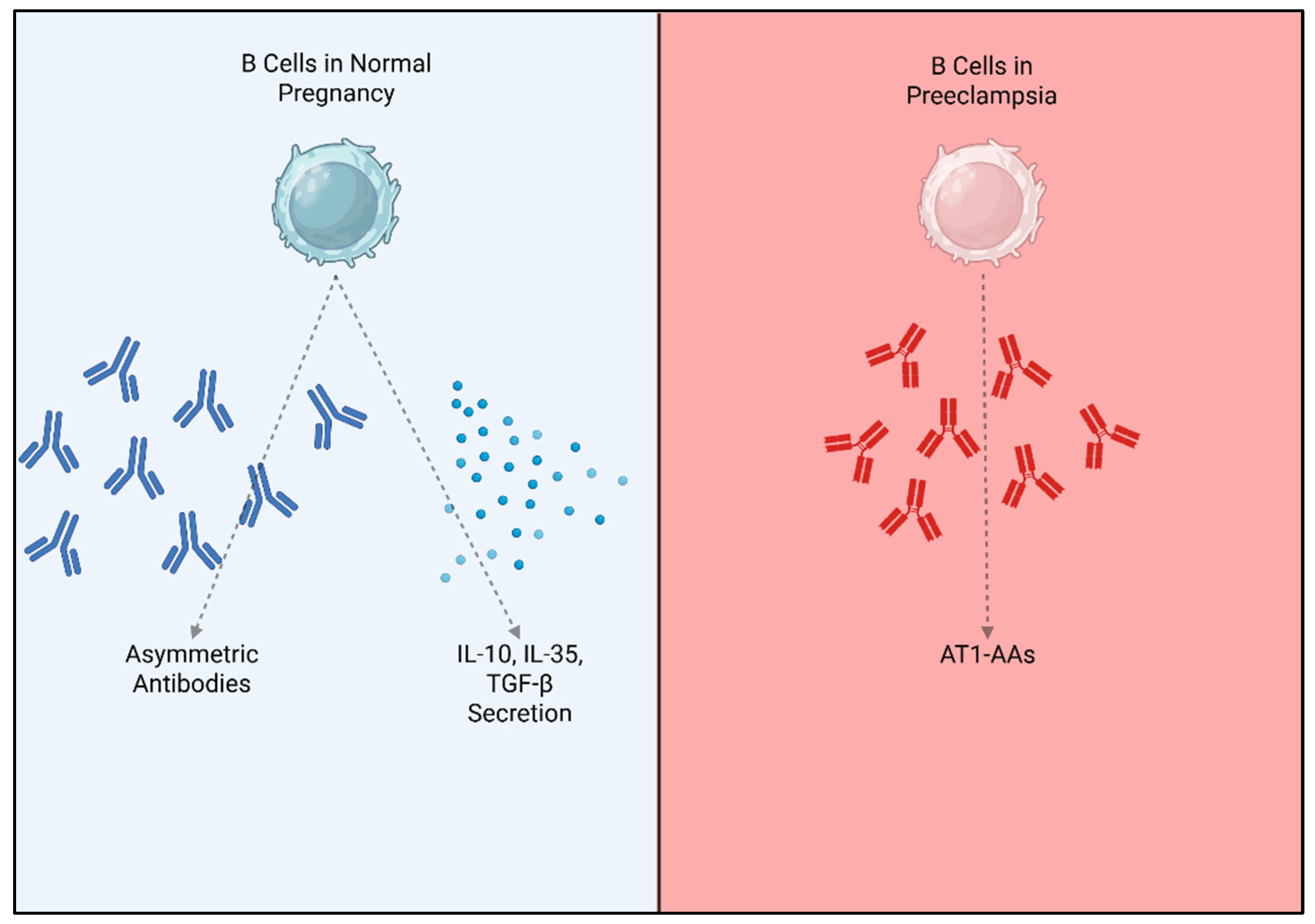
5.1. B Cells in Normal Pregnancy
5.2. B Cells in Preeclampsia
6. Macrophages
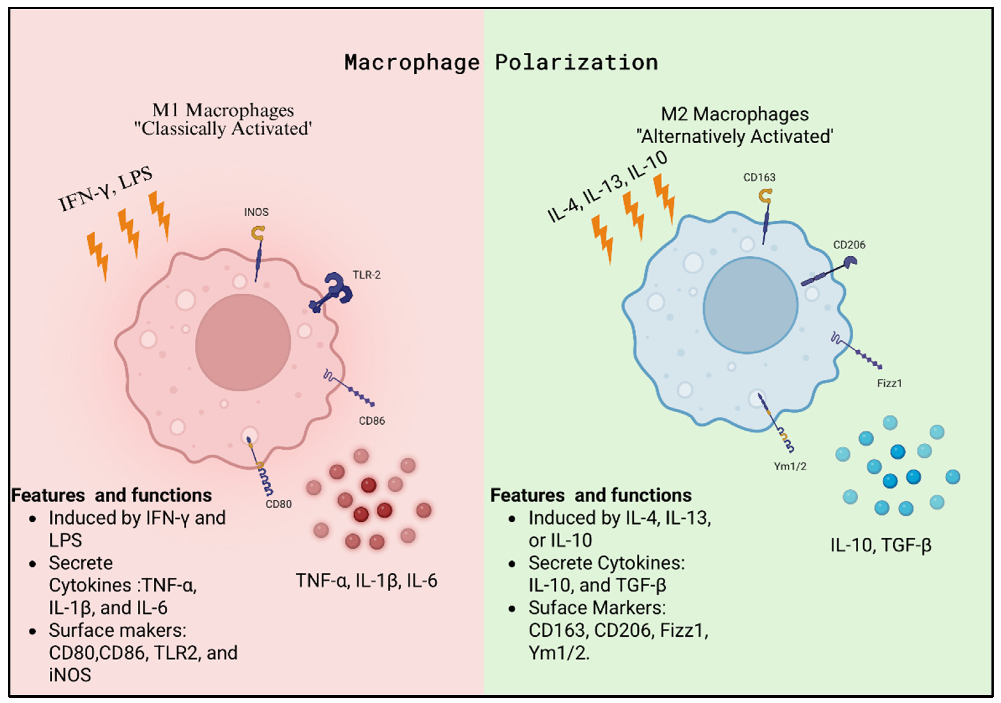
6.1. Macrophages in Normal Pregnancy
6.2. Macrophages in Preeclampsia
7. Natural Killer Cells
7.1. Natural Killer Cells in Normal Pregnancy
7.2. Natural Killer Cells in Preeclampsia
8. Sub-Characterizations of Preeclampsia
9. Clinical Context
10. Conclusions
Author Contributions
Funding
Data Availability Statement
Acknowledgments
Conflicts of Interest
References
- Brown, M.A.; Magee, L.A.; Kenny, L.C.; Karumanchi, S.A.; McCarthy, F.P.; Saito, S.; Hall, D.R.; Warren, C.E.; Adoyi, G.; Ishaku, S.; et al. Hypertensive disorders of pregnancy: ISSHP classification, diagnosis, and management recommendations for international practice. Hypertension 2018, 72, 24–43. [Google Scholar] [CrossRef] [Scilit] [PubMed]
- Gestational hypertension and preeclampsia: ACOG practice bulletin, number 222. Obstet. Gynecol. 2020, 135, e237–e260. [CrossRef] [Scilit]
- Shiozaki, A.; Saito, S. Risk factors for preeclampsia. In Preeclampsia: Basic, Genomic, and Clinical; Springer: Singapore, 2018; pp. 3–25. [Google Scholar]
- Dekker, G.A. Risk factors for preeclampsia. Clin. Obstet. Gynecol. 1999, 42, 422. [Google Scholar] [CrossRef] [Scilit]
- Deer, E.; Herrock, O.; Campbell, N.; Cornelius, D.; Fitzgerald, S.; Amaral, L.M.; LaMarca, B. The role of immune cells and mediators in preeclampsia. Nat. Rev. Nephrol. 2023, 19, 257–270. [Google Scholar] [CrossRef] [Scilit]
- Redman, C.W.; Sacks, G.P.; Sargent, I.L. Preeclampsia: An excessive maternal inflammatory response to pregnancy. Am. J. Obstet. Gynecol. 1999, 180, 499–506. [Google Scholar] [CrossRef] [Scilit]
- Chiarello, D.I.; Abad, C.; Rojas, D.; Toledo, F.; Vázquez, C.M.; Mate, A.; Sobrevia, L.; Marín, R. Oxidative stress: Normal pregnancy versus preeclampsia. Biochim. Biophys. Acta Mol. Basis Dis. 2020, 1866, 165354. [Google Scholar] [CrossRef] [Scilit]
- Rustveld, L.O.; Kelsey, S.F.; Sharma, R. Association between maternal infections and preeclampsia: A systematic review of epidemiologic studies. Matern. Child Health J. 2008, 12, 223–242. [Google Scholar] [CrossRef] [Scilit]
- Yang, L.; Wang, X.; Zhang, L. Maternal Thymus Adaptations and Hormone Regulation During Pregnancy. Cells 2025, 14, 1534. [Google Scholar] [CrossRef] [Scilit] [PubMed]
- Saito, S.; Shiozaki, A.; Nakashima, A.; Sakai, M.; Sasaki, Y. The role of the immune system in preeclampsia. Mol. Asp. Med. 2007, 28, 192–209. [Google Scholar] [CrossRef] [Scilit]
- Fukui, A.; Yokota, M.; Funamizu, A.; Nakamua, R.; Fukuhara, R.; Yamada, K.; Kimura, H.; Fukuyama, A.; Kamoi, M.; Tanaka, K.; et al. Changes of NK cells in preeclampsia. Am. J. Reprod. Immunol. 2012, 67, 278–286. [Google Scholar] [CrossRef] [Scilit] [PubMed]
- Campbell, N.; Deer, E.; Herrock, O.; LaMarca, B. The Role of Different Lymphoid Cell Populations in Preeclampsia Pathophysiology. Kidney360 2022, 3, 1785–1794. [Google Scholar] [CrossRef] [Scilit]
- Saito, S.; Nakashima, A.; Shima, T.; Ito, M. Th1/Th2/Th17 and regulatory T-cell paradigm in pregnancy. Am. J. Reprod. Immunol. 2010, 63, 601–610. [Google Scholar] [CrossRef] [Scilit]
- Saito, S.; Tsuda, S.; Nakashima, A. T cell immunity and the etiology and pathogenesis of preeclampsia. J. Reprod. Immunol. 2023, 159, 104125. [Google Scholar] [CrossRef] [Scilit]
- Schonkeren, D.; van der Hoorn, M.-L.; Khedoe, P.; Swings, G.; van Beelen, E.; Claas, F.; van Kooten, C.; de Heer, E.; Scherjon, S. Differential Distribution and Phenotype of Decidual Macrophages in Preeclamptic versus Control Pregnancies. Am. J. Pathol. 2011, 178, 709–717. [Google Scholar] [CrossRef] [Scilit]
- Wallukat, G.; Homuth, V.; Fischer, T.; Lindschau, C.; Horstkamp, B.; Jüpner, A.; Baur, E.; Nissen, E.; Vetter, K.; Neichel, D. Patients with preeclampsia develop agonistic autoantibodies against the angiotensin AT 1 receptor. J. Clin. Investig. 1999, 103, 945–952. [Google Scholar] [CrossRef] [Scilit]
- Campbell, N.; LaMarca, B.; Cunningham, M.W., Jr. The role of agonistic autoantibodies to the angiotensin II type 1 receptor (AT1-AA) in pathophysiology of preeclampsia. Curr. Pharm. Biotechnol. 2018, 19, 781–785. [Google Scholar] [CrossRef] [Scilit]
- Jensen, F.; Wallukat, G.; Costa, S.-D.; Dechend, R.; Zenclussen, A. Peripheral CD19+ CD5+ B-1a B cells are increased in patients with preeclampsia and secrete AT1-AA antibodies. J. Reprod. Immunol. 2012, 1, 110–111. [Google Scholar] [CrossRef] [Scilit]
- Zhu, X.; Zhu, J. CD4 T helper cell subsets and related human immunological disorders. Int. J. Mol. Sci. 2020, 21, 8011. [Google Scholar] [CrossRef] [Scilit] [PubMed]
- Piccinni, M.-P.; Raghupathy, R.; Saito, S.; Szekeres-Bartho, J. Cytokines, hormones and cellular regulatory mechanisms favoring successful reproduction. Front. Immunol. 2021, 12, 717808. [Google Scholar] [CrossRef] [Scilit] [PubMed]
- Crants, S.; Yin, S.; Andrusier, M.A.; Reddy, R.; Ginsburg, E.S. The Role of Cytokines in Early Pregnancy: Fertilization, Implantation, and Maintenance. Curr. Obstet. Gynecol. Rep. 2024, 13, 59–65. [Google Scholar] [CrossRef] [Scilit]
- dos Santos Fagundes, I.; Brendler, E.P.; Nunes Erthal, I.; Eder Ribeiro, R.J.; Caron-Lienert, R.S.; Machado, D.C.; Pinheiro da Costa, B.E.; Poli-de-Figueiredo, C.E. Total Th1/Th2 cytokines profile from peripheral blood lymphocytes in normal pregnancy and preeclampsia syndrome. Hypertens. Pregnancy 2022, 41, 15–22. [Google Scholar] [CrossRef] [Scilit]
- Rao, V.A.; Kurian, N.K.; Rao, K.A. Cytokines, NK cells and regulatory T cell functions in normal pregnancy and reproductive failures. Am. J. Reprod. Immunol. 2023, 89, e13667. [Google Scholar] [CrossRef] [Scilit] [PubMed]
- Esparvarinha, M.; Madadi, S.; Aslanian-Kalkhoran, L.; Nickho, H.; Dolati, S.; Pia, H.; Danaii, S.; Taghavi, S.; Yousefi, M. Dominant immune cells in pregnancy and pregnancy complications: T helper cells (TH1/TH2, TH17/Treg cells), NK cells, MDSCs, and the immune checkpoints. Cell Biol. Int. 2023, 47, 507–519. [Google Scholar] [CrossRef] [Scilit] [PubMed]
- Wang, J.; Wen, Z.Q.; Cheng, X.-Y.; Mei, T.Y.; Chen, Z.-F.; Su, L.-X. siRNA-mediated knockdown of T-bet and RORγt contributes to decreased inflammation in pre-eclampsia. Mol. Med. Rep. 2017, 16, 6368–6375. [Google Scholar] [CrossRef] [Scilit] [PubMed]
- Shen, C.; Song, Y.; Fan, W.; Guo, X.; Li, J.; Zhao, R. Changes in expression levels of immune cells and inflammatory cytokines in pre-eclampsia patients before and after delivery. J. Reprod. Immunol. 2023, 156, 103812. [Google Scholar] [CrossRef] [Scilit]
- Ribeiro, V.R.; Romao-Veiga, M.; Nunes, P.R.; Matias, M.L.; Peracoli, J.C.; Peracoli, M.T.S. Vitamin D modulates the transcription factors of T cell subsets to anti-inflammatory and regulatory profiles in preeclampsia. Int. Immunopharmacol. 2021, 101, 108366. [Google Scholar] [CrossRef] [Scilit]
- Liu, A.; Wang, H.; Gao, X. A correlation analysis of Th1/Th2 cells in preeclampsia patients. Int. J. Clin. Exp. Med. 2019, 12, 12868–12873. [Google Scholar]
- Mor, G.; Abrahams, V.M. Potential role of macrophages as immunoregulators of pregnancy. Reprod. Biol. Endocrinol. 2003, 1, 119. [Google Scholar] [CrossRef] [Scilit]
- Geldenhuys, J.; Rossouw, T.M.; Lombaard, H.A.; Ehlers, M.M.; Kock, M.M. Disruption in the regulation of immune responses in the placental subtype of preeclampsia. Front. Immunol. 2018, 9, 1659. [Google Scholar] [CrossRef] [Scilit]
- Workman, C.J.; Szymczak-Workman, A.L.; Collison, L.W.; Pillai, M.R.; Vignali, D.A. The development and function of regulatory T cells. Cell. Mol. Life Sci. 2009, 66, 2603–2622. [Google Scholar] [CrossRef] [Scilit]
- Corthay, A. How do regulatory T cells work? Scand. J. Immunol. 2009, 70, 326–336. [Google Scholar] [CrossRef] [Scilit]
- Sakaguchi, S.; Yamaguchi, T.; Nomura, T.; Ono, M. Regulatory T Cells and Immune Tolerance. Cell 2008, 133, 775–787. [Google Scholar] [CrossRef] [Scilit] [PubMed]
- Josefowicz, S.Z.; Lu, L.-F.; Rudensky, A.Y. Regulatory T Cells: Mechanisms of Differentiation and Function. Annu. Rev. Immunol. 2012, 30, 531–564. [Google Scholar] [CrossRef] [Scilit] [PubMed]
- Wang, K.; Fu, W. Transcriptional regulation of Treg homeostasis and functional specification. Cell. Mol. Life Sci. 2020, 77, 4269–4287. [Google Scholar] [CrossRef] [Scilit]
- Goswami, T.K.; Singh, M.; Dhawan, M.; Mitra, S.; Emran, T.B.; Rabaan, A.A.; Mutair, A.A.; Alawi, Z.A.; Alhumaid, S.; Dhama, K. Regulatory T cells (Tregs) and their therapeutic potential against autoimmune disorders—Advances and challenges. Hum. Vaccin. Immunother. 2022, 18, 2035117. [Google Scholar] [CrossRef] [Scilit]
- Arellano, B.; Graber, D.J.; Sentman, C.L. Regulatory T cell-based therapies for autoimmunity. Discov. Med. 2016, 22, 73–80. [Google Scholar]
- Bettini, M.; Vignali, D.A. Regulatory T cells and inhibitory cytokines in autoimmunity. Curr. Opin. Immunol. 2009, 21, 612–618. [Google Scholar] [CrossRef] [Scilit]
- Astarita, J.L.; Dominguez, C.X.; Tan, C.; Guillen, J.; Pauli, M.L.; Labastida, R.; Valle, J.; Kleinschek, M.; Lyons, J.; Zarrin, A.A. Treg specialization and functions beyond immune suppression. Clin. Exp. Immunol. 2023, 211, 176–183. [Google Scholar] [CrossRef] [Scilit]
- Jugder, B.-E.; Park, E.; Du, L.; Jawale, C.; Popov, N.; Guo, Z.; Bednar, K.J.; Ort, T. Tissue-specific roles of regulatory T cells: Mechanisms of suppression and beyond along with emerging therapeutic insights in autoimmune indications. Front. Immunol. 2025, 16, 1650451. [Google Scholar] [CrossRef] [Scilit]
- Huang, N.; Chi, H.; Qiao, J. Role of Regulatory T Cells in Regulating Fetal-Maternal Immune Tolerance in Healthy Pregnancies and Reproductive Diseases. Front. Immunol. 2020, 11, 1023. [Google Scholar] [CrossRef] [Scilit]
- Rackaityte, E.; Halkias, J. Mechanisms of Fetal T Cell Tolerance and Immune Regulation. Front. Immunol. 2020, 11, 1023. [Google Scholar] [CrossRef] [Scilit] [PubMed]
- Wang, W.; Zhao, Y.; Zhou, X.; Sung, N.; Chen, L.; Zhang, X.; Ma, D.; Zhu, P.; Kwak-Kim, J. Dynamic changes in regulatory T cells during normal pregnancy, recurrent pregnancy loss, and gestational diabetes. J. Reprod. Immunol. 2022, 150, 103492. [Google Scholar] [CrossRef] [Scilit]
- Alanazi, H.; Zhang, Y.; Fatunbi, J.; Luu, T.; Kwak-Kim, J. The impact of reproductive hormones on T cell immunity; normal and assisted reproductive cycles. J. Reprod. Immunol. 2024, 165, 104295. [Google Scholar] [CrossRef] [Scilit]
- Li, Q.H.; Zhao, Q.Y.; Yang, W.J.; Jiang, A.F.; Ren, C.E.; Meng, Y.H. Beyond Immune Balance: The Pivotal Role of Decidual Regulatory T Cells in Unexplained Recurrent Spontaneous Abortion. J. Inflamm. Res. 2024, 17, 2697–2710. [Google Scholar] [CrossRef] [Scilit]
- Zenclussen, A.C. Regulatory T cells in pregnancy. Springer Semin. Immunopathol. 2006, 28, 31–39. [Google Scholar] [CrossRef] [Scilit]
- Jørgensen, N.; Persson, G.; Hviid, T.V.F. The Tolerogenic Function of Regulatory T Cells in Pregnancy and Cancer. Front. Immunol. 2019, 10, 911. [Google Scholar] [CrossRef] [Scilit] [PubMed]
- Pan, Y.; Zhou, H.; Sun, Z.; Zhu, Y.; Zhang, Z.; Han, J.; Liu, Y.; Wang, Q. Regulatory T cells in solid tumor immunotherapy: Effect, mechanism and clinical application. Cell Death Dis. 2025, 16, 277. [Google Scholar] [CrossRef] [Scilit]
- Schmidt, A.; Oberle, N.; Krammer, P.H. Molecular mechanisms of treg-mediated T cell suppression. Front. Immunol. 2012, 3, 51. [Google Scholar] [CrossRef] [Scilit]
- Wang, S.; Li, M.; Sun, F.; Chen, C.; Ye, J.; Li, D.; Qian, J.; Du, M. Th17/Treg-cell balance in the peripheral blood of pregnant females with a history of recurrent spontaneous abortion receiving progesterone or cyclosporine A. Exp. Ther. Med. 2021, 21, 37. [Google Scholar] [CrossRef] [Scilit]
- Figueiredo, A.S.; Schumacher, A. The T helper type 17/regulatory T cell paradigm in pregnancy. Immunology 2016, 148, 13–21, Erratum in Immunology 2019, 156, 213. [Google Scholar] [CrossRef] [Scilit] [PubMed]
- Li, Z.; Si, P.; Meng, T.; Zhao, X.; Zhu, C.; Zhang, D.; Meng, S.; Li, N.; Liu, R.; Ni, T.; et al. CCR8+ decidual regulatory T cells maintain maternal-fetal immune tolerance during early pregnancy. Sci. Immunol. 2025, 10, eado2463. [Google Scholar] [CrossRef] [Scilit]
- Koucky, M.; Lastuvka, Z.; Koprivova, H.; Cindrova-Davies, T.; Hrdy, J.; Cerna, K.; Calda, P. Reduced number of regulatory T cells in maternal circulation precede idiopathic spontaneous preterm labor in a subset of patients. Am. J. Obstet. Gynecol. 2025, 232, 222.e1–222.e11. [Google Scholar] [CrossRef] [Scilit]
- Guan, D.; Sun, W.; Gao, M.; Chen, Z.; Ma, X. Immunologic insights in recurrent spontaneous abortion: Molecular mechanisms and therapeutic interventions. Biomed. Pharmacother. 2024, 177, 117082. [Google Scholar] [CrossRef] [Scilit]
- Miller, D.; Motomura, K.; Galaz, J.; Gershater, M.; Lee, E.D.; Romero, R.; Gomez-Lopez, N. Cellular immune responses in the pathophysiology of preeclampsia. J. Leukoc. Biol. 2022, 111, 237–260. [Google Scholar] [CrossRef] [Scilit] [PubMed]
- Robertson, S.A.; Green, E.S.; Care, A.S.; Moldenhauer, L.M.; Prins, J.R.; Hull, M.L.; Barry, S.C.; Dekker, G. Therapeutic Potential of Regulatory T Cells in Preeclampsia-Opportunities and Challenges. Front. Immunol. 2019, 10, 478. [Google Scholar] [CrossRef] [Scilit]
- Toldi, G.; Svec, P.; Vásárhelyi, B.; Mészáros, G.; Rigó, J.; Tulassay, T.; Treszl, A. Decreased number of FoxP3+ regulatory T cells in preeclampsia. Acta Obstet. Gynecol. Scand. 2008, 87, 1229–1233. [Google Scholar] [CrossRef] [Scilit] [PubMed]
- Eghbal-Fard, S.; Yousefi, M.; Heydarlou, H.; Ahmadi, M.; Taghavi, S.; Movasaghpour, A.; Jadidi-Niaragh, F.; Yousefi, B.; Dolati, S.; Hojjat-Farsangi, M.; et al. The imbalance of Th17/Treg axis involved in the pathogenesis of preeclampsia. J. Cell. Physiol. 2019, 234, 5106–5116. [Google Scholar] [CrossRef] [Scilit]
- Hosking, S.L.; Moldenhauer, L.M.; Tran, H.M.; Chan, H.Y.; Groome, H.M.; Lovell, E.A.K.; Green, E.S.; O’Hara, S.E.; Roberts, C.T.; Foyle, K.L.; et al. Treg cells promote decidual vascular remodeling and modulate uterine NK cells in pregnant mice. JCI Insight 2025, 10, e169836. [Google Scholar] [CrossRef] [Scilit] [PubMed]
- Christensen, A.P.; Bert, S.; Nadkarni, S. Immune Mechanisms of Vascular Disease in Preeclampsia. Arterioscler. Thromb. Vasc. Biol. 2025, 45, 1715–1723. [Google Scholar] [CrossRef] [Scilit]
- Cornelius, D.C.; Amaral, L.M.; Harmon, A.; Wallace, K.; Thomas, A.J.; Campbell, N.; Scott, J.; Herse, F.; Haase, N.; Moseley, J.; et al. An increased population of regulatory T cells improves the pathophysiology of placental ischemia in a rat model of preeclampsia. Am. J. Physiol. Regul. Integr. Comp. Physiol. 2015, 309, R884-891. [Google Scholar] [CrossRef] [Scilit]
- Motomura, K.; Miller, D.; Galaz, J.; Liu, T.N.; Romero, R.; Gomez-Lopez, N. The effects of progesterone on immune cellular function at the maternal-fetal interface and in maternal circulation. J. Steroid Biochem. Mol. Biol. 2023, 229, 106254. [Google Scholar] [CrossRef] [Scilit]
- Ohkura, N.; Hamaguchi, M.; Morikawa, H.; Sugimura, K.; Tanaka, A.; Ito, Y.; Osaki, M.; Tanaka, Y.; Yamashita, R.; Nakano, N.; et al. T cell receptor stimulation-induced epigenetic changes and Foxp3 expression are independent and complementary events required for Treg cell development. Immunity 2012, 37, 785–799. [Google Scholar] [CrossRef] [Scilit] [PubMed]
- Riaz, F.; Huang, Z.; Pan, F. Targeting post-translational modifications of Foxp3: A new paradigm for regulatory T cell-specific therapy. Front. Immunol. 2023, 14, 1280741. [Google Scholar] [CrossRef] [Scilit] [PubMed]
- Li, J.; Wang, F.P.; She, W.M.; Yang, C.Q.; Li, L.; Tu, C.T.; Wang, J.Y.; Jiang, W. Enhanced high-mobility group box 1 (HMGB1) modulates regulatory T cells (Treg)/T helper 17 (Th17) balance via toll-like receptor (TLR)-4-interleukin (IL)-6 pathway in patients with chronic hepatitis B. J. Viral Hepat. 2014, 21, 129–140. [Google Scholar] [CrossRef] [Scilit]
- Tesmer, L.A.; Lundy, S.K.; Sarkar, S.; Fox, D.A. Th17 cells in human disease. Immunol. Rev. 2008, 223, 87–113. [Google Scholar] [CrossRef] [Scilit] [PubMed]
- Shields, C.A.; McCalmon, M.; Ibrahim, T.; White, D.L.; Williams, J.M.; LaMarca, B.; Cornelius, D.C. Placental ischemia-stimulated T-helper 17 cells induce preeclampsia-associated cytolytic natural killer cells during pregnancy. Am. J. Physiol. Regul. Integr. Comp. Physiol. 2018, 315, R336–R343. [Google Scholar] [CrossRef] [Scilit]
- Osborne, L.M.; Brar, A.; Klein, S.L. The role of Th17 cells in the pathophysiology of pregnancy and perinatal mood and anxiety disorders. Brain Behav. Immun. 2019, 76, 7–16. [Google Scholar] [CrossRef] [Scilit]
- Beurel, E.; Harrington, L.E.; Jope, R.S. Inflammatory T helper 17 cells promote depression-like behavior in mice. Biol. Psychiatry 2013, 73, 622–630. [Google Scholar] [CrossRef] [Scilit]
- Fu, B.; Tian, Z.; Wei, H. TH17 cells in human recurrent pregnancy loss and pre-eclampsia. Cell. Mol. Immunol. 2014, 11, 564–570. [Google Scholar] [CrossRef] [Scilit]
- Murphy, T.M.; O’Donovan, A.; Mullins, N.; O’Farrelly, C.; McCann, A.; Malone, K. Anxiety is associated with higher levels of global DNA methylation and altered expression of epigenetic and interleukin-6 genes. Psychiatr. Genet. 2015, 25, 71–78. [Google Scholar] [CrossRef] [Scilit]
- Fu, B.; Li, X.; Sun, R.; Tong, X.; Ling, B.; Tian, Z.; Wei, H. Natural killer cells promote immune tolerance by regulating inflammatory TH17 cells at the human maternal-fetal interface. Proc. Natl. Acad. Sci. USA 2013, 110, E231-240. [Google Scholar] [CrossRef] [Scilit]
- Mor, G.; Cardenas, I.; Abrahams, V.; Guller, S. Inflammation and pregnancy: The role of the immune system at the implantation site. Ann. N. Y. Acad. Sci. 2011, 1221, 80–87. [Google Scholar] [CrossRef] [Scilit]
- Wu, H.X.; Jin, L.P.; Xu, B.; Liang, S.S.; Li, D.J. Decidual stromal cells recruit Th17 cells into decidua to promote proliferation and invasion of human trophoblast cells by secreting IL-17. Cell. Mol. Immunol. 2014, 11, 253–262. [Google Scholar] [CrossRef] [Scilit]
- Nakashima, A.; Ito, M.; Yoneda, S.; Shiozaki, A.; Hidaka, T.; Saito, S. Circulating and decidual Th17 cell levels in healthy pregnancy. Am. J. Reprod. Immunol. 2010, 63, 104–109. [Google Scholar] [CrossRef] [Scilit]
- Pongcharoen, S.; Somran, J.; Sritippayawan, S.; Niumsup, P.; Chanchan, P.; Butkhamchot, P.; Tatiwat, P.; Kunngurn, S.; Searle, R.F. Interleukin-17 expression in the human placenta. Placenta 2007, 28, 59–63. [Google Scholar] [CrossRef] [Scilit]
- Morcillo-Martín-Romo, P.; Valverde-Pozo, J.; Ortiz-Bueno, M.; Arnone, M.; Espinar-Barranco, L.; Espinar-Barranco, C.; García-Rubiño, M.E. The Role of NK Cells in Cancer Immunotherapy: Mechanisms, Evasion Strategies, and Therapeutic Advances. Biomedicines 2025, 13, 857. [Google Scholar] [CrossRef] [Scilit]
- Zhang, X.; Wei, H. Role of Decidual Natural Killer Cells in Human Pregnancy and Related Pregnancy Complications. Front. Immunol. 2021, 12, 728291. [Google Scholar] [CrossRef] [Scilit]
- Toldi, G.; Rigó, J., Jr.; Stenczer, B.; Vásárhelyi, B.; Molvarec, A. Increased prevalence of IL-17-producing peripheral blood lymphocytes in pre-eclampsia. Am. J. Reprod. Immunol. 2011, 66, 223–229. [Google Scholar] [CrossRef] [Scilit] [PubMed]
- Darmochwal-Kolarz, D.; Kludka-Sternik, M.; Tabarkiewicz, J.; Kolarz, B.; Rolinski, J.; Leszczynska-Gorzelak, B.; Oleszczuk, J. The predominance of Th17 lymphocytes and decreased number and function of Treg cells in preeclampsia. J. Reprod. Immunol. 2012, 93, 75–81. [Google Scholar] [CrossRef] [Scilit] [PubMed]
- Cunningham, M.W.; Jayaram, A.; Deer, E.; Amaral, L.M.; Vaka, V.R.; Ibrahim, T.; Cornelius, D.C.; LaMarca, B. Tumor necrosis factor alpha (TNF-α) blockade improves natural killer cell (NK) activation, hypertension, and mitochondrial oxidative stress in a preclinical rat model of preeclampsia. Hypertens. Pregnancy 2020, 39, 399–404. [Google Scholar] [CrossRef] [Scilit] [PubMed]
- LeBien, T.W.; Tedder, T.F. B lymphocytes: How they develop and function. Blood J. Am. Soc. Hematol. 2008, 112, 1570–1580. [Google Scholar] [CrossRef] [Scilit]
- Cyster, J.G.; Allen, C.D. B cell responses: Cell interaction dynamics and decisions. Cell 2019, 177, 524–540. [Google Scholar] [CrossRef] [Scilit] [PubMed]
- Hoffman, W.; Lakkis, F.G.; Chalasani, G. B cells, antibodies, and more. Clin. J. Am. Soc. Nephrol. 2016, 11, 137–154. [Google Scholar] [CrossRef] [Scilit]
- Griffin, D.O.; Holodick, N.E.; Rothstein, T.L. Human B1 cells in umbilical cord and adult peripheral blood express the novel phenotype CD20+ CD27+ CD43+ CD70−. J. Exp. Med. 2011, 208, 67–80. [Google Scholar] [CrossRef] [Scilit] [PubMed]
- Muzzio, D.; Zenclussen, A.C.; Jensen, F. The role of B cells in pregnancy: The good and the bad. Am. J. Reprod. Immunol. 2013, 69, 408–412. [Google Scholar] [CrossRef] [Scilit]
- Martin, F.; Kearney, J.F. Marginal-zone B cells. Nat. Rev. Immunol. 2002, 2, 323–335. [Google Scholar] [CrossRef] [Scilit]
- Monroe, J.G.; Dorshkind, K. Fate decisions regulating bone marrow and peripheral B lymphocyte development. Adv. Immunol. 2007, 95, 1–50. [Google Scholar]
- Lorek, D.; Kedzierska, A.E.; Slawek, A.; Chelmonska-Soyta, A. Expression of Toll-like receptors and costimulatory molecules in splenic B cells in a normal and abortion-prone murine pregnancy model. Am. J. Reprod. Immunol. 2019, 82, e13148. [Google Scholar] [CrossRef] [Scilit]
- Wang, L.; Jiang, P.; Zhao, S.; Liu, H.; Liu, L.; Mor, G.; Liu, C.; Liao, A. The dynamic profile and potential function of B-cell subsets during pregnancy. Cell. Mol. Immunol. 2021, 18, 1082–1084. [Google Scholar] [CrossRef] [Scilit] [PubMed]
- Tongio, M.; Berrebi, A.; Mayer, S. A study of lymphocytotoxic antibodies in multiparous women having had at least four pregnancies. Tissue Antigens 1972, 2, 378–388. [Google Scholar] [CrossRef] [Scilit]
- Vives, J.; Gelabert, A.; Castillo, R. HLA antibodies and period of gestation: Decline in frequency of positive sera during last trimester. Tissue Antigens 1976, 7, 209–212. [Google Scholar] [CrossRef] [Scilit]
- Canellada, A.; Färber, A.; Zenclussen, A.; Gentile, T.; Dokmetjian, J.; Keil, A.; Blois, S.; Miranda, S.; Berod, L.; Gutierrez, G. Interleukin regulation of asymmetric antibody synthesized by isolated placental B cells. Am. J. Reprod. Immunol. 2002, 48, 275–282. [Google Scholar] [CrossRef] [Scilit]
- Zenclussen, A.C.; Gentile, T.; Margni, R.; Kortebani, G.; Mazzolli, A. Asymmetric antibodies and pregnancy. Am. J. Reprod. Immunol. 2001, 45, 289–294. [Google Scholar] [CrossRef] [Scilit]
- Yang, X.; Zhang, C.; Chen, G.; Sun, C.; Li, J. Antibodies: The major participants in maternal–fetal interaction. J. Obstet. Gynaecol. Res. 2019, 45, 39–46. [Google Scholar] [CrossRef] [Scilit]
- Zeng, B.; Kwak-Kim, J.; Liu, Y.; Liao, A.H. Treg Cells Are Negatively Correlated with Increased Memory B Cells in Pre-eclampsia While Maintaining Suppressive Function on Autologous B-Cell Proliferation. Am. J. Reprod. Immunol. 2013, 70, 454–463. [Google Scholar] [CrossRef] [Scilit] [PubMed]
- Ota, K.; Dambaeva, S.; Lee, J.; Gilman-Sachs, A.; Beaman, K.; Kwak-Kim, J. Persistent high levels of IgM antiphospholipid antibodies in a patient with recurrent pregnancy losses and rheumatoid arthritis. Am. J. Reprod. Immunol. 2014, 71, 286–292. [Google Scholar] [CrossRef] [Scilit]
- Carbone, J.; Sarmiento, E.; Gallego, A.; Lanio, N.; Navarro, J.; García, S.; Fernandez-Cruz, E. Peripheral blood T-and B-cell immunophenotypic abnormalities in selected women with unexplained recurrent miscarriage. J. Reprod. Immunol. 2016, 113, 50–53. [Google Scholar] [CrossRef] [Scilit]
- Dutta, S.; Sengupta, P.; Haque, N. Reproductive immunomodulatory functions of B cells in pregnancy: B cells are critical players in pregnancy that fine-tune immune tolerance and response, by crosstalks with gestational hormones. Int. Rev. Immunol. 2020, 39, 53–66. [Google Scholar] [CrossRef] [Scilit]
- Wu, H.; Su, Z.; Barnie, P.A. The role of B regulatory (B10) cells in inflammatory disorders and their potential as therapeutic targets. Int. Immunopharmacol. 2020, 78, 106111. [Google Scholar] [CrossRef] [Scilit] [PubMed]
- Carter, N.A.; Vasconcellos, R.; Rosser, E.C.; Tulone, C.; Muñoz-Suano, A.; Kamanaka, M.; Ehrenstein, M.R.; Flavell, R.A.; Mauri, C. Mice lacking endogenous IL-10–producing regulatory B cells develop exacerbated disease and present with an increased frequency of Th1/Th17 but a decrease in regulatory T cells. J. Immunol. 2011, 186, 5569–5579. [Google Scholar] [CrossRef] [Scilit] [PubMed]
- Liu, J.; Chen, X.; Hao, S.; Zhao, H.; Pang, L.; Wang, L.; Ren, H.; Wang, C.; Mao, H. Human chorionic gonadotropin and IL-35 contribute to the maintenance of peripheral immune tolerance during pregnancy through mediating the generation of IL-10+ or IL-35+ Breg cells. Exp. Cell Res. 2019, 383, 111513. [Google Scholar] [CrossRef] [Scilit]
- LaMarca, B.; Wallace, K.; Granger, J. Role of angiotensin II type I receptor agonistic autoantibodies (AT1-AA) in preeclampsia. Curr. Opin. Pharmacol. 2011, 11, 175–179. [Google Scholar] [CrossRef] [Scilit]
- LaMarca, B.; Wallace, K.; Herse, F.; Wallukat, G.; Martin, J.N., Jr.; Weimer, A.; Dechend, R. Hypertension in response to placental ischemia during pregnancy: Role of B lymphocytes. Hypertension 2011, 57, 865–871. [Google Scholar] [CrossRef] [Scilit]
- Cornelius, D.C.; Castillo, J.; Porter, J.; Amaral, L.M.; Campbell, N.; Paige, A.; Thomas, A.J.; Harmon, A.; Cunningham, M.W., Jr.; Wallace, K. Blockade of CD40 ligand for intercellular communication reduces hypertension, placental oxidative stress, and AT1-AA in response to adoptive transfer of CD4+ T lymphocytes from RUPP rats. Am. J. Physiol. Regul. Integr. Comp. Physiol. 2015, 309, R1243–R1250. [Google Scholar] [CrossRef] [Scilit] [PubMed]
- Herrock, O.T.; Deer, E.; Amaral, L.M.; Campbell, N.; Lemon, J.; Ingram, N.; Cornelius, D.C.; Turner, T.; Fitzgerald, S.; Ibrahim, T. B2 cells Contribute to the Pathophysiology of Hypertension in Response to Placental Ischemia. Am. J. Physiol.-Ren. Physiol. 2022, 324, F179–F192. [Google Scholar] [CrossRef] [Scilit] [PubMed]
- Schneider, P. The role of APRIL and BAFF in lymphocyte activation. Curr. Opin. Immunol. 2005, 17, 282–289. [Google Scholar] [CrossRef] [Scilit]
- Herrock, O.; Deer, E.; Amaral, L.M.; Campbell, N.; Whitney, D.; Ingram, N.; Cornelius, D.C.; Turner, T.; Hardy-Hardin, J.N.; Booz, G.W. Inhibiting B cell activating factor attenuates preeclamptic symptoms in placental ischemic rats. Am. J. Reprod. Immunol. 2023, 89, e13693. [Google Scholar] [CrossRef] [Scilit] [PubMed]
- Thomas Herrock, O.; Campbell, N.; Deer, E.; Amaral, L.M.; Whitney, D.; Morris, R.; Wallace, K.; Warren Turner, T.; Cleveland, E.H.; Belk, S. Preeclamptic placental CD19+ B cells are causal to hypertension during pregnancy. Hypertension 2025, 82, 894–903. [Google Scholar] [CrossRef] [Scilit]
- Salby, S.B.; Persson, G.; Pedersen, N.H.; Turan, G.; Kimmerslev, L.; Finne, K.F.; Weisdorf, I.; Lebech, M.; Hviid, T.V.F. Reduced expression of programmed cell death protein 1 on peripheral regulatory B cells in pre-eclampsia–Signs of impaired immune suppression. J. Reprod. Immunol. 2025, 168, 104426. [Google Scholar] [CrossRef] [Scilit]
- Wang, S.; Liu, Y.; Liang, Y.; Sun, L.; Du, X.; Shi, Y.; Meng, J. Excessive Immune Activation and the Correlation with Decreased Expression of PD-1 at the Maternal–Fetal Interface in Preeclampsia. Reprod. Sci. 2023, 30, 192–202. [Google Scholar] [CrossRef] [Scilit]
- Meggyes, M.; Miko, E.; Szigeti, B.; Farkas, N.; Szereday, L. The importance of the PD-1/PD-L1 pathway at the maternal-fetal interface. BMC Pregnancy Childbirth 2019, 19, 74. [Google Scholar] [CrossRef] [Scilit]
- Solise, D.; Campbell, N.; Ashraf, U.; Herrock, O.; Crudup, B.; Mallette, J.; Willis, A.; Rawls, A.Z.; Turner, T.; Cockrell, K. Inhibition of angiotensin II type 1 receptor agonistic autoantibodies by direct binding does not impact reduced uterine perfusion pressure offspring birthweight and blood pressure at adulthood. Am. J. Obstet. Gynecol. MFM 2023, 5, 100945. [Google Scholar] [CrossRef] [Scilit] [PubMed]
- Campbell, N.; Deer, E.; Solise, D.; Cornelius, D.C.; Turner, T.; Amaral, L.M.; Herrock, O.; Jordan, A.; Shukla, S.; Ibrahim, T. AT1-AA Is Produced in Offspring in Response to Placental Ischemia and Is Lowered by B-Cell Depletion Without Compromising Overall Offspring Health. J. Am. Heart Assoc. 2024, 13, e031417. [Google Scholar] [CrossRef] [Scilit]
- Hubel, C.A.; Wallukat, G.; Wolf, M.; Herse, F.; Rajakumar, A.; Roberts, J.M.; Markovic, N.; Thadhani, R.; Luft, F.C.; Dechend, R. Agonistic angiotensin II type 1 receptor autoantibodies in postpartum women with a history of preeclampsia. Hypertension 2007, 49, 612–617. [Google Scholar] [CrossRef] [Scilit]
- Rieber-Mohn, A.B.; Sugulle, M.; Wallukat, G.; Alnæs-Katjavivi, P.; Størvold, G.L.; Bolstad, N.; Redman, C.W.; Dechend, R.; Staff, A.C. Auto-antibodies against the angiotensin II type I receptor in women with uteroplacental acute atherosis and preeclampsia at delivery and several years postpartum. J. Reprod. Immunol. 2018, 128, 23–29. [Google Scholar] [CrossRef] [Scilit]
- Guilliams, M.; Mildner, A.; Yona, S. Developmental and Functional Heterogeneity of Monocytes. Immunity 2018, 49, 595–613. [Google Scholar] [CrossRef] [Scilit]
- Lendeckel, U.; Venz, S.; Wolke, C. Macrophages: Shapes and functions. ChemTexts 2022, 8, 12. [Google Scholar] [CrossRef] [Scilit] [PubMed]
- Davies, L.C.; Jenkins, S.J.; Allen, J.E.; Taylor, P.R. Tissue-resident macrophages. Nat. Immunol. 2013, 14, 986–995. [Google Scholar] [CrossRef] [Scilit] [PubMed]
- Gordon, S.; Plüddemann, A. Tissue macrophages: Heterogeneity and functions. BMC Biol. 2017, 15, 53. [Google Scholar] [CrossRef] [Scilit]
- Canton, J. Phagosome maturation in polarized macrophages. J. Leukoc. Biol. 2014, 96, 729–738. [Google Scholar] [CrossRef] [Scilit]
- Yao, Y.; Xu, X.-H.; Jin, L. Macrophage Polarization in Physiological and Pathological Pregnancy. Front. Immunol. 2019, 10, 792. [Google Scholar] [CrossRef] [Scilit]
- Sica, A.; Mantovani, A. Macrophage plasticity and polarization: In vivo veritas. J. Clin. Investig. 2012, 122, 787–795. [Google Scholar] [CrossRef] [Scilit]
- Mantovani, A.; Sica, A.; Sozzani, S.; Allavena, P.; Vecchi, A.; Locati, M. The chemokine system in diverse forms of macrophage activation and polarization. Trends Immunol. 2004, 25, 677–686. [Google Scholar] [CrossRef] [Scilit]
- Porta, C.; Riboldi, E.; Ippolito, A.; Sica, A. Molecular and epigenetic basis of macrophage polarized activation. Semin. Immunol. 2015, 27, 237–248. [Google Scholar] [CrossRef] [Scilit]
- Weisser, S.B.; McLarren, K.W.; Kuroda, E.; Sly, L.M. Generation and characterization of murine alternatively activated macrophages. In Basic Cell Culture Protocols; Springer: Berlin/Heidelberg, Germany, 2012; pp. 225–239. [Google Scholar]
- True, H.; Blanton, M.; Sureshchandra, S.; Messaoudi, I. Monocytes and macrophages in pregnancy: The good, the bad, and the ugly. Immunol. Rev. 2022, 308, 77–92. [Google Scholar] [CrossRef] [Scilit]
- Jena, M.K.; Nayak, N.; Chen, K.; Nayak, N.R. Role of macrophages in pregnancy and related complications. Arch. Immunol. Ther. Exp. 2019, 67, 295–309. [Google Scholar] [CrossRef] [Scilit] [PubMed]
- Lash, G.E.; Pitman, H.; Morgan, H.L.; Innes, B.A.; Agwu, C.N.; Bulmer, J.N. Decidual macrophages: Key regulators of vascular remodeling in human pregnancy. J. Leucoc. Biol. 2016, 100, 315–325. [Google Scholar] [CrossRef] [Scilit] [PubMed]
- Tang, Z.; Niven-Fairchild, T.; Tadesse, S.; Norwitz, E.R.; Buhimschi, C.S.; Buhimschi, I.A.; Guller, S. Glucocorticoids enhance CD163 expression in placental Hofbauer cells. Endocrinology 2013, 154, 471–482. [Google Scholar] [CrossRef] [Scilit] [PubMed]
- Ingman, K.; Cookson, V.; Jones, C.; Aplin, J. Characterisation of Hofbauer cells in first and second trimester placenta: Incidence, phenotype, survival in vitro and motility. Placenta 2010, 31, 535–544. [Google Scholar] [CrossRef] [Scilit]
- Loegl, J.; Hiden, U.; Nussbaumer, E.; Schliefsteiner, C.; Cvitic, S.; Lang, I.; Wadsack, C.; Huppertz, B.; Desoye, G. Hofbauer cells of M2a, M2b and M2c polarization may regulate feto-placental angiogenesis. Reproduction 2016, 152, 447–455. [Google Scholar] [CrossRef] [Scilit]
- Takahashi, K.; Naito, M.; Katabuchi, H.; Higashi, K. Development, differentiation, and maturation of macrophages in the chorionic villi of mouse placenta with special reference to the origin of Hofbauer cells. J. Leukoc. Biol. 1991, 50, 57–68. [Google Scholar] [CrossRef] [Scilit]
- Vishnyakova, P.; Elchaninov, A.; Fatkhudinov, T.; Sukhikh, G. Role of the Monocyte–Macrophage System in Normal Pregnancy and Preeclampsia. Int. J. Mol. Sci. 2019, 20, 3695. [Google Scholar] [CrossRef] [Scilit]
- Faas, M.M.; Spaans, F.; De Vos, P. Monocytes and macrophages in pregnancy and pre-eclampsia. Front. Immunol. 2014, 5, 298. [Google Scholar] [CrossRef] [Scilit]
- Liu, J.; Geng, X.; Hou, J.; Wu, G. New insights into M1/M2 macrophages: Key modulators in cancer progression. Cancer Cell Int. 2021, 21, 389. [Google Scholar] [CrossRef] [Scilit]
- Harmon, A.; Cornelius, D.; Amaral, L.; Paige, A.; Herse, F.; Ibrahim, T.; Wallukat, G.; Faulkner, J.; Moseley, J.; Dechend, R.; et al. IL-10 supplementation increases Tregs and decreases hypertension in the RUPP rat model of preeclampsia. Hypertens. Pregnancy 2015, 34, 291–306. [Google Scholar] [CrossRef] [Scilit] [PubMed]
- Raymond, D.; Peterson, E. A critical review of early-onset and late-onset preeclampsia. Obstet. Gynecol. Surv. 2011, 66, 497–506. [Google Scholar] [CrossRef] [Scilit]
- Amaral, L.M.; Wallace, K.; Owens, M.; LaMarca, B. Pathophysiology and current clinical management of preeclampsia. Curr. Hypertens. Rep. 2017, 19, 61. [Google Scholar] [CrossRef] [Scilit] [PubMed]
- Lei, J.; Vermillion, M.; Jia, B.; Xie, H.; Xie, L.; McLane, M.; Sheffield, J.; Pekosz, A.; Brown, A.; Klein, S.; et al. IL-1 receptor antagonist therapy mitigates placental dysfunction and perinatal injury following Zika virus infection. JCI Insight 2019, 4, e122678. [Google Scholar] [CrossRef] [Scilit] [PubMed]
- Nadeau-Vallée, M.; Chin, P.; Belarbi, L.; Brien, M.-E.; Pundir, S.; Berryer, M.; Beaudry-Richard, A.; Madaan, A.; Sharkey, D.; Lupien-Meilleur, A.; et al. Antenatal Suppression of IL-1 Protects against Inflammation-Induced Fetal Injury and Improves Neonatal and Developmental Outcomes in Mice. J. Immunol. 2017, 198, 2047–2062. [Google Scholar] [CrossRef] [Scilit]
- Phipps, E.A.; Thadhani, R.; Benzing, T.; Karumanchi, S.A. Pre-eclampsia: Pathogenesis, novel diagnostics and therapies. Nat. Rev. Nephrol. 2019, 15, 275–289, Correction in Nat. Rev. Nephrol. 2019, 15, 386–386. [Google Scholar] [CrossRef] [Scilit]
- Jabrane-Ferrat, N. Features of human decidual NK cells in healthy pregnancy and during viral infection. Front. Immunol. 2019, 10, 1397. [Google Scholar] [CrossRef] [Scilit] [PubMed]
- Saito, S.; Morii, T.; Enomoto, M.; Sakakura, S.; Nishikawa, K.; Narita, N.; Ichijo, M. The Effect of Interleukin 2 and Transforming Growth Factor-β2 (TGF-β2) on the Proliferation and Natural Killer Activity of Decidual CD16− CD56bright Natural Killer Cells. Cell. Immunol. 1993, 152, 605–613. [Google Scholar] [CrossRef] [Scilit] [PubMed]
- Goodridge, J.P.; Jacobs, B.; Saetersmoen, M.L.; Clement, D.; Hammer, Q.; Clancy, T.; Skarpen, E.; Brech, A.; Landskron, J.; Grimm, C.; et al. Remodeling of secretory lysosomes during education tunes functional potential in NK cells. Nat. Commun. 2019, 10, 514. [Google Scholar] [CrossRef] [Scilit]
- Chen, X.; Man, G.C.W.; Liu, Y.; Wu, F.; Huang, J.; Li, T.C.; Wang, C.C. Physiological and pathological angiogenesis in endometrium at the time of embryo implantation. Am. J. Reprod. Immunol. 2017, 78, e12693. [Google Scholar] [CrossRef] [Scilit]
- Manaster, I.; Mandelboim, O. The unique properties of human NK cells in the uterine mucosa. Placenta 2008, 29, S60–S66. [Google Scholar] [CrossRef] [Scilit]
- Yang, F.; Zheng, Q.; Jin, L. Dynamic Function and Composition Changes of Immune Cells During Normal and Pathological Pregnancy at the Maternal-Fetal Interface. Front. Immunol. 2019, 10, 2317. [Google Scholar] [CrossRef] [Scilit]
- Wei, X.; Yang, X. The central role of natural killer cells in preeclampsia. Front. Immunol. 2023, 14, 1009867. [Google Scholar] [CrossRef] [Scilit] [PubMed]
- Cartwright, J.E.; James-Allan, L.; Buckley, R.J.; Wallace, A.E. The role of decidual NK cells in pregnancies with impaired vascular remodelling. J. Reprod. Immunol. 2017, 119, 81–84. [Google Scholar] [CrossRef] [Scilit]
- Wang, Y.; Xu, B.; Li, M.Q.; Li, D.J.; Jin, L.P. IL-22 secreted by decidual stromal cells and NK cells promotes the survival of human trophoblasts. Int. J. Clin. Exp. Pathol. 2013, 6, 1781–1790. [Google Scholar]
- Ma, L.; Li, G.; Cao, G.; Zhu, Y.; Du, M.R.; Zhao, Y.; Wang, H.; Liu, Y.; Yang, Y.; Li, Y.X.; et al. dNK cells facilitate the interaction between trophoblastic and endothelial cells via VEGF-C and HGF. Immunol. Cell Biol. 2017, 95, 695–704. [Google Scholar] [CrossRef] [Scilit]
- Du, M.; Wang, W.; Huang, L.; Guan, X.; Lin, W.; Yao, J.; Li, L. Natural killer cells in the pathogenesis of preeclampsia: A double-edged sword. J. Matern. Fetal Neonatal Med. 2022, 35, 1028–1035. [Google Scholar] [CrossRef] [Scilit]
- Bachmayer, N.; Rafik Hamad, R.; Liszka, L.; Bremme, K.; Sverremark-Ekström, E. Aberrant uterine natural killer (NK)-cell expression and altered placental and serum levels of the NK-cell promoting cytokine interleukin-12 in pre-eclampsia. Am. J. Reprod. Immunol. 2006, 56, 292–301. [Google Scholar] [CrossRef] [Scilit] [PubMed]
- Williams, P.J.; Bulmer, J.N.; Searle, R.F.; Innes, B.A.; Robson, S.C. Altered decidual leucocyte populations in the placental bed in pre-eclampsia and foetal growth restriction: A comparison with late normal pregnancy. Reproduction 2009, 138, 177–184. [Google Scholar] [CrossRef] [Scilit]
- Milosevic-Stevanovic, J.; Krstic, M.; Radovic-Janosevic, D.; Popovic, J.; Tasic, M.; Stojnev, S. Number of decidual natural killer cells & macrophages in pre-eclampsia. Indian J. Med. Res. 2016, 144, 823–830. [Google Scholar] [CrossRef] [Scilit]
- Lockwood, C.J.; Huang, S.J.; Chen, C.P.; Huang, Y.; Xu, J.; Faramarzi, S.; Kayisli, O.; Kayisli, U.; Koopman, L.; Smedts, D.; et al. Decidual cell regulation of natural killer cell-recruiting chemokines: Implications for the pathogenesis and prediction of preeclampsia. Am. J. Pathol. 2013, 183, 841–856, Correction in Am. J. Pathol. 2013, 183, 1698. [Google Scholar] [CrossRef] [Scilit]
- Al Omar, S.; Flanagan, B.F.; Almehmadi, M.; Christmas, S.E. The effects of IL-17 upon human natural killer cells. Cytokine 2013, 62, 123–130. [Google Scholar] [CrossRef] [Scilit]
- Pazmany, L.; Mandelboim, O.; Vales-Gomez, M.; Davis, D.M.; Becker, T.C.; Reyburn, H.T.; Seebach, J.D.; Hill, J.A.; Strominger, J.L. Human leucocyte antigen-G and its recognition by natural killer cells. J. Reprod. Immunol. 1999, 43, 127–137. [Google Scholar] [CrossRef] [Scilit] [PubMed]
- Apps, R.; Murphy, S.P.; Fernando, R.; Gardner, L.; Ahad, T.; Moffett, A. Human leucocyte antigen (HLA) expression of primary trophoblast cells and placental cell lines, determined using single antigen beads to characterize allotype specificities of anti-HLA antibodies. Immunology 2009, 127, 26–39. [Google Scholar] [CrossRef] [Scilit]
- Tilburgs, T.; Crespo, Â.C.; van der Zwan, A.; Rybalov, B.; Raj, T.; Stranger, B.; Gardner, L.; Moffett, A.; Strominger, J.L. Human HLA-G+ extravillous trophoblasts: Immune-activating cells that interact with decidual leukocytes. Proc. Natl. Acad. Sci. USA 2015, 112, 7219–7224. [Google Scholar] [CrossRef] [Scilit] [PubMed]
- Von Dadelszen, P.; Magee, L.A.; Roberts, J.M. Subclassification of preeclampsia. Hypertens. Pregnancy 2003, 22, 143–148. [Google Scholar] [CrossRef] [Scilit]
- Nevalainen, J.; Skarp, S.; Savolainen, E.-R.; Ryynänen, M.; Järvenpää, J. Intrauterine growth restriction and placental gene expression in severe preeclampsia, comparing early-onset and late-onset forms. J. Perinat. Med. 2017, 45, 869–877. [Google Scholar] [CrossRef] [Scilit] [PubMed]
- van Rijn, B.B.; Franx, A.; Steegers, E.A.; de Groot, C.J.; Bertina, R.M.; Pasterkamp, G.; Voorbij, H.A.; Bruinse, H.W.; Roest, M. Maternal TLR4 and NOD2 gene variants, pro-inflammatory phenotype and susceptibility to early-onset preeclampsia and HELLP syndrome. PLoS ONE 2008, 3, e1865. [Google Scholar] [CrossRef] [Scilit]
- Rolnik, D.L.; Wright, D.; Poon, L.C.; O’Gorman, N.; Syngelaki, A.; de Paco Matallana, C.; Akolekar, R.; Cicero, S.; Janga, D.; Singh, M. Aspirin versus placebo in pregnancies at high risk for preterm preeclampsia. N. Engl. J. Med. 2017, 377, 613–622. [Google Scholar] [CrossRef] [Scilit]
- Cunningham, M.W., Jr.; Castillo, J.; Ibrahim, T.; Cornelius, D.C.; Campbell, N.; Amaral, L.; Vaka, V.R.; Usry, N.; Williams, J.M.; LaMarca, B. AT1-AA (angiotensin II type 1 receptor agonistic autoantibody) blockade prevents preeclamptic symptoms in placental ischemic rats. Hypertension 2018, 71, 886–893. [Google Scholar] [CrossRef] [Scilit] [PubMed]
- Cottrell, J.N.; Witcher, A.C.; Comley, K.; Cunningham, M.W., Jr.; Ibrahim, T.; Cornelius, D.C.; LaMarca, B.; Amaral, L.M. Progesterone-induced blocking factor improves blood pressure, inflammation, and pup weight in response to reduced uterine perfusion pressure (RUPP). Am. J. Physiol. Regul. Integr. Comp. Physiol. 2021, 320, R719–R727. [Google Scholar] [CrossRef] [Scilit] [PubMed]


Disclaimer/Publisher’s Note: The statements, opinions and data contained in all publications are solely those of the individual author(s) and contributor(s) and not of MDPI and/or the editor(s). MDPI and/or the editor(s) disclaim responsibility for any injury to people or property resulting from any ideas, methods, instructions or products referred to in the content. |
© 2025 by the authors. Licensee MDPI, Basel, Switzerland. This article is an open access article distributed under the terms and conditions of the Creative Commons Attribution (CC BY) license.
Share and Cite
Campbell, N.; Robbins, M.; Nembaware, H.; Deer, E.; Cornelius, D.; LaMarca, B. Immune Cells in Preeclampsia. Int. J. Mol. Sci. 2026, 27, 74. https://doi.org/10.3390/ijms27010074
Campbell N, Robbins M, Nembaware H, Deer E, Cornelius D, LaMarca B. Immune Cells in Preeclampsia. International Journal of Molecular Sciences. 2026; 27(1):74. https://doi.org/10.3390/ijms27010074
Chicago/Turabian StyleCampbell, Nathan, Marcus Robbins, Hellen Nembaware, Evangeline Deer, Denise Cornelius, and Babbette LaMarca. 2026. "Immune Cells in Preeclampsia" International Journal of Molecular Sciences 27, no. 1: 74. https://doi.org/10.3390/ijms27010074
APA StyleCampbell, N., Robbins, M., Nembaware, H., Deer, E., Cornelius, D., & LaMarca, B. (2026). Immune Cells in Preeclampsia. International Journal of Molecular Sciences, 27(1), 74. https://doi.org/10.3390/ijms27010074