Article

Mitogen-Activated Protein Kinases 3/6 Reduce Auxin Signaling via Stabilizing Indoleacetic Acid-Induced Proteins 8/9 in Plant Abiotic Stress Adaptation

1
The Key Laboratory of Plant Development and Environmental Adaptation Biology, Ministry of Education, School of Life Sciences, Shandong University, Qingdao 266237, China
2
Shandong Academy of Grape, Shandong Academy of Agricultural Sciences, Jinan 250100, China
3
Shandong Peanut Research Institute, Shandong Academy of Agricultural Sciences, Qingdao 266100, China
*
Authors to whom correspondence should be addressed.
Int. J. Mol. Sci. 2025, 26(5), 1964; https://doi.org/10.3390/ijms26051964
Submission received: 15 January 2025 / Revised: 11 February 2025 / Accepted: 20 February 2025 / Published: 24 February 2025
(This article belongs to the Special Issue New Insights into Environmental Stresses and Plants)

Abstract

:
The balance between plant growth and stress response is a key issue in the field of biology. In this process, mitogen-activated protein kinase 3 (MPK3) and MPK6 contribute to the construction of plants’ defense system during stress tolerance, while auxin, a growth-promoting hormone, is the key to maintaining plant growth. Nevertheless, the antagonistic or cooperative relationship between MPK3/6-mediated stress response and auxin-mediated plant growth remains unclear. Here, we demonstrate that stress-activated MPK3/6 interact with the auxin signaling repressors indoleacetic acid-induced protein 8 (IAA8) and IAA9, two key targets for regulating the auxin signaling output during stress responses. Protein phosphorylation mass spectrometry followed by a co-analysis with in vitro phosphorylation experiments revealed that MPK3/6 phosphorylated the S91, T94, and S152 residues of IAA8 and the S88 residue of IAA9. Phosphorylation significantly enhanced the protein stability of IAA8/9, thereby maintaining basal auxin signaling during plant stress adaptation. Collectively, MPK3/6-IAA8/9 are key modules that are turned on during plant stress adaptation to precisely reduce auxin signaling output, thereby preventing plants from improper vigorous growth under stress conditions.

1. Introduction

Nearly half of crop yield losses are attributed to environmental pressures [1], including abiotic stresses (such as drought, extreme temperatures, and high salinity) and biotic stresses (such as pathogen and insect infections). For example, high salinity affects leaf and shoot growth, seed germination, crop productivity, disrupted metabolism, and growth inhibition [1,2]. Drought stress induces poor growth performance in plants, decreases leaf water content, reduces cellular turgor pressure, and lowers the transpiration rate [3]. To survive and reproduce in constantly changing environments, plants must precisely balance their growth and stress responses [4]. When confronted with abiotic stresses, plants slow down their growth and evolve specific adaptations to allow growth by reprogramming their gene-regulatory networks [5,6,7]. The mechanism by which plants coordinate the balance between external environmental signals and their intrinsic growth signals to respond to abiotic stresses remains the object of in-depth investigations.
As a key signaling module downstream of receptor-like kinases (RLKs), the mitogen-activated protein (MAP) kinase cascades function as molecular switches in perceiving various upstream stress or developmental signals, ultimately regulating plant growth or stress adaptation [8]. A typical MAPK cascade signaling module consists of MAP kinase kinase kinase (MAP3K, MKKK, or MEKK), MAP kinase kinase (MAP2K, MKK, or MEK), and MAP kinase (MAPK or MPK) [9]. There are 80 MKKKs, 10 MKKs, and 20 MPKs in Arabidopsis based on similarities in the amino acid sequence [10]. MPK3 and MPK6 are instrumental in plant immunity and stress responses [11]. Moreover, MPK3/6 play pivotal roles in almost all aspects of plant growth and development, including gametophyte development, embryogenesis, senescence, hypocotyl elongation, lateral root formation, and organ abscission [8,12,13]. Therefore, MPK3/6 is a potential balancing factor for plant growth and stress response.
As a growth-promoting phytohormone, auxin plays an important role in plant growth and development, including root growth [14,15], hypocotyl elongation [13], organogenesis [16], lateral root formation [17,18], and stress responses [7,10]. The core components of the canonical auxin signaling pathway comprise auxin receptors such as transport inhibitor-resistant 1 (TIR1) and auxin signaling F-BOXs (AFBs), the transcriptional repressors called auxin/indole-3-3 acetic acids (Aux/IAAs), and the transcription factors called auxin response factors (ARFs) [19]. Under low-auxin conditions, Aux/IAAs interact with ARFs and inhibit their transcriptional activity, whereas auxin facilitates the binding of Aux/IAAs into TIR1/AFBs, leading to the ubiquitination and degradation of Aux/IAAs, thereby removing inhibition on ARFs-regulated auxin-responsive genes [19]. Importantly, MPKs are also vital players in auxin-regulated plant growth, such as root stem cell niche identity, hypocotyl elongation, and lateral root formation [12,13,15,17,20].
Nevertheless, how MPK3/6, as a stress-adapting factor, and auxin, as a growth-promoting factor, cooperate or antagonize to enhance stress adaptation in plants remains a mystery. In this study, we found the interactions between MPK3/6 and the auxin signaling repressors IAA8 and IAA9. We also demonstrated that stress-activated MPK3/6 phosphorylate IAA8 at the S91, T94, and S152 residues and phosphorylate IAA9 at the S88 residue, thereby enhancing protein stabilization. In summary, this study suggests that MPK3/6 downregulate auxin signaling in plants by stabilizing the IAA8/9 proteins in plant abiotic stress adaptation.

2. Results

2.1. Auxin Signaling Output Is Tightly Controlled in Plant Abiotic Stress Responses

Auxin is well established as the primary regulator of plant growth and development [19], positioning it as a likely intermediary in stress-reduced plant growth. As expected, using the DR5:GUS reporter system as an auxin signaling response marker, we observed that 1-naphthaleneacetic acid (NAA) significantly enhanced histochemical GUS staining signals in roots, while other stress simulations, including NaCl, abscisic acid (ABA), H2O2, and mannitol, reduced GUS signals in roots (Figure 1). These results are consistent with the previous speculation that the growth-promoting hormone auxin may be a key node hormone that is tightly regulated in plant stress adaptation [7]. Reduced auxin signaling output leads to basal growth of plants under stress conditions, which may be a key strategy for plant tolerance to stress.

2.2. MPK3/6 Interact with Many Auxin Signaling Components

As key factors in plant defense during stress adaptation, MPK3/6 are potential explanations for reduced auxin signaling output under stress conditions. Excitingly, Zhu et al. observed enhanced DR5:GUS signals in the double-mutants MPK3SR (mpk3 mpk6 MPK3pro:MPK3TG) and MPK6SR (mpk3 mpk6 MPK6pro:MPK6TG), as well as in the double-mutant mkk4 mkk5, strongly supporting our inference [21]. Therefore, we examined the interactions between MPK3/6 and various auxin signaling components, including the auxin receptor transport inhibitor response 1 (TIR1), auxin signaling repressors auxin/indole-3-3 acetic acids (Aux/IAAs), Aux/IAA-interacting protein TOPLESS (TPL), and the core auxin-activated transcription factors called auxin response factors (ARFs). Our yeast two-hybrid (Y2H) assays performed in yeasts, bimolecular fluorescence complementation (BiFC) assays in Arabidopsis protoplasts, and co-immunoprecipitation (Co-IP) experiments in Arabidopsis protoplasts revealed that MPK3/6 interact with TIR1, IAA8, IAA16, IAA32, and IAA34 (Figure 2 and Figure S1). transport inhibitor response 1.
Four of the five MPK3/6-interacting proteins belonged to the Aux/IAA family, implying that Aux/IAAs may be key targets for MPK3/6 regulation of auxin signaling. Therefore, Aux/IAAs became our main point of interest and were selected for further molecular mechanism elucidation. Nonetheless, although IAA32 and IAA34 are potential MPK3/6 phosphorylation targets, IAA32 and IAA34 are specifically expressed in the apical hook and regulate the formation of the apical hook [22]. In this study, we mainly sought to elucidate the molecular mechanism by which MPK3/6 reduce auxin signaling in roots under stress conditions. Therefore, we temporarily excluded IAA32/34 and focused on IAA8/16 in this study.

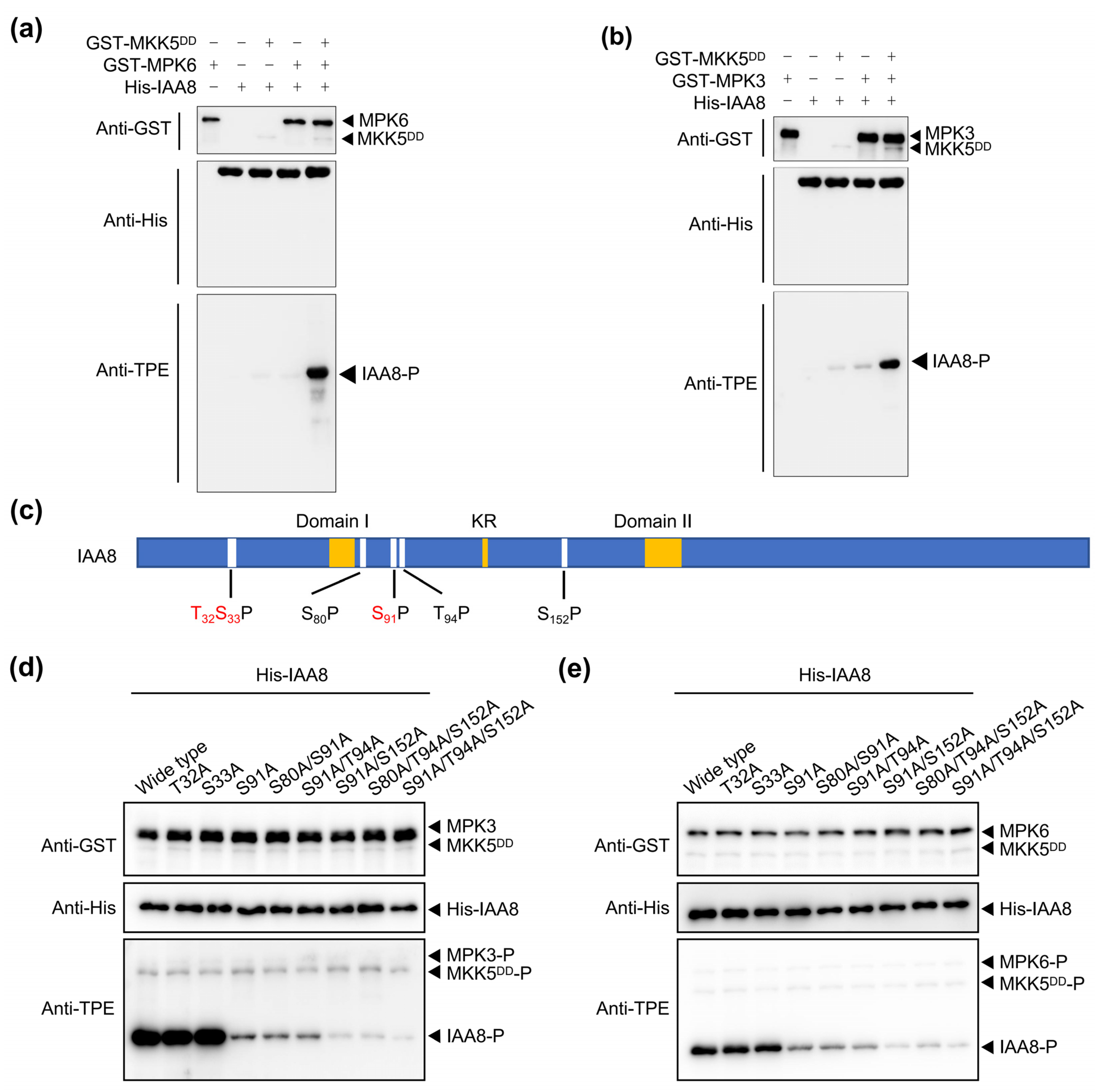
2.3. MPK3/6 Phosphorylate IAA8 at S91, T94, and S152 Residues

Since stress-activated MPK3/6 interact with IAA8/16 (Figure 2 and Figure S1), we hypothesized that MPK3/6 might phosphorylate IAA8/16, thereby reducing auxin signaling. To explore this possibility, we purified recombinant glutathione S-transferase (GST)-tagged proteins GST-MKK5DD, GST-MPK3, and GST-MPK6 and histidine (His)-tagged proteins His-IAA8 and His-IAA16 in E. coli BL21 cells and performed in vitro phosphorylation assays. The anti-thiophosphate ester-specific antibody (anti-TPE), which specifically recognizes phosphorylated proteins [13,23,24], revealed the phosphorylation of MPK6 and IAA8 in the presence of MKK5DD (constitutionally activated MKK5, used to activate MPK3/6), while MPK6 alone did not phosphorylate IAA8 (Figure 3a). We obtained the same results with MPK3 (Figure 3b), indicating that MKK5-activated MPK3 and MPK6 in turn phosphorylated IAA8. Unlike the intense phosphorylation of IAA8, the MKK5DD-MPK3/6 module could not phosphorylate IAA16 (Figure 3a,b and Figure S2), suggesting that IAA8, not IAA16, is the node where MPK3/6 regulate auxin signaling under stress conditions.
To further identify the IAA8 site(s) phosphorylated by MPK3/6, we performed a phosphorylation mass spectrometry assay. Three potential phosphorylation sites (T32, S33, and S91) within the amino acid sequence of IAA8 were identified (Figure 3c and Figure S3). Then, we mutated T32, S33, or S91 to the non-phosphorylated form residue alanine in IAA8T32A, IAA8S33A, or IAA8S91A, respectively, and repeated the in vitro phosphorylation assays. Mutation of T32 or S33 did not affect the IAA8 phosphorylation level, whereas mutation of the S91 residue remarkably reduced the phosphorylation level of IAA8 (Figure 3d,e), indicating that S91 was the main phosphorylation residue phosphorylated by MPK3/6. Although the phosphorylation intensity of IAA8S91A was significantly reduced, it could still be phosphorylated by MPK3/6, indicating that S91 was not the only phosphorylation site (Figure 3d,e).
Since MPK3/6 recognize typical serine–proline (SP) and threonine–proline (TP) motifs [13,25], we searched for other potential MPK3/6 recognition motifs in the IAA8 protein sequence and successfully obtained three motifs: S80P, T94P, and S152P (Figure 3c). On the basis of S91A, we performed various combinations of mutations at these three sites, and the results of in vitro phosphorylation experiments showed that T94 and S152 residues could also be phosphorylated by MPK3/6 (Figure 3d,e). In summary, MPK3/6 phosphorylated the S91, T94, and S152 residues of IAA8, among which S91 was the primary residue.

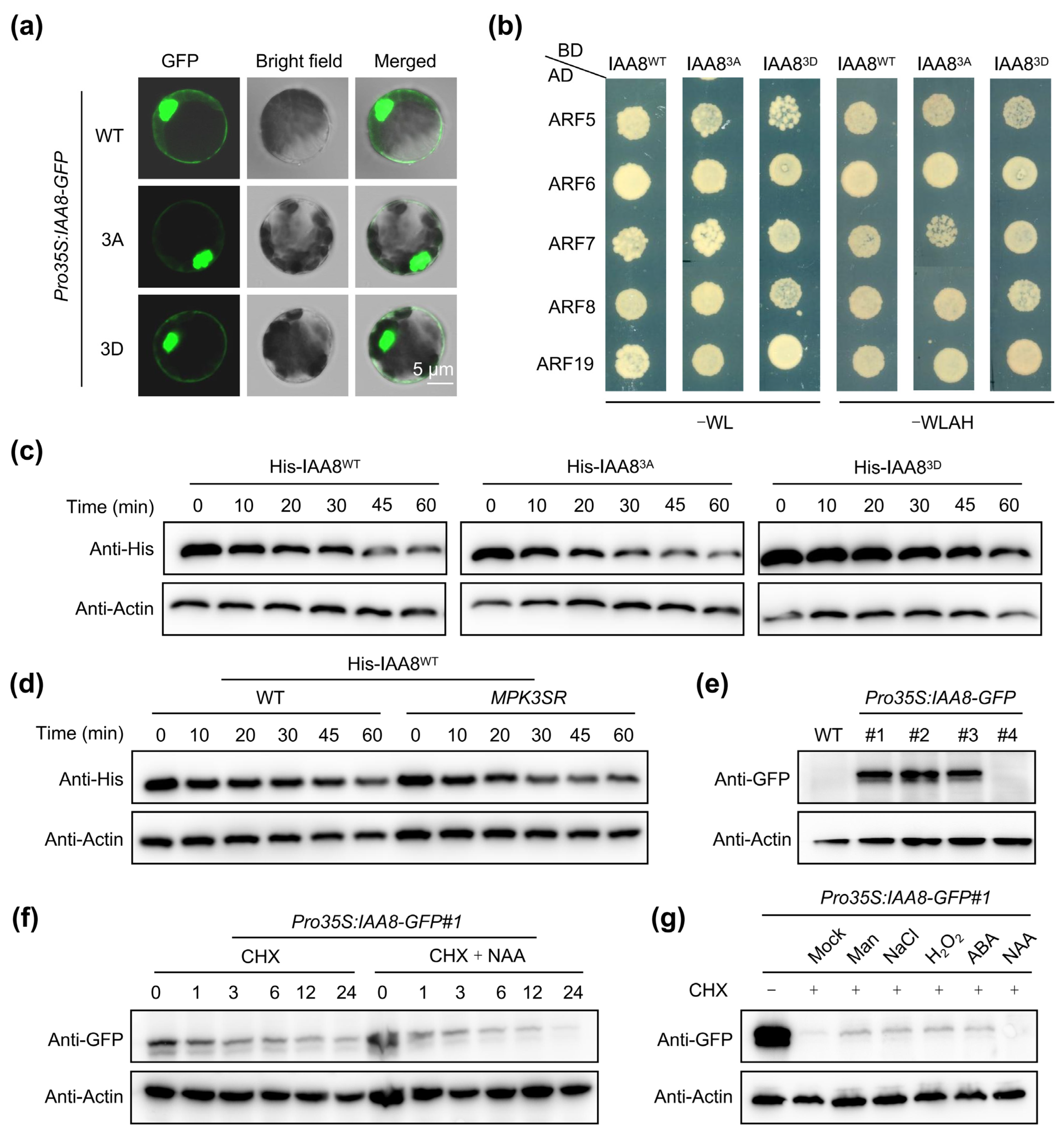
2.4. MPK3/6-Mediated Phosphorylation Stabilizes IAA8

Phosphorylation on the target protein usually influences its cellular localization, interaction intensity with other proteins, protein/DNA-binding ability, enzymatic activity, or protein stability [10,12,17,20,26]. To detect whether the MPK3/6-mediated phosphorylation of IAA8 affects its cellular localization, we expressed wild-type IAA8 (IAA8WT-GFP), a phosphomimic IAA8 variant, whereby the S91, T94, and S152 residues were mutated to aspartic acid (D) in IAA83D, and a non-phosphorylated IAA8 variant, whereby the S91, T94, and S152 residues were mutated to alanine (A) in IAA83A in Arabidopsis mesophyll protoplast cells, respectively. We found that the phosphorylation state of IAA8 did not affect its cellular localization (Figure 4a). Importantly, Aux/IAAs interact with ARFs and then repress the transcription of the ARF-regulated genes [19,27], promoting us to test whether MPK3/6-mediated phosphorylation of IAA8 affected its interaction with ARFs. In the Y2H experiment, IAA8WT, IAA83A, and IAA83D showed no difference in the interaction intensity with the auxin-activated ARFs, including ARF5, ARF6, ARF7, ARF8, and ARF19 (Figure 4b), indicating that MPK3/6-mediated phosphorylation did not affect the interaction between IAA8 and ARFs.
To further investigate whether this phosphorylation modification affected the protein stability of IAA8, we incubated and calculated the degradation rates of the recombinant His-IAA8WT, His-IAA83A, and His-IAA83D in the protein extracts from WT seedlings in cell-free assays. Compared with His-IAA8WT, the degradation of His-IAA83A was significantly elevated, whereas His-IAA83D was very stable (Figure 4c), suggesting that phosphorylation modification stabilized the IAA8 protein. Furthermore, we incubated the recombinant His-IAA8WT in the protein extracts from WT and MPK3SR, whereby the embryo lethality of the mpk3 mpk6 double-mutant was rescued by a version of MPK3 whose activity was repressed by 4-amino-1-tert-butyl-3-(1′-naphthyl) pyrazolo[3,4-d] pyrimidine (NA-PP1) [13,28]. We found that the His-IAA8WT proteins were more unstable in the protein extracts from MPK3SR seedlings than in those from WT (Figure 4d). Collectively, MPK3/6-mediated phosphorylation enhanced the protein stability of IAA8.
As a negative regulator of auxin signaling, IAA8 was degraded in the presence of NAA (Figure 4e,f), confirming the importance of its protein quantity for auxin signaling output. Stress-activated MPK3/6 significantly enhanced the protein stability of IAA8, prompting us to further investigate the effects of various stresses on the protein stability of IAA8. Not surprisingly, multiple treatments significantly increased the protein stability of IAA8 (Figure 4g). These results suggest that abiotic stresses reduce auxin signaling in plants by stabilizing the auxin signaling repressor IAA8, a process which relies on phosphorylation modification mediated by two crucial kinases, MPK3 and MPK6.

2.5. MPK6 Phosphorylates IAA9 at S88 Residue
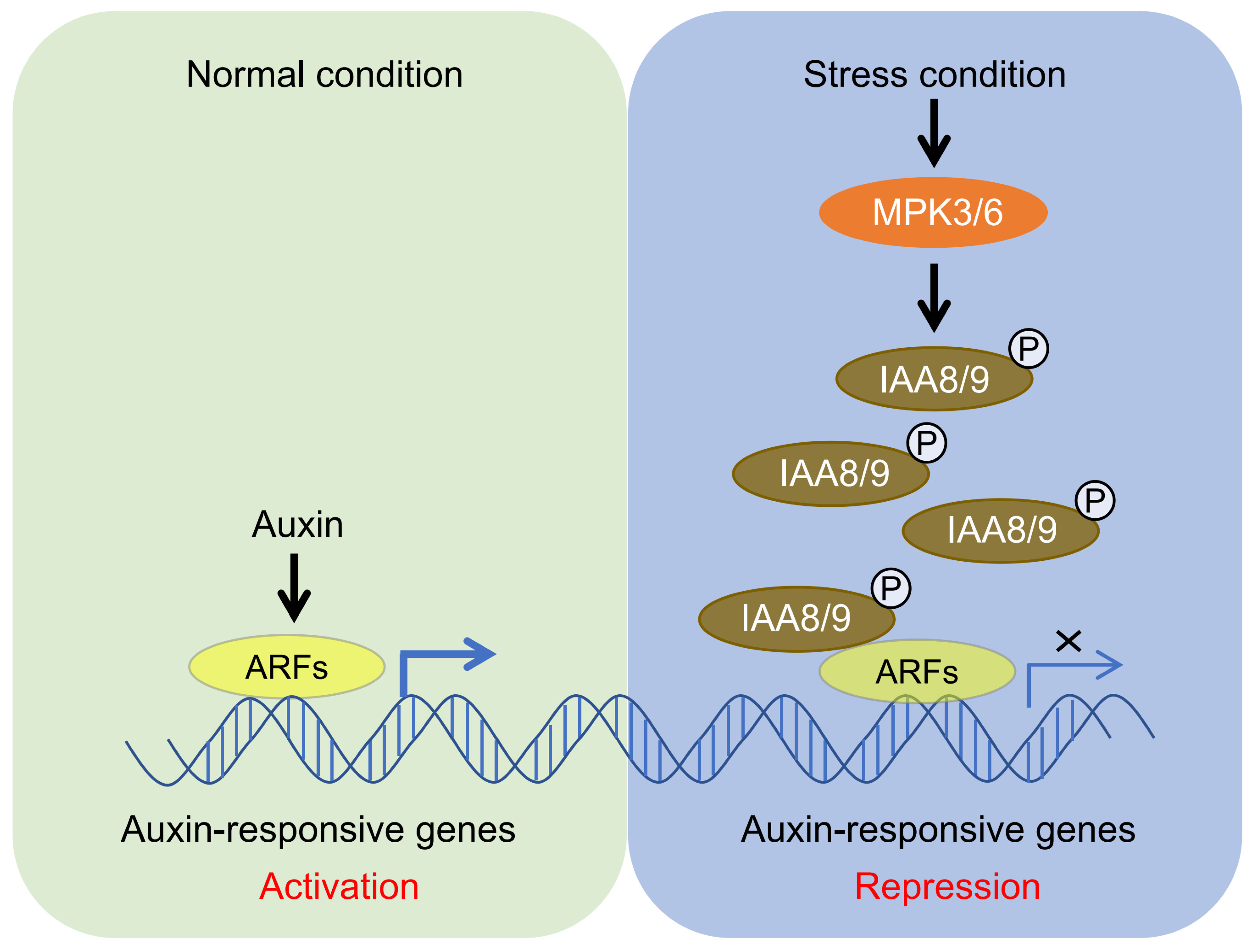
IAA9 shares the closest homology with IAA8, and protein sequence alignment reveals that an SP motif (S88P) in IAA9 is homologous to the SP motif (S91P, the key phosphorylation residue) in IAA8 (Figure 5a). This motif is only observed in IAA8 and IAA9 (Figure 5a), suggesting that MPK3/6 might also phosphorylate IAA9. Subsequently, we performed an in vitro phosphorylation assay and found that MPK6, but not MPK3, could phosphorylate IAA9 (Figure 5b,c). Furthermore, mutation of the S88 residue to Ala (IAA9S88A) significantly reduced the phosphorylation of IAA9 mediated by MPK6 (Figure 5b), indicating that S88 was the major site for the MPK6-mediated phosphorylation of IAA9.
Homologous proteins IAA8 and IAA9 share the same phosphorylation site, suggesting that MPK-mediated phosphorylation may have the same effect on IAA8 and IAA9, namely enhancing their protein stability. Then, we incubated the recombinant His-IAA9WT in the protein extracts from WT and MPK3SR seedlings in the cell-free assays. The degradation rate of His-IAA9WT was faster in MPK3SR than in the WT (Figure 5d), indicating that MPK6 enhanced the protein stability of IAA9.

3. Discussion

Abiotic stresses, such as high salinity, low temperature, and drought, seriously affect plant growth and crop production [29]. When faced with abiotic stresses, plants slow down their growth and evolve specific adaptations to allow growth by reprogramming their gene-regulatory networks [5,6]. The plant hormone auxin plays a crucial role in plant growth and development, and previous studies have shown that abiotic stresses affect auxin synthesis, transport, and signaling transduction [30,31,32]. In this study, we showed that various abiotic stresses inhibited auxin signaling (Figure 1), confirming the speculation that abiotic stresses suppress auxin signaling [7].
A previous study demonstrated that mIAA8, a functionally gain-of-function mutant of IAA8, interacts with ARF6/8 to inhibit their transcriptional activity, subsequently reducing the production of jasmonic acid (JA) and resulting in defects in floral organ development [33]. The MPK-mediated phosphorylation of IAA8 disturbs the expression of flower development-related genes to inhibit flower development under heat stress [34]. Cold signals and reactive oxygen species (ROS) promote the accumulation of IAA8 to inhibit the transcriptional activity of ARFs, and, in turn, IAA8 represses the transcription of ABA Insensitive 3 (ABI3) to facilitate seed germination [35]. Here, we revealed that various abiotic stresses reduced auxin signaling by stabilizing IAA8/9 (Figure 1 and Figure 4g), which expanded our understanding of IAA8/9 in plant response to stress. IAA8 may be the key for plants to actively reduce auxin signaling in response to stress, so the activation of MPK3/6-IAA8/9 modules may be necessary for plant stress adaptation.
MPK3/6 are essential for both plant growth and stress response [25,30,34,36,37,38,39], making them critical for balancing these two functions. Importantly, MPK3/6 can phosphorylate IAA15 to regulate lateral root formation under drought stress conditions [39] and can also phosphorylate IAA8 to regulate flower development under heat stress conditions [35]. Here, we found that MPK3/6 not only phosphorylate IAA8 and IAA9 but also interact with TIR1, IAA16, IAA32, and IAA34 in Y2H assays (Figure 2a), implying that there may be more substrates phosphorylated by MPK3/6. TIR1 is a component of the ubiquitin ligase (E3) complex SKP1-CUL-FBP (SCF) and receives the auxin signal to recruit Aux/IAA repressors to the SCFTIR1 complex for ubiquitination and degradation [7]. TIR1/AFBs also play crucial roles in response to abiotic and biotic stresses and are regulated post-translationally by other proteins [40]. Heat shock protein 90 (HSP90) interacts with and stabilizes TIR1 during plant development regulation [41]. The FERONIA receptor kinase–NADPH oxidase signaling pathway regulates the auxin-mediated oxidation of TIR1/AFB2 and their subsequent nuclear import [42]. Here, we found interactions between MPK3/6 and TIR1 (Figure 2a and Figure S1), implying that the stress-activated MPK3/6 may phosphorylate TIR1, thereby modulating the subcellular localization of TIR1. IAA32 and IAA34 belong to non-canonical Aux/IAAs, which lack a TIR1-binding domain [22]. Auxin-activated TMK1 C-terminus (kinase domain) phosphorylates IAA32 and IAA34 to regulate apical hook formation and maintenance [22]. Therefore, we hypothesized that MPK3/6 might also phosphorylate IAA32 and IAA34 to regulate apical hook development in response to abiotic stresses. Notably, MPK3/6 phosphorylate IAA9 at the S88 residue (Figure 5b), which is conserved in IAA8 at the S91 residue (Figure 5a). This residue is absent in other IAAs (Figure 5a), indicating that MPK3/6 may phosphorylate IAA32 and IAA34 at other sites. The function of MPK3/6-IAA32/34 modules in plant abiotic stress adaptation is worthy of further investigation.
In summary, this study reveals that, when plants are exposed to various stresses, activated MPK3/6 phosphorylate IAA8/9 and enhance their protein stability to reduce auxin signaling in plants (Figure 6). This study provides some new perspectives to understand how plants balance plant growth and stress response by regulating MPK3/6-mediated IAA8/9 protein levels.

4. Materials and Methods

4.1. Plant Materials and Growth Conditions

All plant materials used in this study were from the Arabidopsis thaliana (L.) Columbia-0 background. Arabidopsis seeds from WT and transgenic lines DR5:GUS [14], MPK3SR (mpk3 mpk6 ProMPK3:MPK3TG) [28], and Pro35S:IAA8-GFP were used for gene expression and function analyses. Pro35S:IAA8-GFP was generated in this study, and the coding sequence of IAA8 was cloned into the pSuper1300-GFP vector (Yaji Biological, Shanghai, China) to construct Pro35S:IAA8-GFP using the primers listed in Table S1. The floral-dip method with Agrobacterium tumefaciens strain GV3101 (Tsingke, Beijing, China) was used to perform the transformation of Arabidopsis, and homozygous transgenic plants were selected on half-strength MS medium (1.5% [w/v] sucrose and 0.85% [w/v] agar) supplemented with 40 mg L−1 hygromycin and confirmed by Western blot assays. For stress treatments, 7-day-old DR5:GUS transgenic lines were treated with 10 µM 1-naphthaleneacetic acid (NAA), 200 mM NaCl, 200 mM mannitol, 50 μM abscisic acid (ABA), or 5 mM H2O2 for 6 h. All selected Arabidopsis plants were grown in a controlled growth chamber under long-day conditions (16 h light/8 h dark) at 22 °C.

4.2. Yeast Two-Hybrid Assay (Y2H)

Protein interaction assays in yeast were performed according to the protocol for the Matchmaker Gold Yeast Two-Hybrid System (GAL4 based; Takara, Beijing, China). The coding sequences of the target genes were cloned into pGBKT7 and pGADT7 vectors and transformed into the yeast strain AH109 according to the protocol. Positive transformants were selected on a synthetic defined (SD) medium lacking leucine (Leu) and tryptophan (Trp) and grown at 30 °C for 2 days. Physical interactions were indicated by the ability of cells to grow on SD medium without Leu, Trp, His, and Ade for 2 to 3 days after plating. The primers used in the Y2H assays are listed in Table S1.

4.3. Bimolecular Fluorescence Complementation (BiFC) Assay in Arabidopsis Mesophyll Protoplasts

BiFC assays were performed as previously described [13]. The CDS of MPK3 or MPK6 was cloned into the pSAT6-nEYFP-C1 vector to construct MPK3-YFPN or MPK6-YFPN, and the coding sequence of TIR1 or IAAs was cloned into the pSAT6-cEYFP-C1 vector (Yaji Biological, Shanghai, China) to construct TIR1-YFPC or IAAs-YFPC. The protoplasts were prepared as previously described [13]. The plasmid combinations were transfected into Arabidopsis mesophyll protoplasts and then cultured at 25 °C for 12 h in the dark. The GFP signals were detected using a confocal microscope (Zeiss LSM 900, Oberkochen, Germany) with a 488 nm laser excitation wavelength and a 500–550 nm emission wavelength. The primers used for the BiFC assay are listed in Table S1.

4.4. Co-Immunoprecipitation (Co-IP) Assay in Arabidopsis Mesophyll Protoplasts

A Co-IP assay was performed as previously described [17] to detect the interactions between MPKs and TIR1 or IAAs. The CDS of MPK3, MPK6, TIR1, or IAA16 was cloned into the pSuper1300-Myc vector to generate MPK3-Myc, MPK6-Myc, TIR1-Myc, or IAA16-Myc; the CDS of MPK3, MPK6, or IAA8 was cloned into the pSuper1300-GFP vector to construct MPK3-GFP, MPK6-GFP, or IAA8-GFP. The protoplasts were prepared as previously described [13]. The various plasmid combinations were co-transfected into Arabidopsis mesophyll protoplasts and then cultured at 25 °C for 12 h in the dark. The anti-GFP antibody (Chromo Tek, 1:100 dilution, Munich, Germany) was used to immunoprecipitate IAA8-GFP, and the anti-Myc antibody (ABclonal, 1:5000 dilution, Boston, MA, USA) was used to detect the protein level of MPK3-Myc or MPK6-Myc. The anti-Myc antibody (Chromo Tek, 1:100 dilution, Germany) was used to immunoprecipitate IAA16-Myc or TIR1-Myc, and the anti-GFP antibody (ABclonal, 1:5000 dilution, Boston, MA, USA) was used to detect the protein level of MPK3-GFP or MPK6-GFP. The primers used for the Co-IP assay are listed in Table S1.

4.5. In Vitro Kinase Assay

The in vitro kinase assay was performed as previously described [13]. The coding sequences of GST-MKK5T215D/S221D (MKK5DD) [38], GST-MPK6, and GST-MPK3 were cloned into the pGEX-4T-1 vector to add a GST tag. The CDS of IAA8, IAA8T32A, IAA8S33A, IAA8S91A, IAA8S80A/S91A, IAA8S91A/T94A, IAA8S91A/S152A, IAA8S80A/T94A/S152A, IAA8S91A/T94A/S152A, IAA9, IAA9S88A, or IAA16 was cloned into the pET30a vector to add an His tag. The production of recombinant proteins was induced in E. coli BL21 cells with 0.4 mM isopropyl β-D-1-thiogalactopyranoside (IPTG) at 16 °C for 12 h and purified with glutathione beads (Smart-Lifesciences, Changzhou, China) or Ni IDA beads (Smart-Lifesciences, Changzhou, China). The in vitro kinase assay was performed as described previously [36]. In brief, 0.1 μg of recombinant GST-MKK5DD, 0.2 μg of GST-MPK3 or GST-MPK6, and 2 μg of His-IAA8, the mutant His-IAA8, or His-IAA16 were incubated in a 30 μL reaction buffer (10 mM MgCl2, 1 mM adenosine 5′-O-[3-thiotriphosphate], 50 mM tris-HCl [pH 7.5], and 1 mM DTT) at 30 °C for 1 h. Then, 20 mM EDTA and 1.5 mM p-nitrobenzylmesylate (ABclonal, Boston, MA, USA) were added and incubated at 25 °C for another 1 h, and boiled samples with 5× loading buffer were separated by SDS-PAGE and analyzed by immunoblotting. The anti-TPE antibody (ABclonal, Boston, MA, USA), which specifically recognizes phosphorylated proteins [23,24], was used to detect whether MPK3 and MPK6 phosphorylated IAA8, the mutant IAA8, IAA9, the mutant IAA9, or IAA16. The anti-GST or anti-His antibody (TransGen, 1:5000 dilution, Beijing, China) was used to determine whether the loading quantity of each protein was consistent. The primers used for the in vitro kinase assay are listed in Table S1.

4.6. Cell-Free Assay

The cell-free assay was performed as previously described [26]. Briefly, total proteins from 7-day-old Arabidopsis WT or MPK3SR seedlings were extracted with the lysis buffer (25 mM Tris-HCl [pH 7.5], 10 mM NaCl, 10 mM MgCl2, 4 mM PMSF, 5 mM DTT, and 10 mM ATP). After centrifugations at 14,000× g at 4 °C, the supernatant was collected. To detect the protein degradation of His-IAA8WT or His-IAA9WT, 100 ng recombinant protein was incubated with 100 μL of total protein at 22 °C. All mock controls used an equal amount of solvent for each drug. Boiled samples were separated by SDS-PAGE and analyzed by immunoblotting. The anti-His antibody was used to detect His-IAA8 or His-IAA9. The anti-actin antibody (ABclonal, 1:5000 dilution, Boston, MA, USA) was used to determine whether the loading quantity of each protein was consistent.

4.7. Protein Stability Detection

We performed a protein stability assay as previously described [26]. In brief, 7-day-old Arabidopsis Pro35S:IAA8-GFP#1 seedlings grown on 1/2 MS medium at 22 °C were treated with 125 μM CHX or 125 μM CHX + 10 μM NAA for 1, 3, 6, 12, and 24 h. Boiled samples were separated by SDS-PAGE and analyzed by immunoblotting. The anti-GFP antibody (TransGen Biotech, Beijing, China) was used to detect IAA8-GFP. The anti-actin antibody was used to determine whether the loading quantity of each protein was consistent.

4.8. β-Glucuronidase (GUS) Staining

Histochemical GUS staining was performed as previously described [14]. Briefly, the root tips of 7-day-old seedlings were incubated in a solution (100 mM sodium phosphate buffer [pH 7.2], 50 μM potassium ferricyanide, 5 mM potassium ferrocyanide, 0.1% (v/v) Triton X-100, 2 mM 5-bromo-4-chloro-3-indolyl-β-D-glucuronide [X-gluc], and 10 mM EDTA) at 37 °C for 3 h. The samples were cleared in chloral hydrate and observed with an Olympus BX53 microscope (Tokyo, Japan).

Supplementary Materials

The following supporting information can be downloaded at https://www.mdpi.com/article/10.3390/ijms26051964/s1.

Author Contributions

Conceptualization, Z.Y., L.Y. and R.G.; data curation, C.W., X.C. and K.L.; formal analysis, H.Z. and X.C.; funding acquisition, K.L., Y.X. and Z.Y.; investigation, C.W., X.C. and W.X.; methodology, C.W. and H.Z.; project administration, Y.X. and R.G.; resources, X.L. and X.C.; software, H.Z.; supervision, Y.X. and R.G.; visualization, L.Y.; and writing—review and editing, Y.X. and Z.Y. All authors have read and agreed to the published version of the manuscript.

Funding

This research was funded by the Natural Science Foundation of the Shandong Province, with grants number ZR2024QC012 and ZR2022QC059, the Natural Science Foundation of Qingdao, with grant number 24-4-4-zrjj-132-jch, the National Natural Science Foundation of China, with grants number 32470326 and 32100266, and the Shandong Province Higher Educational Science and Technology Program, with grant number 2024KJG070.

Institutional Review Board Statement

Not applicable.

Informed Consent Statement

Not applicable.

Data Availability Statement

The plant materials supporting the findings of this study are available from the corresponding author upon request.

Acknowledgments

We thank Shuqun Zhang (Zhejiang University) for sharing published materials and Jie Dai (Shanghai Bioprofile Technology Co., Ltd.) for their technical support in mass spectroscopy.

Conflicts of Interest

The authors declare no conflicts of interest.

Abbreviations

The following abbreviations are used in this manuscript:
MPK3Mitogen-activated protein kinase 3
IAA8Indoleacetic acid-induced protein 8
Aux/IAAAuxin/indole-3-3 acetic acid
ARFAuxin response factor
NAA1-naphthaleneacetic acid
ABAAbscisic acid
JAJasmonic acid
RLKsReceptor-like kinases
MAP3KMAP kinase kinase kinase
MAP2KMAP kinase kinase
TIR1Transport inhibitor response 1
AFBsAuxin signaling F-boxs
MPK3SRmpk3 mpk6 MPK3pro:MPK3TG
MPK6SRmpk3 mpk6 MPK6pro:MPK6TG
TPLTOPLESS
Y2HYeast two-hybrid
BiFCBimolecular fluorescence complementation
Co-IPCo-immunoprecipitation
GSTGlutathione S-transferase
NA-PP14-amino-1-tert-butyl-3-(1′-naphthyl) pyrazolo[3,4-d] pyrimidine
anti-TPEAnti-thiophosphate ester-specific antibody
ROSReactive oxygen species
ABI3ABA-insensitive 3
CHXCycloheximide

References

  1. Bai, Y.; Kissoudis, C.; Yan, Z.; Visser, R.G.F.; van der Linden, G. Plant behaviour under combined stress: Tomato responses to combined salinity and pathogen stress. Plant J. 2018, 93, 781–793. [Google Scholar] [CrossRef]
  2. Zhou, H.; Shi, H.; Yang, Y.; Feng, X.; Chen, X.; Xiao, F.; Lin, H.; Guo, Y. Insights into plant salt stress signaling and tolerance. J. Genet. Genom. 2024, 51, 16–34. [Google Scholar] [CrossRef]
  3. Kumari, V.V.; Roy, A.; Vijayan, R.; Banerjee, P.; Verma, V.C.; Nalia, A.; Pramanik, M.; Mukherjee, B.; Ghosh, A.; Reja, M.H.; et al. Drought and Heat Stress in Cool-Season Food Legumes in Sub-Tropical Regions: Consequences, Adaptation, and Mitigation Strategies. Plants 2021, 10, 1038. [Google Scholar] [CrossRef]
  4. Clauw, P.; Coppens, F.; Korte, A.; Herman, D.; Slabbinck, B.; Dhondt, S.; Van Daele, T.; De Milde, L.; Vermeersch, M.; Maleux, K.; et al. Leaf Growth Response to Mild Drought: Natural Variation in Arabidopsis Sheds Light on Trait Architecture. Plant Cell 2016, 28, 2417–2434. [Google Scholar] [CrossRef] [PubMed]
  5. Claeys, H.; Inzé, D. The agony of choice: How plants balance growth and survival under water-limiting conditions. Plant Physiol. 2013, 162, 1768–1779. [Google Scholar] [CrossRef] [PubMed]
  6. Julkowska, M.M.; Testerink, C. Tuning plant signaling and growth to survive salt. Trends Plant Sci. 2015, 20, 586–594. [Google Scholar] [CrossRef]
  7. Jing, H.; Wilkinson, E.G.; Sageman-Furnas, K.; Strader, L.C. Auxin and abiotic stress responses. J. Exp. Bot. 2023, 74, 7000–7014. [Google Scholar] [CrossRef]
  8. Xu, J.; Zhang, S. Mitogen-activated protein kinase cascades in signaling plant growth and development. Trends Plant Sci. 2015, 20, 56–64. [Google Scholar] [CrossRef] [PubMed]
  9. Jonak, C.; Okrész, L.; Bögre, L.; Hirt, H. Complexity, cross talk and integration of plant MAP kinase signalling. Curr. Opin. Plant Biol. 2002, 5, 415–424. [Google Scholar] [CrossRef]
  10. Jagodzik, P.; Tajdel-Zielinska, M.; Ciesla, A.; Marczak, M.; Ludwikow, A. Mitogen-Activated Protein Kinase Cascades in Plant Hormone Signaling. Front. Plant Sci. 2018, 9, 1387. [Google Scholar] [CrossRef] [PubMed]
  11. Zhang, M.; Su, J.; Zhang, Y.; Xu, J.; Zhang, S. Conveying endogenous and exogenous signals: MAPK cascades in plant growth and defense. Curr. Opin. Plant Biol. 2018, 45, 1–10. [Google Scholar] [CrossRef]
  12. Huang, R.; Zheng, R.; He, J.; Zhou, Z.; Wang, J.; Xiong, Y.; Xu, T. Noncanonical auxin signaling regulates cell division pattern during lateral root development. Proc. Natl. Acad. Sci. USA 2019, 116, 21285–21290. [Google Scholar] [CrossRef]
  13. Yu, Z.; Ma, J.; Zhang, M.; Li, X.; Sun, Y.; Zhang, M.; Ding, Z. Auxin promotes hypocotyl elongation by enhancing BZR1 nuclear accumulation in Arabidopsis. Sci. Adv. 2023, 9, eade2493. [Google Scholar] [CrossRef] [PubMed]
  14. Wang, M.; Li, P.; Ma, Y.; Nie, X.; Grebe, M.; Men, S. Membrane Sterol Composition in Arabidopsis thaliana Affects Root Elongation via Auxin Biosynthesis. Int. J. Mol. Sci. 2021, 22, 437. [Google Scholar] [CrossRef] [PubMed]
  15. Liu, J.J.; Tian, H.Y.; Zhang, M.X.; Sun, Y.; Wang, J.X.; Yu, Q.Q.; Ding, Z.J. STOP1 attenuates the auxin response to maintain root stem cell niche identity. Cell Rep. 2024, 43, 113617. [Google Scholar] [CrossRef] [PubMed]
  16. Braybrook, S.A. Auxin and Organogenesis: Initiation of Organs and Nurturing a Scientific Spirit. Plant Cell 2019, 31, 1397. [Google Scholar] [CrossRef] [PubMed]
  17. Yu, Z.; Qu, X.; Lv, B.; Li, X.; Sui, J.; Yu, Q.; Ding, Z. MAC3A and MAC3B mediate degradation of the transcription factor ERF13 and thus promote lateral root emergence. Plant Cell 2024, 36, 3162–3176. [Google Scholar] [CrossRef] [PubMed]
  18. Yang, Y.; Zhang, L.; Chen, P.; Liang, T.; Li, X.; Liu, H. UV-B photoreceptor UVR8 interacts with MYB73/MYB77 to regulate auxin responses and lateral root development. EMBO J. 2020, 39, e101928. [Google Scholar] [CrossRef]
  19. Yu, Z.; Zhang, F.; Friml, J.; Ding, Z. Auxin signaling: Research advances over the past 30 years. J. Integr. Plant Biol. 2022, 64, 371–392. [Google Scholar] [CrossRef] [PubMed]
  20. Lv, B.S.; Wei, K.J.; Hu, K.Q.; Tian, T.; Zhang, F.; Yu, Z.P.; Zhang, D.J.; Su, Y.H.; Sang, Y.L.; Zhang, X.S.; et al. MPK14-mediated auxin signaling controls lateral root development via ERF13-regulated very-long-chain fatty acid biosynthesis. Mol. Plant 2021, 14, 285–297. [Google Scholar] [CrossRef] [PubMed]
  21. Zhu, Q.K.; Shao, Y.M.; Ge, S.T.; Zhang, M.M.; Zhang, T.S.; Hu, X.T.; Liu, Y.D.; Walker, J.; Zhang, S.Q.; Xu, J. A MAPK cascade downstream of IDA-HAE/HSL2 ligand-receptor pair in lateral root emergence. Nat. Plants 2019, 5, 414–423. [Google Scholar] [CrossRef]
  22. Cao, M.; Chen, R.; Li, P.; Yu, Y.; Zheng, R.; Ge, D.; Zheng, W.; Wang, X.; Gu, Y.; Gelová, Z.; et al. TMK1-mediated auxin signalling regulates differential growth of the apical hook. Nature 2019, 568, 240–243. [Google Scholar] [CrossRef] [PubMed]
  23. Allen, J.J.; Li, M.Q.; Brinkworth, C.S.; Paulson, J.L.; Wang, D.; Hubner, A.; Chou, W.H.; Davis, R.J.; Burlingame, A.L.; Messing, R.O.; et al. A semisynthetic epitope for kinase substrates. Nat. Methods 2007, 4, 511–516. [Google Scholar] [CrossRef] [PubMed]
  24. Leissing, F.; Nomoto, M.; Bocola, M.; Schwaneberg, U.; Tada, Y.; Conrath, U.; Beckers, G.J.M. Substrate thiophosphorylation by Arabidopsis mitogen-activated protein kinases. BMC Plant Biol. 2016, 16, 48. [Google Scholar] [CrossRef] [PubMed]
  25. Zhang, M.; Zhang, S. Mitogen-activated protein kinase cascades in plant signaling. J. Integr. Plant Biol. 2022, 64, 301–341. [Google Scholar] [CrossRef] [PubMed]
  26. Zhang, L.; Yu, Z.P.; Xu, Y.; Yu, M.; Ren, Y.; Zhang, S.Z.; Yang, G.D.; Huang, J.G.; Yan, K.; Zheng, C.C.; et al. Regulation of the stability and ABA import activity of NRT1.2/NPF4.6 by CEPR2-mediated phosphorylation in. Mol. Plant 2021, 14, 633–646. [Google Scholar] [CrossRef] [PubMed]
  27. Cancé, C.; Martin-Arevalillo, R.; Boubekeur, K.; Dumas, R. Auxin response factors are keys to the many auxin doors. New Phytol. 2022, 235, 402–419. [Google Scholar] [CrossRef]
  28. Su, J.; Zhang, M.; Zhang, L.; Sun, T.; Liu, Y.; Lukowitz, W.; Xu, J.; Zhang, S. Regulation of Stomatal Immunity by Interdependent Functions of a Pathogen-Responsive MPK3/MPK6 Cascade and Abscisic Acid. Plant Cell 2017, 29, 526–542. [Google Scholar] [CrossRef] [PubMed]
  29. Lama, S.; Broda, M.; Abbas, Z.; Vaneechoutte, D.; Belt, K.; Säll, T.; Vandepoele, K.; Van Aken, O. Neofunctionalization of Mitochondrial Proteins and Incorporation into Signaling Networks in Plants. Mol. Biol. Evol. 2019, 36, 974–989. [Google Scholar] [CrossRef] [PubMed]
  30. Kovtun, Y.; Chiu, W.L.; Tena, G.; Sheen, J. Functional analysis of oxidative stress-activated mitogen-activated protein kinase cascade in plants. Proc. Natl. Acad. Sci. USA 2000, 97, 2940–2945. [Google Scholar] [CrossRef] [PubMed]
  31. Lee, J.S.; Wang, S.; Sritubtim, S.; Chen, J.G.; Ellis, B.E. Arabidopsis mitogen-activated protein kinase MPK12 interacts with the MAPK phosphatase IBR5 and regulates auxin signaling. Plant J. 2009, 57, 975–985. [Google Scholar] [CrossRef] [PubMed]
  32. Dory, M.; Hatzimasoura, E.; Kállai, B.M.; Nagy, S.K.; Jäger, K.; Darula, Z.; Nádai, T.V.; Mészáros, T.; López-Juez, E.; Barnabás, B.; et al. Coevolving MAPK and PID phosphosites indicate an ancient environmental control of PIN auxin transporters in land plants. FEBS Lett. 2018, 592, 89–102. [Google Scholar] [CrossRef] [PubMed]
  33. Wang, J.; Yan, D.W.; Yuan, T.T.; Gao, X.; Lu, Y.T. A gain-of-function mutation in IAA8 alters Arabidopsis floral organ development by change of jasmonic acid level. Plant Mol. Biol. 2013, 82, 71–83. [Google Scholar] [CrossRef]
  34. Kim, S.H.; Hussain, S.; Pham, H.T.T.; Kadam, U.S.; Bahk, S.; Ramadany, Z.; Lee, J.; Song, Y.H.; Lee, K.O.; Hong, J.C.; et al. Phosphorylation of auxin signaling repressor IAA8 by heat-responsive MPKs causes defective flower development. Plant Physiol. 2024, 196, 2825–2840. [Google Scholar] [CrossRef] [PubMed]
  35. Hussain, S.; Kim, S.H.; Bahk, S.; Ali, A.; Nguyen, X.C.; Yun, D.J.; Chung, W.S. The Auxin Signaling Repressor IAA8 Promotes Seed Germination Through Down-Regulation of ABI3 Transcription in Arabidopsis. Front. Plant Sci. 2020, 11, 111. [Google Scholar] [CrossRef] [PubMed]
  36. Yan, Z.; Wang, J.; Wang, F.; Xie, C.; Lv, B.; Yu, Z.; Dai, S.; Liu, X.; Xia, G.; Tian, H.; et al. MPK3/6-induced degradation of ARR1/10/12 promotes salt tolerance in Arabidopsis. EMBO Rep. 2021, 22, e52457. [Google Scholar] [CrossRef]
  37. Liu, Y.; Leary, E.; Saffaf, O.; Frank Baker, R.; Zhang, S. Overlapping functions of YDA and MAPKKK3/MAPKKK5 upstream of MPK3/MPK6 in plant immunity and growth/development. J. Integr. Plant Biol. 2022, 64, 1531–1542. [Google Scholar] [CrossRef] [PubMed]
  38. Li, H.; Ding, Y.; Shi, Y.; Zhang, X.; Zhang, S.; Gong, Z.; Yang, S. MPK3- and MPK6-Mediated ICE1 Phosphorylation Negatively Regulates ICE1 Stability and Freezing Tolerance in Arabidopsis. Dev. Cell 2017, 43, 630–642.e4. [Google Scholar] [CrossRef] [PubMed]
  39. Kim, S.H.; Bahk, S.; Nguyen, N.T.; Pham, M.L.A.; Kadam, U.S.; Hong, J.C.; Chung, W.S. Phosphorylation of the auxin signaling transcriptional repressor IAA15 by MPKs is required for the suppression of root development under drought stress in Arabidopsis. Nucleic Acids Res. 2022, 50, 10544–10561. [Google Scholar] [CrossRef]
  40. Du, W.; Lu, Y.; Li, Q.; Luo, S.; Shen, S.; Li, N.; Chen, X. TIR1/AFB proteins: Active players in abiotic and biotic stress signaling. Front. Plant Sci. 2022, 13, 1083409. [Google Scholar] [CrossRef] [PubMed]
  41. Muñoz, A.; Mangano, S.; Toribio, R.; Fernández-Calvino, L.; Del Pozo, J.C.; Castellano, M.M. The co-chaperone HOP participates in TIR1 stabilisation and in auxin response in plants. Plant Cell Environ. 2022, 45, 2508–2519. [Google Scholar] [CrossRef] [PubMed]
  42. Lu, B.; Wang, S.; Feng, H.; Wang, J.; Zhang, K.; Li, Y.; Wu, P.; Zhang, M.; Xia, Y.; Peng, C.; et al. FERONIA-mediated TIR1/AFB2 oxidation stimulates auxin signaling in Arabidopsis. Mol. Plant 2024, 17, 772–787. [Google Scholar] [CrossRef] [PubMed]
Figure 1. Abiotic stresses repress auxin signaling. Histochemical β-glucuronidase (GUS) staining with 7-day-old transgenic plants harboring the auxin-responsive reporter gene DR5:GUS treated with 200 mM NaCl, 200 mM mannitol, 50 μM ABA, 5 mM H2O2, or 10 μM NAA. Scale bar, 100 µm. NAA, 1-naphthaleneacetic acid.
Ijms 26 01964 g001
Figure 2. MPK3/6 interact with IAA8. (a) Yeast two-hybrid (Y2H) assay, showing the interactions between MPK3/6 and TIR1, Aux/IAAs, TPL, and ARFs. (b) Bimolecular fluorescence complementation (BiFC) assay performed in Arabidopsis protoplasts, detecting the interactions between MPK3/6 and IAA8. Scale bar, 10 μm. (c) Co-immunoprecipitation (Co-IP) assays performed in Arabidopsis protoplasts, validating the interactions between MPK3/6 and IAA8. IP was performed with an anti-GFP antibody, and interactions with MPK3/6-Myc were detected with an anti-Myc antibody.
Ijms 26 01964 g002
Figure 3. MPK3/6 phosphorylate IAA8. (a,b) In vitro kinase assays showing whether MPK3/6, which are activated by MKK5DD, phosphorylate IAA8. The anti-thiophosphate ester-specific (anti-TPE) antibody was used to visualize the phosphorylated proteins. The anti-GST or anti-His antibody was used to determine whether the loading quantity of each protein was consistent. (c) The schematic diagram illustrates the potential TP/SP motifs within the IAA8 that may be phosphorylated by MPK3/6. (d,e) The candidate phosphorylation sites in IAA8 were confirmed using an in vitro phosphorylation assay.
Ijms 26 01964 g003
Figure 4. MPK3/6-mediated phosphorylation stabilizes IAA8. (a) Confocal images of GFP signals in Arabidopsis protoplasts expressing IAA8WT-GFP, IAA83D-GFP, or IAA83A-GFP. (b) Y2H assay validating the interaction between IAA8WT, IAA83A, or IAA83D and the auxin-activated ARFs. (c) Degradation of the recombinant His-IAA8WT, His-IAA83A, and His-IAA83D in the protein extracts from 8-day-old WT seedlings in cell-free assays. (d) Degradation of His-IAA8WT in the protein extracts from 8-day-old WT seedlings or MPK3SR seedlings in cell-free assays. (e) Protein level of IAA8 in different Pro35S:IAA8-GFP transgenic lines. (f) Protein degradation of IAA8 in Pro35S:IAA8-GFP#1 seedlings treated with or without NAA. (g) Protein degradation of IAA8 in Pro35S:IAA8-GFP#1 seedlings treated with or without various treatments. Seedlings were treated with 125 μM CHX or 125 μM CHX + 200 mM NaCl, 200 mM mannitol, 50 μM ABA, 5 mM H2O2, and 10 μM NAA for 6 h. CHX, cycloheximide.
Ijms 26 01964 g004
Figure 5. MPK6 phosphorylates and enhances the protein stability of IAA9. (a) Amino acid sequence alignments of IAAs in Arabidopsis. (b,c) In vitro kinase assays showing whether MPK3/6, which are activated by MKK5DD, phosphorylate IAA9. The anti-TPE antibody was used to visualize phosphorylated proteins. The anti-GST or anti-His antibody was used to determine whether the loading quantity of each protein was consistent. (d) Protein degradation of the recombinant His-IAA9WT in the protein extracts from 8-day-old WT seedlings or MPK3SR seedlings in cell-free assay.
Ijms 26 01964 g005
Figure 6. Proposed model of MPK3/6-IAA8/9 module in plant stress adaptation. Under normal conditions, as a growth-promoting hormone, auxin maintains the expression of many growth-related genes by activating the key transcription factor ARFs, thus ensuring the normal growth and development of plants. Under stress conditions, MPK3/6-mediated phosphorylation significantly enhances the protein stability of IAA8/9, resulting in basal auxin signaling for plant stress adaptation. ARF, auxin response factor; MPK3, mitogen-activated protein kinase 3; and IAA8, auxin/indole-3-3 acetic acid 8.
Ijms 26 01964 g006
Disclaimer/Publisher’s Note: The statements, opinions and data contained in all publications are solely those of the individual author(s) and contributor(s) and not of MDPI and/or the editor(s). MDPI and/or the editor(s) disclaim responsibility for any injury to people or property resulting from any ideas, methods, instructions or products referred to in the content.

