Case Report

Novel EVI5::BRAF Gene Fusion in Infantile Fibrosarcoma: A Case Report and Review of Literature

by
Judith González-López
1,
Luis Alberto Rubio-Martínez
1,
Miryam Atarés
2,
José Vicente Amaya
3,
Maria Carmen Huart
1,
Empar Mayordomo-Aranda
1 and
Francisco Giner
1,4,*
1
Pathology Department, Hospital Universitari i Politècnic La Fe, 46026 València, Spain
2
Radiology Department, Hospital Universitari i Politècnic La Fe, 46026 València, Spain
3
Orthopaedics and Traumatology Department, Hospital Universitari i Politècnic La Fe, 46026 València, Spain
4
Pathology Department, Universitat de València, Avinguda Blasco Ibáñez, 15, 46010 València, Spain
*
Author to whom correspondence should be addressed.
Int. J. Mol. Sci. 2025, 26(3), 1182; https://doi.org/10.3390/ijms26031182
Submission received: 13 December 2024 / Revised: 23 January 2025 / Accepted: 27 January 2025 / Published: 29 January 2025
(This article belongs to the Section Molecular Pathology, Diagnostics, and Therapeutics)

Abstract

:
Infantile Fibrosarcoma is a malignant tumor of fibroblastic origin, typically found in early childhood, locally aggressive, and characterized by molecular alterations that activate tyrosine kinase signaling, primarily the ETV6::NTRK3 fusion. In recent years, a series of fusions different from the classic one have been described, including NTRK1, RAF1, and BRAF. In this paper, we present a case of IFS with a novel EVI5::BRAF fusion. We observed a spindle cell neoplasm growing in intertwined fascicles within a fibrous stroma, without the formation of an immature osteoid matrix. Weak and focal immunoreactivity for S100 was observed. SATB2 exhibited diffuse and intense staining, with focal expression of osteonectin and negativity for caldesmon, Smooth Muscle Actin, desmin, GFAP, SOX10, MelanA, panTRK, and HMB45. The Ki67 index was 7%, and the tumor harbored an EVI5::BRAF genetic fusion. To the best of our knowledge, the EVI5::BRAF fusion has not yet been described in BRAF fusions in IFS. Nevertheless, further studies are needed to define the prognostic features of these emerging BRAF sarcomas, along with new anti-BRAF therapeutic approaches.

1. Introduction

Pediatric mesenchymal tumors encompass a heterogeneous group of benign and malignant pathologies, both de novo (90%) and associated with hereditary syndromes (10%). Soft tissue sarcomas are more common in children than in adults, with an incidence that increases with age from 0.9 cases per 100,000 person-years in children under 10 years old to 18.2 cases per 100,000 person-years in older children. In children under 15 years old, the most common sarcoma is rhabdomyosarcoma (95–97% of cases), while the remaining 3–5% includes both low-grade tumors (rarely metastatic or locally aggressive) and high-grade tumors [1].
This group includes both typical pediatric sarcomas and other “adult-type” sarcomas that can appear in childhood. One of the typical pediatric tumors, Infantile Fibrosarcoma (IFS), is a malignant tumor of fibroblastic origin, typically found in early childhood, locally very aggressive, and characterized by molecular alterations that activate tyrosine kinase signaling, primarily the ETV6::NTRK3 fusion. Despite its local aggressiveness, metastases are infrequent (8–15%), and the 10-year survival rate is 90% with surgical and chemotherapeutic treatment. Local recurrence occurs in 25–40% of cases with affected margins. These tumors exhibit a poorly characteristic immunohistochemical profile, with variable expression of CD34, S100, Smooth Muscle Actin, and desmin. PanTRK may be positive in cases where mutations are present [1].
In recent years, a series of IFSs with fusions different from the classic one have been described, involving other tyrosine kinase genes, including NTRK1, NTRK2, RET, MET, and RAF1 [1]. Among these new fusions is BRAF, a gene that encodes proteins from the RAF family of protein kinases that regulate the MAP kinase/ERK signaling pathway. This pathway promotes cell proliferation and survival, and its activation induces tumorigenesis [2]. Although BRAF has functions typically associated with carcinomas, its role as an inducer of mesenchymal neoplasms has also been described. These neoplasms characteristically present a “spindle cell” or “epithelioid” pattern and are NTRK-negative [2]. This group includes not only IFSs but also mandibular [3] and central nervous system sarcomas [4,5], as well as myxoinflammatory myofibroblastic sarcomas, glomus tumors, stromal tumors of the kidney, and gastrointestinal stromal tumors (GISTs) [6].
Treatment consists of surgery and chemotherapy. Despite being locally aggressive, metastasis is rare (10%), and the 10-year survival rate exceeds 90%. The most relevant factor for recurrences (which occur in 25–40% of cases) is resection with clear margins, which is why surgery is often aggressive. Given the development of new therapeutic targets against NTRK and BRAF, the identification of these molecular alterations is of vital importance for the prognosis and treatment of patients. [1].
In this paper, we present a case of IFS with a novel EVI5::BRAF fusion and discuss its clinical, histological, immunohistochemical, and molecular features.

2. Case Presentation

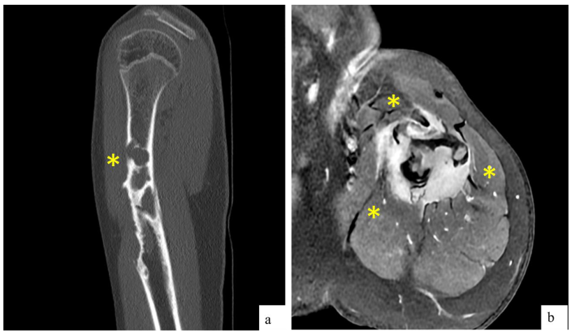
2.1. Clinical and Radiological Findings

A 14-year-old male experienced intense pain in his left arm after a sudden movement while descending a slide at a water park. He sought emergency care and was diagnosed with a pathological fracture of the left humerus. The patient denied any prior symptoms before the accident. An arm sling was applied by orthopedics, and 20 days later, he returned for a follow-up magnetic resonance imaging (MRI) scan.
The MRI revealed a pathological fracture of the proximal humerus with angulation, located over a poorly defined lytic lesion in the proximal diaphysis, measuring approximately 17 × 72 mm. There was a cystic area of 13 mm in its proximal portion, cortical erosion, and intense contrast uptake. The findings suggested a differential diagnosis of fibrous dysplasia (FD), an aneurysmal bone cyst (ABC), Ewing sarcoma (ES), and osteosarcoma (OS). Additionally, there was edema and contrast uptake in the adjacent musculature, which could be secondary to inflammatory changes due to the fracture, although infiltration could not be ruled out (Figure 1). Given these findings, the patient was referred to our center and was admitted for further evaluation.
A core needle biopsy (CNB) was performed. Three cores were obtained, with a diagnosis of “low-grade sarcoma”. A thoracoabdominal–pelvic computed tomography (CT) scan was performed, showing no signs of distant metastasis. After discussion in the sarcoma committee, a decision was made to proceed with radical surgery, including resection and reconstruction with a vascularized fibula graft. Medical Oncology determined that there was no indication for Neoadjuvant Chemotherapy (NCT) or Radiotherapy (NCRT).
During surgery, 13 cm of the humerus was resected along with the coracobrachialis muscle and parts of the deltoid and brachialis anterior muscles. Intraoperative samples were sent to the Pathology Department, confirming free margins. The humerus was reconstructed using a free osteocutaneous fibula flap from the right leg (intramedullary in the proximal humerus and trench technique in the distal humerus). After the surgery, the patient was discharged without complications, with no indication for adjuvant therapy from Medical Oncology.

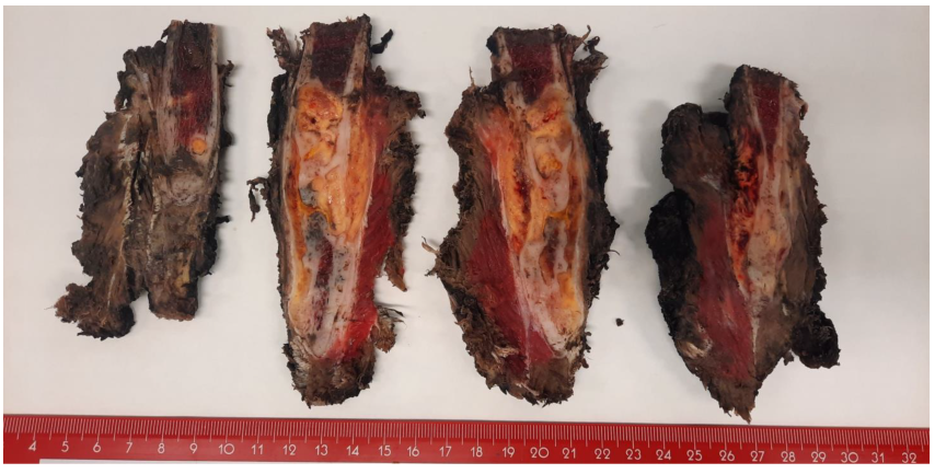
2.2. Macroscopic Findings

The resection specimen of the humerus measured 14 × 5.4 × 4.6 cm. After staining the margins and sectioning the specimen, a poorly defined yellowish lesion measuring 4.7 × 2.3 cm was observed within the medullary cavity, expanding into the surrounding soft tissues. The lesion was located 2.6 cm from the proximal margin, 1.7 cm from the distal margin, and 1 cm from the circumferential margin, all of which were macroscopically free of the lesion (Figure 2).

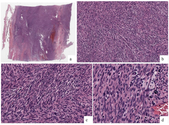
2.3. Microscopic Findings

The lesion exhibited a “fish spine” pattern in the medullary cavity, composed of atypical cells with moderate pleomorphism and medium-to-large size. These cells were arranged in long, highly intertwined fascicles, breaking through the cortical bone and extending into the soft tissues (Figure 3a,b). The nuclei were oval to elongated, hyperchromatic, and granular with nuclear pseudo-inclusions, and the cytoplasm was elongated and eosinophilic (Figure 3c). An inflammatory component, predominantly mononuclear and plasmacytic, was observed between the tumor fascicles and around the blood vessels. There were 5 mitoses in 10 high-power fields (Figure 3d). No bone trabeculae, osteoid formation, or areas of necrosis were present within the lesion.
Based on the histological findings and the results of the previous biopsy, the diagnosis was oriented towards IFS. However, other diagnoses based on age, appearance and location such as low-grade osteosarcoma (LGO), inflammatory myofibroblastic tumor (IMT), malignant peripheral nerve sheath tumor (MPNST), or melanoma were also considered. Taking into account these differential diagnoses, microscopic findings and immunohistochemical stainings were crucial, in addition to Next-Generation Sequencing (NGS).

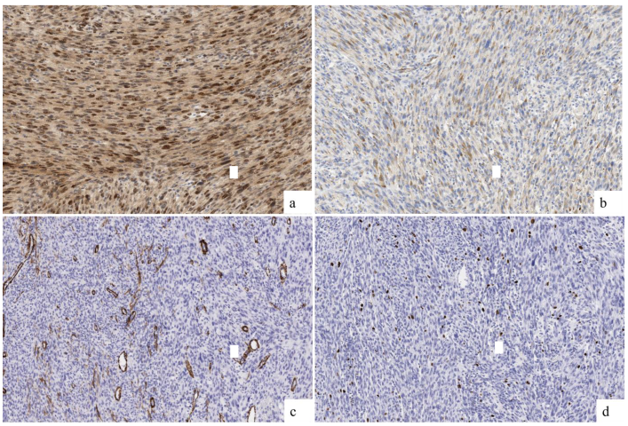
2.4. Immunohistochemical Findings

Weak and focal immunoreactivity was observed for S100. SATB2 expressed a diffuse and intense staining, with focal expression of osteonectin and negativity for caldesmon, Smooth Muscle Actin (SMA), desmin, GFAP, SOX10, MelanA, panTRK, and HMB45. H3K27me3 was not valuable, and the Ki67 index was 7% (Figure 4 and Table 1). According to the Fédération Nationale des Centres de Lutte Contre le Cancer (FNLCC) criteria [7], the score was 4, corresponding to a G2 grade (Dedifferentiation 3/3, mitosis 1/3, and necrosis 0/2). Table 1 includes the antibodies used for diagnosis.

2.5. Fluorescence In Situ Hybridization (FISH) and Next-Generation Sequencing (NGS) Results

Given the suspicion of pediatric fibrosarcoma and the negativity for panTRK, fluorescence in situ hybridization (FISH) was performed for the BRAF gene, revealing an EVI5::BRAF fusion. This rearrangement was later studied by Next-Generation Sequencing (Figure 5), confirming the fusion.
It is important to note that although the tumor expressed both SATB2 and osteonectin, a diagnosis of LGS was ruled out due to the cytomorphological features, lack of osteoid matrix and absence of MDM2 amplification. All these findings supported the IFS diagnosis considering SATB2 and osteonectin expression as an unspecific staining. Additionally, a diagnosis of IMT was ruled out due to the aggressive local behavior of the lesion and the absence of ALK immunexpression and translocation. IFS can also present with a prominent lymphoid inflammatory component [1].
Table 2 includes a clinical and pathological summary of the case.

3. Discussion

Among the BRAF fusions described in IFS are SEPT7::BRAF, SEPT9::BRAF, SEPT11::BRAF, ERC1::BRAF, PDE10A::BRAF, CUX1::BRAF and KIAA1549::BRAF [2,7,8,9]. Kao et al. [9], in his series of Infantile Fibrosarcomas, describes five cases with BRAF fusions in patients of varying ages (from 2 days to 16 years), with three tumors located in the pelvis, one in the T6 vertebra, and another in the retroperitoneum. The tumors exhibited a ‘spindle cell’ morphology with few mitoses and patchy and focal expression for SMA, with an absence of expression for desmin and S100. It is important to note that these tumors are not associated with the upregulation of BRAF mRNA, so the immunohistochemistry of BRAF is not reliable.
EVI5 (Ecotropic Viral Integration Site 5) is a gene that enables GTPase activator activity and small GTPase binding activity, and it is involved in the positive regulation of GTPase activity and retrograde transport from the endosome to the Golgi. It functions as a regulator of cell cycle progression by stabilizing the FBXO5 protein and promoting cyclin-A accumulation during interphase [10]. EVI5 was discovered by X. Liao et al. [11] in 1995 and defined as a common site of retroviral integration in T-cell lymphomas in mice. Since then, it has been described as a regulatory gene involved in both proliferation and metastasis in tumors such as non-small-cell lung carcinoma [12] and hepatocellular carcinoma [13], as well as in neuroblastoma, where chromosomal translocation may influence its origin. [14]. To the best of our knowledge, the EVI5::BRAF fusion has been only described in congenital melanocytic nevi [15].
Kao et al. [9] describes that these “IFS-like” tumors appear in unusual clinical settings, such as older age groups or intra-abdominal locations, unlike classic IFS, which is more common in the early years of life and in the extremities. It is important to consider that the radiological appearance of the tumor is highly variable, potentially presenting as ovoid, nodular, or fascicular, with irregular contrast uptake and poorly defined margins. In this context, core needle biopsy is crucial and enables a definitive diagnosis to guide therapeutic decision-making [16]. The Penning series (2021) includes 14 tumors with BRAF fusions, making it the largest series reported to date. Similar to Kao, these tumors are defined as “IFS-like” [6].
These tumors have similar prognoses, making it complex to determine whether they correspond to variants of IFS or distinct entities. However, in the latest classification of pediatric tumors by the WHO (2022), they are considered as IFS, and BRAF fusions are included alongside the classic NTRK ones [1]. These genetic alterations are also described as oncogenic drivers [6].
It is important to highlight that while tumors harboring the BRAF V600E mutation have shown a good response to anti-BRAF therapy, those with other mutations or fusions have demonstrated resistance or paradoxical activation. Consequently, new clinical trials are currently under development, focusing primarily on MEK/BRAF inhibitors [6]. In recent years, theranostics has emerged as a novel form of precision medicine that integrates diagnostics with therapeutics, aiming to develop more personalized alternatives for each patient through the use of nanoparticles. In both the present and the future, these and other innovative therapeutic alternatives can be applied even in difficult and singular cases, such as the one proposed, extending beyond immunology and genetics [17].
To the best of our knowledge, EVI5::BRAF has not been described in BRAF fusions in IFS yet, making this case the first in the literature. Nevertheless, further studies are needed to define the prognostic features of these emergent BRAF sarcomas. This will not only help to guide diagnosis in cases where the same fusion is detected, but it will also serve as a foundation for the development of new therapeutic targets in the future.

Author Contributions

Conceptualization, F.G. and J.G.-L.; methodology, F.G., J.G.-L. and E.M.-A.; software, L.A.R.-M.; validation L.A.R.-M. and M.C.H.; formal analysis, M.C.H.; investigation, F.G., J.G.-L., L.A.R.-M. and E.M.-A.; resources, L.A.R.-M. and M.A.; data curation, F.G. and J.G.-L.; writing—original draft preparation, J.G.-L.; writing—review and editing, F.G.; visualization, M.A.; supervision, J.V.A.; project administration, J.V.A.; funding acquisition, Not applicable. All authors have read and agreed to the published version of the manuscript.

Funding

This research received no funding.

Institutional Review Board Statement

This study did not require ethical approval.

Informed Consent Statement

Not applicable.

Data Availability Statement

Data is unavailable due to privacy or ethical restrictions.

Conflicts of Interest

The authors declare no conflicts of interest.

Abbreviations

IFSInfantile Fibrosarcoma
ETV6ETS Variant Transcription Factor 6
NTRK3Neurotrophic Receptor Tyrosine Kinase 3
NTRK1Neurotrophic Receptor Tyrosine Kinase 1
NTRK2Neurotrophic Receptor Tyrosine Kinase 2
RETRearranged During Transfection Proto-Oncogene
METMET Proto-Oncogene, Receptor Tyrosine Kinase
RAF1RAF Proto-Oncogene Serine/Threonine-Protein Kinase 1
BRAFB-Raf Proto-Oncogene Serine/Threonine-Protein Kinase
MRIMagnetic Resonance Imaging
FDFibrous Dysplasia
ABCAneurysmal Bone Cyst
ESEwing Sarcoma
OSOsteosarcoma
CNBCore Needle Biopsy
CTComputed Tomography
MRIMagnetic Resonance Imaging
NCTNeoadjuvant Chemotherapy
NCRTNeoadjuvant Radiotherapy
LGOLow-Grade Osteosarcoma
IMTInflammatory Myofibroblastic Tumor
NGSNext-Generation Sequencing
SMASmooth Muscle Actin
FNLCCFédération Nationale des Centres de Lutte Contre le Cancer
FISHFluorescence In Situ Hybridization
SEPT7Septin 7
SEPT9Septin 9
SEPT11Septin 11
ERC1ELKS/RAB6-Interacting/CAST Family Member 1
PDE10Phosphodiesterase 10
CUX1Cut-Like Homeobox 1
EVI5Ecotropic Viral Integration Site 5
WHOWorld Health Organization
MDM2Mouse Double Minute 2 Homolog
ALKAnaplastic Lymphoma Kinase

References

  1. WHO Classification of Tumours Editorial Board. Paediatric Tumours [Internet]. In WHO Classification of Tumours Series, 5th ed.; International Agency for Research on Cancer: Lyon, France, 2022; Volume 7, Available online: https://tumourclassification.iarc.who.int/chapters/44 (accessed on 15 August 2024).
  2. Abualola, R.A.; Al-Zaid, T. KIAA1549-BRAF Gene Fusion Spindle Cell Sarcoma with Infantile Fibrosarcoma-Like Pattern in a Pediatric Patient: A Case Report. Cureus 2024, 16, e51981. [Google Scholar] [CrossRef] [PubMed] [PubMed Central]
  3. Zhou, P.; Liu, W.; Zheng, J.; Zhang, H.; Luo, J. Case report: Primary sarcoma of the mandible with a novel SLMAP-BRAF fusion. Front. Oncol. 2024, 14, 1369046. [Google Scholar] [CrossRef] [PubMed] [PubMed Central]
  4. Cecchi, R.; Guptil, D.; Haslett, N.; Hristov, A.; Bledsoe, J.R.; Tsai, H.; DeWitt, J.; Ferris, S.P. Primary CNS histiocytic sarcoma: Two case reports highlighting a novel MIGA2::BRAF gene fusion and genome-wide DNA methylation profiling results. J. Neuropathol. Exp. Neurol. 2024, 83, 882–886. [Google Scholar] [CrossRef] [PubMed]
  5. Zhang, L.; Zhang, G.; Zheng, H.; Jiang, B.; Ju, Y.; Duan, Q.; An, L.; Shi, H. A rare case of primary central nervous system histiocytic sarcoma harboring a novel ARHGAP45::BRAF fusion: A case report and literature review. Brain Tumor Pathol. 2024, 41, 18–29. [Google Scholar] [CrossRef] [PubMed]
  6. Penning, A.J.; Al-Ibraheemi, A.; Michal, M.; Larsen, B.T.; Cho, S.J.; Lockwood, C.M.; Paulson, V.A.; Liu, Y.J.; Plank, L.; Fritchie, K.; et al. Novel BRAF gene fusions and activating point mutations in spindle cell sarcomas with histologic overlap with infantile fibrosarcoma. Mod. Pathol. 2021, 34, 1530–1540. [Google Scholar] [CrossRef] [PubMed]
  7. Gourmel, A.; Rouette, A.; Benlimame, N.; El-Jalbout, R.; Dubé, M.; Théorêt, Y.; Piché, N.; Labonté, S.; Sinnett, D.; Cellot, S.; et al. Durable Response to Trametinib in an Infant with ERC1-BRAF Infantile Fibrosarcoma-Like Tumor: A Case Report and Literature Review of BRAF-Altered Infantile Fibrosarcoma-Like Tumors. JCO Precis. Oncol. 2022, 6, e2200200. [Google Scholar] [CrossRef] [PubMed]
  8. Hughes, C.E.; Correa, H.; Benedetti, D.J.; Smith, B.; Sumegi, J.; Bridge, J. Second Report of PDE10A-BRAF Fusion in Pediatric Spindle Cell Sarcoma with Infantile Fibrosarcoma-Like Morphology Suggesting PDE10A-BRAF Fusion Is a Recurrent Event. Pediatr. Dev. Pathol. 2021, 24, 554–558. [Google Scholar] [CrossRef] [PubMed] [PubMed Central]
  9. Kao, Y.C.; Fletcher, C.D.M.; Alaggio, R.; Wexler, L.; Zhang, L.; Sung, Y.S.; Orhan, D.; Chang, W.C.; Swanson, D.; Dickson, B.C.; et al. Recurrent BRAF Gene Fusions in a Subset of Pediatric Spindle Cell Sarcomas: Expanding the Genetic Spectrum of Tumors with Overlapping Features with Infantile Fibrosarcoma. Am. J. Surg. Pathol. 2018, 42, 28–38. [Google Scholar] [CrossRef] [PubMed] [PubMed Central]
  10. Stelzer, G.; Rosen, R.; Plaschkes, I.; Zimmerman, S.; Twik, M.; Fishilevich, S.; Iny Stein, T.; Nudel, R.; Lieder, I.; Mazor, Y.; et al. The GeneCards Suite: From Gene Data Mining to Disease Genome Sequence Analyses. Curr. Protoc. Bioinform. 2016, 54, 1.30.1–1.30.33. [Google Scholar] [CrossRef] [PubMed]
  11. Liao, X.; Buchberg, A.M.; Jenkins, N.A.; Copeland, N.G. Evi-5, a common site of retroviral integration in AKXD T-cell lymphomas, maps near Gfi-1 on mouse chromosome 5. J. Virol. 1995, 69, 7132–7137. [Google Scholar] [CrossRef] [PubMed] [PubMed Central]
  12. Cai, T.; Zhou, J.; Zeng, Y.; Du, W.; Zhang, Y.; Liu, T.; Fu, Y.; Huang, J.; Qian, Q.; Zhu, J.; et al. EVI5 is an oncogene that regulates the proliferation and metastasis of NSCLC cells. J. Exp. Clin. Cancer Res. 2020, 39, 84. [Google Scholar] [CrossRef] [PubMed]
  13. Tang, J.; Ou, J.; Xu, C.; Yi, C.; Xue, F.; Xu, L.; Lai, F.; Tang, J.; Li, S.; Kang, T.; et al. EVI5 is a novel independent prognostic predictor in hepatocellular carcinoma after radical hepatectomy. Oncol. Rep. 2017, 38, 2251–2258. [Google Scholar] [CrossRef] [PubMed]
  14. Vandepoele, K.; Andries, V.; van Roy, F. The NBPF1 promoter has been recruited from the unrelated EVI5 gene before simian radiation. Mol. Biol. Evol. 2009, 26, 1321–1332. [Google Scholar] [CrossRef] [PubMed]
  15. Martin, S.B.; Polubothu, S.; Bruzos, A.L.; Kelly, G.; Horswell, S.; Sauvadet, A.; Bryant, D.; Zecchin, D.; Riachi, M.; Michailidis, F.; et al. Mosaic BRAF Fusions Are a Recurrent Cause of Congenital Melanocytic Nevi Targetable by MAPK Pathway Inhibition. J. Investig. Dermatol. 2024, 144, 593–600.e7. [Google Scholar] [CrossRef] [PubMed]
  16. Sedaghat, S.; Schmitz, F.; Krieger, A.; Reichardt, B. Appearance of recurrent adult fibrosarcoma of the soft tissue and loco-regional post-treatment changes on MRI follow-up. Eur. J. Plast. Surg. 2021, 44, 97–102. [Google Scholar] [CrossRef]
  17. Basirinia, G.; Ali, M.; Comelli, A.; Sperandeo, A.; Piana, S.; Alongi, P.; Longo, C.; Di Raimondo, D.; Tuttolomondo, A.; Benfante, V. Theranostic Approaches for Gastric Cancer: An Overview of In Vitro and In Vivo Investigations. Cancers 2024, 16, 3323. [Google Scholar] [CrossRef] [PubMed]
Figure 1. (a) CT findings: Sagital reconstruction showing diaphyseal lytic lesion with cortical erosion, causing bone remodeling. (b) MRI findings: Post-contrast transverse T1 sequence showing surrounding soft tissue mass with homogeneous and intense contrast uptake. In both images, the lesion is delineated between the yellow asterisks.
Ijms 26 01182 g001
Figure 2. Macroscopic findings. A poorly defined yellowish lesion measuring 4.7 × 2.3 cm was observed within the medullary cavity, expanding into the surrounding soft tissues.
Ijms 26 01182 g002
Figure 3. Microscopy findings. (a) The lesion occupied the medullary cavity, breaking through the cortex and extending into the surrounding soft tissues (HE; 4×). (b) Diffuse ‘herringbone’ pattern (HE; 10×). (c) Elongated and interwoven fascicles composed of atypical cells with moderate pleomorphism and medium size, with hyperchromatic nuclei and poorly defined cytoplasm. (HE; 20×). (d) Isolated mitoses were observed, with a notable lymphoid inflammatory component. No osteoid formation and areas of necrosis were present within the lesion. (HE, 40×).
Ijms 26 01182 g003
Figure 4. Immunohistochemical findings. (a) Diffuse positivity for SATB2 (20×). (b) Weak and focal positivity for osteonectin (20×). (c) Absence of expression for Smooth Muscle Actin (20×). (d) Ki67 index was established in 7% (20×).
Ijms 26 01182 g004
Figure 5. FISH and NGS results. (a) FISH shows BRAF fusion. (b) NGS shows the fusion point between EVI5 and BRAF. Made with the SeqOne® in vitro diagnostic medical device.
Ijms 26 01182 g005
Table 1. Main antibodies and staining pattern used.
AntibodySourceCloneStaining Pattern
S100DakoPolyclonalDiffuse (Nuclear)
SATB2ZetaZR167Diffuse (Nuclear)
CaldesmonDakoh_CDNegative
Smooth Muscle ActinDakoHHF-35Negative
DesminDakoD33Negative
OsteonectinLeicaG-15-G12Focal (Cytoplasmic)
GFAPDakoPolyclonalNegative
SOX10BiocareBC34Negative
MelanADakoA103Negative
HMB45DakoHMB-45Negative
H3K27me3Gennova/BiocareC36B11Not evaluable
Ki67DakoMIB-17%
ALKDakoALK1Negative
Table 2. Clinical and pathological summary. SMA: Smooth Muscle Actin. Mt: metastases.
AgeSexSiteS100SMASATB2Ki67MtGeneticsTreatmentOutcome
14MaleLeft humerusWeak, focalNegativeIntense, difusse7%NoEVI5::BRAFResection and reconstructionNo evidence of disease
Disclaimer/Publisher’s Note: The statements, opinions and data contained in all publications are solely those of the individual author(s) and contributor(s) and not of MDPI and/or the editor(s). MDPI and/or the editor(s) disclaim responsibility for any injury to people or property resulting from any ideas, methods, instructions or products referred to in the content.

