Article

Altered Neuroinflammatory Transcriptomic Profile in the Hippocampal Dentate Gyrus Three Weeks After Lateral Fluid Percussion Injury in Rats

1
Department of Neurosurgery, University of Cincinnati College of Medicine, Cincinnati, OH 45267, USA
2
Department of Neurology and Rehabilitation Medicine, University of Cincinnati College of Medicine, Cincinnati, OH 45267, USA
*
Author to whom correspondence should be addressed.
These authors contributed equally to this work.
Int. J. Mol. Sci. 2025, 26(18), 9140; https://doi.org/10.3390/ijms26189140
Submission received: 31 July 2025 / Revised: 10 September 2025 / Accepted: 12 September 2025 / Published: 19 September 2025
(This article belongs to the Special Issue Molecular Advances in Neurologic and Neurodegenerative Disorders)

Abstract

Traumatic brain injury (TBI) is a major source of disability worldwide, with cognitive and memory deficits being pervasive after injury. The hippocampus, a major structure involved in learning and memory, is particularly vulnerable to TBI, and cellular dysfunction within the hippocampal dentate gyrus is believed to be a major contributor to cognitive deficits after TBI. However, there is little known about the transcriptomic changes occurring directly within the dentate gyrus at subacute-to-chronic timepoints after TBI. To address this, we performed bulk RNA sequencing and single-nucleus RNA sequencing of the isolated dentate gyrus three weeks after lateral fluid percussion injury in male rats. We report here that there is evidence of an ongoing neuroinflammatory response marked by increased neuroinflammatory genes that implicate various neuroinflammatory pathways that are associated with a subset of microglia and astrocyte populations.

1. Introduction

Traumatic brain injury (TBI) is estimated to affect sixty-nine million people annually, with long-term implications regarding cognitive abilities after injury [1,2]. Clinically, cognitive and memory impairments are commonly reported after TBI [3,4,5] and are recapitulated in pre-clinical models of TBI [6,7,8]. Cognitive dysfunction is associated with TBI-induced changes to the hippocampus [9], and the hippocampal dentate gyrus is particularly vulnerable to TBI [10,11,12]. Given the dentate gyrus’s role in cognition, learning, and memory (reviewed in [13]), dentate gyrus dysfunction likely plays an important role in the cognitive and memory deficits observed after TBI. Early after TBI, there is a loss of hippocampal neural progenitor cells within the subgranular zone of the dentate gyrus, followed by increased neurogenesis from 24 h to 7 days post-injury [10,14,15,16], with reduced astrogliogenesis [15].
Under healthy conditions, glial cells play important roles within the dentate gyrus. Retention and integration of new-born neurons are mediated by local microglia and astrocytes [17,18]. Microglia are necessary for survival of neuronal stem cells [19], and interplay between microglia and astrocytes facilitates synaptic pruning [20]. In the context of TBI, astrocytes and microglia within the dentate gyrus demonstrate a robust response to TBI associated with the neuroinflammatory response to injury [21,22,23]. Neuroinflammatory changes are well documented after brain injury and are believed to be a key driver of long-term cognitive dysfunction (reviewed in [24]). Hippocampal transcriptional changes associated with an ongoing immune response occur at early, subacute, and chronic timepoints post-injury [25,26], and transcriptional changes in animals have been correlated with long-term TBI-associated neurodegenerative sequelae [27].
Researchers have long recognized the importance of the dentate gyrus after TBI, and, in the past decade, a plethora of publications have examined the TBI-induced transcriptional changes exhibited in animal brains, and specifically the hippocampus [28,29,30,31,32]. However, the majority of such studies focus on the acute phase after injury, mostly obtaining data within 24 h to 7 days post-injury. Importantly, little is known about changes in gene expression at timepoints after 2 weeks post-injury, especially in the isolated dentate gyrus [15,33].
Given the enduring effects of TBI on cognitive function, we aimed to utilize both bulk and single-nucleus RNA sequencing techniques to examine the transcriptional changes in the isolated dentate gyrus at three weeks after TBI. To our knowledge, this is the first single-nucleus study to investigate the whole tissue dentate gyrus specifically after rat TBI, providing a greater level of regional resolution within the injured hippocampus. Briefly, our data suggest altered gene expression associated with an ongoing neuroinflammatory response within specific populations of microglia and astrocytes at three weeks post-TBI.

2. Results

Male Sprague-Dawley rats underwent a craniectomy, and, three days afterward, underwent a lateral fluid percussion injury (LFPI) to induce TBI. As an additional confirmation of injury, spontaneous righting reflex time (RRT) was assessed. Brain-injured rats had significantly longer RRT compared to sham when compared with a two-tailed unpaired t-test (p = 0.0146, t = 3.221, df = 7; Figure 1). The rats were euthanized three weeks after injury, and the dentate gyrus was dissected out and processed for bulk RNA-seq (ntotal = 6) or single-nucleus RNA-seq (snRNA-seq; ntotal = 4; Figure 1).

2.1. Bulk RNA Sequencing Reveals a Neuroinflammatory Response in the Dentate Gyrus Three Weeks After LFPI

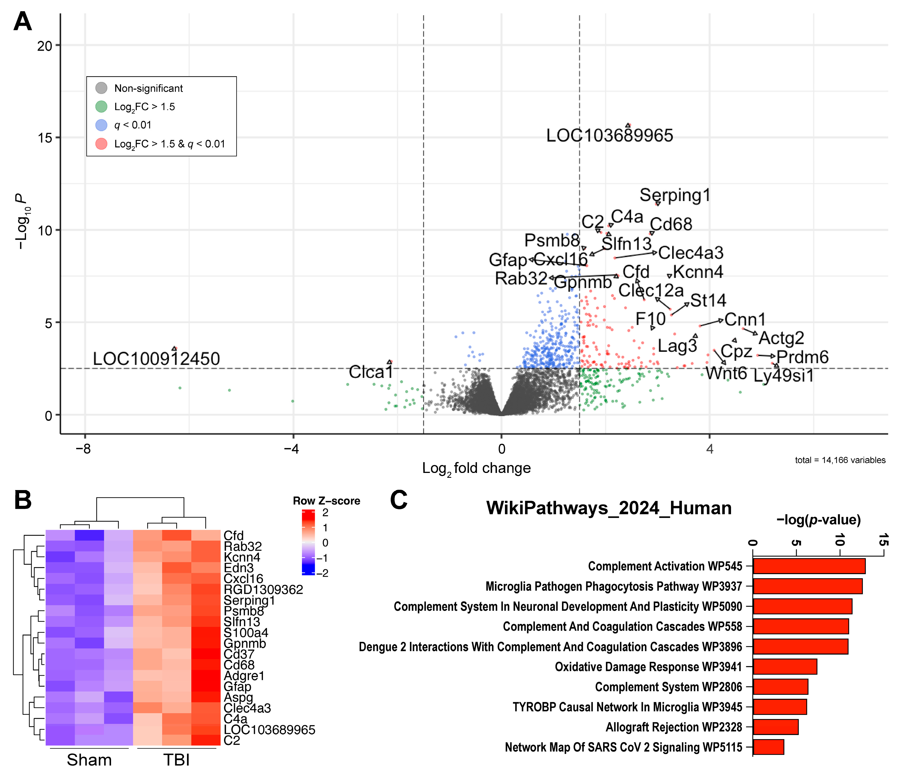
As a result of our bulk RNA sequencing, a total of fourteen-thousand-three-hundred-sixty-eight genes were identified, with four-hundred-seventy-six having a q-value < 0.01 (Benjamini and Hochberg). Of these, one-hundred-twenty-eight genes were differentially expressed (q < 0.01; Log2FC > 1.5). Of these differentially expressed genes (DEGs), one-hundred-twenty-six were upregulated in TBI and two were downregulated (Figure 2A). Hierarchical clustering with complete linkage shows that injured and sham rats cluster separately, and that rats within the same injury group are similar to each other (Figure 2B). Notably, genes associated with inflammatory processes were upregulated in brain-injured rats, with the top three DEGs based on p-value being LOC103689965 (p = 3.21 × 10⁻¹³, Log2FC = 2.47), Serping1 (serpin family G member 1; q = 3.11 × 10−09, Log2FC = 2.97), and C4a (q = 3.10× 10−08, Log2FC = 2.06), which encode for the proteins ‘Complement C4-like’, ‘C1 inhibitor’, and the acidic form of ‘Complement Factor 4’. The two downregulated genes, LOC10091245 (q = 0.00178, Log2FC = −6.25) and Clca1 (q = 0.005616, Log2FC = −2.12), encode for SMCO3 and CLCA1 (Chloride Channel Accessory 1), respectively. While SMCO3’s function has not been well characterized, Clca1 is of potential interest as it has been implicated in glutamate-induced cell death within the hippocampus [34]. Under ischemic conditions, extrasynaptic N-methyl-D-aspartate (NMDA) activation increases Clca1 gene expression within hours and has been shown to induce cell death in vivo [35]. The implications of the downregulation of Clca1 gene expression three weeks after LFPI remain to be determined.
A pathway analysis through Enrichr using the “WikiPathways 2024 Human” dataset implicated multiple complement-related pathways. Of the top 10 pathways identified by Enrichr, half are directly associated with complement signaling (Table 1). Additionally, multiple complement genes are implicated as part of the “Microglia Pathogen Phagocytosis pathway”. These findings suggest an ongoing inflammatory response within the dentate gyrus at three weeks after injury.

2.2. A Subset of Dentate Gyrus Microglia Express Genes Associated with an Activated Phenotype

Because bulk RNA sequencing does not provide information on the cellular source of observed transcriptional changes, we chose to perform additional single-nucleus RNA sequencing experiments to identify the sources of neuroinflammation in the dentate gyrus. Due to the prominent change in inflammatory-related genes observed in our bulk RNA sequencing, we chose to focus our single-nucleus RNA sequencing data analysis on microglia and astrocytes as these were the most likely source of the observed changes in complement-related genes [36,37]. Under pathological conditions, both microglia and astrocytes have been implicated in clearing debris [38], synaptic remodeling [39,40], and progressive neurodegeneration (reviewed in [41,42]).
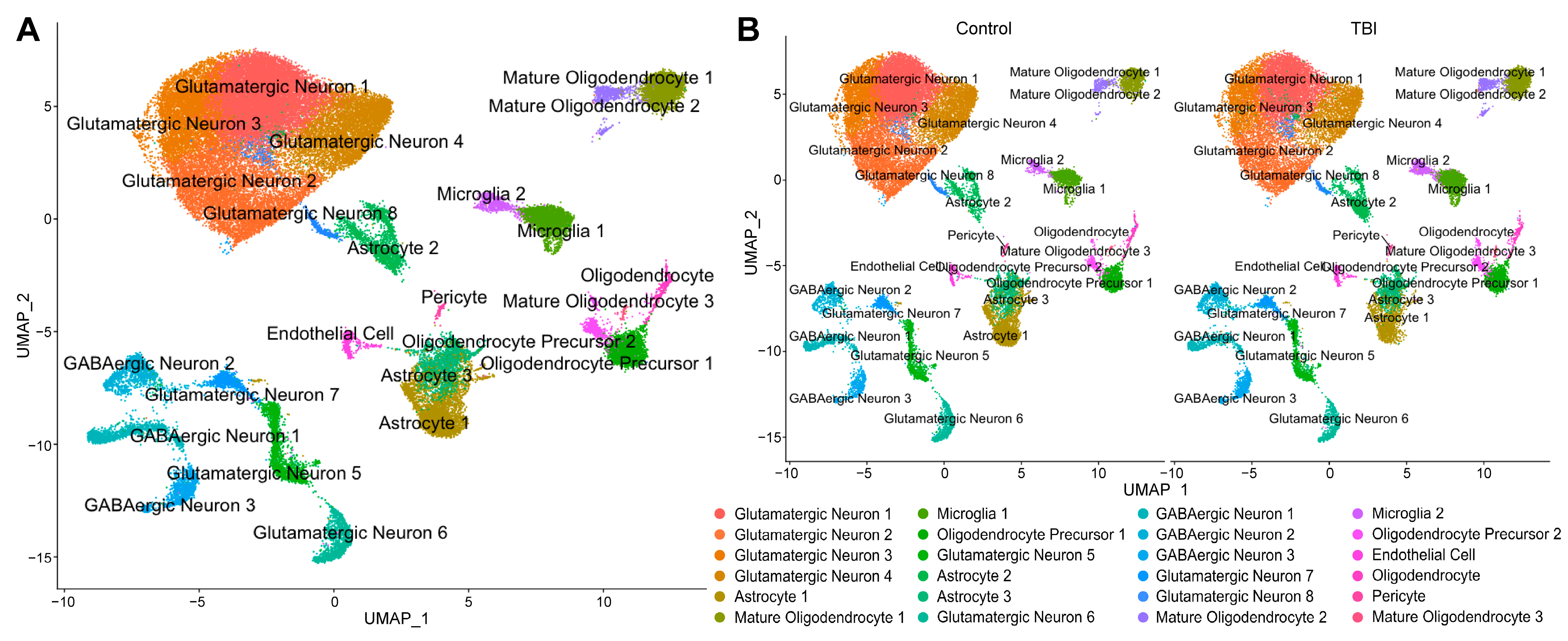
Prior to quality control and filtering (detailed below), 55,863 nuclei were present in our samples, and, afterwards, 55,123 nuclei were utilized for analysis. A Uniform Manifold Approximation and Projection (UMAP) graph was generated using Seurat, and 24 distinct clusters were identified (Figure 3A). Using the online tool, “Annotation of Cell Types” (ACT) [43], clusters were labeled as astrocytes, microglia, oligodendrocytes, oligodendrocyte precursor cells, mature oligodendrocytes, pericytes, endothelial cells, GABAergic neurons, and glutamatergic neurons. Two clusters were identified as microglia, and three clusters were identified as astrocytes. When the UMAP was split by injury condition (Figure 3B), there were some appreciable differences in the number and distribution of nuclei within the microglial and astrocyte clusters (Table A1). The TBI animals had a greater number of nuclei associated with both microglial clusters (Microglia 1 and Microglia 2), while the astrocyte nuclei were lower in TBI animals in Astrocyte 1 and 3 but were higher in TBI animals in Astrocyte 2 when compared to sham.
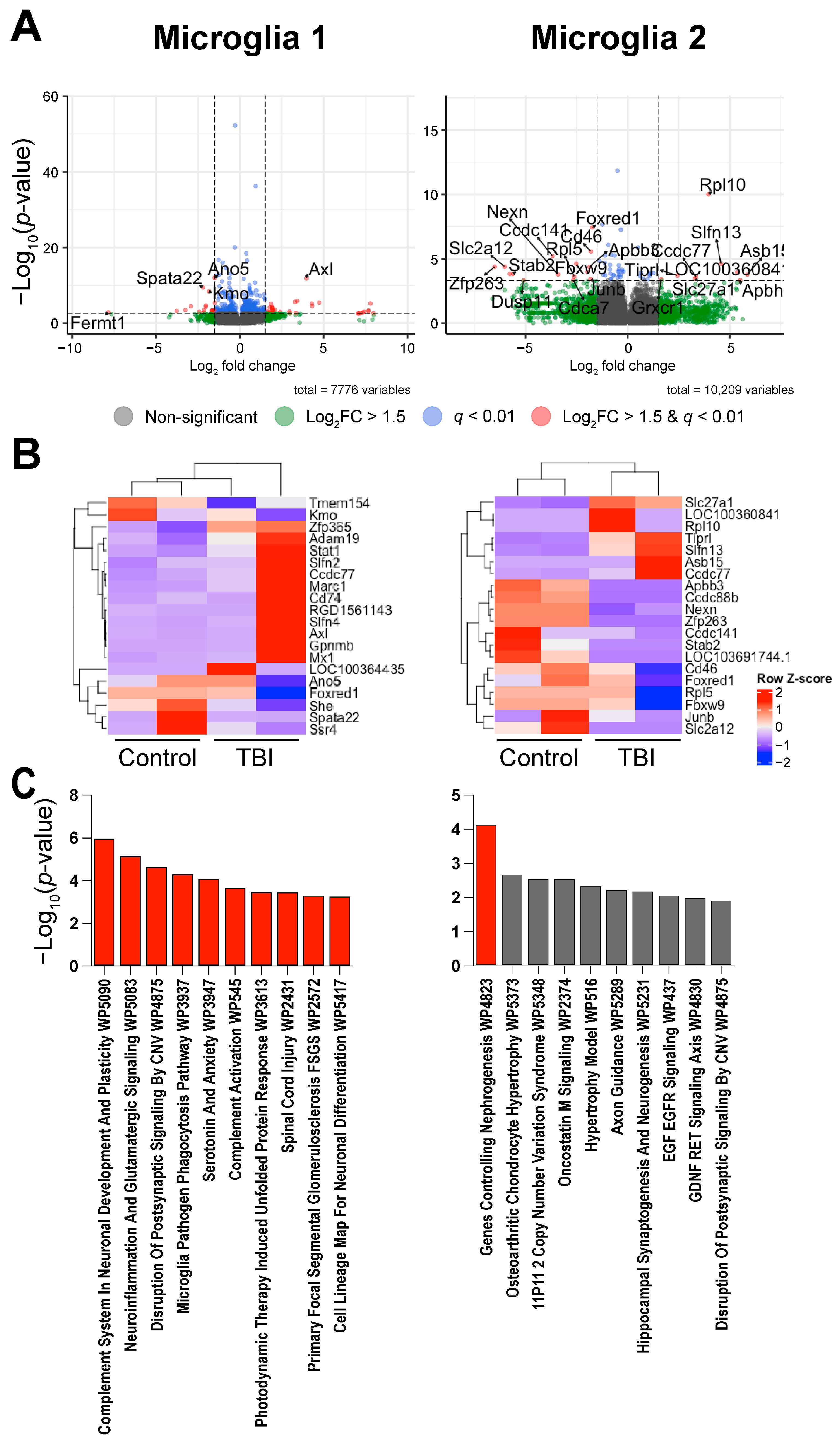
Differential expression analysis between the control and TBI rats revealed that fifty-four genes were differentially expressed in Microglia 1 (thirty-four upregulated and twenty downregulated in TBI; Table 2) and twenty-four genes in Microglia 2 (nine upregulated and fifteen downregulated in TBI). The top five DEGs based on p-value were Ano5, Axl, Spata22, Kmo, and Slfn4 for Microglia 1 and Rpl10, Foxred1, Cd46, Ccdc141, and Ccdc88b for Microglia 2. A hierarchical clustering analysis by complete linkage clustering (Figure 4) did not show clear clustering between the injury and control groups for Microglia 1 but did cluster by injury condition for Microglia 2. Similar to what was observed in our bulk signaling, pathway analysis shows that the DEGs associated with Microglia 1 are enriched for complement signaling. The most significant pathway (Table 3) identified was “Complement System in Neuronal Development and Plasticity” (p = 1.03 × 10⁻⁶). However, when looking at the complement-associated DEGs, it is evident that several complement genes are decreased in our TBI animals compared to control animals, in opposition to what was observed in our bulk RNA sequencing. This included a reduction in the C1qa, C1qb, and C1qc components of the C1q subunit that helps form the C1 complex and is associated with classical complement signaling [44]. The second most significant is “Neuroinflammation and Glutamatergic Signaling” (p = 6.59 × 10⁻⁶). Only one pathway was found to be significant in the Microglia 2 cluster after multiple testing corrections, “Genes controlling Nephrogenesis” (p = 6.85 × 10⁻⁵).
Despite the discrepancy between our bulk and single-nucleus data, several inflammatory-associated genes were upregulated in Microglia 1, suggesting that there is still an inflammatory response present within the dentate gyrus at three weeks post-TBI. These genes include three genes that are associated with reactive microglia: Axl (Log2FC = 3.97, p = 1.41 × 10⁻¹²), Cd74 (Log2FC = 1.84, p = 7.92 × 10⁻⁶), and Stat1 (Log2FC = 1.77, p = 7.10 × 10⁻⁵) [45,46,47].
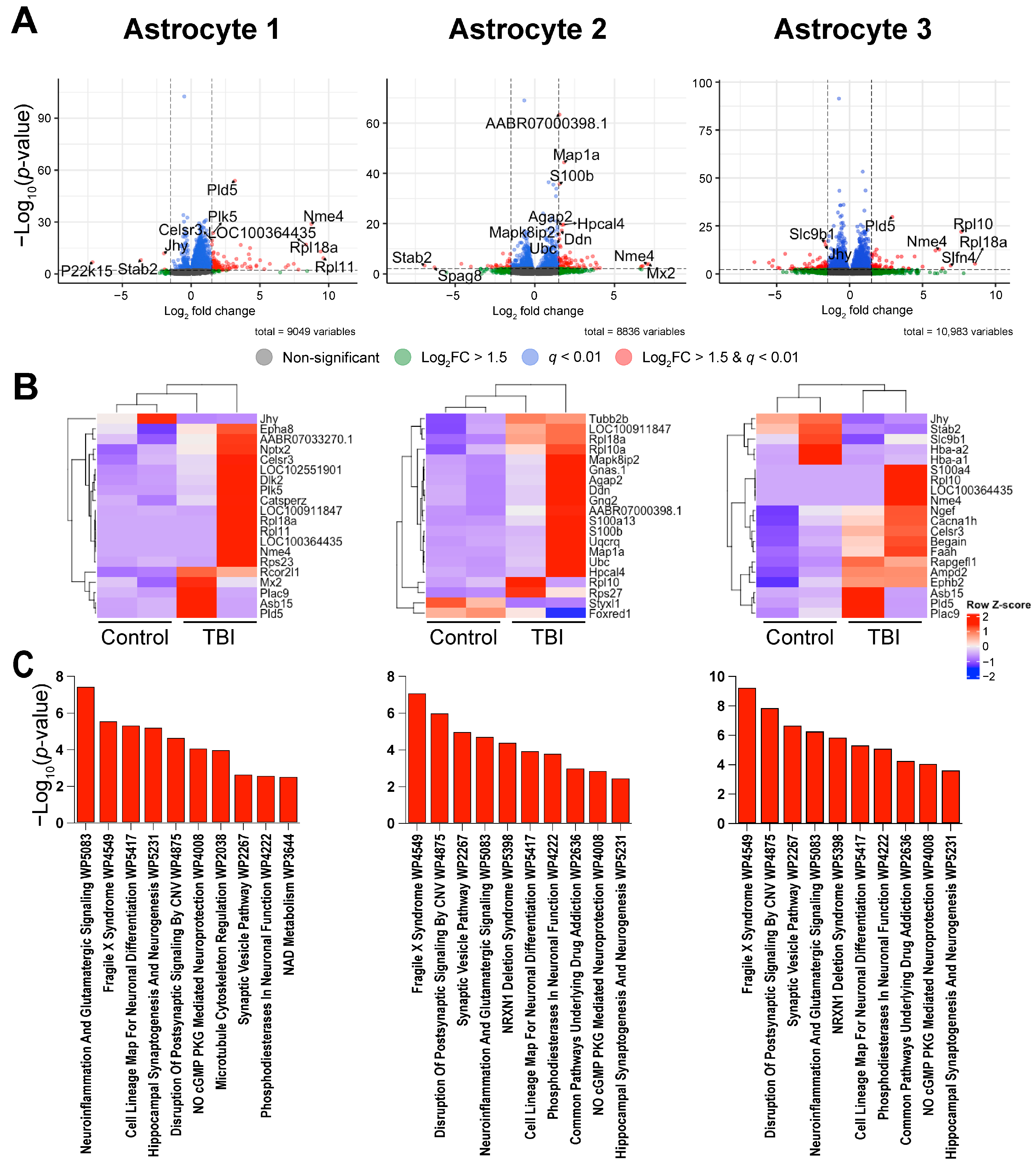
For the astrocyte clusters (Figure 5), one-hundred-ten DEGs were identified in Astrocyte 1 (one-hundred-five upregulated and five downregulated; Table 4), one-hundred-four DEGs were identified in Astrocyte 2 (seventy-six upregulated and twenty-eight downregulated), and one-hundred-thirty-five DEGs were identified in Astrocyte 3 (sixty upregulated and seventy-five downregulated). A hierarchical clustering analysis using complete linkage clustering (Figure 5B) did not show clear clustering between injury groups in the clusters for Astrocyte 1 and Astrocyte 2 but did cluster by injury condition in Astrocyte 3. A pathway analysis for all three astrocyte clusters suggests changes in genes associated with neuroinflammation and altered synaptic signaling (Table 5).
There was a disproportionate amount of upregulated DEGs in our bulk RNA sequencing data compared to most microglial and astrocyte clusters in our single-nucleus dataset. This skew towards upregulated genes in our bulk data may be due to our exclusion of neuronal cell populations from our complete analysis. As post-mitotic cells, neurons have high transcriptional activity compared to other cell types in the brain [48]. This may also reflect changes in the abundance of specific mRNA transcripts outside the nucleus due to post-translational modifications that can influence mRNA abundance and longevity [49]. Alternatively, it may be due to technical differences between bulk and single-nucleus RNA sequencing. Bulk sequencing averages out the expression of genes across cell types, and as such the differential regulation of the same gene between different cell types can eliminate changes that are appreciable at single-cell resolutions. Additionally, single-nucleus RNA sequencing generally captures a smaller subset of available RNA because it does not include RNAs found outside of the nucleus [50].
Taken together, there is evidence for an overall increase in neuroinflammation-related genes despite lacking complete accordance on the pathways that are increased at three weeks after TBI. Ultimately, the differences in these datasets underscore the complementary nature of bulk RNA sequencing and single-nucleus sequencing technologies in untangling the complex neuroinflammatory response to TBI.

3. Discussion

Given the high incidence of TBI and its enduring implications on cognitive function, our aim was to assess the transcriptional changes that cells in the dentate gyrus of the hippocampus undergo at three weeks after TBI. Briefly, our data showed a significant upregulation in the transcription for components of the complement system in the bulk-sequenced specimens. Due to the overrepresentation of inflammatory pathways in our bulk sequencing, we chose to focus on microglial and astrocyte clusters when analyzing our single-nucleus RNA-seq dataset. The microglia DEGs were enriched for pathways associated with phagocytosis, while the astrocytes exhibited enrichment in pathways for neuroinflammation and glutamatergic signaling as well as hippocampal synaptogenesis and neurogenesis.
Our bulk RNA sequencing findings are consistent with a number of studies in clinical and animal models that demonstrated a role for complement activation after TBI. Under healthy conditions, the complement system is a component of the innate immune system, which comprises a family of proteins that activate one another in an amplification cascade, usually culminating in inflammatory signaling, opsonization, or removal of foreign bodies or cellular debris [51]. The complement system performs these roles in the brain, protecting it from infection [52] and removing fragments of apoptotic cells as well as aggregated proteins [53,54]. More interestingly, it contributes to synaptic pruning by way of astrocytes, which express the complement proteins, opsonizing specific synapses for phagocytosis by microglia [36,37,55]. Additionally, it plays a role in neurogenesis, with some mediators (C3 and C5) encouraging maturation and migration and others (C3d and CR2) inhibiting them [56,57,58].
In contrast, the complement system takes on a more pro-inflammatory role in the brain after trauma, contributing to secondary injury. Bellander et al. found increased presence of complement components C1q, C3b, C3d, and the membrane attack complex (MAC) composed of C5b-9 in the penumbra around the contused brain tissue [59], whereas Kossmann et al. found increased levels of C3 and factor B (fB) in the CSF of TBI patients [60]. Moreover, numerous studies in animal models have shown supporting findings by manipulating the complement system after TBI. Kaczorowski et al. inhibited the system with the use of soluble CR1 (sCR1) and found a decrease in neutrophil infiltration into brain tissue [61]. Rancan et al. showed that transgenic mice expressing a soluble complement inhibitor (sCrry) experienced less neurologic impairment after TBI compared to wild-type mice [62]. Leinhase et al. built on this by injecting brain-injured mice with recombinant sCrry, finding that inhibiting the complement system in this way allowed for decreased tissue destruction in the hippocampus and better neurologic function [63], showing great promise for this system as a potential therapeutic target after TBI.
Nevertheless, with the rise of sequencing information technologies in the recent past, there is great utility in examining these system dynamics as they occur without experimental intervention, leaving room to uncover their molecular and cellular interactions more broadly. This is of particular interest as the complement system has been shown to effect change by interacting with astrocytic and microglial cell populations [36,37,54,55,64]. Indeed, transcriptomic studies, focusing mostly on the acute phase after TBI, have found increases in astrocytic and microglial cell populations along with upregulation of inflammatory pathways, including chemokine and interleukin signaling [28,65]. A similar pattern of activation and inflammatory function was detected by Arneson et al., who also interestingly uncovered an increase in calcium/calmodulin signaling by astrocytes, a finding mirrored by our data in which all three astrocyte groups showed upregulation of the Camk2a and Camk2b genes [31]. Further linking astrocytic and microglial interactions with complement activation, Zheng et al. found communication between microglia and endothelial cells via components of the complement system (C1qa-Cd93) [30]. Other studies found significant amplification of transcribed genes that were flagging pathway terms, including “complement and coagulation cascades” and “innate immunity”, in their analyses [64,66]. One of note even identified this upregulation of complement at a timepoint closer to our own, 14 days after injury, and yet another at much later timepoints up to 2 years after injury [65,67].
Interestingly, while the list of DEGs from our Microglia 1 single-nucleus cluster was significantly enriched for complement-related genes, several of the associated genes were downregulated compared to our control animals. Specifically, genes associated with the components of the C1q subunit of the C1 complex were downregulated, suggesting that the classical complement pathway was reduced in Microglia 1 nuclei. At one day and two weeks following LFPI, Catta-Preta et al. assessed the transcriptional changes within the rat hippocampus and found that a subset of genes associated with inflammatory pathways were upregulated at both timepoints, while others had a delayed upregulation [26]. In that study, several genes relating to complement signaling were persistently upregulated at both timepoints, while other complement genes were associated with delayed increase, including C4a, which we observed in our bulk sequencing but not our single-nucleus RNA sequencing. This study utilized bulk RNA sequencing, making it most comparable to our bulk RNA sequencing dataset and suggesting that complement signaling may be increased persistently up to three weeks post-injury.
Our results show the prevalence of complement activation in our bulk-sequenced data and are consistent with the literature. Our exploration of microglial and astrocytic activation in the single-nucleus-sequenced data provide further evidence of neuroinflammation, but either shows a downregulation or lacks many of the genes associated with complement activation that were observed in our bulk RNA sequencing. This discrepancy may be due to the differences between the two sequencing technologies [68,69] or typical localization of mRNA transcripts, but it is also possible that the observed changes in complement could be attributed to an unexpected cell type.
Delving into the astrocytic nuclei changes shows significant enrichment in pathways such as “neuroinflammation and glutamatergic signaling” but also in “hippocampal synaptogenesis and neurogenesis” by way of upregulation of genes including Camk2a, Camk2b, Nrxn1, and Ptpn6. This becomes more intriguing in the context of some transcriptomic studies after TBI, one of which found increased activity of the “innate immunity” pathways but also in those for “axon guidance”, and another found Nup62 responsible for increasing cell division [29,32]. Additionally, transcriptional changes occurring in the chronic setting after TBI at 30 and 90 days post-injury, as noted by Makinde et al., were associated with pathways for “synaptic plasticity” and “regulation of long-term synaptic potentiation” [70]. Indeed, investigating some of our implicated genes shows that numerous studies have implicated Camk2a, Camk2b, and Nrxn1 in neurogenesis and neuroplasticity through neuritic branching, pruning, and assembly [71,72,73,74,75]. Further, these astrocytic changes could be linked to complement upregulation through the overexpression of Ptpn6 in our data, which was found to be linked to C1s [76], and to neuroplasticity with the overexpression of the closely related PTPN5 [70].
In our Astrocyte 2 cluster, the genes Map1a and Mapk8ip2 were upregulated in the TBI animals compared to sham. These genes are important for microtubule maintenance [77,78]. However, microtubule changes in astrocytes remain relatively poorly understood [79]. In TBI-associated neurodegenerative pathologies, microtubule disturbances often occur with tau-related changes in neurons, but glial tau pathology has been observed in hippocampal astrocytes [80,81]. Tau-related changes can happen within one day after TBI in tauopathy mouse models [82], and accumulated tau has been shown to be a potent immune target and contribute to synapse loss [83].
We noted in our hierarchical clustering of Microglia 1 (Figure 4B) and Astrocytes 1 and 2 (Figure 5A,B) that the TBI animals did not cluster together with complete linkage clustering. This may be due to outliers present within our datasets as complete linkage clustering utilizes the maximum dissimilarity value between clusters and therefore is vulnerable to outliers [84]. However, it could also be due to variability in TBI severity.
We recognize that there are limitations to the present study. Notably, our results were obtained from a small group of animals. Only male rats were utilized, so the generalizability to female rats may be limited. Additionally, for our single-nucleus RNA sequencing data, we only focused on changes to microglia and astrocytes due to the prominent change in inflammatory-related genes observed in our bulk RNA sequencing data. While we chose to focus on the two microglial populations that we identified, it is necessary to point out that single-nucleus RNA sequencing may lack the sensitivity to observe all the microglial transcriptional states relevant to TBI-induced neuroinflammation [85]. There are likely important changes occurring in the other cell populations that we have identified, and differentially expressed genes from all clusters can be found in the Supplementary Materials. Neuronal populations can likely be further localized within the dentate gyrus [86], but this was outside the scope of our study. Furthermore, there was a disproportionate amount of upregulated DEGs in our bulk RNA sequencing data compared to most microglial and astrocyte clusters in our single-nucleus dataset. This skew towards upregulated genes in our bulk data may be due to our exclusion of neuronal cell populations from our complete analysis. As post-mitotic cells, neurons have high transcriptional activity compared to other cell types in the brain [48]. This may also reflect changes in the abundance of specific mRNA transcripts outside the nucleus due to post-translational modifications that can influence mRNA abundance and longevity [49]. Alternatively, it may be due to technical differences between bulk and single-nucleus RNA sequencing. Bulk sequencing averages out the expression of genes across cell types, and as such differential regulation of the same gene between different cell types can eliminate changes that are appreciable at single-cell resolutions. Additionally, single-nucleus RNA sequencing generally captures a smaller subset of available RNA because it does not include RNAs found outside of the nucleus [50]. The dataset is publicly available for further investigation at the Gene Expression Omnibus (GSE307917), and the differentially expressed genes from this study are found in the Supplementary Materials.
To our knowledge, only a single other study has assessed changes to the isolated dentate gyrus after TBI. Bielefeld et al. assessed nestin-positive cells that were isolated from the dentate gyrus 15 days post-injury by flow cytometry followed by single-cell RNA sequencing, with a primary focus on neuronal stem cell differentiation into astrocytes and neurons in male mice [15]. This study found that TBI switched neural stem cell differentiation to favor neurogenesis over astrogliogenesis, in opposition to what occurs in the healthy brain [15]. Our study utilized whole tissue from the isolated dentate gyrus without prior flow sorting. Despite our recognized shortcomings, we believe that our findings are congruent with other studies that used non-sequencing techniques, and that our study adds further evidence for prolonged neuroinflammatory changes within the dentate gyrus after TBI.
In conclusion, the aim of our study was to assess the transcriptional changes that occur in the dentate gyrus at three weeks after injury both at the global and cellular levels. Our data suggests evidence of increased neuroinflammatory response at three weeks after TBI, but our findings differed based on the sequencing technology utilized. Generally, our findings are consistent with those of other transcriptomic studies that identified roles for complement as well as other pathways for both neuroinflammation and neurogenesis. More work is warranted to further elucidate whether these pathways contribute to a detrimental or protective neuroinflammatory response.

4. Materials and Methods

4.1. Animals and Experimental Design

Male Sprague-Dawley rats (248–476 g at injury, approx. 8–15 weeks old) were obtained from Envigo (Indianapolis, IN, USA). Rats were allowed to acclimate within the University of Cincinnati animal vivarium for at least one week prior to any procedures. Vivarium temperature was maintained at a constant temperature, and animals were initially housed in pairs on a 14/10 h light/dark cycle in filter top cages with food and water available ad libitum. Following surgical procedures (detailed below), rats were singly housed with enrichment until euthanasia. As described in more detail below, rats received TBI, were euthanized three weeks after injury, and the dentate gyrus was dissected out and processed for bulk RNA-seq (nSham = 3, nTBI = 3) or single-nucleus RNA-seq (nControl = 2, nTBI = 2). For the single-nucleus study, one sham and one naïve animal were grouped into a single control group.
All animal experiments were approved by the University of Cincinnati’s Institutional Animal Care and Use Committee, and all experiments conformed with the National Institute of Health ‘Guide for Care and Use of Laboratory Animals’ [87].

4.2. Lateral Fluid Percussion Injury Model

TBI was induced using a LFPI model, as previously described [88]. In brief, animals were anesthetized using isoflurane (4% induction, 2–3% maintenance) and a 4 mm diameter craniectomy was made with a trephine hand-drill centered over the right parietal cortex at 4 mm lateral to midline and 2.5 mm caudal to bregma. A Luer-lock hub was affixed to the skull using cyanoacrylate adhesive (Super Glue, Loctite, Rocky Hill, CT, USA) to form a water-tight seal. A set screw was placed on the left frontal bone, and dental cement was used to further adhere the hub to the skull. The hub was filled with sterile saline and a Luer-lock cap was screwed on.
Animals received a single dose of extended-release buprenorphine (ZooPharm, Fort Collins, CO, USA) via subcutaneous injection for analgesic. Three days after the initial craniectomy, rats were anesthetized with isoflurane (4% for 5 min) and attached to the Luer-lock end of the LFPI device. Animals were checked for minimal consciousness using a toe-pinch, and, upon first retraction in response to toe pinch, a moderate TBI was induced by delivering a fluid pulse to the intact dura (2.14 atm; stdev = 0.11). Following injury, animals were removed from the device and spontaneous RRT was recorded as an additional confirmation of injury severity. After righting, animals were immediately re-anesthetized using 4% isoflurane and the Luer-lock hub was removed, and their incision was closed. Sham animals underwent all surgical procedures except for fluid pressure pulse delivery. Naïve animals did not undergo any surgical procedures.

4.3. Euthanasia and Tissue Acquisition

Three weeks following TBI, animals were immobilized using DecapiCone (Braintree Scientific, Braintree, MA, USA) and euthanized by rapid decapitation using a guillotine. The whole brain was dissected out, and the hippocampal dentate gyrus was isolated as described previously [89]. In brief, the brain was removed and placed into ice-cold 1x phosphate-buffered saline (PBS). Under a dissection microscope, brains were hemisectioned at the sagittal suture, and the medial side of the hippocampus was exposed to visualize the dentate gyrus. A 27G needle tip was then used to isolate the ipsilateral dentate gyrus. Tissue was immediately flash frozen and stored at −80 °C until processing for bulk (n = 3/group) or single-nucleus (n = 2/group) RNA sequencing.

4.4. Bulk RNA-Seq Sample Preparation and Analysis

The isolated dentate gyrus was placed in RNAlater-Ice (Ambion, Austin, TX, USA; cat# AM7030) and stored at −20 °C until sequencing by the University of Cincinnati Genomics Epigenomics and Sequencing core for extraction, processing, and sequencing. RNA-seq library preparation was conducted using NEBNext Ultra II Directional RNA Library Prep Kit (New England Biolabs, Ipswich, MA, USA; cat#E7760), as per manufacturer instructions, and polyA RNA sequencing was conducted using an Illumina NextSeq 2000 (Illumina, Inc., San Diego, CA, USA) with paired end reads (2 × 61bp). Adapter sequences (AGATCGGAAGAGCACACGTC; AdapterRead2: AGATCGGAAGAGCGTCGTGT) were trimmed from reads and demultiplexed prior to exporting FASTQ files from Illumina BaseSpace (Illumina, Inc., San Diego, CA, USA).
Using the ExpressAnalyst.ca platform [90] (within a Docker container on Mac OS: Apple M3 Pro, 36GB memory), FASTQ paired-end reads were uploaded and processed and mapped to the reference for Rattus norvegicus (GCF_000001895) using Kallisto under the default settings (min # reads per gene = 20). After mapping, the RNA count table was uploaded to ExpressAnalyst’s web-browser-based tools for differential expression analysis. Data was first filtered and normalized. Unannotated features were filtered out. Across the 6 samples, 31,715 features were initially identified by ExpressAnalyst, and 87% were successfully annotated (27,678 features). On average, samples had 3.02 × 10⁷ counts prior to filtering and normalization. Low-abundance samples were excluded using the sum method with a low abundance filter threshold of 4 (default). Features with a variance percentile rank lower than 15 were excluded so that only genes expressed across conditions were included. Samples were normalized by Log2 counts per million (logCPM) transformation for visualizations only (Figure A1).
Differential expression analysis was conducted using DESeq2 within ExpressAnalyst, comparing injury condition (sham vs. TBI) as the primary factor with no secondary factor. Unnormalized counts were input into DESeq2 as normalization is integrated into the DESeq2 workflow. DEGs with a p < 0.05 were adjusted for multiple testing within GraphPad Prism (version 10.0.3; GraphPad Software Inc., Boston, MA, USA) based on FDR (Q = 1%; Benjamini and Hochberg), and a q < 0.05 was considered significant. Genes with an absolute Log2FC ≥ 1.5 were considered to be significantly differentially expressed. DEGs were plotted as volcano plots using the EnahancedVolcano (v1.22.0) package within R (version 4.4.1) [91]. To generate heatmaps, z-scores were calculated for the top 20 DEGs ranked by p-value and were plotted using the ComplexHeatmap (v2.20.0) package [92]. Hierarchical clustering within heatmaps was conducted utilizing ComplexHeatmap’s default complete linkage method.

4.5. Single-Nucleus RNA-Seq Sample Preparation and Analysis

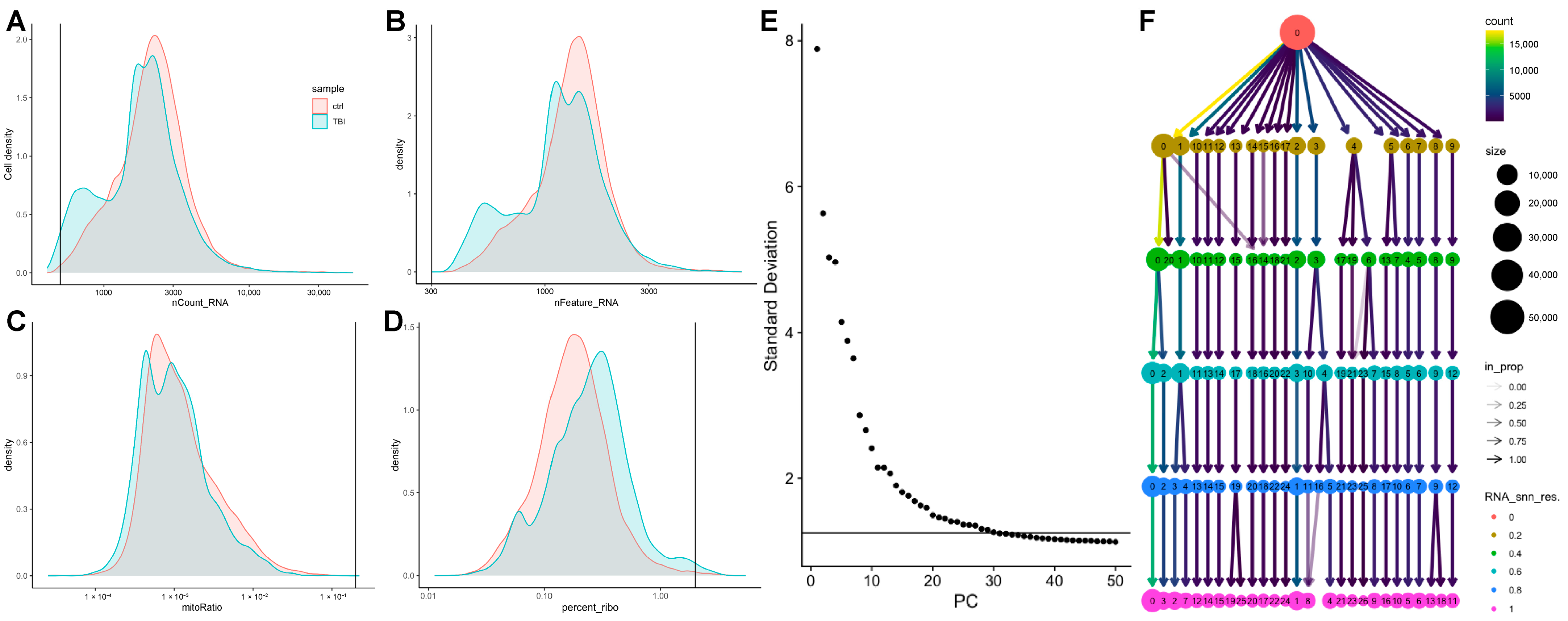
Nuclei were isolated from the dentate gyrus using the Nuclei EZ prep kit (NUC101; Sigma-Aldrich, St. Louis, MO, USA) as per manufacturer instructions, and each animal was processed individually. Isolated nuclei were submitted for sequencing by the Cincinnati Children’s Hospital and Medical Center Single Cell Genomics Core and were sequenced using the 10x Genomics platform (3’ v3.1 Single Index). Raw reads were aligned to the rat genome (Rnor 6.0) using Cell Ranger (v6.1.2), including both exons and introns. Matrices from Cell Ranger were imported into R (version 4.4.1),, where they were merged. Quality control was conducted using the Seurat (v5.1.0) and SeuratObject (v5.0.2) packages based on established workflows [93,94] (Figure A2). Prior to filtering, 55,863 nuclei were present in our samples. Nuclei were filtered out if they contained less than 500 unique molecular identifiers (UMI), less than 300 features, less 0.8 log10GenesPerUMI, greater than 5% mitochondrial genes, or greater than 2% ribosomal genes. Following filtering, 55,123 nuclei remained for analysis.
The filtered data was then log-normalized with a scaling factor of 10,000. Data was scaled to prevent highly expressed genes from dominating downstream analysis. The first 25 principal components were utilized for dimensional reduction by principal component analysis (PCA) based on the elbow plot leveling out between 20 and 30 principal components (Figure A2E). A UMAP was generated at a resolution of 0.6. A resolution of 0.6 was chosen based on the relative stability across resolutions visualized with a Clustree (Figure A2F) [95] while providing a biologically reasonable number of clusters that were not overly fragmented. Data was integrated using canonical correlation analysis (CCA). CCA identifies conserved sources of variation across groups as a way to identify genes most likely to distinguish cell types [96]. Twenty-four unique clusters were identified, and marker genes for each cluster were identified. The top 30 marker genes for each cluster were imported into the online tool “Annotation of Cell Types” (ACT) [43]. Within ACT, marker genes were compared against a reference database for the mouse hippocampus and the mouse whole brain as no rat-specific reference was available. For several neuronal clusters, inputting the marker genes against the mouse hippocampal and whole-brain datasets resulted in different cluster identification dependent upon the tissue dataset used. In those instances, canonical marker genes were utilized. Cell types containing Slc17a7 or Slc17a6, the genes encoding VGLUT1 and VGLUT2, respectively, and lacking traditional GABAergic markers, Gabra1, Gad1, and Gad2, were labeled as glutamatergic neurons, while clusters with the inverse were labeled as GABAergic neurons. Oligodendrocyte maturity was determined automatically using ACT and represents differences in marker genes associated with the extent of myelination (reviewed in [97]). As such, our “Oligodendrocyte” cluster represents non-myelinating oligodendrocytes, while the “Mature Oligodendrocyte” clusters represent myelinating oligodendrocytes.
Based on our findings from the bulk RNA-seq data, we chose to focus our analysis on microglial and astrocytic clusters. Within these clusters, control (sham or naïve) and TBI animals were compared by differential expression analysis using a non-parametric Wilcoxon Rank Sum test (default in Seurat). Differentially expressed genes with an unadjusted p < 0.05 were adjusted for multiple testing within GraphPad Prism (version 10.0.3; GraphPad Software Inc., Boston, MA, USA) based on FDR (Q = 1%; Benjamini and Hochberg). Genes with q < 0.05 with a Log2FC > 1.5 were considered significant DEGs and were visualized as volcano plots using the Enhanced Volcano Package (v1.22.0).
Heatmaps were generated by subsetting the microglial and astrocytic clusters and performing pseudobulking using the AggregateExpression function within Seurat. The top 20 DEGs (q < 0.05 and Log2FC > 1.5) based on p-value were identified and z-scores were calculated to generate heatmaps using the ComplexHeatmap (v2.20.0) package. Hierarchical clustering was conducted utilizing ComplexHeatmap’s default complete linkage method.

4.6. Pathway Analysis

For both the bulk and single-nucleus RNA-seq datasets, we identified signaling pathways associated with our DEGs using iLINCS [98]. The gene names of all DEGs with Log2FC and p-values were submitted to iLINCS using the ‘Signatures’ tool. To be more inclusive of DEGs for pathway analysis, all DEGs were passed to iLINCS regardless of Log2FC. As such, iLINCS default absolute Log2FC > 0.5 was used for pathway analysis inclusion. The top 100 recognized DEGs were used as a signature and were then passed to Enrichr [99] within iLINCS. Within Enrichr, we utilized the ‘WikiPathways 2024 Human’ database [100] to identify potential pathways impacted by TBI. Figures were generated within GraphPad Prism by taking the −log (p-value) for each pathway. Enrichr calculates p-values using a Fisher exact test and adjuste for FDR using Benjamini and Hochberg and a significance of q < 0.05 was considered significant.

Supplementary Materials

The following supporting information can be downloaded at https://www.mdpi.com/article/10.3390/ijms26189140/s1.

Author Contributions

A.J.D.: software, validation, formal analysis, investigation, data curation, writing—original draft, writing—review and editing, visualization. Y.A.: methodology, software, formal analysis, investigation, writing—original draft. R.K.: software, formal analysis, investigation. T.M.H.: investigation. F.V.B.: investigation. J.L.M.: conceptualization, methodology, investigation, resources, supervision, project administration. L.B.N.: conceptualization, methodology, validation, formal analysis, investigation, resources, writing—review and editing, supervision, project administration, funding acquisition. All authors have read and agreed to the published version of the manuscript.

Funding

The research was funded by a grant from the Local Initiative for Excellence Foundation (to L.B.N.) and by the National Institutes of Health/National Institute of Neurological Disorders and Stroke, grant number K08NS110988 (to L.B.N.). The APC was funded by the University of Cincinnati.

Institution Review Board Statement

The animal study protocol was approved by the Institutional Animal Care and Use Committee of the University of Cincinnati (protocol #19-09-04-01, approved 14 November 2019).

Data Availability Statement

Raw data and processed data are available on Gene Expression Omnibus (GSE307917), and the Cell Ranger script and all relevant R-code are available at https://github.com/NgwenyaLab/DeSana_and_Alfawares_2025 (accessed on 30 July 2025).

Acknowledgments

We would like to acknowledge Robert Smith (University of Toledo) and Nathan Salomonis (Cincinnati Children’s Hospital Medical Center) for their helpful discussion in the initial design and analysis of the single-nucleus RNA sequencing data. This research was made possible, in part, using the Cincinnati Children’s Single Cell Genomics Facility (RRID:SCR_022653).

Conflicts of Interest

The authors declare no conflicts of interest.

Abbreviations

The following abbreviations are used in this manuscript:
TBITraumatic Brain Injury
LFPILateral Fluid Percussion Injury
RRTRighting Reflex Time
UMAPUniform Manifold Approximation and Projection
DEGDifferentially Expressed Gene
ACTAnnotation of Cell Type
MACMembrane Attack Complex
fBFactor B
sCR1Soluble CR1
sCrrySoluble Complement Inhibitor
PBSPhosphate-Buffered Saline
logCPMLog2 Counts Per Million
log2FCLog2 Fold Change
UMIUnique Molecular Identifier
PCAPrincipal Component Analysis
CCACanonical Correlation Analysis
StdevStandard Deviation
RNA-seqRibonucleic Acid Sequencing

Appendix A

Table A1. Nuclei counts per cluster for each injury condition.
Cluster Name Nuclei Counts
(Control)
Nuclei Counts (TBI) Control % of Total TBI % of Total
Astrocyte 1230713088.18%4.86%
Astrocyte 275311442.67%4.25%
Astrocyte 37436962.63%2.59%
Endothelial Cell1972370.70%0.88%
GABAergic Neuron 16237022.21%2.61%
GABAergic Neuron 25316811.88%2.53%
GABAergic Neuron 34606991.63%2.60%
Glutamatergic Neuron 16610534823.43%19.88%
Glutamatergic Neuron 23936309613.95%11.51%
Glutamatergic Neuron 33082222010.92%8.25%
Glutamatergic Neuron 42949232710.45%8.65%
Glutamatergic Neuron 59029963.20%3.70%
Glutamatergic Neuron 66726622.38%2.46%
Glutamatergic Neuron 74193771.48%1.40%
Glutamatergic Neuron 83424421.21%1.64%
Mature Oligodendrocyte 184816673.01%6.20%
Mature Oligodendrocyte 22634580.93%1.70%
Mature Oligodendrocyte 319300.07%0.11%
Microglia 190016113.19%5.99%
Microglia 21884470.67%1.66%
Oligodendrocyte1551940.55%0.72%
Oligodendrocyte Precursor 199511283.53%4.19%
Oligodendrocyte Precursor 22513690.89%1.37%
Pericyte71680.25%0.25%
Nuclei counts per cluster by injury condition: Table contains the number of nuclei present in each cluster on the UMAP for control and TBI animals. The two right-most columns contain the number of nuclei in a given cluster divided by the total number of nuclei for each injury condition, represented as a percentage.
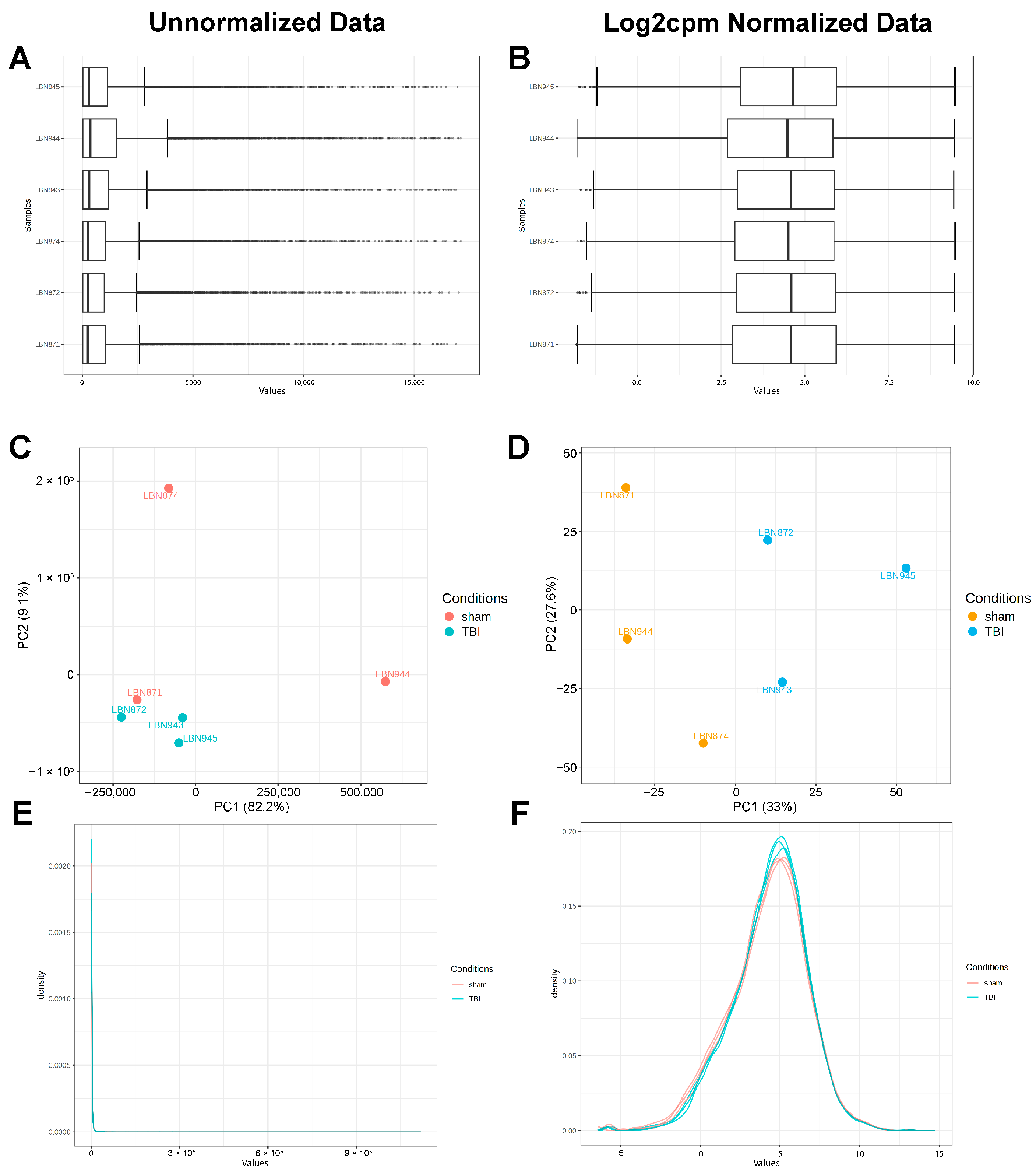
Figure A1. Normalization of bulk RNA sequencing data: Data normalization by Log2 counts per million: within ExpressAnalyst, data was normalized using Log2 counts per million (Log2cpm). (A) Distribution of read counts prior to normalization and (B) after Log2cpm transformation. (C) PCA plot showing contributions of PCs prior to normalization and (D) after normalization. (E) Plot of read density prior to normalization and (F) after Log2cpm transformation.
Ijms 26 09140 g0a1
Figure A2. Quality control for single-nucleus RNA sequencing: Prior to normalization, data was assessed for several common quality control metrics. The traditional cutoffs were utilized: (A) the distribution of UMI (nCount_RNA) for control and TBI animals. The vertical line represents the traditional cutoff point (500) used to exclude nuclei that have not been sequenced deeply enough. (B) The distribution of genes (nFeature_RNA) with the vertical line representing the cutoff of 300 features for the exclusion of nuclei. (C) Distribution of the ratio of mitochondrial genes expressed to non-mitochondrial genes detected per nucleus, with the vertical line representing the cutoff for poor-quality samples (mitoRatio > 0.2). (D) Distribution of the ratio of ribosomal genes, with the vertical line representing the cutoff for poor-quality samples (percent_ribo > 2%). (E) Elbow plot heuristic utilized to determine the number of principal components to be used in making the UMAP. Contribution of each principal component levels out between 20 and 30 principal components, and as such 25 principal components were used for the UMAP. (F) Clustree diagram representing clustering differences based on resolution. A resolution of 0.6 was chosen based on the relative stability across resolutions while providing a biologically reasonable number of clusters without overly fragmenting clusters.
Ijms 26 09140 g0a2

References

  1. Dewan, M.C.; Rattani, A.; Gupta, S.; Baticulon, R.E.; Hung, Y.C.; Punchak, M.; Agrawal, A.; Adeleye, A.O.; Shrime, M.G.; Rubiano, A.M.; et al. Estimating the global incidence of traumatic brain injury. J. Neurosurg. 2019, 130, 1080–1097. [Google Scholar] [CrossRef]
  2. Lai, J.Q.; Shi, Y.C.; Lin, S.; Chen, X.R. Metabolic disorders on cognitive dysfunction after traumatic brain injury. Trends Endocrinol. Metab. 2022, 33, 451–462. [Google Scholar] [CrossRef]
  3. Lambez, B.; Vakil, E. The effectiveness of memory remediation strategies after traumatic brain injury: Systematic review and meta-analysis. Ann. Phys. Rehabil. Med. 2021, 64, 101530. [Google Scholar] [CrossRef] [PubMed]
  4. Jamora, C.W.; Young, A.; Ruff, R.M. Comparison of subjective cognitive complaints with neuropsychological tests in individuals with mild vs more severe traumatic brain injuries. Brain Inj. 2012, 26, 36–47. [Google Scholar] [CrossRef] [PubMed]
  5. Bay, E.; Kalpakjian, C.; Giordani, B. Determinants of subjective memory complaints in community-dwelling adults with mild-to-moderate traumatic brain injury. Brain Inj. 2012, 26, 941–949. [Google Scholar] [CrossRef] [PubMed]
  6. McGuire, J.L.; Correll, E.A.; Lowery, A.C.; Rhame, K.; Anwar, F.N.; McCullumsmith, R.E.; Ngwenya, L.B. Pioglitazone improves working memory performance when administered in chronic TBI. Neurobiol. Dis. 2019, 132, 104611. [Google Scholar] [CrossRef]
  7. Thompson, H.J.; LeBold, D.G.; Marklund, N.; Morales, D.M.; Hagner, A.P.; McIntosh, T.K. Cognitive evaluation of traumatically brain-injured rats using serial testing in the Morris water maze. Restor. Neurol. Neurosci. 2006, 24, 109–114. [Google Scholar] [CrossRef]
  8. Whiting, M.D.; Hamm, R.J. Traumatic Brain Injury Produces Delay-Dependent Memory Impairment in Rats. J. Neurotrauma 2006, 23, 1529–1534. [Google Scholar] [CrossRef]
  9. Hicks, R.R.; Smith, D.H.; Lowenstein, D.H.; Saint Marie, R.; McIntosh, T.K. Mild experimental brain injury in the rat induces cognitive deficits associated with regional neuronal loss in the hippocampus. J. Neurotrauma 1993, 10, 405–414. [Google Scholar] [CrossRef]
  10. Dash, P.K.; Mach, S.A.; Moore, A.N. Enhanced neurogenesis in the rodent hippocampus following traumatic brain injury. J. Neurosci. Res. 2001, 63, 313–319. [Google Scholar] [CrossRef]
  11. Correll, E.A.; Ramser, B.J.; Knott, M.V.; McCullumsmith, R.E.; McGuire, J.L.; Ngwenya, L.B. Deficits in pattern separation and dentate gyrus proliferation after rodent lateral fluid percussion injury. IBRO Neurosci. Rep. 2021, 10, 31–41. [Google Scholar] [CrossRef]
  12. Ngwenya, L.B.; Danzer, S.C. Impact of Traumatic Brain Injury on Neurogenesis. Front. Neurosci. 2018, 12, 1014. [Google Scholar] [CrossRef]
  13. Borzello, M.; Ramirez, S.; Treves, A.; Lee, I.; Scharfman, H.; Stark, C.; Knierim, J.J.; Rangel, L.M. Assessments of dentate gyrus function: Discoveries and debates. Nat. Rev. Neurosci. 2023, 24, 502–517. [Google Scholar] [CrossRef]
  14. Neuberger, E.J.; Swietek, B.; Corrubia, L.; Prasanna, A.; Santhakumar, V. Enhanced Dentate Neurogenesis after Brain Injury Undermines Long-Term Neurogenic Potential and Promotes Seizure Susceptibility. Stem Cell Rep. 2017, 9, 972–984. [Google Scholar] [CrossRef]
  15. Bielefeld, P.; Martirosyan, A.; Martín-Suárez, S.; Apresyan, A.; Meerhoff, G.F.; Pestana, F.; Poovathingal, S.; Reijner, N.; Koning, W.; Clement, R.A.; et al. Traumatic brain injury promotes neurogenesis at the cost of astrogliogenesis in the adult hippocampus of male mice. Nat. Commun. 2024, 15, 5222. [Google Scholar] [CrossRef] [PubMed]
  16. Urrea, C.; Castellanos, D.A.; Sagen, J.; Tsoulfas, P.; Bramlett, H.M.; Dietrich, W.D. Widespread cellular proliferation and focal neurogenesis after traumatic brain injury in the rat. Restor. Neurol. Neurosci. 2007, 25, 65–76. [Google Scholar] [CrossRef]
  17. Diaz-Aparicio, I.; Paris, I.; Sierra-Torre, V.; Plaza-Zabala, A.; Rodríguez-Iglesias, N.; Márquez-Ropero, M.; Beccari, S.; Huguet, P.; Abiega, O.; Alberdi, E.; et al. Microglia Actively Remodel Adult Hippocampal Neurogenesis through the Phagocytosis Secretome. J. Neurosci. 2020, 40, 1453. [Google Scholar] [CrossRef] [PubMed]
  18. Sultan, S.; Li, L.; Moss, J.; Petrelli, F.; Cassé, F.; Gebara, E.; Lopatar, J.; Pfrieger, F.W.; Bezzi, P.; Bischofberger, J.; et al. Synaptic Integration of Adult-Born Hippocampal Neurons Is Locally Controlled by Astrocytes. Neuron 2015, 88, 957–972. [Google Scholar] [CrossRef]
  19. Kreisel, T.; Wolf, B.; Keshet, E.; Licht, T. Unique role for dentate gyrus microglia in neuroblast survival and in VEGF-induced activation. Glia 2019, 67, 594–618. [Google Scholar] [CrossRef] [PubMed]
  20. Dejanovic, B.; Wu, T.; Tsai, M.C.; Graykowski, D.; Gandham, V.D.; Rose, C.M.; Bakalarski, C.E.; Ngu, H.; Wang, Y.; Pandey, S.; et al. Complement C1q-dependent excitatory and inhibitory synapse elimination by astrocytes and microglia in Alzheimer’s disease mouse models. Nat. Aging 2022, 2, 837–850. [Google Scholar] [CrossRef]
  21. Caplan, H.W.; Cardenas, F.; Gudenkauf, F.; Zelnick, P.; Xue, H.; Cox, C.S.; Bedi, S.S. Spatiotemporal Distribution of Microglia After Traumatic Brain Injury in Male Mice. ASN Neuro 2020, 12, 1759091420911770. [Google Scholar] [CrossRef]
  22. Robinson, C.; Apgar, C.; Shapiro, L.A. Astrocyte Hypertrophy Contributes to Aberrant Neurogenesis after Traumatic Brain Injury. Neural Plast. 2016, 2016, 1347987. [Google Scholar] [CrossRef]
  23. Littlejohn, E.L.; DeSana, A.J.; Williams, H.C.; Chapman, R.T.; Joseph, B.; Juras, J.A.; Saatman, K.E. IGF1-Stimulated Posttraumatic Hippocampal Remodeling Is Not Dependent on mTOR. Front. Cell Dev. Biol. 2021, 9, 663456. [Google Scholar] [CrossRef]
  24. Simon, D.W.; McGeachy, M.J.; Bayır, H.; Clark, R.S.B.; Loane, D.J.; Kochanek, P.M. The far-reaching scope of neuroinflammation after traumatic brain injury. Nat. Rev. Neurol. 2017, 13, 171–191, Erratum in Nat. Rev. Neurol. 2017, 13, 572. [Google Scholar] [CrossRef]
  25. Boone, D.R.; Weisz, H.A.; Willey, H.E.; Torres, K.E.O.; Falduto, M.T.; Sinha, M.; Spratt, H.; Bolding, I.J.; Johnson, K.M.; Parsley, M.A.; et al. Traumatic brain injury induces long-lasting changes in immune and regenerative signaling. PLoS ONE 2019, 14, e0214741. [Google Scholar] [CrossRef]
  26. Catta-Preta, R.; Zdilar, I.; Jenner, B.; Doisy, E.T.; Tercovich, K.; Nord, A.S.; Gurkoff, G.G. Transcriptional Pathology Evolves over Time in Rat Hippocampus after Lateral Fluid Percussion Traumatic Brain Injury. Neurotrauma Rep. 2021, 2, 512–525. [Google Scholar] [CrossRef] [PubMed]
  27. Meng, Q.; Zhuang, Y.; Ying, Z.; Agrawal, R.; Yang, X.; Gomez-Pinilla, F. Traumatic Brain Injury Induces Genome-Wide Transcriptomic, Methylomic, and Network Perturbations in Brain and Blood Predicting Neurological Disorders. EBioMedicine 2017, 16, 184–194. [Google Scholar] [CrossRef] [PubMed]
  28. Xing, J.; Ren, L.; Xu, H.; Zhao, L.; Wang, Z.H.; Hu, G.D.; Wei, Z.L. Single-Cell RNA Sequencing Reveals Cellular and Transcriptional Changes Associated With Traumatic Brain Injury. Front. Genet. 2022, 13, 861428. [Google Scholar] [CrossRef]
  29. Zhao, J.; Wang, W.; Yan, K.; Zhao, H.; Zhang, Z.; Wang, Y.; Zhu, W.; Chen, S. RNA-seq reveals Nup62 as a potential regulator for cell division after traumatic brain injury in mice hippocampus. PeerJ 2023, 11, e14913. [Google Scholar] [CrossRef] [PubMed]
  30. Zheng, R.Z.; Xing, J.; Huang, Q.; Yang, X.T.; Zhao, C.Y.; Li, X.Y. Integration of single-cell and bulk RNA sequencing data reveals key cell types and regulators in traumatic brain injury. Math. Biosci. Eng. 2021, 18, 1201–1214. [Google Scholar] [CrossRef]
  31. Arneson, D.; Zhang, G.; Ying, Z.; Zhuang, Y.; Byun, H.R.; Ahn, I.S.; Gomez-Pinilla, F.; Yang, X. Single cell molecular alterations reveal target cells and pathways of concussive brain injury. Nat. Commun. 2018, 9, 3894. [Google Scholar] [CrossRef]
  32. Chen, Z.; Wang, H.; Zhong, J.; Yang, J.; Darwazeh, R.; Tian, X.; Huang, Z.; Jiang, L.; Cheng, C.; Wu, Y.; et al. Significant changes in circular RNA in the mouse cerebral cortex around an injury site after traumatic brain injury. Exp. Neurol. 2019, 313, 37–48. [Google Scholar] [CrossRef] [PubMed]
  33. Puhakka, N.; Bot, A.M.; Vuokila, N.; Debski, K.J.; Lukasiuk, K.; Pitkänen, A. Chronically dysregulated NOTCH1 interactome in the dentate gyrus after traumatic brain injury. PLoS ONE 2017, 12, e0172521. [Google Scholar] [CrossRef]
  34. Zhang, S.-J.; Steijaert, M.N.; Lau, D.; Schütz, G.; Delucinge-Vivier, C.; Descombes, P.; Bading, H. Decoding NMDA Receptor Signaling: Identification of Genomic Programs Specifying Neuronal Survival and Death. Neuron 2007, 53, 549–562. [Google Scholar] [CrossRef]
  35. Wahl, A.S.; Buchthal, B.; Rode, F.; Bomholt, S.F.; Freitag, H.E.; Hardingham, G.E.; Rønn, L.C.B.; Bading, H. Hypoxic/ischemic conditions induce expression of the putative pro-death gene Clca1 via activation of extrasynaptic N-methyl-d-aspartate receptors. Neuroscience 2009, 158, 344–352. [Google Scholar] [CrossRef]
  36. Stevens, B.; Allen, N.J.; Vazquez, L.E.; Howell, G.R.; Christopherson, K.S.; Nouri, N.; Micheva, K.D.; Mehalow, A.K.; Huberman, A.D.; Stafford, B.; et al. The classical complement cascade mediates CNS synapse elimination. Cell 2007, 131, 1164–1178. [Google Scholar] [CrossRef]
  37. Schafer, D.P.; Lehrman, E.K.; Kautzman, A.G.; Koyama, R.; Mardinly, A.R.; Yamasaki, R.; Ransohoff, R.M.; Greenberg, M.E.; Barres, B.A.; Stevens, B. Microglia sculpt postnatal neural circuits in an activity and complement-dependent manner. Neuron 2012, 74, 691–705. [Google Scholar] [CrossRef] [PubMed]
  38. Zhou, T.; Li, Y.; Li, X.; Zeng, F.; Rao, Y.; He, Y.; Wang, Y.; Liu, M.; Li, D.; Xu, Z.; et al. Microglial debris is cleared by astrocytes via C4b-facilitated phagocytosis and degraded via RUBICON-dependent noncanonical autophagy in mice. Nat. Commun. 2022, 13, 6233. [Google Scholar] [CrossRef]
  39. Kim, S.K.; Nabekura, J.; Koizumi, S. Astrocyte-mediated synapse remodeling in the pathological brain. Glia 2017, 65, 1719–1727. [Google Scholar] [CrossRef] [PubMed]
  40. Krukowski, K.; Nolan, A.; Becker, M.; Picard, K.; Vernoux, N.; Frias, E.S.; Feng, X.; Tremblay, M.-E.; Rosi, S. Novel microglia-mediated mechanisms underlying synaptic loss and cognitive impairment after traumatic brain injury. Brain Behav. Immun. 2021, 98, 122–135. [Google Scholar] [CrossRef]
  41. Gao, C.; Jiang, J.; Tan, Y.; Chen, S. Microglia in neurodegenerative diseases: Mechanism and potential therapeutic targets. Signal Transduct. Target. Ther. 2023, 8, 359. [Google Scholar] [CrossRef]
  42. Brandebura, A.N.; Paumier, A.; Onur, T.S.; Allen, N.J. Astrocyte contribution to dysfunction, risk and progression in neurodegenerative disorders. Nat. Rev. Neurosci. 2023, 24, 23–39. [Google Scholar] [CrossRef] [PubMed]
  43. Quan, F.; Liang, X.; Cheng, M.; Yang, H.; Liu, K.; He, S.; Sun, S.; Deng, M.; He, Y.; Liu, W.; et al. Annotation of cell types (ACT): A convenient web server for cell type annotation. Genome Med. 2023, 15, 91. [Google Scholar] [CrossRef]
  44. Sontheimer, R.D.; Racila, E.; Racila, D.M. C1q: Its Functions within the Innate and Adaptive Immune Responses and its Role in Lupus Autoimmunity. J. Investig. Dermatol. 2005, 125, 14–23. [Google Scholar] [CrossRef]
  45. Cao, C.; Liu, T.; Peng, L.; Li, L.; Xu, Z.; Li, X.; Chen, G.; Li, H.; Bai, L. Targeting CD74 in microglia to modulate experimental cerebral ischemia and reperfusion injury: Insights from Single-Cell and bulk transcriptomics. Mol. Brain 2025, 18, 46. [Google Scholar] [CrossRef]
  46. Jahn, J.; Bollensdorf, A.; Kalischer, C.; Piecha, R.; Weiß-Müller, J.; Potru, P.S.; Ruß, T.; Spittau, B. Microglial CD74 Expression Is Regulated by TGFβ Signaling. Int. J. Mol. Sci. 2022, 23, 10247. [Google Scholar] [CrossRef]
  47. Zhao, Y.; Ma, C.; Chen, C.; Li, S.; Wang, Y.; Yang, T.; Stetler, R.A.; Bennett, M.V.L.; Dixon, C.E.; Chen, J.; et al. STAT1 Contributes to Microglial/Macrophage Inflammation and Neurological Dysfunction in a Mouse Model of Traumatic Brain Injury. J. Neurosci. 2022, 42, 7466. [Google Scholar] [CrossRef]
  48. Yap, E.-L.; Greenberg, M.E. Activity-Regulated Transcription: Bridging the Gap between Neural Activity and Behavior. Neuron 2018, 100, 330–348. [Google Scholar] [CrossRef] [PubMed]
  49. D’Sa, K.; Guelfi, S.; Vandrovcova, J.; Reynolds, R.H.; Zhang, D.; Hardy, J.; Botía, J.A.; Weale, M.E.; Taliun, S.A.G.; Small, K.S.; et al. Analysis of subcellular RNA fractions demonstrates significant genetic regulation of gene expression in human brain post-transcriptionally. Sci. Rep. 2023, 13, 13874. [Google Scholar] [CrossRef] [PubMed]
  50. Grindberg, R.V.; Yee-Greenbaum, J.L.; McConnell, M.J.; Novotny, M.; O’Shaughnessy, A.L.; Lambert, G.M.; Araúzo-Bravo, M.J.; Lee, J.; Fishman, M.; Robbins, G.E.; et al. RNA-sequencing from single nuclei. Proc. Natl. Acad. Sci. USA 2013, 110, 19802–19807. [Google Scholar] [CrossRef]
  51. Hammad, A.; Westacott, L.; Zaben, M. The role of the complement system in traumatic brain injury: A review. J. Neuroinflamm. 2018, 15, 24, Correction in J. Neuroinflamm. 2018, 15, 59. [Google Scholar] [CrossRef]
  52. Stahel, P.F.; Barnum, S.R. Bacterial meningitis: Complement gene expression in the central nervous system. Immunopharmacology 1997, 38, 65–72. [Google Scholar] [CrossRef]
  53. Alawieh, A.; Elvington, A.; Tomlinson, S. Complement in the Homeostatic and Ischemic Brain. Front. Immunol. 2015, 6, 417. [Google Scholar] [CrossRef] [PubMed]
  54. Rogers, J.; Strohmeyer, R.; Kovelowski, C.J.; Li, R. Microglia and inflammatory mechanisms in the clearance of amyloid beta peptide. Glia 2002, 40, 260–269. [Google Scholar] [CrossRef]
  55. Stephan, A.H.; Barres, B.A.; Stevens, B. The complement system: An unexpected role in synaptic pruning during development and disease. Annu. Rev. Neurosci. 2012, 35, 369–389. [Google Scholar] [CrossRef]
  56. Rahpeymai, Y.; Hietala, M.A.; Wilhelmsson, U.; Fotheringham, A.; Davies, I.; Nilsson, A.K.; Zwirner, J.; Wetsel, R.A.; Gerard, C.; Pekny, M.; et al. Complement: A novel factor in basal and ischemia-induced neurogenesis. EMBO J. 2006, 25, 1364–1374. [Google Scholar] [CrossRef] [PubMed]
  57. Shinjyo, N.; Ståhlberg, A.; Dragunow, M.; Pekny, M.; Pekna, M. Complement-derived anaphylatoxin C3a regulates in vitro differentiation and migration of neural progenitor cells. Stem Cells 2009, 27, 2824–2832. [Google Scholar] [CrossRef] [PubMed]
  58. Moriyama, M.; Fukuhara, T.; Britschgi, M.; He, Y.; Narasimhan, R.; Villeda, S.; Molina, H.; Huber, B.T.; Holers, M.; Wyss-Coray, T. Complement receptor 2 is expressed in neural progenitor cells and regulates adult hippocampal neurogenesis. J. Neurosci. 2011, 31, 3981–3989. [Google Scholar] [CrossRef]
  59. Bellander, B.M.; Singhrao, S.K.; Ohlsson, M.; Mattsson, P.; Svensson, M. Complement activation in the human brain after traumatic head injury. J. Neurotrauma 2001, 18, 1295–1311. [Google Scholar] [CrossRef]
  60. Kossmann, T.; Stahel, P.F.; Morganti-Kossmann, M.C.; Jones, J.L.; Barnum, S.R. Elevated levels of the complement components C3 and factor B in ventricular cerebrospinal fluid of patients with traumatic brain injury. J. Neuroimmunol. 1997, 73, 63–69. [Google Scholar] [CrossRef]
  61. Kaczorowski, S.L.; Schiding, J.K.; Toth, C.A.; Kochanek, P.M. Effect of soluble complement receptor-1 on neutrophil accumulation after traumatic brain injury in rats. J. Cereb. Blood Flow Metab. 1995, 15, 860–864. [Google Scholar] [CrossRef]
  62. Rancan, M.; Morganti-Kossmann, M.C.; Barnum, S.R.; Saft, S.; Schmidt, O.I.; Ertel, W.; Stahel, P.F. Central nervous system-targeted complement inhibition mediates neuroprotection after closed head injury in transgenic mice. J. Cereb. Blood Flow Metab. 2003, 23, 1070–1074. [Google Scholar] [CrossRef]
  63. Leinhase, I.; Schmidt, O.I.; Thurman, J.M.; Hossini, A.M.; Rozanski, M.; Taha, M.E.; Scheffler, A.; John, T.; Smith, W.R.; Holers, V.M.; et al. Pharmacological complement inhibition at the C3 convertase level promotes neuronal survival, neuroprotective intracerebral gene expression, and neurological outcome after traumatic brain injury. Exp. Neurol. 2006, 199, 454–464. [Google Scholar] [CrossRef]
  64. Todd, B.P.; Chimenti, M.S.; Luo, Z.; Ferguson, P.J.; Bassuk, A.G.; Newell, E.A. Traumatic brain injury results in unique microglial and astrocyte transcriptomes enriched for type I interferon response. J. Neuroinflamm. 2021, 18, 151. [Google Scholar] [CrossRef]
  65. Izzy, S.; Liu, Q.; Fang, Z.; Lule, S.; Wu, L.; Chung, J.Y.; Sarro-Schwartz, A.; Brown-Whalen, A.; Perner, C.; Hickman, S.E.; et al. Time-Dependent Changes in Microglia Transcriptional Networks Following Traumatic Brain Injury. Front. Cell Neurosci. 2019, 13, 307. [Google Scholar] [CrossRef]
  66. Zhong, J.; Jiang, L.; Cheng, C.; Huang, Z.; Zhang, H.; Liu, H.; He, J.; Cao, F.; Peng, J.; Jiang, Y.; et al. Altered expression of long non-coding RNA and mRNA in mouse cortex after traumatic brain injury. Brain Res. 2016, 1646, 589–600. [Google Scholar] [CrossRef]
  67. Toutonji, A.; Mandava, M.; Guglietta, S.; Tomlinson, S. Chronic complement dysregulation drives neuroinflammation after traumatic brain injury: A transcriptomic study. Acta Neuropathol. Commun. 2021, 9, 126. [Google Scholar] [CrossRef] [PubMed]
  68. Haque, A.; Engel, J.; Teichmann, S.A.; Lönnberg, T. A practical guide to single-cell RNA-sequencing for biomedical research and clinical applications. Genome Med. 2017, 9, 75. [Google Scholar] [CrossRef] [PubMed]
  69. Kim, N.; Kang, H.; Jo, A.; Yoo, S.A.; Lee, H.O. Perspectives on single-nucleus RNA sequencing in different cell types and tissues. J. Pathol. Transl. Med. 2023, 57, 52–59. [Google Scholar] [CrossRef]
  70. Makinde, H.M.; Just, T.B.; Gadhvi, G.T.; Winter, D.R.; Schwulst, S.J. Microglia Adopt Longitudinal Transcriptional Changes After Traumatic Brain Injury. J. Surg. Res. 2020, 246, 113–122. [Google Scholar] [CrossRef] [PubMed]
  71. Malinow, R.; Schulman, H.; Tsien, R.W. Inhibition of postsynaptic PKC or CaMKII blocks induction but not expression of LTP. Science 1989, 245, 862–866. [Google Scholar] [CrossRef]
  72. Fink, C.C.; Bayer, K.U.; Myers, J.W.; Ferrell, J.E., Jr.; Schulman, H.; Meyer, T. Selective regulation of neurite extension and synapse formation by the beta but not the alpha isoform of CaMKII. Neuron 2003, 39, 283–297. [Google Scholar] [CrossRef] [PubMed]
  73. Silva, A.J.; Paylor, R.; Wehner, J.M.; Tonegawa, S. Impaired spatial learning in alpha-calcium-calmodulin kinase II mutant mice. Science 1992, 257, 206–211. [Google Scholar] [CrossRef] [PubMed]
  74. Puram, S.V.; Kim, A.H.; Ikeuchi, Y.; Wilson-Grady, J.T.; Merdes, A.; Gygi, S.P.; Bonni, A. A CaMKIIβ signaling pathway at the centrosome regulates dendrite patterning in the brain. Nat. Neurosci. 2011, 14, 973–983. [Google Scholar] [CrossRef]
  75. Borghi, R.; Trivisano, M.; Specchio, N.; Tartaglia, M.; Compagnucci, C. Understanding the pathogenetic mechanisms underlying altered neuronal function associated with CAMK2B mutations. Neurosci. Biobehav. Rev. 2023, 152, 105299. [Google Scholar] [CrossRef]
  76. Paban, V.; Ogier, M.; Chambon, C.; Fernandez, N.; Davidsson, J.; Risling, M.; Alescio-Lautier, B. Molecular gene expression following blunt and rotational models of traumatic brain injury parallel injuries associated with stroke and depression. J. Transl. Sci. 2016, 2. [Google Scholar] [CrossRef]
  77. Halpain, S.; Dehmelt, L. The MAP1 family of microtubule-associated proteins. Genome Biol. 2006, 7, 224. [Google Scholar] [CrossRef]
  78. Giza, J.; Urbanski, M.J.; Prestori, F.; Bandyopadhyay, B.; Yam, A.; Friedrich, V.; Kelley, K.; D’Angelo, E.; Goldfarb, M. Behavioral and cerebellar transmission deficits in mice lacking the autism-linked gene islet brain-2. J. Neurosci. 2010, 30, 14805–14816. [Google Scholar] [CrossRef]
  79. Villablanca, C.; Vidal, R.; Gonzalez-Billault, C. Are cytoskeleton changes observed in astrocytes functionally linked to aging? Brain Res. Bull. 2023, 196, 59–67. [Google Scholar] [CrossRef] [PubMed]
  80. Richetin, K.; Steullet, P.; Pachoud, M.; Perbet, R.; Parietti, E.; Maheswaran, M.; Eddarkaoui, S.; Bégard, S.; Pythoud, C.; Rey, M.; et al. Tau accumulation in astrocytes of the dentate gyrus induces neuronal dysfunction and memory deficits in Alzheimer’s disease. Nat. Neurosci. 2020, 23, 1567–1579. [Google Scholar] [CrossRef]
  81. Milenkovic, I.; Petrov, T.; Kovacs, G.G. Patterns of Hippocampal Tau Pathology Differentiate Neurodegenerative Dementias. Dement. Geriatr. Cogn. Disord. 2014, 38, 375–388. [Google Scholar] [CrossRef]
  82. Edwards, G., 3rd; Zhao, J.; Dash, P.K.; Soto, C.; Moreno-Gonzalez, I. Traumatic Brain Injury Induces Tau Aggregation and Spreading. J. Neurotrauma 2020, 37, 80–92. [Google Scholar] [CrossRef] [PubMed]
  83. Dejanovic, B.; Huntley, M.A.; De Mazière, A.; Meilandt, W.J.; Wu, T.; Srinivasan, K.; Jiang, Z.; Gandham, V.; Friedman, B.A.; Ngu, H.; et al. Changes in the Synaptic Proteome in Tauopathy and Rescue of Tau-Induced Synapse Loss by C1q Antibodies. Neuron 2018, 100, 1322–1336.e7. [Google Scholar] [CrossRef]
  84. Milligan, G.W. An examination of the effect of six types of error perturbation on fifteen clustering algorithms. Psychometrika 1980, 45, 325–342. [Google Scholar] [CrossRef]
  85. Thrupp, N.; Sala Frigerio, C.; Wolfs, L.; Skene, N.G.; Fattorelli, N.; Poovathingal, S.; Fourne, Y.; Matthews, P.M.; Theys, T.; Mancuso, R.; et al. Single-Nucleus RNA-Seq Is Not Suitable for Detection of Microglial Activation Genes in Humans. Cell Rep. 2020, 32, 108189. [Google Scholar] [CrossRef] [PubMed]
  86. Cembrowski, M.S.; Wang, L.; Sugino, K.; Shields, B.C.; Spruston, N. Hipposeq: A comprehensive RNA-seq database of gene expression in hippocampal principal neurons. eLife 2016, 5, e14997. [Google Scholar] [CrossRef] [PubMed]
  87. National Research Council. Guide for the Care and Use of Laboratory Animals: Eighth Edition; The National Academies Press: Washington, DC, USA, 2011; p. 246.
  88. Best, F.V.; Hartings, J.A.; Ngwenya, L.B. Repetitive cortical spreading depolarizations are prolonged early after experimental traumatic brain injury. Exp. Neurol. 2025, 385, 115120. [Google Scholar] [CrossRef]
  89. Hagihara, H.; Toyama, K.; Yamasaki, N.; Miyakawa, T. Dissection of hippocampal dentate gyrus from adult mouse. J. Vis. Exp. 2009, 1543. [Google Scholar] [CrossRef]
  90. Liu, P.; Ewald, J.; Pang, Z.; Legrand, E.; Jeon, Y.S.; Sangiovanni, J.; Hacariz, O.; Zhou, G.; Head, J.A.; Basu, N.; et al. ExpressAnalyst: A unified platform for RNA-sequencing analysis in non-model species. Nat. Commun. 2023, 14, 2995. [Google Scholar] [CrossRef]
  91. Blighe, K.; Rana, S.; Lewis, M. EnhancedVolcano: Publication-ready volcano plots with enhanced colouring and labeling. R Package Version 1.22.0 2024. [Google Scholar] [CrossRef]
  92. Gu, Z.; Eils, R.; Schlesner, M. Complex heatmaps reveal patterns and correlations in multidimensional genomic data. Bioinformatics 2016, 32, 2847–2849. [Google Scholar] [CrossRef]
  93. Harvard Chan Bioinformatics Core. Introduction to Single-Cell RNA-seq. Available online: https://github.com/hbctraining (accessed on 1 July 2024).
  94. Hao, Y.; Stuart, T.; Kowalski, M.H.; Choudhary, S.; Hoffman, P.; Hartman, A.; Srivastava, A.; Molla, G.; Madad, S.; Fernandez-Granda, C.; et al. Dictionary learning for integrative, multimodal and scalable single-cell analysis. Nat. Biotechnol. 2024, 42, 293–304. [Google Scholar] [CrossRef]
  95. Zappia, L.; Oshlack, A. Clustering trees: A visualization for evaluating clusterings at multiple resolutions. GigaScience 2018, 7, giy083. [Google Scholar] [CrossRef]
  96. Butler, A.; Hoffman, P.; Smibert, P.; Papalexi, E.; Satija, R. Integrating single-cell transcriptomic data across different conditions, technologies, and species. Nat. Biotechnol. 2018, 36, 411–420. [Google Scholar] [CrossRef]
  97. Sock, E.; Wegner, M. Transcriptional control of myelination and remyelination. Glia 2019, 67, 2153–2165. [Google Scholar] [CrossRef] [PubMed]
  98. Pilarczyk, M.; Fazel-Najafabadi, M.; Kouril, M.; Shamsaei, B.; Vasiliauskas, J.; Niu, W.; Mahi, N.; Zhang, L.; Clark, N.A.; Ren, Y.; et al. Connecting omics signatures and revealing biological mechanisms with iLINCS. Nat. Commun. 2022, 13, 4678. [Google Scholar] [CrossRef] [PubMed]
  99. Chen, E.Y.; Tan, C.M.; Kou, Y.; Duan, Q.; Wang, Z.; Meirelles, G.V.; Clark, N.R.; Ma’ayan, A. Enrichr: Interactive and collaborative HTML5 gene list enrichment analysis tool. BMC Bioinform. 2013, 14, 128. [Google Scholar] [CrossRef] [PubMed]
  100. Agrawal, A.; Balcı, H.; Hanspers, K.; Coort, S.L.; Martens, M.; Slenter, D.N.; Ehrhart, F.; Digles, D.; Waagmeester, A.; Wassink, I.; et al. WikiPathways 2024: Next generation pathway database. Nucleic Acids Res. 2024, 52, D679–D689. [Google Scholar] [CrossRef]
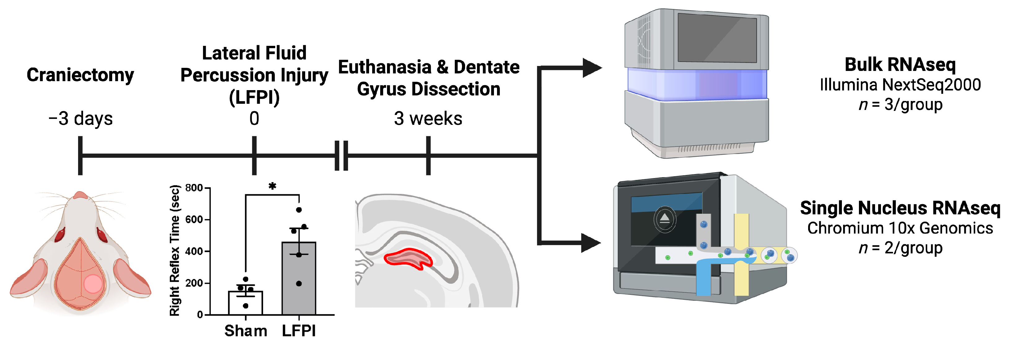
Figure 1. Experimental timeline: Rats underwent a craniectomy three days prior to TBI induced by lateral fluid percussion injury (LFPI) or sham injury. TBI rats had significantly increased righting reflex time compared to sham animals (mean ± standard error; * corresponds with a p < 0.05; p = 0.0146). Three weeks post-injury, rats were euthanized, and the ipsilateral hippocampal dentate gyrus was dissected out and processed for bulk RNA sequencing (NextSeq2000; Illumina, Inc., San Diego, CA, USA) or single-nucleus RNA sequencing (Chromium 10x Genomics; 10x Genomics, Inc., Pleasanton, CA, USA). Created in BioRender. DeSana, A. (2025) https://BioRender.com/eyfapfv (accessed on 30 July 2025).
Ijms 26 09140 g001
Figure 2. Differentially expressed genes suggest upregulation of genes involved in neuroinflammation and complement activation: following TBI, 126 genes were upregulated in TBI rats, with 2 genes downregulated compared to sham injured rats. (A) Volcano plot representing differentially expressed genes (q < 0.01 and Log2FC > 1.5), where each dot represents an individual gene. Genes with a Log2FC > 0 represent an increase in TBI rats compared to sham rats, and Log2FC < 0 represents a decrease in TBI compared to sham. (B) Hierarchical clustering with complete linkage shows a separation in gene expression between sham and TBI rats, with various genes involved in neuroinflammation upregulated with injury. (C) Pathway analysis mapped to the ‘WikiPathways 2024 Human’ database further suggests neuroinflammatory activation in the dentate gyrus at 3 weeks post-TBI, and several of the implicated pathways involve complement activation.
Ijms 26 09140 g002
Figure 3. Single-nucleus RNA sequencing clustering: (A) UMAP clustering of nuclei shows 24 distinct clusters that were assigned to glutamatergic neurons, GABAergic neurons, astrocytes, microglia, oligodendrocyte precursor cells, oligodendrocytes, mature oligodendrocytes, endothelial cells, and pericytes. (B) When the UMAP was split by injury condition, some differences in intercluster relationships are appreciable.
Ijms 26 09140 g003
Figure 4. Single-nucleus RNA sequencing suggests microglial involvement in complement signaling: (A) differential expression analysis within each microglial cluster revealed significantly up- and downregulated genes (q < 0.01 and Log2FC > 1.5) between TBI and control animals, with (B) hierarchical clustering with complete linkage showing a clear separation between control and TBI rats in the Microglia 2 cluster, with less consistent separation between groups in the Microglia 1 cluster. (C) Pathway analysis revealed that the nuclei from Microglia 1 likely represent a pro-inflammatory microglial population with evidence of complement signaling, while Microglia 2 likely represents a homeostatic microglial population. Red bars represent significant findings by Fisher exact test with Benjamini and Hochberg correction: q < 0.05, while gray bars represent non-significant findings.
Ijms 26 09140 g004
Figure 5. Changes in astrocyte gene expression three weeks after TBI: (A) differential expression analysis within each cluster revealed significantly up- and downregulated genes (q < 0.01 and Log2FC > 1.5) within the three astrocyte clusters between control and TBI animals. (B) Hierarchical clustering analysis showed inconsistent separation between the Astrocyte 1 and Astrocyte 2 clusters but clear separation in the Astrocyte 3 cluster. (C) Pathway analysis suggests that all three astrocyte clusters show evidence of increased neuroinflammation and altered signaling. Red bars represent significant findings by Fisher exact test with Benjamini and Hochberg correction: q < 0.05.
Ijms 26 09140 g005
Table 1. Bulk RNA-seq pathway analysis.
TermDEGs in Dataset/Genes in Pathwayp-ValueAdjusted p-Value (q-Value)Odds RatioCombined ScoreDEGs in Pathway
Complement Activation WP5458/239.66 × 10−142.05 × 10−11120.543612.29Cfd; C1qb; C1qa; C4a; C1s; C1r; C2; C1qc
Microglia Pathogen Phagocytosis Pathway WP39379/402.23 × 10−132.36 × 10−1166.321931.98C1qb; C1qq; Tyrobp; Ncf1; Arpc1b; Cyba; Ptpn6; Vav1; C1qc
Complement System In Neuronal Development And Plasticity WP509011/1063.30 × 10−122.33 × 10−1026.98713.36C1qb; Cfd; C4a; C1qa; Tgfb; C1s; C1r; Axl; Serping1; C2; C1qc
Complement And Coagulation Cascades WP5589/588.09 × 10−124.03 × 10−1041.921070.62Cfd; C1qb; C1qa; C1s; F10; C1r; Serping1; C2; C1qc
Dengue 2 Interactions With Complement And Coagulation Cascades WP38969/599.50 × 10−124.03 × 10−1041.081042.51Cfd; C1qb; C1qa; C1s; F10; C1r; Serping1; C2; C1qc
Oxidative Damage Response WP39416/403.51 × 10−81.24 × 10−638.96668.74C1qb; C1qa; C1s; C1r; C2; C1qc
Complement System WP28067/953.68 × 10−71.12 × 10−517.71262.38Cfd; C4a; C1s; F10; Serping1; Icam1; C2
TYROBP Causal Network In Microglia WP39456/625.17 × 10−71.37 × 10−523.63342.04Tyrobp; Npc2; Cd37; Capg; Cxcl16; C1qc
Allograft Rejection WP23286/904.70 × 10−61.11 × 10−415.73192.98C1qb; C4a; C1qa; Tgfb1; C2; C1qc
Network Map of SARS-CoV 2 Signaling WP51157/2542.24 × 10−40.004741646.2652.61Lgals3bp; Trpm2; C1s; C1r; Ctsz; Ptpn6; Cxcl16
Top 10 pathways identified by Enrichr using the WikiPathways 2024 Human dataset. p-values computed within Enrichr by Fisher exact test, with p-values adjusted for multiple testing using Benjamini–Hochberg method. Odds ratios were derived by Enrichr based on comparisons against computed mean ranks from random gene sets, as analyzed by Fisher exact test. Combined scores were computed by Enrichr from the Fisher exact test by multiplying the −log (p-value) by the z-score of the deviation from the expected rank.
Table 2. Top 10 differentially expressed genes for microglia clusters.
Gene Namep-Valueq-ValueAvg Log2FCPct.1 (Control)Pct.2 (TBI)
Microglia 1Ano51.02 × 10−121.30 × 10⁻¹⁰−1.520.1360.055
Axl1.41 × 10−121.53 × 10−103.970.0060.065
Spata223.94 × 10−102.73 × 10−08−2.210.0590.013
Kmo4.18 × 10−92.28 × 10⁻⁷−1.850.0590.016
Slfn41.45 × 10−63.40 × 10−53.410.0140.054
Mx12.38 × 10−64.97 × 10−53.280.0040.034
Marc13.75 × 10−67.23 × 10−54.720.0010.025
Tmem1544.44 × 10−68.46 × 10⁻⁵−1.510.0390.011
LOC1003644356.30 × 10−61.13 × 10−47.7700.019
Gpnmb6.69 × 10−61.19 × 10−44.270.0020.026
Microglia 2Rpl109.75 × 10−116.39 × 10−83.9500.018
Foxred13.83 × 10−81.25 × 10⁻⁵−1.750.2130.069
Cd462.73 × 10−64.47 × 10⁻⁴−1.800.1860.047
Ccdc1416.03 × 10−66.58 × 10⁻⁴−3.690.0320.002
Ccdc88b2.44 × 10−52.02 × 10⁻³−2.530.090.013
Slfn132.47 × 10−52.02 × 10−34.570.0050.101
Zfp2634.17 × 10−52.73 × 10⁻³−6.520.0370
Slc2a124.17 × 10−52.73 × 10⁻³−6.030.0370
Apbb34.40 × 10−52.74 × 10⁻³−1.880.160.04
Fbxw91.06 × 10−44.78 × 10⁻³−1.980.1060.025
Top 10 DEGs for microglial clusters. A complete list of DEGs can be found in the Supplementary Materials.
Table 3. Pathway analysis for microglial clusters for single-nucleus RNA-seq.
TermDEGs in Dataset/Genes in Pathwayp-ValueAdjusted p-Value (q-Value)Odds RatioCombined ScoreDEGs in Pathway
Microglia 1Complement System In Neuronal Development And Plasticity WP50907/1061.03 × 10−62.07 × 10−415.05207.58C1qb; C1qa; Axl; Mbp; Itgav; Mertk; C1qc
Neuroinflammation And Glutamatergic Signaling WP50837/1406.59 × 10−66.66 × 10−411.19133.45Camk2b; Gria2; Camk2D; Slc17a7; Disc1; Grm1; Gls
Disruption Of Postsynaptic Signaling By CNV WP48754/332.15 × 10−51.45 × 10−328.55306.79Camk2b; Camk2d; Nrxn3; Grm1
Microglia Pathogen Phagocytosis Pathway WP39374/404.68 × 10−52.36 × 10−322.99229.20C1qb; Vav3; C1qa; C1qc
Serotonin And Anxiety WP39473/177.84 × 10−53.17 × 10−343.93415.32Camk2b; Ppp3ca; Grm1
Complement Activation WP5453/232.00 × 10−46.72 × 10−330.74261.88C1qb; C1qa; C1qc
Photodynamic Therapy-Induced Unfolded Protein Response WP36133/273.25 × 10−48.30 × 10−325.61205.71Hspa5; Calr; Hsp90b1
Spinal Cord Injury WP24315/1193.29 × 10−48.30 × 10−39.1373.26C1qb; Fkbp1a; Ppp3ca; Ptpra; Mbp
Primary Focal Segmental Glomerulosclerosis FSGS WP25724/724.66 × 10−41.05 × 10−212.1593.21Camk2b; Trpc6; Ctsl; Itgav
Cell Lineage Map For Neuronal Differentiation WP54175/1325.29 × 10−41.07 × 10−28.1961.82Map2; Mbp; Slc17a7; S100b; Gls
Microglia 2Genes Controlling Nephrogenesis WP48234/446.85 × 10−51.10 × 10−220.69198.37Robo2; Notch2; Gli3; Vegfa
Osteoarthritic Chondrocyte Hypertrophy WP53733/502.00 × 10−31.11 × 10−113.0681.16Jund; Junb; Vegfa
11P11 2 Copy Number Variation Syndrome WP53483/562.77 × 10−31.11 × 10−111.5868.18B4gat1; Nrxn1; Gli3
Oncostatin M Signaling WP23743/562.77 × 10−31.11 × 10−111.5868.18Jund; Junb; Vegfa
Hypertrophy Model WP5162/204.43 × 10−31.42 × 10−122.54122.14Jund; Vegfa
Axon Guidance WP52893/725.64 × 10−31.45 × 10−18.8946.03Robo2; Robo3; Lrrc4c
Hippocampal Synaptogenesis And Neurogenesis WP52312/246.36 × 10−31.45 × 10−118.4493.27Nrxn1; Ncam1
EGF EGFR Signaling WP4374/1598.36 × 10−31.67 × 10−15.3125.40Ptprr; Jund; Inpp5d; Ncoa3
GDNF RET Signaling Axis WP48302/309.83 × 10−31.75 × 10−114.4866.95Robo2; Gli3
Disruption of Postsynaptic Signaling by CNV WP48752/331.18 × 10−21.89 × 10−113.0858.06Nrxn1; Dlgap1
Pathway analysis from microglial clusters for single-nucleus RNA-seq: For Microglia 1 and Microglia 2 clusters, the top 10 pathways identified by Enrichr using the WikiPathways 2024 Human dataset. p-values computed within Enrichr by Fisher exact test, with p-values adjusted for multiple testing using Benjamini–Hochberg method. Odds ratios were derived by Enrichr based on comparisons against computed mean ranks from random gene sets, as analyzed by Fisher exact test. Combined scores were computed by Enrichr from the Fisher exact test by multiplying the −log (p-value) by the z-score of the deviation from the expected rank.
Table 4. Top 10 differentially expressed genes for astrocyte clusters.
Gene Namep-Valueq-ValueAvg Log2FCPct.1 (Control)Pct.2 (TBI)
Astrocyte 1Pld51.29 × 10−543.51 × 10−513.180.020.157
Nme41.32 × 10−297.18 × 10−278.7300.044
Plk52.35 × 10−245.81 × 10−221.600.0430.148
Celsr36.06 × 10−205.89 × 10−181.520.0250.098
LOC1003644351.22 × 10−177.04 × 10−168.3600.02
Dlk21.32 × 10−177.48 × 10−161.520.0260.091
Rps232.04 × 10−171.12 × 10−153.610.0040.087
Asb151.18 × 10−154.04 × 10−144.280.0010.026
Rpl18a1.09 × 10−132.39 × 10−129.4100.063
Epha82.69 × 10−135.5 × 10−121.640.0270.084
Astrocyte 2AABR07000398.14.15 × 10−644.61 × 10−611.550.6240.824
Map1a4.42 × 10−453.28 × 10−421.830.260.551
S100b1.82 × 10−368.09 × 10−341.550.2350.486
Hpcal42.49 × 10−204.26 × 10−181.810.1420.316
Agap23.42 × 10−205.43 × 10−181.710.1580.332
Ddn4.92 × 10−185.76 × 10−161.690.10.247
Mapk8ip22.06 × 10−161.64 × 10−141.510.1160.265
Ubc1.36 × 10−148.40 × 10−131.550.10.23
Rpl105.95 × 10−122.50 × 10−101.580.0050.028
Gnas.11.92 × 10−117.11 × 10−101.620.0680.166
Astrocyte 3Pld52.25 × 10−307.50 × 10−282.930.0510.256
Rpl101.16 × 10−221.78 × 10−207.6500.106
Slc9b12.17 × 10−171.38 × 10⁻¹⁵−1.710.2440.085
Jhy1.16 × 10−155.95 × 10⁻¹⁴−1.630.2370.083
Begain1.67 × 10−135.96 × 10−121.850.1010.257
Nme41.91 × 10−136.76 × 10−126.1400.049
LOC1003644356.10 × 10−131.98 × 10−115.8900.022
Rapgefl16.24 × 10−121.63 × 10−101.730.1120.264
Hba-a23.53 × 10−117.71 × 10⁻¹⁰−4.420.0770.006
Cacna1h5.23 × 10−111.10 × 10−91.500.1020.239
Top 10 DEGs for astrocyte clusters. A complete list of DEGs can be found in the Supplementary Materials.
Table 5. Pathway analysis of astrocyte clusters for single-nucleus RNA-seq.
TermDEGs in Dataset/Genes in Pathwayp-ValueAdjusted p-Value (q-Value)Odds RatioCombined ScoreDEGs in Pathway
Astrocyte 1Neuroinflammation And Glutamatergic Signaling WP50839/1403.45 × 10−85.08 × 10−614.93256.43Camk2b; Camk2a; Slc1a2; Slc1a3; Nsmf; Slc17a7; Il6r; Camkk1; Grin1
Fragile X Syndrome WP45497/1222.65 × 10−61.94 × 10−412.95166.31Camk2b; Map1b; Camk2a; Agap2; Dlgap3; Dnm; Grin1
Cell Lineage Map For Neuronal Differentiation WP54177/1324.47 × 10−62.13 × 10−411.91146.68Slc1a2; Rimbp2; Slc1a3; Slc17a7; Bsn; Cacng2; Grin1
Hippocampal Synaptogenesis And Neurogenesis WP52314/245.79 × 10−62.13 × 10−441.42499.47Camk2b; Nrxn1; Camk2a; Camkk1
Disruption Of Postsynaptic Signaling By CNV WP48754/332.15 × 10−56.33 × 10−428.55306.79Camk2b; Nrxn1; Camk2a; Grin1
NO cGMP PKG Mediated Neuroprotection WP40084/468.17 × 10−52.00 × 10−319.70185.43Camk2b; Camk2a; Pde2a; Grin1
Microtubule Cytoskeleton Regulation WP20384/489.67 × 10−52.03 × 10−318.80173.82Tiam1; Ntrk3; Map1b; Ephb2
Synaptic Vesicle Pathway WP22673/512.12 × 10−33.90 × 10−212.7978.73Slc1a3; Slc17a7; Dnm1
Phosphodiesterases In Neuronal Function WP42223/542.50 × 10−34.08 × 10−212.0472.12Camk2a; Pde2A; Grin1
NAD Metabolism WP36442/162.84 × 10−34.17 × 10−228.99170.01Nt5e; Nmnat2
Astrocyte 2Disruption Of Postsynaptic Signaling By CNV WP48756/331.33 × 10−82.37 × 10−646.98851.93Camk2b; Nlgn1; Dlg2; Nrxn1; Nrxn3; Dlgap1
Neuroinflammation And Glutamatergic Signaling WP50838/1405.10 × 10−74.54 × 10−513.02188.67Camk2b; Grm5; Cfl1; Nsmf; Calm1; Adcy8; Glul; Gfap
Cell Lineage Map For Neuronal Differentiation WP54177/1324.47 × 10−62.65 × 10−411.91146.68Nlgn1; Dlg2; Aqp4; Mbp; S100b; Glul; Gfap
ADHD And Autism ASD Pathways WP542010/3701.64 × 10−57.31 × 10−46.0366.44Gabrb1; Grm5; Nlgn1; Dlg2; Syt1; Nrxn1; Nrxn3; Dlgap1; Calm1; Glul
Common Pathways Underlying Drug Addiction WP26364/415.17 × 10−51.84 × 10−322.37220.78Grm5; Adcy8; Calm1; Actb
Calcium Regulation In Cardiac Cells WP5366/1511.11 × 10−43.31 × 10−38.7079.15Camk2b; Ywhab; Calm1; Adcy8; Calm2; Kcnj3
Myometrial Relaxation And Contraction Pathways WP2896/1561.33 × 10−43.39 × 10−38.4074.99Camk2b; Ywhab; Calm1; Adcy8; Calm2; Actb
Hippocampal Synaptogenesis And Neurogenesis WP52313/242.27 × 10−45.06 × 10−329.28245.59Camk2b; Nrxn1; Calm1
Glial Cell Differentiation WP22762/75.11 × 10−41.01 × 10−281.20615.40Plb1; Mbp
Sudden Infant Death Syndrome SIDS Susceptibility Pathways WP7065/1571.16 × 10−32.06 × 10−26.8446.24Ywhab; Plp1; Aqp4; Aldoa; Sptbn1
Astrocyte 3Fragile X Syndrome WP454910/1225.35 × 10−107.70 × 10−819.63419.10Cyfip2; Gria1; Camk2b; Ppp3ca; Grin2a; Map1b; Camk2a; Agap2; Dnm1; Grin1
Disruption Of Postsynaptic Signaling By CNV WP48756/331.33 × 10−89.60 × 10−746.98851.93Camk2b; Ryr2; Grin2a; Nrxn1; Camk2a; Grin1
Synaptic Vesicle Pathway WP22676/512.02 × 10−79.67 × 10−628.16434.20Snap25; Slc1a3; Atp1a2; Slc17a7; Cplx2; Dnm1
Neuroinflammation And Glutamatergic Signaling WP50838/1405.10 × 10−71.84 × 10−513.02188.67Gria1; Camk2b; Grin2a; Camk2a; Slc1a2; Slc1a3; Slc17a7; Grin1
NRXN1 Deletion Syndrome WP53984/171.33 × 10−63.84 × 10−563.74862.34Gria1; Grin2a; Nrxn1; Grin1
Cell Lineage Map For Neuronal Differentiation WP54177/1324.47 × 10−61.07 × 10−411.91146.68Snap25; Slc1a2; Rimbp2; Slc1a3; Slc17a7; Bsn; Grin1
Phosphodiesterases In Neuronal Function WP42225/547.36 × 10−61.51 × 10−421.32252.03Gria1; Pde11A; Grin2A; Camk2a; Grin1
Common Pathways Underlying Drug Addiction WP26364/415.17 × 10−59.30 × 10−422.37220.78Gria1; Grin2a; Camk2a; Grin1
NO cGMP PKG Mediated Neuroprotection WP40084/468.17 × 10−51.31 × 10−319.70185.43Camk2b; Grin2A; Camk2a; Grin1
Hippocampal Synaptogenesis And Neurogenesis WP52313/242.27 × 10−43.28 × 10−329.28245.59CAamk2b; Nrxn1; Camk2a
Pathway analysis from astrocyte clusters for single-nucleus RNA-seq: for Astrocyte 1, Astrocyte 2, and Astrocyte 3 clusters, the top 10 pathways identified by Enrichr using the WikiPathways 2024 Human dataset. p-values computed within Enrichr by Fisher exact test, with p-values adjusted for multiple testing using Benjamini–Hochberg method. Odds ratios were derived by Enrichr based on comparisons against computed mean ranks from random gene sets, as analyzed by Fisher exact test. Combined scores were computed by Enrichr from the Fisher exact test by multiplying the −log (p-value) by the z-score of the deviation from the expected rank.
Disclaimer/Publisher’s Note: The statements, opinions and data contained in all publications are solely those of the individual author(s) and contributor(s) and not of MDPI and/or the editor(s). MDPI and/or the editor(s) disclaim responsibility for any injury to people or property resulting from any ideas, methods, instructions or products referred to in the content.