Review

Fantastic Ferulic Acid Esterases and Their Functions

by
Savvina Leontakianakou
,
Patrick Adlercreutz
and
Eva Nordberg Karlsson
*
Division of Biotechnology and Applied Microbiology, Department of Process and Life Science Engineering, Lund University, P.O. Box 124, SE-22100 Lund, Sweden
*
Author to whom correspondence should be addressed.
Int. J. Mol. Sci. 2025, 26(15), 7474; https://doi.org/10.3390/ijms26157474
Submission received: 19 June 2025 / Revised: 25 July 2025 / Accepted: 31 July 2025 / Published: 2 August 2025
(This article belongs to the Special Issue The Characterization and Application of Enzymes in Bioprocesses)

Abstract

Ferulic acid (FA) is one of the most abundant hydroxycinnamic acids found in plant cell walls. Its dehydrodimers play an important role in maintaining the structural rigidity of the plant cell wall. Ferulic acid esterases (FAEs) act as debranching enzymes, cleaving the ester bond between FA and the substituted carbohydrate moieties in FA-containing polysaccharides in the plant cell wall. This enzymatic reaction facilitates the degradation of lignocellulosic materials and is crucial for the efficient utilization of biomass resources. This review focuses on the occurrence of ferulic acid in nature and its different forms and outlines the various classification systems of FAEs, their substrate specificity, and the synergistic interactions of these enzymes with other CAZymes. Additionally, it highlights the various methods that have been developed for detecting hydroxycinnamic acids and estimating the enzyme activity, as well as the versatile applications of ferulic acid.

Graphical Abstract

1. Introduction

Agricultural residues derived from plant cell walls can be converted to fermentable sugars that can be used in production of biofuels and chemicals. Today’s concern with developing alternatives from biobased sources has enhanced the interest in such materials and increased the amount of research in that field [1]. Lignin and hemicellulose together account for 40–70% of plant biomass by weight [2]. However, the complete degradation of lignocellulosic material is challenging due to its complex composition and the strong interactions between its components, which make it resistant to hydrolysis. Ferulic acid (FA) further contributes to this recalcitrance by acting as a structural component, substituting carbohydrate moieties in the xylan chain, the primary constituent of hemicellulose.
Ferulic acid, or its IUPAC name 3-methoxy-4-hydroxycinnamic acid, is covalently bound to polysaccharides such as xylan, galactoglucomannan, and pectin. It plays a crucial role in cross-linking polysaccharide chains and linking hemicellulose to lignin through radical coupling with monolignols in ferulated xylan [2].
Feruloyl esterases (E.C. 3.1.1.73), also known as ferulic acid esterases (FAEs), cinnamoyl esterases, cinnamoyl ester hydrolases, and p-coumaroyl esterases, belong to the carboxyl esterase family. These enzymes break the bond between FA (or other hydroxycinnamic acids) and polysaccharides. They play a key role in biomass degradation by working synergistically with other enzymes to release sugar monomers. Due to their catalytic abilities, FAEs have widespread applications in industries such as pharmaceuticals, agriculture, and the biofuels industry.

2. Origin and Occurrence of Different Ferulic Acid Forms

FA belongs to the broader category of hydroxycinnamic acids, alongside p-coumaric acid, caffeic acid, and sinapic acid (see Figure 1). These compounds form the largest class of phenolic compounds, which are extensively studied for their antioxidant activity and their role in preventing diseases, including cancer, cardiovascular disorders, and neurodegenerative conditions [3,4].
Ferulic acid is often found linked to polysaccharides, primarily glucuronoarabinoxylan (GAX) and, more frequently, arabinoxylan (AX), as glucuronic acid is not widely found in cereal grains. GAX consists of β-(1→4)-linked xylose units that are branched via glycosidic bonds to glucuronic or methyl glucuronic acid at the C(O)2 position, and α-L-arabinosyl at the C(O)2 and/or C(O)3 positions. In AX, glucuronic acid is absent, and the structure primarily comprises β-(1→4)-linked xylopyranosyl units with α-L-arabinosyl substitutions at the C(O)2 and/or C(O)3 positions.
In monocotyledons, particularly in the Poaceae family (e.g., wheat, maize, barley, and rice), ferulic acid has been extensively studied [5]. Here, ferulic acid is linked through its carboxyl group to the C(O)5 of arabinose, forming 3-O-(5-O-trans-feruloyl-α-L-arabinofuranosyl)-(1→3)-D-xylose (Figure 2a). In bamboo shoots, ferulic acid connects to the xylose backbone, forming O-(4-O-trans-feruloyl-α-D-xylopyranosyl)-(1→6)-D-glucopyranose [6] (Figure 2b).
Aside from ferulic acid, p-coumaric acid is also present in plant cell walls in smaller amounts. Interestingly, in bamboo shoots, p-coumaric acid is linked to arabinose via an ester bond, forming 3-O-[5-O-(trans-p-coumaroyl)-α-L-arabinofuranosyl]-(1→3)-O-β-D-xylopyranosyl-(1→4)-D-xylopyranose (Figure 2c). Both ferulic and p-coumaric acids exist predominantly in their trans-isomeric forms. Cis-ferulic acid has been reported in significantly smaller amounts, comprising approximately 2–18% of the total ferulic acid content in maize roots [7]. However, it remains understudied, primarily due to challenges in separating the cis and trans isomers [8].
In dicotyledons, ferulic acid is found in smaller amounts compared to monocotyledons. It serves as a side chain of arabinan and galactan in sugar beet pulp [9] and spinach. The latter can also be esterified with a p-coumaric acid in the primary cell wall [10]. In galactan, ferulic acid forms an ester bond at the C(O)6 position, while in arabinan, it forms ester bonds at the C(O)2 or C(O)3 positions [11]. Evidence of its presence have been found in Chinese water chestnut, carrots, glass wort, and pine hypocotyl cell walls [12,13,14,15].

3. Ferulate Dimers and Cross-Links

Ferulic acid has a bifunctional role in plant cell walls. Ferulate can form dimers either through the aromatic ring or the aliphatic chain. These dimers result in cross-links between polysaccharide–polysaccharide chains or polysaccharide–lignin chains, influencing the accessibility and digestibility of the cell wall [16].
The general structure of ferulate consists of the ferulic acid aromatic ring which is linked via an ether bond to lignin, while the carboxyl group forms an ester bond with the polysaccharide chain [17]. Interestingly, ferulates act as initiation sites for the lignification process through direct covalent binding to monolignols such as coniferyl and sinapyl alcohols [18].
The formation of ferulate dimers in polysaccharides can occur through two pathways: photochemically [19] and via peroxidase-catalyzed oxidative coupling. The first method produces cyclodimers [20,21], whereas radical-mediated dimerization results in a wide range of dehydrodimers [18].
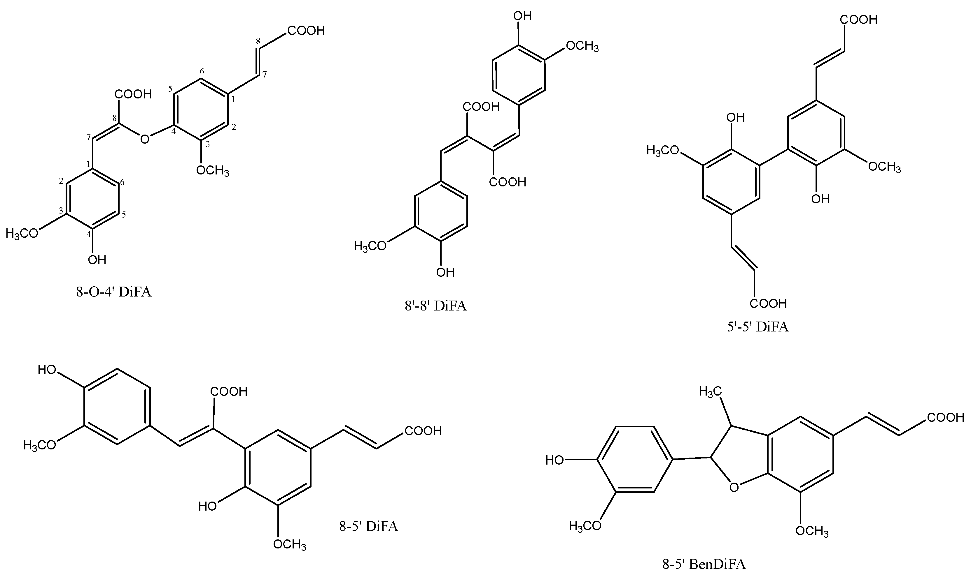
The five main type of dehydrodimers (Figure 3) found in nature are [18]:
  • 8-O-4′-DiFA:(Z)-β-{4-[(E)-2-carboxyvinyl]-2-methoxyphenoxy}-4-hydroxy-3-methoxy-cinnamic acid;
  • 8-5′ DiFA: (E,E)-4,4′-dihydroxy-3,5′-dimethoxy-β,3′-bicinnamic acid;
  • 8-5′-BenDiFA: in the benzofuran form;
  • 8-8′-DiFA: (E,E)-4,4′-dihydroxy-3,3′-dimethoxy-β,β′-bicinnamic acid;
  • 5-5′ DiFA: (E,E)-4,4′-dihydroxy-5,5′dimethoxy-β,3′-bicinnamic acid.
The exact type of dehydrodimers in a sample can be determined after their isolation (using enzymatic degradation or chemical saponification) and depends on the species that are being treated. Diferulates are present in a wide range of cereals such as maize, wheat, spelt, rice, wild rice, barley, rye, oats, and millet. For example, Bartolome et al. reported that in alkali- or enzyme-treated spent barley grain, 5′-5′ DiFA was the predominant diferulate [22]. It is estimated that the diferulate content is 8–39 times higher in the insoluble dietary fiber fraction than in the soluble one. The proportions of various dimers are different in the two fractions, with 8-5′ DiFA being predominant in the insoluble fraction and 8-8′ DiFA in the soluble [23]. Hatfield reported that model substrates of feruloylated arabinose mainly resulted in formation of 8-5′ DiFA and 8-5′ DiFA benzofuran [18].
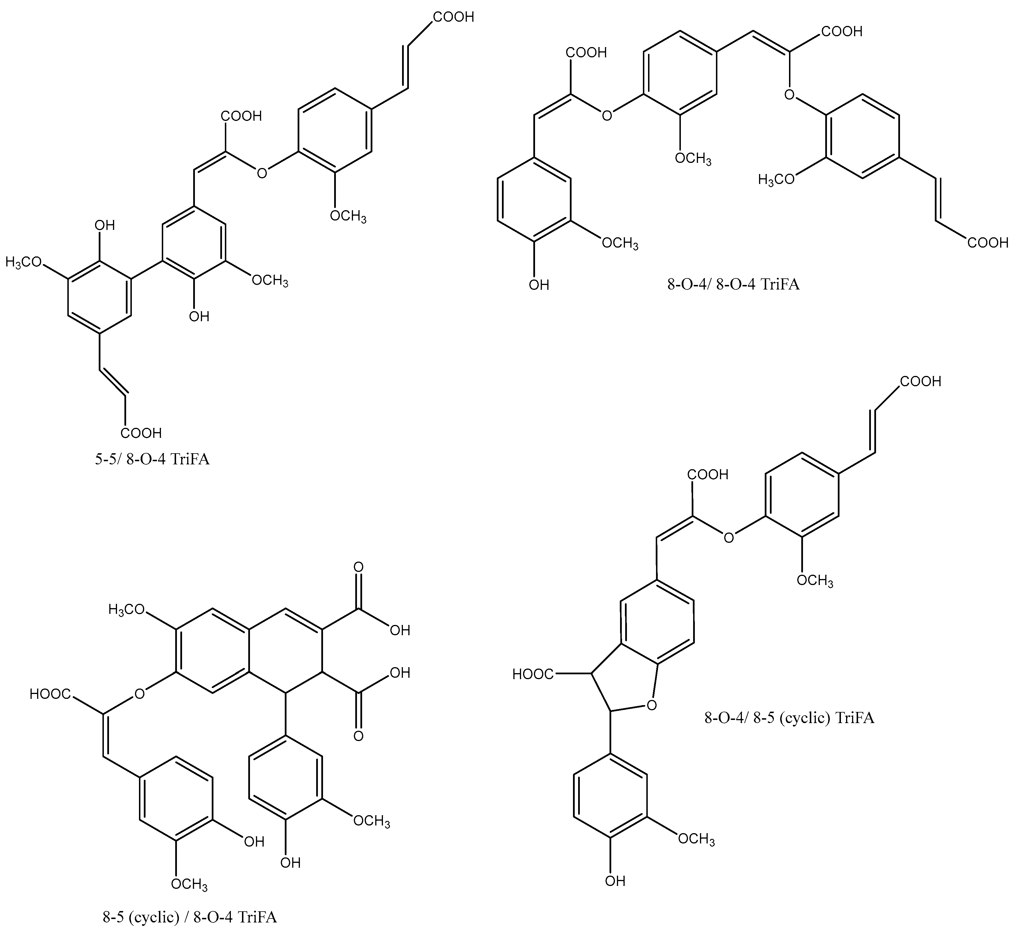
Oxidative coupling is not only limited to the formation of dehydrodimers. Several findings support the presence of both dehydrotrimers (Figure 4) and dehydrotetramers in maize bran [24,25,26,27,28,29]. These structures include the 5-5/8-O-4-coupled dehydrotrimer [27], which is hypothesized to be the prevailing trimer in corn bran, the 8-O-4/8-O-4-dehydrotrimer, and the 8-8 (cyclic)/8-O-4-dehydrotrimer [24]. The isolation of 8-O-4/8-O-4- and 8-8(cyclic)/8-O-4-coupled dehydrotrimers suggests that radical-coupled ferulate dehydrotrimers could potentially lead to cross-linking of three polysaccharide chains [24].
The two tetramers that have been isolated today are probably formed by 4-O-5 coupling of two DiFA 8-5′ (cyclic), and by 5-5′ DiFA−DiFA coupling [29].
Hydroxycinnamic acids were also widely hypothesized to covalently cross-link with Tyr and Cys residues in the cell wall proteins [30]. Proteins have multiple roles in the cell wall, such as transport, inhibitory functions, and structural purposes. Their secretion influences their solubility state, and these insoluble proteins are hypothesized to have ionic or salt interactions with polysaccharides [30]. Although no evidence that these linkages exist in nature has been reported yet, in vitro experiments have been conducted, and covalent coupling between proteins and polysaccharides is expected to result in new macromolecules with functional properties. FA could act as a substrate for the formation of FA oligomers in the presence of horseradish peroxidase (HRP) and H2O2, and this degree of oligomerization increases when the FA is bound to Gly-Tyr-Gly tripeptide [16,31]. In 2004, Boeriu et al. achieved cross-linking between protein and an FA polysaccharide. In the presence of HRP and H2O2, they enzymatically conjugated arabinoxylan to β-casein, which contains Tyr residues [32].

4. Feruloyl (Ferulic Acid) Esterases and Their Classification

FAEs are an important subclass of carboxylic esterases (E.C. 3.1.1), since they facilitate the degradation of plant cell walls via hydrolysis of the ester bond. The release of the ferulate ester groups can result in breaking of the cross-link between hemicellulose chains or between hemicellulose and lignin. FAEs usually exist in a monomeric form with a single catalytic site, and in some cases contain a carbohydrate-binding module (CBM). The presence of a CBM in FAEs has been shown to improve the catalytic efficiency of the protein [33,34,35,36,37,38]. Multiple attempts have been made to systematically classify these enzymes, and different systems have been developed over the years when trying to address their diversity. Below we summarize the main aspects, key features, and limitations of these systems (Table 1).
FAEs as carbohydrate-active enzymes are initially classified under the Carbohydrate Esterase Family 1 (CE1) in the CAZy database. This classification, with the criteria of sequence similarity and being within the same CE family, includes trehalose 6-O-mycolyltransferase (EC 2.3.1.122), diacylglycerol O-acyltransferase (EC 2.3.1.20) carboxylic ester hydrolase (EC 3.1.1), and acetyl xylan esterase (EC 3.1.1.72) [43]. Based on the CAZy database, additionally to CE1, one FAE is classified in CE15 as a 4-O-methyl-glucuronoyl esterase/feruloyl esterase (FI6_CE15).
A widely used FAE classification system is the one proposed by Crepin et al. in 2004 [39]. The criteria used for clustering were primary sequence identity and activity against synthetic substrates and diferulate. Based on these criteria, four subclasses were proposed: A, B, C, and D.
Four model substrates were used to estimate activity and were the methyl esters of the acids. These types of hydroxycinnamic acids are methyl ferulate (MFA), methyl caffeate (MCA), methyl sinapate (MSA), and methyl p-coumarate (MpCA). All enzymes from types C and D were active on all synthetic substrates, whereas type A was not active on MCA, and type B was not active on MSA. Additionally, type B and C enzymes were unable to release ferulic acid dehydrodimers from model and complex substrates [39].
A phylogenetic tree was constructed based on the available sequences, highlighting their similarity to other enzyme families. Type A enzymes resemble lipases, type B is similar to acetyl xylan esterases, type C is related to chlorogenate esterase and tannase, and type D shares similarities with xylanases. In addition, six sequences clustered outside this subclassification scheme, suggesting the existence of a fifth class, E.
The system, however, has certain limitations, which are acknowledged by the authors. These include a lack of comparable activity data, a reliance on primary sequence identity, and gaps in the knowledge regarding the type of biomass that triggers the gene expression of these proteins [39].
In 2008, the presence of FAEs in fungal genomes was extensively investigated by Benoit et al [40]. Starting with a strict threshold 1E70 of BlastP and continuing with phylogenetic analysis, multiple alignment (MUSCLE), and tree reconstruction methods, they proposed seven FAE subfamilies. Biochemically characterized members were included in three of these subfamilies (SF). The biochemical properties of these enzymes appeared to be different, suggesting that the SF described different classes of FAEs [40]. However, the number of FAE genes varies significantly, even among closely related species. This suggests that FAEs evolved from different esterase families and do not necessarily have a common ancestor. Some of these diverse esterase families that FAEs descent from have been classified in specific SFs, such as SF1-4 from tannases, SF6 from acetyl xylan esterase, and SF7 from lipases. However, in comparison to Crepin’s system, Type D FAEs are missing in Benoit’s analysis.
In parallel, in 2011, Udatha et al. [41] conducted a descriptor-based computational analysis that led to a broader classification of FAEs from fungi, bacteria, and plants. This system categorized FAEs into 12 families, with further subclassification within each family to distinguish substrate specificities [41].
Benoit’s work on fungal genes was later expanded by Dilokpimol et al. in 2016 [42]. They investigated the evolutionary relationships between the fungal FAE families and other related enzyme families such as acetyl xylan esterases, lipases, and tannases. Their phylogenetic tree analysis resulted in 13 FAE subfamilies. Some subfamilies, such as SF1–SF3 and SF5–SF7, remained consistent with earlier classifications, while SF8–SF13 represented newly identified evolutionary groups, highlighting the diversity within FAEs [42]. This work managed to provide a complete classification of fungal FAEs and illustrated that despite the catalytic triad Ser-His-Asp being conserved, FAEs do not have a common ancestor.
Moreover, this updated phylogenetic analysis demonstrated that Crepin’s ABCD system does not align well with the evolutionary relationships of FAEs. Some subfamilies are predominantly associated with a single type (A, B, C, or D), while others include multiple types, indicating greater complexity than previously understood.

5. Catalytic Mechanism and Well-Characterized FAEs

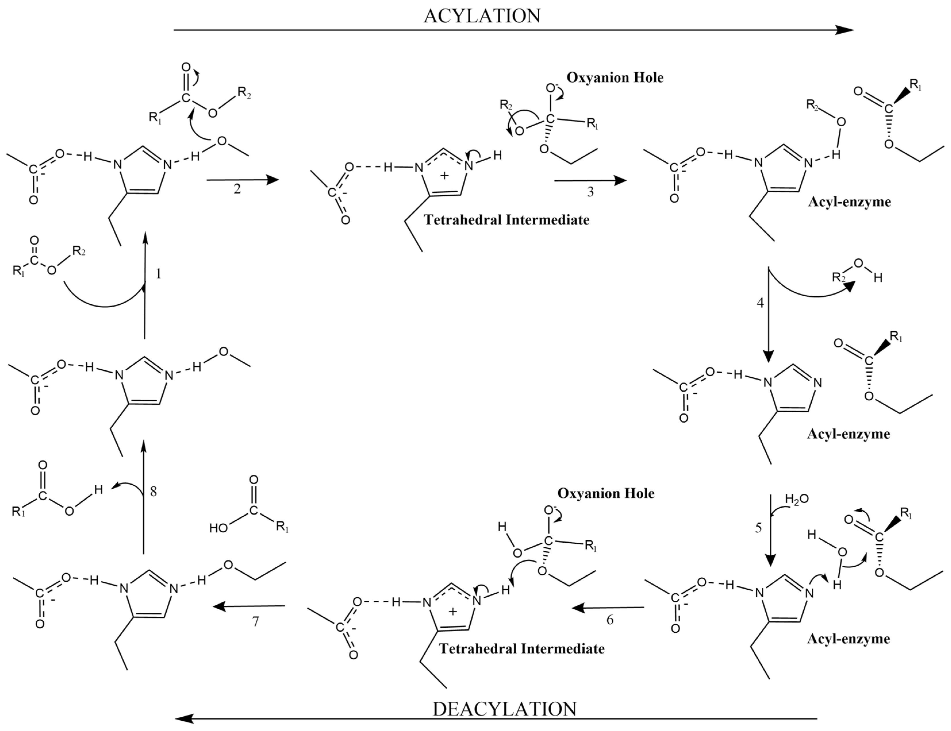
The catalytic mechanism of FAEs has been widely studied and is similar to that adopted by lipases, following the mechanism of serine hydrolases (Figure 5). The catalytic triad consists of Ser-His-Asp/Glu, and the consensus sequence GXSXG is surrounding the Ser. In brief, the hydroxyl group of serine (nucleophile) attacks the carbonyl carbon of the ester. This results in the formation of a tetrahedral intermediate, which then collapses to generate an acyl-enzyme intermediate, with the acyl moiety being covalently bound to the enzyme.
In the second step, a water molecule acts as a nucleophile, hydrolyzing the acyl-enzyme intermediate. Along with the protonation of the ester oxygen by His, this leads to the release of the acid in the deacylation step. In both steps, tetrahedral intermediates are formed and are energetically stabilized by the oxyanion hole, where two NH groups form hydrogen bonds with the carbonyl oxygen [44]. Specifically, for FAEs, the catalytic acid can interact directly with the para-hydroxy group, forming a hydrogen bond. Upon mutation, the esterase activity is maintained, but the recognition on hydroxycinnamate acids is abolished [45]. In some cases the same hydrogen bond is hypothesized to restrict the activity only on monomeric FA and prevent the recognition of dimers [46].
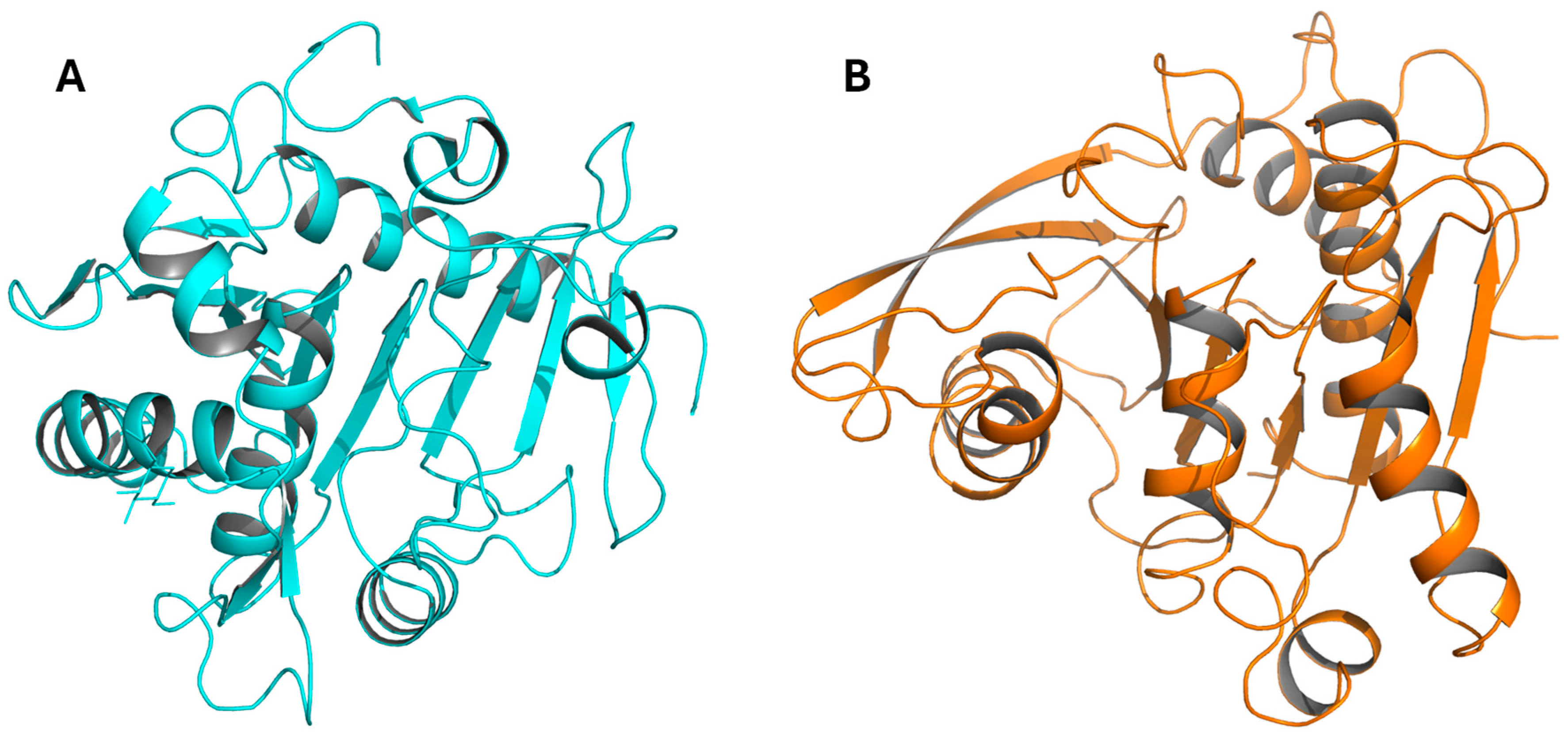
Currently, in the BRENDA database (brenda-enzyme.info) on functional and molecular information of enzymes, only 13 PDB entries and structure links are available, and all these enzymes have an α/β fold. Only few detailed studies on FAEs are available and originate from Aspergillus niger [47,48,49,50,51,52] and Clostridium thermocellum [53,54,55] (Figure 6). For example, AnFAE from A. niger is a α/β hydrolase, with the catalytic triad being described above [48,49,51]. This protein has a significant sequence similarity of 37% to a lipase from Rhizomucor miehi but does not exhibit any lipase activity [51,52]. Another detailed study on FAEs concerns enzymes designated XynY and XynZ [53,54,55], originating from Clostridium thermocellum. They exhibit a canonical α/β-hydrolase fold based on an eight-stranded β-sheet surrounded by α-helices. Despite this, they exhibit a poor structural homology with AnFAE, although the catalytic site of all three enzymes is well conserved.
A number of different FAEs are characterized to varying degrees (see Table 2) and occur either as monomers or dimers. Some FAEs have interesting and distinct features, including activity in the presence of various metal ions or activity under more extreme conditions (pH and/or temperature).
A metal ion-tolerant, 55kDa monomeric FAE from the mushroom Lactarius hatsudake (LhFAE) was characterized in 2016. LhFAE maintained 80% of the initial enzyme activity in the 5.0 mM presence of K+, Al3+, Pb2+, Cd2+, Mn2+, Mg2+, Zn2+, and Ca2+ ions [59].
Stability at extreme pH values has also been targeted. In 2005, Topakas et al. reported an FAE, designated StFaeC, from Sporotrichum thermophile, which was active on all methyl hydroxycinnamic acids and classified as type C. StFaeC was a dimer in solution and had an optimal pH at 6.0, but still retained 50% of its activity at pH 10 [57]. A low-pH-tolerant FAE, PpFAE, was isolated from Penicillium piceum. PpFAE exhibited activity at a broad pH range of 2.0–8.0 with an optimum at 3.0, while the optimum temperature was 70 °C [58]. As most FAEs exhibit optima under less harsh conditions (often pH 6–7 and temperature up to 50 °C), the high acidic thermal stability suggests wider application fields for this enzyme.
Further work includes the engineering of FAEs. In 2022, Yang et al. reported a new FAE from Geobacillus thermoglucosidasius and performed site-directed mutagenesis to enhance thermostability. Although the mutations increased the Tm, the enzyme activity decreased for all variants. The most favorable outcome (highest Tm) was observed in a double mutant. In this variant, an arginine substitution led to salt bridge formation and reduced flexibility in the cap region, while an asparagine substitution improved the hydrophilicity of the protein surface [61]. This indicates that the flexibility of the catalytic region is of importance for the activity, which was corroborated for a novel FAE from Lactobacillus acidophilus, designated as LaFae [62]. In this dimeric protein, a phenylalanine-to-alanine mutation near the catalytic site increased the activity. This variant probably exhibits enhanced flexibility of the cap domain, facilitating substrate binding [62].
Accessibility to the catalytic site may be another factor of importance. In 2022, Kasmaei et al. reported a new type of FAE homodimer in LbFAE from Lentilactobacillus buchneri. This enzyme exhibits a unique dimerization profile, and the active site of each monomer is located internally, facing the other protomer. This enzyme can only accommodate a single FA and is not active on dehydrodimers [45]. This contrasts with the findings of Lai et al. (2011), in whose study the characterized dimeric protein from Lactobacillus johnsonii N6.2 (LJ0536) demonstrated a solvent-exposed, open canal, large catalytic site [60].
These diverse structural and functional studies emphasize the versatility of FAEs across different microbial sources. The number of structurally resolved enzymes is still limited. However recent advances in protein engineering and characterization highlight FAEs’ potential for industrial and biotechnological applications.

6. FAEs in (Trans)Esterification

Various studies have demonstrated the versatility of fungal FAEs in a wide variety of reaction media containing mainly non-aqueous solvents, enabling efficient esterification and transesterification of phenolic acids with simple alcohols and with carbohydrates. For some applications, free ferulic acid is the most useful substance, but in other cases, derivatives of ferulic acid have more promising properties. Therefore, derivatization of ferulic acid, for example by esterification, is of great interest. Some of these cases are highlighted in the following section.
Giuliani et al. used an FAE to esterify ferulic acid with 1-pentanol in a microemulsion containing n-hexane, water, and the surfactant cetyltrimethylammoniumbromide [68]. A high yield was obtained (up to 60%), but due to the low solubility of ferulic acid in the system, the reactions were carried out at concentrations below 1 mM.
In 2003, Topakas et al. purified and characterized an FAE-II from Fusarium oxysporum that retained its catalytic activity in a surfactantless microemulsion containing n-hexane, 1-propanol, and water, in which the solubility of cinnamic acid derivatives was up to 100 times higher. This type of microemulsion is also called a ternary solvent system and has been used in several subsequent studies of FAE-catalyzed synthesis reactions. The F. oxysporum enzyme catalyzed the esterification of various phenolic acids but not cinnamic acid, indicating the necessity of a C-4 substitution on the phenyl ring [64].
In 2005, the same group reported the first enzymatic feruloylation of a carbohydrate, L-arabinose, with Sporotrichum thermophile FAE type C (StFaeC), in another ternary system consisting of n-hexane/t-butanol/water [57]. Other ternary systems were used for the feruloylation of di- and oligo-saccharides (n-hexane, 2-butanone, and MES–NaOH buffer), using an FAE from a commercial multi-enzyme preparation as a catalyst [69]. This composition was slightly altered for the synthesis of feruloylated galactobiose, where 2-butanone was exchanged with 1,4-dioxane [69].
Optimization of butyl hydroxycinnamate esters by transesterification of methyl esters catalyzed by a novel type C FAE from Aspergillus ochraceus was carried out in a ternary system containing isooctane/butanol/water [65]. This enzyme was quite specific for butanol, while other alcohols were converted at much lower rates. Synthesis of feruloyl glycerol esters was achieved in a solvent-free system of just ferulic acid in a mixture of glycerol and water using re-AoFaeA as a catalyst, achieving a yield of 60.3% [70].
Successful FAE-catalyzed transesterification reactions in aqueous/organic two-phase systems were carried out using Talaromyces wortmannii FAEs as catalysts [66]. In the reaction between vinyl ferulate and prenol, just n-hexane with a small addition of water was used as the reaction medium, and in the reaction between vinyl ferulate and L-arabinose, a small amount of DMSO was added as well. The reactions were optimized, giving yields of 87.5% for prenol and 56.2% for L-arabinose. In the case of prenyl ferulate synthesis, the aqueous phase was recovered after the reaction, making it possible to reuse the enzyme, a procedure which was repeated for six consecutive cycles.

7. Synergistic Degradation and Bifunctional Esterases

Given the complexity of the plant cell wall, it is natural to assume that different types of enzymes are necessary for its efficient degradation. The increased activity of enzymes when combined with others from different classes is referred to as synergy. There are multiple reports of FAEs working synergistically with various carbohydrate-degrading enzymes to break down different biomass side streams. FAEs have demonstrated synergistic effects with xylanases [47,57,59,70], cellulases [58], and laccases. The combination of AnXyn11A and AnFaeA from Aspergillus niger BE-2 increased the initial release of ferulic acid (FA) from destarched wheat bran from 16.8% to 61% when added simultaneously [71]. In addition, an engineered bifunctional xylanase/feruloyl esterase (XynII-Fae) from Prevotella ruminicola 23 showed synergistic effects with commercial cellulase for the delignification of corn stover [67]. In this engineered bifunctional protein, each reaction was catalyzed by separate domains, with a TIM barrel and α/β hydrolase folding, respectively. A recent study from Schmitz et al., conducted in 2022 [72], examined the degree of synergy for multiple commercial carbohydrate enzymes in different biomass types, including corn bran, soluble oat bran, insoluble oat bran, and oat hull. The study found varying levels of synergy depending on the substrate [72]. In this work, FAEs showed synergy with laccase and xylanases from GH families 5, 10, and 11. However, not all side streams exhibited this effect, suggesting that the composition and nature of the biomass play a determining role in the degree of degradation [72].
The chemical complexity of plant biomass has directed evolution towards enzymes with dual functionality. This dual activity can occur within the same type of bond, such as esters, or span across different bond types, like ester and glycosidic bonds. For example, Dilokpimol et al. (2022) reported three novel feruloyl/acetyl xylan esterases (FXEs) that are capable of efficiently releasing both ferulic acid and acetic acid from wheat arabinoxylan [73]. Earlier, in 2000, Blum et al. demonstrated that XynY and XynZ, components of the Clostridium thermocellum cellulosome, exhibited FAE activity originating from their C-terminal and N-terminal domains, respectively, in addition to their established xylanase activity. The xylanase function in these proteins is associated with GH family 10, while the feruloyl activity originates from distinct catalytic domains [55]. Here again, the enzyme is not a single fold carrying out both activities, it is a multi-domain structure with dual functionality. More recently, Schmitz et al. (2023) reported a bifunctional arabinoxylanase/feruloyl esterase belonging to GH family 5, in which both activities share the same active site. In that case, the recognition of the arabinose substitution is specifically required for xylanase activity [74].

8. Estimation of Enzymatic Activity and Detection of FA

The catalytic activity of FAEs can be studied using either complex natural substrates or synthetic model compounds. When complex substrates are used, the released FA is monitored. High-Performance Liquid Chromatography (HPLC) with a C-18 reverse-phase column is commonly employed for quantitative analysis, typically using UV detection at 320 nm [75]. Gas Chromatography–Mass Spectrometry (GC-MS) is also suitable for the detection, quantification, and separation of geometrical isomers of FA [76]. Kvasnička et al. separated 12 phenolic acids including FA, with capillary zone electrophoresis (CZE) using HPLC with UV detection [77]. There are multiply reports of Liquid Chromatography–Tandem Mass Spectrometry (LC-MS/MS) being used for the identification and quantification of FA in a diverse range of substrates, such as Chinese herbal medicine [78], rat plasma [79], and milk [80].
Qualitative detection can be achieved with thin-layer chromatography TLC [81] and high-performance thin-layer chromatography (HPTLC), which have been proven effective for the separation and detection of ferulic acid and vanillin [82].
In terms of synthetic substrates, methyl esters of hydroxycinnamic acids are frequently used, particularly for enzyme classification into types A, B, C, and D. Methyl ferulate (MFA), methyl caffeate (MCA), methyl sinapate (MSA), and methyl p-coumarate (MpCA) are incubated with the enzyme, and the hydrolysis products are analyzed via HPLC, providing a detailed estimation of the enzyme activity with these different substrates [39,48,83]. Another widely used method involves the use of p-nitrophenyl (pNP) esters, such as pNP-ferulate or pNP-acetate, in spectrophotometric assays [83]. These allow for real-time monitoring of enzyme activity by measuring the release of p-nitrophenol at 410 nm.
Frequently, FAEs exhibit dual activity as feruloyl esterases and acetyl xylan esterases. It is therefore of interest to test the enzymatic activity and preference in both pNP-ferulate and pNP-acetate [73,74,84]. These synthetic substrate assays are advantageous for enzyme screenings, since they are compatible with high-throughput methods and kinetic analysis and can facilitate FAE classification. Nevertheless, it is important to note that they do not reflect the complexity found in nature. Therefore, high activity on synthetic substrates does not necessarily translate to efficient activity on plant cell wall materials.

9. Applications of FAEs and FA

FAEs’ applications can be viewed in two ways. One focuses on their enzymatic effect on biomass, where degrading the side chain enhances the accessibility of hydrolytic enzymes to cellulosic and hemicellulosic substrates and lignin–polysaccharide chains (described in the Section 7). The other considers FA as a valuable product with diverse applications in pharmaceuticals, cosmetics, food, feed, food packaging, and biogels [85].
In pharmaceuticals and cosmetics, FA is widely used for its antioxidant, anti-inflammatory, anti-aging, and photoprotective properties [85,86]. It is an important ingredient in sunscreens and skin-whitening products due to its ability to inhibit melanin accumulation, protect against UV radiation, and prevent skin photoaging [86,87,88].
Additionally, FA exhibits anti-cancer properties by inhibiting reactive oxygen species (ROS), protecting cellular components from oxidative damage, and regulating intracellular signaling related to proliferation, apoptosis, and metastasis. FA and its derivatives also offer antithrombotic, immune-regulating, and neuroprotective benefits, aiding in the treatment of cardiovascular and cerebrovascular diseases [89]. Studies further suggest wound-healing properties, demonstrated in experiments on diabetic rats [90,91]. These benefits derive from FA’s free radical scavenging ability and reduction in oxidative damage [85]. FA can be used as substrate in enzymatic decarboxylation reactions producing styrene derivatives, which can be converted further to various products, such as biobased polymers [92,93,94].
In the food industry, FA is a precursor for vanillin production [95], a flavoring compound that is also used in drug delivery systems and as battery energy storage through vanillin-cross-linked chitosan electrodes [96].
FA is also incorporated into fibrous films for fruit preservation, where its core–sheath-structured fibers provide both immediate and sustained antioxidant release, helping to prevent oxidation, reduce water loss, and extend shelf life [97]. Despite its broad potential across various fields, FA’s practical applications remain limited due to its low water solubility [85].

10. Conclusions

This review aims to provide a comprehensive overview of FAEs and ferulic acid (FA). It explores the various natural forms and cross-links of ferulic acid, emphasizing the compound’s structural complexity. All FAEs are serine hydrolases, playing an important role in the effective valorization of biomass, alone or in synergy with other enzymes. The released ferulic acid has broad applications across the pharmaceutical, food, and chemical industries. FAEs are also used as biosynthetic catalysts in (trans)esterification reactions involving non-aqueous solvents. By evaluating the current classification systems and the structural diversity of these enzymes, this review highlights the need for further research to fully elucidate their structure–function relationships and to deepen our understanding of the complex patterns of nature to which they correspond.

Author Contributions

Conceptualization, E.N.K. and S.L.; writing—original draft preparation, S.L.; writing—review and editing, S.L., P.A. and E.N.K. visualization, S.L.; supervision, P.A. and E.N.K.; project administration, E.N.K.; funding acquisition E.N.K. All authors have read and agreed to the published version of the manuscript.

Funding

This research was funded by the EU Horizon 2020 project EnXylaScope, grant number 101000831, and the Horizon EUROPE project BIONEER, grant number 101157779.

Conflicts of Interest

The authors declare no conflicts of interest.

Abbreviations

The following abbreviations are used in this manuscript:
FAFerulic Acid
FAEFerulic Acid Esterase
CAZymesCarbohydrate-Active Enzymes
GAXGlucuronoarabinoxylan
AXArabinoxylan
DiFADi-Ferulic Acid
BenDiFABenzofuran Form of Di-Ferulic Acid
HRPHorseradish Peroxidase
CBMCarbohydrate-Binding Module
CE1Carbohydrate Esterase Family 1
MFAMethyl Ferulate
MCAMethyl Caffeate
MSAMethyl Sinapate
MpCAMethyl p-Coumarate
GHGlycoside Hydrolase Family
HPLCHigh-Performance Liquid Chromatography
GC-MSGas Chromatography–Mass Spectrometry
CZECapillary Zone Electrophoresis
LC-MS/MSLiquid Chromatography–Tandem Mass Spectrometry
TLCThin-Layer Chromatography
HPTLCHigh-Performance Thin-Layer Chromatography
pNPPara Nitrophenol
ROSReactive Oxygen Species

References

  1. Saha, B.C. Hemicellulose bioconversion. J. Ind. Microbiol. Biotechnol. 2003, 30, 279–291. [Google Scholar] [CrossRef]
  2. Terrett, O.M.; Dupree, P. Covalent interactions between lignin and hemicelluloses in plant secondary cell walls. Curr. Opin. Biotechnol. 2019, 56, 97–104. [Google Scholar] [CrossRef]
  3. Natella, F.; Nardini, M.; Giannetti, I.; Dattilo, C.; Scaccini, C. Coffee drinking influences plasma antioxidant capacity in humans. J. Agric. Food Chem. 2002, 50, 6211–6216. [Google Scholar] [CrossRef]
  4. Johnston, K.L.; Clifford, M.N.; Morgan, L.M. Coffee acutely modifies gastrointestinal hormone secretion and glucose tolerance in humans: Glycemic effects of chlorogenic acid and caffeine. Am. J. Clin. Nutr. 2003, 78, 728–733. [Google Scholar] [CrossRef]
  5. Scheller, H.V.; Ulvskov, P. Hemicelluloses. Annu. Rev. Plant Biol. 2010, 61, 263–289. [Google Scholar] [CrossRef] [PubMed]
  6. Ishii, T.; Hiroi, T. Linkage of phenolic acids to cell-wall polysaccharides of bamboo shoot. Carbohydr. Res. 1990, 206, 297–310. [Google Scholar] [CrossRef] [PubMed]
  7. Locher, R.; Martin, H.V.; Grison, R.; Pilet, P.E. Cell wall-bound trans-and cis-ferulic acids in growing maize roots. Physiol. Plant. 1994, 90, 734–738. [Google Scholar] [CrossRef]
  8. Tang, Y.; Hao, J.; Fan, C.; Cao, X. Preparative separation of high-purity trans-and cis-ferulic acid from wheat bran by pH-zone-refining counter-current chromatography. J. Chromatogr. A 2021, 1636, 461772. [Google Scholar] [CrossRef]
  9. Rombouts, F.M.; Thibault, J.-F. Feruloylated pectic substances from sugar-beet pulp. Carbohydr. Res. 1986, 154, 177–187. [Google Scholar] [CrossRef]
  10. Fry, S.C. Feruloylated pectins from the primary cell wall: Their structure and possible functions. Planta 1983, 157, 111–123. [Google Scholar] [CrossRef]
  11. Colquhoun, I.J.; Ralet, M.-C.; Thibault, J.-F.; Faulds, C.B.; Williamson, G. Structure identification of feruloylated oligosaccharides from sugar-beet pulp by NMR spectroscopy. Carbohydr. Res. 1994, 263, 243–256. [Google Scholar] [CrossRef]
  12. Parr, A.J.; Waldron, K.W.; Ng, A.; Parker, M.L. The wall-bound phenolics of Chinese water chestnut (Eleocharis dulcis). J. Sci. Food Agric. 1996, 71, 501–507. [Google Scholar] [CrossRef]
  13. Massiot, P.; Rouau, X.; Thibault, J.-F. Characterisation of the extractable pectins and hemicelluloses of the cell wall of carrot. Carbohydr. Res. 1988, 172, 229–242. [Google Scholar] [CrossRef]
  14. Renard, C.; Champenois, Y.; Thibault, J.-F. Characterisation of the extractable pectins and hemicelluloses of the cell wall of glasswort, Salicornia ramosissima. Carbohydr. Polym. 1993, 22, 239–245. [Google Scholar] [CrossRef]
  15. Sanchez, M.; Pena, M.J.; Revilla, G.; Zarra, I. Changes in dehydrodiferulic acids and peroxidase activity against ferulic acid associated with cell walls during growth of Pinus pinaster hypocotyl. Plant Physiol. 1996, 111, 941–946. [Google Scholar] [CrossRef]
  16. Ward, G.; Hadar, Y.; Bilkis, I.; Konstantinovsky, L.; Dosoretz, C.G. Initial steps of ferulic acid polymerization by lignin peroxidase. J. Biol. Chem. 2001, 276, 18734–18741. [Google Scholar] [CrossRef]
  17. Nordkvist, E.; Salomonsson, A.C.; Åman, P. Distribution of insoluble bound phenolic acids in barley grain. J. Sci. Food Agric. 1984, 35, 657–661. [Google Scholar] [CrossRef]
  18. Hatfield, R.D.; Ralph, J.; Grabber, J.H. Cell wall cross-linking by ferulates and diferulates in grasses. J. Sci. Food Agric. 1999, 79, 403–407. [Google Scholar] [CrossRef]
  19. Ford, C.W.; Hartley, R.D. GC/MS characterisation of cyclodimers from p-coumaric and ferulic acids by photodimerisation—A possible factor influencing cell wall biodegradability. J. Sci. Food Agric. 1989, 46, 301–310. [Google Scholar] [CrossRef]
  20. Ford, C.W.; Hartley, R.D. Cyclodimers of p-coumaric and ferulic acids in the cell walls of tropical grasses. J. Sci. Food Agric. 1990, 50, 29–43. [Google Scholar] [CrossRef]
  21. Hartley, R.D.; Whatley, F.R.; Harris, P.J. 4, 4′-Dihydroxytruxillic acid as a component of cell walls of Lolium multiflorum. Phytochemistry 1988, 27, 349–351. [Google Scholar] [CrossRef]
  22. Bartolome, B.; Faulds, C.B.; Kroon, P.A.; Waldron, K.; Gilbert, H.J.; Hazlewood, G.; Williamson, G. An Aspergillus niger esterase (ferulic acid esterase III) and a recombinant Pseudomonas fluorescens subsp. cellulosa esterase (Xy1D) release a 5-5′ferulic dehydrodimer (diferulic acid) from barley and wheat cell walls. Appl. Environ. Microbiol. 1997, 63, 208–212. [Google Scholar] [CrossRef] [PubMed]
  23. Bunzel, M.; Ralph, J.; Marita, J.M.; Hatfield, R.D.; Steinhart, H. Diferulates as structural components in soluble and insoluble cereal dietary fibre. J. Sci. Food Agric. 2001, 81, 653–660. [Google Scholar] [CrossRef]
  24. Funk, C.; Ralph, J.; Steinhart, H.; Bunzel, M. Isolation and structural characterisation of 8–O–4/8–O–4-and 8–8/8–O–4-coupled dehydrotriferulic acids from maize bran. Phytochemistry 2005, 66, 363–371. [Google Scholar] [CrossRef]
  25. Bunzel, M.; Ralph, J.; Funk, C.; Steinhart, H. Structural elucidation of new ferulic acid-containing phenolic dimers and trimers isolated from maize bran. Tetrahedron Lett. 2005, 46, 5845–5850. [Google Scholar] [CrossRef]
  26. Rouau, X.; Cheynier, V.; Surget, A.; Gloux, D.; Barron, C.; Meudec, E.; Louis-Montero, J.; Criton, M. A dehydrotrimer of ferulic acid from maize bran. Phytochemistry 2003, 63, 899–903. [Google Scholar] [CrossRef]
  27. Bunzel, M.; Ralph, J.; Funk, C.; Steinhart, H. Isolation and identification of a ferulic acid dehydrotrimer from saponified maize bran insoluble fiber. Eur. Food Res. Technol. 2003, 217, 128–133. [Google Scholar] [CrossRef]
  28. Bunzel, M.; Ralph, J.; Brüning, P.; Steinhart, H. Structural identification of dehydrotriferulic and dehydrotetraferulic acids isolated from insoluble maize bran fiber. J. Agric. Food Chem. 2006, 54, 6409–6418. [Google Scholar] [CrossRef]
  29. Bunzel, M.; Heuermann, B.; Kim, H.; Ralph, J. Peroxidase-catalyzed oligomerization of ferulic acid esters. J. Agric. Food Chem. 2008, 56, 10368–10375. [Google Scholar] [CrossRef] [PubMed]
  30. Iiyama, K.; Lam, T.B.-T.; Stone, B.A. Covalent cross-links in the cell wall. Plant Physiol. 1994, 104, 315. [Google Scholar] [CrossRef] [PubMed]
  31. Oudgenoeg, G.; Dirksen, E.; Ingemann, S.; Hilhorst, R.; Gruppen, H.; Boeriu, C.G.; Piersma, S.R.; van Berkel, W.J.; Laane, C.; Voragen, A.G. Horseradish peroxidase-catalyzed oligomerization of ferulic acid on a template of a tyrosine-containing tripeptide. J. Biol. Chem. 2002, 277, 21332–21340. [Google Scholar] [CrossRef] [PubMed]
  32. Boeriu, C.G.; Oudgenoeg, G.; Spekking, W.T.J.; Berendsen, L.B.; Vancon, L.; Boumans, H.; Gruppen, H.; van Berkel, W.J.; Laane, C.; Voragen, A.G. Horseradish peroxidase-catalyzed cross-linking of feruloylated arabinoxylans with β-casein. J. Agric. Food Chem. 2004, 52, 6633–6639. [Google Scholar] [CrossRef]
  33. Ferreira, L.; Wood, T.M.; Williamson, G.; Faulds, C.; Hazlewood, G.P.; Black, G.W.; Gilbert, H.J. A modular esterase from Pseudomonas fluorescens subsp. cellulosa contains a non-catalytic cellulose-binding domain. Biochem. J. 1993, 294, 349–355. [Google Scholar] [CrossRef]
  34. Kroon, P.A.; Williamson, G.; Fish, N.M.; Archer, D.B.; Belshaw, N.J. A modular esterase from Penicillium funiculosum which releases ferulic acid from plant cell walls and binds crystalline cellulose contains a carbohydrate binding module. Eur. J. Biochem. 2000, 267, 6740–6752. [Google Scholar] [CrossRef]
  35. Holck, J.; Fredslund, F.; Møller, M.S.; Brask, J.; Krogh, K.B.; Lange, L.; Welner, D.H.; Svensson, B.; Meyer, A.S.; Wilkens, C. A carbohydrate-binding family 48 module enables feruloyl esterase action on polymeric arabinoxylan. J. Biol. Chem. 2019, 294, 17339–17353. [Google Scholar] [CrossRef]
  36. Mamiya, A.; Sakka, M.; Kosugi, A.; Katsuzaki, H.; Tanaka, A.; Kunitake, E.; Kimura, T.; Sakka, K. Significance of a family-6 carbohydrate-binding module in a modular feruloyl esterase for removing ferulic acid from insoluble wheat arabinoxylan. Enzym. Microb. Technol. 2020, 138, 109546. [Google Scholar] [CrossRef] [PubMed]
  37. Holck, J.; Djajadi, D.T.; Brask, J.; Pilgaard, B.; Krogh, K.B.; Meyer, A.S.; Lange, L.; Wilkens, C. Novel xylanolytic triple domain enzyme targeted at feruloylated arabinoxylan degradation. Enzym. Microb. Technol. 2019, 129, 109353. [Google Scholar] [CrossRef]
  38. Lin, S.; Hunt, C.J.; Holck, J.; Brask, J.; Krogh, K.B.; Meyer, A.S.; Wilkens, C.; Agger, J.W. Fungal feruloyl esterases can catalyze release of diferulic acids from complex arabinoxylan. Int. J. Biol. Macromol. 2023, 232, 123365. [Google Scholar] [CrossRef]
  39. Crepin, V.F.; Faulds, C.B.; Connerton, I. Functional classification of the microbial feruloyl esterases. Appl. Microbiol. Biotechnol. 2004, 63, 647–652. [Google Scholar] [CrossRef]
  40. Benoit, I.; Danchin, E.G.; Bleichrodt, R.-J.; de Vries, R.P. Biotechnological applications and potential of fungal feruloyl esterases based on prevalence, classification and biochemical diversity. Biotechnol. Lett. 2008, 30, 387–396. [Google Scholar] [CrossRef] [PubMed]
  41. Udatha, G. Descriptor-based computational analysis reveals a novel classification scheme for feruloyl esterase enzyme families. In Proceedings of the International Conference on Bioinformatics, Kuala Lumpur, Malaysia, 30 November 2011–2 December 2011. [Google Scholar]
  42. Dilokpimol, A.; Mäkelä, M.R.; Aguilar-Pontes, M.V.; Benoit-Gelber, I.; Hildén, K.S.; de Vries, R.P. Diversity of fungal feruloyl esterases: Updated phylogenetic classification, properties, and industrial applications. Biotechnol. Biofuels 2016, 9, 231. [Google Scholar] [CrossRef]
  43. Lombard, V.; Henrissat, B.; Garron, M.-L. CAZac: An activity descriptor for carbohydrate-active enzymes. Nucleic Acids Res. 2025, 53, D625–D633. [Google Scholar] [CrossRef]
  44. Faber, K.; Faber, K. Biotransformations in Organic Chemistry: A Textbook; Springer: Heidelberg, Germany, 2004; Volume 6. [Google Scholar]
  45. Kasmaei, K.M.; Kalyani, D.C.; Reichenbach, T.; Jiménez-Quero, A.; Vilaplana, F.; Divne, C. Crystal structure of the feruloyl esterase from Lentilactobacillus buchneri reveals a novel homodimeric state. Front. Microbiol. 2022, 13, 1050160. [Google Scholar] [CrossRef]
  46. Faulds, C.B.; Molina, R.; Gonzalez, R.; Husband, F.; Juge, N.; Sanz-Aparicio, J.; Hermoso, J.A. Probing the determinants of substrate specificity of a feruloyl esterase, AnFaeA, from Aspergillus niger. FEBS J. 2005, 272, 4362–4371. [Google Scholar] [CrossRef]
  47. Dilokpimol, A.; Mäkelä, M.R.; Mansouri, S.; Belova, O.; Waterstraat, M.; Bunzel, M.; de Vries, R.P.; Hildén, K.S. Expanding the feruloyl esterase gene family of Aspergillus niger by characterization of a feruloyl esterase, FaeC. New Biotechnol. 2017, 37, 200–209. [Google Scholar] [CrossRef] [PubMed]
  48. Faulds, C.B.; Williamson, G. Purification and characterization of a ferulic acid esterase (FAE-III) from Aspergillus niger: Specificity for the phenolic moiety and binding to microcrystalline cellulose. Microbiology 1994, 140, 779–787. [Google Scholar] [CrossRef]
  49. De Vries, R.P.; Michelsen, B.; Poulsen, C.; Kroon, P.; Van Den Heuvel, R.; Faulds, C.; Williamson, G.; Van Den Hombergh, J.; Visser, J. The faeA genes from Aspergillus niger and Aspergillus tubingensis encode ferulic acid esterases involved in degradation of complex cell wall polysaccharides. Appl. Environ. Microbiol. 1997, 63, 4638–4644. [Google Scholar] [CrossRef]
  50. de Vries, R.P.; vanKuyk, P.A.; Kester, H.C.; Visser, J. The Aspergillus niger faeB gene encodes a second feruloyl esterase involved in pectin and xylan degradation and is specifically induced in the presence of aromatic compounds. Biochem. J. 2002, 363, 377–386. [Google Scholar] [CrossRef]
  51. Hermoso, J.A.; Sanz-Aparicio, J.; Molina, R.; Juge, N.; González, R.; Faulds, C.B. The crystal structure of feruloyl esterase A from Aspergillus niger suggests evolutive functional convergence in feruloyl esterase family. J. Mol. Biol. 2004, 338, 495–506. [Google Scholar] [CrossRef] [PubMed]
  52. Aliwan, F.O.; Kroon, P.A.; Faulds, C.B.; Pickersgill, R.; Williamson, G. Ferulic acid esterase-III from Aspergillus niger does not exhibit lipase activity. J. Sci. Food Agric. 1999, 79, 457–459. [Google Scholar] [CrossRef]
  53. Prates, J.A.; Tarbouriech, N.; Charnock, S.J.; Fontes, C.M.; Ferreira, L.S.M.; Davies, G.J. The structure of the feruloyl esterase module of xylanase 10B from Clostridium thermocellum provides insights into substrate recognition. Structure 2001, 9, 1183–1190. [Google Scholar] [CrossRef]
  54. Schubot, F.D.; Kataeva, I.A.; Blum, D.L.; Shah, A.K.; Ljungdahl, L.G.; Rose, J.P.; Wang, B.-C. Structural basis for the substrate specificity of the feruloyl esterase domain of the cellulosomal xylanase Z from Clostridium thermocellum. Biochemistry 2001, 40, 12524–12532. [Google Scholar] [CrossRef]
  55. Blum, D.L.; Kataeva, I.A.; Li, X.-L.; Ljungdahl, L.G. Feruloyl esterase activity of the Clostridium thermocellum cellulosome can be attributed to previously unknown domains of XynY and XynZ. J. Bacteriol. 2000, 182, 1346–1351. [Google Scholar] [CrossRef]
  56. Suzuki, K.; Hori, A.; Kawamoto, K.; Thangudu, R.R.; Ishida, T.; Igarashi, K.; Samejima, M.; Yamada, C.; Arakawa, T.; Wakagi, T. Crystal structure of a feruloyl esterase belonging to the tannase family: A disulfide bond near a catalytic triad. Proteins Struct. Funct. Bioinform. 2014, 82, 2857–2867. [Google Scholar] [CrossRef] [PubMed]
  57. Topakas, E.; Vafiadi, C.; Stamatis, H.; Christakopoulos, P. Sporotrichum thermophile type C feruloyl esterase (StFaeC): Purification, characterization, and its use for phenolic acid (sugar) ester synthesis. Enzym. Microb. Technol. 2005, 36, 729–736. [Google Scholar] [CrossRef]
  58. Gao, L.; Wang, M.; Chen, S.; Zhang, D. Biochemical characterization of a novel feruloyl esterase from Penicillium piceum and its application in biomass bioconversion. J. Mol. Catal. B Enzym. 2016, 133, S388–S394. [Google Scholar] [CrossRef]
  59. Wang, L.; Li, Z.; Zhu, M.; Meng, L.; Wang, H.; Ng, T.B. An acidic feruloyl esterase from the mushroom Lactarius hatsudake: A potential animal feed supplement. Int. J. Biol. Macromol. 2016, 93, 290–295. [Google Scholar] [CrossRef]
  60. Lai, K.-K.; Stogios, P.J.; Vu, C.; Xu, X.; Cui, H.; Molloy, S.; Savchenko, A.; Yakunin, A.; Gonzalez, C.F. An inserted α/β subdomain shapes the catalytic pocket of Lactobacillus johnsonii cinnamoyl esterase. PLoS ONE 2011, 6, e23269. [Google Scholar] [CrossRef]
  61. Yang, W.; Sun, L.; Dong, P.; Chen, Y.; Zhang, H.; Huang, X.; Wu, L.; Chen, L.; Jing, D.; Wu, Y. Structure-guided rational design of the Geobacillus thermoglucosidasius feruloyl esterase GthFAE to improve its thermostability. Biochem. Biophys. Res. Commun. 2022, 600, 117–122. [Google Scholar] [CrossRef] [PubMed]
  62. Jeon, S.; Hwang, J.; Do, H.; Le, L.T.H.L.; Lee, C.W.; Yoo, W.; Lee, M.J.; Shin, S.C.; Kim, K.K.; Kim, H.-W. Feruloyl Esterase (La Fae) from Lactobacillus acidophilus: Structural Insights and Functional Characterization for Application in Ferulic Acid Production. Int. J. Mol. Sci. 2023, 24, 11170. [Google Scholar] [CrossRef]
  63. Gruninger, R.J.; Cote, C.; McAllister, T.A.; Abbott, D.W. Contributions of a unique β-clamp to substrate recognition illuminates the molecular basis of exolysis in ferulic acid esterases. Biochem. J. 2016, 473, 839–849. [Google Scholar] [CrossRef]
  64. Topakas, E.; Stamatis, H.; Biely, P.; Kekos, D.; Macris, B.; Christakopoulos, P. Purification and characterization of a feruloyl esterase from Fusarium oxysporum catalyzing esterification of phenolic acids in ternary water–organic solvent mixtures. J. Biotechnol. 2003, 102, 33–44. [Google Scholar] [CrossRef]
  65. Romero-Borbón, E.; Grajales-Hernández, D.; Armendáriz-Ruiz, M.; Ramírez-Velasco, L.; Rodríguez-González, J.A.; Cira-Chávez, L.A.; Estrada-Alvarado, M.I.; Mateos-Díaz, J.C. Type C feruloyl esterase from Aspergillus ochraceus: A butanol specific biocatalyst for the synthesis of hydroxycinnamates in a ternary solvent system. Electron. J. Biotechnol. 2018, 35, 1–9. [Google Scholar] [CrossRef]
  66. Antonopoulou, I.; Iancu, L.; Jütten, P.; Piechot, A.; Rova, U.; Christakopoulos, P. Screening of novel feruloyl esterases from Talaromyces wortmannii for the development of efficient and sustainable syntheses of feruloyl derivatives. Enzym. Microb. Technol. 2019, 120, 124–135. [Google Scholar] [CrossRef] [PubMed]
  67. Wang, H.; Qi, X.; Gao, S.; Zhang, Y.; An, Y. Biochemical characterization of an engineered bifunctional xylanase/feruloyl esterase and its synergistic effects with cellulase on lignocellulose hydrolysis. Bioresour. Technol. 2022, 355, 127244. [Google Scholar] [CrossRef]
  68. Giuliani, S.; Piana, C.; Setti, L.; Hochkoeppler, A.; Pifferi, P.; Williamson, G.; Faulds, C. Synthesis of pentylferulate by a feruloyl esterase from Aspergillus niger using water-in-oil microemulsions. Biotechnol. Lett. 2001, 23, 325–330. [Google Scholar] [CrossRef]
  69. Couto, J.; St-Louis, R.; Karboune, S. Optimization of feruloyl esterase-catalyzed synthesis of feruloylated oligosaccharides by response surface methodology. J. Mol. Catal. B Enzym. 2011, 73, 53–62. [Google Scholar] [CrossRef]
  70. Zeng, Y.; Yin, X.; Wu, M.-C.; Yu, T.; Feng, F.; Zhu, T.-D.; Pang, Q.-F. Expression of a novel feruloyl esterase from Aspergillus oryzae in Pichia pastoris with esterification activity. J. Mol. Catal. B Enzym. 2014, 110, 140–146. [Google Scholar] [CrossRef]
  71. Wu, H.; Li, H.; Xue, Y.; Luo, G.; Gan, L.; Liu, J.; Mao, L.; Long, M. High efficiency co-production of ferulic acid and xylooligosaccharides from wheat bran by recombinant xylanase and feruloyl esterase. Biochem. Eng. J. 2017, 120, 41–48. [Google Scholar] [CrossRef]
  72. Schmitz, E.; Leontakianakou, S.; Norlander, S.; Karlsson, E.N.; Adlercreutz, P. Lignocellulose degradation for the bioeconomy: The potential of enzyme synergies between xylanases, ferulic acid esterase and laccase for the production of arabinoxylo-oligosaccharides. Bioresour. Technol. 2022, 343, 126114. [Google Scholar] [CrossRef]
  73. Dilokpimol, A.; Verkerk, B.; Li, X.; Bellemare, A.; Lavallee, M.; Frommhagen, M.; Underlin, E.N.; Kabel, M.A.; Powlowski, J.; Tsang, A. Screening of novel fungal Carbohydrate Esterase family 1 enzymes identifies three novel dual feruloyl/acetyl xylan esterases. FEBS Lett. 2022, 596, 1932–1943. [Google Scholar] [CrossRef]
  74. Schmitz, E.; Leontakianakou, S.; Adlercreutz, P.; Nordberg Karlsson, E.; Linares-Pastén, J.A. Novel function of CtXyn5A from Acetivibrio thermocellus: Dual arabinoxylanase and feruloyl esterase activity in the same active site. ChemBioChem 2023, 24, e202200667. [Google Scholar] [CrossRef]
  75. Pussayanawin, V.; Wetzel, D.L. High-performance liquid chromatographi determination of ferulic acid in wheat milling fractions as a measure of bran contamination. J. Chromatogr. A 1987, 391, 243–255. [Google Scholar] [CrossRef] [PubMed]
  76. Razboršek, M.I.; Ivanović, M.; Kolar, M. Validated stability-indicating GC-MS method for characterization of forced degradation products of trans-caffeic acid and trans-ferulic acid. Molecules 2021, 26, 2475. [Google Scholar] [CrossRef] [PubMed]
  77. Kvasnička, F.; Čopíková, J.; Ševčík, R.; Krátká, J.; Syntytsia, A.; Voldřich, M. Determination of phenolic acids by capillary zone electrophoresis and HPLC. Cent. Eur. J. Chem. 2008, 6, 410–418. [Google Scholar] [CrossRef]
  78. Jong, T.-T.; Lee, M.-R.; Chiang, Y.-C.; Chiang, S.-T. Using LC/MS/MS to determine matrine, oxymatrine, ferulic acid, mangiferin, and glycyrrhizin in the Chinese medicinal preparations Shiau-feng-saan and Dang-guei-nian-tong-tang. J. Pharm. Biomed. Anal. 2006, 40, 472–477. [Google Scholar] [CrossRef]
  79. Li, N.; Liu, C.; Mi, S.; Wang, N.; Zheng, X.; Li, Y.; Huang, X.; He, S.; Chen, H.; Xu, X. Simultaneous determination of oleanolic acid, p-coumaric acid, ferulic acid, kaemperol and quercetin in rat plasma by LC–MS-MS and application to a pharmacokinetic study of Oldenlandia diffusa extract in rats. J. Chromatogr. Sci. 2012, 50, 885–892. [Google Scholar] [CrossRef] [PubMed]
  80. Waterstraat, M.; Hildebrand, A.; Rosler, M.; Bunzel, M. Development of a QuEChERS-based stable-isotope dilution LC-MS/MS method to quantitate ferulic acid and its main microbial and hepatic metabolites in milk. J. Agric. Food Chem. 2016, 64, 8667–8677. [Google Scholar] [CrossRef]
  81. Sharma, O.P.; Bhat, T.K.; Singh, B. Thin-layer chromatography of gallic acid, methyl gallate, pyrogallol, phloroglucinol, catechol, resorcinol, hydroquinone, catechin, epicatechin, cinnamic acid, p-coumaric acid, ferulic acid and tannic acid. J. Chromatogr. A 1998, 822, 167–171. [Google Scholar] [CrossRef]
  82. Hingse, S.S.; Digole, S.B.; Annapure, U.S. Method development for simultaneous detection of ferulic acid and vanillin using high-performance thin layer chromatography. J. Anal. Sci. Technol. 2014, 5, 21. [Google Scholar] [CrossRef]
  83. Hashimoto, K.; Kaneko, S.; Yoshida, M. Extracellular carbohydrate esterase from the basidiomycete Coprinopsis cinerea released ferulic and acetic acids from xylan. Biosci. Biotechnol. Biochem. 2010, 74, 1722–1724. [Google Scholar] [CrossRef]
  84. Shi, Q.; Ma, J.; Abdel-Hamid, A.M.; Li, Y.; Zhong, P.; Wang, D.; Sun, Z.; Tu, T.; Zhu, W.; Cheng, Y. Mining of latent feruloyl esterase resources in rumen and insight into dual-functional feruloyl esterase-xylanase from Pecoramyces ruminantium F1. Bioresour. Technol. 2025, 418, 131854. [Google Scholar] [CrossRef]
  85. Zheng, M.; Liu, Y.; Zhang, G.; Yang, Z.; Xu, W.; Chen, Q. The antioxidant properties, metabolism, application and mechanism of ferulic acid in medicine, food, cosmetics, livestock and poultry. Antioxidants 2024, 13, 853. [Google Scholar] [CrossRef]
  86. Janus, E.; Pinheiro, L.R.; Nowak, A.; Kucharska, E.; Świątek, E.; Podolak, N.; Perużyńska, M.; Piotrowska, K.; Duchnik, W.; Kucharski, Ł. New ferulic acid and amino acid derivatives with increased cosmeceutical and pharmaceutical potential. Pharmaceutics 2022, 15, 117. [Google Scholar] [CrossRef]
  87. Neopane, D.; Ansari, V.A.; Singh, A. Ferulic acid: Signaling pathways in aging. Drug Res. 2023, 73, 318–324. [Google Scholar] [CrossRef] [PubMed]
  88. Lin, F.-H.; Lin, J.-Y.; Gupta, R.D.; Tournas, J.A.; Burch, J.A.; Selim, M.A.; Monteiro-Riviere, N.A.; Grichnik, J.M.; Zielinski, J.; Pinnell, S.R. Ferulic acid stabilizes a solution of vitamins C and E and doubles its photoprotection of skin. J. Investig. Dermatol. 2005, 125, 826–832. [Google Scholar] [CrossRef] [PubMed]
  89. Zhang, X.-X.; Zhao, D.-S.; Wang, J.; Zhou, H.; Wang, L.; Mao, J.-L.; He, J.-X. The treatment of cardiovascular diseases: A review of ferulic acid and its derivatives. Die Pharm. -Int. J. Pharm. Sci. 2021, 76, 55–60. [Google Scholar]
  90. Dwivedi, S.; Singh, D.; Deshmukh, P.T.; Soni, R.; Trivedi, R. Healing potential of ferulic acid on dermal wound in diabetic animals. Asian J. Mol. Model 2015, 1, 1–16. [Google Scholar]
  91. Ghaisas, M.M.; Kshirsagar, S.B.; Sahane, R.S. Evaluation of wound healing activity of ferulic acid in diabetic rats. Int. Wound J. 2014, 11, 523–532. [Google Scholar] [CrossRef]
  92. Myrtollari, K.; Calderini, E.; Kracher, D.; Schöngaßner, T.; Galusic, S.; Slavica, A.; Taden, A.; Mokos, D.; Schrüfer, A.; Wirnsberger, G. Stability Increase of Phenolic Acid Decarboxylase by a Combination of Protein and Solvent Engineering Unlocks Applications at Elevated Temperatures. ACS Sustain. Chem. Eng. 2024, 12, 3575–3584. [Google Scholar] [CrossRef]
  93. Petermeier, P.; Bittner, J.P.; Jonsson, T.; de María, P.D.; Byström, E.; Kara, S. Integrated preservation of water activity as key to intensified chemoenzymatic synthesis of bio-based styrene derivatives. Commun. Chem. 2024, 7, 57. [Google Scholar] [CrossRef]
  94. Myrtollari, K.; Chánique, A.M.; Kracher, D.; Herrera, D.P.; Gutierrez-Benavente, J.; Schüller, A.; Kourist, R. Engineering Substrate Acceptance of Resveratrol O-Methyltransferase from Vitis vinifera for the Selective Synthesis of O-Methyl Protected Biobased Hydroxystyrenes. ChemCatChem 2025, 17, e202402027. [Google Scholar] [CrossRef]
  95. Kaur, B.; Chakraborty, D.; Kumar, B. Phenolic biotransformations during conversion of ferulic acid to vanillin by lactic acid bacteria. BioMed Res. Int. 2013, 2013, 590359. [Google Scholar] [CrossRef] [PubMed]
  96. Liaqat, F.; Xu, L.; Khazi, M.I.; Ali, S.; Rahman, M.U.; Zhu, D. Extraction, purification, and applications of vanillin: A review of recent advances and challenges. Ind. Crops Prod. 2023, 204, 117372. [Google Scholar] [CrossRef]
  97. Huang, X.; Jiang, W.; Zhou, J.; Yu, D.-G.; Liu, H. The applications of ferulic-acid-loaded fibrous films for fruit preservation. Polymers 2022, 14, 4947. [Google Scholar] [CrossRef] [PubMed]
Figure 1. Illustration of the four main hydroxycinnamic acids, p-coumaric acid, caffeic acid, ferulic acid, and sinapic acid, highlighting their structural similarities and different functional groups on the phenyl ring.
Ijms 26 07474 g001
Figure 2. Hydroxycinnamic acids bound to a polysaccharide chain: (a) ferulic acid (FA) attached to arabinose, which is linked to xylose; (b) FA directly bound to xylose, which is connected to glucose; and (c) p-Coumaric acid attached to arabinose, which is linked to xylose.
Ijms 26 07474 g002
Figure 3. Structures of FA dehydrodimers.
Ijms 26 07474 g003
Figure 4. Structures of FA trimers.
Ijms 26 07474 g004
Figure 5. Schematic view of the serine hydrolases mechanism. The figure illustrates all the steps for carbohydrate hydrolysis and the formation of a tetrahedral intermediate, which is stabilized by the oxyanion hole and then collapses from the formation of the acyl enzyme complex during acylation. The addition of water initiates the reverse process of deacylation.
Ijms 26 07474 g005
Figure 6. Cartoon diagrams of AnFAE, PDB 1UZA (A), and XynZ, PDB 1JJF (B), highlighting the low overall structural homology between the two enzymes, which are both classified as FAEs.
Ijms 26 07474 g006
Table 1. FAE classification systems.
Classification SystemDescriptionMain FeaturesLimitations
CAZy (CE1)Groups FAEs into the CE1 based on sequence similarity and enzymatic catalytic mechanism.Standardized database for carbohydrate-active enzymes.Does not consider the functional diversity of FAEs.
Crepin (2004) [39]Categorizes FAEs into types A, B, C, D, and putative E based on sequence similarity and substrate specificity.Correlations based on sequence similarities and specificity on synthetic methyl esters.Based on limited data; it can cluster unrelated enzymes within the same group.
Benoit (2008) [40] Seven fungal FAE subfamilies based on phylogenetic analysis of fungal genomes.Identifies evolutionary relationships within fungal species.Limited to fungal genomic data. Excludes bacterial FAEs.
Udatha (2011) [41]Twelve FAE families based on sequence and structure from fungi, bacteria, and plants.Validation via computational and experimental methods.Lacks functional correlation for some enzymes.
Dilokpimol (2016) [42]Thirteen subfamilies of fungal FAEs based on phylogenetics and substrate specificity.Divides fungal FAEs into subfamilies within CE1 based on biochemical data from new fungal genomes.Error-prone in grouping bacterial and fungal FAEs together.
Table 2. Summary of well-characterized FAEs.
EnzymeOrganismOligomeric StatepH Opt.Temp Opt.Interesting Characteristics
AnFAEAAspergillus nigerMonomer6.0 *37 °C *Tyr80 and Trp260 mutations to accommodate bigger substrates [46] (PDB 2BJH).
AoFAEBAspergillus oryzaeDimer7.0 *37 °C *C residues neighboring the catalytic S and H form a disulfide bond (motif found in tannases), which is crucial for activity [56] (PDB 3WMT).
XynZClostridium thermocellumNot mentioned6.060 °CCBM containing FAEs that are active with ΔCBM truncation but inactive when unknown (non-catalytic domain) is omitted [55].
StFaeCSporotrichum thermophileDimer6.055 °CType C FAE, active on all methyl hydroxycinnamic acids [57].
PpFAEPenicillium piceumNot mentioned3.070 °CFAE type C with broad pH activity range 2.0–8.0 [58].
LhFAELactarius hatsudakeMonomer4.030 °CMetal ion-tolerant: retains 80% of activity in 5 mM metal ions; Mn2+ boosts activity to 114% [59].
LbFAELentilactobacillus buchneriDimer6.540 °CInternally facing active site; inactive on dehydrodimers [45].
LJ0536Lactobacillus johnsonii N6.2Dimer--Open canal catalytic site; solvent exposed [60].
GthFAEGeobacillus thermoglucosidasiusDimer8.550 °CMutagenesis increased Tm, but activity decreased in variants [61].
LaFaeLactobacillus acidophilusDimer8.025–37 °CPhe→Ala mutation near catalytic site increased activity; cap domain flexibility [62].
AmCE1Anaeromyces mucronatusMonomer7.2 *25 °C *Structurally based loop domain “β-clamp” responsible for exolytic activity [63] (PDB 5CXU).
FAE-IIFusarium oxysporumNot mentioned7.045 °CSynergistic interaction with xylanase; active in a ternary solvent system [64].
AocFaeCAspergillus ochraceusMonomer6.540 °CButanol-specific biocatalyst, with 5x higher butyl caffeate synthesis rate compared to type B FAE from A. niger [65].
Fae125Talaromyces wortmanniiNot mentioned4.7/4.724.5 °C/38.9 °CThe optimal values of pH and temperature correspond to the synthesis of PFA and AFA, respectively [66] **.
XynII-FaePrevotella ruminicola 23Not mentioned7.040 °CBifunctional xylanase/feruloyl esterase, with each activity originating from a distinct domain [67].
* Optimizing conditions in these cases were not part of the published work, and the values mentioned in the table correspond to the ones used to determine the enzymatic activity. ** PFA: prenyl ferulate; AFA: L-arabinose ferulate.
Disclaimer/Publisher’s Note: The statements, opinions and data contained in all publications are solely those of the individual author(s) and contributor(s) and not of MDPI and/or the editor(s). MDPI and/or the editor(s) disclaim responsibility for any injury to people or property resulting from any ideas, methods, instructions or products referred to in the content.