
Physicochemical and Computational Study of the Encapsulation of Resv-4′-LA and Resv-4′-DHA Lipophenols by Natural and HP-β-CDs

 ,
, 
 ,
,  , , ,
, , ,  and
and 
Abstract

1. Introduction
2. Results and Discussion
2.1. LipoResv CMC
2.2. Stoichiometry and Kc of the Complexes LipoResv and CDs
2.3. Particle Size in Relation to CMC and Complex Formation
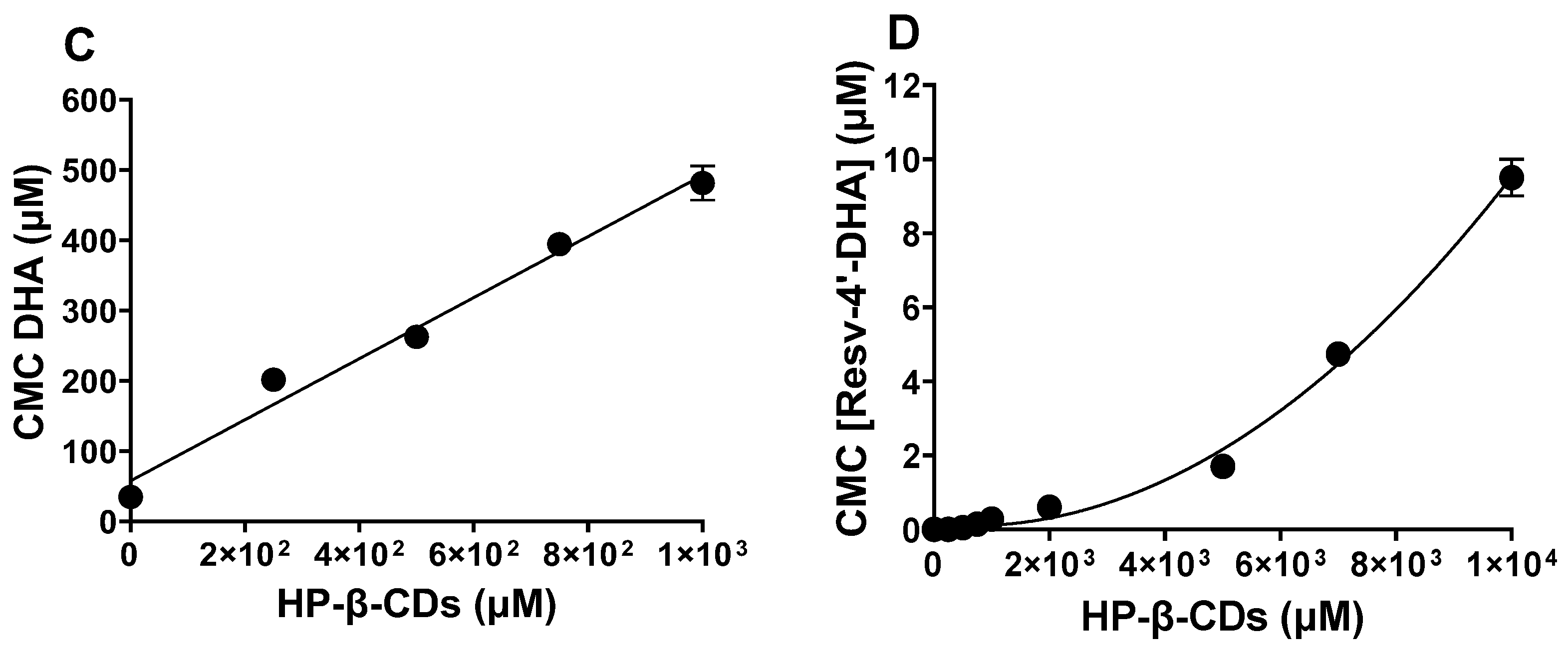
 ) and 600 nm (Figure 5,
  ), respectively. However, when the LipoResv concentration increased to 50 µM, the particle size for Resv-4′-LA increased to 622 nm (Figure 5,
  ), (1.4-fold), and for Resv-4′-DHA, it increased to 2989 nm (Figure 5,
  ), (5-fold). These findings suggested that Resv-4′-LA exhibited a greater aqueous solubility compared to Resv-4′-DHA. This interpretation aligns with the CMC0 values obtained in the absence of CDs, which were 6 µM for Resv-4′-LA (Figure 2C) and 0.001 µM for Resv-4′-DHA (Figure 2D). The lower CMC0 of Resv-4′-DHA (6000-fold) indicated that micelle formation occurred at a much lower concentration than in the case of Resv-4′-LA due to the lower aqueous solubility of Resv-4′-DHA (Table 1).
2.4. Influence of pH, Temperature, and Ionic Strength on LipoResv CMC and Complexation Process
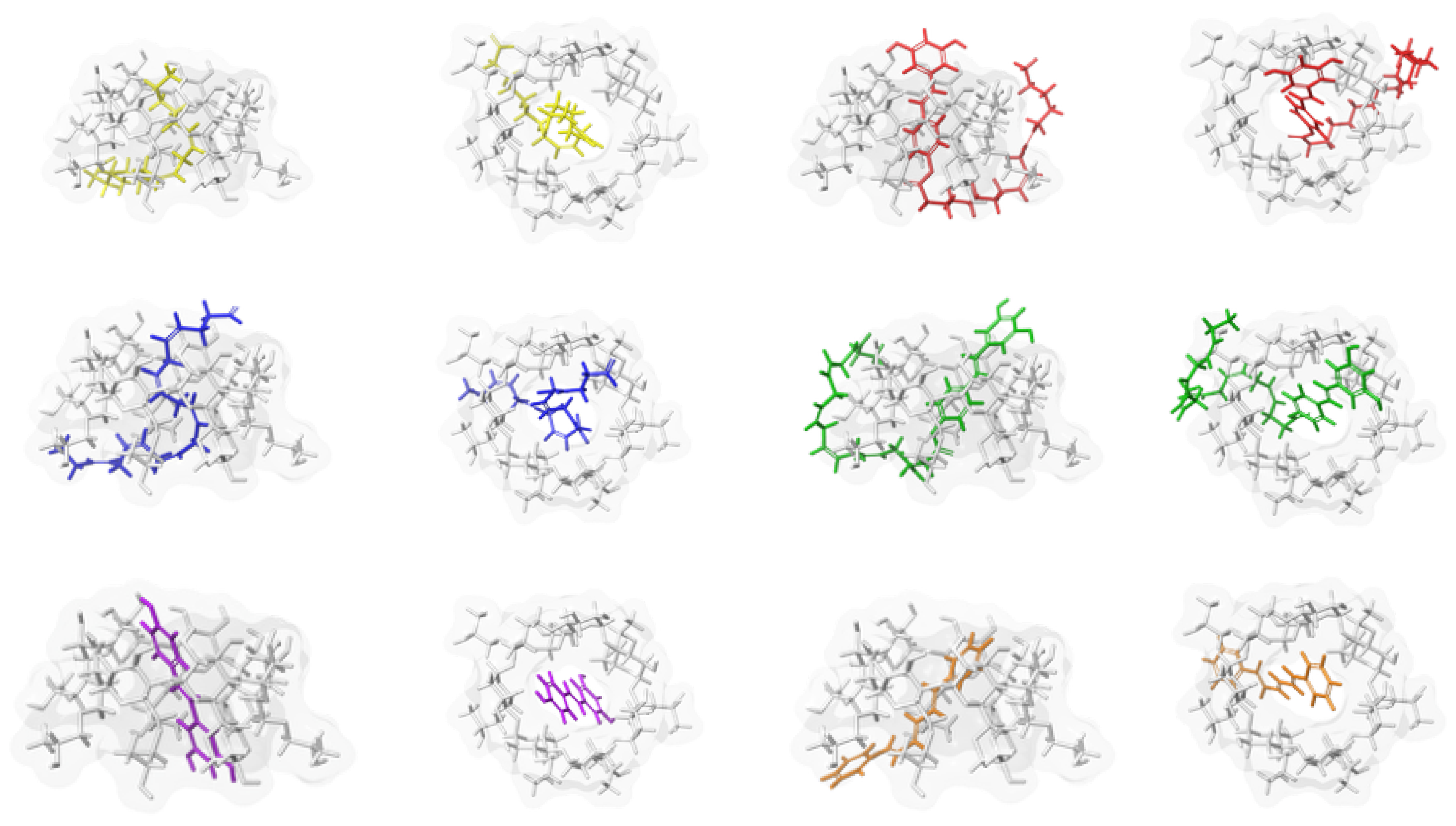
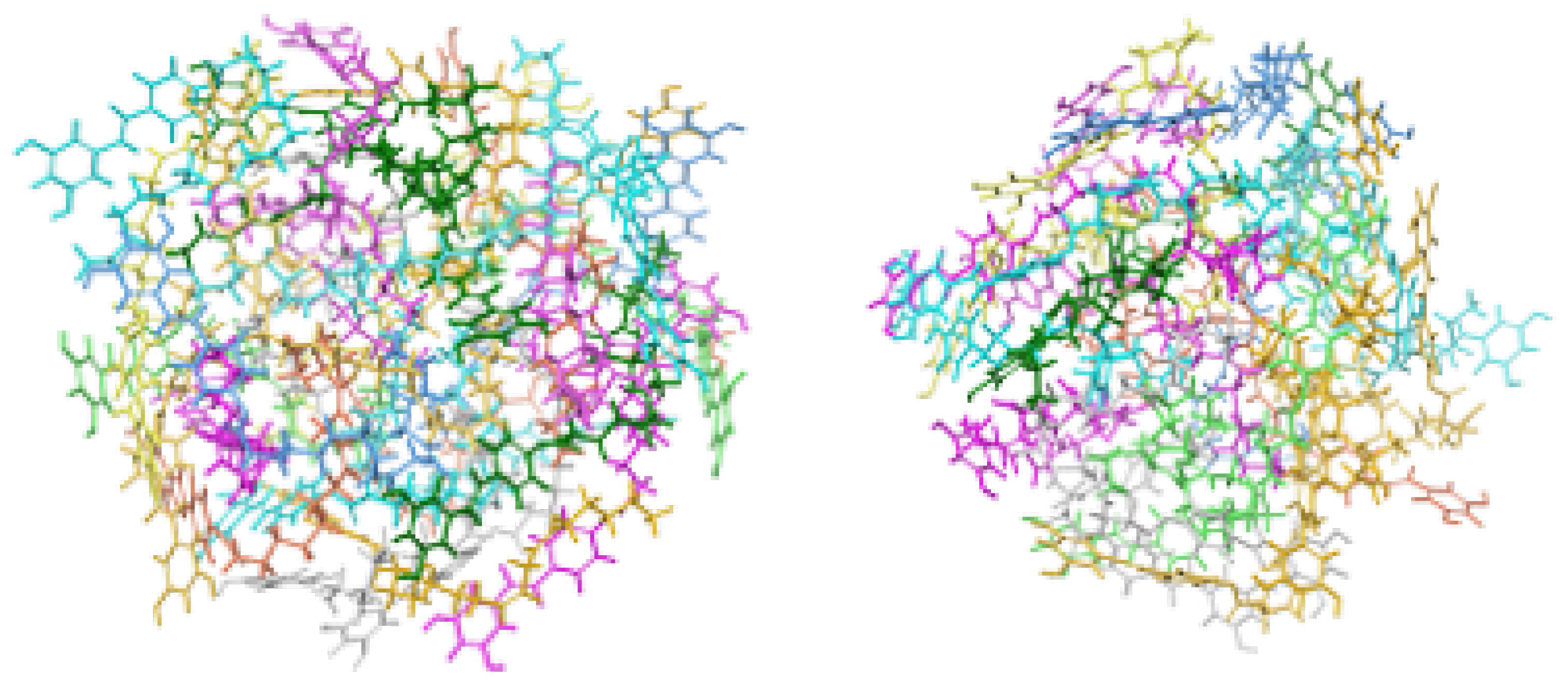
2.5. Computational Models
3. Materials and Methods
3.1. Materials
3.2. Chemo–Enzymatic Synthesis of LipoResv
3.3. Fluorimetric Determination of CMC
3.4. Determination of Complexation Stoichiometry and Equilibrium Constant Between CDs and Fatty Acid or Liporesv
3.5. Determination of Particle Size by DLS
3.6. Molecular Modelling
4. Conclusions
Limitations and Future Outlook
Supplementary Materials
Author Contributions
Funding
Institutional Review Board Statement
Informed Consent Statement
Data Availability Statement
Conflicts of Interest
References
- Tomas-Barberan, F.A.; Andres-Lacueva, C. Polyphenols and health: Current state and progress. J. Agric. Food Chem. 2012, 60, 8773–8775. [Google Scholar] [CrossRef]
- Shahidi, F.; Ambigaipalan, P. Omega-3 polyunsaturated fatty acids and their health benefits. Annu. Rev. Food Sci. Technol. 2018, 9, 345–381. [Google Scholar] [CrossRef]
- Crauste, C.; Rosell, M.; Durand, T.; Vercauteren, J. Omega-3 polyunsaturated lipophenols, how and why? Biochimie 2016, 120, 62–74. [Google Scholar] [CrossRef] [PubMed]
- Crauste, C.; Vercauteren, J.; Veas, F.; Durand, T.; Blondeau, N. Uses of Lipophenolic Compounds. U.S. Patent No. 11,419,841, 23 August 2022. [Google Scholar]
- Moine, E.; Brabet, P.; Guillou, L.; Durand, T.; Vercauteren, J.; Crauste, C. New Lipophenol Antioxidants Reduce Oxidative Damage in Retina Pigment Epithelial Cells. Antioxidants 2018, 7, 197. [Google Scholar] [CrossRef] [PubMed]
- Shamseddin, A.; Crauste, C.; Durand, E.; Villeneuve, P.; Dubois, G.; Pavlickova, T.; Durand, T.; Vercauteren, J.; Veas, F. Resveratrol-Linoleate Protects from Exacerbated Endothelial Permeability via a Drastic Inhibition of the MMP-9 Activity. Biosci. Rep. 2018, 38, BSR20171712. [Google Scholar] [CrossRef] [PubMed]
- Silva, V.; Pórfido, J.L. Interacción de lípidos con el agua y formación de estructuras empaquetadas. In Análisis Estructural Y Funcional De Macromoléculas, 1st ed.; Córsico, B., Falomir-Lockhart, L.J., Franchini, G.R., Scaglia, N., Eds.; Universidad Nacional de La Plata: Buenos Aires, Argentina, 2013; pp. 193–219. [Google Scholar] [CrossRef]
- Göktürk, S.; Çalışkan, E.; Talman, R.Y.; Var, U. A study on solubilization of poorly soluble drugs by cyclodextrins and micelles: Complexation and binding characteristics of sulfamethoxazole and trimethoprim. Sci. World J. 2012, 1, 718791. [Google Scholar] [CrossRef]
- Von Krbek, L.K.S.; Schalley, C.A.; Thordarson, P. Assessing cooperativity in supramolecular systems. Chem. Soc. Rev. 2017, 46, 2622–2637. [Google Scholar] [CrossRef]
- Shalaby, K.S.; Ismail, M.I.; Lamprecht, A. Cyclodextrin Complex Formation with Water-Soluble Drugs: Conclusions from Isothermal Titration Calorimetry and Molecular Modeling. AAPS Pharm. Sci. Tech. 2021, 22, 232. [Google Scholar] [CrossRef]
- Pérez-Abril, M.; Lucas-Abellán, C.; Castillo-Sánchez, J.; Pérez-Sánchez, H.; Cerón Carrasco, J.P.; Fortea, M.I.; Gabaldón, J.A.; Núñez-Delicado, E. Systematic investigation and molecular modelling of complexation between several groups of flavonoids and HP-β-cyclodextrins. J. Funct. Foods 2017, 36, 122–131. [Google Scholar] [CrossRef]
- Martín, V.I.; Ostos, F.J.; Angulo, M.; Márquez, A.M.; López-Cornejo, P.; López-López, M.; Carmona, A.T.; Moyá, M.L. Host-guest interactions between cyclodextrins and surfactants with functional groups at the end of the hydrophobic tail. J. Colloid Interface Sci. 2017, 491, 336–348. [Google Scholar] [CrossRef]
- Del Valle, E.M. Cyclodextrins and their uses: A review. Process Biochem. 2004, 39, 1033–1046. [Google Scholar] [CrossRef]
- Saokham, P.; Muankaew, C.; Jansook, P.; Loftsson, T. Solubility of cyclodextrins and drug/cyclodextrin complexes. Molecules 2018, 23, 1161. [Google Scholar] [CrossRef] [PubMed]
- Nicolaescu, O.E.; Ionescu, C.; Samide, A.; Tigae, C.; Spînu, C.I.; Oprea, B. Advancements in Cyclodextrin Complexes with Bioactive Secondary Metabolites and Their Pharmaceutical Applications. Pharmaceutics 2025, 17, 506. [Google Scholar] [CrossRef] [PubMed]
- Pellicer, J.A.; Rodríguez-López, M.I.; Fortea, I.; Gabaldón, J.A.; Lucas-Abellán, C.; Mercader-Ros, M.T.; Serrano-Martínez, A.; Núñez-Delicado, E.; Cosma, P.; Fini, P.; et al. Removing of Direct Red 83:1 using α- and HP-α-CDs polymerized with epichlorohydrin: Kinetic and equilibrium studies. Dyes Pigm. 2018, 149, 736–746. [Google Scholar] [CrossRef]
- Pellicer, J.A.; Rodríguez-López, M.I.; Fortea, I.; Lucas-Abellán, C.; Mercader-Ros, M.T.; López-Miranda, S.; Gómez-López, V.M.; Semeraro, P.; Cosma, P.; Fini, P.; et al. Adsorption Properties of β- and Hydroxypropyl-β-Cyclodextrins Cross-Linked with Epichlorohydrin in Aqueous Solution. A Sustainable Recycling Strategy in Textile Dyeing Process. Polymers 2019, 11, 252. [Google Scholar] [CrossRef]
- Chodankar, D.; Vora, A.; Kanhed, A. β-cyclodextrin and its derivatives: Application in wastewater treatment. Environ. Sci. Pollut. Res. Int. 2022, 29, 1585–1604. [Google Scholar] [CrossRef]
- Gómez-Morte, T.; Gómez-López, V.M.; Lucas-Abellán, C.; Martínez-Alcalá, I.; Ayuso, M.; Martínez-López, S.; Montemurro, N.; Pérez, S.; Barceló, D.; Fini, P.; et al. Removal and toxicity evaluation of a diverse group of drugs from water by a cyclodextrin polymer/pulsed light system. J. Hazard. Mater. 2021, 402, 123504. [Google Scholar] [CrossRef]
- Romita, R.; Rizzi, V.; Gubitosa, J.; Gabaldón, J.A.; Fortea-Gorbe, M.I.; Gómez-Morte, T.; Gómez-López, V.M.; Fini, P.; Cosma, P. Cyclodextrin polymers and salts: An Eco-Friendly combination to modulate the removal of sulfamethoxazole from water and its release. Chemosphere 2021, 283, 131238. [Google Scholar] [CrossRef]
- Lucas-Abellán, C.; Gabaldón-Hernández, J.A.; Penalva, J.; Fortea, M.I.; Núñez-Delicado, E. Preparation and characterization of the inclusion complex of chlorpyrifos in cyclodextrins to improve insecticide formulations. J. Agric. Food Chem. 2008, 56, 8081–8085. [Google Scholar] [CrossRef]
- Rodríguez-López, M.I.; Mercader-Ros, M.T.; López-Miranda, S.; Pellicer, J.A.; Pérez-Garrido, A.; Pérez-Sánchez, H.; Núñez-Delicado, E.; Gabaldón, J.A. Thorough characterization and stability of HP-β-cyclodextrin thymol inclusion complexes prepared by microwave technology: A required approach to a successful application in food industry. J. Sci. Food Agric. 2019, 99, 1322–1333. [Google Scholar] [CrossRef]
- Lucas-Abellán, C.; Pérez-Abril, M.; Castillo, J.; Serrano, A.; Mercader-Ros, M.T.; Fortea, M.I.; Gabaldón, J.A.; Núñez-Delicado, E. Effect of temperature, pH, β- and HP-β-CDs on the solubility and stability of flavanones: Naringenin and hesperetin. LWT-Food Sci. Technol. 2019, 108, 233–239. [Google Scholar] [CrossRef]
- Moulik, S.P.; Rakshit, A.K.; Naskar, B. Evaluation of non-ambiguous critical micelle concentration of surfactants in relation to solution behaviors of pure and mixed surfactant systems: A physicochemical documentary and analysis. J. Surfactants Deterg. 2021, 24, 535–549. [Google Scholar] [CrossRef]
- Funasaki, N.; Ishikawa, S.; Neya, S. 1:1 and 1:2 complexes between long-chain surfactant and α-cyclodextrin studied by NMR. J. Phys. Chem. B. 2004, 108, 9593–9598. [Google Scholar] [CrossRef]
- Scholz, N.; Behnke, T.; Resch-Genger, U. Determination of the critical micelle concentration of neutral and ionic surfactants with fluorometry, conductometry, and surface tension—A method comparison. J. Fluoresc. 2018, 28, 465–476. [Google Scholar] [CrossRef] [PubMed]
- Al-Soufi, W.; Piñeiro, L.; Novo, M. A model for monomer and micellar concentrations in surfactant solutions: Application to conductivity, NMR, diffusion, and surface tension data. J. Colloid Interface Sci. 2012, 370, 102–110. [Google Scholar] [CrossRef]
- Cabaleiro-Lago, C.; García-Río, L.; Hervés, P.; Mejuto, J.C.; Pérez-Juste, J. In search of fully uncomplexed cyclodextrin in the presence of micellar aggregates. J. Phys. Chem. B. 2006, 110, 15831–15838. [Google Scholar] [CrossRef]
- Perinelli, D.R.; Cespi, M.; Lorusso, N.; Palmieri, G.F.; Bonacucina, G.; Blasi, P. Surfactant self-assembling and critical micelle concentration: One approach fits all? Langmuir 2020, 36, 5745–5753. [Google Scholar] [CrossRef]
- Bru, R.; López-Nicolás, J.; García-Carmona, F. Aggregation of polyunsaturated fatty acids in the presence of cyclodextrins. Colloid. Surf. A. 1995, 97, 263–269. [Google Scholar] [CrossRef]
- Matencio, A.; Hernández-Gil, C.J.G.; García-Carmona, F.; López-Nicolás, J.M. Physicochemical, thermal and computational study of the encapsulation of rumenic acid by natural and modified cyclodextrins. Food Chem. 2017, 216, 289–295. [Google Scholar] [CrossRef]
- Degrand, L.; Garcia, R.; Urion, K.C.; Guiga, W. Dynamic light scattering for the determination of linoleic acid critical micelle concentration. Effect of pH, ionic strength, and ethanol. J. Mol. Liq. 2023, 388, 122670. [Google Scholar] [CrossRef]
- Dorrego, B.; Garcia-Rio, L.; Hervés, P.; Leis, J.R.; Mejuto, J.C.; Pérez-Juste, J. Changes in the fraction of uncomplexed cyclodextrin in equilibrium with the micellar system as a result of balance between micellization and cyclodextrin−surfactant complexation. Cationic alkylammonium surfactants. J. Phys. Chem. B. 2001, 105, 4912–4920. [Google Scholar] [CrossRef]
- Suvarna, V.; Bore, B.; Bhawar, C.; Mallya, R. Complexation of phytochemicals with cyclodextrins and their derivatives-an update. Biomed. Pharmacother. 2022, 149, 112862. [Google Scholar] [CrossRef]
- De Lisi, R.; Milioto, S.; Muratore, N. Thermodynamic evidence of cyclodextrin− micelle interactions. J. Phys. Chem. B. 2002, 106, 8944–8953. [Google Scholar] [CrossRef]
- Matencio, A.; García-Carmona, F.; López-Nicolás, J.M. Aggregation of t10, c12 conjugated linoleic Acid in presence of natural and modified cyclodextrins. A physicochemical, thermal and computational analysis. Chem. Phys. Lipids. 2017, 204, 57–64. [Google Scholar] [CrossRef]
- Ondo, D. Thermodynamic study on complexation of long-chain fatty acid anions with α-cyclodextrin in water. J. Mol. Liq. 2020, 311, 113172. [Google Scholar] [CrossRef]
- Connors, K.A. The stability of cyclodextrin complexes in solution. Chem. Rev. 1997, 97, 1325–1358. [Google Scholar] [CrossRef] [PubMed]
- Triamchaisri, N.; Toochinda, P.; Lawtrakul, L. Structural Investigation of Beta-Cyclodextrin Complexes with Cannabidiol and Delta-9-Tetrahydrocannabinol in 1: 1 and 2: 1 Host-Guest Stoichiometry: Molecular Docking and Density Functional Calculations. Int. J. Mol. Sci. 2023, 24, 1525. [Google Scholar] [CrossRef] [PubMed]
- Dorrego, A.B.; García-Río, L.; Hervés, P.; Leis, J.R.; Mejuto, J.C.; Pérez-Juste, J. Micellization versus cyclodextrin–surfactant complexation. Angew. Chem. Int. Ed. 2000, 39, 2945–2948. [Google Scholar] [CrossRef]
- Namani, T.; Ishikawa, T.; Morigaki, K.; Walde, P. Vesicles from docosahexaenoic acid. Colloids Surf B Biointerfaces 2007, 54, 118–123. [Google Scholar] [CrossRef]
- Vinarov, Z.; Katev, V.; Radeva, D.; Tcholakova, S.; Denkov, N.D. Micellar solubilization of poorly water-soluble drugs: Effect of surfactant and solubilizate molecular structure. Drug Dev. Ind. Pharm. 2018, 44, 677–686. [Google Scholar] [CrossRef]
- Ghosh, A.; Kanti Seth, S.; Purkayastha, P. Controlled formation of hydrated micelles by the intervention of cyclodextrins. Chem. Plus. Chem. 2019, 84, 130–135. [Google Scholar] [CrossRef]
- Jiang, Y.B.; Wang, X.J. Direct evidence for β-cyclodextrin-induced aggregation of ionic surfactant below critical micelle concentration. Appl. Spectrosc. 1994, 48, 1428–1431. [Google Scholar] [CrossRef]
- Tsianou, M.; Fajalia, A.I. Cyclodextrins and surfactants in aqueous solution above the critical micelle concentration: Where are the cyclodextrins located? Langmuir 2014, 30, 13754–13764. [Google Scholar] [CrossRef] [PubMed]
- Hinze, W.L. Fluorescence in Organized Assemblies. In Encyclopedia of Analytical Chemistry: Applications, Theory and Instrumentation, 1st ed.; Meyers, R.A., Warner, I.M., Eds.; John Wiley & Sons, Ltd.: Hoboken, NJ, USA, 2006; pp. 1–84. [Google Scholar] [CrossRef]
- Samuelsen, L.; Holm, R.; Schönbeck, C. Simultaneous determination of cyclodextrin stability constants as a function of pH and temperature—A tool for drug formulation and process design. J. Drug Deliv. Sci. Tec. 2021, 65, 102675. [Google Scholar] [CrossRef]
- Kinart, Z. Stability of the Inclusion Complexes of Dodecanoic Acid with α-Cyclodextrin, β-Cyclodextrin and 2-HP-β-Cyclodextrin. Molecules 2023, 28, 3113. [Google Scholar] [CrossRef] [PubMed]
- Alopaeus, J.F.; Hagesæther, E.; Tho, I. Micellisation mechanism and behaviour of Soluplus®–furosemide micelles: Preformulation studies of an oral nanocarrier-based system. Pharm 2019, 12, 15. [Google Scholar] [CrossRef]
- Greenidge, P.A.; Kramer, C.; Mozziconacci, J.C.; Sherman, W. Improving docking results via reranking of ensembles of ligand poses in multiple X-ray protein conformations with MM-GBSA. J. Chem. Inf. Model. 2014, 54, 2697–2717. [Google Scholar] [CrossRef]
- Hopkins, A.L.; Keserü, G.M.; Leeson, P.D.; Rees, D.C.; Reynolds, C.H. The role of ligand efficiency metrics in drug discovery. Nat. Rev. Drug Discov. 2014, 13, 105–121. [Google Scholar] [CrossRef]
- Maffucci, I.; Contini, A. Explicit ligand hydration shells improve the correlation between MM-PB/GBSA binding energies and experimental activities. J. Chem. Theory Comput. 2013, 9, 2706–2717. [Google Scholar] [CrossRef]
- Suárez, D.; Díaz, N. Affinity Calculations of Cyclodextrin Host–Guest Complexes: Assessment of Strengths and Weaknesses of End-Point Free Energy Methods. J. Chem. Inf. Model. 2019, 59, 421–440. [Google Scholar] [CrossRef]
- Maibaum, L.; Dinner, A.R.; Chandler, D. Micelle formation and the hydrophobic effect. J. Phys. Chem. B. 2004, 108, 6778–6781. [Google Scholar] [CrossRef]
- Rawicz, W.; Olbrich, K.C.; McIntosh, T.; Needham, D.; Evans, E. Effect of chain length and unsaturation on elasticity of lipid bilayers. Biophys. J. 2000, 79, 328–339. [Google Scholar] [CrossRef] [PubMed]
- Chattopadhyay, A.; London, E. Fluorimetric determination of critical micelle concentration avoiding interference from detergent charge. Anal. Biochem. 1984, 139, 408–412. [Google Scholar] [CrossRef] [PubMed]
- Lopez-Nícolas, J.M.; Bru, R.; Sánchez-Ferrer, A.; García-Carmona, F. Use of ‘soluble lipids’ for biochemical processes: Linoleic acid-cyclodextrin inclusion complexes in aqueous solutions. Biochem. J. 1995, 308, 151–154. [Google Scholar] [CrossRef] [PubMed]
- Schrödinger, L. Schrödinger Release 2024 4: Maestro, LigPrep, Glide, Prime, Desmond; Schrödinger, LLC: New York, NY, USA, 2024. [Google Scholar]
- Lu, C.; Wu, C.; Ghoreishi, D.; Chen, W.; Wang, L.; Damm, W.; Ross, G.A.; Dahlgren, M.K.; Russell, E.; Von Bargen, C.D.; et al. OPLS4: Improving force field accuracy on challenging regimes of chemical space. J. Chem. Theory Comput. 2021, 17, 4291–4300. [Google Scholar] [CrossRef]
- Friesner, R.A.; Banks, J.L.; Murphy, R.B.; Halgren, T.A.; Klicic, J.J.; Mainz, D.T.; Repasky, M.P.; Knoll, E.H.; Shelley, M.; Perry, J.K.; et al. Glide: A new approach for rapid, accurate docking and scoring. 1. Method and assessment of docking accuracy. J. Med. Chem. 2004, 47, 1739–1749. [Google Scholar] [CrossRef]
- Lyne, P.D.; Lamb, M.L.; Saeh, J.C. Accurate prediction of the relative potencies of members of a series of kinase inhibitors using molecular docking and MM-GBSA scoring. J. Med. Chem. 2006, 49, 4805–4808. [Google Scholar] [CrossRef]
- Fereidounpour, P.; Steinmann, C.; Larsen, K.L. Prediction of the free energy of binding for cyclodextrin-steroid complexes: Phase solubility and molecular dynamics studies. J. Incl. Phenom. Macrocycl. Chem. 2024, 104, 535–546. [Google Scholar] [CrossRef]
- Bowers, K.J.; Chow, E.; Xu, H.; Dror, R.O.; Eastwood, M.P.; Gregersen, B.A.; Klepeis, J.L.; Kolossvary, I.; Moraes, M.A.; Sacerdoti, F.D.; et al. Scalable algorithms for molecular dynamics simulations on commodity clusters. In Proceedings of the 2006 ACM/IEEE Conference on Supercomputing, Tampa, FL, USA, 11–17 November 2006; p. 84. [Google Scholar] [CrossRef]
 LA,
LA,  0.25 mM HP-β-CDs/LA; (B)
  DHA,
DHA,  0.25 mM HP-β-CDs/DHA; (C)
  Resv-4′-LA,
  0.25 mM HP-β-CDs/Resv-4′-LA; and (D)
  Resv-4′-DHA,
  0.25 mM HP-β-CDs/Resv-4′-DHA.
LA, 0.25 mM HP-β-CDs/LA; (B) DHA, 0.25 mM HP-β-CDs/DHA; (C) Resv-4′-LA, 0.25 mM HP-β-CDs/Resv-4′-LA; and (D) Resv-4′-DHA, 0.25 mM HP-β-CDs/Resv-4′-DHA.
0.25 mM HP-β-CDs/Resv-4′-DHA.
LA, 0.25 mM HP-β-CDs/LA; (B) DHA, 0.25 mM HP-β-CDs/DHA; (C) Resv-4′-LA, 0.25 mM HP-β-CDs/Resv-4′-LA; and (D) Resv-4′-DHA, 0.25 mM HP-β-CDs/Resv-4′-DHA.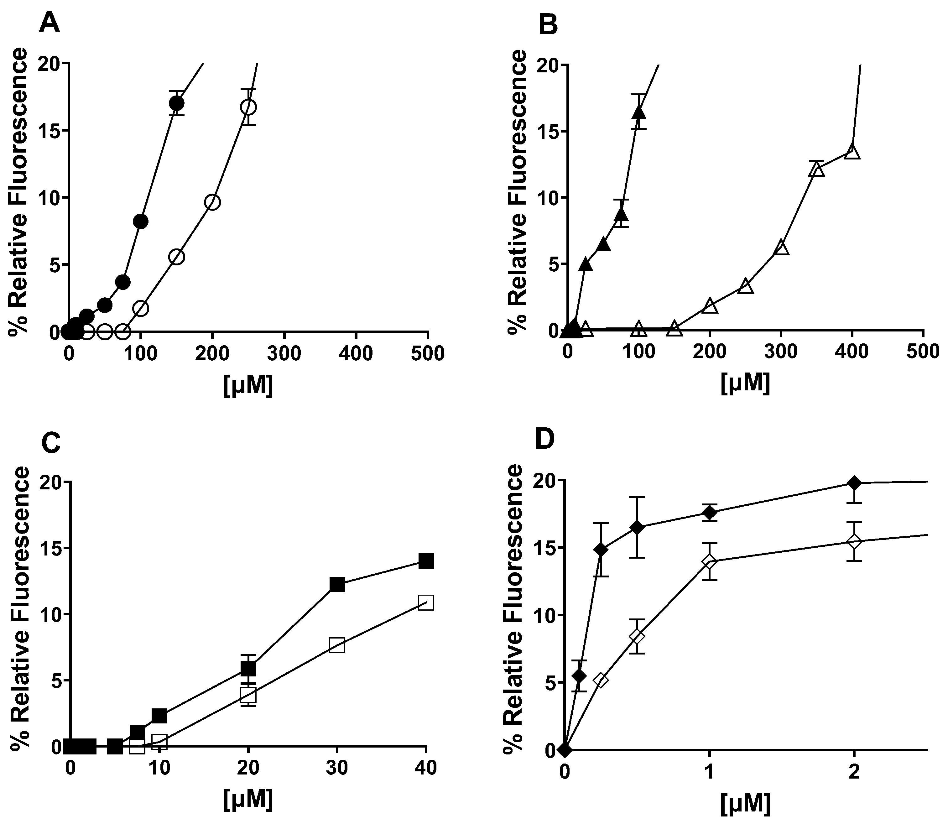 0 mM HP-β-CDs; 1 mM HP-β-CDs; 2 mM HP-β-CDs;
  5 mM HP-β-CDs; and 10 mM HP-β-CDs.
0 mM HP-β-CDs; 1 mM HP-β-CDs; 2 mM HP-β-CDs; 5 mM HP-β-CDs; and 10 mM HP-β-CDs.
5 mM HP-β-CDs; and 10 mM HP-β-CDs.
0 mM HP-β-CDs; 1 mM HP-β-CDs; 2 mM HP-β-CDs; 5 mM HP-β-CDs; and 10 mM HP-β-CDs.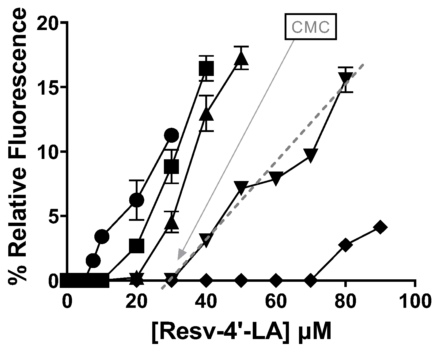

 ), Resv-4′-LA 50 µM (
 ), Resv-4′-DHA 10 µM (
 ), and Resv-4′-DHA 50 µM (
 ) in PBS at 35 °C and pH 7.0.
), Resv-4′-LA 50 µM (), Resv-4′-DHA 10 µM (), and Resv-4′-DHA 50 µM () in PBS at 35 °C and pH 7.0.
) in PBS at 35 °C and pH 7.0.
), Resv-4′-LA 50 µM (), Resv-4′-DHA 10 µM (), and Resv-4′-DHA 50 µM () in PBS at 35 °C and pH 7.0.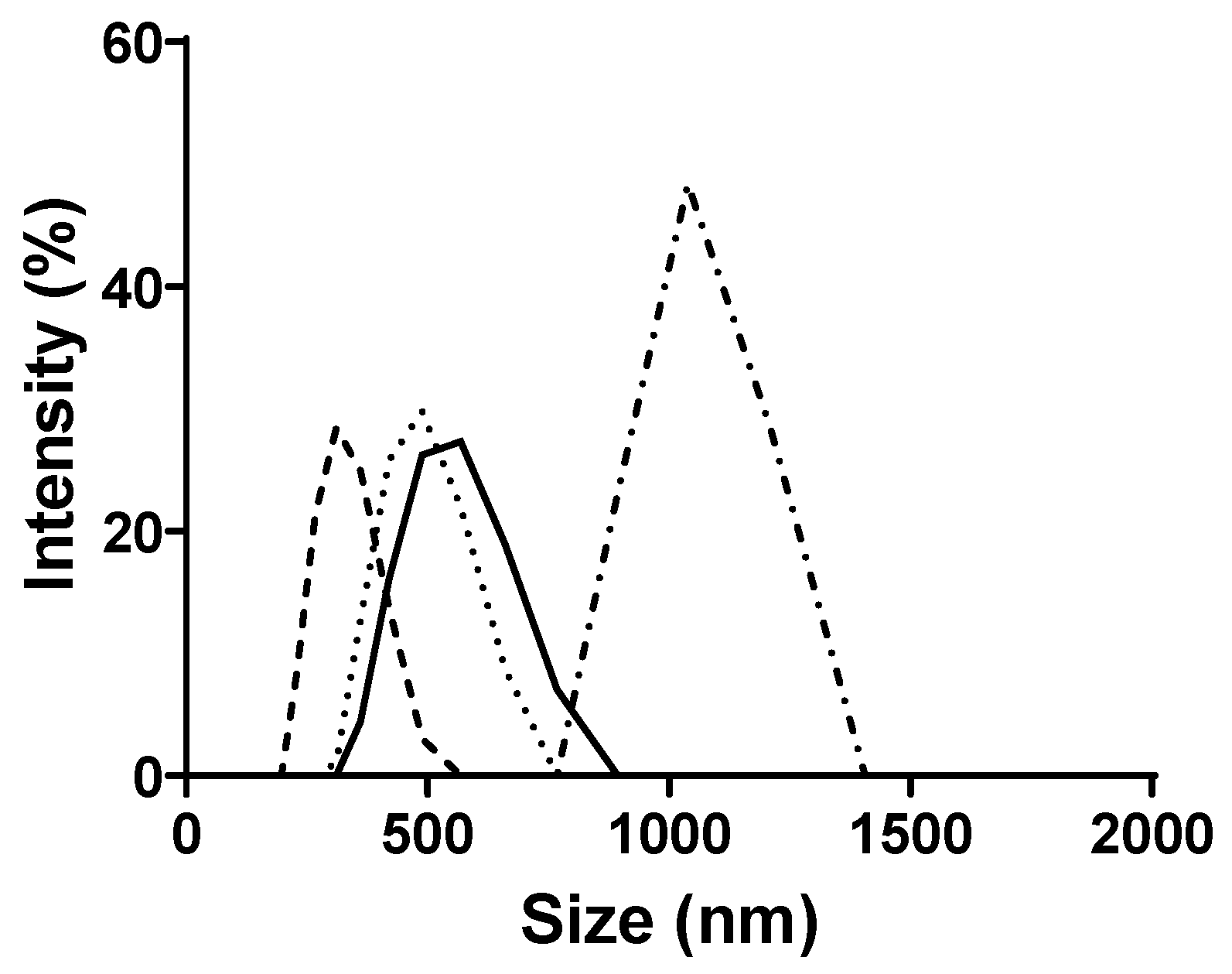
 ), Resv-4′-LA 50 µM with 10 mM HP-β-CDs (
 ) in PBS, Resv-4′-LA 50 µM (
 ), and Resv-4′-LA 50 µM with 10 mM HP-β-CDs (
 ) in MilliQ water at 35 °C and pH 7.0.
), Resv-4′-LA 50 µM with 10 mM HP-β-CDs () in PBS, Resv-4′-LA 50 µM (), and Resv-4′-LA 50 µM with 10 mM HP-β-CDs () in MilliQ water at 35 °C and pH 7.0.
) in MilliQ water at 35 °C and pH 7.0.
), Resv-4′-LA 50 µM with 10 mM HP-β-CDs () in PBS, Resv-4′-LA 50 µM (), and Resv-4′-LA 50 µM with 10 mM HP-β-CDs () in MilliQ water at 35 °C and pH 7.0.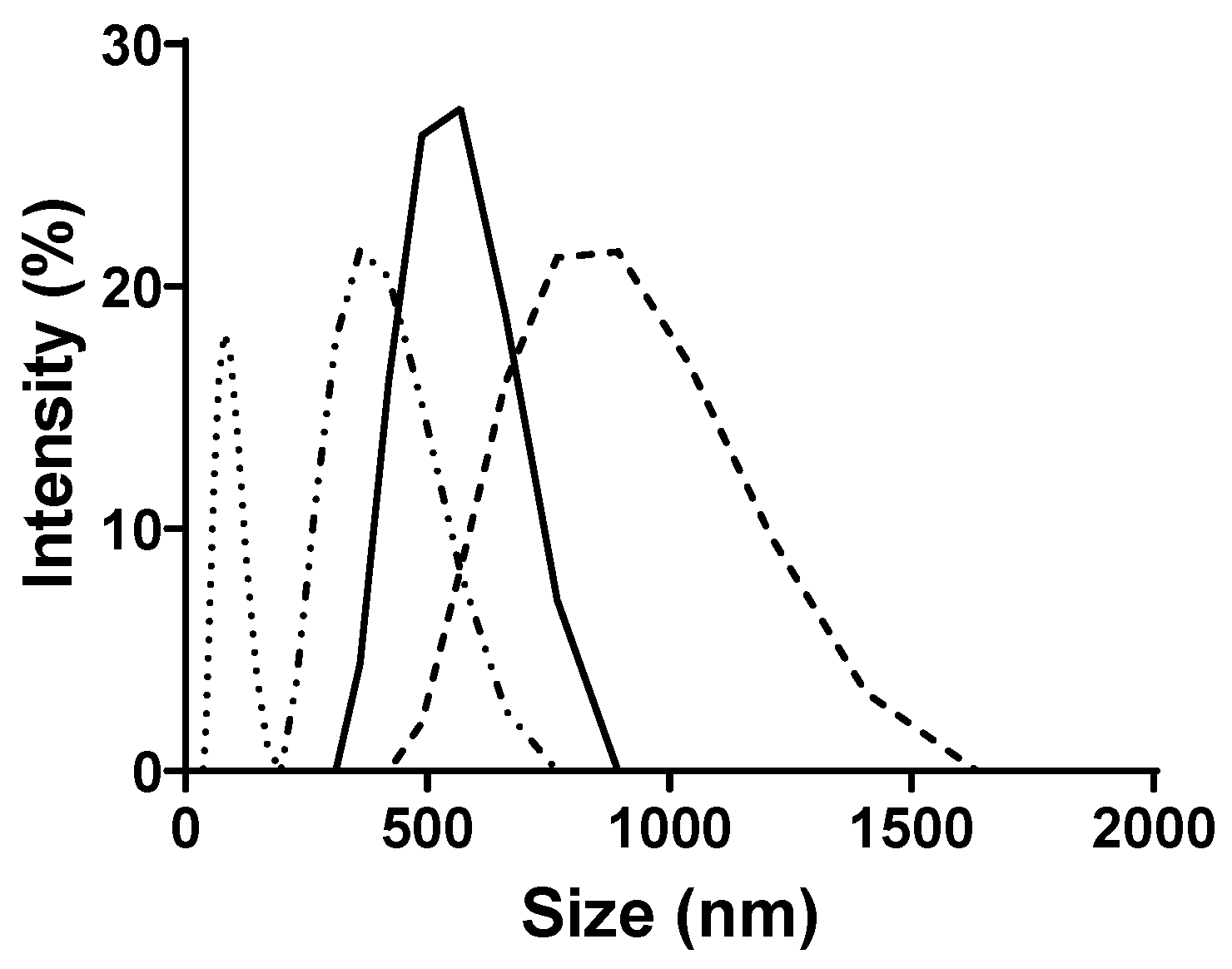

| LipoResv | Concentration (µM) | HP-β-CDs (mM) | Size (nm) | %RSD | PDI | CMC (µM) | KC (M−1) |
|---|---|---|---|---|---|---|---|
| Resv-4′-LA | 10 | 0 10 | 438 89 | 2.64 5.08 | 0.42 0.57 | 6 | 719 |
| 50 | 0 10 | 622 894 | 0.17 1.13 | 0.21 0.37 | |||
| Resv-4′-DHA | 10 | 0 10 | 600 943 | 1.76 7.75 | 0.31 0.46 | 0.001 | K1: 17; K2: 0.18 |
| 50 | 0 10 | 2989 3835 | 3.18 10.37 | 0.73 0.95 |
| LipoResv | Temperature (°C) | pH | Medium | CMC (µM) | Kc (M−1) |
|---|---|---|---|---|---|
| Resv-4′-LA | 35 | 7.0 | PBS | 6.00 | 720 |
| 25 | 7.0 | PBS | 0.27 | 8157 | |
| 15 | 7.0 | PBS | 0.14 | 10,432 | |
| 35 | 2.0 | PBS | 0.54 | 4347 | |
| 35 | 7.0 | MilliQ | 0.11 | 42,535 | |
| Resv-4′-DHA | 35 | 7.0 | PBS | 1 × 10⁻³ | K1: 17; K2: 0.18 |
| 25 | 7.0 | PBS | 6 × 10⁻⁴ | K1: 787; K2: 0.10 | |
| 15 | 7.0 | PBS | 3 × 10⁻⁴ | K1: 898; K2: 0.33 | |
| 35 | 2.0 | PBS | 5 × 10⁻⁴ | K1: 97; K2: 0.45 | |
| 35 | 7.0 | MilliQ | 5 × 10⁻⁴ | K1: 707; K2: 0.33 |
Disclaimer/Publisher’s Note: The statements, opinions and data contained in all publications are solely those of the individual author(s) and contributor(s) and not of MDPI and/or the editor(s). MDPI and/or the editor(s) disclaim responsibility for any injury to people or property resulting from any ideas, methods, instructions or products referred to in the content. |
© 2025 by the authors. Licensee MDPI, Basel, Switzerland. This article is an open access article distributed under the terms and conditions of the Creative Commons Attribution (CC BY) license (https://creativecommons.org/licenses/by/4.0/).
Share and Cite
Hernández-Heredia, A.B.; Silva-Cullishpuma, D.A.; Cerón-Carrasco, J.P.; Gil-Izquierdo, Á.; Lehoux, J.; Faion, L.; Crauste, C.; Durand, T.; Gabaldón, J.A.; Núñez-Delicado, E. Physicochemical and Computational Study of the Encapsulation of Resv-4′-LA and Resv-4′-DHA Lipophenols by Natural and HP-β-CDs. Int. J. Mol. Sci. 2025, 26, 7454. https://doi.org/10.3390/ijms26157454
Hernández-Heredia AB, Silva-Cullishpuma DA, Cerón-Carrasco JP, Gil-Izquierdo Á, Lehoux J, Faion L, Crauste C, Durand T, Gabaldón JA, Núñez-Delicado E. Physicochemical and Computational Study of the Encapsulation of Resv-4′-LA and Resv-4′-DHA Lipophenols by Natural and HP-β-CDs. International Journal of Molecular Sciences. 2025; 26(15):7454. https://doi.org/10.3390/ijms26157454
Chicago/Turabian StyleHernández-Heredia, Ana Belén, Dennis Alexander Silva-Cullishpuma, José Pedro Cerón-Carrasco, Ángel Gil-Izquierdo, Jordan Lehoux, Léo Faion, Céline Crauste, Thierry Durand, José Antonio Gabaldón, and Estrella Núñez-Delicado. 2025. "Physicochemical and Computational Study of the Encapsulation of Resv-4′-LA and Resv-4′-DHA Lipophenols by Natural and HP-β-CDs" International Journal of Molecular Sciences 26, no. 15: 7454. https://doi.org/10.3390/ijms26157454
APA StyleHernández-Heredia, A. B., Silva-Cullishpuma, D. A., Cerón-Carrasco, J. P., Gil-Izquierdo, Á., Lehoux, J., Faion, L., Crauste, C., Durand, T., Gabaldón, J. A., & Núñez-Delicado, E. (2025). Physicochemical and Computational Study of the Encapsulation of Resv-4′-LA and Resv-4′-DHA Lipophenols by Natural and HP-β-CDs. International Journal of Molecular Sciences, 26(15), 7454. https://doi.org/10.3390/ijms26157454