Article

Clostridia and Enteroviruses as Synergistic Triggers of Type 1 Diabetes Mellitus

1
Department of Physiology, Michigan State University, East Lansing, MI 48824, USA
2
Department of Microbiology and Molecular Genetics, Michigan State University, East Lansing, MI 48824, USA
3
Department of Chemistry, University of Chicago, Chicago, IL 60637, USA
*
Author to whom correspondence should be addressed.
Int. J. Mol. Sci. 2023, 24(9), 8336; https://doi.org/10.3390/ijms24098336
Submission received: 27 March 2023 / Revised: 24 April 2023 / Accepted: 2 May 2023 / Published: 6 May 2023
(This article belongs to the Special Issue Editorial Board Members’ Collection Series: Autoimmune Diseases)

Abstract

:
What triggers type 1 diabetes mellitus (T1DM)? One common assumption is that triggers are individual microbes that mimic autoantibody targets such as insulin (INS). However, most microbes highly associated with T1DM pathogenesis, such as coxsackieviruses (COX), lack INS mimicry and have failed to induce T1DM in animal models. Using proteomic similarity search techniques, we found that COX actually mimicked the INS receptor (INSR). Clostridia were the best mimics of INS. Clostridia antibodies cross-reacted with INS in ELISA experiments, confirming mimicry. COX antibodies cross-reacted with INSR. Clostridia antibodies further bound to COX antibodies as idiotype–anti-idiotype pairs conserving INS–INSR complementarity. Ultraviolet spectrometry studies demonstrated that INS-like Clostridia peptides bound to INSR-like COX peptides. These complementary peptides were also recognized as antigens by T cell receptor sequences derived from T1DM patients. Finally, most sera from T1DM patients bound strongly to inactivated Clostridium sporogenes, while most sera from healthy individuals did not; T1DM sera also exhibited evidence of anti-idiotype antibodies against idiotypic INS, glutamic acid decarboxylase, and protein tyrosine phosphatase non-receptor (islet antigen-2) antibodies. These results suggest that T1DM is triggered by combined enterovirus-Clostridium (and possibly combined Epstein–Barr-virus-Streptococcal) infections, and the probable rate of such co-infections approximates the rate of new T1DM diagnoses.

Graphical Abstract

1. Introduction

Type 1 diabetes mellitus (T1DM) is an autoimmune disease in which antibodies and T cells target a range of host autoantigens associated with beta cell production of insulin (INS), resulting in loss of INS production and consequent hyperglycemia. While much of the focus of T1DM research is on the autoimmune targeting of INS itself [1,2,3], antibodies and T cells also target glutamic acid decarboxylases (GAD) [1,2,3], protein tyrosine phosphatase non-receptor types (related to islet-associated protein or PTPN-IA-2) [1,2,3], the INS receptor (INSR) [4,5,6,7,8], and glucagon [9,10]. This combination of autoantigenic targets helps to explain why pancreatic beta cells are particular targets of T1DM pathogenesis. However, the major mystery concerning T1DM pathogenesis is the disease’s etiology: what triggers the autoimmunity directed at these pancreatic targets?
Determining the causes of autoimmune diseases such as T1DM has turned out to be a recalcitrant problem. Despite over a century of epidemiological and experimental studies of autoimmunity, the natural cause of no human autoimmune disease has yet to be discovered. It is generally believed that predisposition to autoimmune diseases is determined by genetic factors but that infectious (or other environmental) factors are required to trigger the disease process (e.g., [11,12,13,14,15,16,17]). Epidemiological methods in conjunction with individual patient case reports are generally used to try to identify what these infectious triggers may be. The general assumption is that causative microbes present antigens to the immune system that mimic the host autoantigens that the disease subsequently targets.
The onset of T1DM has been associated epidemiologically with both viral and bacterial infections, and the best clinical correlations for the onset of T1DM are probably the coxsackieviruses (COX), both A and B strains [18,19,20,21,22,23]. However, other enteroviruses [18,23,24,25], such as rubella, mumps, rotaviruses, cytomegalovirus (CMV), Epstein–Barr virus (EBV), and hepatitis C virus (HCV), have also been associated with T1DM initiation [13,26,27,28,29]. Bacterial infections associated with onset of T1DM include Bordatella pertussis and Mycobacterium species [30,31] and Helicobacter pylori [32]. Studies using T cells specific for the INS B chain, which is often considered to be the main target of T1DM autoimmunity, have identified Streptococci, Clostridia, Escherichia coli, and Pseudomonas [33] as potential mimics. T cells specific for GAD65 identified Streptococci, Staphylococci, Haemophilus, Legionella, and Chlamydia as the most likely triggers [34]. Most recently, significant differences in gut microbiota between children who have just been diagnosed with T1DM and those who have not suggest that intestinal bacteria may also play a critical role in triggering or regulating the development of diabetes [35,36,37,38,39,40]. The focus on the gut microbiome has led to the identification of Parabacteroides distasonis as a possible trigger of T1DM because its bacterial antigens activated both human T cell clones from T1DM patients and T cell hybridomas from nonobese diabetic (NOD) mice specific to the INS B chain residues 9–23 [40]. However, P. distasonis was not identified by previous studies of T cells reactive to INS [41,42]. Another study identified peptides from Bacteroides fragilis and Clostridium asparigiforme as potent activators of human T1DM T cells responsive to pre-pro-INS [41], of which only Clostridia were identified in previous studies [41,42].
Unfortunately, the numerous agents associated with T1DM leave significant questions regarding the sufficiency and necessity of any one microbe as a trigger for diabetes, a problem that has persisted for decades [28,43]. Attempts to model the onset of T1DM using individual infectious agents from the list above have thus far failed. No one has been able to produce T1DM in any animal using any of the single infectious agent listed above. COX and CMV exacerbate or accelerate disease in rodents already producing autoantibodies (e.g., NOD mice) or pretreated with the pancreatic toxin streptozotocin [21,44] but produce only transient pancreatic pathology in select strains of non-diabetic mice [45]. Monkeys infected with coxsackie B virus types 3 and 4 also develop transient pancreatitis but fail to develop chronic diabetes [46]. Cross-reactivity between COX antigens (strains B1-B6) and human T1DM antibodies reactive to either pro-INS or GAD could not be identified [47,48,49,50,51,52,53]. COX antibodies do not appear to be cross-reactive with INS, nor are INS antibodies cross-reactive with COX [54]. Moreover, although evidence of COX infections appears in temporal relationships with subsequent T1DM diagnosis [50,55,56], at least one live enterovirus vaccine—oral polio—is not associated with any increased risk of T1DM, even among genetically high-risk individuals [57,58]. However, COX infections have been linked to the development of INS receptor antibodies [59].
Even more confusingly, some putative triggers of T1DM have actually prevented the development of T1DM in animal models (reviewed in [60]). For example, pertussis vaccine protected streptozotocin-treated CD-1 mice against developing diabetes [61]; immunization with Mycobacterium. leprae [62], Bacillus Calmette-Guerin (BCG) [63], or Clostridium butyricum [64] also prevented diabetes in NOD mice [65].
Taken as a whole, the results summarized above demonstrate that mono-infectious approaches to modeling T1DM have universally failed or yielded results that seem to contradict the role of any particular microbe as a cause of the disease. These failures led Horwitz et al. [20,66,67] to suggest that the role of COX may not be as direct triggers of T1DM via molecular mimicry but rather as bystander infections supporting some other infectious agent. As Filippi and von Herrath [68] suggested, “This could be explained by the fact that viral association with T1D will likely be multifactorial”.
So, perhaps the difficulty identifying “the cause” of T1DM stems from the assumption that “the cause” is mono-factorial and resides in the unique identification of one of the microbes listed above as “the” T1DM trigger. However, perhaps no single microbe is both necessary and sufficient. Perhaps a new paradigm is required, based on multiple, concurrent infections as autoimmune disease initiators stimulating the production of complementary or synergistic sets of autoantibodies and TCR directed at multiple targets simultaneously.
The major aim of our research is to explore the possibility that sets of microbes may cooperate to induce T1DM. This aim requires a shift in the types of experiments and logic employed to test possible T1DM etiologies. The standard approach to elucidating autoimmune disease etiology is based on Koch’s postulates, which assume a single etiological agent. A concurrent-infection model requires that two or more etiological agents be involved. The use of multiple agents is consistent with the observation that there are multiple autoantigen targets in T1DM. Therefore, one aim of our research is to use proteonomic methods to identify microbes that mimic the key autoantigens in T1DM, INS, and INSR. A second aim is to use the mimicry data as the basis for experimentally testing whether antibodies against the identified microbes cross-react with INS and INSR. Because INS and INSR are molecularly complementary, logically it follows that the microbial antigens inducing cross-reactive immunity to INS and INSR will also be complementary to each other. Thus, a third aim is to test for possible antigen complementarity. Three such tests are presented. The first test is based on the proposition that some microbial antigens mimicking INS and INSR will bind to each other just as INS and INSR themselves bind to each other. A second test is predicated on the assumption that antibodies against these INS and INSR mimics will act like idiotype–anti-idiotype pairs. A third test is whether or not T cell receptor sequences from T1DM patients that have previously been demonstrated to bind to INS and INSR peptides also recognize these microbial antigens. The final aim is to test whether sera from T1DM patients recognize the microbial antigens identified in testing the previous aims.
The results consistently reveal that COX mimic INSR antigens; Clostridia mimic INS; and that the resulting immune responses whether based on animal-derived antibodies, human TCR sequences, or T1DM sera, involve idiotype–anti-idiotype relationships to sets of complementary antigens. No set of control antibodies displayed a similar range of interactions. These results suggest a new role for COX and other enteroviruses (inducing INSR, rather than INS, cross-reactivity) in T1DM etiology; identify Clostridia as the triggers of INS cross-reactivity for the first time; and provide the first evidence suggesting a multifactorial, synergistic mechanism for T1DM etiology.

2. Results

2.1. Proteomic Search for Microbial Similarities to T1DM Autoantigens

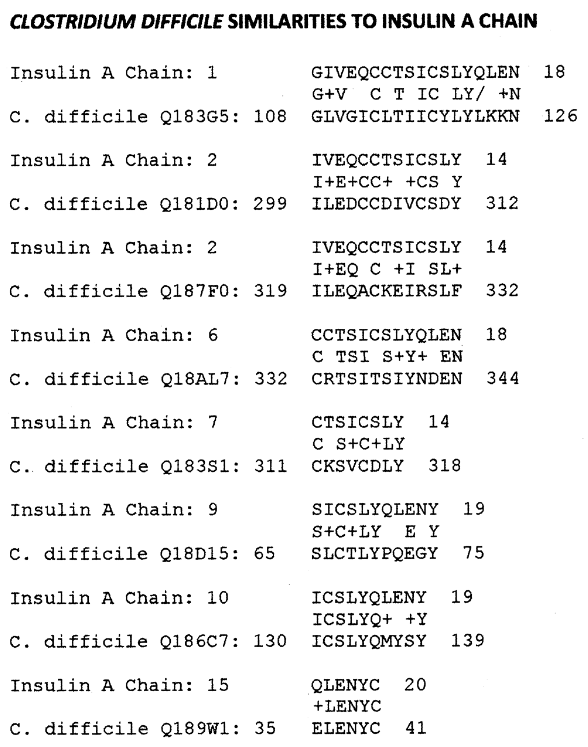
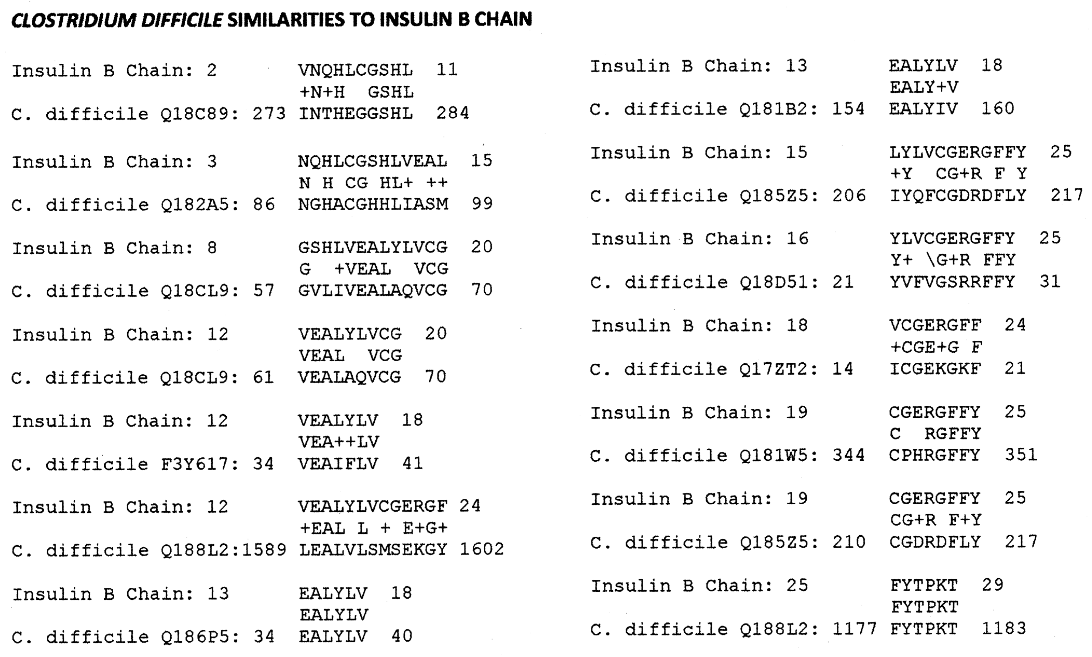
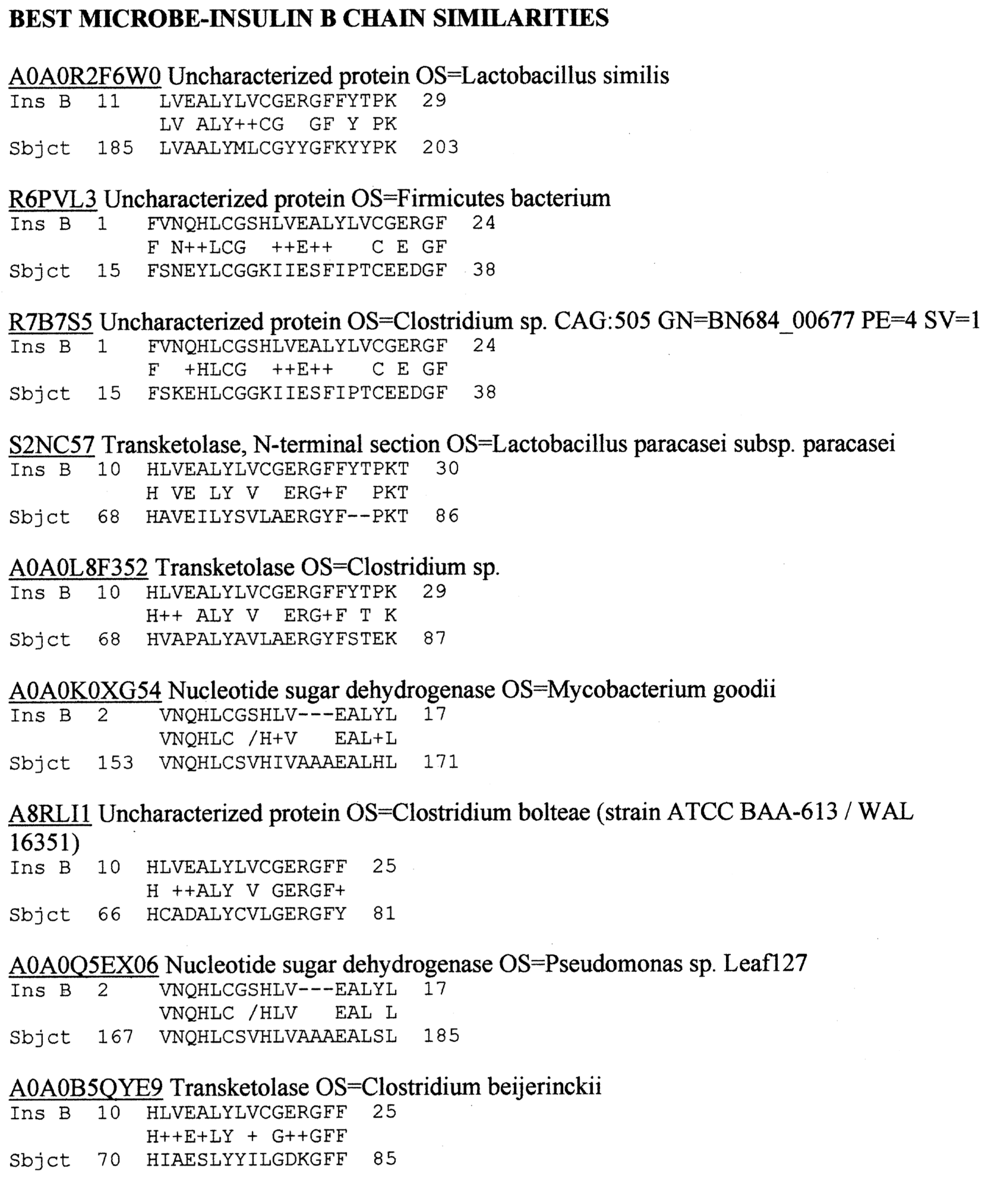
Our investigation began with a simple strategy, which was to search for the best similarity matches (as defined by high rates of identity over short regions of ten or twelve amino acids identified using BLOSUM80) between INS, INSR, and the bacteria and viruses listed in the UniProtKB databases of microbial proteins. We relied on the internal statistical algorithms of BLASTP to provide the best matches. Matches involving microbes that do not infect or that live as commensal organisms in human beings were eliminated from the search results as being unlikely to induce active immunity. Figure 1 and Figure 2 display the key findings.
As Figure 1 and Figure 2 demonstrate, only Clostridium species repeatedly appeared as significantly similar to human INS A and B chains. A more targeted similarity search with BLASTP comparing INS A and B chains with pathogenic Clostridia (C. clostridioforme, C. difficile, C. perfingens, C. sordelli, C. tetani) revealed a much broader set of INS similarities in each case. Only the C. difficile similarities are presented here as a representative case (Appendix B). Similarities to the INS B chain were much more common than those to the INS A chain, which is consistent with the B chain being the main target of autoantibodies in T1DM. No significant similarities (defined as at least six identities in a sequence of ten amino acids) were found between any virus, including COX, and either the INS A or B chains.
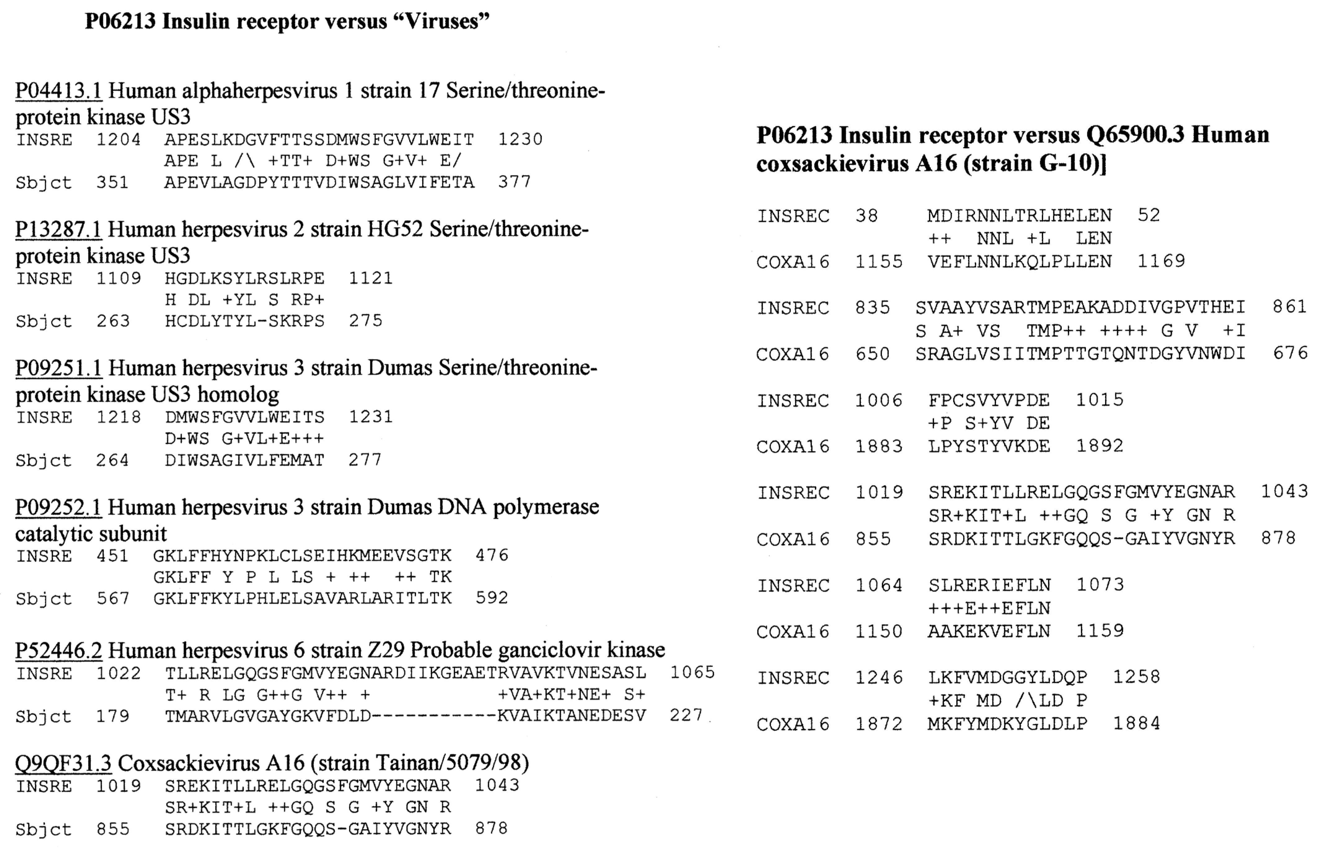
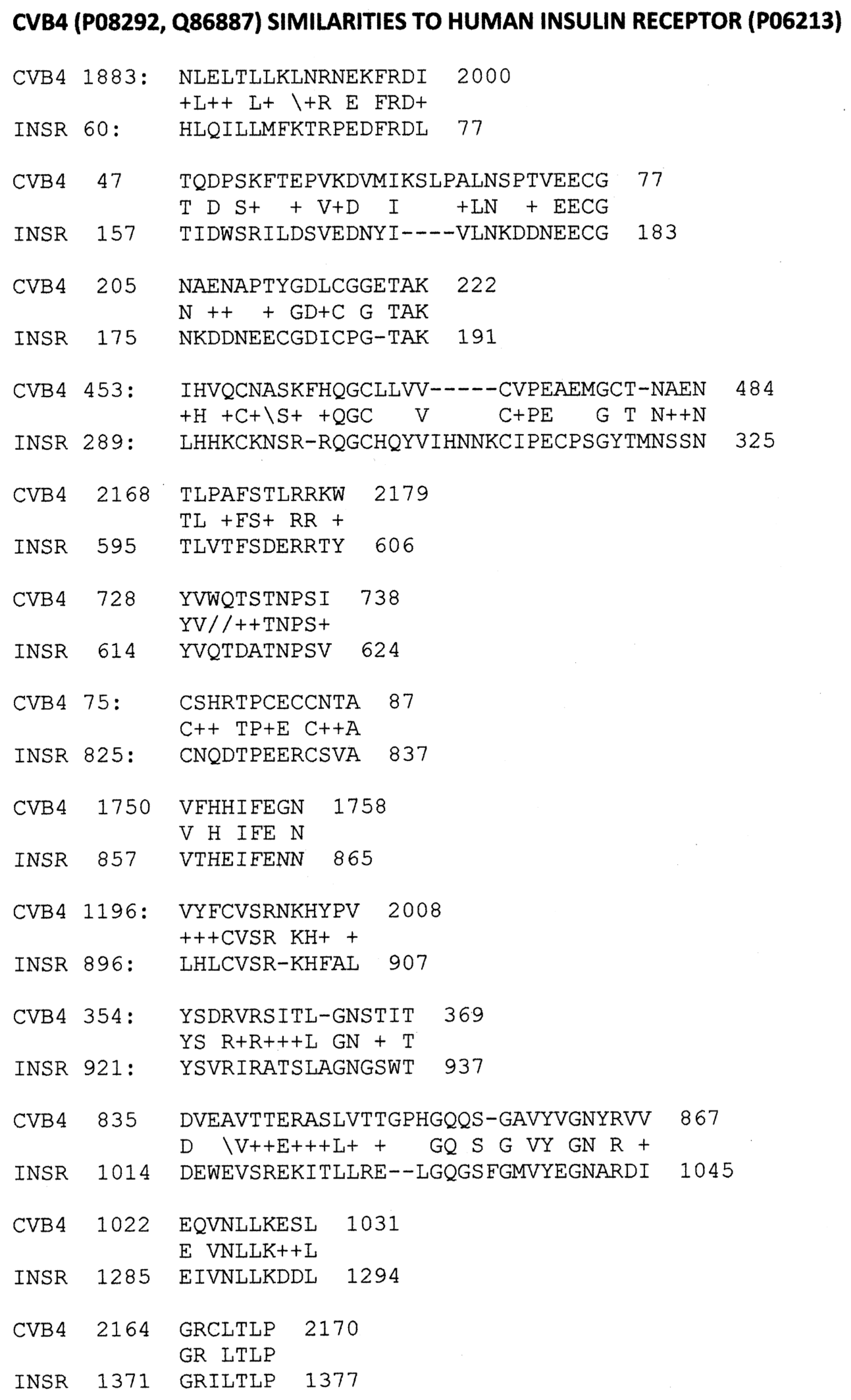
Because COX has very consistently been associated with T1DM using many types of methods [18,19,20,21,22,23,24,25], the possibility that it was responsible for triggering INSR rather than INS cross-reactivity was investigated. A BLASTP search of INSR against the entire SwissProt virus database revealed that among the top 100 virus matches, only five types of human viruses had significant similarities to INSR: Human herpesviruses (HHV) 1, 2, 3, and 6 and coxsackieviruses (Appendix A). HHV1, 2, and 6 are not among the viruses frequently associated epidemiologically with T1DM (see Section 1), while HHV3 (varicella zoster virus) is almost certainly not a significant trigger of T1DM, as demonstrated by the fact that T1DM cases continued to increase following almost universal vaccination against this virus. Thus, we focused our further efforts on coxsackieviruses for which many very significant INSR similarities were found. The results for type B4 are typical of B-type COX and are provided in Figure 3, while the lesser number of significant matches for COX A16 (typical of A-type COX) are illustrated in Appendix B.
BLASTP similarity searches were also performed on protein tyrosine phosphatase non-receptor type 1 (PTPN(IA-2)) and glutamic acid decarboxylase 65 (GAD65) because these are the two most common intracellular T1DM targets (Appendix C). Many significant similarities exist between C. difficile proteins and both PTPN(IA-2) and GAD65. No significant similarities were found between COX and PTPN(IA-2), and only three similarities were found to GAD65 (Appendix C). Thus, Clostridia are much more likely triggers of anti-PTPRN and anti-GAD65 antibodies or TCR than are COX.
In sum, pathogenic Clostridia were found to have the most abundant similarities to human INS of any bacteria associated with human health that are currently in the SwissProtKB database. The only other bacteria to rise to an equivalent degree of similarity were Lactobacilli, Bacteroides fragilis, Mycobacterium goodie, and Pseudomonas species, but these are all commensal components of the human microbiome and unlikely to trigger autoimmunity. Clostridia also displayed many significant similarities to two other major targets of autoimmunity in T1DM, PTPN(IA2), and GAD65. COX did not display significant similarities to INS or PTPN(IA2), and only three to GAD65, but it did display many significant similarities to INSR. These results suggest that COX may initiate an immune response to INSR rather than to INS, while Clostridia, which have not previously been investigated in this regard, may trigger the observed responses to INS, PTPN(IA2), and/or GAD65. According to the results, some herpes viruses may also contribute to T1DM risk. These predictions are tested in the next sections.

2.2. Microbial Antibody Binding to INS, INSR, and Peptides Derived from INSR

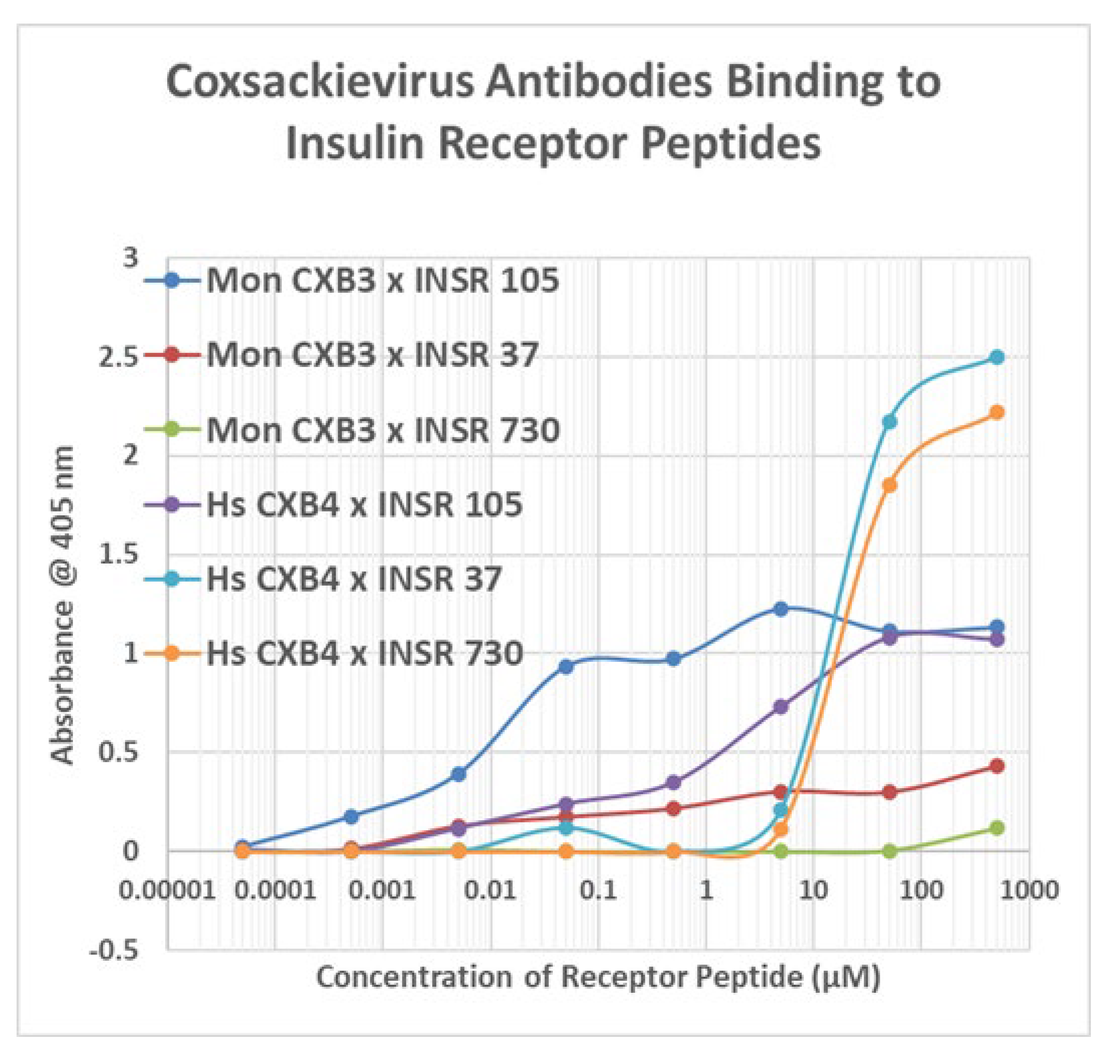
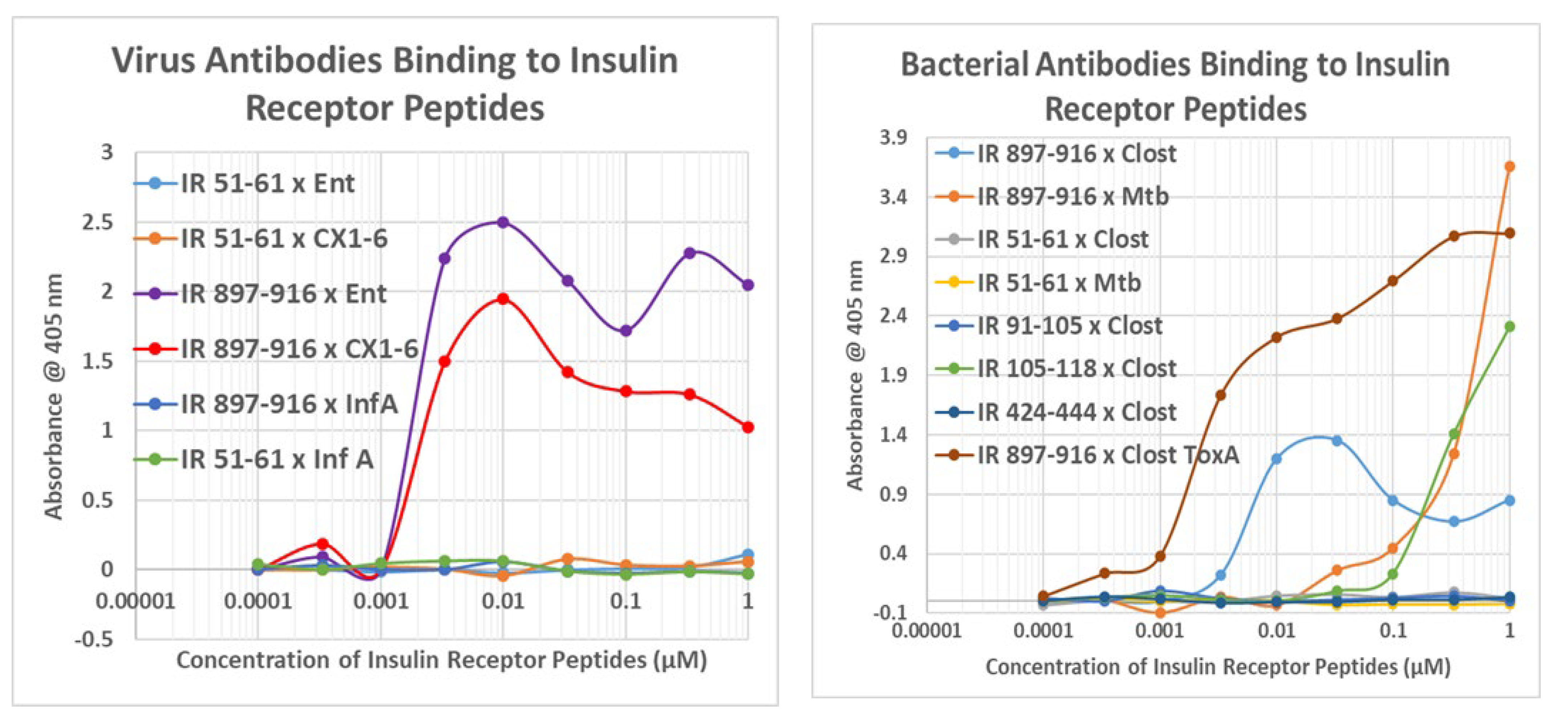
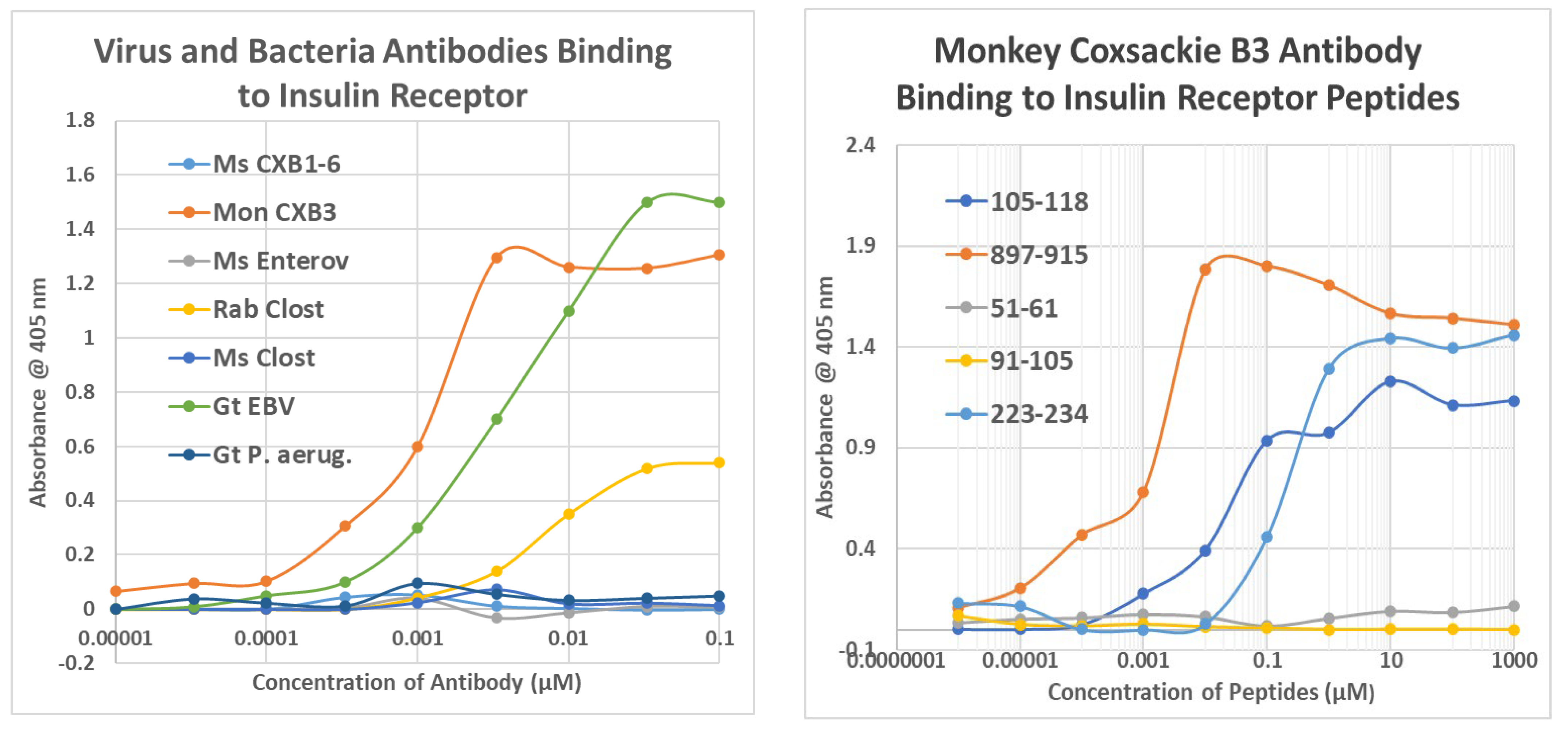
As a first test of the proteomic search results, we explored the ability of antibodies against COX, Clostridia, and a range of other viruses and bacteria to bind to INS, INSR, and peptides derived from the INSR using quantitative enzyme-linked immunoadsorption assays (ELISA). The regions tested for antibody binding were in the extracellular portion of the receptor; although some of the COX-INSR similarities reported in the previous section were in the transmembrane and intracellular portions of the receptor, these were assumed to be inaccessible to antibodies. The results generally confirmed the proteomic findings and are summarized in Figure 4 and Figure 5. COX antibodies, whether derived from monkey, horse, or mouse, consistently recognized INSR as a target with significant affinity, while Clostridium antibodies did not (Figure 4). In general, COX antibodies were specific for regions of INSR that are associated with INS binding. This observation may have two different kinds of importance. One is that COX binding to INSR may interfere with INS binding during T1DM, producing some degree of INS resistance. Secondly, the specificity for INS-binding regions may indicate that some COX antibodies mimic INS itself. This possibility is tested below. Note, however, that COX antibodies derived from different species displayed varying patterns of INSR peptide specificities, suggesting that INSR autoantigenicity may have a genetic component. Note also that although some Clostridium antibodies recognized one INSR region (amino acid positions 897–915)—apparently a highly cross-reactive sequence (see Figure 4)—this cross-reactivity was not sufficient to result in recognition by these Clostridium antibodies (nor Epstein–Barr virus antibodies—Figure 5) also cross-reacting with whole INSR, suggesting that this particular region is perhaps not very accessible to antibodies. None of the antibodies tested recognized glucagon, glucagon receptor peptides, or the beta 2 adrenergic receptor (Figure 4).
Figure 5 illustrates the fact that no other virus antibody tested (influenza A and B viruses, cytomegalovirus, adenoviruses, or herpes simplex virus type 1), except for the Epstein–Barr virus (EBV), cross-reacted with INS, glucagon, or INSR. EBV antibodies recognized INSR 897–915 and the whole INSR. Of the bacterial antibodies tested (Enterococci, Pseudomonas aeruginosa, Mycobacterium tuberculosis, Staphylococcus aureus, and group A Streptococci), only group A Streptococci recognized INS as a target, and once again, while P. aeruginosa and M. tuberculosis recognized several INSR peptides, they did not bind significantly to whole INSR. None of the antibodies tested recognized glucagon, glucagon receptor peptides, or the beta 2 adrenergic receptor.
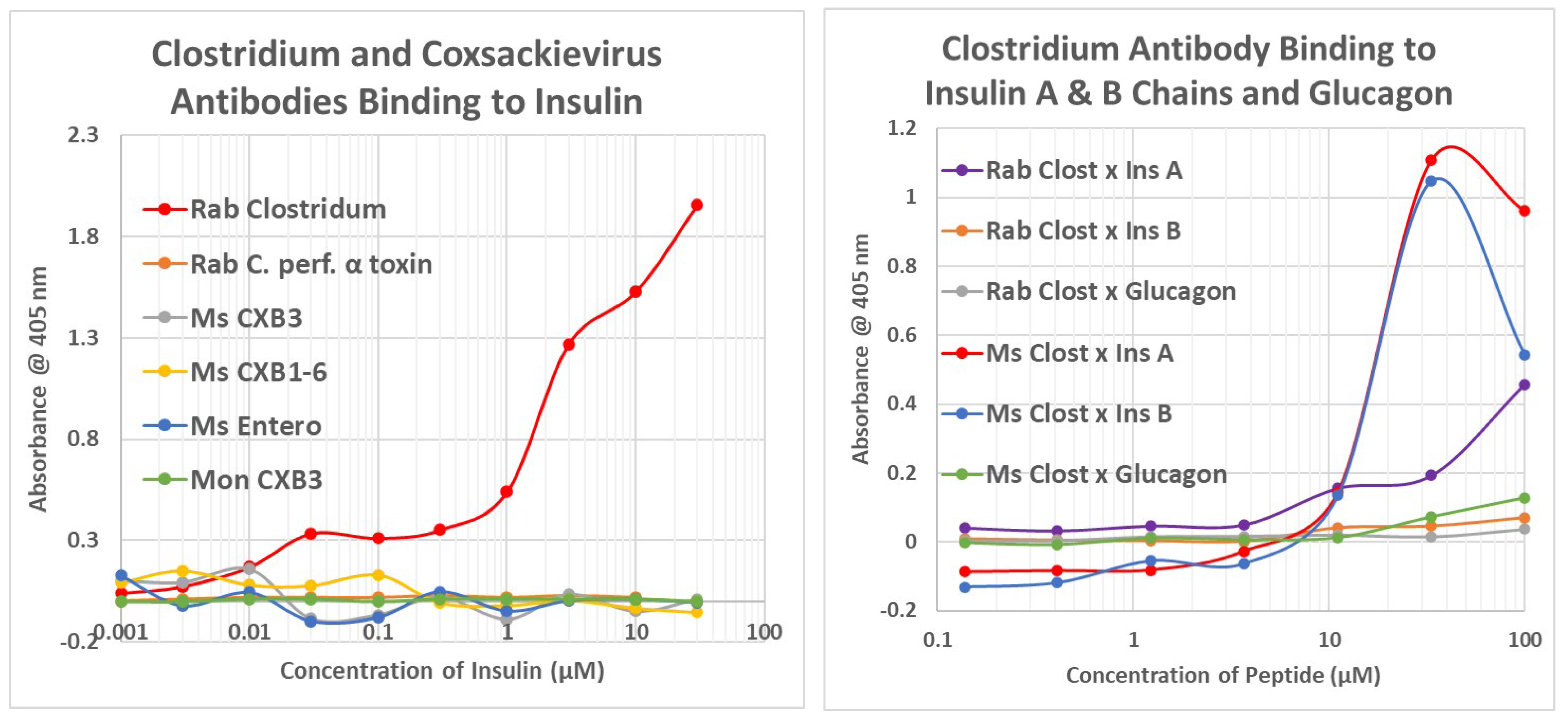
Figure 6 and Appendix D provide examples of many of the key results summarized in Figure 4 and Figure 5 (Kd values) as well as a wide range of negative results. These results clearly demonstrate that cross-reactivity is not a function of the species source of the antibodies. COX, regardless of the species in which the antibodies were raised, and enterovirus antibodies uniformly bound INSR and some of its peptide regions. Figure 7 expands on the data summarized in Figure 4 by demonstrating that Clostridia antibodies cross-react with INS A and B chains (Kd > 15 µM), though with less affinity than to intact INS. Clostridia antibodies bind to INS with both high affinity (Kd 10 nM ) and lower affinity (1.5 µM) interaction.

2.3. Complementarity between Virus Antibodies and Bacterial Antibodies
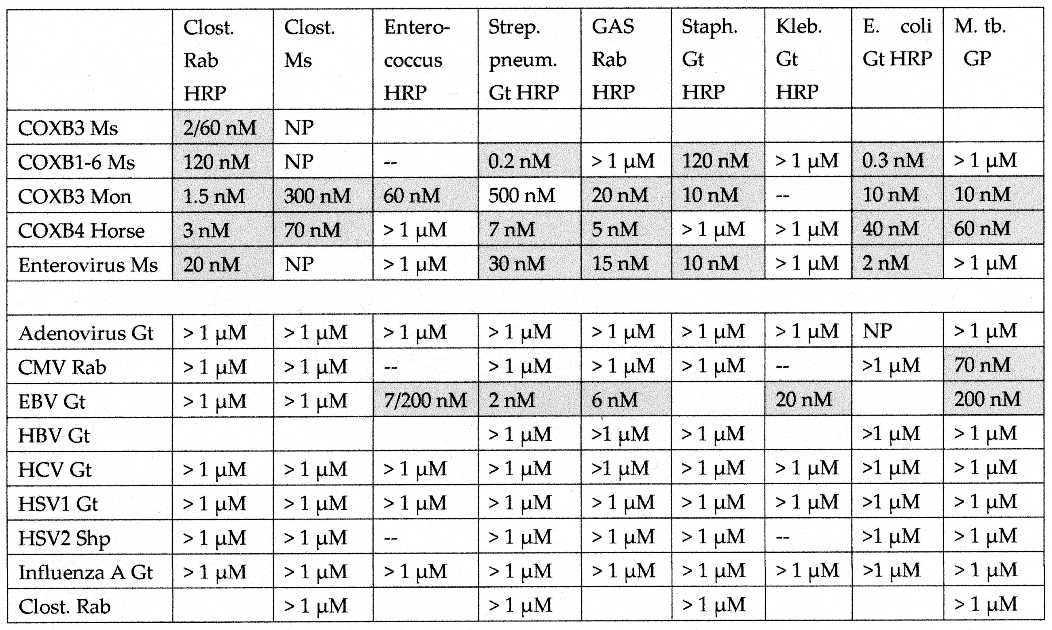
Because Clostridia antibodies cross-react with INS and COX/enterovirus antibodies cross-react with INSR, and because INS and INSR are molecularly complementary to each other, one implication is that some Clostridia antibodies will be complementary to some COX antibodies, i.e., they will interact like idiotype–anti-idiotype pairs. This possibility was investigated using double-antibody ELISA (DA-ELISA) in which one antibody replaces the antigen in a standard ELISA and is bound to the ELISA plate following serial dilution; a second antibody induced in a different species of animal than the first is then applied at a constant concentration, and a horseradish peroxidase-labeled antibody against the species of the second antibody is then used to determine whether the second antibody has bound to the first. In some cases, a horseradish peroxidase-labelled (HRP) antibody was used for the second antibody, making it possible to measure binding to an antibody derived from the same species as the HRP-labelled antibody. We predicted that some Clostridia antibodies would bind specifically to some COX antibodies. The results are summarized in Figure 8 and Figure 9 and illustrated in Figure 10 and Appendix E.
Figure 8. Summary of the binding constants (Kd) from double-antibody ELISA experiments involving a virus antibody potentially binding to a bacterial antibody. See Figure 10 and Appendix E for examples of the binding curves. The binding constants were derived from the inflection points of the curves. COXB = COX type B; CMV = cytomegalovirus; HBV = hepatitis B virus; HCV = hepatitis C virus; HSV = herpes simplex virus; Clost = Clostridium; Strep. pneum. = Streptococcus pneumoniae; GAS = group A Streptococci; Staph = Staphylococcus aureus; Kleb. = Klebsiella; E. coli = Escherichia coli; M. tb. = Mycobacterium tuberculosis; Rab = rabbit; Ms = mouse; Gt = goat; Shp = sheep. Where two numbers appear in any particular box, this indicates that the binding curve was doubly inflected, the first number providing the binding constant of the high affinity part of the curve, and the second number providing the binding constant of the low affinity part of the curve. NP = combination not possible.
Ijms 24 08336 g008
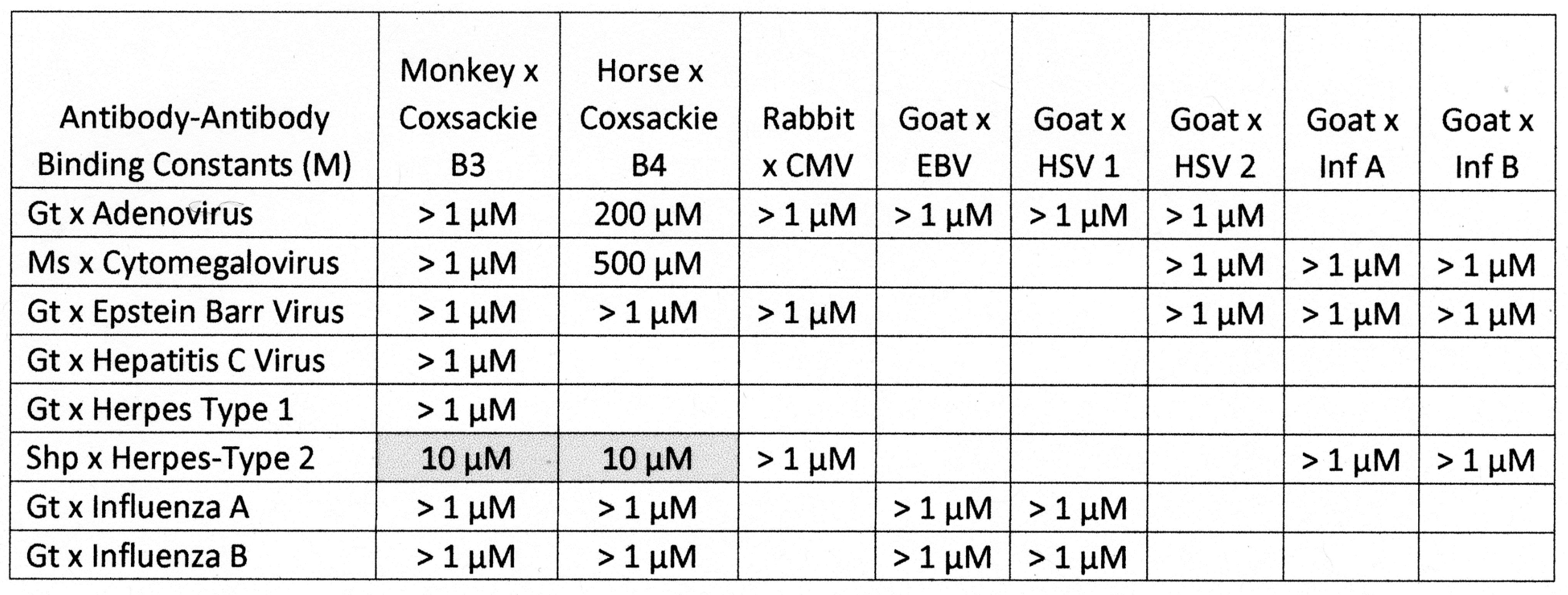
Briefly, all of the COX antibodies tested bound significantly (i.e., with nanomolar affinity) to at least one Clostridium antibody (not all permutations could be tested because some of the antibodies against each microbe were derived from mice). None of the COX antibodies bound to Klebsiella antibodies and only one bound to Enterococcus antibodies, but most bound to Streptococcal, Staphylococcal, Escherichia coli (E. coli), and Mycobacterium tuberculosis (Mtb) antibodies. Clostridia antibodies were more selective, binding only to COX and enterovirus antibodies but not to any of the antibodies against adenoviruses, cytomegalovirus (CMV), Epstein–Barr virus (EBV), hepatitis C virus, herpes simplex type 1, herpes simplex type 2, influenza A virus, or the SARS-CoV-2 spike protein. Of these control antibodies, only those against EBV and SARS-CoV-2 bound significantly to several of the bacterial antibodies (Enterococci, Streptococci, Staphylococci, Klebsiella, and E. coli). The many negative interactions observed for most of the antibody combinations summarized in Figure 8 and Figure 9 illustrate the point that binding is not associated with the species of animal in which the antibodies are raised and, additionally, that virus–bacteria antibody interactions appear to be much more common than virus–virus antibody interactions (Figure 9). No concerted effort was made to investigate bacteria–bacteria antibody interactions, but some results are provided in Figure 8.
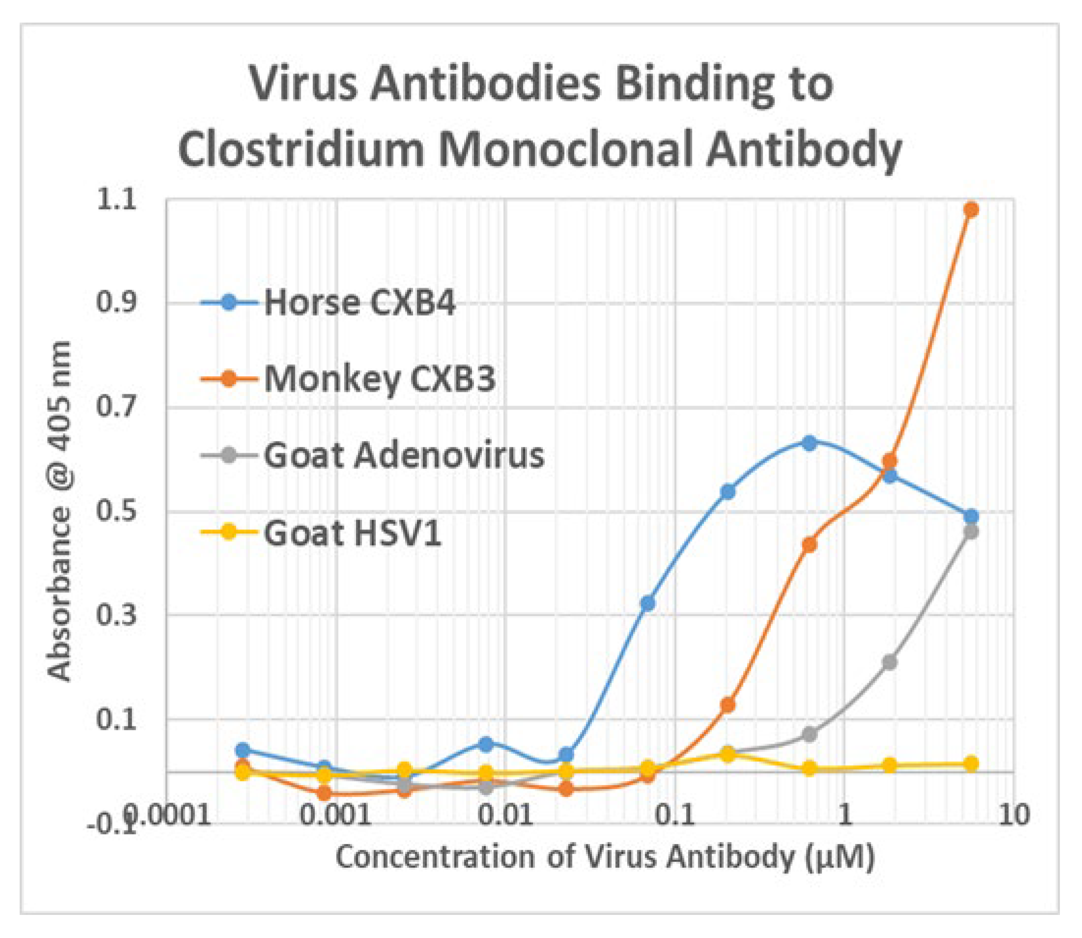
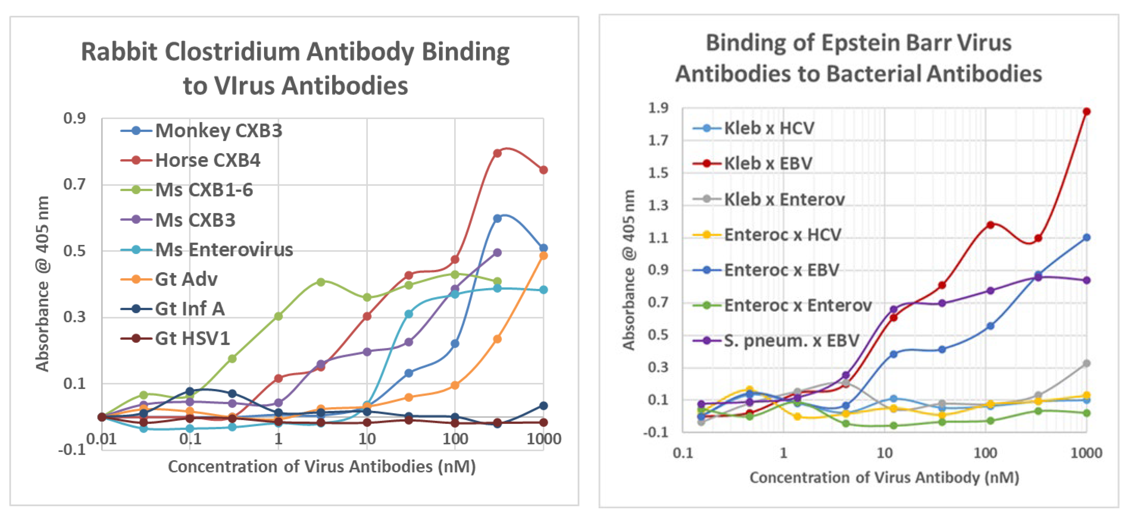
Examples of the binding curves from which Figure 8 and Figure 9 were derived are provided in Figure 10 and Appendix E. These emphasize the specificity of the binding between COX and Clostridia antibodies, although it is also notable that Epstein–Barr virus (EBV) antibodies bound significantly to a number of bacterial antibodies (Figure 9 and Figure 10), particularly in light of the fact that EBV antibodies also recognized INSR and two of the INSR peptides (Figure 10). Notably, one of the bacterial antibodies to which EBV antibodies bound—Streptococcal antibodies—also cross-reacted with INS (Figure 10). Thus, a combination of EBV–Streptococci may mimic the COX–Clostridia combination in terms of recognizing both INS and INSR, thereby resulting in antibodies that have an idiotype–anti-idiotype relationship.

2.4. Idiotype–Anti-Idiotype Relationships between Microbial Antibodies and INS, INS Receptor, GAD-65, or PTPN(IA-2) Antibodies

One limitation of the previous set of DA-ELISA experiments is that they do not demonstrate directly that binding between virus and bacterial antibodies involves recognition of INSR and INS epitopes. One way to determine whether the complementarity specifically may involve INSR and INS epitopes is to utilize specific INSR and INS antibodies in place of either a virus or bacterial antibody. In the following experiments, COX and Clostridia antibodies were therefore tested for their ability to recognize INSR or INS antibodies as well as antibodies to GAD-65 and PTPN(IA-2).
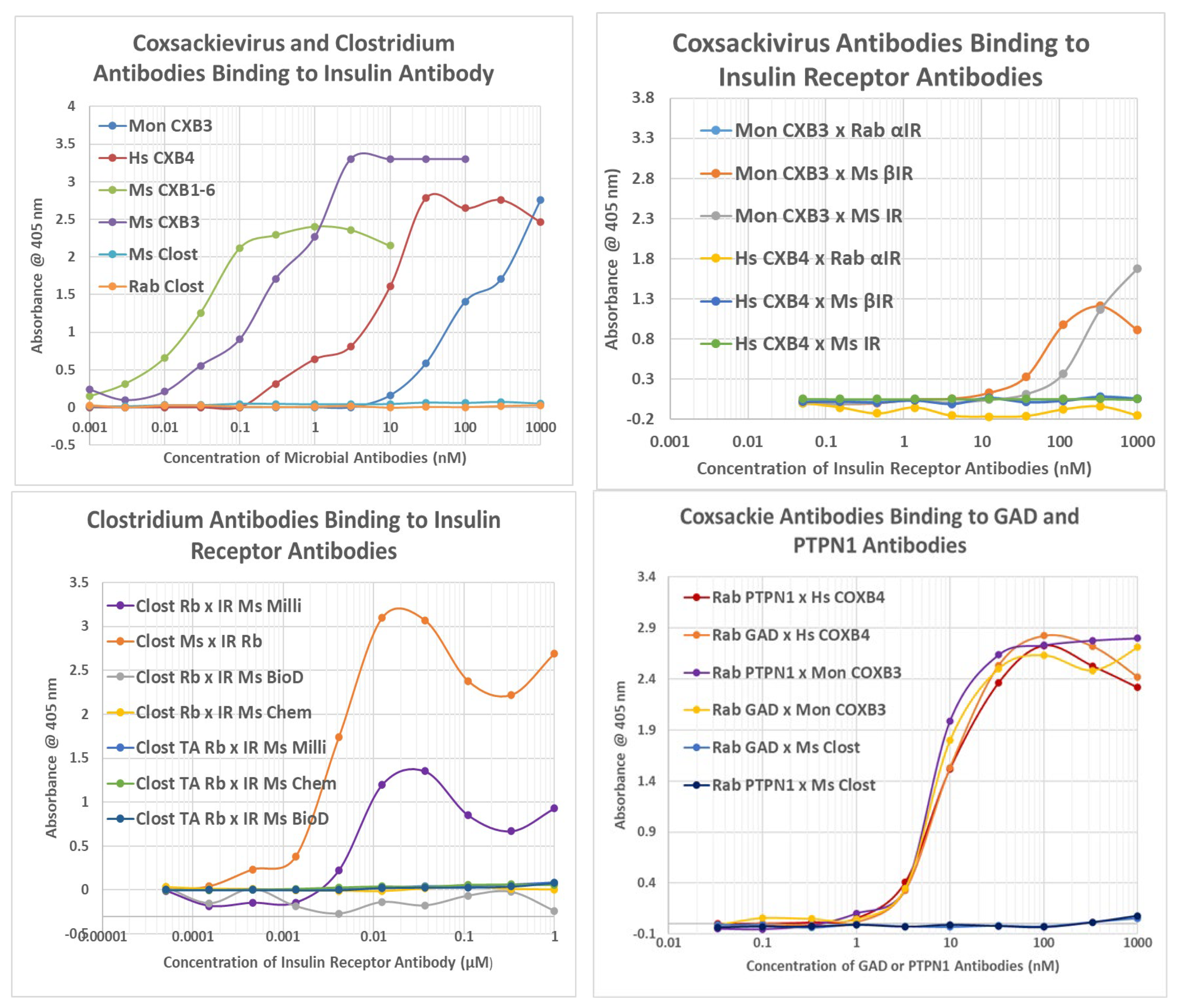
All of the COX antibodies recognized the INS antibody, but none of the Clostridia antibodies did so (Figure 11). Conversely, most of the COX antibodies failed to recognize INSR antibodies (Figure 11), while Clostridia antibodies bound to all of the INSR antibodies (Figure 11). These results are consistent with COX mimicking INSR antigens, so that its antibodies mimic INS and therefore bind to antibodies against INS (which mimic INSR). These results are also consistent with Clostridia mimicking INS, so that its antibodies mimic INSR and bind to antibodies against INSR (which mimic INS). The weak binding of monkey anti-COX antibodies to INSR antibodies may be due to the fact that several of the regions of the INSR that its antibodies recognize are INS mimics [69,70].
Additionally, COX antibodies bound to GAD-65 and PTPN(IA-2) antibodies, but Clostridia antibodies did not (Figure 11). This result is once again consistent with the proteonomic data provided above, indicating that Clostridia proteins mimic GAD-65 and PTPN(IA-2) but COX proteins do not (Appendix C). Antibodies against GAD-65 and PTPN(IA-2) should therefore behave like antibodies against Clostridia. Thus, Clostridia antibodies mimic the behavior of INS antibodies, which in turn mimic the behavior of GAD-65 antibodies and PTPN(IA-2) antibodies, in terms of binding to COX antibodies.

2.5. Complementarity of COX and Clostridium Antigens

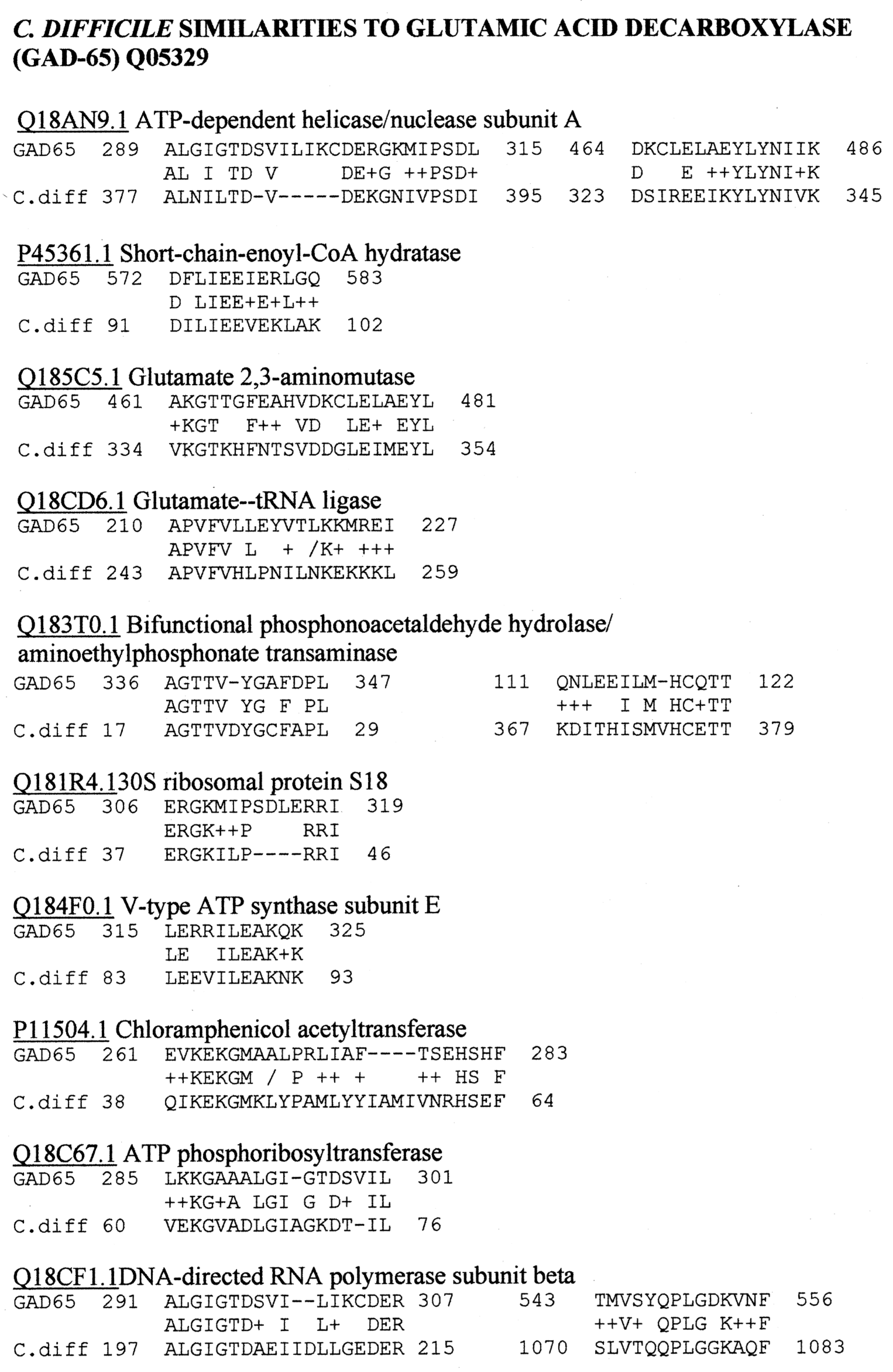
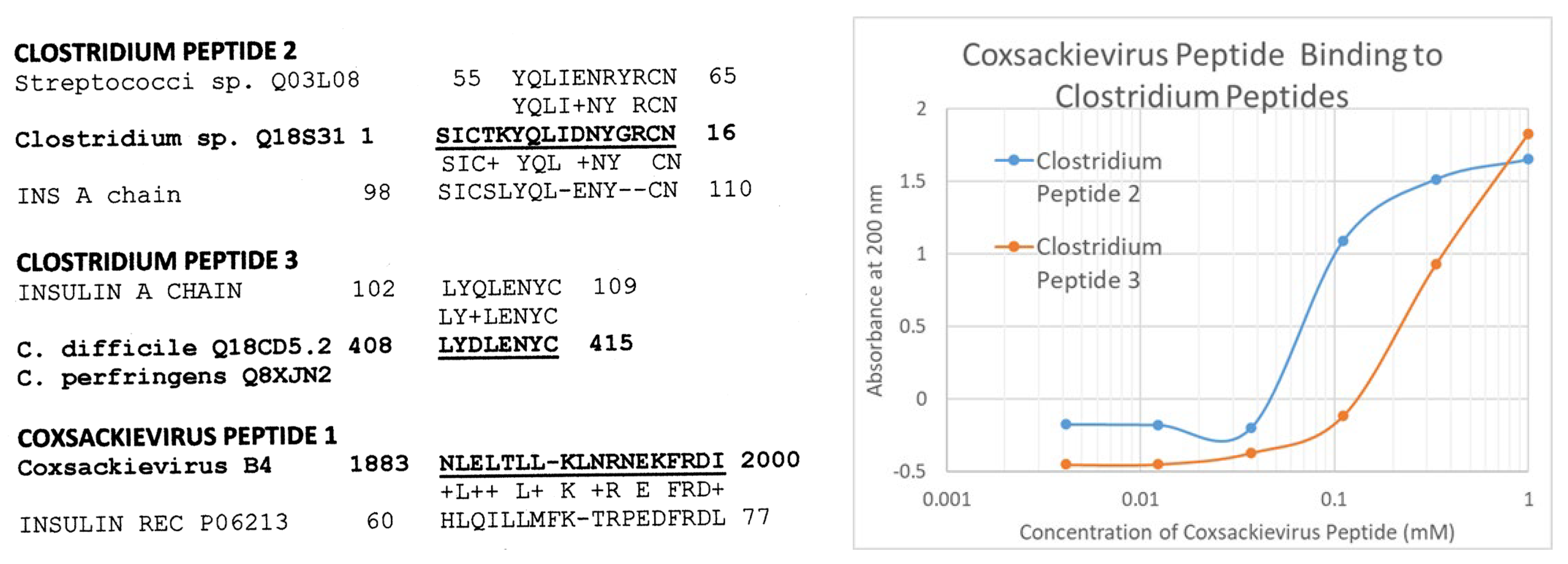
Because some COX antigens mimic INSR sequences and some Clostridia antigens mimic INS, we synthesized several microbial peptide sequences that mimicked these human proteins and used ultraviolet spectroscopy to determine whether they bound to each other, as would be the case if they were complementary antigens. Figure 12 provides the peptide sequences and their similarities to either INS or INSR. Clostridium similarities to the INS A chain were specifically chosen because the role of the A chain in the induction of T1DM is generally ignored in favor of the B chain. Figure 12 also demonstrates that a COX peptide mimicking INSR does bind to both Clostridium (INS A chain mimic) peptides.
The object of this particular set of experiments was not to provide a complete investigation of the range of potential complementarity peptides or proteins that may exist in COX and Clostridia antigens, but merely to test one of the more unusual predictions that follows from the previous results. The fact that this small sample of peptides did yield evidence of complementary antigens suggests the need for a much broader and more complete analysis of COX–Clostridium antigen complementarities.

2.6. T1DM T Cell Receptor Recognition of COX and Clostridium Antigens

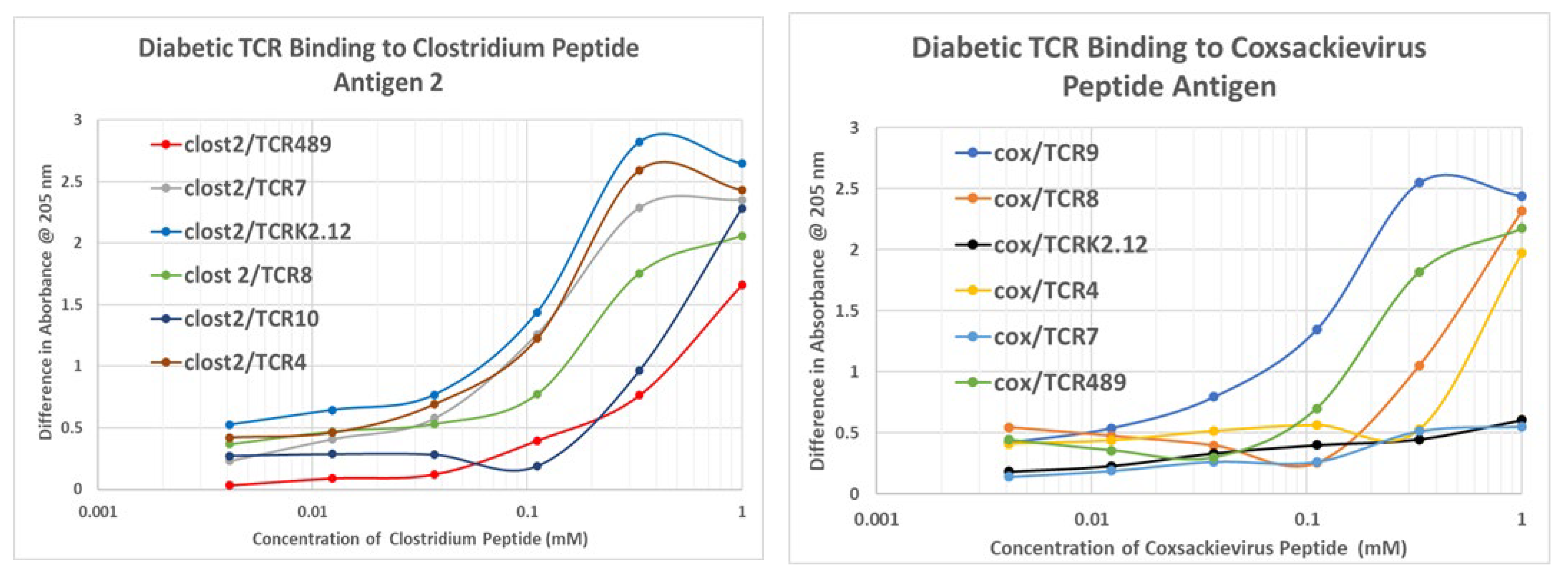
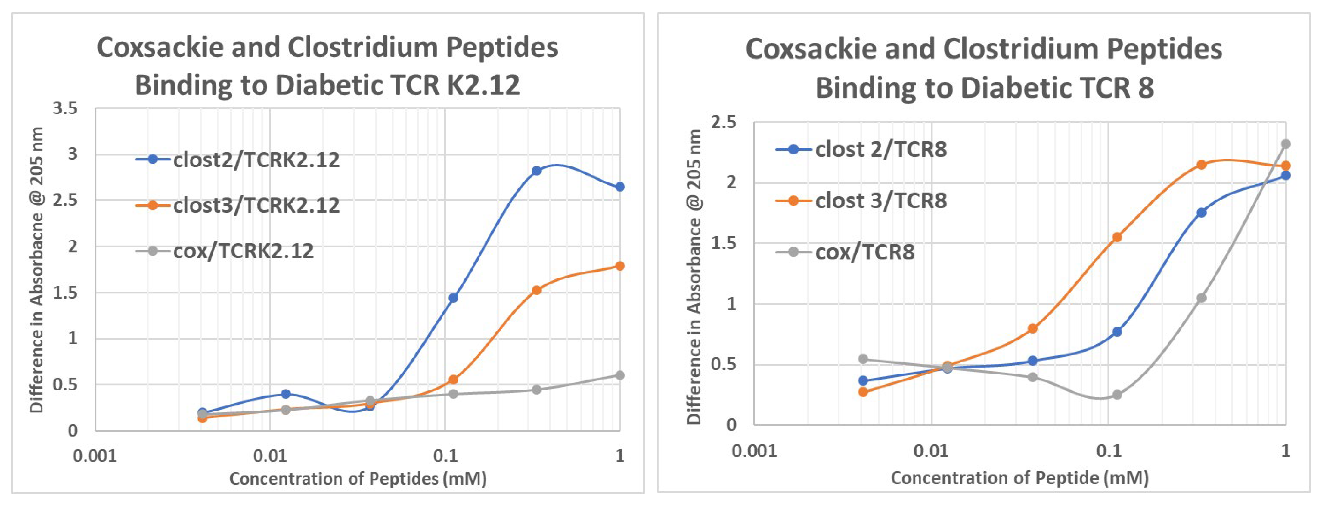
Previous investigators have sequenced the T cell receptor (TCR) sequences that are expanded in T1DM patients with particular specificities for INS or INSR [61,62]. In light of the results above demonstrating that INS-like peptides from Clostridia and INSR-like peptides from COX can mimic INS–INSR binding, we investigated whether TCR recognizing INS or INSR would also recognize these microbially-derived peptides. The TCR sequences were synthesized (sequences provided in Table 1). TCR 1, 2, and 4 were from one patient [61]; TCR 8, 9, and 10 were from a second patient [71]; and TCR K2.4, K2.12, and K2.16 were from a third [72]. Ultraviolet light spectrometry was used to determine whether each TCR sequence recognized the COX and Clostridia peptides used in the previous set of experiments (Figure 12). The results are shown in Table 1, Figure 13, and Appendix F.
It has previously been demonstrated that these TCR occur in complementary groups for the individual patients from whom they are derived, and that at least two of these TCR pairs bind to each other within each patient group, therefore acting like idiotype–anti-idiotype TCR [73,74]. Thus, the fact that the COX peptide, which mimics INSR, binds best to TCR that mimic INS (TCR 9 and TCR 4, 8,and 9) (Table 1) confirms the mimicry of the peptide for INSR. Notably, other INSR peptides (105–118 and 897–915) also bind to these TCR in a very similar pattern. In contrast, the Clostridium peptides mimic INS binding to the INSR-like TCR sequences. The data on INS and INSR peptide binding to these TCR is derived from our previous study [71]. In sum, the hypervariable regions of TCR derived from clones expanded during T1DM recognize a COX peptide similarly to their recognition of INSR peptides, while these TCR recognize Clostridium peptides similarly to the way they do INS. These results are consistent with the other experiments described above.

2.7. Human T1DM Sera Binding to INS and Clostridium sporogenes
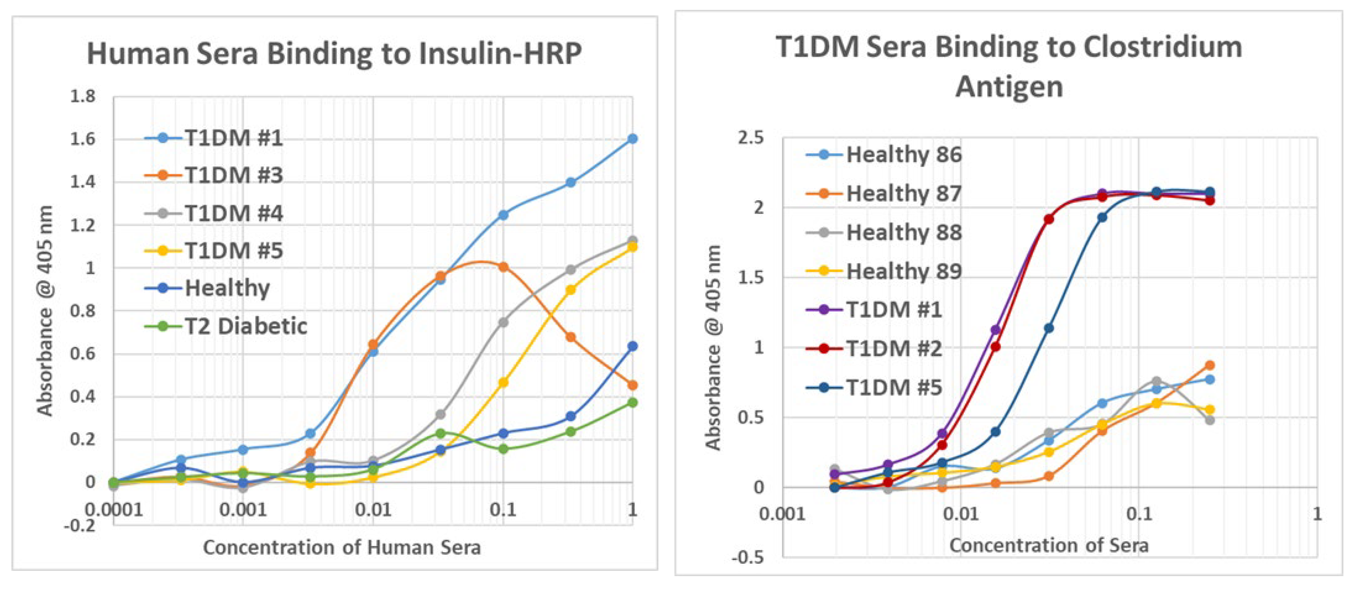
The final set of experiments we performed utilized sera from human T1DM patients, type 2 diabetic patients, and healthy human controls. These sera were tested first for their ability to bind to INS. All of the T1DM sera did so, but healthy individuals and type 2 diabetic sera displayed significantly less INS binding (at least an order of magnitude, and often more than two orders of magnitude less than the T1DM sera) (Appendix G).
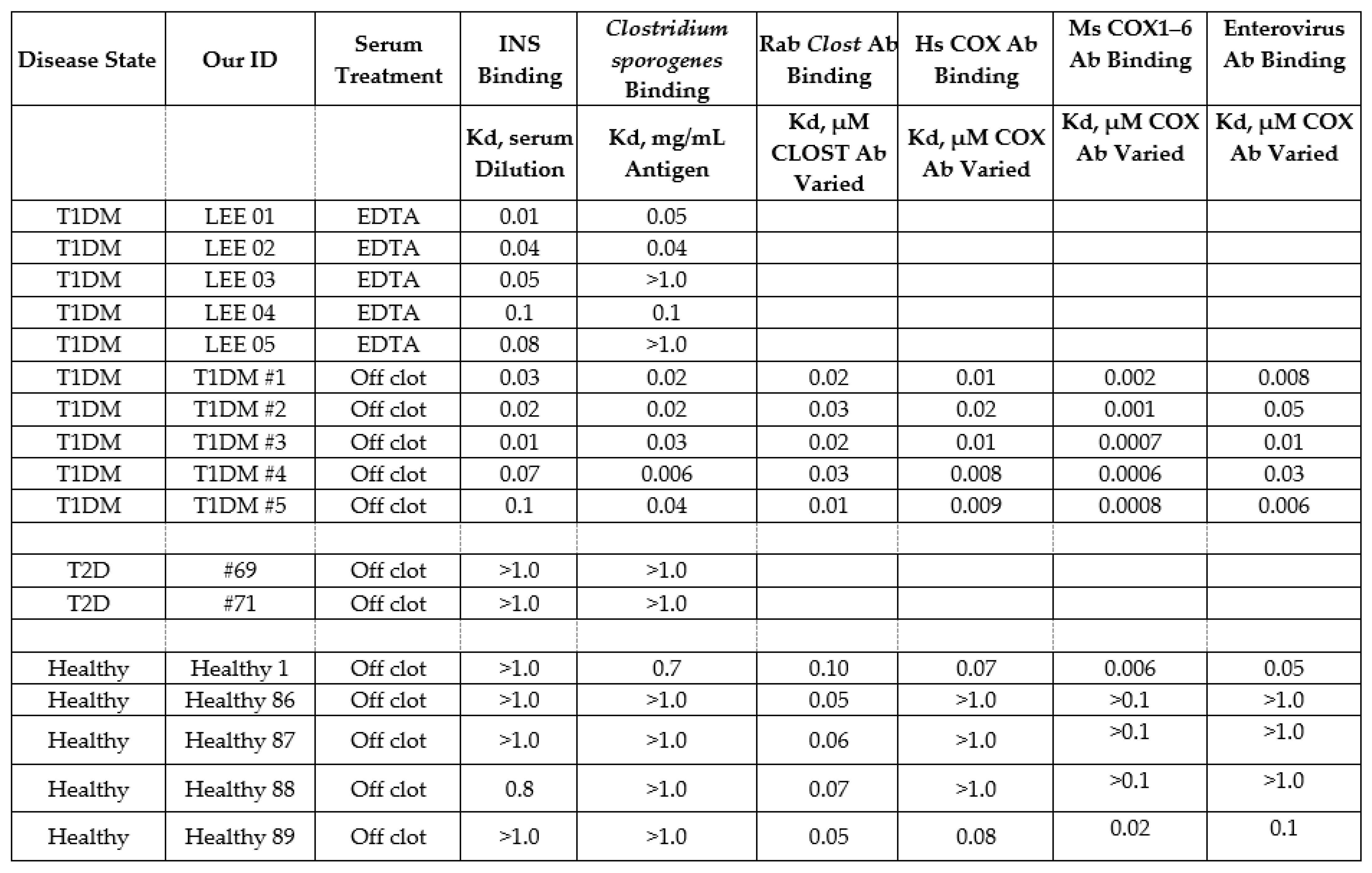
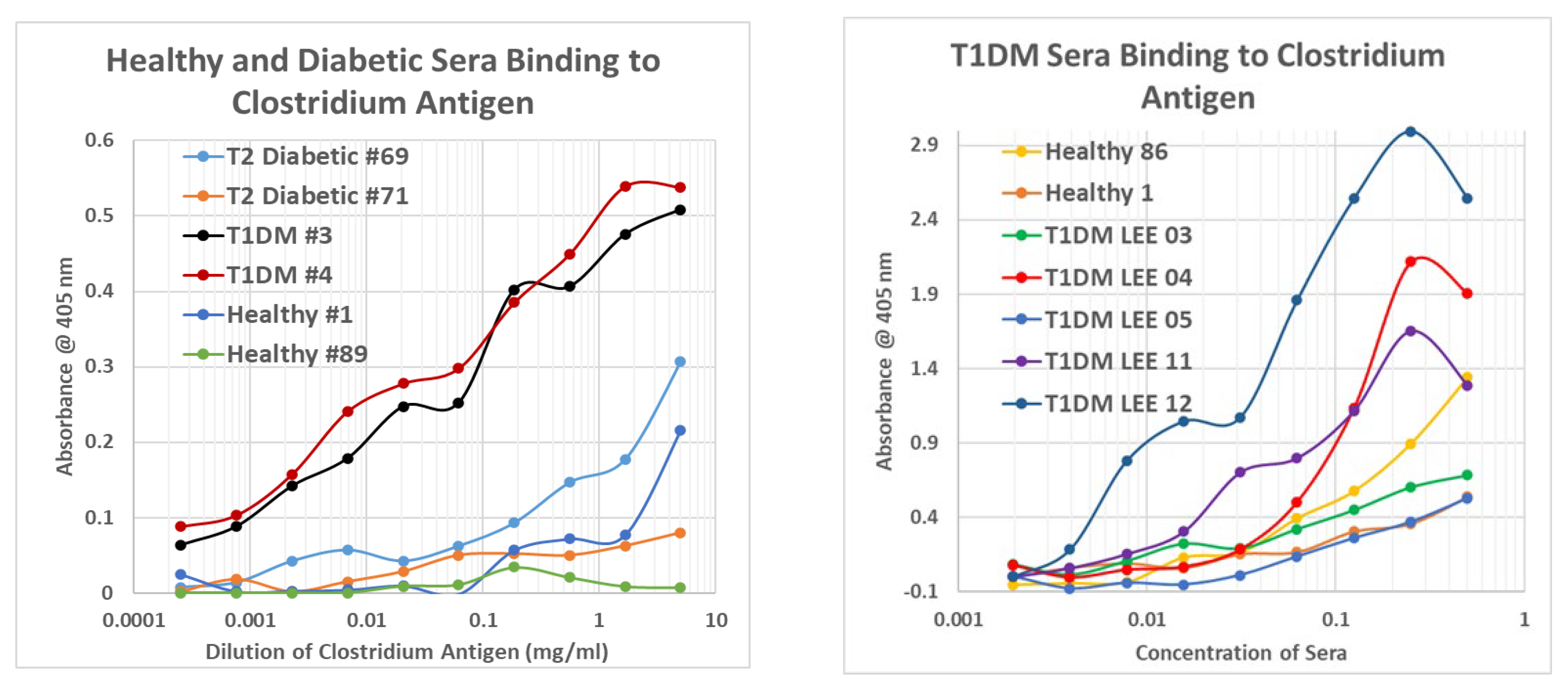
The sera were then tested for binding to inactivated Clostridium sporogenes that is being developed by GN Neutriceuticals SDN BHD in Malaysia in collaboration with Nanyang Technological University Singapore as a possible cancer therapy [75]. These C. sporogenes experiments were run in two different ways that gave equivalent results. The first (Figure 14, LEFT) was to vary the concentration of the Clostridium antigen from 10 mg/mL to less than a thousandth of a mg/mL, keeping the serum concentration constant at 1/100. All T1DM sera bound to the Clostridium antigen in a generally linear concentration-dependent manner that displayed binding to antigen concentrations significantly less than 1/10,000 mg/mL while healthy control and type 2 diabetic sera displayed binding only at the highest concentrations of antigen (above 1/10 mg/mL).
Figure 14. Results of ELISA experiments demonstrating binding of sera from type 1 diabetic patients (T1DM), type 2 diabetic patients (T2 diabetic), and healthy controls to inactivated Clostridium sporogenes antigen. LEFT: The sera were held constant and the concentration of Closridium antigen was varied. RIGHT: the concentration of Clostridium antigen was held constant and concentration of sera was varied.
Ijms 24 08336 g014
The second method involved using a constant concentration of Clostridium antigen (0.1 mg/mL) and varying the concentration of the sera by serial dilution (Figure 14, RIGHT). These experiments yielded classic S-shaped binding curves. In these experiments, while most of the T1DM sera bound significantly more to the antigen with curves that had lower binding constants, a few of the sera (Figure 14, RIGHT) that had been treated with ethylenediaminetetraacetic acid (EDTA) to prevent clotting did not bind the antigen better than did some of the healthy controls, at least one of which demonstrated significant binding to the antigen. Additional data are presented in Appendix G, and results are summarized in Figure 15.
Figure 15. Summary of human sera binding to insulin (INS), Clostridium sporogenes antigen, rabbit (Rab) anti-Clostridium antibody (Ab), horse (Hs) and mouse (Ms) anti-coxsackievirus (COX) antibodies, and enterovirus antibodies. EDTA = etheylenediaminetetraacetic acid.
Ijms 24 08336 g015
There are three possibilities to consider in interpreting these data. One is that the EDTA processing of the one set of T1DM sera may have interfered with antigen recognition. This possibility is given some credence by the fact that most of the T1DM off-the-clot sera displayed lower binding constants (indicating better affinity) than the EDTA-treated sera. Another possibility is that Clostridia were not involved in triggering some of the T1DM cases so that their sera lacked Clostridia antibodies. A third possibility is that nearly everyone is exposed to Clostridia at some time in their lives so that most develop lasting antibodies to the species that wane in some T1DM patients and are robust in some type 2 and healthy individuals. These possibilities are not mutually exclusive.
These data, combined with the TCR data above, are the first evidence clearly linking robust immune responses to Clostridia antigens in most T1DM patients and few non-T1DM patients. The data do not exclude the possibility that other microbes are also involved in triggering T1DM, and they are compatible with the likelihood that some people exposed to Clostridium antigens do not go on to develop T1DM. There is more detail on these points in Section 3 below.

2.8. Human Sera Binding to COX and Clostridia Antibodies
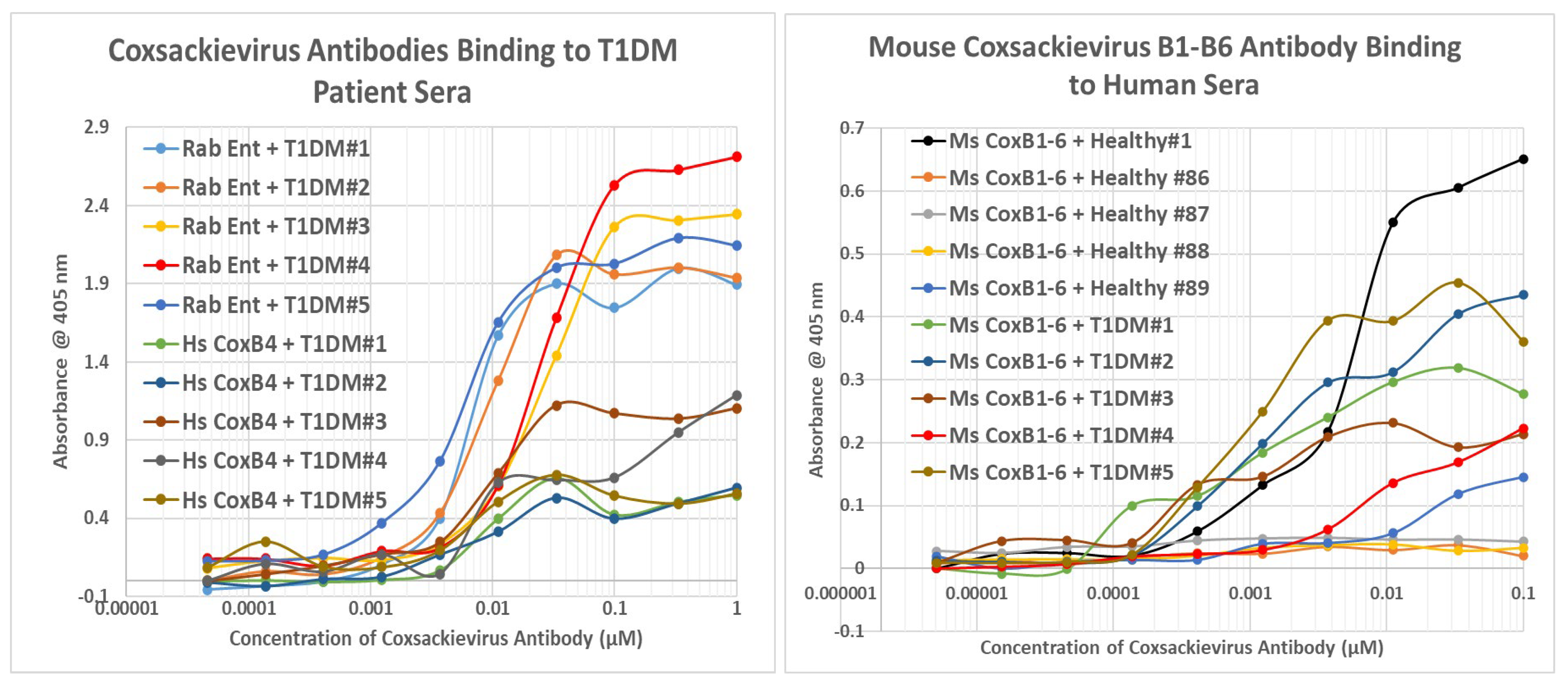
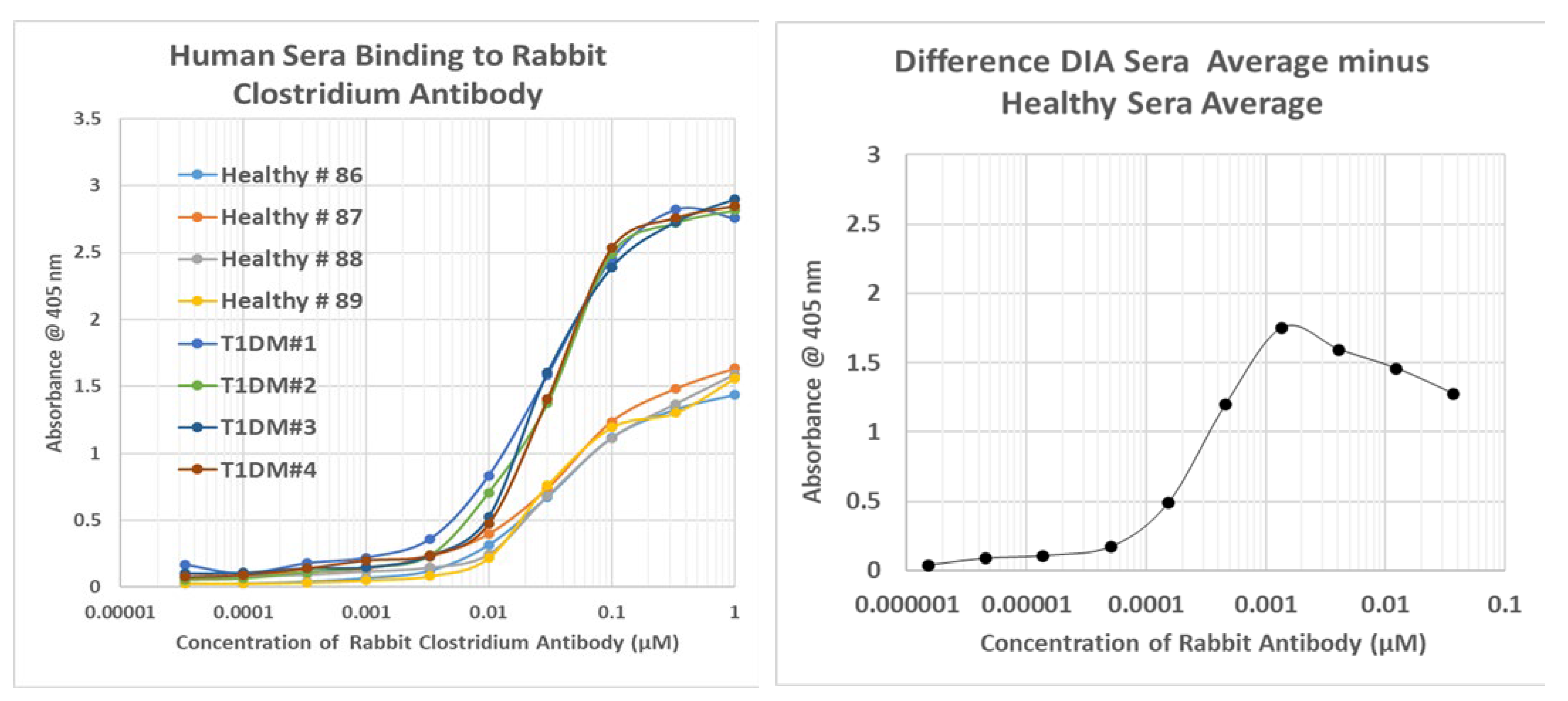
Because we were unable to locate a source of reasonably pure COX antigen, we devised a pair of work-around experiments. On the basis that some Clostridium antigens are complementary to COX antigens (Table 1 and Figure 13), that some Clostridium antibodies are complementary to COX antibodies (Figure 8 and Figure 10), and that T1DM sera contain antibodies against Clostridia (Figure 14 and Figure 15), it follows that T1DM sera should contain antibodies that react to COX antibodies (i.e., are anti-idiotypic to COX antibodies). In fact, T1DM sera do contain antibodies that bind to COX antibodies (Figure 15 and Figure 16). These sera also contain antibodies that recognize Clostridium antibodies (Figure 15 and Appendix G), suggesting that these T1DM sera contain anti-idiotype antibodies derived from idiotypic responses to both COX and Clostridia antigens. Binding to these COX and Clostridium antibodies was much less for healthy individuals than for T1DM patients, with a few exceptions, as illustrated in Figure 15 and Figure 16 and Appendix G.
Figure 16. Results of double antibody ELISA experiments involving the binding of human healthy or type 1 diabetic patient (T1DM) sera to enterovirus (Ent) or COX (Cox) antibodies derived from rabbit (Rab), horse (Hs), or mouse (Ms).
Ijms 24 08336 g016

3. Discussion

3.1. Detailed Summary and Interpretation of Results

Section 2.1 demonstrated that pathogenic Clostridia species mimic INS, PTPN(IA2), and GAD65 better than any other human pathogen or commensal microbe, while COX was the most likely T1DM-associated mimic of INSR, a result consistent with a previous similarity study that found very significant similarities between INS and PTPN(IA-2) [74]. Lactobacilli and Bifidobacteria might play a similar role, but their lack of pathogenicity may mediate their autoimmunogenic potential. Section 2.2 demonstrated that, as predicted, COX antibodies bound to INSR and some of its constituent peptides, while Clostridia antibodies (and most of the other microbial antibodies tested) did not. Conversely, Clostridia antibodies bound to INS, but COX antibodies (and most of the other microbial antibodies tested) did not. Very few other microbial antibodies tested had any affinity for either INS or INSR. The notable exceptions were Streptococcal antibodies binding to INS and Epstein–Barr virus (EBV) antibodies recognizing INSR peptides. Notably, COX antibodies were demonstrated to be complementary to Clostridium antibodies and EBV antibodies to Staphylococcal antibodies, mimicking INS–INSR complementarity (Section 2.3).
Section 2.4 then demonstrated that because COX antibodies mimic INS by binding to INSR, COX antibodies also bind to INS antibodies. Similarly, because Clostridia antibodies mimic INSR by binding to INS, Clostridia antibodies bind to INSR antibodies. It follows that at least some COX antibodies are not only complementary to Clostridia antibodies, as demonstrated in the previous Section, but that this complementarity involves INS- and INSR-specific idiotypes. It was further demonstrated that, as predicted from the similarity data in Section 2.1, PTPN(IA-2) and GAD antibodies could be substituted for INS and Clostridia antibodies, yielding the same binding to COX antibodies. The basic logic of these experiments is summarized in Figure 17.
The complementarity of INSR-like COX sequences and INS-like Clostridia sequences were demonstrated using U.V. spectroscopy in Section 2.5 and Section 2.6, and it was then shown that TCR derived from T1DM patients recognized these COX and Clostridia peptides as antigens, providing direct evidence of autoimmunity against both microbes in three sets of patient TCR. These results indicated that each patient had TCR against both INS–Clostridia and against INSR–COX. Similar results were obtained using sera from T1DM patients, which recognized inactivated Clostridium sporogenes and COX antibodies (Section 2.7 and Section 2.8), thus demonstrating the existence within the sera of anti-idiotype antibodies against both Clostridia antibodies and COX antibodies. Not surprisingly, given the prevalence of the microbes, some of the healthy and T2D control sera also recognized Clostridium antigen, Clostridium antibodies, and COX antibodies, though generally with lower affinities than the T1DM sera (Figure 15).
In short, Clostridia antigens mimic INS; both are in turn mimicked by some INSR antibodies as well as some COX antibodies. COX antigens mimic INSR so that COX antibodies mimic INS as well as INS-like Clostridia antigens. Thus, COX antigens are complementary to Clostridia antigens; COX antibodies are complementary to (idiotype–anti-idiotype) Clostridia antibodies; which means that COX antigens mimic antibodies against Clostridia as well as INSR, while Clostridia antigens mimic antibodies against COX as well as INS (Figure 17). The result is that the immune system loses the ability to differentiate between “self” and “non-self” because any simultaneous response to both COX and Clostridia necessitates an active response against its own antibodies (and TCR) as well INS and INSR. Moreover, each antibody mimics one of the microbial antigens, creating further confusion. A combination of EBV with Staphylococci may induce similar self–non-self confusion, leading to T1DM.

3.2. Relationship of the Experimental Findings to Previous Results

The experimental findings reported here are consistent with much of the previously published literature concerning T1DM etiology and pathogenesis. While a variety of viruses and bacteria have been associated with T1DM onset (reviewed in [76]), enteroviruses, and in particular COX, have been the ones most consistently identified through methods ranging from direct virus isolation to antibody cross-reactivity to microbiome studies [12,13,14,15,16,17,18,19,19,20,21,22,23,48,49,50,51,52,53,54,55,56,76,77,78,79,80,81]. Our results also make a strong case of a role for enteroviruses in T1DM etiology but suggest that the main target of enterovirus antibodies is not INS (as most previous research has attempted to demonstrate) but rather INSR, a T1DM target other investigators have previously reported [4,5,6,7,8,9,54,82,83,84,85,86]. Additionally, previous studies have found that COX does not elicit anti-INS antibodies, nor antibodies against GAD or PTPN(IA-2) [38,51,53,54,87,88,89,90,91]. These results are consistent with our previous report that GAD, PTPN(IA-2), and INS share many homologies [73,74] and the results shown in Figure 11 demonstrating that COX antibodies are complementary to, rather than mimics of, GAD and PTPN(IA-2) antibodies.
What is missing from the enterovirus story is how INS becomes a primary target of autoimmunity in T1DM. Given the importance of INS autoantibodies [40,41,42,66,92,93,94,95,96,97,98], as well as anti-GAD and anti-PTPN(IA-2) autoantibodies and TCR [99,100,101,102,103], in T1DM, and the failure of COX antibodies to recognize these antigens, a critical question is whether there needs to be a second microbe that triggers concomitant anti-INS autoimmunity. Our results strongly implicate Clostridia for this role.
While few studies have directly linked the presence of a Clostridium infection to initiation of T1DM [104,105], a very large number of studies have implicated dysregulation in the number and types of gut Clostridia. In particular, Bifidobacteria, Bacteroides, and Lactobacilli all decrease significantly, while the number of Clostridia increases and is directly correlated with the degree of glucose dysregulation observed in the patient [26,37,106,107,108,109,110,111]. Thus, it is plausible that Clostridia plays a role in T1DM etiology, either to produce bystander activation of the immune system or, as is more likely in view of the complementarity to COX demonstrated here, by synergizing with COX via the production of complementary immune responses. Microbiome constituents such as Lactobacilli, Bifidobacteria, and Bacteroides may become accidental targets of the resulting autoimmunity because they, too, express antigens that mimic INS (Figure 1 and Figure 2) [112]. Additionally, our results are consistent with the observation that pro-INS and GAD65 are both recognized as antigens by the same T cells [1,38,39,113] so that Clostridia could initiate autoimmunity against both antigens via shared mimicry and/or epitope drift.

3.3. Animal Models

Two animal models support the theory that T1DM has a multifactorial etiology involving a combination of viral and bacterial infections and, in particular, that Clostridia has a role in triggering the disease.
T1DM can be triggered in Lewis rats by infecting them with Kilham rat virus (KRV). Studies found that KRV infection significantly increased the abundance of intestinal Bifidobacterium and Clostridium species, indicating a possible synergism between the KRV and these bacteria. Furthermore, treating KRV-infected rats with a combination of trimethoprim and sulfamethoxazole (Sulfatrim) beginning on the day of infection prevented the increase in Bifidobacterium and Clostridium abundance, and also T1DM development [114].
T1DM can also arise “spontaneously” in genetically-predisposed nonobese diabetic (NOD) mice. However, several studies suggest that bacteria, and in particular Clostridia, are involved in triggering diabetes. Tanca et al. [115], for example, found that NOD mice, compared with genetically modified NOD mice protected from T1D (Eα16/NOD), differed in the significant depletion of commensal Clostridial butyrate biosynthesis species. Consistent with this finding, Jia et al. [64,116] demonstrated that supplementing the gut microbiome of NOD mice with the probiotic Clostridium butyricum protected them against diabetes onset. Fecal transplants from non-NOD mice into NOD mice also prevented onset of T1DM, specifically increasing colonization by commensal Clostridia species [117]. Conversely, vancomycin-treated NOD mice were much more prone to develop T1DM than non-treated NOD mice, and the accelerated risk was again associated with a significant decrease in commensal Clostridia species in the gut providing a niche for pathogenic forms [118].

3.4. Epidemiology of Clostridium and Enterovirus Infections

The epidemiology of Clostridium and enterovirus infections is also consistent with the possibility that T1DM is a result of their co-infection. Such a multifactorial mechanism helps to explain one of the great mysteries of autoimmune disease epidemiology, and that of T1DM in particular, which is why many of the putative triggers are so common and the incidence of disease so rare.
Overt Clostridium difficile infections are relatively common, particularly in children, with an incidence of 4 in 1000 American children or 6 per 10,000 patient days [119]. The rate is approximately half that in most European nations, and adults contract Clostridia infections at somewhat lower rates worldwide [120]. However, studies of asymptomatic C. difficile carriage demonstrate that approximately 12% of children less than 18 years of age are infected with non-toxigenic variants, while 6% carry toxigenic variants (reviewed in [121]). In most countries, there is no seasonal variation in incidence [122].
There are also approximately 10 million (1 in 35) COX infections each year in the U.S., the majority among infants and children, with similar rates in most other nations [123,124]; however, asymptomatic carriage of enteroviruses as a whole is approximately 5% among children under 18 worldwide [125]. Cases follow cyclical patterns that vary seasonally across the globe. In North America, cases tend to increase sharply during summer and again in late fall, with a peak in August [126], while, for instance in China, the peak is in January–February [127]. Like Clostridia, COX infections also occur in adults at slightly lower rates than in children [123,124,125,126,127].
The epidemiology of COX and Clostridium infections broadly correspond to T1DM epidemiology. The incidence of new T1DM diagnoses is approximately equal among individuals less than 19 years of age and in those above 19 years of age [128,129], making children approximately four or five times more likely to develop T1DM than adults in any given year of life. This epidemiology is consistent with the majority of COX and Clostridia infections occurring in children. Additionally, the seasonal incidence of new T1DM diagnoses correlates reasonably well with COX incidence. The peak of new T1DM diagnoses in China occurs between December and February [130], which corresponds to the January peak in COX infections. In the U.S., there are two peaks, one of them in August and the other in early winter [130], also corresponding again to the variations in COX incidence. The incidence of new T1DM diagnoses in the southern hemisphere tends to be inverted from that in the northern hemisphere [130], which again corresponds with peak incidences of COX infections [126]. Thus, although a genetic component to T1DM susceptibility is well-recognized among children with relatives with T1DM [131,132], and their risk can be documented by the development of an increasing number and diversity of T1DM-related autoantibodies over many months or years preceding T1DM diagnosis [133,134], the seasonality of new T1DM diagnoses suggest that triggering full-blown autoimmune disease, even against this genetic background, may require an appropriate combination of infectious triggers.
Note, however, that the putative triggers of T1DM identified here—COX with Clostridia—are very common infections, while T1DM is very rare. The reported rates of diagnosed infections and asymptomatic carriage of COX (or enteroviruses more generally) and Clostridia raise serious problems for any T1DM mechanism that is based on a mono-infectious trigger model. The estimated number of new, annual T1DM diagnoses worldwide is many orders of magnitude less than the number of new enterovirus and Clostridia infections. Estimates of the annual incidence of new T1DM diagnoses in the United States range from approximately 40,000 cases, or 1.2/10,000 individuals [128] to approximately 60,000 cases, or 2.0/10,000 individuals [129], the latter figure being typical of most of the rest of the world [129]. While genetic predisposition certainly accounts for some of T1DM risk [131,132,135], it is important to stress that 90% of new T1DM cases have no known relative with T1DM or any defined genetic risk [136], and known genetic risk factors appear to be involved in fewer new cases each year [137].
The incidence of new T1DM diagnoses is much more in line with a dual, concurrent-infection model of etiology. Assuming 12% of children have C. difficile carriage and 5% contract COX each year, then 60/10,000 would be exposed to both in a single year. However, the dual, concurrent-infection model requires that both infections be present simultaneously. Therefore, it seems reasonable to divide the 60/10,000 figure by 52 to yield the probability that an individual would contract both infections during the same week. This yields a probability of 1.2/10,000, which approximates the actual incidence of new T1DM diagnoses. Multiple Clostridia species might be involved as triggers of T1DM, so this number might increase, but on the other hand, a requirement for active (or activated) infection with at least one microbe (see the KRV rat model above) might decrease the probability.
Our data also hint at a combination of EBV with Streptococci as possible co-triggers of T1DM, which would have a probability of occurring concurrently at approximately the same order of magnitude of occurrence as COX + Clostridia. The essential point is that a dual-infection model gets the probability of a new T1DM diagnoses within the right order-of-magnitude estimation, whereas any single-agent model is several orders of magnitude off. Variations in the geographic incidence of T1DM would then be a function of the seasonal variations in the infections and their specific co-incidences in that location.

3.5. Prevention Implications of Complementary Antigen Theory

If both COX and Clostridia are necessary to trigger T1DM, then several implications concerning the prevention of T1DM follow. One is that anyone diagnosed with either an enterovirus infection or a Clostridium infection should be tested for the complementary infection. In light of the fact that T1DM could be prevented in KRV-infected rats with a combination of trimethoprim and sulfamethoxazole (Sulfatrim), beginning on the day of KRV infection [114], if both COX and Clostridia infections are present, appropriate antibiotic therapy might be an effective T1DM preventative. The assumption that the clinically obvious infection is the only infection present may be putting patients at risk for post-vaccinal autoimmune complications such as T1DM.
Next, vaccination against COX should be effective in preventing most cases of T1DM. Each individual vaccine should be completely safe in light of Figure 17 because any host cross-reactive epitopes will either be non-antigenic or tolerized. Only in the rare instance that an individual contracts a Clostridium infection concurrently with their COX vaccination would there be a risk of triggering post-vaccinal T1DM (and thus individuals should be screened before vaccination). COX vaccines for this purpose are already under development [138], but these are being developed under the assumption of a mono-infectious etiology for T1DM acting by means of one of three mechanisms: “Beta-cell death may be primarily induced by CVB [COX type B] itself, possibly in the context of poor immune protection, or secondarily provoked by T-cell responses against CVB-infected beta cells. The possible involvement of epitope mimicry mechanisms skewing the physiological anti-viral response toward autoimmunity has also been suggested… Understanding which [mechanisms] are at play is critical to maximize the odds of success of CVB vaccination, and to develop suitable tools to monitor the efficacy of immunization and its intermingling with autoimmune onset or prevention” [138]. If, in fact, T1DM etiology involves complementary antigens, then the safety of COX vaccines may require antigen deletion of regions mimicking INSR and other TIDM autoantigen sequences identified here.
A third approach would be to develop Clostridium vaccines, which should be safe for the same reasons provided above for COX vaccines but carry the same caveat regarding COX co-infection. Such Clostridia vaccines are also in development, although at present for different purposes [75,139,140,141,142]. The use of such vaccines again depends upon understanding the mechanism by which Clostridium is involved in T1DM etiology and including (or excluding) appropriate antigenic regions. For example, our data suggest (but certainly do not prove) that Clostridium toxin A is not involved in T1DM pathogenesis so that a toxin-based vaccine might be particularly safe from the perspective of preventing Clostridium infections without risk of T1DM as a post-vaccinal side effect. However, if non-toxin-producing strains of Clostridia can trigger T1DM, then a toxin-based vaccine may not be optimal for preventing T1DM.
Whether COX or Clostridia-based, optimization of T1DM prevention strategies rely, in the end, on having an appropriate animal model with which to test such strategies.

3.6. The Need for New Animal Models of T1DM

If T1DM is, in fact, triggered by a combination of COX and Clostridium infections (or EBV + Streptococci), then new animal models of the disease need to be developed, not least in order to test potential preventative approaches such as those described in the previous section. Such models might be implemented in several ways. One would be to infect susceptible animals with combinations of COX and Clostridia. An alternative would be to inoculate animals with combinations of inactivated COX and inactivated Clostridia. Success of the second type of experiment would also demonstrate that the pathogenesis is immunologically mediated (and therefore purely autoimmune) rather than requiring damage to the pancreas due to active infection. This second type of experiment might also be used to screen COX and Clostridia vaccines for their potential synergy and thus be used as a screen to increase their safety or to warn against their co-administration. Finally, another way to develop a novel T1DM animal model would be to inoculate animals with combinations of COX polyclonal antibodies and Clostridia polyclonal antibodies induced in the same species as that inoculated. According to the antigenic complementarity demonstrated in this paper, the resulting immune complexes should mimic the key complementary antigens triggering T1DM, and their idiotypes should therefore function equally as antigenic epitopes to initiate an autoimmune process.
Animal models should also explore the possibility suggested by the data we have provided here that an EBV–Streptococci combination may trigger T1DM.

3.7. Limitations of the Study

This study has limitations. One obvious one that has just been addressed is the lack of an animal model to test whether a combination of COX and Clostridia induces T1DM. The previous section lays out three ways to test this prediction.
A second limitation is that we tested our human sera only for binding to C. sporogenes rather than pathogenic species of Clostridia such as C. perfringens, C. difficile, etc., and may therefore have missed important cross-reactivity to additional Clostridia antigens or found cross-reactivities that do not extrapolate to other Clostridia species. Similarly, we were unable to obtain even relatively pure whole-COX antigens and therefore could not directly demonstrate whether our human T1DM sera were positive for COX antibody. COX antibody presence was inferred from a demonstration of anti-idiotypic responses to Clostridia antibodies combined with a demonstration of COX–Clostridia antibody anti-idiotype.
A third limitation is that the actual molecular complementarity between COX and Clostridium antigens has only been tested in the most cursory way in the present study using two pairs of peptides. Clearly much additional work needs to be performed to characterize this antigenic complementarity.
Similarly, the role of TCR in mediating T1DM through both antigenic complementarity and TCR idiotype–anti-idiotype interactions has only been explored cursorily here. While much more comprehensive studies have previously been published [72,73], the role of TCR complementarity needs much further investigation. The possibility that the TCR sequences expanded in T1DM may identify not just the particular microbes triggering the disease but also the specific antigenic sequences that are involved also needs further research. Both of these possibilities therefore stand as important predictions that can be used to test the validity and utility of the complementary antigen theory presented here.
Additionally, the COX–Clostridia combination and its corresponding anti-idiotype may not be the sole triggers of T1DM. Our data also suggest that an EBV–Streptococci combination be investigated more thoroughly. Additionally, Bifidobacteria and Lactobacilli both appeared as possible INS mimics in our similarity studies (Figure 1 and Figure 2) and have also been implicated in many T1DM microbiome studies (Section 3.2 and Section 3.3). Unfortunately, no antibodies against these bacteria could be located and therefore their possible synergy with COX or other viruses could not be explored.
Correspondingly, various viruses other than COX and EBV have also been associated with T1DM initiation, including CMV [89,143,144,145] and rotaviruses [145,146]. Thus, it is possible, if not likely, that other microbial combinations (probably bacterial–viral but possibly bacterial–bacterial or viral–viral) can also express complementary antigens that mimic human glucose-regulatory proteins and peptides and thus induce some form of T1DM. This possibility is supported by the fact that not all of the T1DM sera tested in this study reacted strongly to Clostridia antigens (Section 2.7) and some cross-reactivity was observed between non-COX virus antibodies or bacteria to INSR peptides (Section 2.2). Thus, it is important not to over-interpret the data presented here as meaning that a COX–Clostridium combination is the only possible cause of T1DM or predict that COX and/or Clostridium vaccines will prevent all T1DM cases in the future.
Finally, there is thus far no direct evidence from human studies (either clinical or epidemiological) demonstrating that individuals newly diagnosed with T1DM have recently been exposed to both enteroviruses and Clostridia infections. Such studies will be needed to test the hypothesis proposed here.

4. Materials and Methods

4.1. Similarity Searches

Protein sequences for use in similarity searches were obtained from UniProtKB (https://www.uniprot.org/help/uniprotkb) accessed between 28 August 2018 and 17 July 2022. BLASTP (version 2.2.31+) on the www.expasy.org server. BLOSUM80 (1 June 2021–17 July 2022) was used to identify the type of short, continuous sequences approximately ten to fifteen amino acids in length that are presented by Human Leukocyte Antigens (HLA) to T and B cells [147,148]. The E value was set to 1; filter low complexity regions on; no gaps; 100 best scoring and best alignments to show. Only matches that had a Waterman–Eggert score of at least 50, an E value of less than 1.0, and which contained a sequence of ten amino acids in which at least six were identical, were counted as sufficiently similar to induce possible cross-reactive immunity; this criterion is based on substantial experimental research demonstrating that sequences exhibiting at least this degree of similarity have a >85% probability of being cross-reactive under experimental conditions [149,150,151,152].

4.2. Experimental Protocols

ELISA and double-antibody ELISA (DA-ELISA) were employed to investigate whether the similarity searches yielded immunologically valuable information.
Enzyme-linked immunosorbent assay (ELISA) was used to investigate cross-reactivities between microbial antibodies and diabetes-related proteins. The diabetes-related protein was diluted in pH 7.4 phosphate buffer to a concentration of 10 µM. This standard solution was then diluted by ten-fold steps to approximately 10−14 M. Two wells received only phosphate buffer as controls. An amount of 100 µL of each protein dilution was added in duplicate to wells of a Costar round-bottomed 96-well ELISA plate and incubated for one hour. The excess protein was triply washed out using a 1% Tween 20 solution (in phosphate buffer) and a plate washer. Next, 200 µL of blocking agent (2% polyvinylalcohol in phosphate buffer) was added to every well, incubated for an hour, and then triply washed. (PVA was used rather than bovine serum albumin or ovalbumin because these proteins were found to cross-react with some of the antibodies used in our experiments.) An antibody against a microbe (at 1 mg/mL concentration) was then diluted to 1/200 in phosphate buffer, and 100 µL was added to every well. The antibody was incubated for an hour and then triply washed. A species-appropriate horseradish peroxidase-linked secondary antibody was then, at a dilution of 1/1000, incubated for an hour, and triply washed. Finally, 100 µL of ABTS reagent (Chemicon via SigmAldrich, St. Louis, MO, USA) was added, incubated for 30 min, and the plate read at 405 nm in a Spectramax UV-VIS scanning spectrophotometer (Molecular Devices, San Jose, CA, USA) Data were gathered using Spectramax software (Molecular Devices, San Jose, CA, USA) and then analyzed using Excel (Microsoft, San Jose, CA, USA). Analysis essentially consisted of subtracting non-specific binding to the buffer-only wells from the protein-containing wells and plotting the amount of antibody binding (as measured by absorbance at 405 nm) as a function of protein concentration.
Double antibody ELISA (DA-ELISA) was used to investigate possible antigenic complementarity between the antibodies used in the study. DA-ELISA differs from ELISA in that the protein laid down in the 96-well plate in the initial step of an ELISA is substituted with an antibody. A second antibody (from a different species) is tested for its ability to bind to the first. The ability of the second antibody to bind to the first is then monitored using peroxidase-linked antibody against the species from which the second antibody is derived. As in the ELISA protocol, the first antibody is made up at a concentration of approximately 10 µM (assuming IgG antibodies have a molecular weight of 150,000 daltons) and then serially diluted by factors of ten. The rest of the protocol is the same. In some cases, it is possible to use an HRP-conjugated antibody as the second antibody in the process, in which case the third step can be skipped. The use of HRP-conjugated antibodies in the second step permits the first and second antibodies to be from the same species.

4.3. Antigens

INS receptor peptides and glucagon receptor peptides were synthesized to at least 95% purity by mass spectrometry by RS Synthesis (St. Louis, MO, USA). See Figure 8 and Figure 9 for sequences. Inactivated Clostridium sporogenes was obtained from Professors Moumita Rakshit and Swee Hin Teoh of Nanyang Technological University, Singapore, where it is being developed by GN Neutriceuticals SDN BHD in Malaysia as a possible cancer therapy [75]. Proteins and peptides are listed in Table 2.

4.4. Antibodies

Antibodies against viruses are listed in Table 3; against bacteria in Table 4; against proteins in Table 5; and secondary antibodies are listed in Table 6.
Monkey COX B4 was observed using anti-human secondary antibody.

4.5. Human Sera

Human sera sources are listed in Table 7.

Author Contributions

Conceptualization, R.R.-B.; methodology, R.R.-B.; validation, R.R.-B., K.C., J.H., A.Z. and M.P.; formal analysis, R.R.-B., K.C., J.H., A.Z., M.P. and M.T.; investigation, K.C., J.H., A.Z., M.P. and M.T.; resources, R.R.-B.; data curation, K.C., J.H., A.Z., M.P. and M.T.; writing—original draft preparation, R.R.-B.; writing—review and editing, K.C.; visualization, K.C., J.H., A.Z., M.P. and M.T.; supervision, R.R.-B.; project administration, R.R.-B.; funding acquisition, R.R.-B. All authors have read and agreed to the published version of the manuscript.

Funding

This research received no external funding.

Institutional Review Board Statement

Not relevant: all patient sera were obtained through commercial sources.

Informed Consent Statement

Not relevant: all patient sera were obtained through commercial sources that anonymized the donors.

Data Availability Statement

The majority of the data compiled for this study is presented herein; all data not explicitly described are available on request from the primary investigator.

Conflicts of Interest

The authors declare no conflict of interest.

Appendix A

Figure A1. BLASTP similarities between human INS A chain and C. difficile.
Ijms 24 08336 g0a1
Figure A2. BLASTP similarities between human INS B chain and C. difficile.
Ijms 24 08336 g0a2

Appendix B

Figure A3. Similarities between viruses and human insulin receptor.
Ijms 24 08336 g0a3

Appendix C

Figure A4. A selection of BLASTP similarities between C. difficile proteins and protein tyrosine phosphatase non-receptor type 1. Many additional significant similarities exist.
Ijms 24 08336 g0a4
Figure A5. A selection of BLASTP similarities between C. difficile proteins and glutamic acid decarboxylase type 65. Many additional significant similarities exist.
Ijms 24 08336 g0a5
Figure A6. BLASTP similarities between glutamic acid decarboxylase type 65 and COX type B4 (which is typical of the other COX). These are all the significant similarities that were found (compared with results for GAD-65 and Clostridia above).
Ijms 24 08336 g0a6

Appendix D

Figure A7. Comparison of monkey (Mon) coxsackievirus (CX) antibodies and horse (Hs) CX antibodies binding to IR peptides. Numbers indicate the amino acid sequence of the INS receptor from which the peptides were derived.
Ijms 24 08336 g0a7
Figure A8. Results of ELISA studies concerning virus and bacteria antibodies binding to human INS receptor peptides. LEFT: Enterovirus (Ent), COX types B1-6 (CX1-6), and influenza A virus (Inf A) antibodies binding to INS receptor (IR) peptides identified by their sequence numbers. RIGHT: Clostridium (Clost), and Mycobacterium tuberculosis (Mtb) antibodies binding to INS receptor peptides identified by their sequence numbers. ToxA = toxin A.
Ijms 24 08336 g0a8

Appendix E

Figure A9. Representative binding curves resulting from double antibody ELISA experiments involving Clostridium monoclonal antibodies binding to coxsackievirus (CX) type B3 and type B4 antibodies, adenovirus antibodies, and herpes simplex type 1 (HSV1) antibodies.
Ijms 24 08336 g0a9
Figure A10. Some of the many control combinations of virus–bacterial antibody pairs tested by double antibody ELISA and summarized in Figure 12 and Figure 13. S. pneum. = Streptococcus pneumoniae; S. aureus = Staphylococcus aureus; Clost = Clostridium; Adv = adenovirus; CMV = cytomegalovirus; EBV = Epstein–Barr virus; HSV1 = herpes simplex type 1 virus; Ms = mouse; Rbt = rabbig; Gt = goat.
Ijms 24 08336 g0a10

Appendix F

Figure A11. Ultraviolet spectrometry study of COX (cox) peptide (which mimics the INS receptor) and Clostridium peptides 2 and 3 (clost2, clost3) (which mimic INS), binding to T cell receptors (TCR) that mimic a range of peptides (see Table 1 for details). The binding constants listed in Table 1 were estimated from the inflection points of the curves.
Ijms 24 08336 g0a11

Appendix G

Figure A12. TIDM and Control Sera binding to human insulin and Clostridium antigen. LEFT: Results of ELISA experiments demonstrating binding of sera from type 1 diabetic patients (T1DMand healthy controls to INS conjugated to horseradish peroxidase (INS–HRP). RIGHT: Results of ELISA experiments demonstrating binding of sera from type 1 diabetic patients (T1DM), and healthy controls to inactivated Clostridium sporogenes antigen. In these experiments, the concentration of Clostridium antigen was held constant, and concentration of sera was varied.
Ijms 24 08336 g0a12
Figure A13. Results of double antibody ELISA experiments involving the binding of human healthy or type 1 diabetic patient (T1DM) sera to rabbit Clostridium antibodies (LEFT). Although all sera bound Clostridium antibodies, the T1DM sera bound significant more than the healthy sera (RIGHT).
Ijms 24 08336 g0a13

References

  1. Mallone, R.; Brezar, V.; Boitard, C. T cell recognition of autoantigens in human type 1 diabetes: Clinical perspectives. Clin. Dev. Immunol. 2011, 2011, 513210. [Google Scholar] [CrossRef] [PubMed]
  2. Krischer, J.P.; Liu, X.; Lernmark, Å.; Hagopian, W.A.; Rewers, M.J.; She, J.X.; Toppari, J.; Ziegler, A.G.; Akolkar, B.; TEDDY Study Group. Predictors of the Initiation of Islet Autoimmunity and Progression to Multiple Autoantibodies and Clinical Diabetes: The TEDDY Study. Diabetes Care 2022, 45, 2271–2281. [Google Scholar] [CrossRef] [PubMed]
  3. Bauer, W.; Veijola, R.; Lempainen, J.; Kiviniemi, M.; Härkönen, T.; Toppari, J.; Knip, M.; Gyenesei, A.; Ilonen, J. Age at Seroconversion, HLA Genotype, and Specificity of Autoantibodies in Progression of Islet Autoimmunity in Childhood. J. Clin. Endocrinol. Metab. 2019, 104, 4521–4530. [Google Scholar] [CrossRef] [PubMed]
  4. Kim, S.W.; Won, H.K.; Seok, H.; Lee, B.W.; Jung, C.H.; Lee, W.J.; Kim, J.H. High prevalence of both anti-INS and anti-INS receptor antibodies in Korean patients with INS autoimmune syndrome. Diabetes Res. Clin. Pract. 2012, 98, e12–e15. [Google Scholar] [CrossRef]
  5. Blackard, W.G.; Anderson, J.H.; Mullinax, F. Anti-INS receptor antibodies and diabetes. Ann. Intern. Med. 1977, 86, 584–585. [Google Scholar] [CrossRef]
  6. Ludwig, S.M.; Faiman, C.; Dean, H.J. INS and INS-receptor autoantibodies in children with newly diagnosed IDDM before INS therapy. Diabetes 1987, 36, 420–425. [Google Scholar] [CrossRef]
  7. Kahn, C.R.; Kasuga, M.; King, G.L.; Grunfeld, C. Autoantibodies to INS receptors in man: Immunological determinants and mechanism of action. In Ciba Foundation Symposium; Wiley: Hoboken, NJ, USA, 1982; pp. 91–113. [Google Scholar] [CrossRef]
  8. Rochet, N.; Sadoul, J.L.; Ferrua, B.; Kubar, J.; Tanti, J.F.; Bougnères, P.; Vialettes, B.; Van Obberghen, E.; Le Marchand-Brustel, Y.; Freychet, P. Autoantibodies to the INS receptor are infrequent findings in type 1 (INS-dependent) diabetes mellitus of recent onset. Diabetologia 1990, 33, 411–416. [Google Scholar] [CrossRef]
  9. Gergely, A.; Korányl, L.; Halmos, T.; Zsombók, M.; Péterfy, F.; Csizér, F.; Salamon, F.; Takó, J. Anti-glucagon antibodies in diabetes mellitus. Ann. Immunol. Hung. 1973, 17, 231–233. [Google Scholar]
  10. Asplin, C.M.; Hollander, P.; Pecoraro, R.E.; Brodsky, J.; Palmer, J.P. INS, pancreatic polypeptide, and glucagon antibodies in INS-dependent diabetes mellitus. Diabetes Care 1981, 4, 337–342. [Google Scholar] [CrossRef]
  11. Caforio, A.L.; Iliceto, S. Genetically determined myocarditis: Clinical presentation and immunological characteristics. Curr. Opin. Cardiol. 2008, 23, 219–226. [Google Scholar] [CrossRef]
  12. Li, H.S.; Ligons, D.L.; Rose, N.R. Genetic complexity of autoimmune myocarditis. Autoimmun. Rev. 2008, 7, 168–173. [Google Scholar] [CrossRef] [PubMed]
  13. Van Belle, T.L.; Coppieters, K.T.; von Herrath, M.G. Type 1 diabetes: Etiology, immunology, and therapeutic strategies. Physiol. Rev. 2011, 91, 79–118. [Google Scholar] [CrossRef] [PubMed]
  14. Guilherme, L.; Köhler, K.F.; Postol, E.; Kalil, J. Genes, autoimmunity and pathogenesis of rheumatic heart disease. Ann. Pediatr. Cardiol. 2011, 4, 13–21. [Google Scholar] [CrossRef] [PubMed]
  15. Pugliese, A. The multiple origins of Type 1 diabetes. Diabet. Med. 2013, 30, 135–146. [Google Scholar] [CrossRef] [PubMed]
  16. Buschard, K. The etiology and pathogenesis of type 1 diabetes—A personal, non-systematic review of possible causes, and interventions. Front. Endocrinol. 2022, 13, 876470. [Google Scholar] [CrossRef] [PubMed]
  17. Mannering, S.I.; Pathiraja, V.; Kay, T.W. The case for an autoimmune aetiology of type 1 diabetes. Clin. Exp. Immunol. 2016, 183, 8–15. [Google Scholar] [CrossRef] [PubMed]
  18. Hober, D.; Sauter, P. Pathogenesis of type 1 diabetes mellitus: Interplay between enterovirus and host. Nat. Rev. Endocrinol. 2010, 6, 279–289. [Google Scholar] [CrossRef]
  19. Jaidane, H.; Hober, D. Role of COX B4 in the pathogenesis of type 1 diabetes. Diabetes Metab. 2008, 34, 537–548. [Google Scholar] [CrossRef]
  20. Richer, M.J.; Horwitz, M.S. COX infection as an environmental factor in the etiology of type 1 diabetes. Autoimmun. Rev. 2009, 8, 611–615. [Google Scholar] [CrossRef]
  21. Zipris, D. Epidemiology of type 1 diabetes and what animal models teach us about the role of viruses in disease mechanisms. Clin. Immunol. 2009, 131, 11–23. [Google Scholar] [CrossRef]
  22. Harms, R.Z.; Ostlund, K.R.; Cabrera, M.S.; Edwards, E.; Fisher, M.; Sarvetnick, N. Confirmation and Identification of Biomarkers Implicating Environmental Triggers in the Pathogenesis of Type 1 Diabetes. Front. Immunol. 2020, 11, 1922. [Google Scholar] [CrossRef] [PubMed]
  23. Yang, S.; Zhao, B.; Zhang, Z.; Dai, X.; Zhang, Y.; Cui, L. Association between enterovirus infection and clinical type 1 diabetes mellitus: Systematic review and meta-analysis of observational studies. Epidemiol. Infect. 2021, 150, e23. [Google Scholar] [CrossRef] [PubMed]
  24. Roivainen, M.; Knip, M.; Hyöty, H.; Kulmala, P.; Hiltunen, M.; Vähäsalo, P.; Hovi, T.; Akerblom, H.K.; Childhood Diabetes in Finland (DiMe) Study Group. Several different enterovirus serotypes can be associated with prediabetic autoimmune episodes and onset of overt IDDM. J. Med. Virol. 1998, 56, 74–78. [Google Scholar] [CrossRef]
  25. Yeung, W.C.; Rawlinson, W.D.; Craig, M.E. Enterovirus infection and type 1 diabetes mellitus: Systematic review and meta-analysis of observational molecular studies. BMJ 2011, 342, d35. [Google Scholar] [CrossRef] [PubMed]
  26. Goldberg, E.; Krause, I. Infection and type 1 diabetes mellitus—A two edged sword? Autoimmun. Rev. 2009, 8, 682–686. [Google Scholar] [CrossRef] [PubMed]
  27. Yoon, J.W.; Jun, H.S. Viruses in type 1 diabetes: Brief review. ILAR J. 2004, 45, 343–348. [Google Scholar] [CrossRef]
  28. Lammi, N.; Karvonen, M.; Tuomilehto, J. Do microbes have a causal role in type 1 diabetes? Med. Sci. Monit. 2005, 11, RA63–RA69. [Google Scholar]
  29. Laverdant, C.; Fromantin, M. Le diabète de l’hépatite virale. A propos de 20 observations émanant d’un groupement de 4,456 malades [Diabetes after viral hepatitis. 20 cases out of a group of 4,456 patients]. Ann. Gastroenterol. Hepatol. 1970, 6, 101–109. (In French) [Google Scholar]
  30. Sechi, L.A.; Rosu, V.; Pacifico, A.; Fadda, G.; Ahmed, N.; Zanetti, S. Humoral immune responses of type 1 diabetes patients to Mycobacterium avium subsp. paratuberculosis lend support to the infectious trigger hypothesis. Clin. Vaccine Immunol. 2008, 15, 320–326. [Google Scholar] [CrossRef]
  31. Rosu, V.; Ahmed, N.; Paccagnini, D.; Gerlach, G.; Fadda, G.; Hasnain, S.E.; Zanetti, S.; Sechi, L.A. Specific immunoassays confirm association of Mycobacterium avium Subsp. paratuberculosis with Type-1 but not Type-2 diabetes mellitus. PLoS ONE 2009, 4, e4386. [Google Scholar] [CrossRef]
  32. Marietti, M.; Gasbarrini, A.; Saracco, G.; Pellicano, R. Helicobacter pylori infection and diabetes mellitus: The 2013 state of art. Panminerva Med. 2013, 55, 277–281. [Google Scholar] [PubMed]
  33. Whalley, T.; Dolton, G.; Brown, P.E.; Wall, A.; Wooldridge, L.; van den Berg, H.; Fuller, A.; Hopkins, J.R.; Crowther, M.D.; Attaf, M.; et al. GPU-Accelerated Discovery of Pathogen-Derived Molecular Mimics of a T-Cell INS Epitope. Front. Immunol. 2020, 11, 296. [Google Scholar] [CrossRef] [PubMed]
  34. Uemura, Y.; Senju, S.; Maenaka, K.; Iwai, L.K.; Fujii, S.; Tabata, H.; Tsukamoto, H.; Hirata, S.; Chen, Y.Z.; Nishimura, Y. Systematic analysis of the combinatorial nature of epitopes recognized by TCR leads to identification of mimicry epitopes for glutamic acid decarboxylase 65-specific TCRs. J. Immunol. 2003, 170, 947–960. [Google Scholar] [CrossRef] [PubMed]
  35. Giongo, A.; Gano, K.A.; Crabb, D.B.; Mukherjee, N.; Novelo, L.L.; Casella, G.; Drew, J.C.; Ilonen, J.; Knip, M.; Hyöty, H.; et al. Toward defining the autoimmune microbiome for type 1 diabetes. ISME J. 2011, 5, 82–91. [Google Scholar] [CrossRef]
  36. Brown, C.T.; Davis-Richardson, A.G.; Giongo, A.; Gano, K.A.; Crabb, D.B.; Mukherjee, N.; Casella, G.; Drew, J.C.; Ilonen, J.; Knip, M.; et al. Gut microbiome metagenomics analysis suggests a functional model for the development of autoimmunity for type 1 diabetes. PLoS ONE 2011, 610, e25792. [Google Scholar] [CrossRef]
  37. De Goffau, M.C.; Luopajärvi, K.; Knip, M.; Ilonen, J.; Ruohtula, T.; Härkönen, T.; Orivuori, L.; Hakala, S.; Welling, G.W.; Harmsen, H.J.; et al. Fecal microbiota composition differs between children with beta-cell autoimmunity and those without. Diabetes 2013, 62, 1238–1244. [Google Scholar] [CrossRef]
  38. Vaarala, O. Human intestinal microbiota and Type 1 Diabetes. Curr. Diabetes Rep. 2013, 13, 601–607. [Google Scholar] [CrossRef]
  39. Murri, M.; Leiva, L.; Gomez-Zumaquero, J.M.; Tinahones, F.J.; Cardona, F. Gut microbiota in children with type 1 diabetes differs from that in healthy children: A case-control study. BMC Med. 2013, 11, 46. Available online: http://www.biomedcentral.com/1741-7015/11/46 (accessed on 28 November 2022). [CrossRef]
  40. Majumdar, S.; Lin, Y.; Bettini, M.L. Host-microbiota interactions shaping T-cell response and tolerance in type 1 diabetes. Front. Immunol. 2022, 13, 974178. [Google Scholar] [CrossRef]
  41. Girdhar, K.; Huang, Q.; Chow, I.T.; Vatanen, T.; Brady, C.; Raisingani, A.; Autissier, P.; Atkinson, M.A.; Kwok, W.W.; Kahn, C.R.; et al. A gut microbial peptide and molecular mimicry in the pathogenesis of type 1 diabetes. Proc. Natl. Acad. Sci. USA 2022, 119, e2120028119. [Google Scholar] [CrossRef]
  42. Cole, D.K.; Bulek, A.M.; Dolton, G.; Schauenberg, A.J.; Szomolay, B.; Rittase, W.; Trimby, A.; Jothikumar, P.; Fuller, A.; Skowera, A.; et al. Hotspot autoimmune T cell receptor binding underlies pathogen and INS peptide cross-reactivity. J. Clin. Investig. 2016, 126, 3626. [Google Scholar] [CrossRef] [PubMed]
  43. Von Herrath, M.G.; Holz, A.; Homann, D.; Oldstone, M.B. Role of viruses in type I diabetes. Semin. Immunol. 1998, 10, 87–100. [Google Scholar] [CrossRef] [PubMed]
  44. Babu, P.-G.; John, T.-J. Alteration of immune response to coxsackie B3 virus by streptozotocin in dual-aetiology diabetes mellitus in mouse. Indian J. Exp. Biol. 1987, 25, 17–21. [Google Scholar]
  45. Cook, S.H.; Loria, R.M.; Madge, G.E. Host factors in COX B4-induced pancreopathy. Lab Investig. 1982, 46, 377–382. [Google Scholar]
  46. Yoon, J.W.; London, W.T.; Curfman, B.L.; Brown, R.L.; Notkins, A.L. Coxsackie virus B4 produces transient diabetes in nonhuman primates. Diabetes 1986, 35, 712–716. [Google Scholar] [CrossRef] [PubMed]
  47. Richter, W.; Mertens, T.; Schoel, B.; Muir, P.; Ritzkowsky, A.; Scherbaum, W.A.; Boehm, B.O. Sequence homology of the diabetes-associated autoantigen glutamate decarboxylase with coxsackie B4-2C protein and heat shock protein 60 mediates no molecular mimicry of autoantibodies. J. Exp. Med. 1994, 180, 721–726. [Google Scholar] [CrossRef]
  48. Vreugdenhil, G.R.; Batstra, M.R.; Aanstoot, H.J.; Melchers, W.J.; Galama, J.M. Analysis of antibody responses against coxsackie virus B4 protein 2C and the diabetes autoantigen GAD(65). J. Med. Virol. 1999, 59, 256–261. [Google Scholar] [CrossRef]
  49. Marttila, J.; Juhela, S.; Vaarala, O.; Hyöty, H.; Roivainen, M.; Hinkkanen, A.; Vilja, P.; Simell, O.; Ilonen, J. Responses of COX B4-specific T-cell lines to 2C protein-characterization of epitopes with special reference to the GAD65 homology region. Virology 2001, 284, 131–141. [Google Scholar] [CrossRef]
  50. Sioofy-Khojine, A.B.; Lehtonen, J.; Nurminen, N.; Laitinen, O.H.; Oikarinen, S.; Huhtala, H.; Pakkanen, O.; Ruokoranta, T.; Hankaniemi, M.M.; Toppari, J.; et al. COX B1 infections are associated with the initiation of INS-driven autoimmunity that progresses to type 1 diabetes. Diabetologia 2018, 61, 1193–1202. [Google Scholar] [CrossRef]
  51. Kukreja, A.; Maclaren, N.K. Current cases in which epitope mimicry is considered as a component cause of autoimmune disease: Immune-mediated (type 1) diabetes. Cell. Mol. Life Sci. 2000, 57, 534–541. [Google Scholar] [CrossRef]
  52. Sarugeri, E.; Dozio, N.; Meschi, F.; Pastore, M.R.; Bonifacio, E. T cell responses to type 1 diabetes related peptides sharing homologous regions. J. Mol. Med. 2001, 79, 213–220. [Google Scholar] [CrossRef] [PubMed]
  53. Lönnrot, M.; Hyöty, H.; Knip, M.; Roivainen, M.; Kulmala, P.; Leinikki, P.; Akerblom, H.K.; Childhood Diabetes in Finland Study Group. Antibody cross-reactivity induced by the homologous regions in glutamic acid decarboxylase (GAD65) and 2C protein of COX B4. Clin. Exp. Immunol. 1996, 104, 398–405. [Google Scholar] [CrossRef] [PubMed]
  54. Yoon, J.W.; Onodera, T.; Jenson, A.B.; Notkins, A.L. Virus-induced diabetes mellitus. XI. Replication of coxsackie B3 virus in human pancreatic beta cell cultures. Diabetes 1978, 27, 778–781. [Google Scholar] [CrossRef]
  55. Honkanen, H.; Oikarinen, S.; Nurminen, N.; Laitinen, O.H.; Huhtala, H.; Lehtonen, J.; Ruokoranta, T.; Hankaniemi, M.M.; Lecouturier, V.; Almond, J.W.; et al. Detection of enteroviruses in stools precedes islet autoimmunity by several months: Possible evidence for slowly operating mechanisms in virus-induced autoimmunity. Diabetologia 2017, 60, 424–431. [Google Scholar] [CrossRef]
  56. Morse, Z.J.; Simister, R.L.; Crowe, S.A.; Horwitz, M.S.; Osborne, L.C. Virus induced dysbiosis promotes type 1 diabetes onset. Front. Immunol. 2023, 14, 1096323. [Google Scholar] [CrossRef] [PubMed]
  57. Viskari, H.; Oikarinen, S.; Hoppu, S.; Vuorinen, T.; Huhtala, H.; Toppari, J.; Veijola, R.; Ilonen, J.; Knip, M.; Hyöty, H. Live attenuated enterovirus vaccine (OPV) is not associated with islet autoimmunity in children with genetic susceptibility to type 1 diabetes: Prospective cohort study. Diabetologia 2018, 61, 203–209. [Google Scholar] [CrossRef]
  58. Rasquinha, M.T.; Lasrado, N.; Sur, M.; Mone, K.; Qiu, H.; Riethoven, J.J.; Sobel, R.A.; Reddy, J. A Monovalent Mt10-CVB3 Vaccine Prevents CVB4-Accelerated Type 1 Diabetes in NOD Mice. Vaccines 2022, 11, 76. [Google Scholar] [CrossRef]
  59. el-Reshaid, K.; al-Mofti, S.; Stepic, N.R. Induction of INS resistance by autoantibodies to INS receptors following on an acute Coxsackie B4 infection. Diabetes Res. Clin. Pract. 1994, 25, 207–210. [Google Scholar] [CrossRef]
  60. Boettler, T.; von Herrath, M. Protection against or triggering of Type 1 diabetes? Different roles for viral infections. Expert Rev. Clin. Immunol. 2011, 7, 45–53. [Google Scholar] [CrossRef]
  61. Huang, S.W.; Taylor, G.; Basid, A. The effect of pertussis vaccine on the INS-dependent diabetes induced by streptozotocin in ice. Pediatr. Res. 1984, 18, 221–226. [Google Scholar] [CrossRef]
  62. Nomaguchi, H.; Yogi, Y.; Kawatsu, K.; Okamura, H.; Ozawa, Y.; Kasatani, T. Prevention of diabetes in non-obese diabetic mice by a single immunization with Mycobacterium leprae. Nihon Hansenbyo Gakkai Zasshi 2002, 71, 31–38. [Google Scholar] [CrossRef] [PubMed]
  63. Silveira, P.A.; Baxter, A.G. The NOD mouse as a model of SLE. Autoimmunity 2001, 34, 53–64. [Google Scholar] [CrossRef] [PubMed]
  64. Jia, L.; Shan, K.; Pan, L.L.; Feng, N.; Lv, Z.; Sun, Y.; Li, J.; Wu, C.; Zhang, H.; Chen, W.; et al. Clostridium butyricum CGMCC0313.1 Protects against Autoimmune Diabetes by Modulating Intestinal Immune Homeostasis and Inducing Pancreatic Regulatory T Cells. Front. Immunol. 2017, 8, 1345. [Google Scholar] [CrossRef] [PubMed]
  65. Dovi, K.S.; Bajinka, O.; Conteh, I. Evidence and possible mechanisms of probiotics in the management of type 1 diabetes mellitus. J. Diabetes Metab. Disord. 2022, 21, 1081–1094. [Google Scholar] [CrossRef]
  66. Horwitz, M.S.; Bradley, L.M.; Harbertson, J.; Krahl, T.; Lee, J.; Sarvetnick, N. Diabetes induced by Coxsackie virus: Initiation by bystander damage and not molecular mimicry. Nat. Med. 1998, 4, 781–785. [Google Scholar] [CrossRef]
  67. Sané, F.; Moumna, I.; Hober, D. Group B COX and autoimmunity: Focus on Type 1 diabetes. Expert Rev. Clin. Immunol. 2011, 7, 357–366. [Google Scholar] [CrossRef]
  68. Filippi, C.; von Herrath, M. How viral infections affect the autoimmune process leading to type 1 diabetes. Cell. Immunol. 2005, 233, 125–132. [Google Scholar] [CrossRef]
  69. Root-Bernstein, R. Molecular complementarity III. peptide complementarity as a basis for peptide receptor evolution: A bioinformatic case study of INS, glucagon and gastrin. J. Theor. Biol. 2002, 218, 71–84. [Google Scholar] [CrossRef]
  70. Rhinesmith, T.; Turkette, T.; Root-Bernstein, R. Rapid Non-Enzymatic Glycation of the INS Receptor under Hyperglycemic Conditions Inhibits INS Binding In Vitro: Implications for INS Resistance. Int. J. Mol. Sci. 2017, 18, 2602. [Google Scholar] [CrossRef]
  71. Kent, S.C.; Chen, Y.; Bregoll, L.; Clemmings, S.M.; Kenyon, N.S.; Ricordi, C.; Hering, B.J.; Hafler, D.A. Expanded T cells from pancreatic lymph nodes of type 1 diabetic subjects recognize an INS epitope. Nature 2005, 435, 224–228. [Google Scholar] [CrossRef]
  72. Durinovic-Bello, I.; Steinle, A.; Ziegler, A.G.; Schendel, D.J. HLA-DQ-restricted, islet-specific T-cell clones of a type I diabetic patient. T-cell receptor sequence similarities to insulitis-inducing T-cells of nonobese diabetic mice. Diabetes 1994, 43, 1318–1325. [Google Scholar] [CrossRef] [PubMed]
  73. Root-Bernstein, R. Autoreactive T-cell receptor (Vbeta/D/Jbeta) sequences in diabetes are homologous to INS, glucagon, the INS receptor, and the glucagon receptor. J. Mol. Recognit. 2009, 22, 177–187. [Google Scholar] [CrossRef] [PubMed]
  74. Root-Bernstein, R.; Podufaly, A. T Cell Receptor Variable Regions in Diabetes Bind to Each Other, to INS, Glucagon or INS Receptor, and to Their Antibodies. Open Autoimmun. J. 2012, 4, 10–22. [Google Scholar] [CrossRef]
  75. Bhave, M.S.; Hassanbhai, A.M.; Anand, P.; Luo, K.Q.; Teoh, S.H. Effect of Heat-Inactivated Clostridium sporogenes and Its Conditioned Media on 3-Dimensional Colorectal Cancer Cell Models. Sci. Rep. 2015, 5, 15681. [Google Scholar] [CrossRef]
  76. Kondrashova, A.; Hyöty, H. Role of viruses and other microbes in the pathogenesis of type 1 diabetes. Int. Rev. Immunol. 2014, 33, 284–295. [Google Scholar] [CrossRef]
  77. Real-Fernández, F.; Gallo, A.; Nuti, F.; Altamore, L.; Del Vescovo, G.G.; Traldi, P.; Ragazzi, E.; Rovero, P.; Lapolla, A.; Papini, A.M. Cross-reactive peptide epitopes of Enterovirus Coxsackie B4 and human glutamic acid decarboxylase detecting anti-bodies in latent autoimmune diabetes in adults versus type 1 diabetes. Clin. Chim. Acta 2021, 515, 73–79. [Google Scholar] [CrossRef]
  78. Alhazmi, A.; Nekoua, M.P.; Mercier, A.; Vergez, I.; Sane, F.; Alidjinou, E.K.; Hober, D. Combating coxsackievirus B infections. Rev. Med. Virol. 2023, 33, e2406. [Google Scholar] [CrossRef]
  79. Dotta, F.; Censini, S.; van Halteren, A.G.; Marselli, L.; Masini, M.; Dionisi, S.; Mosca, F.; Boggi, U.; Muda, A.O.; Del Prato, S.; et al. Coxsackie B4 virus infection of beta cells and natural killer cell insulitis in recent-onset type 1 diabetic patients. Proc. Natl. Acad. Sci. USA 2007, 104, 5115–5120. [Google Scholar] [CrossRef]
  80. Rodriguez-Calvo, T. Enterovirus infection and type 1 diabetes: Unraveling the crime scene. Clin. Exp. Immunol. 2019, 195, 15–24. [Google Scholar] [CrossRef]
  81. Ylipaasto, P.; Klingel, K.; Lindberg, A.M.; Otonkoski, T.; Kandolf, R.; Hovi, T.; Roivainen, M. Enterovirus infection in human pancreatic islet cells, islet tropism in vivo and receptor involvement in cultured islet beta cells. Diabetologia 2004, 47, 225–239. [Google Scholar] [CrossRef]
  82. Kim, K.W.; Horton, J.L.; Pang, C.N.I.; Jain, K.; Leung, P.; Isaacs, S.R.; Bull, R.A.; Luciani, F.; Wilkins, M.R.; Catteau, J.; et al. Higher abundance of enterovirus A species in the gut of children with islet auto-immunity. Sci. Rep. 2019, 9, 1749. [Google Scholar] [CrossRef]
  83. Batarseh, H.; Thompson, R.A.; Odugbesan, O.; Barnett, A.H. INS receptor antibodies in diabetes mellitus. Clin. Exp. Immunol. 1988, 71, 85–90. [Google Scholar] [PubMed]
  84. Fonseca, V.; Khokher, M.A.; Dandona, P. INS receptor antibodies causing steroid responsive diabetes mellitus in a patient with myositis. Br. Med. J. (Clin. Res. Ed.) 1984, 288, 1578. [Google Scholar] [CrossRef] [PubMed]
  85. Maron, R.; Elias, D.; de Jongh, B.M.; Bruining, G.J.; van Rood, J.J.; Schechter, Y.; Cohen, I.R. Autoantibodies to the INS receptor in juvenile onset INS-dependent diabetes. Nature 1983, 303, 817–818. [Google Scholar] [CrossRef]
  86. Elias, D.; Bone, A.J.; Baird, J.D.; Cooke, A.; Cohen, I.R. INS-mimicking anti-idioptypic antibodies in development of spontaneous autoimmune diabetes in BB/E rats. Diabetes 1990, 39, 1467–1471. [Google Scholar] [CrossRef] [PubMed]
  87. Afonso, G.; Mallone, R. Infectious triggers in type 1 diabetes: Is there a case for epitope mimicry? Diabetes Obes. Metab. 2013, 15 (Suppl. S3), 82–88. [Google Scholar] [CrossRef] [PubMed]
  88. Roep, B.O.; Hiemstra, H.S.; Schloot, N.C.; De Vries, R.R.; Chaudhuri, A.; Behan, P.O.; Drijfhout, J.W. Molecular mimicry in type 1 diabetes: Immune cross-reactivity between islet autoantigen and human cytomegalovirus but not Coxsackie virus. Ann. N. Y. Acad. Sci. 2002, 958, 163–165. [Google Scholar] [CrossRef]
  89. Schloot, N.C.; Willemen, S.J.; Duinkerken, G.; Drijfhout, J.W.; de Vries, R.R.; Roep, B.O. Molecular mimicry in type 1 diabetes mellitus revisited: T-cell clones to GAD65 peptides with sequence homology to Coxsackie or proINS peptides do not crossreact with homologous counterpart. Hum. Immunol. 2001, 62, 299–309. [Google Scholar] [CrossRef]
  90. Vreugdenhil, G.R.; Geluk, A.; Ottenhoff, T.H.; Melchers, W.J.; Roep, B.O.; Galama, J.M. Molecular mimicry in diabetes mellitus: The homologous domain in coxsackie B virus protein 2C and islet autoantigen GAD65 is highly conserved in the coxsackie B-like enteroviruses and binds to the diabetes associated HLA-DR3 molecule. Diabetologia 1998, 41, 40–46. [Google Scholar] [CrossRef]
  91. Hou, J.; Said, C.; Franchi, D.; Dockstader, P.; Chatterjee, N.K. Antibodies to glutamic acid decarboxylase and P2-C peptides in sera from coxsackie virus B4-infected mice and IDDM patients. Diabetes 1994, 43, 1260–1266. [Google Scholar] [CrossRef]
  92. Horwitz, M.S.; Ilic, A.; Fine, C.; Rodriguez, E.; Sarvetnick, N. Presented antigen from damaged pancreatic beta cells activates autoreactive T cells in virus-mediated autoimmune diabetes. J. Clin. Investig. 2002, 109, 79–87. [Google Scholar] [CrossRef]
  93. Narendran, P.; Williams, A.J.; Elsegood, K.; Leech, N.J.; Dayan, C.M. Humoral and cellular immune responses to proINS in adults with newly diagnosed type 1 diabetes. Diabetes Metab. Res. Rev. 2003, 19, 52–59. [Google Scholar] [CrossRef] [PubMed]
  94. Griffin, A.; Zhao, C.W.; Wegmann, K.W.; Hickley, W.F. Experimental autoimmune insulitis. Induction by T lymphocytes specific for a peptide of proINS. Am. J. Pathol. 1995, 147, 845–857. [Google Scholar] [PubMed]
  95. Schloot, N.C.; Willemen, S.; Duinkerken, G.; de Vries, R.R.; Roep, B.O. Cloned T cells from a recent onset IDDM patient reactive with INS B-chain. J. Autoimmun. 1998, 11, 169–175. [Google Scholar] [CrossRef] [PubMed]
  96. Nakayama, M.; Abiru, N.; Moriyama, H.; Babaya, N.; Liu, E.; Miao, D.; Yu, L.; Wegmann, D.R.; Hutton, J.C.; Elliott, J.F.; et al. Prime role for an INS epitope in the development of type 1 diabetes in NOD mice. Nature 2005, 435, 220–223. [Google Scholar] [CrossRef] [PubMed]
  97. Pinkse, G.G.; Tysma, O.H.; Bergen, C.A.; Kester, M.G.; Ossendorp, F.; van Veelen, P.A.; Keymeulen, B.; Pipeleers, D.; Drijfhout, J.W.; Roep, B.O. Autoreactive CD8 T cells associated with beta cell destruction in type 1 diabetes. Proc. Natl. Acad. Sci. USA 2005, 102, 18425–18430. [Google Scholar] [CrossRef]
  98. Pinkse, G.G.; Boitard, C.; Tree, T.I.; Peakman, M.; Roep, B.O. HLA class I epitope discovery in type 1 diabetes: Independent and reproducible identification of proINS epitopes of CD8 T cells-report of the IDS T Cell Workshop Committee. Ann. N. Y. Acad. Sci. 2006, 1079, 19–23. [Google Scholar] [CrossRef]
  99. Rabin, D.U.; Pleasic, S.M.; Shapiro, J.A.; Yoo-Warren, H.; Oles, J.; Hicks, J.M.; Goldstein, D.E.; Rae, P.M. Islet cell antigen 512 is a diabetes-specific islet autoantigen related to protein tyrosine phosphatases. J. Immunol. 1994, 152, 3183–3188. [Google Scholar] [CrossRef]
  100. Jarchum, I.; Baker, J.C.; Yamada, T.; Takaki, T.; Marron, M.P.; Serreze, D.V.; DiLorenzo, T.P. In vivo cytotoxicity of INS-specific CD8+ T-cells in HLA-A*0201 transgenic NOD mice. Diabetes 2007, 56, 2551–2560. [Google Scholar] [CrossRef]
  101. Bonifacio, E.; Lampasona, V.; Genovese, S.; Ferrari, M.; Bosi, E. Identification of protein tyrosine phosphotase-like IA-2 (islet cell antigen 512) as the INS-dependent diabetes-related 37/40K autoantigen and a target of islet-cell antibodies. J. Immunol. 1995, 155, 5419–5426. [Google Scholar] [CrossRef]
  102. Solimena, M.; Dirkx RJr Hermel, J.-M.; Pleasic-Williams, S.; Shapiro, J.A.; Caron, L.; Rabin, D.U. ICA 512, an autoantigen of type I diabetes, is an intrinsic membrane protein of neurosecretory granules. EMBO J. 1996, 15, 2102–2114. [Google Scholar] [CrossRef] [PubMed]
  103. Honeyman, M.C.; Stone, N.L.; Harrison, L.C. T-cell epitopes in type 1 diabetes autoantigen tyrosine phosphatase IA-2: Potential for mimicry with rotavirus and other environmental agents. Mol. Med. 1998, 4, 231–239. [Google Scholar] [CrossRef]
  104. Iddings, A.C.; Shenoi, A.N.; Morales Pozzo, A.; Kiessling, S.G. Hemolytic uremic syndrome complicated by Clostridium septicum bacteremia and new-onset type 1 diabetes mellitus. Report of a case. Clin. Nephrol. 2017, 87, 207–211. [Google Scholar] [CrossRef]
  105. Mirzai, S.; Rifai, A.O.; Webb, S.; Rifai, K.; Reiner, A. Probable Clostridium septicum pneumocephalus in a user of natural remedies with newly diagnosed diabetes mellitus type 1. IDCases 2019, 17, e00581. [Google Scholar] [CrossRef]
  106. Jamshidi, P.; Hasanzadeh, S.; Tahvildari, A.; Farsi, Y.; Arbabi, M.; Mota, J.F.; Sechi, L.A.; Nasiri, M.J. Is there any association be-tween gut microbiota and type 1 diabetes? A systematic review. Gut Pathog. 2019, 11, 49. [Google Scholar] [CrossRef] [PubMed]
  107. Zhao, M.; Xu, S.; Cavagnaro, M.J.; Zhang, W.; Shi, J. Quantitative Analysis and Visualization of the Interaction Between Intestinal Microbiota and Type 1 Diabetes in Children Based on Multi-Databases. Front. Pediatr. 2021, 9, 752250. [Google Scholar] [CrossRef] [PubMed]
  108. Cinek, O.; Kramna, L.; Mazankova, K.; Odeh, R.; Alassaf, A.; Ibekwe, M.U.; Ahmadov, G.; Elmahi, B.M.E.; Mekki, H.; Lebl, J.; et al. The bacteriome at the onset of type 1 diabetes: A study from four geographically distant African and Asian countries. Diabetes Res. Clin. Pract. 2018, 144, 51–62. [Google Scholar] [CrossRef]
  109. Vatanen, T.; Franzosa, E.A.; Schwager, R.; Tripathi, S.; Arthur, T.D.; Vehik, K.; Lernmark, Å.; Hagopian, W.A.; Rewers, M.J.; She, J.X.; et al. The human gut microbiome in early-onset type 1 diabetes from the TEDDY study. Nature 2018, 562, 589–594. [Google Scholar] [CrossRef]
  110. Endesfelder, D.; Engel, M.; Davis-Richardson, A.G.; Ardissone, A.N.; Achenbach, P.; Hummel, S.; Winkler, C.; Atkinson, M.; Schatz, D.; Triplett, E.; et al. Towards a functional hypothesis relating anti-islet cell autoimmunity to the dietary impact on microbial communities and butyrate production. Microbiome 2016, 4, 17. [Google Scholar] [CrossRef]
  111. Traversi, D.; Scaioli, G.; Rabbone, I.; Carletto, G.; Ferro, A.; Franchitti, E.; Carrera, D.; Savastio, S.; Cadario, F.; Siliquini, R.; et al. Gut microbiota, behavior, and nutrition after type 1 diabetes diagnosis: A longitudinal study for supporting data in the metabolic control. Front. Nutr. 2022, 9, 968068. [Google Scholar] [CrossRef]
  112. Root-Bernstein, R. Autoimmunity and the microbiome: T-cell receptor mimicry of “self” and microbial antigens mediates self tolerance in holobionts: The concepts of “holoim-munity” (TcR-mediated tolerance for the holobiont) and “holoautoimmunity” (loss of tol-erance for the holobiont) are introduced. Bioessays 2016, 38, 1068–1083. [Google Scholar] [CrossRef] [PubMed]
  113. Rudy, G.; Stone, N.; Harrison, L.C.; Colman, P.G.; McNair, P.; Brusic, V.; French, M.B.; Honeyman, M.C.; Tait, B.; Lew, A.M. Similar peptides from two beta cell autoantigens, proINS and glutamic acid decarboxylase, stimulate T cells of individuals at risk for INS-dependent diabetes. Mol. Med. 1995, 1, 625–633. [Google Scholar] [CrossRef] [PubMed]
  114. Hara, N.; Alkanani, A.K.; Ir, D.; Robertson, C.E.; Wagner, B.D.; Frank, D.N.; Zipris, D. Prevention of virus-induced type 1 diabetes with antibiotic therapy. J. Immunol. 2012, 189, 3805–3814. [Google Scholar] [CrossRef] [PubMed]
  115. Di Lorenzo, T.P.; Peakman, M.; Roep, B.O. Translational mini-review series on type 1 diabetes: Systematic analysis of T cell epitopes in autoimmune diabetes. Clin. Exp. Immunol. 2007, 148, 1–16. [Google Scholar] [CrossRef]
  116. Tanca, A.; Palomba, A.; Fraumene, C.; Manghina, V.; Silverman, M.; Uzzau, S. Clostridial Butyrate Biosynthesis Enzymes Are Significantly Depleted in the Gut Microbiota of Nonobese Diabetic Mice. mSphere 2018, 3, e00492-18. [Google Scholar] [CrossRef]
  117. Jia, L.; Li, D.; Feng, N.; Shamoon, M.; Sun, Z.; Ding, L.; Zhang, H.; Chen, W.; Sun, J.; Chen, Y.Q. Anti-diabetic Effects of Clostridium butyricum CGMCC0313.1 through Promoting the Growth of Gut Butyrate-producing Bacteria in Type 2 Diabetic Mice. Sci. Rep. 2017, 7, 7046. [Google Scholar] [CrossRef]
  118. Zhou, Y.; Li, Y.Y.; Liu, Y. Effect of fecal microbiota transplantation on type 1 diabetes mellitus in non-obese diabetic mice and its underlying mechanism. Zhonghua Yi Xue Za Zhi 2022, 102, 1224–1231. (In Chinese) [Google Scholar] [CrossRef]
  119. Brown, K.; Godovannyi, A.; Ma, C.; Zhang, Y.; Ahmadi-Vand, Z.; Dai, C.; Gorzelak, M.A.; Chan, Y.; Chan, J.M.; Lochner, A.; et al. Prolonged antibiotic treatment induces a diabetogenic intestinal microbiome that accelerates diabetes in NOD mice. ISME J. 2016, 10, 321–332. [Google Scholar] [CrossRef]
  120. Depestel, D.D.; Aronoff, D.M. Epidemiology of Clostridium difficile infection. J. Pharm. Pract. 2013, 26, 464–475. [Google Scholar] [CrossRef]
  121. Finn, E.; Andersson, F.L.; Madin-Warburton, M. Burden of Clostridioides difficile infection (CDI)—A systematic review of the epidemiology of primary and recurrent CDI. BMC Infect. Dis. 2021, 21, 456. [Google Scholar] [CrossRef]
  122. Tougas, S.R.; Lodha, N.; Vandermeer, B.; Lorenzetti, D.L.; Tarr, P.I.; Tarr, G.A.M.; Chui, L.; Vanderkooi, O.G.; Freedman, S.B. Prevalence of Detection of Clostridioides difficile Among Asymptomatic Children: A Systematic Review and Meta-analysis. JAMA Pediatr. 2021, 175, e212328. [Google Scholar] [CrossRef] [PubMed]
  123. Brennhofer, S.A.; Rogawski McQuade, E.T.; Liu, J.; Guerrant, R.L.; Platts-Mills, J.A.; Warren, C.A. Clostridioides difficile colonization among very young children in resource-limited settings. Clin. Microbiol. Infect. 2022, 28, 996–1002. [Google Scholar] [CrossRef]
  124. Romero, J.R. Pediatric group B COX infections. Curr. Top. Microbiol. Immunol. 2008, 323, 223–239. [Google Scholar] [CrossRef]
  125. Khetsuriani, N.; Lamonte-Fowlkes, A.; Oberst, S.; Pallansch, M.A.; Centers for Disease Control and Prevention. Enterovirus surveillance—United States, 1970–2005. MMWR Surveill. Summ. 2006, 55, 1–20. [Google Scholar] [PubMed]
  126. Brouwer, L.; Moreni, G.; Wolthers, K.C.; Pajkrt, D. World-Wide Prevalence and Genotype Distribution of Enteroviruses. Viruses 2021, 13, 434. [Google Scholar] [CrossRef] [PubMed]
  127. Guo, J.; Cao, Z.; Liu, H.; Xu, J.; Zhao, L.; Gao, L.; Zuo, Z.; Song, Y.; Han, Z.; Zhang, Y.; et al. Epidemiology of hand, foot, and mouth disease and the genetic characteristics of COX A16 in Taiyuan, Shanxi, China from 2010 to 2021. Front. Cell. Infect. Microbiol. 2022, 12, 1040414. [Google Scholar] [CrossRef] [PubMed]
  128. Leslie, R.D.; Evans-Molina, C.; Freund-Brown, J.; Buzzetti, R.; Dabelea, D.; Gillespie, K.M.; Goland, R.; Jones, A.G.; Kacher, M.; Phillips, L.S.; et al. Adult-Onset Type 1 Diabetes: Current Understanding and Challenges. Diabetes Care 2021, 44, 2449–2456. [Google Scholar] [CrossRef]
  129. Mobasseri, M.; Shirmohammadi, M.; Amiri, T.; Vahed, N.; Hosseini Fard, H.; Ghojazadeh, M. Prevalence and incidence of type 1 diabetes in the world: A systematic review and meta-analysis. Health Promot. Perspect. 2020, 10, 98–115. [Google Scholar] [CrossRef]
  130. Moltchanova, E.V.; Schreier, N.; Lammi, N.; Karvonen, M. Seasonal variation of diagnosis of Type 1 diabetes mellitus in children worldwide. Diabet. Med. 2009, 26, 673–678. [Google Scholar] [CrossRef]
  131. Ricci, S.; Perugia, F.; Piccini, B.; Lodi, L.; Pegoraro, F.; Giovannini, M.; Rombolà, G.; Perferi, G.; Toni, S.; Azzari, C. DR4/DQ2 haplotype confers susceptibility to T1DM with early clinical disease onset: A retrospective analysis in a tertiary-care hospital in Italy. PLoS ONE 2022, 17, e0276896. [Google Scholar] [CrossRef]
  132. Morran, M.P.; Vonberg, A.; Khadra, A.; Pietropaolo, M. Immunogenetics of type 1 diabetes mellitus. Mol. Asp. Med. 2015, 42, 42–60. [Google Scholar] [CrossRef] [PubMed]
  133. Ziegler, A.G.; Kick, K.; Bonifacio, E.; Haupt, F.; Hippich, M.; Dunstheimer, D.; Lang, M.; Laub, O.; Warncke, K.; Lange, K.; et al. Yield of a Public Health Screening of Children for Islet Autoantibodies in Bavaria, Germany. JAMA 2020, 323, 339–351. [Google Scholar] [CrossRef] [PubMed]
  134. Raab, J.; Haupt, F.; Scholz, M.; Matzke, C.; Warncke, K.; Lange, K.; Assfalg, R.; Weininger, K.; Wittich, S.; Löbner, S.; et al. Capillary blood islet autoantibody screening for identifying pre-type 1 diabetes in the general population: Design and initial results of the Fr1da study. BMJ Open 2016, 6, e011144. [Google Scholar] [CrossRef] [PubMed]
  135. Redondo, M.J.; Steck, A.K.; Pugliese, A. Genetics of type 1 diabetes. Pediatr. Diabetes 2018, 19, 346–353. [Google Scholar] [CrossRef] [PubMed]
  136. Steck, A.K.; Rewers, M.J. Genetics of type 1 diabetes. Clin. Chem. 2011, 57, 176–185. [Google Scholar] [CrossRef]
  137. Steck, A.K.; Armstrong, T.K.; Babu, S.R.; Eisenbarth, G.S.; Type 1 Diabetes Genetics Consortium. Stepwise or linear decrease in penetrance of type 1 diabetes with lower-risk HLA genotypes over the past 40 years. Diabetes 2011, 60, 1045–1049. [Google Scholar] [CrossRef]
  138. Carré, A.; Vecchio, F.; Flodstrom-Tullberg, M.; You, S.; Mallone, R. COX and type 1 diabetes: Diabetogenic mechanisms and implications for prevention. Endocr. Rev. 2023, bnad007. [Google Scholar] [CrossRef]
  139. Lan, H.; Hosomi, K.; Kunisawa, J. Clostridium perfringens enterotoxin-based protein engineering for the vaccine design and delivery system. Vaccine 2019, 37, 6232–6239. [Google Scholar] [CrossRef]
  140. Hosomi, K.; Hinenoya, A.; Suzuki, H.; Nagatake, T.; Nishino, T.; Tojima, Y.; Hirata, S.I.; Matsunaga, A.; Kondoh, M.; Yamasaki, S.; et al. Development of a bivalent food poisoning vaccine: Augmented antigenicity of the C-terminus of Clostridium perfringens enterotoxin by fusion with the B subunit of Escherichia coli Shiga toxin 2. Int. Immunol. 2019, 31, 91–100. [Google Scholar] [CrossRef]
  141. Rodrigues, R.R.; Ferreira, M.R.A.; Kremer, F.S.; Donassolo, R.A.; Júnior, C.M.; Alves, M.L.F.; Conceição, F.R. Recombinant Vaccine Design against Clostridium spp. Toxins Using Immunoinformatics Tools. Methods Mol. Biol. 2022, 2412, 457–470. [Google Scholar] [CrossRef]
  142. Saadh, M.J.; Lafi, F.F.; Dahadha, A.A.; Albannan, M.S. Immunogenicity of a newly developed vaccine against Clostridium perfringens alpha-toxin in rabbits and cattle. Vet. World 2022, 15, 1617–1623. [Google Scholar] [CrossRef] [PubMed]
  143. Nicoletti, F.; Scalia, G.; Lunetta, M.; Condorelli, F.; Di Mauro, M.; Barcellini, W.; Stracuzzi, S.; Pagano, M.; Meroni, P.L. Correlation between islet cell antibodies and anti-cytomegalovirus IgM and IgG antibodies in healthy first-degree relatives of type 1 (INS-dependent) diabetic patients. Clin. Immunol. Immunopathol. 1990, 55, 139–147. [Google Scholar] [CrossRef] [PubMed]
  144. Yoneda, S.; Imagawa, A.; Fukui, K.; Uno, S.; Kozawa, J.; Sakai, M.; Yumioka, T.; Iwahashi, H.; Shimomura, I. A Histological Study of Fulminant Type 1 Diabetes Mellitus Related to Human Cytomegalovirus Reactivation. J. Clin. Endocrinol. Metab. 2017, 102, 2394–2400. [Google Scholar] [CrossRef] [PubMed]
  145. Aarnisalo, J.; Veijola, R.; Vainionpää, R.; Simell, O.; Knip, M.; Ilonen, J. Cytomegalovirus infection in early infancy: Risk of induction and progression of autoimmunity associated with type 1 diabetes. Diabetologia 2008, 51, 769–772. [Google Scholar] [CrossRef]
  146. Ataei-Pirkooh, A.; Tehrani, M.; Keyvani, H.; Esghaei, M.; Tavakoli, A.; Nikmanesh, B.; Farahmand, M.; Ghaffari, H.; Monavari, S.H. Rotavirus Infection Enhances Levels of Autoantibodies Against Islet Cell Antigens GAD65 and IA-2 in Children with Type 1 Diabetes. Fetal Pediatr. Pathol. 2019, 38, 103–111. [Google Scholar] [CrossRef]
  147. Rudensky, Y.; Preston-Hurlburt, P.; Hong, S.C.; Barlow, A.; Janeway, C.A., Jr. Sequence analysis of peptides bound to MHC class II molecules. Nature 1991, 353, 622–627. [Google Scholar] [CrossRef]
  148. Suri, A.; Lovitch, S.B.; Unanue, E.R. The wide diversity and complexity of peptides bound to class II MHC molecules. Curr. Opin. Immunol. 2006, 18, 70–77. [Google Scholar] [CrossRef]
  149. Cunningham, M.W.; McCormack, J.M.; Fenderson, P.G.; Ho, M.K.; Beachey, E.H.; Dale, J.B. Human and murine antibodies cross-reactive with streptococcal M protein and myosin recognize the sequence GLN-LYS-SER-LYS-GLN in M protein. J. Immunol. 1989, 143, 2677–2683. [Google Scholar] [CrossRef]
  150. Root-Bernstein, R. Human Immunodeficiency Virus Proteins Mimic Human T Cell Receptors Inducing Cross-Reactive Antibodies. Int. J. Mol. Sci. 2017, 18, 2091. [Google Scholar] [CrossRef]
  151. Root-Bernstein, R.; Vonck, J.; Podufaly, A. Antigenic complementarity between coxsackie virus and streptococcus in the induction of rheumatic heart disease and autoimmune myocarditis. Autoimmunity 2009, 42, 1–16. [Google Scholar] [CrossRef]
  152. Root-Bernstein, R.; Huber, J.; Ziehl, A. Complementary Sets of Autoantibodies Induced by SARS-CoV-2, Adenovirus and Bacterial Antigens Cross-React with Human Blood Protein Antigens in COVID-19 Coagulopathies. Int. J. Mol. Sci. 2022, 23, 11500. [Google Scholar] [CrossRef] [PubMed]
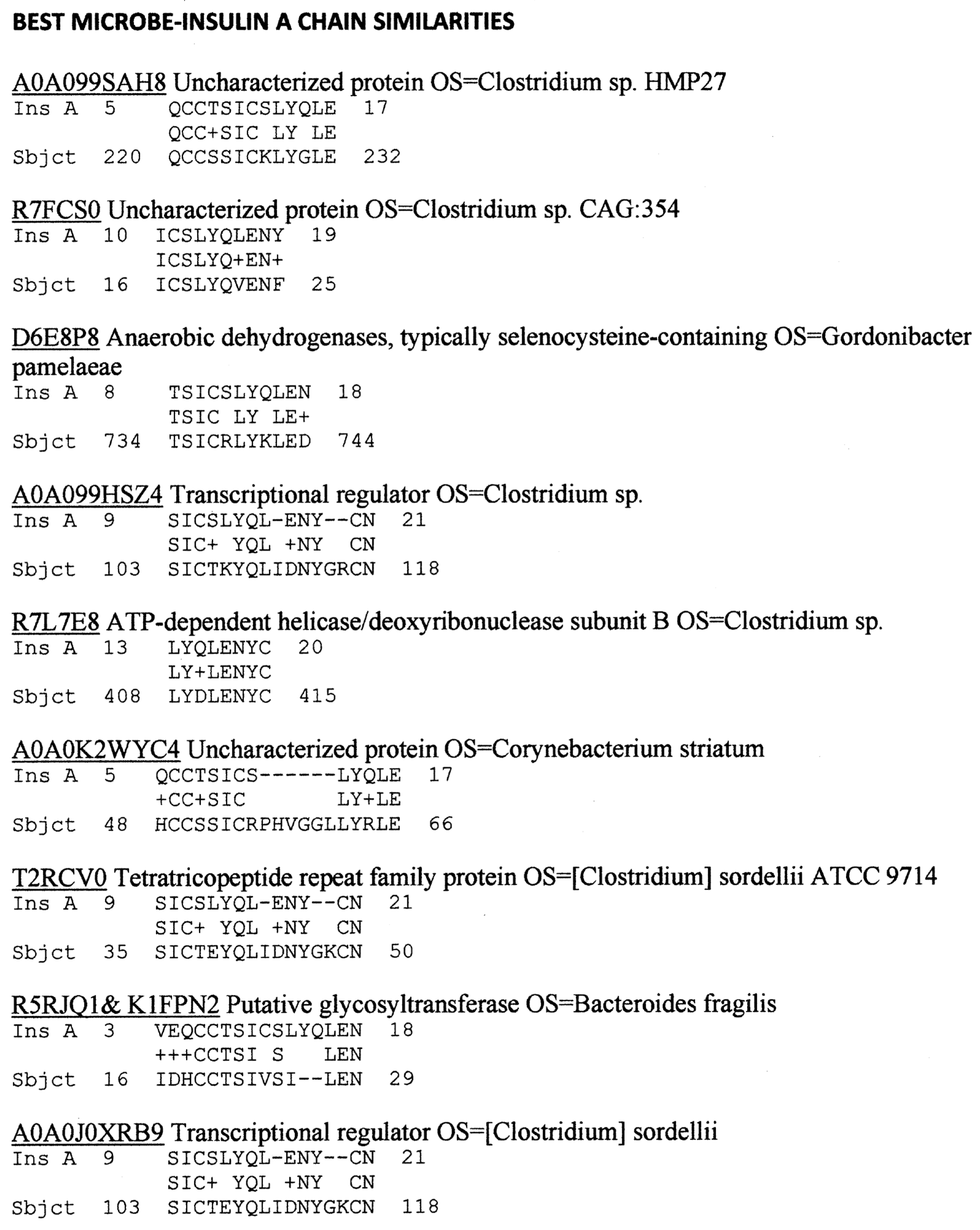
Figure 1. Results of BLASTP similarity search comparing the human INS A chain sequence with the UNIPROTKB bacterial database curated for human pathogens and commensals.
Ijms 24 08336 g001
Figure 2. Results of BLASTP similarity search comparing the human INS B chain sequence with the UNIPROTKB bacterial database curated for human pathogens and commensals.
Ijms 24 08336 g002
Figure 3. BLASTP similarities between COX type B4 and the human INS receptor.
Ijms 24 08336 g003
Figure 4. Summary of quantitative ELISA experiments regarding COX and Clostridia antibody binding to INS, glucagon, the INS receptor (IR), IR peptides, glucagon receptor peptides, and the beta 2 adrenergic receptor. Blue highlighted sequences are ones overlapping the COX-INS receptor similarities listed in Figure 3. Green highlighted figures are sequences with significant INS binding. Gray highlighted figures are sequences that displayed significant antibody binding. Results are binding constants (Kd) in micromoles determined by the inflection points of each curve. A zero (0) indicates that binding was not measurable (in practice, Kd > 1 micromolar). The IR sequences are listed by the numerical position in the IR sequence and followed by the single-letter amino acid sequence. HRP = horseradish peroxidase conjugated; α = “anti-“; Cox = coxsackivierus; Mabs = monoclonal antibodies (mouse); Rab = rabbit antibody; Clost = Clostridium; Tox A = Clostridium toxin A.
Ijms 24 08336 g004
Figure 5. Binding of virus and bacterium antibodies to INS, glucagon, the INS receptor (INSR), and INSR peptides. EBV = Epstein–Barr virus; Inf A = Influenza A virus; Inf B = Influenza B virus; CMV = cytomegalovirus; Adeno = adenovirus; HSV1 = human herpes simplex virus type 1; Pseud aerug = Pseudomonas aeruginosa; Mycob tuber = Mycobacterium tuberculosis; Staph aureus = Staphylococcus aureus; Strep A = group A Stretpcococci. See Figure 8 for an explanation of the rest of the abbreviations and formalisms.
Ijms 24 08336 g005
Figure 6. Results of ELISA studies concerning binding to the INS receptor (IR) and its peptides. LEFT: Monkey (Mon) anti-coxsackievirus type B3 (CXB3), Epstein–Barr virus (EBV) antibody and to a much lesser extent, Clostridium antibodies, bind to the human insulin receptor. RIGHT: Further characterization of monkey anti-CXB3 antibody binding to peptides derived from the human INSR. Ms = mouse; Rab = rabbit; CX = coxsackievirus; Enterov = enterovirus; EBV = Epstein–Barr virus; Clost = Clostridium; P. aerug. = Pseudomonas aeruginosa. Numbers indicate the amino acid sequence of the INS receptor from which the peptides were derived. Further data are provided in Appendix D.
Ijms 24 08336 g006
Figure 7. Results of ELISA studies concerning binding to insulin (INS). LEFT: Clostridium antibody, but not coxsackievirus (X) antibodies, bind to INS. RIGHT: Clostridium antibodies bind to the INS A and B chains but not to glucagon. Rab = rabbit; Ms = mouse; C. perf toxin = Clostridium perfringens alpha toxin; Cox = COX; Clost = Clostridium; Ins = INS; A = A chain; B = B chain.
Ijms 24 08336 g007
Figure 9. Summary of the binding constants (Kd) from double-antibody ELISA experiments involving a virus antibody potentially binding to another virus antibody. See Figure 14, Figure 15 and Figure 16 for examples. COXB = COX type B; CMV = cytomegalovirus; HBV = hepatitis B virus; HCOX = hepatitis C virus; HSV = herpes simplex virus; Ms = mouse; Gt = goat; Shp = sheep.
Ijms 24 08336 g009
Figure 10. Representative binding curves resulting from double antibody ELISA experiments involving virus and bacterium antibodies. LEFT: Rabbit Clostridium antibodies bound to all of the COX antibodies tested as well as an enterovirus antibody but did not bind to other virus antibodies. RIGHT: Additional control combinations of virus–bacterial antibody pairs tested by double antibody ELISA and summarized in Appendix E. CXB = Coxsackievirus; Adv = adenovirus; Inf A = influenza A virus, HSV1 = herpes simplex type 1 virus; Ms = mouse; Gt = goat; Hs = horse; Mon = monkey; Enteroc = Enterococcus faecium; S. pneum. = Streptococcus pneumoniae; Adv = adenovirus; EBV = Epstein–Barr virus; HCOX = hepatitis C virus; Enterov = enterovirus. See Figure 8 and Figure 9 for species of each antibody.
Ijms 24 08336 g010
Figure 11. Results of double antibody ELISA experiments. TOP LEFT: Coxsackievirus (CX) and Clostridium antibodies binding to INS antibody. TOP RIGHT: CX and Clostridium antibodies binding to several INS receptor antibodies. BOTTOM LEFT: Clostridia antibodies binding to INS receptor antibodies (IR-Ab). Rb IR-Ab were tested against Ms Clostridia, while Ms IR-Ab were tested against Rb Clostridia. BOTTOM RIGHT: Binding of COX type B (COXB) or Clostridium (Clost) antibody binding to glutamic acid decarboxylase (GAD-65) antibody or protein tyrosine phosphatase non-receptor type 1 (PTPN(IA2)) antibody. Clost = Clostridium; TA = toxin A; Rb = rabbit; Ms = mouse; Milli = Millipore CXB = Coxsackievirus type B; IR = INS receptor (alpha and beta indicating the chain to which the antibody is specific); Mon = monkey; Hs = horse; Rab = rabbit; Ms = mouse; Biod = Biodesign; Chem = Chemicon (see Tables of antibodies in Section 4).
Ijms 24 08336 g011
Figure 12. Two Clostidium peptides that mimic the INS A chain (INS A) and one COX peptide that mimics the INS receptor (INS Rec) were synthesized (sequences and similarities to the LEFT) and tested for their ability to bind to each other using ultraviolet spectrometry (RIGHT). Kd was 60 µM for Clostridium peptide 2 and 200 µM for peptide 3.
Ijms 24 08336 g012
Figure 13. Ultraviolet spectrometry study of COX (cox) peptide (which mimics the INS receptor) and Clostridium peptides 2 and 3 (clost2, clost3) (which mimic INS), binding to T cell receptors (TCR) K2.12 (which mimics the INS receptor) and TCR8 (which mimics the glucagon receptor). The binding constants listed in Table 1 were estimated from the inflection points of the curves.
Ijms 24 08336 g013
Figure 17. Summary of the various types of complementarity and mimicry revealed by the experiments performed here. INS = insulin; INSR = INS receptor; Ab = antibody; TCR = T cell receptor; Clost = Clostridium; COX = coxsackievirus. The arrow link mimics. Steric fits represent complementarity between antibodies with their antigens as well as between pairs of antigens (e.g., COX and Clost or INS and INSR).
Ijms 24 08336 g017
Table 1. Summary of ultraviolet spectrometry experiments concerning binding of T cell receptor sequences expanded in type 1 diabetic patients (TCR DIA) to a COX peptide (Cox Pep) that mimics the INS receptor (Ins Rec), two Clostridium peptides (Clost Pep) that mimic INS, two INS receptor peptides identified by their amino acid sequence numbers, and INS itself. Gluc = glucagon-like sequence; GR = glucagon receptor-like sequence; IR = INS receptor-like sequence; Ins = INS-like sequence. Some TCR sequences mimic regions of more than one protein.
Binding Constants (µM) Cox Pep #1Ins Rec 105–118Ins Rec 897–915Clost Pep #2Clost Pep #3INS
TCR DIA 1 (Gluc) CASSIYLCSVEATRAD40012530010010080
TCR DIA 2, K2.16 (GR) CASSLAVIRT1000>1000>1000110>10,000>1000
TCR DIA 4, K2.4 (IR) CASSLATSGGGSDTQ1000>1000>10001307015
TCR DIA 7 (IR) CASSFRRVTDTQ>10,000>1000>100012010070
TCR DIA 8 (Ins, GR) CASSQVRLAGGGEQ75012013030070130
TCR DIA 9 (Ins, GR) CASSLGQGETEAFF10015014550010023
TCR DIA 10 (IR) CASRNLGLNTE2000110140100080130
TCR DIA 4,8,9 (Ins, IR, GR) DSALYLCASSLG200>1000>10003000150>1000
TCR DIA K2.12 (IR) CASSDRLGNQPQH>10,000120>100010020075
Table 2. List of proteins and peptides used in the research and their suppliers.
PRODUCT NAMESPECIESSUPPLIERPRODUCT #Purity
Insulin (INS)Human (recombinant)Sigma-AldrichI2643100%
INS A chain, oxidizedBovineSigma-AldrichI1633>80%
INS B chain, oxidizedBovineSigma-AldrichI1764>95%
INS C chainHuman (recombinant)Sigma-AldrichC9781>95%
INS ReceptorHuman (recombinant)R&D Systems1544-IR>95%
GlucagonHuman (recombinant)Sigma-AldrichG2044100%
Beta 2 Adrenergic ReceptorRat (recombinant)MyBioScourceMBS7111177>90%
Table 3. List of primary antibodies against viruses used in this study and their suppliers.
VIRUS ANTIBODIESSPECIESSUPPLIERPRODUCT #
Adenovirus GoatMilliporeAB1056
COX B3 MonkeyATCC (NIH Reference Reagent)VO31-501-563
COX B4HorseATCC (NIH Reference Reagent)VO30-501-560
COX B3MouseMilliporeMAB948
Coxsackie Virus B1-B6 BlendMouseMilliporeMAB9410
CytomegalovirusGoatBiodesign InternationalB562756
Cytomegalovirus MouseBiodesign InternationalC65861M
Enterovirus pan VP3MouseMyBioSourceMBS319564
Epstein-Barr VirusRabbitInvitrogenPA5-115471
Hepatitis C Virus core AntigenRabbitInvitrogenPA1-4113
Herpes Simplex Virus Type 1GoatInvitrogenPA1-7493
Herpes Simplex Virus Type 1/2 RabbitInvitrogenPA1-7214
Influenza A HRPGoatBiodesign InternationalB65243G
Influenza B HRPRabbitBiodesign InternationalB653446
Table 4. List of primary bacteria antibodies used in this study and their suppliers.
BACTERIA ANTIBODIESSPECIESSUPPLIERPRODUCT #
ClostridiaRabbitInvitrogenPA1-7210
Clostridium sp. HRPRabbitUS BiologicalC5853-25C
Clostridium alpha toxin-HRPRabbitBiossBs-2273R-HRP
Enterococcus HRPRabbitInvitrogenPA1-73122
Escherichia coliGoatabcamAB13627
Klebsiella pneumoniae HRPRabbitInvitrogen PA1-73176
Mycobacterium tuberculosisRabbitABD SerotecOBT0947
Mycobacterium tuberculosisGuinea PigMyBioSourceMBS315001
Pseudomonas aeruginosaGuinea PigBiodesign InternationalB47578P
Staphylococcus aureusRabbitInvitrogenPA1-7246
Staphylococcus aureus HRPRabbitInvitrogenPA1-73173
Streptococcus Group AGoatInvitrogenPA1-7249
Streptococcus Group A HRPRabbitAcris AntibodiesBP2026HRP
Streptococcus pneumoniaeRabbitBiodesign InternationalB65831R
Streptococcus pneumoniaeRabbitInvitrogenPA1-7259
Table 5. List of protein antibodies used in this study and their suppliers.
PROTEIN ANTIBODIESSPECIESSUPPLIERPRODUCT #
Glutamic acid decarboxylase 65RabbitMilliporeABN101
Insulin (INS)RabbitSigmaHPA004932
INS ReceptorMouseChemiconMAB105
INS Receptor alpha RabbitBiodesign InternationalK54244R
INS Receptor alphaMouseBiodesign International K54241M
INS Receptor betaMouseMillipore 05-1104
PTPN(IA-2)RabbitSigmaHPA007179
Table 6. List of secondary horseradish peroxidase-labelled (HRP) antibodies used in this study and their suppliers.
SECONDARY ANTIBODIESSPECIESSUPPLIERPRODUCT #
Anti-Guinea Pig-HRPRabbitabcamAB6771
Anti-Horse IgG-HRPGoatSanta Cruze BiotechnologySC-2448
Anti-Human IgG-HRPGoatSigmaAO170
Anti-Mouse IgG-HRPGoatSigma-AldrichA9917
Anti-Rabbit IgGHRPGoatInvitrogen 65-6120
Table 7. List of human sera used in this study and their suppliers.
Disease StateSupplierOur ID Sample IDAgeSexRaceHbA1c %Serum Treatment
T1DMLee BiosolutionsLEE 0109E5731
A1c-50.11
39MCaucasian11.5EDTA
T1DMLee BiosolutionsLEE 0209E5731 A1c-50.1236FCaucasian14.4EDTA
T1DMLee BiosolutionsLEE 0309E5731 A1c-0329FCaucasian10.3EDTA
T1DMLee BiosolutionsLEE 0409E5731 A1c-0430MCaucasian10.5EDTA
T1DMLee BiosolutionsLEE 0509E5731 A1c-0524FCaucasian11.3EDTA
T1DMInnovative ResesarchT1DM #1HMN88906961MCaucasian Off the clot
T1DMInnovative ResesarchT1DM #2HMN88907036MAsian Off the clot
T1DMInnovative ResesarchT1DM #3HMN88907156FCaucasian Off the clot
T1DMInnovative ResesarchT1DM #4HMN88909058MHispanic Off the clot
T1DMInnovative ResesarchT1DM #5HMN88909149FAfrican American Off the clot
T2DZenBio#69SER-DPLE2ML MCaucasian Off the clot
T2DZenBio#71SER-DPLE2ML FAfrican Amderican Off the clot
HealthyZen-BioHealthy 1HSER-2ML FCaucasian Off the clot
HealthyInnovative ResesarchHealthy 8639521-08646MAfrican American Off the clot
HealthyInnovative ResesarchHealthy 8739521-08736FAfrican American Off the clot
HealthyInnovative ResesarchHealthy 8839521-08840MAfrican American Off the clot
HealthyInnovative ResesarchHealthy 8939521-08959MAfrican American Off the clot
Disclaimer/Publisher’s Note: The statements, opinions and data contained in all publications are solely those of the individual author(s) and contributor(s) and not of MDPI and/or the editor(s). MDPI and/or the editor(s) disclaim responsibility for any injury to people or property resulting from any ideas, methods, instructions or products referred to in the content.